35TH ANNUAL MIDWEST/SOUTHEAST PHOTOSYNTHESIS MEETING · 35th annual midwest/southeast...
68
35 TH ANNUAL MIDWEST/SOUTHEAST PHOTOSYNTHESIS MEETING TURKEY RUN STATE PARK MARSHALL, INDIANA NOVEMBER 13 th –15 th , 2009 PROGRAM AND ABSTRACTS
Transcript of 35TH ANNUAL MIDWEST/SOUTHEAST PHOTOSYNTHESIS MEETING · 35th annual midwest/southeast...
35TH ANNUAL MIDWEST/SOUTHEAST
PHOTOSYNTHESIS MEETING
TURKEY RUN STATE PARK
MARSHALL, INDIANA
NOVEMBER 13th–15th, 2009
PROGRAM AND ABSTRACTS

Original Cover Art: I.N.K. b-town/09
35TH ANNUAL MIDWEST/SOUTHEAST
PHOTOSYNTHESIS MEETING
TURKEY RUN STATE PARK MARSHALL, INDIANA
NOVEMBER 13th–15th, 2009
PROGRAM AND ABSTRACTS
THIS YEAR’S ORGANIZERS:
YULIA PUSHKAR DEPARTMENT OF PHYSICS PURDUE UNIVERSITY
DAVID KEHOE DEPARTMENT OF BIOLOGY INDIANA UNIVERSITY
2
OPENING SESSION: FRIDAY, NOVEMBER 13 CHAIR: David Kehoe 7:20 PM Opening Remarks and Welcome 7:25 PM Yulia Pushka; Introduction of Plenary Lecturer 7:30 PM Plenary Lecture: Dr. John Golbeck, Pennsylvania State University
BIOHYBRID SYSTEMS IN SOLAR BIOFUEL PRODUCTION 8:30 PM Dr. Robert E. Blankenship, Washington University
PHOTOSYNTHETIC ANTENNA SYSTEMS: THE PLACE WHERE LIGHT INTERFACES WITH BIOLOGY
9:00 PM Dr. Terry Bricker, Louisiana State University
DOCUMENTATION OF SIGNIFICANT ELECTRON TRANSPORT DEFECTS ON THE REDUCING-SIDE OF PHOTOSYSTEM II UPON REMOVAL OF THE PsbP AND PsbQ EXTRINSIC PROTEINS
9:30 PM MIXER, POSTER MOUNTING AND VIEWING
SESSION II: SATURDAY, NOVEMBER 14
CHARGE TRANSFER CHAIR: Toivo Kallas 9:00 AM Joseph (Kuo-Hsiang) Tang, Yue Hai, Xueyang Feng, Yinjie J.
Tang, and Robert E. Blankenship (Washington University)
ENERGY AND CARBON METABOLISM IN ROSEOBACTER DENITRIFICANS AND HELIOBACTERIUM MODESTICALDUM
3
9:25 AM S. S. Hasan, S. D. Zakharov, E. Yamashita, H. Böhme, and W. A.
Cramer (Purdue University)
EXCITONIC INTERACTION BETWEEN HEMES bn AND bp IN THE CYTOCHROME b6f COMPLEX
9:50 AM Coffee Break 10:20 AM Sandy Zuleger, Alina Ott, David Rivera, Brant Kedrowski, and
Toivo Kallas (University of Wisconsin, Oshkosh)
A SYNTHESIS PRECURSOR, 4(1H)-QUINOLONE, OF THE CYTOCHROME bf QUINONE-REDUCTASE SITE INHIBITOR NQNO IS AN EFFECTIVE INHIBITOR OF THE QUINOL-OXIDASE SITE
10:45 AM Patrick Bell and Robert E. Blankenship (Washington University)
INVESTIGATION OF THE PHOTOSYNTHETIC ELECTRON TRANSPORT CARRIERS, CYTOCHROME C6 AND PLASTOCYANIN, IN ACARYOCHLORIS MARINA
11:10 AM BREAK FOR LUNCH
SATURDAY AFTERNOON POSTER VIEWING AND LEISURE TIME
4
SESSION III: SATURDAY, NOVEMBER 14 ENERGY TRANSFER
CHAIR: Yulia Pushkar 7:30 PM Dugan Hayes, Gitt P. Panitchayangkoon, Kelly A. Fransted, Justin
R. Caram, and Gregory S. Engel (University of Chicago)
THE MICROSCOPIC MECHANISM OF COHERENCE ENERGY TRANSFER IN FMO
7:55 PM Gitt Panitchayangkoon, Kelly A. Fransted, Dugan K. Hayes, Justin
R. Caram, and Gregory S. Engel (University of Chicago)
TEMPERATURE DEPENDENCE OF QUANTUM COHERENCE TRANSFER IN THE FENNA-MATTHEWS-OLSON COMPLEX
8:20 PM Dariusz M. Niedzwiedzki, Aaron M. Collins and Robert E.
Blankenship (Washington University)
CAROTENOID-TO BACTERIOCHLOROPHYLL ENERGY TRANSFER IN THE LIGHT-HARVESTING REACTION CENTER COMPLEX (LH-RC) FROM Roseiflexus castenholzIi
8:45 PM Andrian Gutu and David M. Kehoe (Indiana University)
INTEGRATION OF LIGHT COLOR AND NUTRIENT SIGNALS IN THE REGULATION OF LIGHT HARVESTING GENES
9:10 PM MIXER, POSTER VIEWING
5
SESSION IV: SUNDAY, NOVEMBER 15
REGULATION AND BIOTECHNOLOGY CHAIR: Qingfang He 9:00 AM Anindita Bandyopadhyay, Jana Stöckel, Himadri B. Pakrasi
(Washington University)
ELUCIDATING THE MECHANISM OF HYDROGEN PRODUCTION IN CYANOTHECE 51142, A UNICELLULAR, DIAZOTROPHIC PHOTOAUTOTROPH
9:25 AM Jana Stöckel, Jon M. Jacobs, Thanura Elvitigala, Michelle Liberton,
Eric A. Welsh, Ashoka D. Polpitiya, Marina A. Gritsenko, Carrie D. Nicora, David W. Koppenaal, Richard D. Smith, and Himadri B. Pakrasi (Washington University)
INSIGHTS INTO DIURNAL RHYTHMS OF CYANOTHECE SP. ATCC 51142: THE PROTEOME SIDE OF THE STORY
9:50 AM Coffee Break 10:10 PM Jing Zhang, Stephen C. Grace and Qingfang He (University of
Arkansas Little Rock)
CONSTRUCTION OF CYANOBACTERIAL STRAINS EXPRESSING THE COUMARATE 3-HYDROXYLASE FROM ARABIDOPSIS
10:35 AM Will Kovac, George L. Weir IV, Justin Zangl, Kraig Short,
Matthew Nelson and Toivo Kallas (University of Wisconsin Oshkosh)
MICROARRAY GLOBAL GENE-EXPRESSION PROFILING AND TRANSCRIPTION START-SITE MAPPING OF SYNECHOCOCCUS PCC 7002: OPEN SOURCE TOOLS FOR DATA MINING AND ANALYSIS
11:00 AM PRESENTATION OF AWARDS AND CLOSING REMARKS
6
PRESENTATION ABSTRACTS
(IN THE ORDER OF PRESENTATION WITHIN THE PROGRAM)
7
OPENING SESSION: FRIDAY, NOVEMBER 13
PLENARY LECTURE
BIOHYBRID SYSTEMS IN SOLAR BIOFUEL PRODUCTION
Carolyn Lubner1, Paulo Silva2, Donald A. Bryant2, and John H. Golbeck1,2 1Department of Chemistry; 2Department of Biochemistry; The Pennsylvania State University,
University Park, PA 16802 [email protected]
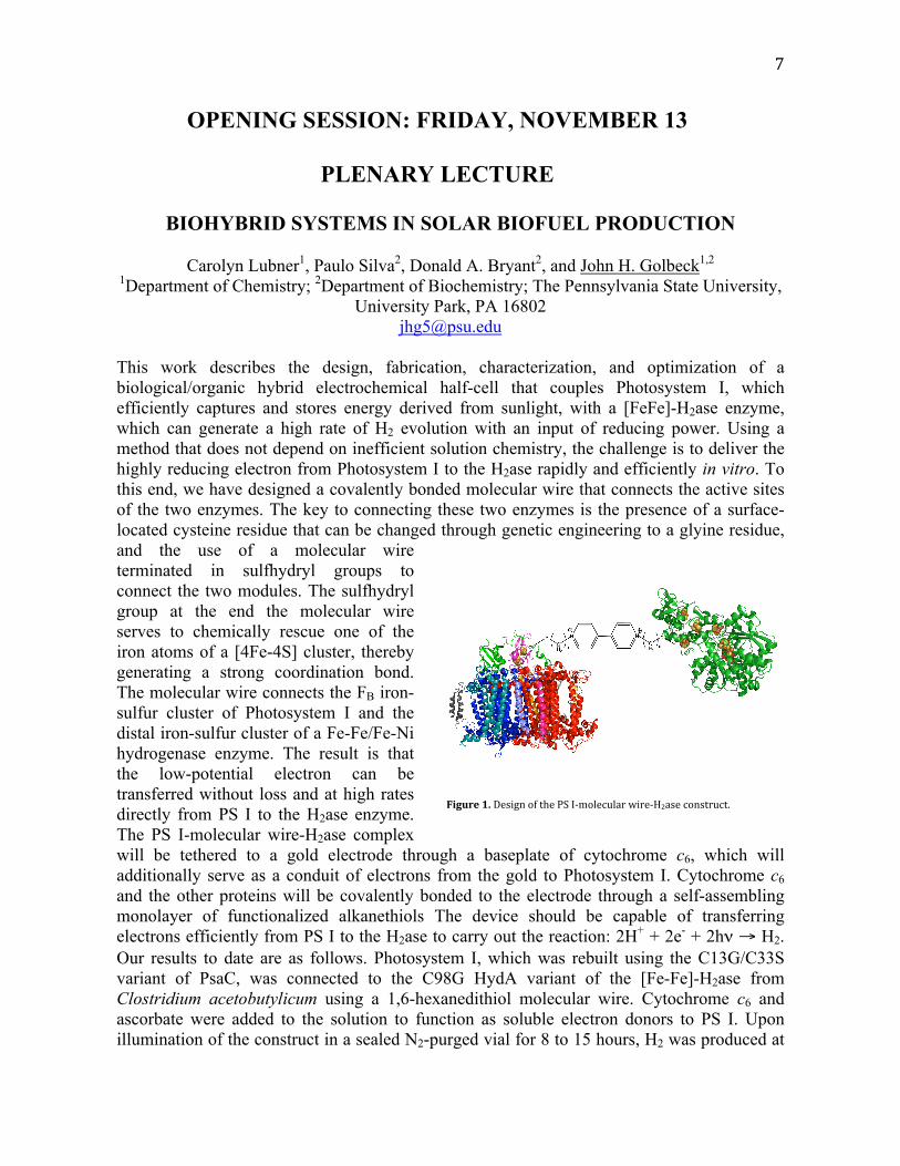
This work describes the design, fabrication, characterization, and optimization of a biological/organic hybrid electrochemical half-cell that couples Photosystem I, which efficiently captures and stores energy derived from sunlight, with a [FeFe]-H2ase enzyme, which can generate a high rate of H2 evolution with an input of reducing power. Using a method that does not depend on inefficient solution chemistry, the challenge is to deliver the highly reducing electron from Photosystem I to the H2ase rapidly and efficiently in vitro. To this end, we have designed a covalently bonded molecular wire that connects the active sites of the two enzymes. The key to connecting these two enzymes is the presence of a surface-located cysteine residue that can be changed through genetic engineering to a glyine residue, and the use of a molecular wire terminated in sulfhydryl groups to connect the two modules. The sulfhydryl group at the end the molecular wire serves to chemically rescue one of the iron atoms of a [4Fe-4S] cluster, thereby generating a strong coordination bond. The molecular wire connects the FB iron-sulfur cluster of Photosystem I and the distal iron-sulfur cluster of a Fe-Fe/Fe-Ni hydrogenase enzyme. The result is that the low-potential electron can be transferred without loss and at high rates directly from PS I to the H2ase enzyme. The PS I-molecular wire-H2ase complex will be tethered to a gold electrode through a baseplate of cytochrome c6, which will additionally serve as a conduit of electrons from the gold to Photosystem I. Cytochrome c6 and the other proteins will be covalently bonded to the electrode through a self-assembling monolayer of functionalized alkanethiols The device should be capable of transferring electrons efficiently from PS I to the H2ase to carry out the reaction: 2H+ + 2e- + 2hν → H2. Our results to date are as follows. Photosystem I, which was rebuilt using the C13G/C33S variant of PsaC, was connected to the C98G HydA variant of the [Fe-Fe]-H2ase from Clostridium acetobutylicum using a 1,6-hexanedithiol molecular wire. Cytochrome c6 and ascorbate were added to the solution to function as soluble electron donors to PS I. Upon illumination of the construct in a sealed N2-purged vial for 8 to 15 hours, H2 was produced at
!
!
"#$%&'!()!"#$%&'!()!*+#!,-!./0(1#23145!6%5#/784$#!2('$*532*9!

8
rates ranging from 0.3 to 2.1 µmol H2 mg Chl-1 h-1, depending on the sample. After a rough optimization of solution conditions, the rate increased approximately two-fold to 3.9 µmol H2 mg Chl-1 h-1. Control experiments were performed to verify light-induced H2 production. The controls included the absence of the following substrates: light, rebuilt PS I with variant PsaC, variant [FeFe]-H2ase, and 1,6-hexane dithiol; as well as the substitution of wild-type PS I and wild-type [FeFe]-H2ase. All of the controls failed to generate H2. We are in the process of optimizing conditions to maximize the rate. Funded by the US DOE (ER46222).
9
PHOTOSYNTHETIC ANTENNA SYSTEMS: THE PLACE WHERE LIGHT INTERFACES WITH BIOLOGY Robert E. Blankenship Departments of Biology and Chemistry, Washington University in St. Louis Campus Box 1137, One Brookings Drive, St. Louis, MO 63130 All photosynthetic organisms contain a light-gathering antenna system, which functions to collect light and transfer energy to the reaction center complex where electron transfer reactions take place. Our work centers on the antenna complexes found in green photosynthetic bacteria, which include chlorosomes, the Fenna-Matthews-Olson (FMO) antenna protein and integral-membrane antenna and reaction center complexes. All of these complexes are involved in the light-energy collection process in these organisms, which are adapted for life in very low light intensities. Chlorosomes are ellipsoidal structures attached to the cytoplasmic side of the inner cell membrane. These antenna complexes provide a very large absorption cross section for light capture. Evidence is overwhelming that the chlorosome represents a very different type of antenna from that found in any other photosynthetic system yet studied. Chlorosomes do not contain traditional pigment-proteins, in which the pigments bind to specific sites on proteins. These systems are of interest from both a basic science perspective of what is the structure of this unique class of photosynthetic antennas and how they work so efficiently, as well as more applied aspects in which the principles of self organization and extraordinary pigment properties that characterize these systems are used in a bio-mimetic approach to devise artificial light-energy capture systems. Recent work involves studies on the structure of the FMO antenna complex and the architecture of the membrane that includes the chlorosome, FMO protein and reaction center. Additional work involves using chlorosomes as part of bio-hybrid systems in which the biological complex feeds energy to an inorganic semiconductor substrate such as titanium dioxide.
10
DOCUMENTATION OF SIGNIFICANT ELECTRON TRANSPORT DEFECTS ON THE REDUCINGSIDE OF PHOTOSYSTEM II UPON REMOVAL OF THE PsbP AND PsbQ EXTRINSIC PROTEINS Johnna L. Roose, Laurie K. Frankel and Terry M. Bricker, Department of Biological Sciences, Biochemistry and Molecular Biology Section, Louisiana State University, Baton Rouge, LA 70803 The Photosystem II extrinsic proteins PsbO, PsbP and PsbQ are required for efficient oxygen‐evolving activity under physiological conditions. In this study, we have used fluorescence decay kinetics to quantitatively probe Photosystem II electron transport upon depletion of these components by standard salt washing protocols. Our results indicate that in addition to the expected oxidizing‐side defects, removal of PsbP and PsbQ with 2 M NaCl significantly slows the rate of electron transfer from QA‐ to QB. Electron transfer from QA‐ to QB in Photosystem II reaction centers with an occupied QB site was slowed by a factor of 12, while electron transport from QA‐ to QB in centers with an unoccupied QB site was slowed by a factor of 6. Subsequent removal of the PsbO protein by treatment with 200 mM NaCl + 2.6 M urea did not induce further reducing‐side alterations. Our results demonstrate that studies attributing defects observed upon PsbP and PsbQ removal solely to the oxidizing side must be viewed with caution.

11
SESSION II: SATURDAY MORNING
CHARGE TRANSFER
ENERGY AND CARBON METABOLISM IN ROSEOBACTER DENITRIFICANS AND HELIOBACTERIUM MODESTICALDUM Joseph (Kuo‐Hsiang) Tang1, Yue Hai1, Xueyang Feng2, Yinjie J. Tang2, Robert E. Blankenship1
1Departments of Biology and Chemistry, 2Department of Energy, Environment and Chemical Engineering, Washington University in St. Louis, St. Louis, MO 63130 We report carbon assimilation, carbon metabolism and energy metabolism in two photoheterotrophic bacteria, the aerobic anoxygenic α‐proteobacterium Roseobacter denitrificans, and the anaerobic anoxygenic Gram‐positive bacterium Heliobacterium modesticaldum. Neither of these organisms is capable of photoautotrophic growth. For R. denitrificans, we identified non‐autotrophic anaplerotic pathways for CO2‐assimilation, and the Entner‐Doudoroff (ED) and non‐oxidative pentose phosphate pathways for carbohydrate metabolism and nucleic acids biosynthesis, respectively. For H. modesticaldum, we identified two nonautotrophic CO2‐assimilation pathways, additional nutrients for enhancing the growth of H. modesticaldum, pigment production during dark growth, and acetate production during phototrophic and dark growth. The proposed energy metabolism pathways of H. modesticaldum during phototrophic and dark growth will be discussed. References: 1. Tang, K.‐H., Feng, X., Tang, Y.J., and Blankenship, R.E. (2009) carbohydrate metabolism and carbon fixation in Roseobacter denitrificans OCh114. PLoS One, 4: e7233 1‐12. 2. Tang, K.‐H., Yue, H., and Blankenship, R.E. Energy metabolism of Heliobacterium modesticaldum during phototrophic and chemotrophic growth. Submitted

12
EXCITONIC INTERACTION BETWEEN HEMES bn AND bp IN THE
CYTOCHROME b6f COMPLEX.
S. S. Hasan, S. D. Zakharov, E. Yamashitaa, H. Böhme*, and W. A. Cramer. Dept. of
Biological Sciences, Purdue University, West Lafayette, IN 47907, USA; a Institute of Protein
Research, Osaka University, Osaka 560-0043, Japan.
The cytochrome b6f complex of oxygenic photosynthesis is a hetero-oligomeric protein
complex involved in electron and proton transfer. Like the bc1 complex, b6f participates in
electron transfer coupled proton translocation by utilizing two b-type hemes (bp and bn) in a
Q-cycle mechanism. Heme cn is bound close to heme bn and is covalently linked to the
cytochrome b6 polypetide1, 2, and 3. π-π* electronic transitions of the heme porphyrin ring of
reduced cytochrome b6 are associated with the Soret band at 431-432 nm4. Circular dichroism
spectroscopy of the crystallizable b6f complex reveals the presence of a bi-lobed spectrum in
the dithionite minus ascorbate sample, with a node close to the 431-432 nm Soret peak.
Similar spectra were previously published from earlier studies of the bc1 complexes5, 6 and 7 and
the b6f complex from Chlamydomonas reinhardtii8. From the crystal structure of the
Mastigocladus laminosus cytochrome b6f complex (PDB 2E74)9, the distance between the Fe
atoms of hemes bp and bn is 20.8 Å, the separation between the Fe atoms of the two hemes bp
is 22.2 Å, and that between the two hemes bn is 35.0 Å. Heme bn and cn are separated by 10.0
Å Fe-Fe and 4 Å edge to edge. It is inferred that the source of the splitting is the interaction of
the bp and bn hemes. Supported by NIH GM-18457.
*deceased; 1Kurisu et al. 2003; 2Stroebel et al. 2003; 3Baniulis et al. 2009; 4Eaton and Hofrichter, 1981; 5Degli Esposti et al. 1987; 6Degli Esposti et al. 1989; 7Palmer and Degli-Esposti, 1994; 8Schoepp et al. 2000; 9Yamashita et al. 2007.
13
A SYNTHESIS PRECURSOR, 4(1H)QUINOLONE, OF THE CYTOCHROME bf QUINONEREDUCTASE SITE INHIBITOR NQNO IS AN EFFECTIVE INHIBITOR OF THE QUINOL
OXIDASE SITE Sandy Zuleger1, Alina Ott2, David Rivera3, Brant Kedrowski1, Toivo Kallas4. Departments
of 1Chemistry & 4Biology‐Microbiology, Univ. Wisconsin, Oshkosh; 2Department of Biology, Univ. Wisconsin, Stevens Point; 3Department of Biology, Univ. Puerto Rico,
Bayamón Photosynthesis is catalyzed by thylakoid membrane protein complexes that allow separation of charges in conversion of solar radiation into chemical energy as ATP and reducing power as NADPH. In this process, the cytochrome bf complex generates a proton gradient for ATP synthesis and functions in redox sensing and signaling. Our goal was to synthesize the quinone‐reductase site (Qn) inhibitor, 2‐nonyl‐1‐hydroxy‐4(1H)‐quinolone (NQNO), and use it to investigate electron transfer pathways and redox signaling in the cyanobacterium Synechococcus PCC 7002. Cyanobacteria perform approximately 25% of global photosynthesis and hold great potential for biofuels applications. There are few useful inhibitors of cyanobacterial cytochrome bf complexes. NQNO binds the quinone‐reductase (Qn) site and slows electron flow through the low potential chain. Tridecylstigmatellin (TDS), a classical inhibitor of the quinol‐oxidation (Qp) site, is largely ineffective in cyanobacterial bf complexes because of a constrained portal for access to this site (Yamashita et al., 2007 JMB 370, 39). The first syntheses of hydroxy‐quinolones was performed by Cornforth and James who synthesized HQNO (a derivative with a seven carbon tail). We used their procedure as a basis for producing NQNO. The synthesis involved a series of steps, beginning by reacting decanoic acid with oxalyl chloride and most noteworthy a column chromatography step to separate two isomers with similar polarities. A precursor to NQNO, 4(1H)‐quinolone, differs from NQNO only by a hydrogen rather than a hydroxyl group at position one of the quinolone ring. In kinetics experiments with a BioLogic JTS‐10 spectrophotometer, the 4HQ precursor, in contrast to NQNO, slowed cytochrome f/c6 reduction by ~10 fold and resulted in b‐heme oxidation rather than reduction. These and other data indicate that 4HQ binds to the cytochrome bf Qp‐site and inhibits quinol oxidation at this site rather than quinone reduction at the Qn site. This indicates that these two inhibitors (NQNO and 4HQ) can be used to selectively reduce or oxidize the cytochrome bf low‐potential chain and will be interesting for studies of electron transfer and redox signaling in cyanobacteria.

14
INVESTIGATION OF THE PHOTOSYNTHETIC ELECTRON TRANSPORT CARRIERS, CYTOCHROME C6 AND PLASTOCYANIN, IN ACARYOCHLORIS MARINA
Patrick Bell and Robert E. Blankenship Department of Chemistry, Washington University, St. Louis MO 63130
Acaryochloris marina, a unicellular marine cyanobacterium, is unique among
oxygenic phototrophs in that it uses chlorophyll d as its primary photosynthetic pigment, which absorbs light at longer wavelengths compared to other chlorophylls. This unique pigment has made A. marina a species of interest for evolutionary analysis and possible bioenergy applications. This has led our lab to begin investigating the proteins and complex involved in its photosynthetic apparatus, including cytochrome c6 and plastocyanin. Cytochrome c6 and plastocyanin are functionally interchangeable proteins involved in photosynthetic electron transport in cyanobacteria and algae. In many cyanobacteria and algae, plastocyanin is expressed under copper replete conditions, while cytochrome c6 is expressed under copper depleted conditions. In A. marina’s genome, there are two genes coding for cytochrome c6 and one gene coding for plastocyanin. Under our growth conditions in iron‐enriched Marine BG‐11 media only one cytochrome c6 from the genome was expressed and subsequently purified by ammonium sulfate fractionation and ion exchange and gel filtration chromatography. This protein was characterized by mass spectroscopy, CD spectroscopy, isoelectrofocusing, and redox potentiometry and was found to be a typical small (8.87 kDa), alpha helical, acidic (pI < 4), and high potential (Em = +327mV vs NHE) cytochrome c6 in relation to cytochrome c6 of other cyanobacteria.
To investigate the expression of cytochrome c6 and plastocyanin, A. marina was then grown in Marine BG‐11 media under increasing copper and iron concentrations. RT‐PCR analysis of the gene expression levels for both cytochrome c6 and plastocyanin under these different growth conditions will be presented. In addition, efforts in the cloning and expression of plastocyanin and the second cytochrome c6 encoded in the genome into E. coli will be discussed.
15
SESSION III: SATURDAY EVENING
ENERGY TRANSFER
THE MICROSCOPIC MECHANISM OF COHERENCE ENERGY TRANSFER IN FMO Dugan Hayes, Gitt P. Panitchayangkoon, Kelly A. Fransted, Justin R. Caram, and Gregory S. Engel, Department of Chemistry, University of Chicago, Chicago, Illinois
Antenna complexes effectively increase the absorption spectrum and cross section of photosynthetic reaction centers, and photosynthetic organisms have evolved to transport excitations through these complexes with almost unitary quantum efficiency so that every photon absorbed can be used to do chemistry. Energy transfer in these systems involves both population (classical) and coherent (quantum) dynamics, and an understanding of how these two mechanisms cooperate to optimize efficiency could direct new approaches for designing synthetic systems such as photovoltaic devices. In systems of strongly coupled chromophores, the energy of an excitation can be transferred reversibly between excitons while maintaining delocalization as a quantum wavepacket moves throughout the complex. Because the system remains in a quantum superposition state, coherence transfer allows an excitation to sample different pathways throughout the system simultaneously, ensuring that the energy will reach its target and thus improving the overall efficiency of transfer. Recent work by Engel et al. has shown that this coherent mechanism plays an important role in energy transfer in the Fenna-Matthews-Olson antenna complex of green sulfur bacteria, but it is unclear which properties of the protein bath are particularly important in preserving coherence during population transfer. Random fluctuations in the transition energies of chromophores caused by interactions with the vibrational modes of the protein bath can assist transfer between excitons, but these interactions tend to collapse the coherence of an excited state. If coherence transfer is instead mediated by evolutionarily tuned, site-specific chromophore-protein interactions, destroying this delicate interplay by crosslinking the scaffold should destroy the coherent dynamics. To this end, we have “stiffened” the FMO complex by crosslinking the protein with a carbodiimide and observed the effect of this modification on the lifetime of excited-state coherences in the system using ultrafast two-dimensional electronic spectroscopy. This technique resolves the linear absorption peaks of the complex into a second dimension, allowing electronic couplings between excitons to be directly observed as cross peaks in a frequency‐frequency plot. Beating in the amplitude of these peaks results from the periodic interference of excited‐state wavefunctions during the waiting time, thus providing a measure of the lifetime of coherences. The data from this experiment and implications for evolution of this mechanism will be presented.
16
TEMPERATURE DEPENDENCE OF QUANTUM COHERENCE TRANSFER IN THE FENNA-MATTHEWS-OLSON COMPLEX Gitt Panitchayangkoon, Kelly A. Fransted, Dugan K. Hayes, Justin R. Caram, and Gregory S. Engel Department of Chemistry, The University of Chicago, Chicago, Illinois. Energy transfer within the Fenna-Matthews-Olson (FMO) complex and other pigment-containing proteins has been described as an incoherent and discrete process using Förster theory. Within this framework, the excitation energy is funneled downhill toward the reaction center in a random walk motion. In 2007, Engel et al. showed that this mechanism was incomplete by providing direct evidence of coherent energy transfer within the FMO complex in the form of a long-lived quantum beating at 77K. However, the existence and importance of coherent energy transfer at room temperature is still unknown. Now, using the same two-dimensional photon echo technique, we extend the experiment to 125K and 150K in an attempt to better understand this energy transfer mechanism. Preliminary result and its implications for coherence transfer at room temperature will be discussed.
17
CAROTENOIDTO BACHRERIOCHLOROPHYLL ENERGY TRANSFER IN THE LIGHTHARVESTING REACTION CENTER COMPLEX (LHRC) FROM ROSEIFLEXUS CASTENHOLZII
Dariusz M. Niedzwiedzki, Aaron M. Collins and Robert E. Blankenship. Departments of Biology and Chemistry, Washington University in Saint Louis, One
Brookings Drive, Saint Louis, MO, 63130
Roseiflexus castenholzii is a recently discovered thermophilic filamentous photosynthetic
bacterium, able to grow photoheterotrophically in anoxygenic conditions in light and in
presence of oxygen in the dark. This bacterium grows symbiotically with neighboring
autotrophic cyanobacteria. The photosynthetic apparatus of that organism is atypical for
filamentous bacteria. It contains a Reaction Center (RC) attached with a tetraheme c‐
type cytochrome, both surrounded by Light Harvesting complex (LH) and lacks
chlorosomes. The most dominant photosynthetic pigment is Bacteriochlorophyll‐a (BChl
a), however previous research done on whole cells has shown the presence of
carotenoids from the group of γ‐carotene derivatives. Carotenoids are a broad group of
molecules naturally synthesized by photosynthetic organisms and are known to play
diverse biological functions. They absorb light between 450‐550 nm in the range where
(B)Chls are not efficient absorbers and then transfer this energy to (B)Chls but also play
the role of photoprotector and quench either directly excessive excited states of (B)Chls
or harmful oxygen species.
So far, almost no work has been done to investigate what kinds of γ‐carotene derivatives
are bound to LH‐RC complex in Roseiflexus c., and what role they play in there. This work
focuses on carotenoids present in LH‐RC obtained from bacteria grown under both
anaerobic and aerobic conditions. Under such conditions, a different set of carotenoids is
synthesized and noticeably affects the spectral properties of the LH‐RC. The carotenoids
present in LH‐RC where indentified by combining HPLC, mass spectroscopy and
spectrophotometry. The detailed photophysical properties of identified carotenoids
were analyzed by femtosecond time‐resolved transient absorption spectroscopy. This
was done for individual pigments in organic solvents as well for pigments bound to LH‐
RCs and helps to understand their biological role in these complexes.
18
INTEGRATION OF LIGHT COLOR AND NUTRIENT SIGNALS IN THE
REGULATION OF LIGHT HARVESTING GENES
Andrian Gutu and David M. Kehoe, Department of Biology, Indiana University, 1001 E. 3rd
St., Bloomington, IN 47405
We are interested in understanding the regulation of the biogenesis of phycobilisomes (PBS),
which are highly abundant protein complexes used to capture and transmit wavelengths of
light unavailable for chlorophyll a to photosystems I and II. The structure and composition of
PBS are well tuned to the available light environment. During growth in sulfur replete
conditions, the filamentous cyanobacterium Fremyella diplosiphon produces two major types
of PBS, in red light containing phycocyanin (PC1 and PC2), and in green light containing
phycocyanin (PC1) and phycoerythrin. Red light specifically activates the expression of PC2
genes at the transcriptional level, whereas PC1 is constitutively expressed in red and green
light. In addition to light color acclimation, PBS have the potential to act as storage structures
for essential macronutrients such as sulfur and nitrogen via a well-described bleaching
response. F. diplosiphon is capable of an interesting variation of the bleaching response.
When sulfur levels decrease in the medium, cells stop accumulating PC1 and PC2 in their
PBS and start to produce a form of phycocyanin called PC3, which possesses very few sulfur
containing amino acids. This low-sulfur response may be a mechanism though which cells
recycle the sulfur contained in their PBS proteins and use it in other cellular processes while
continuing to grow and efficiently harvest light energy for photosynthesis. Thus, PC1/PC2
containing PBS may be light harvesting and sulfur-storage structures. PC3 accumulates when
sulfur becomes limiting in the medium, regardless of light color. We have found that PC3
induction is primarily controlled at the transcriptional level, whereas the loss of PC1 and PC2
occurs post-transcriptionally. For at least PC1, this is due to the decreased stability of its
RNA. Furthermore, the specific down-regulation of PC1 and PC2 is dependent on the
expression of an intergenic region within the operon encoding PC3 and its linkers. The
regulation of the operon encoding PC3 by sulfur availability is being investigated by
screening for loss-of-function mutants in low sulfur conditions. We have isolated several such
mutants and their characterization is underway.
19
SESSION IV: SUNDAY MORNING
REGULATION AND BIOTECHNOLOGY
ELUCIDATING THE MECHANISM OF HYDROGEN PRODUCTION IN CYANOTHECE 51142, A UNICELLULAR, DIAZOTROPHIC PHOTOAUTOTROPH
Anindita Bandyopadhyay, Jana Stöckel, Himadri B. Pakrasi
Department of Biology, Washington University, St. Louis, MO 63130 Biohydrogen derived from a direct and efficient conversion of solar energy is being considered as an attractive sustainable alternative to fossil fuel. Photosynthetic microbes equipped with the hydrogenase and / or nitrogenase enzyme systems implicated in hydrogen metabolism are capable of photobiological hydrogen production. Cyanothece 51142 is a unicellular, diazotrophic cyanobacteria recognized for its metabolic versatility. This cyanobacterium can perform photosynthesis and nitrogen fixation, two physiologically incompatible processes within the same cell by temporally separating them. In contrast to all known unicellular microbes which metabolize hydrogen under strictly anaerobic conditions, wild type Cyanothece 51142 exhibits high specific rates of hydrogen production (>150 µmoles of H2.mg Chl-1.hr-1) under aerobic conditions. These rates are an order of magnitude higher compared to any known wild type photosynthetic hydrogen producing strain. Studies revealed that hydrogen production in this cyanobacterium is largely mediated by the nitrogenase enzyme system. Solar energy stored in glycogen granules during the day provides ATP and reductants for the nitrogenase enzyme to function at night. The rates of production can be increased significantly by providing external carbon sources and by substituting molecular nitrogen in the head space of incubation bottles with argon. A batch culture of Cyanothece cells supplemented with glycerol can produce upto 850 ml H2 / L culture over a period of two days. Systems level investigations are being carried out to identify cellular factors and regulatory mechanisms that influence the high rates of hydrogen production in this organism. This work was supported by funding from U.S. Department of Energy (DE-FG02-08ER64694)

20
INSIGHTS INTO DIURNAL RHYTHMS OF CYANOTHECE SP. ATCC 51142: THE PROTEOME SIDE OF THE STORY
Jana Stöckel*, Jon M. Jacobs§, Thanura Elvitigala*, Michelle Liberton*, Eric A. Welsh‖, Ashoka D. Polpitiya§, Marina A. Gritsenko§, Carrie D. Nicora§, David W. Koppenaal§, Richard D. Smith§, and Himadri B. Pakrasi* *Department of Biology, Washington University, St. Louis, MO 63130, USA; §Pacific Northwest National Laboratory, Richland, WA 99352, USA; ‖Pfizer, St. Louis, MO 63141, USA Cyanothece sp. ATCC 51142 is a marine diazotrophic cyanobacterium notable for its ability to perform oxygenic photosynthesis and dinitrogen fixation in the same single cell. Previous transcriptional analysis uncovered that the existence of these incompatible cellular processes largely depends on tightly synchronized expression programs involving ~30% of genes in the genome. To expand upon current knowledge, we have utilized the high-throughput accurate mass and time (AMT) tag approach and examined the impact of diurnal rhythms on the protein levels in Cyanothece 51142. We identified a total of 3,616 proteins with high confidence, which accounts for ~68% of the predicted proteins based on the completely sequenced Cyanothece 51142 genome. About 77% of identified proteins could be assigned to functional categories. Quantitative proteome analysis uncovered that ~3% of the proteins exhibit oscillations in their abundance under alternating light-dark conditions. The majority of these cyclic proteins are associated to central intermediary metabolism, photosynthesis as well as biosynthesis of cofactors. Our data also suggest that diurnal changes in activities of several enzymes are mainly controlled by turnover of related cofactors and key components, but not entire protein complexes. Furthermore, integration of global proteomics and transcriptomic data revealed that posttranscriptional events are important to facilitate temporal regulation in Cyanothece 51142. This analysis is the first comprehensive report on global proteomics in a unicellular diazotrophic cyanobacterium and uncovers novel findings about diurnal rhythms. This work is part of a Membrane Biology Scientific Grand Challenge project at the W. R. Wiley Environmental Molecular Science Laboratory, a national scientific user facility sponsored by the U.S. Department of Energy's Office of Biological and Environmental Research program (Pacific Northwest National Laboratory).
21
CONSTRUCTION OF CYANOBACTERIAL STRAINS EXPRESSING THE COUMARATE 3HYDROXYLASE FROM ARABIDOPSIS Jing Zhang, Stephen C. Grace and Qingfang He, Department of Applied Science, University of Arkansas Little Rock, AR 72204, USA
Caffeic acid, which exhibits strong anticancer activities, is a natural phenolic compound found in plants in varying amount. There are three enzymes involved in biosynthesis of caffeic acid in plants (i.e., phenylalanine ammonia lyase, cinnamate 4‐hydroxylase and coumarate 3‐hydroxylase), in addition to the 4‐coumarate‐CoA ligase, which generally believed activates coumaric acid before the 3‐hydroxylation of its ring takes place. However, coumarate 3‐hydroxylase expressed in yeast can convert coumarate to caffeate presumably without activation. The research focuses on construction of mutants expressing the coumarate 3‐hydroxylase (encoded by the ref8 gene) from Arabidopsis in the cyanobacterium Synechocystis PCC 6803. The ref8 gene was placed under the control of three different Synechocystis promoters (the promoters of the psaA, psbA2, hliA genes), respectively. The chimeric genes constructed in an expression vector were introduced into Synechocystis via genetic transformation, generating three mutants in which the chimeric ref8 genes were inserted in a neutral site on the Synechocystis genome. Positive transformants have been obtained as confirmed by PCR analysis. A chimeric gene coding for a NADPH‐ cytochrome P450 reductase (CPR)‐‐the electron donor protein for several oxygenase enzymes including coumarate 3‐hydroxylase‐‐was also constructed. The chimeric CPR gene with the psbA2 promoter will be transformed into Synechocystis for co‐expression with the chimeric ref8 genes. Northern blot analyses showed that the ref8 gene was transcribed in two of the stains (psbA2‐ C3H and hliA‐ C3H) grown under normal growth light conditions. SDS‐PAGE showed a novel protein (possibly the REF8 protein) accumulated in two ref8‐containing strains (psaA‐ C3H and hliA‐C3H) but not in the wildtype. These studies provide foundations for the production of caffeic acid in Synechosytis, a microalgal system with strong potential for bioproduction.

22
MICROARRAY GLOBAL GENEEXPRESSION PROFILING AND TRANSCRIPTION START-SITE MAPPING OF SYNECHOCOCCUS PCC 7002: OPEN SOURCE TOOLS
FOR DATA MINING AND ANALYSIS
Will Kovac, George L. Weir IV, Justin Zangl, Kraig Short, Matthew Nelson, & Toivo Kallas. Department of Biology-Microbiology, University of Wisconsin Oshkosh, Oshkosh WI 54901 Cyanobacteria capture solar energy and convert it into energy storing macromolecules. This process holds immense potential for carbon‐neutral biofuels applications. Our goal is to understand the regulation of electron transfer and metabolic pathways that cyanobacteria use to adapt to changing environments and that may be engineered for biofuels. We are using oligonucleotide microarrays to explore global gene expression responses in electron transfer mutants and in cyanobacteria exposed to environmental pertubations. Each of our current arrays contains ~66,000 within‐gene probes (seven probes for most genes) and ~6,000 high‐density probes for up‐stream un‐translated regions (UTR) of ~100 selected electron transfer and metabolism genes. This strategy allows global gene expression profiling and in addition, identification of transcription start‐sites of these genes. Regions upstream of the defined start‐sites can then be mined for common promoter motifs and other sites that may be important in regulatory mechanisms. We used the UTR ‘tiling’ probe data, MySQL, custom written Java scripts and BioConductor R to infer the start sites and possible regulatory regions of the selected genes. Subsequent microarray designs will include UTR probes for all predicted genes. Findings from these studies will contribute to understanding of regulatory mechanisms of electron transport and metabolic pathways used by cyanobacteria for adaptation to their natural environments and for manipulations of gene expression for biofuels pathways.

23
POSTER PRESENTATIONS
(LISTED ALPHABETICALLY BY FIRST AUTHOR)
24
P1
EQUILIBRIA IN THE bc1 COMPLEX ARE NOT MODIFIED BY INTER-DIMERIC FORCES William J. Beeler, Sangjin Hong and Antony R. Crofts Department of Biochemistry, University of Illinois at Urbana Champaign, Urbana, IL 61801 Thermodynamic and kinetic studies of the bc1 complex have established the equilibrium constants determining the distribution of electrons in the high and low potential chains that act as acceptors for the rate-limiting bifurcated reaction at the Qo-site. Early studies leading to the modified Q-cycle mechanism were interpreted in the context of a monomeric mechanism, and a simple simulation of a monomeric Q-cycle had shown that the equilibria at pH 7 in the high potential chain could be mimicked in a computer program. Others however have suggested that the electron distribution is controlled by inter-dimeric forces that operate to restrict function to a single monomer. We have been able to use the variation of the Em value of the Rieske center with pH to explore the electron distribution on varying one component. We then used the program to simulate the reaction, and compare the outcome with the observed kinetics. Since the simulated and the observed kinetics matched, we could conclude that the model of the program accounts for the results in all cases tested. Since at high pH, the total cytochrome c1 and c2 were observed to turnover, the results show that both monomers are functional, and the distribution of electrons is accounted for by a model that assumes that the two monomers function independently.
25
P2
ENGINEERING AND CHARACTERIZATION OF A NOVEL HYDROGENASE MIMIC
Shana L. Bender, Karen Mulfort, Lisa Utschig, Oleg Poluektov, and David Tiede
Chemical Science and Engineering Division, Argonne National Laboratory
Operated by UChicago Argonne, LLC
9700 South Cass Avenue Lemont, IL 60439
Photochemical production of hydrogen has received much attention in recent years because of the potential to produce hydrogen from water by coupling a photosensitizer to a hydrogen catalyst. The most efficient hydrogen catalysts are the hydrogenases found in various organisms; however a major drawback of these enzymes is their sensitivity to oxygen. Oxygen has been shown to inactive the protein by oxidizing the metal cofactor, and also affects the equilibrium of the chemical reaction. It is therefore advantageous to design hydrogenase mimics that are not oxygen sensitive. The first row transition metals show promise for synthetic hydrogen catalysts and are of special interest due to their abundance in nature. Co (II) bis‐glyoxime (cobaloxime) complexes have previously been shown to produce hydrogen electrochemically, and coupled to a photosensitizer, to produce hydrogen photochemically. Cobaloximes have relatively good catalytic activity compared to other first row transition metal catalysts, and have low over‐potential. However, most of the research conducted for these metal catalysts systems are in organic solvents that require the addition of a sacrificial electron donor, and the addition of organic acids. To circumvent these shortfalls, we have analyzed the physical properties of cobaloxime in aqueous environments and have engineered a novel hydrogenase mimic using Co (II) bis‐glyoxime as the cofactor. Electrochemical data as well as EPR spectra will be presented that outline the structural characteristics and ligand coordination effects on the cobaloxime complexes. This work underlies the prospects of engineering novel enzymes for biomimetic hybrid catalyst systems and the potential of coupling these engineered systems to photosynthetic proteins for solar fuel production.
26
P3 LIGHT COLOR REGULATED POSTTRANSCRIPTIONAL CONTROL OF THE cpeCDESTR OPERON DURING COMPLEMENTARY CHROMATIC ACCLIMATION IN FREMYELLA DIPLOSIPHON Ryan P. Bezy and David M. Kehoe, Department of Biology, Indiana University, 1001 E. Third St., Bloomington, IN 47405 The freshwater cyanobacterium Fremyella diplosiphon is able to reversibly change between red‐light absorbing phycocyanin and green‐light absorbing phycoerythrin in response to the ratio of ambient red and green light. This process, called complementary chromatic acclimation (CCA), allows this organism to maximize its photosynthetic efficiency according to its environment through the restructuring of its light‐harvesting antennae, called phycobilisomes (PBS). At least two separate photosensory systems control CCA; the Rca system, which functions in red light to control red‐light gene induction and inhibit green‐light induced genes, and the Cgi system, a system controlling only green‐light induced genes that is a current subject of study in our laboratory. The green‐light up regulation of the cpeCDESTR operon, which encodes proteins necessary for the production of green‐light‐absorbing PBS, is critical during CCA for a number of reasons. Encoded within the operon is CpeR, an activator that is both necessary and sufficient for expression of a number of additional green‐light induced genes. We have previously shown that the Rca system transcriptionally represses this operon in red light conditions via a promoter element called the L Box. Here we present evidence that the Cgi system also represses the cpeCDESTR operon during red‐light growth. However, this system acts post‐transcriptionally, apparently through a specific region of the 5’ leader of cpeCDESTR. This control is not manifested by differential stability of cpeCDESTR RNA in red versus green light and thus is most likely to be controlled by a transcriptional attenuation mechanism. Further analysis of the kinetics and mechanism underlying the Cgi response will be presented. Overall, our findings support the hypothesis that the combined transcriptional and post‐transcriptional regulation of cpeCDESTR is a critical step in the control of CCA, where two separate sensory systems feed in to control the light regulation of cpeCDESTR and, through CpeR, additional green light expressed genes.
27
P4 INVESTIGATING THE NOVEL REGULATION AND FUNCTION OF A PUTATIVE CYANOBACTERIOCHROME OF FREMYELLA DIPLOSIPHON Adam Bussell and David M. Kehoe, Department of Biology, 1001 E. Third St., Indiana University, Bloomington, IN 47405 Complementary chromatic acclimation (CCA) has been characterized primarily in the freshwater filamentous cyanobacteria Fremyella diplosiphon. During CCA Fremyella acclimates to environmental light color changes by altering its phycobilisomes (PBS) light‐harvesting structures. The ratio of many PBS proteins, including phycocyanin, phycoerythrin, and their linkers, change in response to changes in the ratio of red light (RL) to green light (GL) in the environment. A two‐component signal transduction system (termed Rca, for regulator for complementary chromatic adaptation) transcriptionally regulates the operons encoding phycocyanin and phycoerythrin and is composed of the photoreceptor RcaE and two response regulators, RcaF and RcaC. A direct repeat named the L Box (5’TTGCACAN4TTGCACA3’) has been identified within the promoter regions of several CCA‐regulated genes to function as a CCA regulatory element. All available data suggest that the response regulator RcaC binds to the L Box regulatory element predominantly during growth in RL to transcriptionally control these genes. We have analyzed the recently sequenced genome of F. diplosiphon and identified a number of additional L Boxes. One of these is, upstream of a putative cyanobacteriochrome gene. We have examined the expression of this putative cyanobacteriochrome, called RcaP, using quantitative polymerase chain reaction (QPCR). Our analysis of the expression of rcaP during growth of wild‐type cells in RL and GL has revealed that its RNA accumulation is light dependent and that RNA accumulation during growth in GL is approximately eight‐fold higher than during growth in RL. This increase in GL‐dependent RNA accumulation is absent in both rcaE and rcaC mutant lines, demonstrating that the light color regulation of rcaP is controlled through the Rca system. RcaP contains several putative chromophore‐binding domains, and I will conduct further studies to determine whether this protein is capable of binding one or more chromophores. I will also use allelic replacement of rcaP, in conjunction with transcriptome analysis, to determine the potential role(s) of RcaP during CCA. These studies may identify rcaP as the first cyanobacteriochrome that is regulated by another phytochrome‐class photoreceptor and raise the possibility that interactions between members of this class of photoreceptors are commonplace in prokaryotes.
28
P5 TYPE I REACTION CENTER FROM GREEN SULFUR BACTERIUM Chlorobium tepidum: IS Chl a A PRIMARY ELECTRON ACCEPTOR?
Adrien Chauvet1, Bharat Jagannathan2, John H. Golbeck2 and Sergei Savikhin1
1Purdue University, West Lafayette, IN, USA
2Penn State University, University Park, PA, USA
The green sulfur bacterium Chlorobium tepidum has one of the simplest type I reaction center (RC) complexes. While its structure is still unknown, biochemical and protein sequence analyses suggest that it is similar to photosystem I (PS I), with two BChl a forming a special pair P840, four Chl a serving as pairs of accessory and primary electron acceptor (A0) pigments and 14 BChl a constituting as an immediate RC antenna. This is a dramatic simplification compared to PS I RC, where 90 Chl a antenna pigments serve as antenna and 6 additional Chl a molecules function as electron transfer cofactors. The resulting spectral congestion has prevented direct visualization of ultrafast electron transfer processes within PS I RC and even the sequence of primary electron transfer processes in PS I is still under debate. The suggested presence of two types of pigments in RC from Chlorobium tepidum removes spectral congestion and opens a way to directly visualize electron transfer steps in type I RC using ultrafast spectroscopy, since the Chl a and BChl a pigments absorb at 670 nm and 800 nm, respectively. To confirm the proposed functional role of Chl a as electron transfer cofactor we performed extensive ultrafast optical pump-probe experiments on different preparations of RC complexes from Chlorobium tepidum, revealing energy/electron transfer rates between different groups of pigments. Surprisingly, we found that 60 % of the Chl a pigments do not transfer excitation energy to the BChl a antenna or to P840, which indicates that these pigments must be >20Å away from any other BChl a pigment and thus argues against the suggested presence of 4 Chl a in the reaction center core (RCC) complex. Additional analysis were done in order to redefine the BChl a/ Chl a ratio and suggest a higher number of Chl a present per RC. Fluorescence analysis at room and low temperature reveals strong interaction between these active Chl a and the RCC, and we discuss the possible location and function of Chl a in the RC.
29
P6 CCS2 ENCODES A NOVEL ASSEMBLY FACTOR OF PLASTID CYTOCHROMES c IN Chlamydomonas reinhardtii Sara Cline and Patrice Hamel, Plant Cellular and Molecular Biology, The Ohio State University, Columbus, OH 43210 The c-type cytochromes are a class of heme containing electron shuttles in which the heme co-factor is covalently attached to the apoprotein via thioether bonds at a CXXCH motif that define the heme binding site. Cytochromes c occur in mitochondrial and chloroplast energy-transducing membrane systems as well as the bacterial plasma membrane and are involved in generating the protonmotive force across these membranes. The molecular details of their assembly are not clearly understood and at least 3 systems of c-type cytochrome maturation have been recognized (system I,II and III). We use the green alga Chlamydomonas reinhardtii as a prototype to unravel the system II maturation pathway of c-type cytochromes in the chloroplast. Six nuclear loci (CCS1 to CCS6) and one plastid locus (ccsA) have been isolated as cytochrome c assembly mutants (ccs) in Chlamydomonas based on their restricted growth in phototrophic conditions and defect in the conversion of apoforms of cytochromes f and c6 to their respective holoforms. Of the six nuclear genes, defined genetically, only CCS1, CCS2 and CCS4 have been cloned. CcsA and Ccs1 are proposed to function in a heme delivery/handling pathway from stroma to lumen but the activity of the other CCS components is currently unknown. Ccs2 has no obvious motif in its sequence to suggest an activity in the assembly pathway and molecular analysis of the ccs2-1 through -5 alleles suggest that the C-terminal is required for function. Site-directed mutagenesis should reveal important residues and novel motifs.
30
P7 LIGHT HARVESTING AND ELECTRON TRANSFER IN ROSEIFLEXUS CASTENHOLZII Aaron M. Collins1, Christine Kirmaier1, Dewey Holten1 and Robert E. Blankenship1,2 Departments of Chemistry1 and Biology2, Washington University, St. Louis, MO, 63110 Photosynthetic organisms have evolved diverse antennas to harvest light of various qualities and intensities. For example, anoxygenic phototrophs can have bacteriochlorophyll (BChl) Qy antenna absorption bands ranging from about 700‐1100 nm. This broad range of usable wavelengths has allowed many organisms to thrive in unique environments. Roseiflexus castenholzii is a niche‐adapted, filamentous anoxygenic phototroph that lacks chlorosomes, the dominant antenna found in all green bacteria. Light‐harvesting is realized only in the membrane with BChl a and a variety of carotenoids. Here, we present a model for the size and organization of the photosynthetic antenna, which has resemblance to both LH1 and LH2 from purple bacteria. Despite the wide distribution of antennas, photochemistry occurs in the reaction center (RC), which can be separated into two groups distinguishable by the identity of the terminal electron acceptor. These are the Fe‐S type or type‐I and the quinone‐type or type ‐II RCs and all known anoxygenic phototrophs have evolved to utilize only one type of RC. R. castenholzii contains a type‐II RC. We have isolated the RC and analyzed the kinetics of electron transfer in the RC at room temperature and 77˚K. We have also determined the midpoint potentials of the P/P+ redox couple as well as the RC‐attached tetraheme cytochrome c that is the immediate donor to the RC. Finally, we present a model for the kinetics and energetics of the isolated RC.
31
P8 PLASTID BIOGENESIS REQUIRES THE OPERATION OF A TRANSTHYLAKOID THIOREDUCING PATHWAY Stéphane Gabilly1, Mohamed Karamoko1, Vincent Corvest1, Sabeeha Merchant2 and Patrice Hamel1
1Department of Plant Cellular and Molecular Biology and Molecular and Cellular Biochemistry, The Ohio State University, Columbus, OH 43210 2 Department of Chemistry and Biochemistry, University of California Los Angeles, Los Angeles, CA 90095 In bacteria, a thiol/disulfide transporter of the CcdA/DsbD family and thiol‐disulfide oxidoreductases define a thio‐reducing pathway involved in the transfer of electrons from NADPH to disulfide in target proteins in the periplasmic space. While disulfide‐dithiol redox chemistry has been well studied in bacteria, the question of how this process is performed in the plastid lumen has yet to be explored. From our study, using Chlamydomonas reinhardtii as model organism, we have identified CCS4, CCS5 and CCDA three novel components that support the participation of a thio‐reduction pathway on the luminal side of the thylakoid membrane. The ccs4 and ccs5 mutants are photosynthetic deficient mutants that exhibit a block in the assembly of cytochrome f and c6, two c‐type cytochromes with covalently attached heme at a CXXCH motif, involved in electron transfer reactions in the thylakoid lumen. 1) CCS4 (C‐type Cytochrome Synthesis 4) is a unique protein involved in the thio‐reduction pathway based on the thiol‐dependent photosynthetic rescue of the ccs4 mutant. The CCS4 gene product is a 93 amino acids protein with a small hydrophobic amino‐terminal part and a hydrophilic carboxyl‐terminal domain with many charged residues and no noticeable motif suggestive of a biochemical activity. Overexpression of the hydrophilic domain can complement, although partially the ccs4 mutant. The involvement of CCS4 in the thioreducing pathway is supported by the fact that overexpression of plastid CCDA, the ortholog of bacterial CcdA/DsbD can suppress the ccs4 photosynthetic deficient phenotype. We postulate that CCS4 interacts with apocytochrome and/or CCDA to facilitate the reduction of the sulfhydryls in the CXXCH motif of apocytochrome c. 2) CCS5 (C‐type Cytochrome Synthesis 5), is a lumen‐facing thioredoxin‐like protein with a thiol‐reducing activity, inferred from the findings that the ccs5 mutant deficient for photosynthesis can be chemically rescued by reduced thiols (such as DTT) and that recombinant CCS5 has the ability to reduce insulin in vitro. CCS5 displays 55% sequence similarity to Arabidopsis thaliana HCF164, a protein previously identified as being involved in cytochrome b6f biogenesis. By similarity to bacteria, we postulate that the cysteines in the CXXCH motif of apocytochrome f and apocytochrome c6 are maintained reduced by the electrons conveyed from CCDA to CCS5/HCF164 prior to the heme attachment in vivo. The emergence of thio‐reducing components for the manufacture of the thylakoid compartment is a novel development in the field of membrane biogenesis.

32
P9 STRUCTURE AND FUNCTION OF THE ALTERNATIVE COMPLEX III IN THE
ELECTRON TRANSFER CHAIN OF CHLOROFLEXUS AURANTIACUS
Xinliu Gao, Yueyong Xin, Jianzhong Wen, Patrick D. Bell, Robert E. Blankenship
Department of Chemistry, Washington University in St. Louis, MO, 63130
The surprising lack of the cytochrome bc complex in Chloroflexus aurantiacus, which
belongs to the phylum of filamentous anoxygenic phototrophs, suggests that a functional
replacement exists to link the cyclic electron transfer chain and to complete the entire
photosynthetic process. Earlier work identified a potential substitute of cytochrome bc
complex, menaquinol: auracyanin oxidoreductase (also named alternative complex III, or
ACIII), an integral membrane protein complex. This complex has been purified, identified
and characterized from C. aurantiacus. Alternative complex III is an integral membrane
protein complex around 300 kDa that consists of 7 subunits including a 113 kDa iron-sulfur
cluster-containing polypeptide, a 25 kDa multi-heme c-containing subunit and a 23 kDa
mono-heme c-containing subunit. In this work, the number of the copy of each subunit and
the number of the heme c in the multi-heme containing subunit have been studied by HPLC
combined with ESI-MS, potentiometric titration and chemical cross-linking combined with
2D SDS-PAGE. The measurement of menaquinol: auracyanin oxidoreductase activity
strongly supports the view that the ACIII functions as an electron carrier in the electron
transfer chain of C. aurantiacus. The lack of sensitivity to the common inhibitors of the
cytochrome bc complex suggests a different catalytic mechanism of the ACIII complex.
33
P10 PIONEERING THE USE OF MULTIEXCITATION ULTRAVIOLET RESONANCE RAMAN SPECTROSCOPY AS APPLIED TO STRUCTURE FUNCTION QUESTIONS IN A MODEL TRANSMEMBRANE REDOX PROTEIN Christopher M. Halsey, Renee D. JiJi and Jason W. Cooley Dept. of Chemistry, University of Missouri, Columbia, Missouri, 65211 UV resonance Raman spectroscopy has been shown to be a valid and valuable tool for the analysis of secondary structure content and protein dynamics. Previously this techniques’ usability has been extended by the identification of the discrete UVRR spectral profile of each secondary structure and aromatic mode as a function of each components discrete excitation profile in the deep UV region [Simpson, J.V.; Balakrishnan, G.; Jiji, R.D.; Analyst. 2009, 134, 138‐147]. This enhanced methodology has extended the estimation of secondary structure contents and changes to proteins whose direct study are considered problematic due to their limited solubilities or very fast structural fluctuations. Membrane spanning proteins, especially those that contain multiple cofactors or those that contain cofactors that are spectroscopically difficult to visualize in reasonable time domains due to poor extinction coefficients represent another spectroscopically problematic area. Previously, UVRR studies of membrane spanning proteins have been limited to the spectral excitation of the sample above 220 nm where aromatic modes are spectrally dominant and secondary structurally sensitive backbone amide modes are almost non‐existent. We will present here our attempts to extend deep UVRR spectral analysis of proteins to a complicated model transmembrane protein, the cytochrome bc1 complex, which satisfies all of the above areas of problematic study. The tentative assignment of unexpected vibrational modes as well as efforts to visualize metal redox states based upon the UVRR signature of the protein will be detailed.
34
P11 BLASTOCHLORIS VIRIDIS REACTION CENTER PURIFICATION AND IDENTIFICATION BY LCMS/MS Jing He, Michael L. Gross, Robert E. Blankenship Department of Chemistry, Washington University in St Louis, One Brookings Drive, Campus Box 1134, St. Louis, MO 63130 The reaction center is a membrane protein complex that carries out the essential function of energy storage in photosynthesis. Detailed structural studies on reaction centers are quite challenging due to its hydrophobicity and difficulty in crystallization. Mass spectrometry is one of the best tools for protein structure analysis. In this work, we use Blastochloris viridis reaction center, whose structure has been determined previously by X‐ray crystallography as a test system, with the long term goal of developing methods for other less well characterized integral membrane protein complexes. We have studied the reaction center by Liquid Chromatography Tandem Mass Spectrometry (LC‐MS/MS) to develop a mass spectrometric strategy for mapping reaction centers. After extraction from the cell, the reaction center was purified by ion exchange chromatography and gel filtration with the detergent N,N‐dimethyldodecylamine‐N‐oxide (LDAO). The identity of the purified reaction center was confirmed by the UV‐Vis spectrum. The intact reaction center was denatured by SDS, separated by SDS‐PAGE gel and digested with trypsin. Some of the tryptic peptide fragments that reside outside the membrane have been identified by LC‐MS/MS.
35
P12 ORGANIZATION OF THE PHOTOSYNTHETIC ELECTRON TRANSFER CHAIN IN THE CHOROMATOPHORES OF RHODOBACTER SPHAEROIDES Sangjin Honga and Antony R. Croftsa,b a Departments of Biochemistry, University of Illinois at Urbana‐Champaign, Urbana, IL 61801 b Center for Biophysics and Computational Biology, University of Illinois at Urbana‐Champaign, Urbana, IL 61801 The fixed stoichiometry of the reaction center to cyt c2 and bc1 complex expected from the supercomplex model is not found in the many engineered strains of Rb. sphaeroides in which the components are overexpressed in trans. The turnover of the components on a rapid time scale indicates a functional stoichiometry that can vary widely. This demonstrates that a supercomplex is not necessary for rapid reaction. In wild type, the anomalous equilibrium constants depending on the light intensity and the type of inhibitors can also be explained more simply in terms of chromatophore heterogeneity in the context of a free diffusion of cyt c2. The heterogeneity model is consistent with the data from mutant strains about the effects of inhibitors on the electron distribution in the high potential chain and with a dimeric organization of bc1 complex.

36
P13 REGULATION OF PHYCOBILISOME PROTEIN CONTENT IN RESPONSE TO LOW SULFUR CONDITIONS IN FREMYELLA DIPLOSIPHON LaDonna M. Jones, Andrian Gutu, and David M. Kehoe, Department of Biology, Indiana University, 1001 East Third Street, Bloomington IN 47405 Fremyella diplosiphon is a cyanobacterial model organism with a fascinating response to sulfur deprivation. When external sulfate levels drop below 180 µM, these cells replace the sulfur rich light harvesting phycobiliprotein isoforms PC1 and PC2 with PC3 phycobiliproteins, which possess less sulfur containing amino acids. Phycobilisomes therefore have the potential to serve as internal sulfur storage structures for use during sulfur limiting conditions in the environment, giving them another function in addition to light harvesting. Our previous work has shown that the genes encoding PC3 are transcriptionally activated during this response, which leads to the down regulation of the PC1‐ and PC2‐encoding genes at the posttranscriptional level. Wild type cells grown in red light and sulfur‐replete conditions have a PC absorption peak at 626 nm due to the PC1/PC2‐containing phycobilisomes. Wild type cells grown in sulfur conditions that fully elicit this response display a PC absorption peak shift to 623 nm, which is due to the PC3‐containing phycobilisomes. We are taking advantage of this peak absorbance difference by screening for Tn5 mutants that fail to produce PC3 and/or fail to eliminate PC1/PC2 in the absence of sulfur. We have isolated and characterized, at the molecular level, a number of mutants whose PC absorption peak shift does not display a wild type response in the absence of sulfur. We are also interested in determining whether the same regulatory system(s) control both the phycobilisome response described above and other F. diplosiphon responses to sulfur limitation. To accomplish this, we have identified potential sulfate transporter genes in the F. diplosiphon genome and determined that their expression is strongly up regulated when external sulfur sources are removed. These genes encode possible periplasmic binding proteins that bind sulfate and then interact with the transmembrane domain of an ABC transporter to translocate sulfate into the cytoplasm. Currently, we are focused on determining if there are common regulatory features controlling the phycobilisome and putative transporter responses to the absence of external sulfur by determining if the putative sulfate transporter genes are also incorrectly regulated in the phycobilisome mutants we have already generated.

37
P14
LTO1, A NEW DISULFIDE BOND FORMING CATALYST IN THE THYLAKOID LUMEN OF ALL PHOTOSYNTETIC EUKARYOTES.
Mohamed Karamokoa, Sara Clinea, Kevin Reddingb and Patrice Hamela
aDepartment of Plant Cellular and Molecular Biology and Department of Molecular and Cellular Biochemistry, The Ohio State University, Columbus, OH 43210
bDepartment of Chemistry & Biochemistry, Arizona State University, Tempe, AZ 85287-1604
Disulfide-dithiol redox chemistry plays a central role in the biogenesis of energy-transducing membrane systems and has been well studied in bacteria. Recent development in the field of membrane biogenesis indicate that the thylakoid lumen is a place of thiol-based redox chemistry, yet the manner by which disulfide bond formation is performed in plastid lumen and the relevant targets of this process are not known. We use a reverse genetic approach to investigate this process in the thylakoid lumen, using the vascular plant Arabidopsis thaliana as model to study plastid biogenesis. LTO1 (Lumen Thiol Oxidase) a protein with thio-oxidizing activity on the luminal side of the thylakoid membrane was identified via homology searches using the sequence of a thiol-oxidizing protein previously recognized in the cyanobacteria, Synechocystis. LTO1 is predicted to be a polytopic membrane protein with redox active domains facing the thylakoid lumen. Making use of an insertional mutant, we characterized a lto1 knock-down in Arabidopsis. Preliminary semi-quantitative RT–PCR indicated that expression of the LTO1 gene in the lto1 knock-down mutant was decreased. The lto1 mutant displays a slow growth phenotype and we are currently measuring photosynthetic parameters to determine the impact of LTO1 loss on the electron transfer chain. In addition, transformation of E. coli mutants deficient for disulfide bond forming proteins DsbA and DsbB (unrelated in sequence to LTO1) with the Arabidopsis LTO1 cDNA results in partial restoration of disulfide bonding activity. This restoration was assessed by monitoring the swimming behavior and alkaline phosphatase activity that both require thio-oxidation in the periplasmic space. Because the thylakoid lumen of chloroplast is an evolutionary descendent of the bacterial periplasmic space we suggest that the relevant targets of action of LTO1 are in the thylakoid lumen. These data indicate that LTO1 is a catalyst involved in disulfide bond formation in the thylakoid lumen.
38
P15 ON OXYGEN CONCENTRATION IN PHOTOSYNTHETIC MEMBRANES Shigeharu Kihara and Sergei Savikhin Production of oxygen by oxygenic photosynthetic organisms is expected to raise oxygen concentration within their photosynthetic membranes above normal aerobic values. This raised level of oxygen may affect function of many proteins within photosynthetic cells. However, experiments on proteins in vitro are usually performed in aerobic (or anaerobic) conditions. Using theory of diffusion and measured oxygen production rates we estimated the excess levels of oxygen in photosynthetic cells. We show that for an individual photosynthetic cell suspended in water oxygen level is essentially the same as that for a non-photosynthetic cell. These data suggest that oxygen protection mechanisms may have evolved after the development of oxygenic photosynthesis in primitive bacteria and was driven by the overall rise of oxygen concentration in the atmosphere. Substantially higher levels of oxygen are estimated, however, in closely packed colonies of photosynthetic bacteria.
39
P16 PARAMETERS CONTROLLING THE RATE OF UBIHYDROQUINONE OXIDATION IN THE PROTONCOUPLED ELECTRON TRANSFER AT THE QOSITE OF THE BC1 COMPLEX Sangmoon Lhee, Antony R. Crofts, and Charles Wilson Department of Biochemistry and Center for Biophysics and Computational Biology, University of Illinois at Urbana‐Champaign, Urbana, IL 61801 The rate limiting reaction of the bc1 complex from Rhodobacter sphaeroides is transfer of the first electron from ubihydroquinone (quinol, QH2) to the [2Fe‐2S] cluster of the Rieske iron sulfur protein (ISP) at the Qo‐site. Formation of the ES‐complex requires participation of two substrates (S), QH2 and ISPox. The configuration of the ES‐complex likely involves the dissociated form of the oxidized ISP (ISPox) docked at the b‐interface on cyt b, in a complex in which His‐161 (bovine sequence) forms a H‐bond with the quinol –OH. A coupled proton and electron transfer occurs along this H‐bond. The rate is much slower than expected from the distance involved, likely because it is controlled by the low probability of finding the proton in the configuration required for electron transfer. The second electron transfer occurs from the semiquinone product of the first electron transfer to heme bL, which passes the electron through heme bH to the Qi‐site and a Q or SQ acceptor. The rate of the second electron transfer becomes limiting in mutant strains in which either the electron or proton transfer is modified. This poster discusses the information available on parameters that determine the rate of the overall reaction from kinetic, structural and mutagenesis studies. A set of parameters including reasonable values for activation energy, reorganization energy, distances between reactants, and driving forces, all consistent with experimental data, explains why the rate is slow, and accounts for the altered kinetics in mutant strains in which the driving force and energy profile are modified, or in mutant strains that interfere with the second electron and proton transfer.

40
P17 SPECTROSCOPIC CHARACTERIZATION OF THE WATER OXIDATION INTERMEDIATES IN THE RU-BASED CATALYSTS FOR ARTIFICIAL PHOTOSYNTHESIS: XAS AND EPR STUDIES Dooshaye Moonshiram 1, Igor Alperovich 2, Johna W. Jurss 3, Javier Concepcion 3 , Thomas Meyer 3, Yulia Pushkar 1
1. Department of Physics, Purdue University, 525 Northwestern avenue, West Lafayette, IN, 479071 2. Research Center for Nanoscale structure of Matter, Southern Federal University, 5 Sorge St., 344090 Rostov-on-Don, Russian Federation 3. Department of Chemistry, University of North Carolina at Chapel Hill, Chapel Hill, NC 27599
Utilization of sun light requires solar capture, light-to-energy conversion and storage. One
effective way to store energy is to convert it into chemical energy by fuel-forming reactions, such as water splitting into hydrogen and oxygen or water reduction of CO2 to methanol, or other hydrocarbons. In photosynthesis, photosystem II (PS II) is a natural catalyst that captures sunlight and couples its energy to drive water splitting with record efficiency. Direct conversion of visible light to chemical energy by a synthetic device made of inorganic, organic or hybrid materials is an attractive method for harvesting sunlight in the form of fuel. This process requires efficient, robust and economically feasible catalysts. However, in the absence of a serendipitous discovery, mechanistic knowledge is required for the design of such catalysts for water oxidation.
Molecular-defined ruthenium complexes oxidize water catalytically and, thus, are serving as model compounds of artificial photosynthesis. We will present characterization of the electronic configurations in intermediates of water oxidation by “blue dimer” cis,cis-[RuIII
2O3 (H2O)2(bpy)4](PF6)4 and recently discovered monomeric Ru-complexes1. Ru L-edge X-ray absorption near edge structure (XANES) spectra provide critical insights on the electronic configuration of the Ru centers in materials and catalysts. The Ru L2,3-edges of “blue dimer” in the stable oxidation states: [RuIII,RuIII] and [RuIII,RuIV] as well as of the proposed “[RuIII,RuIV]- peroxo intermediates” formed prior to oxygen evolution from catalysts will be reported. Considerable differences in the Ru L2 and L3-edge XAS spectra shapes and positions were detected for “blue dimer” in the [RuIII,RuIII] and [RuIII,RuIV] oxidation states. Oxidation of the one out of two Ru atoms resulted in: i) 0.6 eV higher energy shifts of the maximums of the L3 and L2 edges; ii) a change in the splitting of the L3 and L2 edges; iii) a shift in the maximums of the peak located 10 eV above the Ru edge. For spectral simulations we compared several theoretical approaches which utilize the framework of the self-consistent, real space multiple scattering and finite difference methods with and without muffin-tin approximation potentials, as well as multiplet simulations. We found that a combination of the FDMNES2009 and CTM4XAS codes gives an adequate description of the Ru L2,3-edges spectra. Electronic structures derived from spectral simulations are compared with the results of DFT calculations.
Information on the electronic structure of Ru based catalysts of water oxidation is of great importance as it will facilitate the development of new, efficient catalysts for implementation in solar light-to-energy conversion devices.
1. Concepcion, J. J.; Jurss, J. W.; Templeton, J. L.; Meyer, T. J., One Site is Enough. Catalytic Water Oxidation by [Ru(tpy)(bpm)(OH2)]2+ and [Ru(tpy)(bpz)(OH2)]2+ 2008, 130, (49), 16462-16463.
41
P18 REGULATION OF AUTOTROPHIC AND HETEROTROPHIC METABOLISM IN SYNECHOCYSTIS SP. STRAIN PCC 6803 BY TWOCOMPONENT SYSTEM CLUSTERS ON BOTH THE CHROMOSOME AND A PLASMID Sowmya Nagarajan and Louis. A. Sherman. Department of Biological Sciences, Purdue University, West Lafayette, Indiana‐ 47907 The genome of the model freshwater cyanobacterium Synechocystis sp. strain PCC 6803 (Synechocystis) contains 80 ORFs (>2.5%) that code for Two Component System (TCS) proteins that help the cell adapt to environmental changes. We are interested in the TCS in a three‐gene cluster that contains a histidine kinase (Hik31), a DNA‐ binding response regulator (Rre34), and an upstream hypothetical protein (Uhp). Importantly, there are duplicate (>95% identical) operons of this TCS on the chromosome (sll0788‐sll0790) and on the plasmid pSYSX (slr6039‐slr6041). This is the first study of plasmid‐encoded genes in Synechocystis and the presence of these two clusters (with different promoters) raises important questions as to the function and differential regulation of the paralogs. We have constructed deletion mutants lacking all three genes in the putative operon or hik31 alone, on either the chromosome or the plasmid, and on both the chromosome and the plasmid. Methodology included growth under defined physiological conditions, measurements of cell doubling, ultra structural analysis by transmission electron microscopy, spectral analysis of pigments and microarrays. Phenotype analysis suggests that the chromosomal operon is involved in negative control of autotrophic events, whereas the plasmid operon is involved in positive control of heterotrophic events. The deletion of the entire plasmid operon and the deletion of both operons resulted in strains that were unable to grow in the presence of glucose under light‐dark (LD) conditions. The double operon mutant demonstrated the poorest growth under all conditions studied to date. The chromosomal operon mutant and the various hik mutants grow best photoautotrophically in high light and mixotrophically with varied light levels and duration respectively. Microarray analysis of the wild type and the plasmid operon mutant was carried out at L1, D1, L13 and D13 time points after addition of 5mM glucose in a 12L‐12D growth cycle. There was significant impact on transcription in the mutant with the largest number of the 3495 genes being differentially expressed at D13, compared to the wild type. Gene categories that were up regulated included several high affinity transporters, including bicarbonate and sulfate transporters, and chaperones. Several genes involved in photosynthesis and respiration, glycolysis, chemotaxis, and cell division were down‐regulated. The chromosomal operon was also down regulated in the mutant, indicating a regulatory relationship between the two operons. These results suggest the importance of these genes on both the chromosome and the plasmid on the regulation of major metabolic processes. In addition, all three genes in the operon appear to be functionally important. The unique nature of these operons and the intriguing phenotypes of the mutants indicate that this system is a valuable model for the study of regulation. Supported by a grant from the DOE.
42
P19
LOCAL ENVIRONMENTAL CHANGES LINKED TO ELECTRON TRANSFER AND ASSEMBLY EVENTS IN RIBONUCLEOTIDE REDUCTASE
Adam R. Offenbacher, Jun Chen, and Bridgette A. Barry
Department of Chemistry and Biochemistry and the Petit Institute for Bioengineering and
Bioscience, Georgia Institute of Technology, Atlanta GA 30332
The small, homodimeric subunit (β2) of Escherichia coli and other class Ia ribonucleotide reductases (RNRs) contain an antiferromagnetically coupled µ-oxo bridged diiron cluster adjacent to a stable tyrosyl radical (Y122•). Reduction of ribonucleotides is initiated by reversible long-distance proton-coupled electron transfer reactions that generate the reduced Y122 and transiently oxidized active site cysteine. These reactions are believed to be linked to protein structural dynamics, which are capable of inducing physicochemical changes of amino acid residues. Such effects include changes in protonation, hydrogen bonding, pKa, midpoint potential and side chain orientation. In this study, ultraviolet resonance Raman (UVRR) has been utilized to compare the structural differences among different states of the β2 subunit and yield molecular mechanistic insights into the activation and electron transfer processes of the protein. Data presented here demonstrate direct evidence for protonation of histidine upon iron removal at slightly alkaline pH. Unlike the monooxygenase apoproteins, the buried carboxylate-rich environment of Apoβ2 is stabilized by a potential deprotonated tyrosine (Y122) and cation-π interactions involving tryptophan. In addition, subtle structural perturbations of hisitidine and tryptophan are associated with Y122• reduction. We propose that redox changes to Y122 alter the electrostatic environment of the iron cluster, which in turn drive structural changes of the iron site. Take together; these data demonstrate that UVRR is a powerful tool in obtaining detailed molecular changes for the activation and catalysis in redox-active proteins. Supported by NIH GM43273.
43
P20
ENVIRONMENTAL RESPONSES, REDOX REGULATION OF GENE EXPRESSION, AND POTENTIAL BIOFUELS PATHWAYS OF THE PUERTO RICAN, MARINE COASTAL CYANOBACTERIUM SYNECHOCCUS PCC 7002
Alina Ott1, David E. Rivera Aponte2, George L. Weir IV3, Justin Zangl3, Jessica Dorscher3,
Matthew Nelson3 and Toivo Kallas3. 1Department of Biology, University of Wisconsin, Stevens Point; 2Department of Biology, University of Puerto Rico, Bayamón; 3Department of
Biology-Microbiology, University of Wisconsin, Oshkosh Microalgae obtain enormous amounts of solar energy by photosynthesis, converting atmospheric CO2 into carbohydrates. Photosynthesis involves light energy capture and a series of electron transfer reactions mediated by membrane proteins. The cytochrome bf complex lies at the heart of electron transport, serving in linear and cyclic electron flow, respiration in cyanobacteria, and redox signaling that regulates photosynthesis and gene expression. Our goal is to understand how regulation of electron transfer and metabolic pathways allow microalgae such as the Puerto Rican cyanobacterium Synechococcus PCC 7002 to adapt to different environments. Cyanobacteria have persisted on earth for billions of years and are responsible for ~25% of global and ~50% of oceanic photosynthesis. They are well adapted to many extreme and fluctuating environments. In one set of experiments we grew Synechococcus under optimal, high-CO2 photosynthesis and then shifted cultures to either dark aerobic and anaerobic conditions. RNA molecules were extracted, and cDNA copies of these hybridized to high-density, oligonucleotide microarrays to investigate global gene expression responses. Reverse tranascriptase, quantitative PCR was used to examine expression levels of specific genes. An LED, pump-probe spectrophotometer (BioLogic JTS-10) was used to study electron transfer reactions and the role of the plastoquinone (PQ) pool and cytochrome bf complex in redox regulation. These studies showed a steep down-regulation of electron flow of ~40 fold (measured as cytochrome f/c6 reduction) and of most genes during darkness, particularly under anaerobic conditions. Knowledge of these pathways and their regulation will contribute to understanding the intrinsic biology of cyanobacteria and the development of biofuels applications.

44
P21
STRUCTURAL CHANGES OF THE OXYGEN-EVOLVING COMPLEX IN PHOTOSYSTEM II INDUCED BY INHIBITION WITH AMMONIA
Brandon C. Polander and Bridgette A. Barry, School of Chemistry and Biochemistry and the Petit Institute for Bioengineering and Bioscience, Georgia Institute of Technology, Atlanta, GA 30332 Photosystem II (PSII) is responsible for the oxidation of water to molecular oxygen in oxygenic photosynthesis. Water oxidation is accomplished by the generation of a series of charge-separated states at the PSII oxygen-evolving complex (OEC). The OEC is composed of an Mn4Ca cluster and a closely associated chloride ion. Each successive turnover of the PSII reaction center generates an oxidizing equivalent that is stored at the manganese cluster. Four oxidations of the OEC are required before the release O2, and each of these intermediate oxidation states is assigned an Sn state, where n = 0-4. S1 is the dark stable state of PSII, and oxygen release occurs on the transition from S3 to [S4] to S0. Ammonia, an inhibitor of the oxidation of water, has been shown to induce structural changes at the OEC on the S1 to S2 transition. However, there currently exist no available FT-IR data of the higher S states of PSII inhibited with ammonia. In this work, we have employed reaction-induced vibrational spectroscopy to monitor the structural changes attributed to inhibition with ammonia for each S state. In the FT-IR difference spectra of NH3-treated PSII samples, reaction-induced spectral changes are observed on every flash. The difference spectra are perturbed but similar to control data. Our work has shown that conformational changes at the OEC occur on each S state transition, even under inhibiting conditions.
45
P22 EXPRESSION OF RIESKE IRONSULFUR PROTEIN GENES IN SYNECHOCYSTIS PCC 6803 WILD TYPE AND A MUTANT LACKING THE PETC1 RIESKE PROTEIN DURING
PHOTOSYNTHESIS AND DARK ANAEROBIOSIS
Saurabh J. Ranade, Justin Zangl, Matthew Nelson and Toivo Kallas Department of Biology‐Microbiology, University of Wisconsin, Oshkosh
The cyanobacterium Synechocystis sp. PCC 6803 carries three genes (petC1, petC2, and petC3) for different Rieske iron‐sulfur proteins (ISPs). PetC1 is the major Rieske ISP subunit of the cytochrome bf complex. PetC2 can partially replace PetC1. PetC3 has a lower midpoint potential and resides in the cytoplasmic membrane. Summerfield et al. (2008 Biochemistry 47, 12939) found ~2‐fold up‐regulation of petC2 after 2‐6 h exposure to low oxygen. To further investigate the roles of these Rieske ISPs in electron transfer and redox signaling, we used reverse transcriptase quantitative PCR (RT‐qPCR) to follow the expression of these genes in wild type and ∆petC1 strains of Synechocystis grown under high‐CO2 photoautotrophy and after shifts to dark anaerobiosis. PsaC (PS I) and zwf (glucose 6‐phosphate dehydrogenase) genes were used as controls. Microarray studies have been initiated to assess the impact of the ∆PetC1 mutation and dark anaerobiosis on global gene expression. RT‐qPCR data showed up‐regulation of petC2 by ~16 fold during phototrophic growth of the ∆PetC1 mutant consistent with replacement of the PetC1 Rieske ISP by PetC2. Cytochrome bf turnover was ~40‐fold slower and growth ~3 times slower in the mutant as expected for the less efficient PetC2 Rieske ISP. Steep down‐regulation (~64 fold over 4 h) of petC1 occurred in the wild type during dark anaerobic incubation. PetC2 was down‐regulated only slightly and its level exceeded that of petC1 by 2‐16 fold over 2‐12 h suggesting that the PetC2 Rieske ISP may function, for reasons yet unclear, in bf complexes under dark or low oxygen conditions. Surprisingly, petC3 levels were higher than petC1 and petC2 in both strains during phototrophy as well as dark anaerobiosis. These findings suggest that PetC3 has a function independent of the cytochrome bf complex. The function of PetC3 remains unclear but its expression pattern and role seems strikingly different in Synechocystis 6803 than in the marine cyanobacterium Synechococcus 7002. Further data will be discussed.
46
P23 MUTAGENESIS OF GROUPS (Y160 AND R156) INTERACTING WITH CYTOCHROME f HEME PROPIONATE: EFFECTS ON REDOX PEROPERTIES N. Richardson, A. Bhise, J. Staiger, B. Bishop, and D.J. Davis Dept. of Chemistry and Biochemistry, University of Arkasnsas, Fayetteville, AR 72701 Protein functional groups interacting with heme propionates have often been suggested to affect the redox properties of the heme. Structures of cytochrome f show one of the heme propionates to interact with Y160 and R156. To examine whether such interactions influence the redox properties of the cytochrome f heme, we have performed site directed mutagenesis of these sites in the lumenal domain of cytochrome f from Chlamydomonas reinhardtii. A Y160L mutant was found to shift the redox potential of cytochrome f by ‐20‐30 mV. Such a shift could be due to either the loss of a hydrogen bond between the Y160 phenolic group and the heme propionate or due to the loss of aromaticity at this position. To answer this question, we have prepared a Y160F mutant of cytochrome f and found its redox potential to be identical to that of the wild type protein. We conclude that the loss of aromaticity at this position and not the loss of a hydrogen bond to the heme propionate is responsible for the redox potential change in the Y160L mutant. The same heme propionate is also observed to interact electrostatically with R156. A R156L mutant has also been prepared. In contrast to the other mutants in which the reduced form is stable to air, the resulting R156L mutant is highly susceptible to air oxidation, suggesting that the loss of the electrostatic interaction between R156 and the heme propionate significantly alters of the protein’s redox properties.

47
P24
METABOLIC ENGINEERING OF CELL FACTORIES FOR THE PRODUCTION OF RESVERATROL
Ronnie Cato Ridley, Hong Xu, Jing Zhang, Stephen C. Grace and Qingfang He
Department of Applied Science, Department of Biology, University of Arkansas Little Rock 2801 S. University Ave. LR, AR 72209
This project focuses on de novo construction of the biosynthesis pathway of resveratrol (a phytoalexin produced naturally by several plants under attack) in Synechocystis. Phytoalexins are anti-bacterial and anti-fungal chemicals produced by plants as a defense against infection by pathogens. Resveratrol has been reported to have a number of beneficial health effects in humans, such as anti-cancer, anti-viral, neuroprotective, anti-aging and anti-inflammatory effects. Because of these effects and the easy of working with synechocytis our objective is to explore the possibility of using Synechocystis PCC 6803, as a cell factory for the production of resveratrol from p-coumaric acid. The process requires two enzymes for the catalysis of p-coumaric acid to resveratrol. These two enzymes are 4-coumarate: coenzyme A (CoA) ligase(4CL) and stilbene synthase (STS). The biosynthesis of resveratrol from p-coumaric acid begins with the coupling of CoA to the p-coumaric acid molecule by the action of the enzyme 4CL. Subsequently, the intermediate coumaroyl-CoA molecule is converted into resveratrol by the sequential addition of three malonyl-CoA units with the release of carbon dioxide through the enzymatic action of stilbene synthase (STS). The cDNA clones encoding these enzymes will be linked with cyanobacterial promoters and targeted onto the genome of Synechocystis 6803 for the production of resveratrol. We are in the process of constructing three plasmids expressing genes encoding 4-CL and STS under the control of three different Synechocystis promoters (psaA, psbA2, hliA). These plasmids will be transformed into cyanobacteria, and positive mutants identified, that are capable of converting p-coumaric acid to resveratrol.
48
P25 REOXIDATION BY PHOTOSYSTEM II Johnna Roosea, Alan Commetb, Nicholas Boswellb, Charles Yocumb, and Hana Popelkovab, aDept. of Biological Sciences, LSU, Baton Rouge, LA, 70803; bMCD Biology, University of Michigan, Ann Arbor, MI 48109 Chloride is required for PSII‐catalyzed manganese oxidation state advances beyond the S2 state, including the S4 → S0 transition that releases oxygen. One function of the PsbP and PsbQ PSII extrinsic polypeptides is Cl– retention, but the role of PsbO (the manganese stabilizing protein) in Cl– retention is less clear. We have used directed mutations to create truncated PsbO’s that rebind with stoichiometries of 2 or 1 subunit/PSII to probe Cl– retention and PSII electron transfer on the reducing side to assure that the effects of the anion and of PsbO are associated with oxidizing side electron transfer. The data presented here show that PsbO is essential for restricting access of –OH to the manganese site. Samples reconstituted with two PsbO’s exhibit pH optima shifted to higher values; in PsbO‐depleted PSII and PSII reconstituted with 1 PsbO subunit, a similar shift in the pH optimum requires the addition of high Cl– concentrations (up to 100 mM). The absence of an effect of PsbO on fluorescence decay out to 100 ms is interpreted to indicate that the protein has no effect on electron transfer between S1 and YZ●, although the absence of PsbO does increase the lifetime of the S2 state. Fluorescence data also indicate that PsbO has no effect on electron transfer between QA– and QB on the reducing side of PSII.
49
P26 INVESTIGATING THE EXPRESSION OF PUTATIVE CHLOROPHYLL d SYNTHASE IN A CHLOROPHYLL aCONTAINING CYANOBACTERIUM
Jeanne Sheffield and Robert E. Blankenship Departments of Biology and Chemistry, Washington University in St Louis, St Louis, MO 63130
The photosynthetic pigment chlorophyll d, known only to be synthesized by Acaryochloris marina, allows this cyanobacterium to capture additional wavelengths of light that cannot be absorbed by other cyanobacteria, algae or plants. Chlorophyll d’s potential to increase the amount of energy available for absorption has made this pigment interesting to bioenergy research. If plants or algae could be engineered to express chlorophyll d they may be able to perform oxygenic photosynthesis using light that was previously not accessible to them. However, the enzyme that synthesizes chlorophyll d has not yet been identified. A series of experiments performed by our lab and our collaborators has recently pointed to a particular candidate enzyme as chlorophyll d synthase (Swingley et al., 2008; Chen, Willows, Postier, Honchak, Li and Blankenship, unpublished data). To confirm that the candidate enzyme does synthesize chlorophyll d, we will transform the chlorophyll a‐containing cyanobacteria Synechocystis sp PCC 6803 and Anabaena variabilis ATCC 29413 with the candidate gene in collaboration with Teresa Thiel's lab (University of Missouri‐St Louis). We will explore the functionality of the transgenic pigment in the transformants and attempt to manipulate the expression of the chlorophyll d synthase enzyme in order to improve photosynthetic efficiency. These experiments will begin to assess the value of chlorophyll d as a transgenic trait to bioenergy research.
50
P27 TYPE 4 CHROMATIC ACCLIMATION IN SYNECHOCOCCUS SP. RS9916
Animesh Shukla, Andrian Gutu, and David M. Kehoe
Department of Biology, Indiana University, 1001 E. Third Street, Bloomington, IN 47405
The picocyanobacterium Synechococcus is one of the largest genera of unicellular
organisms performing photosynthesis. Synechococcus sp. RS9916 (9916) is a marine
cyanobacteria isolated from the Red Sea. In changing light conditions, this organism
undergoes a process called Type 4 chromatic acclimation (CA4) for optimizing its
photosynthesis efficiency. CA4 involves varying the accumulation of the light harvesting
chromophores phycoerythrobilin and phycouroubilin within phycobilisomes of many
marine cyanobacteria. Phycoerythrobilin, which absorbs green light, accumulates during
growth in green light, while the blue‐absorbing phycouroubilin accumulates in blue
light. This organism has a small genome size (2.6 Mbp) that has been completely
sequenced.
We are developing a genetic system for 9916 to allow allelic replacement via
homologous recombination. Initial results suggest that homologous recombination can
occur in this organism via transformation. We have also used a functional genomics
approach, genome wide tiling microarrays, to better understand the molecular events
occurring during CA4 in 9916. We have compared the differences in the transcriptomes
of cells grown in green light versus blue light in order to uncover the differential
expression of genes during CA4. The goal of the project is to identify genes that are
regulated by CA4 and then take genetic approaches to elucidate the signal transduction
pathways that are responsible for triggering this phenotypic change.
51
P28
ISIA IS REQUIRED FOR FORMATION OF PHOTOSYSTEM I SUPERCOMPLEXES AND EFFICIENT STATE TRANSITION
Qiang Wang, Hong Xu, Camille L. Hall and Qingfang He
Department of Applied Science, University of Arkansas, Little Rock, AR 72204, USA
The photosynthetic apparatus of cyanobacteria is highly vulnerable to iron deficiency and
other stress conditions. Under extensive iron starvation or high light conditions, three
chlorophyll containing-supercomplexes (termed F3 to F5) were observed by sucrose gradient
ultracentrifugation. Analyses of these supercomplexes by 77K fluorescence and western blots
revealed that they all contained IsiA. F3 and F5 were identified as IsiA-PS I supercomplexes,
while F4 was assigned as an IsiA-PS I-PS II supercomplex. RT-PCR analysis of gene
expression showed that isiA transcript was induced by iron depletion, high light stress, and
exogenous oxidants such as methyl viologen (MV) and rose bengal (RB). However, the IsiA
protein accumulation was more selective: it is induced by high light and by treatment with RB
but not by MV, decadienal or peroxinitrite. Deletion of isiA resulted in diminishing of
photosystem I supercomplexes (including PS I trimers and the IsiA-PS I-PS II complex) and a
significant reduction in saturated whole-chain electron transport rate. However, the maximum
PS II activities remained at similar levels as wild type under various light conditions. The
mutant was defective in state transition and was sensitive to high light. The sensitivity of the
mutant to high light was correlated with a higher level of membrane peroxidation. These
results demonstrated that IsiA is required for the formation of PS I trimers and other higher
complexes and it is critical for efficient state transition.

52
P29
WHOLEGENOME EXPRESSION PROFILING AND TRANSCRIPTION STARTSITE MAPPING IN SYNECHOCOCCUS PCC 7002: ROLE OF THE CYTOCHROME bf LOW AND
HIGHPOTENTIAL CHAINS IN REDOX SIGNALING George L. Weir IV1, Justin Zangl1, Kraig Short1, Alina Ott2, David E. Rivera Aponte3, Will Kovac1, Matthew Nelson1 and Toivo Kallas1. 1Department of Biology‐Microbiology, University of Wisconsin Oshkosh; 2Department of Biology, University of Wisconsin,
Stevens Point; 3Department of Biology, University of Puerto Rico, Bayamón In photosynthesis, cyanobacteria capture enormous amounts of solar energy and convert CO2 into biopolymers that support life and hold enormous potential for production of carbon‐neutral biofuels. Photosynthesis involves light‐energy capture by phycobiliproteins and electron flow between photosystems II and I via the plastoquinone pool and cytochrome bf complex. Our goal is to understand how regulation of electron transfer and metabolic pathways allow cyanobacteria to adapt to changing environment and how these pathways might be engineered for biofuels. We used mutants of Synechococcus PCC 7002 to investigate the role of cytochrome bf complex low‐ and high‐potential chains in redox sensing and signaling of adaptive gene expression events. Custom 4‐plex microarrays were used to explore global gene expression responses in these mutants and the wild type during optimal (high CO2) photosynthesis and low CO2 stress. Each 4‐plex slide has 4 microarrays containing 72,000 probes each including ~6,000 high‐density, up‐stream un‐translated region (UTR) probes (each shifted by 6 bases) for ~100 selected genes to map transcription start sites and define regulatory regions. About a hundred genes were differentially expressed (at p<0.01 from three biological replicates) in response to low CO2 including know genes (such as the ccmR regulator) for low‐CO2 adaptation and many for hypothetical proteins. Mutations of the cytochrome bf low‐ and high‐potential chains profoundly altered the expression of hundreds of genes, some unique and some overlapping. Differentially expressed genes included ones related to known regulators and many for hypothetical proteins. Reverse transcriptase quantitative PCR (RT‐qPCR) of selected genes mirrored the trends seen in microarray analyses. UTR probe data are beginning to identify transcriptional start sites associated with gene regulation in Synechococcus PCC 7002 wild type and mutants in selected environments. These findings link electron transfer regulation in the cytochrome bf complex with global gene regulation and contribute to understanding biofuels pathways.
53
P30 MASS OF NATIVE FMO COMPLEX MEASURED BY NATIVE ELECTROSPRAY MASS
SPECTROMETRY Jianzhong Wen1, Hao Zhang2, Weidong Cui2, Michael L. Gross2, Robert E. Blankenship1 1. Departments of Biology and Chemistry, Washington University, St. Louis, MO 63130 2. Department of Chemistry, Washington University, St. Louis, MO 63130 The essential role of mass spectrometry (MS) coupled with the soft ionization processes of either matrix‐assisted laser desorption (MALDI) or electrospray (ES) ionization in the field of proteomics is well established. Proteomic studies have yielded detailed lists of protein components. Relatively little is known, however, of interactions between proteins, ligands or of their spatial arrangement. These interactions, either stable or transient, form the molecular basis of cell function. With large‐scale proteomics initiatives well underway, studying this highest state of protein interaction and molecular recognition therefore represents a crucial part of modern genomics and proteomics. Mass spectrometry not only plays a crucial role in the identification of proteins involved in the intricate interaction networks of the cell, their expression levels and modifications, but also is increasingly involved in the characterization of the non‐covalent complexes formed by interacting partners. As demonstrated in this report using the FMO protein as a model system, recent developments in MS makes it possible to analyze intact protein complexes, allowing the molecular weight of the complex to be recorded, and thus unambiguously defining the stoichiometry of the interacting components.
54
P31 IDENTIFICATION AND PURIFICATION OF HELIOBACTERIUM MODESTICALDUM NADH:MENAQUINONE OXIDOREDUCTASE Hai Yue, Kuo-Hsiang Tang and Robert E. Blankenship1 1. Department of Biology and Chemistry, Washington University in St. Louis, MO63130 Heliobacteria (Hb) are unique anaerobic anoxygenic photoheterotrophs. In terms of carbon metabolism, Hb are obligately heterotrophic. Phylogenetically, Hb are the only phototrophic organisms that group within the bacterial phylum Firmicutes, most of which are Gram positive. Finally, Hb are unique among all phototrophs in that they produce endospores. Hb also contain the simplest known photosystem, with a homodimeric core reaction center (RC) complex and no peripheral antenna. Finally, the heliobacterial bc complex appears to be a hybrid between the bc1 and b6f complexes. With all of these remarkable characteristics, however, our knowledge is limited in terms of the structure and function about almost every component of the Hb photosystem. Based on previous biochemical and particularly genome research, we think that the NADH:menaquinone oxidoreductase (NDH-1) complex should act as an important part in the photosynthetic electron transfer chain and bioenergetics. To characterize NDH-1 in Hb, we have utilized transcriptomics and proteomics methodologies, including RT-PCR, blue-native and SDS-PAGE, mass spectrometry and enzymatic activity assays. Based on some preliminary results, we believe that NDH-1 is expressed with a fairly low level, yet is detectable within the membrane in terms of its NADH dehydrogenase activity. After having identified its existence, we are now trying to develop and optimize a protocol to purify this huge complex and further analyze it with enzymatic and biochemical methodology. At the same time, we will also use spectroscopic and electrochemical methodologies to further broaden our understanding of the heliobacteria.
55
P32
EXPRESSION OF RIESKE IRON-SULFUR PROTEIN AND CYTOCHROME GENES IN NATIVE SYNECHOCOCCUS PCC 7002 AND ELECTRON TRANSFER
MUTANTS DURING PHOTOSYNTHESIS AND DARK ANAEROBIOSIS Justin Zangl1, George L. Weir IV1, Alina Ott2, Saurabh J. Ranade1, David E. Rivera Aponte3,
Jessica Dorschner1, Matthew Nelson1 and Toivo Kallas1. 1Department of Biology-Microbiology, University of Wisconsin, Oshkosh, 2Department of Biology, University of Wisconsin, Stevens Point; 3Department of Biology, University of Puerto Rico, Bayamón
Cyanobacteria, like plants and algae, obtain their energy from photosynthesis. These organisms are extremely important for life on earth because of the high volume of oxygen they produce and the atmospheric carbon dioxide (CO2) they consume during photosynthesis. Photosynthesis involves harvesting of light energy to drive electron transport through a series of reactions in the thylakoid membrane protein complexes photosystems II and I, and the cytochrome bf complex. Our goal is to understand how regulation of electron transfer and metabolic pathways allow cyanobacteria to adapt to changing environments. To address the role of alternative Rieske iron-sulfur proteins (PetC1, PetC2, and PetC3) of the bf complex and cryptic cytochromes, we used reverse transcriptase, quantitative PCR (RT-qPCR) to track the expression of these genes (petC1, -C2, -C3, petJ1, -J2, and cytM) in high- (PetC1-∆2G) and low- (PetB-R214H) potential chain mutants of the bf complex and in cyanobacteria shifted from optimal, high CO2 photosynthesis to dark aerobic and anaerobic conditions. The psaC gene of PS I and the zwf gene for glucose-6-phosphate dehydrogenase (expected higher expression during darkness) were used as controls. Dark anaerobiosis resulted in a steep decline in cytochrome bf turnover (measured as cytochrome f/c6 reduction) of ~40 fold over a 12 h period and corresponding dramatic declines in the expression levels of most of the genes tested with the exception of petC2. These data raise interesting questions as to whether the decline in electron flow results from down-regulation of cytochrome bf proteins or to bf complexes that rely, for reasons unclear, on the less efficient PetC2 Rieske protein. The RT-qPCR data also validated microarray data, which showed global, steep down-regulation of most genes during dark anaerobiosis. Data from high-density, upstream microarray probes will be presented on transcription start site mapping of the selected genes and on striking differences in petC gene transcription patterns relative to Synechocystis PCC 6803.
56
LIST OF PARTICIPANTS Anindita Banerjee Washington Univ. of St. Louis Dept. of Biology Campus Box 1137 One Brookings Dr. St. Louis, MO 63130 314-935-4964 [email protected] William Beeler 1010 St. Second St. Champaign, IL 61820 618-560-8303 [email protected] Patrick Bell Chemistry Dept. Washington Univ. One Brookings Drive Box 1137 St. Louis, MO 63130 314-935-9162 [email protected] Shana Bender Chemical Sciences & Engineering Division Argonne National Lab 9700 Cass Ave. Argonne, IL 60439 630-252-5796 [email protected]
Ryan Bezy 1001 E 3rd St. Dept. of Biology Indiana Univ. Bloomington, IN 47405 Fax: 812-855-6705 [email protected] Robert Blankenship Washington State Univ. One Brookings Dr. Box 1137 St. Louis, MO 63130 314-935-6343 [email protected] Adam Bussell Indiana Univ. 1001 E. Third St., JH A413 Bloomington, IN 47405 [email protected] Chris Cantrell 326231 Georgia Tech Station Atlanta, GA 30337 [email protected] Adrien Chauvet 2431 Neil Armstrong Dr. West Lafayette, IN 47906 765-541-0635 [email protected]
57
Sara Cline 318 W. 12th Ave. Columbus, OH 43210 614-292-1032 Fax: 612-292-6345 [email protected] Aaron M. Collins 1 Brookings Dr. Campus Box 1134 Washington Univ. St. Louis, MO 63130 314-935-6343 [email protected] Jason W. Cooley 125 Chemistry Building 601 S. College Ave. Columbia, MO 65211 Phone 573-884-7525 Fax: 573-882-2754 [email protected] Dan Davis Dept. of Chemistry \& Biochemistry Univ. of Arkansas Fayetteville, AR 72701 479-575-5077 Fax: 479-575-4049 [email protected] Jessica Dorschner Dept. Biology-Microbiology Univ. of Wisconsin-Oshkosh Oshkosh, WI 54901 715-225-2006 [email protected]
Tina M. Dreaden 315 Ferst Dr. Georgia Inst. Tech. Atlanta, GA 30332 404-385-1333 Fax: 404-385-6270 [email protected] Greg Engel 929 East 57th St. GC1S E117 Chicago, IL 60637 773-834-0818 [email protected] Mr. Stephane Gabilly 318 W. 12th Ave. Columbus, OH 43210 614-292-1032 Fax: 614-292-6345 [email protected] Xinliu Gao 1 Brookings Dr. Campus Box 1134 Washington University in St. Louis St. Louis, MO 63130 314-935-9162 [email protected] John H. Golbeck Departments of Chemistry and Biochemistry The Pennsylvania State University University Park, PA 16802 [email protected]

58
Andrian Gutu 1001 E. 3rd St. Dept. of Biology Indiana Univ. Bloomington Bloomington, IN 47405 812-856-5741 Fax: 812-855-6705 [email protected] Christopher Halsey 601 S. College Ave. Chemistry 121 Columbia, MO 65211 816-830-8368 Fax: 573-882-2754 [email protected] Patrice Hamel 318 W 12th Ave. Aronoff Laboratory 500 Columbus, OH 43210 614-292-3817 Fax: 614-292-6345 [email protected] Dan Hartzler Department of Physics, Purdue University 525 Northwester Avenue West Lafayette IN 47907 Phone:765-491-0663 [email protected] S. Saif Hasan B410, Dept. Biol. Sci. Lilly Hall Purdue University W. Lafayette, IN 47907 765-494-4927 [email protected]
Dugan Hayes 929 E 57th St. GC1S E028 Chicago, IL 60637 773-702-6066 [email protected] Qingfang He Dept. of Applied Science Univ. of Arkansas at Little Rock 2801 S. Univ. Ave. Little Rock, AR 72204 501-569-8033 Fax: 501-569-8020 [email protected] Jing He One Brookings Drive Dept. of Chemistry Washington Univ. St. Louis, MO 63130 314-935-6343 [email protected] Sangjin Hong 607 S. Matthews Ave. Davenport Hall Rm 153 Urbana, IL 61801 217-333-7407 Fax: 217-244-6615 [email protected] LaDonna Jones 1001 East 3rd St. Indiana Univ. Bloomington, IN 47405 812-856-5741 [email protected]
59
Toivo Kallas Dept. of Biology-Microbiology Univ. of Wisconsin-Oshkosh Oshkosh, WI 54901 920-424-7084 [email protected] Mohamed Karamoko 318 W. 12th Ave. Columbus, OH 43210 614-247-2481 Fax: 614-292-6345 [email protected] David Kehoe 1001 E. 3rd St. Dept. of Biology Indiana Univ. Bloomington, IN 47405 812-856-5741 [email protected] James Keough 1150 Collier Rd. Unit I-14 Atlanta, GA 30318 404-385-1333 [email protected] Shigeharu Kihara 2530 Derbyshire Court West Lafayette, IN 47906 765-409-2367 [email protected] Dmytro Kosenkov Purdue Univ. Dept. of Chemistry 560 Oval Drive Box 134 W. Lafayette, IN 47907-2084 [email protected]
Will Kovac Dept. Biology-Microbiology Univ. of Wisconsin-Oshkosh Oshkosh, WI 54901 262-880-8349 [email protected] Sangmoon Lhee 607 S. Matthews Ave. 153 Davenport Hall Urbana, IL 61801 217-333-7407 [email protected] Phil Long 929 East 57th St. GC1S E028 Chicago, IL 60637 773-702-6066 [email protected] Emily Malcolm 600 Greensferry Ave. \#304 Atlanta, GA 30314 501-733-2995 [email protected] Aidas J. Mattis 160 Davenport Hall Univ. of Illinois 607 S. Matthews Ave. Urbana, IL 61801 818-209-4430 Fax: 323-395-5724 [email protected]
60
Sowmya Nagarajan Purdue Univ. Dept. Biological Sciences Hansen Hall West Lafayette, IN 47907 765-494-0560 Fax: 765-496-1496 [email protected] Dariusz Niedzwiedzki Dept. of Biology Campus Box 1137 Washington Univ. One Brookings Dr. St. Louis, MO 63130 314-935-9162 [email protected] Adam Offenbacher 315 Ferst Dr. Georgia Inst. Tech. Atlanta, GA 30332 404-385-1333 Fax: 404-385-6270 [email protected] Alina Ott Dept. Biology-Microbiology Univ. of Wisconsin-Oshkosh Oshkosh, WI 54901 608-575-6109 Gitt Panitchayangkoon 929 East 57th St. GC1S E028 Chicago, IL 60637 773-702-6066 [email protected]
Brandon C. Polander Georgia Inst. of Technology 315 Ferst Dr. Atlanta, GA 30332 404-385-1333 Fax: 404-385-6270 [email protected] Yulia Pushkar Department of Physics, Purdue University 525 Northwester Avenue West Lafayette IN 47907 [email protected] Phone:765-4963279 Fax:765-4940706 Saurabh Ranade Dept. Biology-Microbiology Univ. of Wisconsin-Oshkosh Oshkosh, WI 54901 848-565-8435 [email protected] Sergei Savikhin Department of Physics, Purdue University 525 Northwester Avenue West Lafayette IN 47907 [email protected] Phone:765-4943017 Fax:765-4940706 Jeanne Sheffield Blankenship Lab Washington Univ. St. Louis, MO 314-935-6343 [email protected]
61
Louis Sherman Purdue Univ. Dept. Biological Sciences Hansen Hall W. Lafayette, IN 47907 765-494-8106 [email protected] Animesh Shukla 1001 E. Third St., JHA413 Bloomington, IN 47401 812-856-5741 [email protected] Lyudmila Slipchenko Purdue Univ. Dept. of Chemistry 560 Oval Dr. W. Lafayette, IN 47907-2084 765-494-5255 Fax: 765-494-0239 [email protected] Jana Stockel Washington Univ. St. Louis Dept. of Biology Campus Box 1137 One Brookings Dr. St. Louis, MO 63130 314-935-4964 [email protected] Joseph (Kuo-Hsiang) Tang Dept. of Biology \& Chemistry Campus Box 1137 Washington Univ. in St. Louis One Brookings Dr. St. Louis, MO 63130 214-935-6343 [email protected]
George Weir Dept. Biology-Microbiology Univ. of Wisconsin-Oshkosh Oshkosh, WI 54901 920-765-0331 [email protected] Jianzhong Wen 1 Brookings Dr. Campus Box 1134 Washington Univ. St. Louis, MO 63130 314-935-9162 [email protected] Yong Xue 2801 S. Univ. Ave. Little Rock, AR 72204 814-321-2881 [email protected] Charles Yocum Dept. of MCD Biology Univ. of Michigan Ann Arbor, MI 48109-1048 734-647-0897 734-647-0884 [email protected] Hai Yue PO Box 1134 Washington Univ. St. Louis One Brookings Dr. St. Louis, MO 63130 314-372-1156 [email protected]
62
Justin Zangl Dept. Biology-Microbiology Univ. of Wisconsin-Oshkosh Oshkosh, WI 54901 262-993-5718 [email protected] Jing Zhang 2801 S. Univ. Ave. Little Rock, AR 72204 501-683-7341 [email protected] Sandy Zulegar Dept. Biology-Microbiology Univ. of Wisconsin-Oshkosh Oshkosh, WI 54901 715-965-5007 [email protected]

63
We thank our sponsors for their generous support!
The ISPR grant comes from ISPR membership subscriptions. We urge you to renew memberships or become members. Please note that those paid up through 2010 qualify for discounted registration for PS2010 Beijing in August of next year.