Zoonosis Caused by Baylisascaris procyonis - InTechcdn.intechopen.com/pdfs-wm/34776.pdf · Zoonosis...
19
16 Zoonosis Caused by Baylisascaris procyonis José Piñero, Jacob Lorenzo-Morales, Carmen Martín-Navarro, Atteneri López-Arencibia, María Reyes-Batlle and Basilio Valladares University Institute of Tropical Diseases and Public Health of the Canary Islands, Departament of Parasitology, Ecology and Genetics, University of La Laguna, Spain 1. Introduction The raccoon roundworm, Baylisascaris procyonis, is classified under the Phylum Nemathelminthes (the roundworms) and Class Nematoda. It is a member of Family Ascaridae and Superfamily Ascaridoidea, which represents intestinal worms with direct life cycles. Other, more familiar ascarids are Ascaris lumbricoides,Toxocara canis, and Toxocara cati, nematode parasites of humans, dogs, and cats, respectively. Baylisascaris procyonis was first named as Ascaris columnaris and isolated from raccoons in the New York Zoological Park in 1931(McClure, 1933). It was later recognized as a different species (Ascaris procyonis) in raccoons in Europe (Stefanski & Zarnowski, 1951). The genus Baylisascaris was defined by Sprent in 1968 and included eight recognized and two provisional species previously classified as members of theAscaris or Toxascaris genus (Sprent, 1968). The new genus was namedafter H. A. Baylis, formerly member of the British Museum of Natural History, London, United Kingdom.The possibility of human infection was anticipated by Beaver (Beaver, 1969) and later by Kazacos (Kazacos& Boyce, 1989). The marked zoonotic potential of B. procyonis has become apparent only in the last 2 decades. The first confirmed cases of NLM in humans were described to have occurred in two young boys, in 1984 and 1985 (Huff et al., 1984; Fox et al., 1985). This parasite is common in raccoons (Procyon lotor) in North America and Europe and also is frequent in racoons kept in zoos or peltry farms. Other members of this genus are found in bears, skunks, badgers and other carnivores. There is also evidence that dogs can acquire patent B. procyonis infections after scavenging intermediate, hosts. Baylisascaris procyonis is considered the most common cause of clinical larva migrans (LM) in animals, in which it is usually associated with fatal or severe neurological disease. In humans, particularly children, has emerged in recent years as one of the most serious causes of zoonotic visceral, ocular, and neural LM (VLM; OLM; NLM) and has been recognized as a source of severe, often fatal, neurologic disease. 2. Morphology and life cycle B. procyonis biologically and morphologically resembles the intestinal roundwormof dogs Toxocara canis. Adult worms measure are tan-white in color, cylindrical and tapered at both www.intechopen.com
Transcript of Zoonosis Caused by Baylisascaris procyonis - InTechcdn.intechopen.com/pdfs-wm/34776.pdf · Zoonosis...

16
Zoonosis Caused by Baylisascaris procyonis
José Piñero, Jacob Lorenzo-Morales, Carmen Martín-Navarro, Atteneri López-Arencibia, María Reyes-Batlle and Basilio Valladares University Institute of Tropical Diseases and Public Health of the Canary Islands,
Departament of Parasitology, Ecology and Genetics, University of La Laguna, Spain
1. Introduction
The raccoon roundworm, Baylisascaris procyonis, is classified under the Phylum Nemathelminthes (the roundworms) and Class Nematoda. It is a member of Family Ascaridae and Superfamily Ascaridoidea, which represents intestinal worms with direct life cycles. Other, more familiar ascarids are Ascaris lumbricoides,Toxocara canis, and Toxocara cati, nematode parasites of humans, dogs, and cats, respectively.
Baylisascaris procyonis was first named as Ascaris columnaris and isolated from raccoons in the New York Zoological Park in 1931(McClure, 1933). It was later recognized as a different species (Ascaris procyonis) in raccoons in Europe (Stefanski & Zarnowski, 1951). The genus Baylisascaris was defined by Sprent in 1968 and included eight recognized and two provisional species previously classified as members of theAscaris or Toxascaris genus (Sprent, 1968). The new genus was namedafter H. A. Baylis, formerly member of the British Museum of Natural History, London, United Kingdom.The possibility of human infection was anticipated by Beaver (Beaver, 1969) and later by Kazacos (Kazacos& Boyce, 1989). The marked zoonotic potential of B. procyonis has become apparent only in the last 2 decades. The first confirmed cases of NLM in humans were described to have occurred in two young boys, in 1984 and 1985 (Huff et al., 1984; Fox et al., 1985).
This parasite is common in raccoons (Procyon lotor) in North America and Europe and also is frequent in racoons kept in zoos or peltry farms. Other members of this genus are found in bears, skunks, badgers and other carnivores. There is also evidence that dogs can acquire patent B. procyonis infections after scavenging intermediate, hosts.
Baylisascaris procyonis is considered the most common cause of clinical larva migrans (LM) in animals, in which it is usually associated with fatal or severe neurological disease. In humans, particularly children, has emerged in recent years as one of the most serious causes of zoonotic visceral, ocular, and neural LM (VLM; OLM; NLM) and has been recognized as a source of severe, often fatal, neurologic disease.
2. Morphology and life cycle
B. procyonis biologically and morphologically resembles the intestinal roundwormof dogs Toxocara canis. Adult worms measure are tan-white in color, cylindrical and tapered at both
www.intechopen.com

Zoonosis 286
ends. The female reaching 20–22 cm long and the male 9–11 cm long (Kazacos, 2001). Cervical alae are vestigial and inconspicuous, the vulva is located one-fourth to one-third the body length fromthe anterior end, and males possess pericloacal roughened áreas. The egg itself is a typical ascarid egg, although smaller than a Toxocara canis egg, with a thick pitted shell and a large, dark zygote that almost completely fills the shell.The eggs of B. procyonis are ellipsoidal in shape, brown in color, and have a thickshell with a finely granular surface; theyrange in size from 63–88 x 50—70 μm, with most averaging 68–76 x 55–61 μm.
Adult female worms in the small intestine of raccoons and produce between 115,000 and
179,000 eggs/worm/day. In nature, infected raccoons shed an average of 20,000 to 26,000
eggs per gram of feces, with higher shedding rates in juvenile raccoons than in adults, and
can shed in excess of 250,000 eggs per gram of feces. Thus, infected raccoons can shed
millions of B. procyonis eggs daily, leading to widespread and heavy environmental
contamination. The numbers of eggs produced by infected raccoons combined with their
defecation behaviour, ensures that latrine sites will become heavily contaminated. The eggs
possess a sticky proteinaceous outer coat that enables them to adhere to objects and
facilitates transmission. B. procyonis eggs become infective (secondstagelarva) in ∼2–4
weeks, depending on environmental temperature and moisture. B. procyonis eggs are very
resistant to environmental conditions, especially in moist soil. Although they can be killed
eventually by extreme heat and dryness, the eggs survive harsh winters, and under
appropriate conditions, they can remain viable for years, contributing to the long-term
danger posed by latrines.
Young raccoons become infected by ingesting infective eggs, whereas older raccoons
become infected byingesting third-stage larvae (L3’s) in paratecnic hosts, usually rodents.
Young raccoons become infected at an early age by ingesting eggs fromtheir mother’s
contaminated teats or fur, from the contaminated den, or from raccoon latrines near their
den.
In young raccoons, larvae hatching from eggs enter themucosa of the small intestine and
develop there several weeks before reentering the intestinal lumen to mature, the worms
reaching patency in 50–76 days. In older raccoons, larvae from intermediate hosts develop to
adults in the intestinal lumen, reaching patency in 32–38 days.
The higherparasite burden of juvenile raccoons (mean burden, 48 to 62 worms) than in adults (mean burden, 12 to 22 worms) likely reflects differences in mechanisms of infection.
Like other parasite (Toxocara spp.) B. procyonis can be borne by intermediate hosts when eggs are swallowed by a different vertebrate. Most commonly the intermediate host is a rodent, birds or lagomorph. In these animals, parasite eggs hatch in the small intestine, penetrate the intestinal wall and are get to the bloodstream through the liver to the lungs, where they are eventually distributed via the blood to various organs. Larvae eventually become encapsulated within eosinophilic granulomas, where they remain viable until they are ingested by raccoonsor for the lifetime of the host (Kazacos& Boyce, 1989).
B. procyonis larvae often invading the central nevous system (CNS). Invasion of the brain seems to be particularly common in rodents, rabbits, birds and primates. In mice, approximately 5-7% of B. procyonis larvae are estimated to enter the CNS. These larvae can
www.intechopen.com
Zoonosis Caused by Baylisascaris Procyonis 287
cause considerable damage, both from mechanical damage during migration and from the inflammatory reaction they stimulate. Larvae in the eye can damage the retina and other structures. Eventually the larvae encyst, mainly in the connective tissues and muscles.
Fig. 1. Life cycle of Baylisascaris procyonis.
External Environment
(2-4 weeks until infective eggs)
Eggs passed in feces
Small mammals (woodchucks, rabbits, etc.) and birds
In humans, eggs liberate larvae and then penetrate the gut wall and migrate to a wide variety of tissues and cause VLM and OLM.
Larvae encyst
in tissue
Adults in small intestine
www.intechopen.com

Zoonosis 288
Humans are accidental intermediate hosts and infection typically occurs in young children with pica or geophagia after ingestion of infective B. procyonis eggs from environments or items contaminated with raccoon feces (Kazacos, 2000).
In humans, B. procyonis larvae have a tendency to invade the eyes, spinal cord, and brain, causing inflammatory reactions and tissue damage. The result can be blindness, neurological damage, and even death. B. procyonis infection typically results in fatal disease or severe sequelae.
3. Epidemiology
Raccoons, which are the definitive hosts for B. procyonis, are native to the Americas, where
they can be found from Canada to Panama. They were introduced into Europe, the former
U.S.S.R. and Asia for the commercial fur trade and into Japan as pets, and have become
naturalized in some of these areas. The prevalence of B. procyonis infection is high in wild
raccoons in Germany and those kept in zoos or as pets in Japan (Baeur &Gey, 2002; Kazacos,
2001; Miyashita,1993; Sato et al., 2001). Although surveys of feral raccoons in Japan have not
detected this organism, it is possible that some pets released into the wild were infected.
In areas where B. procyonis is common in raccoons, it has much higher prevalence in juvenile
raccoons (> 90%) than in adults (37%–55%). Average parasite intensity ranges from 43 to 52
worms, with juvenile raccoons having a higher mean intensity (48–62, range 1–480) than
adult raccoons (12–22, range1–257) (Snyder & Fitzgerald 1985; Ermer& Fodge 1986; Kazacos,
2001).
In United State B. procyonis roundworms are most prevalent in the midwestern,
northeastern, and Pacific western states. Numerous surveillance studies have been
conducted in the southeastern United States, and parasite are most common in the
mountainous regions of Virginia, Kentucky, and West Virginia (Kazacos, 2001; Souza et al.,
2009). Geographic expansion of B. procyonis roundwormshas been recently documented in
Georgia (Eberhard et al., 2003; Blizzard et al., 2010a) and into northwestern and
southeastern of Florida (Blizzard et al., 2010b).Recently, and study about the prevalence of
B. procyonis in raccoons in Portland, Oregon, showed that 58% of sampled raccoons were
found to be infected with parasite (Yeitz et al., 2009).
In Canada, the prevalence of B. procyonis was estimated in 37.1% of the urban raccoon
population of Winnipeg (Manitoba) (Sexsmith et al., 2009).
In most areas where raccoons occur, there should be no environmental limitationson the
presence of B. procyonis, although conditions for optimal egg development and survival
willvary based on temperature and humidity. Baylisascaris procyonis eggs become infective in
11–14 days at 22°C–25° C and 100% humidity (Sakla et al. 1989), similar to eggs of B.
columnaris (11–16 d) (Berry, 1985). Under natural conditions, with cooler and/or fluctuating
temperatures, egg development will beslower and will take several weeks to months.
Under sufficiently warm but fluctuating temperaturas (e.g., cooler nights), most eggs should reach infectivity in 3–4+ weeks. Embryonated B. procyonis eggs stored 9–12 years at4° C retained their infectivity and central nervous system pathogenicity for mice (Kazacos, 2001). Given adequate moisture, embryonated eggs will last years in the soil, including through
www.intechopen.com

Zoonosis Caused by Baylisascaris Procyonis 289
harsh winters(Kazacos 1986, 1991; Kazacos & Boyce 1989).Conditions of extreme heat and dryness, as occur inbarn lofts and attics in summer months, will kill B. procyoniseggs by desiccation, probably in a few weeks ormonths (Kazacos & Boyce 1989).
The epidemiology of Baylisascaris infection is linked to the defecation habits of raccoons. Presumably for communication or territorial reasons, individuals and groups of raccoons habitually defecate in focal areas called “latrines”, where large amounts of feces and B. procyonis eggs accumulate (Kazacos, 2001; Roussere et al., 2003; Page et al., 1998; Page et al., 1999). Raccoon latrines are found directly on the ground, particularly at the base of trees; along and on the tops of fences; on roofs, decks, and stored firewood; and in outbuildings, attics, and various other locations (Kazacos, 2001; Roussere et al., 2003; Page et al., 1998). Homeowners are often unaware that there are latrines on roofs or hidden elsewhere on their property, thus increasing the risk of exposure to raccoon feces. Moreover, decomposition of the feces can occur rapidly under outdoor conditions, making it less obvious that these areas are contaminated.
The primary risk factors for human B. procyonis infection include contact with raccoon latrines, pica/geophagia, young age (<4 years), and male sex. Older persons who have pica or exhibit geophagia are alsoat risk for significant infection. People are commonly exposed to the eggsof this parasite in peridomestic areas where infected raccoons are common.
4. Pathology
4.1 Infections in humans
Baylisascaris procyonis causes neurologic disease in wild, zoo, and domestic animals as well as human beings. The full clinical spectrum of human baylisascariasis is unknown but includes VLM, NLM, and OLM. In addition, preliminary evidence suggests that asymptomatic infection also occurs.
The severity of CNS disease is related to the number of eggs ingested, the extent and location of larval migration, and the severity of ensuing inflammation and necrosis (Kazacos 2000, 2001).When infective B. procyonis eggs are ingested, infective larvae emerge from the eggs, penetrate the gut, and after migrating through the liver and lungs, become distributed via the bloodstream to various somatic tissues, including skeletal muscles, the viscera, brain, and eyes; here, they continue to migrate and eventually become encapsulated in granulomas (Kazacos, 1997, 2001). The pathogenicity of the larvae is related to their aggressive migratory behavior in the tissues and the fact that they molt and grow considerably during migration (Kazacos, 1997, 2000, 2001; Goldberg et al., 1993). Only 5%–7% of ingested larvae enter the CNS (Kazacos, 2000, 2001); thus, although theyare not neurotropic per se, their large size, aggressive migration, and stimulation of intense eosinophilic inflammatory reactions cause extensive damage to nervous (and ocular) tissues (Kazacos, 1997). Baylisascaris larvae entering the brain migrate there for extended periods before becoming walled off by host reactions.In heavy infections, the brain undergoes postinflammatory atrophy, leading to the progressive neurologic impairment and severe incapacitation seen in surviving patients with neural LM (Gavin et al., 2002a; Rowley et al., 2000a). Pathologic changes are further exacerbated by diagnostic and treatment delays.
The incubation period in humans is uncertain, but NLM may occur as soon as 2 to 4 weeks after ingestion of the eggs.
www.intechopen.com

Zoonosis 290
NLM occurs when the parasites migrate through the CNS, and the symptoms vary with the location and number of the migrating larvae. The initial signs may be mild, with subtle behavioral changes, lethargy, somnolence or irritability, weakness, speech defects and/or mild changes in vision, but they can rapidly become severe. A variety of symptoms including ataxia, paresis or paralysis, developmental regression, tremors, torticollis, nystagmus and coma have been reported. Seizures are common and can be severe. Ocular signs, including blindness, also occur in many cases.
NLM is associated with eosinophilic meningoencephalitis, an elevated peripheral cerebrospinal fluid eosinophilia can be detected in cases of meningoencephalitis.
Some cases of NLM are fatal, almost all surviving patients have been left with serious neurological defects despite treatment. It has been reported only a one case of a child who developed relatively mild symptoms (headache, right arm pain, vomiting, mild upper extremity tremors and dysmetria, progressing to ataxia) and appeared to recover completely.
OLM has been reported more frequently than neural larva migrans, and can occur without neurological signs. Inflammatory and degenerative changes are mainly seen in the retina and optic disk, usually only in one eye. The clinical signs may include transient obscuration of the vision, photophobia, other signs of diffuse unilateral subacute neuroretinitis (DUSN) and loss of vision. Some visual defects can be permanent.
VLM by B. procyonis showed non-specific signs such as low-grade fever, nausea and lethargy. Invasion of the liver can result in hepatomegaly, and migration through the lung may cause symptoms of pneumonitis. A macular rash, seen mainly on the face and trunk, has also been reported. VLM is associated with eosinophilic cardiac pseudotumors (cardiac myofibroblastic tumors with high percentage of eosinophils). Subclinical cases might also occur in infection with B. procyonis.
Human infections with B. procyonis have been documented most often in the U.S., but suspected cases have been reported from Europe, and a patient with neural larva migrans was reported in Canada in 2009. One case of ocular larva migrans in Brazil was reported as a probable B. procyonis infection, but the identification was not definitive. No exposure to raccoons was documented in the latter case, although the patient had been exposed to skunks.
Other Baylisascaris species have been less well studied, but probably occur in most areas where their definitive hosts are found.
Year of published
report Location Age Sex Clinical Risk factor
Diagnostic method
Treatment Outcome
1975 Missouri 18 mo Female Eosinophilic
meningo-encephalitis
Geophagia Serologic
(cross- reacting)
Persistent weakness and spastic right arm and leg
1984 Pennsylvania 10 mo Male Eosinophilic
meningo-encephalitis
Pica Autopsy, serologic
None Death
www.intechopen.com

Zoonosis Caused by Baylisascaris Procyonis 291
Year of published
report Location Age Sex Clinical Risk factor
Diagnostic method
Treatment Outcome
1985 Illinois 18 mo Male Eosinophilic
meningo-encephalitis
Down syndrome and
pica
Autopsy, serologic
Thiabendazole Death
1991 Germany 48 y Female
Diffuse unilateral subacute
neuroretinitis
Ocular sequelae
1992 California 29y Male
Diffuse unilateral subacute
neuroretinitis
Exposure to raccoons
Serologic None Ocular sequelae
1993 Michigan 9 mo Male Pica Serologic Not recorded Neurologic
deficits, cortical blindness
1994 Oregon 21 y Male Encephalopthy Developmental
delay, pica/geophagia
Not recorded Persistent
residual deficits
1994 New York 13 mo Male Eosinophilic
meningo-encephalitis
Pica Serologic Thiabendazole,
ivermectin, and prednisone
Neurologic deficits, cortical blindness, brain
atrophy
1995 Massachusetts 10 y Male Eosinophilic
cardiac pseudotumor
Not date Not date None Death
2000 California 11 mo Male Eosinophilic encephalitis
Pica Serologic
Albendazole and
Methyl-prednisolone
Neurologic deficits, seizures,
profound visual impairment
2000 California 13 mo Male Eosinophilic
meningo-encephalitis
Pica/ geophagia
Brain biopsy,
serologic
Solumedrol and prednisolone
Neurologic deficits,
blindness, seizures, brain
atrophy
2001 Minnesota 13 mo Male Eosinophilic
meningo-encephalitis
Unknown Serologic
Methyl-prednisolone,
vincristine, and thioguanine
Death
2001 Minnesota 19 mo Male Eosinophilic
meningo-encephalitis
Klinefelter síndrome
Serologic Prednisone,
vincristine, and thioguanine
Death
2002 California 17 y Male Eosinophilic
meningo-encephalitis
Developmental delay and geophagia
Brain biopsy,
serologic
Albendazole and
antiinflammatories
Death
2002 California 11 mo Male Eosinophilic
meningo-encephalitis
Pica/ geophagia
Serologic
Albendazole and
antiinflammatories
Neurologic deficits, cortical
blindness, seizures
www.intechopen.com
Zoonosis 292
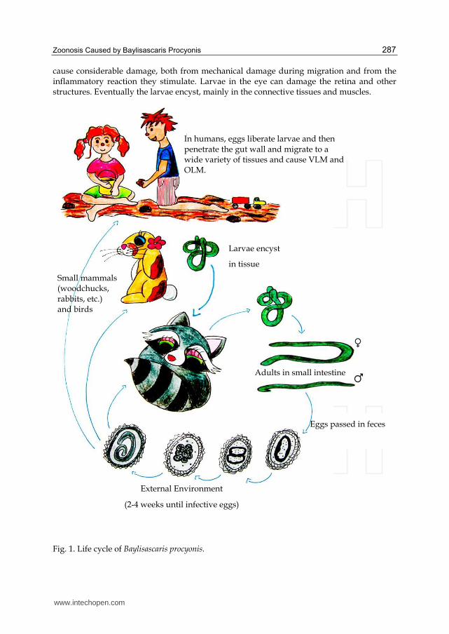
Year of published
report Location Age Sex Clinical Risk factor
Diagnostic method
Treatment Outcome
2002 Illinois 2.5 y Male Progressive encephalo-
pathy
Pica/ geophagia
Serologic Albendazole
and solumedrol
Neurologic deficits,
blindness, generalized spasticity
2002 Illinois 6 y Male
Progressive encephalopathy
, diffuse unilateral subacute
neuroretinitis
Developmental delay,pica/ geophagia
Serologic Albendazole
and prednisone
Neurologic deficits, seizures
2003 Michigan 6 y Male
Diffuse unilateral subacute
neuroretinitis, neurologic
deficits
Pica Serologic Severe
neurologic sequelae
2003 Michigan 2 y Male
Eosinophilic meningo-
encephalitis, chorioretinitis
Pica Serologic Severe
neurologic sequelae
2004 Louisiana 4 y Male Eosinophilic meningitis
Raccoons in neighborhood
Serologic Dexamethasone
albendazole Full recovery
2009 Oregon 17 y Male Eosinophilic
meningo-encephalitis
Geophagia and substance
abuse Serologic
Methyl-prednisolone
Aphasia and memory deficits
2009 Toronto 7 y Male Eosinophilic
meningo-encephailitis
Autism, raccoons in backyard
Serologic
Albendazole, methyl-
prednisone, prednisone
No longer used speech to
communicate, cortical visual impairment,
seizure disorder
2010 New York 12 mo Male Eosinophilic encephalo-
myelitis Geophagia Serologic
Albendazole, prednisone and
methyl-prednisolone
Neurologic deficits
Table 1. Reported human cases caused by Baylisascaris procyonis.
4.2 Infections in animals
Susceptibility to Baylisascaris larva migrans varies among animal groups and species (Wirtz 1982; Sheppard & Kazacos 1997). Animal groups particularly susceptible to parasite NLM include rodents, rabbits, primates, and birds, based on the number of cases and species affected. For example, in 2007 a Baylisascaris procyonis infection in a Moluccan cockatoo (Cacatua moluccensis), was reported. An adult female Moluccan cockatoo was evaluated for a 10-day history of progressive ataxia and weakness. The bird had been exposed intermittently over a 3-day period to a cage that had previously housed juvenile raccoons. Results of diagnostic tests were inconclusive and, despite supportive care, the bird died 7 days after the initial presentation. Histopathologic examination revealed a single nematode larva in the midbrain that was consistent with Baylisascaris species and multifocal granulomas in the left ventricle of the heart (Wolf et al., 2007).
www.intechopen.com

Zoonosis Caused by Baylisascaris Procyonis 293
Some animal groups and species are only marginally susceptible, with limited migration occurring in the intestinal wall or viscera; others appear to be resistant. For example, no cases of B. procyonis NLM have been documented in opossums, which are commonly exposed through foraging at raccoon latrines(Page et al., 1998, 1999), or in adult domestic livestockor zoo hoofstock, which are commonly exposed through contaminated hay. Very limited or no migration was seen in sheep, goats, and swine experimentally infected with B. procyonis (Dubey, 1982; Snyder, 1983; Kazacos & Kazacos 1984). No cases have been documented in cats or raptors, which eat rodents possibly contaminated with eggs and/or containing L3’s. (Kazacos, 2001). The apparent species limitations to Baylisascaris infection should be regarded with caution.
Except in very heavy infections with intestinal obstruction, raccoons infected with B.
procyonis appear clinically normal with no outward signs of infection. Similarly, other
species with Baylisascaris larva migrans usually are asymptomatic if no larvae enter the
brain. The severity and progression of central nervous system disease in NLM depends on
the number of eggs ingested, the number of larvae entering the brain, the location and
extent of migration damage and inflammation in the brain, and the size of the brain.Thus,
clinical disease will vary from mild, insidious, slowly progressive central nervous system
disease with subtle clinical signs to acute, fulminating, rapidly progressive central nervous
system disease with marked clinical signs. Although larvae enter the somatic tissues, eyes,
and brain of some species as early as 3 days postinfection, clinical central nervous system
disease is not usually apparent before 9–10 days postinfection, and in many cases not until
2–4+ weeks postinfection, due to the lag time in causing central nervous system damage and
inflammation (Kazacos, 1997; Sheppard& Kazacos, 1997).
5. Diagnostic
The diagnosis of baylisascariasis is difficult in live patients; there is no widely available,
non-invasive definitive test. The combination of encephalopathy with cerebrospinal fluid
(CSF) and peripheral eosinophilia and diffuse white matter disease on neuroimaging, with
or without eye disease, in a patient from North America or Europe should strongly suggest
the diagnosis of Baylisascaris NLM, and a history of exposure to raccoons or their feces
should be sought.
Unless a brain biopsy is done and a larva is found, antemortem diagnosis usually depends
on serology, with supportive evidence from other tests.
5.1 Serology
In neural larva migrans, antibodies to Baylisascaris can be found in serum and CSF; a rising
titer is usually seen. An enzyme linked immunosorbent assay (ELISA), indirect
immunofluorescence and immunoblotting (Western blotting) have been developed to detect
anti-Baylisascaris antibodies. These serological assays are not commercially available, but
they may be provided by university research laboratories. In the U.S., an ELISA is available
from the Department of Comparative Pathobiology at Purdue University, West Lafayette,
IN. Indirect immunofluorescence tests use frozen sections of B. procyonis third-stage larvae
as an antigen. Enzyme-linked immunosorbent assay and Western blotting use excretory-
www.intechopen.com

Zoonosis 294
secretory products from in vitro cultures of B. procyonis larvae as the antigen (Boyce et al.,
1989). Larval excretory-secretory antigens have been characterized as complex
glycoproteins, with molecular masses of 10 kDa to 200 kDa, that contain several different
sugar residues(Boyce et al, 1989). Protein epitopes of 33-kDa to 45-kDa antigens appear to be
recognized selectively by antibodies from B. procyonis-infected humans and animals but not
by normal human or T. canis antibody-positive sera (Boyce et al, 1989). In addition, children
with clinical B. procyonis NLM are strongly positive for anti-Baylisascaris antibodies in CSF
and serum and have consistently been negative for anti-Toxocara antibodies (Cunningham et
al., 1994; Fox et al., 1985; Gavin et al., 2002; Kazacos, 2001; Moertel et al., 2001; Murray, 2002;
Park et al., 2000; Rowley et al., 2000). In several of these cases, positive B. procyonis serology
was confirmed by brain biopsy or at autopsy (CDC, 2002; Fox et al., 1985; Huff et al., 1984;
Rowley et al., 2000). Acute and convalescent-phase titers characteristically demonstrate
several fold increases in both serum and CSF anti-Baylisascaris antibody levels (Gavin et al.,
2002; Moertel et al., 2001).
Although B. procyonis excretory-secretory (BPES) antigen-based ELISA and Western blot
assays are useful in the immunodiagnosis of this infection, cross-reactivity remains a
major problem. Recently, a recombinant B. procyonis antigen, BpRAG1, was reported for
use in development of improved serological assays for the diagnosis of Baylisascaris larva
migrans (Dangoudoubiyam et al., 2010). In a recent study, authors tested a total of 384
human patient serum samples in a BpRAG1 ELISA, including 20 patients with clinical
Baylisascaris larva migrans, 137 patients with other parasitic infections (8 helminth and 4
protozoan), and 227 with unknown/suspected parasitic infections. A sensitivity of 85%
and specificity of 86.9% was observed with the BpRAG1 ELISA, compared to only 39.4%
specificity with the BPES ELISA. In addition, the BpRAG1 ELISA had a low degree
ofcross-reactivity with antibodies to Toxocara spp. infection (25%), while the BPES antigen
showed 90.6% cross-reactivity. Based on these results, BpRAG1 antigen has a high degree
of sensitivity and specificity and should be very useful and reliable in the diagnosis
and seroepidemiology of Baylisascaris larva migrans by ELISA (Dangoudoubiyam et al.,
2011)
5.2 Laboratory tests
Although no routine laboratory test is considered diagnosticof B. procyonis NLM by itself, a
number of studies provide additional supporting evidence. Most importantly, the presence
of eosinophilia, particularly eosinophilic meningitis,
should alert the physician to the possibility of a parasitic etiology(Lo Re& Gluckman, 2003; Rothenberg, 1998; Weller, 1993). Eosinophils are not normally present in CSF; their presence narrows the differential diagnosis of CNS disease and provides an early or theonly etiologic clue. In documented cases of NLM, the peripheral white blood cell count is usually mildly elevated, but eosinophilia may be marked. Cerebrospinal fluid cell counts may be normal at presentation and generally demonstrate only mild leukocytosis, again with eosinophilia. Notably, even in the absence of pleocytosis, demonstrable CSF eosinophilia may be evident. Because eosinophils are easily missed in unstained or Gram-stained CSF, it may be necessary to request Wright’s orGiemsa stain of cytocentrifuged CSF specimens. In documented cases of NLM, CSF protein is generally normal or only mildly elevated, while
www.intechopen.com

Zoonosis Caused by Baylisascaris Procyonis 295
CSF glucose levels are normal. Although the finding of elevated serum isohemagglutinins, caused by cross-reactions between larval glycoproteins and human blood group antigens, is not specific for baylisascariasis, it does provide an additional clue to the diagnosis (Boyce et al., 1989).
5.3 Other techniques
Imaging techniques and encephalography provide supportive evidence and help rule out
other causes. In ocular larva migrans, an ophthalmoscopic examination may occasionally
reveal large, motile larvae in the retina, as well as choroidioretinitis and other signs of
DUSN. The presence of Baylisascaris larvae in the eye is also suggestive in cases with
neurological signs. Biopsies of the CNS are occasionally definitive, but larvae are often
absent from the sample. A definitive diagnosis can also be made retrospectively from CNS
samples taken at autopsy. Baylisascaris larvae are much larger (up to 80 μm in diameter and
up to 1900 μm long) than Toxocara larvae, and can also be distinguished by their
morphology. However, parasite larvae can be difficult to identify within tissues, and
misidentification is common. In tissues, the third stage larvae of B. procyonis cannot be
differentiated from B. columnaris or B. melis. Epidemiological evidence, such as a history of
exposure to raccoons but not skunks or badgers, can be suggestive.
6. Treatment
The prognosis for B. procyonis NLM is grave with or without treatment; among documented
cases,there are no neurologically intact survivors. In this parasite is very important early
clinical suspicion of raccoon roundworm meningoencephalitis.
The majority of cases have been treated with anthelmintics and corticosteroids. Empirical
anthelmintic treatment with thiabendazole, fenbendazole, tetramisole, or ivermectin has
failed to prevent death or unfavorable outcomes. Animal data suggest that albendazole and
diethylcarbazinehave the best CSF penetration and larvicidal activity (Kazacos, 2001).
Treatment with albendazole is protective in animal models if eggs have been ingested, but symptoms have not yet developed. In humans, albendazole has been used prophylactically after exposure to raccoon latrines or other sources of eggs. Whether it is helpful in patients with clinical signs is uncertain, because the death of the parasite might worsen the inflammation.
Albendazole appears to have the more favorable pharmacologic profile, with good absorption, high serum concentrations of the active metabolite, good penetration across the blood-brain barrier, and minimal toxicity (de Silva et al., 1997; Jung et al., 1990).
Most clinical cases have been treated concurrently with anthelmintics and corticosteroids;
the corticosteroids are used to suppress inflammation caused by the death of the larvae, as
well as to dampen the existing inflammatory response. In a recent case a early intervention
with both, albendazole and steroids, may have contributed to patient’s partial recovery
(Hajek et al., 2009) and a mild case of suspected B procyonis infection, with apparent early
complete response after cerebellar edema, was treated with early corticosteroids and later
albendazole (Pai et al., 2007).
www.intechopen.com

Zoonosis 296
Other supportive therapy may also be given. Recently, an acute eosinophilic meningoencephalitis, caused by Baylisascaris procyonis in a previously healthy teenager with a history of substance abuse, was treated with methylprednisolone; no antihelmintic drugs were administered (Chun et al., 2009).
Laser photocoagulation, systemic corticosteroids and other therapies have been used in
ocular larva migrans (Goldberg et al., 1993; Kazacos et al., 1985).
In many cases, significant damage has already occurred by the time treatment is begun, and
improvement is not seen. The best chance of recovery is expected with a very early
diagnosis and treatment.
7. Prevention and control
In intermediate hosts, the risk of infection can be decreased by avoiding contact with
raccoons, other definitive hosts and their feces. Raccoons can be discouraged from visiting
homes and farms with the same measures that are used to prevent disease in humans.
Infections are difficult to prevent completely in pets allowed outdoors, as the infective eggs
can survive for long periods in the environment. In dogs, monthly heartworm/nematode
preventatives appear to decrease the risk of intestinal infection with B. procyonis. In high-risk
areas, dogs that are not on these preventatives should receive regular fecal examinations to
decrease the risk that they will shed eggs.
In zoos and other facilities, the housing for intermediate hosts should be designed to
minimize exposure to raccoons, skunks and other definitive hosts. Captive raccoons and
skunks should be kept in dedicated cages that can be cleaned, if necessary, with the harsh
methods required to destroy Baylisascaris eggs. They should be tested regularly and
dewormed when necessary, and they should not be fed wild animals that might carry
larvae. Newly acquired definitive hosts should be quarantined and dewormed. Once
contamination has occurred, it can be difficult to remove completely. Intermediate hosts in
exhibits are sometimes treated prophylactically with pyrantel tartrate or ivermectin.
Similarly to humans, animals with recent exposure might also be treated with albendazole
to prevent the development of clinical signs.
In humans, risk of infection is greatest when infants or toddlers with geophagia or pica
come in contact with raccoon latrines or an environment contaminated by infected raccoon
feces. Young infants and toddlers, particularly those with pica or geophagia, should be kept
away from potentially contaminated areas.
Raccoon latrines in and around homes and play areas shouldbe cleaned up and
decontaminated. However, the longevity of B. procyonis eggs and their resistance to
disinfection or decontamination makes successful environmental cleanup difficult. Recently
a publication (Shafir et al., 2011) showed that eggs survived complete desiccation for at least
6 months at room temperature. Total loss of viability was observed after 7 months of
desiccation. Eggs frozen at –15°C for 6 months demonstrated no reduction in viability.
Freeze–thaw, including exposure to 5 cycles, did not affect survival. Although they did not
specifically design experiments to assess the effect of chlorine on inactivation of B. procyonis
eggs, exposure to undiluted household bleach for 90 min toremove the mammillated layer
www.intechopen.com

Zoonosis Caused by Baylisascaris Procyonis 297
did not affect viability. In this study (Shafir et al., 2011) B. procyonis eggs showed alow
thermal death point at <62°C, similar to that reported for other ascarids (Shafir et al., 2007;
Feachem et al., 1983).Heat is by far the best method of killing B. procyonis eggs (Kazacos,
2001). Boiling water, steam-cleaning, flaming, or fire are highly effective and practical
methods for decontamination of large or small areas. The use of direct flames from a
propaneflame-gun is a favored method (Kazacos, 2001). For heavily contaminated areas a
combination of removal and disposal of the top few inches of surface soil with flaming is
most effective.
8. Acknowledgment
Jacob Lorenzo Morales was funded by a “Ramón y Cajal” grant 2011 from the Spanish Ministry of Science and Innovation. Carmen Mª Martín was supported by a Ph.D. grant “Beca de Investigación CajaCanarias para Postgraduados 2010” from the University of La Laguna, Spain. Atteneri López was funded by the grant “Ayudas del Programa de Formación de Personal Investigador para la realización de Tesis Doctorales” from the Agencia Canaria de Investigación, Innovación y Sociedad de la Información from the Canary Islands Government. This work was funded by project RICET (project no. RD06/0021/0005 of the program of Redes Temáticas de Investigación Cooperativa, FIS), Spanish Ministry of Health, Madrid, Spain and the project “Protozoosis emergentes por amebas de vida libre: Aislamiento y caracterización molecular, identificación de cepas transportadas de otros agentes patógenos y búsqueda de quimioterapias” PI10/01298, Spanish Ministry of Science and Innovation, Madrid, Spain. Authors are thankful to Cecilia Arrate for drawing the Baylisascaris procyonis life cycle.
9. References
Anderson, D.C.; Greenwood, R.; Fishman, M. & Kagan, I.G. (1975). Acute infantile
hemiplegia with cerebrospinal fluid eosinophilic pleocytosis: an unusual case of
visceral larva migrans. The Journal of Pediatrics, Vol.86, nº2, (February 1975), pp.
247–249.
Baeur, C. & Gey, A. (1995). Efficacy of six anthelmintics against luminal stages of
Baylisascaris procyonis in naturally infected raccoons (Procyon lotor). Veterinary
Parasitology, Vol.60, nº1-2, (November 1995), pp. 155–159, ISSN 0304-4017
Beaver, P. (1969). The nature of visceral larva migrans. The Journal of Parasitology, Vol.55, nº1,
(February 1969), pp. 3–12, ISSN 0022-3395
Berry, J.F. (1985). Phylogenetic relationship between Baylisascaris spp. Sprent, 1968
(Nematoda: Ascarididae) from skunks, raccoons and groundhogs in southern
Ontario. M.S Thesis. University of Guelph, Guelph, Ontario, 99 pp.
Blizzard, E.L.; Davis, C.L.; Henke, S.; Long, D.B.; Hall, C.A. & Yabsley, M.J. (2010a).
Distribution, prevalence, and genetic characterization of Baylisascaris procyonis in
selected areas of Georgia. The Journal of Parasitology, Vol.96, nº6, (December 2010),
pp. 1128-33, ISSN 0022-3395.
Blizzard, E.L.; Yabsley, M.J.; Beck, M.F. & Harsch, S. (2010b). Geographic expansion of
Baylisascaris procyonis roundworms, Florida, USA. Emerging Infectious Diseases,
Vol.16, nº11, (November 2010), pp. 1803-4, ISSN 1080-6059.
www.intechopen.com

Zoonosis 298
Boyce, W.M.; Asai, D.J.; Wilder, J.K. & Kazacos, K.R. (1989). Physiochemical
characterization and monoclonal and polyclonal antibody recognition of
Baylisascaris procyonis larval excretory-secretory antigens. The Journal of
Parasitology, Vol.75, pp. 540–548.
Centers for Disease Control. 2002. Raccoon roundworm encephalitis—Chicago, Illinois, and
Los Angeles, California, 2000. Morbidity and Mortality Weekly Report, Vol.50, nº51,
(January 2002), pp. 1153–1155, ISSN 0149-2195.
Chun, C.S.; Kazacos, K.R.; Glaser, C.; Bardo, D.; Dangoudoubiyam, S. & Nash, R. (2009).
Global neurologic deficits with baylisascaris encephalitis in a previously healthy
teenager. The Pediatric Infectious Diseases Journal, Vol.28, nº10, (October 2009), pp.
925-7, ISSN 0891-3668.
Cunningham, C.K.; Kazacos, K.R.; Lucas, J.A.; McAuley, J.B.; Wozniak, E.J. & Weiner, L.B.
(1994). Diagnosis and management of Baylisascaris procyonis infection in an infant
with nonfatal meningoencephalitis. Clinical Infectious Diseases, Vol.18, nº6, (June
1994), pp. 868–872, ISSN 1058-4838.
Dangoudoubiyam, S.; Vemulapalli, R.; Hancock, K. & Kazacos, KR. (2010). Molecular
cloning of an immunogenic protein of Baylisascaris procyonis and expression in
Escherichia coli for use in developing improved serodiagnostic assays. Clinical and
Vaccine Immunology, Vol17, nº12, (December 2010), pp. 1933-9, ISSN 1556-6811
Dangoudoubiyam, S.; Vemulapalli, R.; Ndao, M. & Kazacos KR. (2011). A recombinant
antigen-based enzyme-linked immunosorbent assay for specific diagnosis of
Baylisascaris procyonis larva migrans. Clinical and Vaccine Immunology CVI
Accepts, (published online ahead of print on 10 August 2011)
doi:10.1128/CVI.00083-11
De Silva, N.; Guyatt, H. & Bundy, D. (1997). Anthelmintics. A comparative review of their
clinical pharmacology. Drugs Vol.53, nº5, (May 1997), pp. 769–788, ISSN 0012-
6667.
Dubey, J.P. (1982). Baylisascaris procyonis and eimerian infections in raccoons. Journal of the
American Veterinary Medical Association Vol.181, nº11, (December 1982), pp. 1292–
1294, ISSN 0003-1488.
Eberhard, M.L.; Nace, E.K.; Won, K.Y.; Punkosdy, G.A.; Bishop, H.S. & Johnston, S.P. (2003).
Baylisascaris procyonis in the metropolitan Atlanta area. Emerging Infectious Diseases,
Vol9, nº12, (December 2003), pp. 1636–7, ISSN 1080-6059.
Ermer, E.M. & Fodge, J.A. (1986). Occurrence of the raccoon roundworm in raccoons in
western New York. New York Fish and Game Journal, Vol.33, pp. 58–61, ISSN 0028-
7210.
Feachem, R.; Bradley, D.J.; Garelick, H. & Mara, D.D. 1983)Ascaris and ascariasis. In:
Sanitation and disease: health aspects of excreta and wastewater management, John Wiley
& Sons for World Bank Studies in Water Supply and Sanitation, p. 391,
Washington.
Fox, A.S.; Kazacos, K.R.; Gould, N.S.; Heydemann, P.T.; Thomas, C. & Boyer, K.M. (1985).
Fatal eosinophilic meningoencephalitis and visceral larva migrans caused by the
raccoon ascarid Baylisascaris procyonis. The New England Journal of Medicine Vol.312,
nº25, (June 1985), pp. 1619–1623, ISSN 0028-4793.
www.intechopen.com
Zoonosis Caused by Baylisascaris Procyonis 299
Gavin, P.J.; Kazacos, K.R.; Tan, T.Q.; Brinkman, W.B.; Byrd, S.E.; Davis, A.T.; Mets, M.B. &
Shulman, S.T. (2002). Neural larval migrans caused by the raccoon roundworm
Baylisascaris procyonis. The Pediatric Infectious Disease Journal, Vol.21, nº.10, (October
2002), pp. 971–975, ISSN 0891-3668.
Goldberg, M.A.; Kazacos, K.R.; Boyce, W.M.; Ai, E. & Katz, B. (1993). Diffuse unilateral
subacute neuroretinitis: morphometric, serologic, and epidemiologic support for
Baylisascaris as a causative agent. Ophthalmology, Vol.100, nº.11, (November 1993),
pp. 1695–701, ISSN 0161-6420.
Hajek, J.; Yau, Y.; Kertes, P.; Soman, T.; Laughlin, S.; Kanani, R.; Kazacos, K.;
Dangoudoubiyam, S. & Opavsky, M.A. (2009). A child with raccoon roundworm
meningoencephalitis: A pathogen emerging in your own backyard?. The Canadian
Journal of Infectious Diseases & Medical Microbiology, Vol. 20, nº4, (Winter 2009), pp.
177-80, ISSN 1712-9532.
Hoffmann, C.O. & Gottschang, J.L. (1977). Numbers, distribution and movements of a
raccoon population in a suburban residential community. Journal of Mammalogy,
Vol.58, No.4, (November 1977), pp. 623-36, ISSN 0022-2372.
Huff, D.S.; Neafie, R.C.; Binder, M.J.; Leon, G.A.D.; Brown, L.W. & Kazacos, K.R. (1984). The
first fatal Baylisascaris infection in humans. Pediatric Pathology, Vol.2, nº3, (pp. 345–
52, ISSN 0277-0938.
Jung, H.; Hurtado, M.; Sanchez, M.; Medina, M. & Sotelo, J. (1990). Plasma and CSF levels of
albendazole and praziquantel in patients with neurocysticercosis. Clinical
Neuropharmacology, Vol.13, nº6, (December 1990), pp. 559–564, ISSN 0362-5664.
Kazacos, K.R. (1986). Raccoon ascarids as a cause of larva migrans. Parasitology Today, Vol.2,
nº9, (September 1986), pp. 253–255, ISSN 0169-4758.
Kazacos, KR. (1991). Visceral and ocular larva migrans. Seminars in Veterinary Medicine and
Surgery (Small Animal), Vol.6, nº3, (August 1991), pp. 227–235, ISSN 0882-0511.
Kazacos, K.R. (1997). Visceral, ocular, and neural larva migrans. In: Pathology of infectious
diseases, Vol. II. Connor, D.H.; Chandler, F.W.; Schwartz, D.A.; Manz, H.J. & Lack,
E.E., pp. 1459–73, Appleton & Lange, Stamford, CT.
Kazacos, KR. (2000). Protecting children from helminthic zoonoses. Contemporary Pediatrics,
Vol.95, nº3, (Suppl) pp. 1–24, ISSN 8750-0507.
Kazacos, K.R. (2001). Baylisascaris procyonis and related species. In: Parasitic diseases of wild
mammals. 2nd ed. Samuels, W.M.; Pybus, M.J. & Kocans, A.A., pp. 301–41, Iowa State
University Press/Ames, Iowa.
Kazacos, K.R. & Kazacos, E.A. (1984). Experimental infection of domestic swine with
Baylisascaris procyonis from raccoons. American Journal of Veterinary Research, Vol.45,
nº6, (June 1984), pp. 1114–1121, ISSN 0002-9645.
Kazacos, K.R.; Raymond, L.A.; Kazacos, E.A. & Vestre, W.A. (1985). The raccoon ascarid. A
probable cause of human ocular larva migrans. Ophthalmology, Vol.92, nº12,
(December 1985), pp. 1735–1743, ISSN 0161-6420.
Kazacos, K.R. & Boyce, W.M. (1989). Baylisascaris larva migrans. Journal of the American
Veterinary Medical Association, Vol.195, nº7, (October 1989), pp. 894-903, ISSN 0003-
1488.
www.intechopen.com

Zoonosis 300
Lo Re, V.III & Gluckman, S.J. (2003). Eosinophilic meningitis. The American Journal of
Medicine, Vol.114, nº3, (February 2003), pp. 217–223, ISSN 0002-9343.
McClure, G. (1933). Nematode parasites of mammals. From specimens collected in the New
York Zoological Park, 1931. Zoologica (New York) Vol.15 pp. 29–47.
Mets, M.B.; Noble, A.G.; Basti, S.; Gavin, P.; Davis, A.T.; Shulman, S.T. & Kazacos KR.
(2003). Eye findings of diffuse unilateral subacute neuroretinitis and multiple
choroidal infiltrates associated with neural larva migrans due to Baylisascaris
procyonis. American Journal of Ophthalmology,Vol.135, nº6, (June 2003), pp. 888-90,
ISSN 0002-9394.
Miyashita, M. (1993). Prevalence of Baylisascaris procyonis in raccoons in Japan and
experimental infections of the worm to laboratory animals. Journal Urban Living
Health Association, Vol.37, pp. 137–151. (In Japanese.)
Moertel, C.L.; Kazacos, K.R.; Butterfield, J.H.; Kita, H.; Watterson, J. & Gleich, G.J. (2001).
Eosinophil-associated inflammation and elaboration of eosinophil-derived proteins
in two children with raccoon roundworm (Baylisascaris procyonis) encephalitis.
Pediatrics, Vol.108, nº5, (November 2001), pp. E93, ISSN 0031-4005.
Murray, W.J. (2002). Human infections caused by the raccoon roundworm, Baylisascaris
procyonis. Clinical Microbiology Newsletter, Vol.24, nº1, pp. 1-7, ISSN 0196-4399.
Murray, W.J. & Kazacos, K.R. (2004). Raccoon roundworm encephalitis. Clinical Infectious
Diseases, Vol.39, nº10, (November 2004), pp. 1484-92, ISSN 1058-4838.
Page, L.K.; Swihart, R.K. & Kazacos, K.R. (1998). Raccoon latrine structure and its potential
role in transmission of Baylisascaris procyonis to vertebrates. American Midland
Naturalist, Vol.140, nº 1, (July 1998), pp. 180-185, ISSN 0003-0031.
Page, L.K.; Swihart, R.K. & Kazacos, K.R. (1999). Implications of raccoon latrines in the
epizootiology of baylisascariasis. Journal of Wildlife Diseases, Vol.35, nº3, (July 1999),
pp. 474-80, ISSN 0090-3558.
Pai, P.J.; Blackburn, B.G.; Kazacos, K.R.; Warrier, R.P. & Bégué RE. (2007). Full recovery
from Baylisascaris procyonis eosinophilic meningitis. Emerging Infectious Diseases,
Vol.13, nº6, (June 2007), pp. 928-30, ISSN 1080-6040.
Park, S.Y.; Glaser, C.; Murray, W.J.; Kazacos, K.R.; Rowley, H.A.; Fredrick, D.R. & Bass, N.
(2000). Raccoon roundworm (Baylisascaris procyonis) encephalitis: Case report and
field investigation. Pediatrics, Vol.106, nº4, (October 2000), pp. E56, ISSN 0031-
4005.
Perlman, J.E.; Kazacos, K.R.; Imperato, G.H.; Desai, R.U.; Schulman, S.K.; Edwards, J.;
Pontrelli, L.R.; Machado, F.S.; Tanowitz, H.B. & Saffra, N.A. (2010). Baylisascaris
procyonis neural larva migrans in an infant in New York City. Journal of
Neuroparasitology, Vol.1, (June 2010), pp., ISSN 2090-2344
Rothenberg, M.E. (1998). Eosinophilia. The New England Journal of Medicine, Vol.338, nº22,
(May 1998), pp. 1592-600, ISSN 0028-4793.
Roussere, G.P.; Murray, W.J.; Raudenbush, C.B.; Kutilek, M.J.; Levee, D.J. & Kazacos, K.R.
(2003). Raccoon roundworm eggs near homes and risk for larva migrans disease,
California communities. Emerging Infectious Diseases, Vol.9, nº12, (December 2003),
pp. 1516–22, ISSN 1080-6040.
www.intechopen.com

Zoonosis Caused by Baylisascaris Procyonis 301
Rowley, H.A.; Uht, R.M.; Kazacos, K.R.; Sakanari, J.; Wheaton, W.V.; Barkovich, A.J. &
Bollen, A.W. (2000). Radiologic-pathologic findings in raccoon roundworm
(Baylisascaris procyonis) encephalitis. AJNR. American Journal of Neuroradiology,
Vol.21, nº2, (February 2000), pp. 415–420, ISSN 0195-6108.
Sakla, A.A.; Donnelly, J.J.; Khatami, M. & Rockey, J.H. (1989). Baylisascaris procyonis
(Stefanski and Zarnowski, 1951) Ascarididae: Nematoda. I. Embryonic
development and morphogenesis of second stage larvae. Assiut Veterinary Medical
Journal, Vol.21, pp. 68–76.
Sato, H.; Furuoka, H. & Kamiya, H. (2002). First outbreak of Baylisascaris procyonis larva
migrans in rabbits in Japan. Parasitology international,Vol.51, nº1, (March 2002), pp.
105–108, ISSN 1383-5769.
Sexsmith, J.L.; Whiting, T.L.; Gree, C.; Orvis, S.; Berezanski, D.J. & Thompson A.B. (2009).
Prevalence and distribution of Baylisascaris procyonis in urban raccoons (Procyon
lotor) in Winnipeg, Manitoba. The Canadian Veterinary Journal, Vol.50, nº8, (August
2009), pp. 846-50, ISSN 0008-5286.
Shafir, S.C.; Wang, W.; Sorvillo, F.J.; Wise, M.E.; Moore, L., Sorvilo, T. & Eberhard, M.L:
(2007). Thermal death point of Baylisascaris procyonis eggs. Emerging Infectious
Diseases, Vol.13, nº1, (January 2007) pp. 172–3, ISSN 1080-6040.
Shafir, S.C.; Sorvillo, F.J.; Sorvillo, T. & Eberhard, M.L. (2011). Viability of Baylisascaris
procyonis Eggs. Emerging Infectious Diseases, Vol.17, nº7, (July 2011), pp. 1293-5, ISSN
1080-6040.
Sheppard, C.H. & Kazacos, K.R. (1997). Susceptibility of Peromyscus leucopus and Mus
musculus to infection with Baylisascaris procyonis. The Journal of Parasitology, Vol.83,
nº6, (December 1997), pp. 1104–1111, ISSN 0022-3395.
Snyder, D.E. (1983). The prevalence, cross-transmissibility to domestic animals and adult
structure of Baylisascaris procyonis (Nematoda) from Illinois raccoons (Procyon lotor).
Ph.D. Thesis, University of Illinois at Urbana-Champaign, Urbana, IL, 233 pp.
Snyder, D.E. & Fitzgerald, P.R. (1985). The relationship of Baylisascaris procyonis to Illinois
raccoons (Procyon lotor). The Journal of Parasitology, Vol.71, nº5, (October 1985), pp.
596–598, ISSN 0022-3395.
Souza, M.J.; Ramsay, E.C.; Patton, S. & New, J.C. (2009). Baylisascaris procyonis in raccoons
(Procyon lotor) in eastern Tennessee. Journal of Wildlife Diseases, Vol.45, nº4, (October
2009), pp. 1231–4, ISSN 0090-3558.
Sprent, J.F.A. (1968). Notes on Ascaris and Toxascaris, with a definition of Baylisascaris gen.
nov. Parasitology, Vol.58, nº1, (February 1968), pp. 185–198, ISSN 0031-1820.
Stefanski, W. & Zarnowski, E. (1951). Ascaris procyonis n. sp. z jelita szopa (Procyon lotor L.)
Ascaris procyonis n. sp. provenant de l’intestin de Procyon lotor. Ann. Mus. Zool.
Polonici, Vol.14, pp. 199–202.
Weller, P. F. (1993). Eosinophilic meningitis. The American Journal of Medicine, Vol.95, nº3,
(September 1993), pp. 250–253, ISSN 0002-9343.
Wirtz, W.L. (1982). Cerebrospinal nematodiasis due to the raccoon ascarid, Baylisascaris
procyonis. M.S. Thesis, Purdue University, West Lafayette, IN, 90 pp
www.intechopen.com

Zoonosis 302
Wolf, K.N.; Lock, B.; Carpenter, J.W. & Garner, M.M. (2007). Baylisascaris procyonis infection
in a Moluccan cockatoo (Cacatua moluccensis). Journal of Avian Medicine and Surgery,
Vol.21, nº3, (September 2007), pp. 220-5, ISSN 1082-6742.
Yeitz, J.L.; Gillin, C.M.; Bildfell, R.J. & Debess, E.E. (2009). Prevalence of Baylisascaris
procyonis in raccoons (Procyon lotor) in Portland, Oregon, USA. Journal of Wildlife
Diseases, Vol.45, nº1, (January 2009), pp. 14-8, ISSN 0090-3558.
www.intechopen.com
ZoonosisEdited by Dr. Jacob Lorenzo-Morales
ISBN 978-953-51-0479-7Hard cover, 436 pagesPublisher InTechPublished online 04, April, 2012Published in print edition April, 2012
InTech EuropeUniversity Campus STeP Ri Slavka Krautzeka 83/A 51000 Rijeka, Croatia Phone: +385 (51) 770 447 Fax: +385 (51) 686 166www.intechopen.com
InTech ChinaUnit 405, Office Block, Hotel Equatorial Shanghai No.65, Yan An Road (West), Shanghai, 200040, China
Phone: +86-21-62489820 Fax: +86-21-62489821
Zoonotic diseases are mainly caused by bacterial, viral or parasitic agents although "unconventional agents"such as prions could also be involved in causing zoonotic diseases. Many of the zoonotic diseases are a publichealth concern but also affect the production of food of animal origin thus they could cause problems ininternational trade of animal-origin goods. A major factor contributing to the emergence of new zoonoticpathogens in human populations is increased contact between humans and animals. This book provides aninsight on zoonosis and both authors and the editor hope that the work compiled in it would help to raiseawareness and interest in this field. It should also help researchers, clinicians and other readers in theirresearch and clinical usage.
How to referenceIn order to correctly reference this scholarly work, feel free to copy and paste the following:
José Piñero, Jacob Lorenzo-Morales, Carmen Martín-Navarro, Atteneri López-Arencibia, María Reyes-Batlleand Basilio Valladares (2012). Zoonosis Caused by Baylisascaris procyonis, Zoonosis, Dr. Jacob Lorenzo-Morales (Ed.), ISBN: 978-953-51-0479-7, InTech, Available from:http://www.intechopen.com/books/zoonosis/zoonosis-caused-by-baylisascaris-procyonis