ZEISS AngioPlex OCT Angiography - Eyetube.net · ZEISS AngioPlex OCT Angiography Proliferative...
12
ZEISS AngioPlex OCT Angiography Clinical Case Reports
Transcript of ZEISS AngioPlex OCT Angiography - Eyetube.net · ZEISS AngioPlex OCT Angiography Proliferative...
ZEISS AngioPlex OCT AngiographyClinical Case Reports
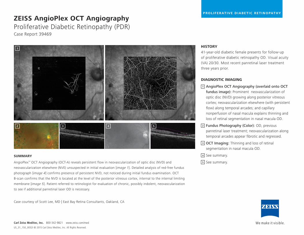
ZEISS AngioPlex OCT Angiography Proliferative Diabetic Retinopathy (PDR) Case Report 39469
HISTORY
41-year-old diabetic female presents for follow-up of proliferative diabetic retinopathy OD. Visual acuity (VA) 20/30. Most recent panretinal laser treatment three years prior.
DIAGNOSTIC IMAGING
1 AngioPlex OCT Angiography (overlaid onto OCT fundus image): Prominent neovascularization of optic disc (NVD) growing along posterior vitreous cortex; neovascularization elsewhere (with persistent flow) along temporal arcades; and capillary nonperfusion of nasal macula explains thinning and loss of retinal segmentation in nasal macula OD.
Fundus Photography (Color): OD, previous panretinal laser treatment; neovascularization along temporal arcades appear fibrotic and regressed.
OCT Imaging: Thinning and loss of retinal segmentation in nasal macula OD.
See summary.
See summary.
Carl Zeiss Meditec, Inc. 800-342-9821 www.zeiss.com/med
US_31_150_0032I © 2015 Carl Zeiss Meditec, Inc. All Rights Reserved.
P R O L I F E R AT I V E D I A B E T I C R E T I N O PAT H Y
SUMMARY
AngioPlex™ OCT Angiography (OCT-A) reveals persistent flow in neovascularization of optic disc (NVD) and
neovascularization elsewhere (NVE) unsuspected in initial evaluation [image 1]. Detailed analysis of red-free fundus
photograph [image 4] confirms presence of persistent NVD, not noticed during initial fundus examination. OCT
B-scan confirms that the NVD is located at the level of the posterior vitreous cortex, internal to the internal limiting
membrane [image 5]. Patient referred to retinologist for evaluation of chronic, possibly indolent, neovascularization
to see if additional panretinal laser OD is necessary.
Case courtesy of Scott Lee, MD | East Bay Retina Consultants, Oakland, CA
1
2 3 4 5
1
2
3
4
5
ZEISS AngioPlex OCT Angiography Central retinal vein occlusion with optic disc neovascularization Case Report 49473
HISTORY
50-year-old male patient received two anti-VEGF injections OD for cystoid macular edema due to central retinal vein occlusion. Patient was lost to follow-up for 8 months, and presents for evaluation of decreased vision of count fingers at one foot, OD
DIAGNOSTIC IMAGING
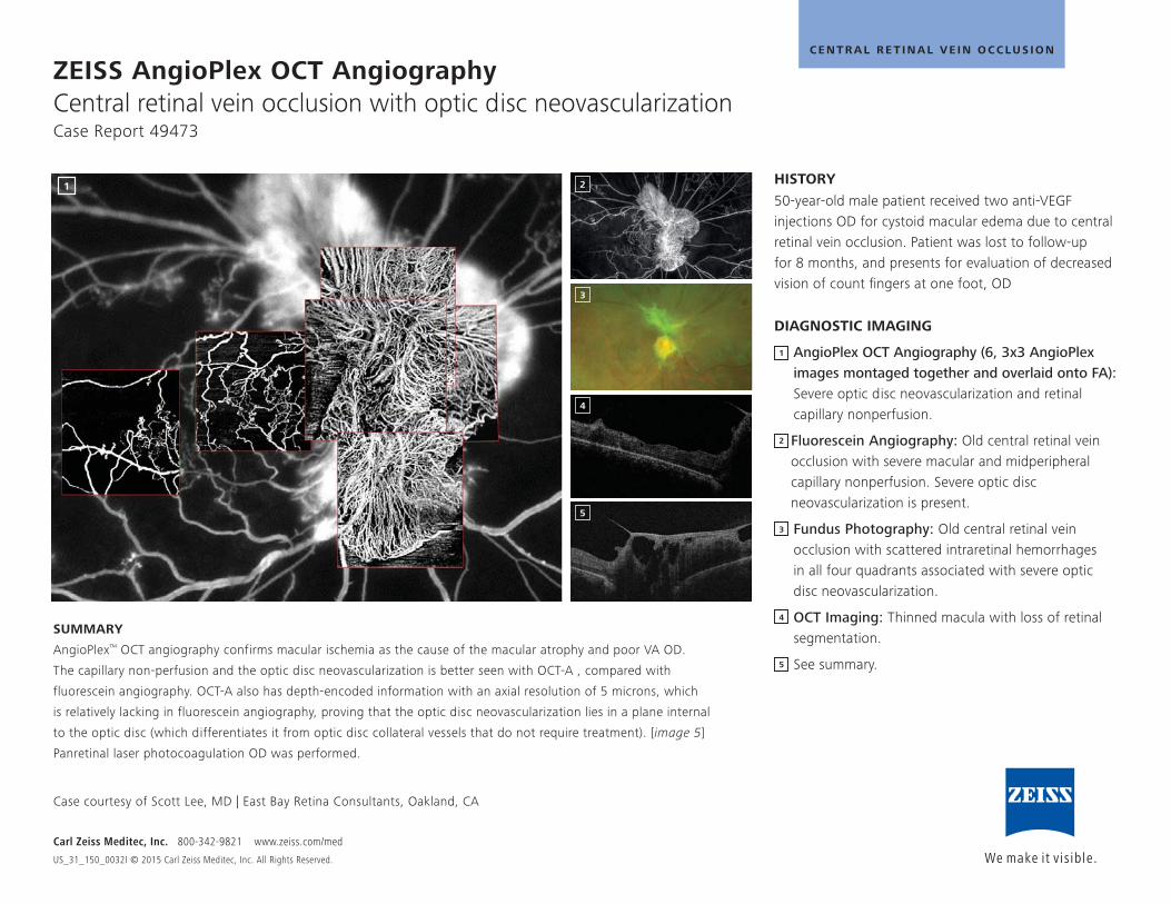
AngioPlex OCT Angiography (6, 3x3 AngioPlex images montaged together and overlaid onto FA): Severe optic disc neovascularization and retinal capillary nonperfusion.
Fluorescein Angiography: Old central retinal vein occlusion with severe macular and midperipheral capillary nonperfusion. Severe optic disc neovascularization is present.
Fundus Photography: Old central retinal vein occlusion with scattered intraretinal hemorrhages in all four quadrants associated with severe optic disc neovascularization.
OCT Imaging: Thinned macula with loss of retinal segmentation.
See summary.
Carl Zeiss Meditec, Inc. 800-342-9821 www.zeiss.com/med
US_31_150_0032I © 2015 Carl Zeiss Meditec, Inc. All Rights Reserved.
C E N T R A L R E T I N A L V E I N O C C LU S I O N
SUMMARY
AngioPlex™ OCT angiography confirms macular ischemia as the cause of the macular atrophy and poor VA OD.
The capillary non-perfusion and the optic disc neovascularization is better seen with OCT-A , compared with
fluorescein angiography. OCT-A also has depth-encoded information with an axial resolution of 5 microns, which
is relatively lacking in fluorescein angiography, proving that the optic disc neovascularization lies in a plane internal
to the optic disc (which differentiates it from optic disc collateral vessels that do not require treatment). [image 5]
Panretinal laser photocoagulation OD was performed.
Case courtesy of Scott Lee, MD | East Bay Retina Consultants, Oakland, CA
1 2
3
4
5
1
2
3
4
5
ZEISS AngioPlex OCT Angiography Proliferative diabetic retinopathy with macular edemaCase Report 48905
HISTORY
49-year-old male diabetic patient presents for anti-VEGF injection #7 for diabetic macular edema OS. Last anti-VEGF injection OS was 4 months prior, and last panretinal laser photocoagulation OS was 8 months prior.
DIAGNOSTIC IMAGING
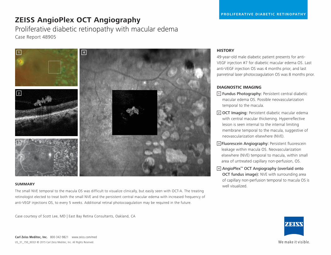
1 Fundus Photography: Persistent central diabetic macular edema OS. Possible neovascularization temporal to the macula.
2 OCT Imaging: Persistent diabetic macular edema with central macular thickening. Hyperreflective lesion is seen internal to the internal limiting membrane temporal to the macula, suggestive of neovascularization elsewhere (NVE).
Fluorescein Angiography: Persistent fluorescein leakage within macula OS. Neovascularization elsewhere (NVE) temporal to macula, within small area of untreated capillary non-perfusion, OS.
AngioPlex™ OCT Angiography (overlaid onto OCT fundus image): NVE with surrounding area of capillary non-perfusion temporal to macula OS is well visualized.
Carl Zeiss Meditec, Inc. 800-342-9821 www.zeiss.com/med
US_31_150_0032I © 2015 Carl Zeiss Meditec, Inc. All Rights Reserved.
SUMMARY
The small NVE temporal to the macula OS was difficult to visualize clinically, but easily seen with OCT-A. The treating
retinologist elected to treat both the small NVE and the persistent central macular edema with increased frequency of
anti-VEGF injections OS, to every 5 weeks. Additional retinal photocoagulation may be required in the future.
Case courtesy of Scott Lee, MD | East Bay Retina Consultants, Oakland, CA
1 4
2
3
1
2
3
4
P R O L I F E R AT I V E D I A B E T I C R E T I N O PAT H Y
ZEISS AngioPlex OCT Angiography Neovascular age-related macular degenerationCase Report 33347
HISTORY
69-year-old male patient, who has been treated with anti-VEGF injections OS for wet age-related macular degeneration (AMD) for 6 years and 7 months, presents for 57th intravitreal anti-VEGF OS. Visual acuity (VA) 20/80 OS. Most recent intravitreal anti-VEGF OS was 7 weeks prior, and first and only photodynamic therapy OS 8 months prior.
DIAGNOSTIC IMAGING
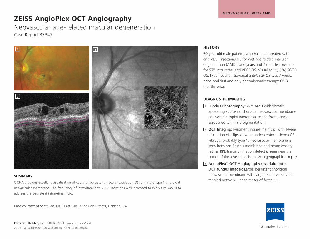
Fundus Photography: Wet AMD with fibrotic appearing subfoveal choroidal neovascular membrane OS. Some atrophy inferonasal to the foveal center associated with mild pigmentation.
OCT Imaging: Persistent intraretinal fluid, with severe disruption of ellipsoid zone under center of fovea OS. Fibrotic, probably type 1, neovascular membrane is seen between Bruch’s membrane and neurosensory retina. RPE transillumination defect is seen near the center of the fovea, consistent with geographic atrophy.
AngioPlex™ OCT Angiography (overlaid onto OCT fundus image): Large, persistent choroidal neovascular membrane with large feeder vessel and tangled network, under center of fovea OS.
Carl Zeiss Meditec, Inc. 800-342-9821 www.zeiss.com/med
US_31_150_0032I © 2015 Carl Zeiss Meditec, Inc. All Rights Reserved.
SUMMARY
OCT-A provides excellent visualization of cause of persistent macular exudation OS: a mature type 1 choroidal
neovascular membrane. The frequency of intravitreal anti-VEGF inejctions was increased to every five weeks to
address the persistent intraretinal fluid.
Case courtesy of Scott Lee, MD | East Bay Retina Consultants, Oakland, CA
1 3
2
N E OVAS C U LA R ( W E T ) A M D
1
2
3
ZEISS AngioPlex OCT Angiography Branch retinal artery occlusionCase Report 46605
HISTORY
81-year-old male patient with history of systemic hypertension, coronary artery disease, and JAK2 mutation positive thrombocytosis presents with 2-day history of sudden onset of visual field loss above fixation OD. Visual acuity (VA) OD 20/30, consistent with 3- nuclear sclerotic cataract OD.
DIAGNOSTIC IMAGING
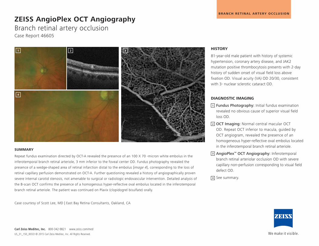
Fundus Photography: Initial fundus examination revealed no obvious cause of superior visual field loss OD.
OCT Imaging: Normal central macular OCT OD. Repeat OCT inferior to macula, guided by OCT angiogram, revealed the presence of an homogeneous hyper-reflective oval embolus located in the inferotemporal branch retinal arteriole.
AngioPlex™ OCT Angiography: Inferotemporal branch retinal arteriolar occlusion OD with severe capillary non-perfusion corresponding to visual field defect OD.
See summary.
Carl Zeiss Meditec, Inc. 800-342-9821 www.zeiss.com/med
US_31_150_0032I © 2015 Carl Zeiss Meditec, Inc. All Rights Reserved.
B R A N C H R E T I N A L A R T E R Y O C C LU S I O N
SUMMARY
Repeat fundus examination directed by OCT-A revealed the presence of an 100 X 70 -micron white embolus in the
inferotemporal branch retinal arteriole, 3 mm inferior to the foveal center OD. Fundus photography revealed the
presence of a wedge-shaped area of retinal infarction distal to the embolus [image 4], corresponding to the loss of
retinal capillary perfusion demonstrated on OCT-A. Further questioning revealed a history of angiographically proven
severe internal carotid stenosis, not amenable to surgical or radiologic endovascular intervention. Detailed analysis of
the B-scan OCT confirms the presence of a homogenous hyper-reflective oval embolus located in the inferotemporal
branch retinal arteriole. The patient was continued on Plavix (clopidogrel bisulfate) orally.
Case courtesy of Scott Lee, MD | East Bay Retina Consultants, Oakland, CA
1 2 3
4
1
2
3
4
ZEISS AngioPlex OCT Angiography Subretinal neovascular membraneCase Report 52990
Carl Zeiss Meditec, Inc. 800-342-9821 www.zeiss.com/med
US_31_150_0032I © 2015 Carl Zeiss Meditec, Inc. All Rights Reserved.
S U B R E T I N A L N E OVAS C U LA R M E M B R A N E
HISTORY
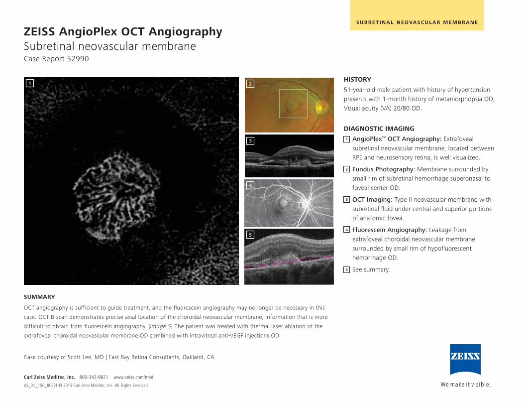
51-year-old male patient with history of hypertension presents with 1-month history of metamorphopsia OD, Visual acuity (VA) 20/80 OD.
DIAGNOSTIC IMAGING
AngioPlex™ OCT Angiography: Extrafoveal subretinal neovascular membrane, located between RPE and neurosensory retina, is well visualized.
Fundus Photography: Membrane surrounded by small rim of subretinal hemorrhage superonasal to foveal center OD.
OCT Imaging: Type II neovascular membrane with subretinal fluid under central and superior portions of anatomic fovea.
Fluorescein Angiography: Leakage from extrafoveal choroidal neovascular membrane surrounded by small rim of hypofluorescent hemorrhage OD.
See summary.
SUMMARY
OCT angiography is sufficient to guide treatment, and the fluorescein angiography may no longer be necessary in this
case. OCT B-scan demonstrates precise axial location of the choroidal neovascular membrane, information that is more
difficult to obtain from fluorescein angiography. [image 5] The patient was treated with thermal laser ablation of the
extrafoveal choroidal neovascular membrane OD combined with intravitreal anti-VEGF injections OD.
Case courtesy of Scott Lee, MD | East Bay Retina Consultants, Oakland, CA
1 2
3
4
5
1
2
3
4
5
ZEISS AngioPlex OCT Angiography Adult-onset foveomacular vitelliform dystrophy (AOFVD)Case Report 34174
HISTORY
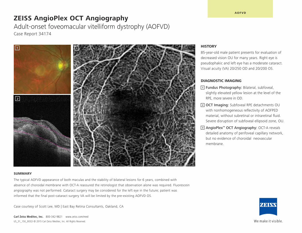
85-year-old male patient presents for evaluation of decreased vision OU for many years. Right eye is pseudophakic and left eye has a moderate cataract. Visual acuity (VA) 20/250 OD and 20/200 OS.
DIAGNOSTIC IMAGING
Fundus Photography: Bilateral, subfoveal, slightly elevated yellow lesion at the level of the RPE, more severe in OD.
OCT Imaging: Subfoveal RPE detachments OU with nonhomogeneous reflectivity of AOFPED material, without subretinal or intraretinal fluid. Severe disruption of subfoveal ellipsoid zone, OU.
AngioPlex™ OCT Angiography: OCT-A reveals detailed anatomy of perifoveal capillary network, but no evidence of choroidal neovascular membrane.
Carl Zeiss Meditec, Inc. 800-342-9821 www.zeiss.com/med
US_31_150_0032I © 2015 Carl Zeiss Meditec, Inc. All Rights Reserved.
AO F V D
SUMMARY
The typical AOFVD appearance of both maculas and the stability of bilateral lesions for 6 years, combined with
absence of choroidal membrane with OCT-A reassured the retinologist that observation alone was required. Fluorescein
angiography was not performed. Cataract surgery may be considered for the left eye in the future; patient was
informed that the final post-cataract surgery VA will be limited by the pre-existing AOFVD OS.
Case courtesy of Scott Lee, MD | East Bay Retina Consultants, Oakland, CA
1
2
3
1
2
3
ZEISS AngioPlex OCT Angiography Central serous chorioretinopathyCase Report 52992
HISTORY
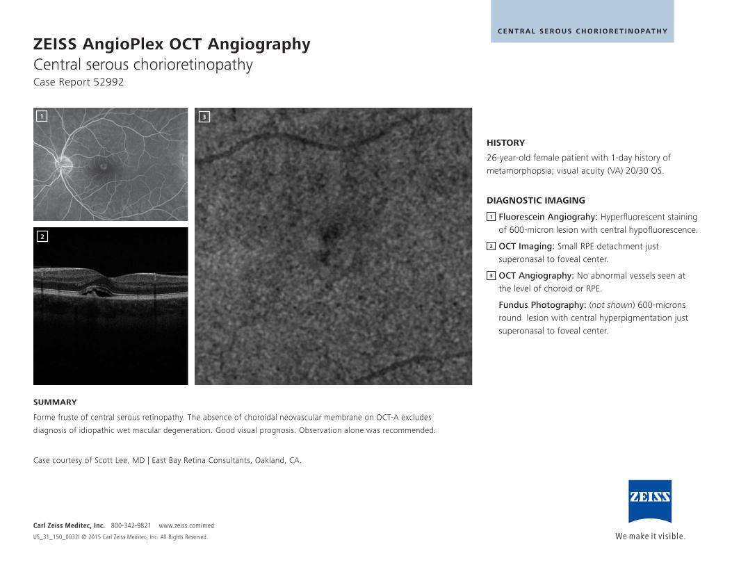
26-year-old female patient with 1-day history of metamorphopsia; visual acuity (VA) 20/30 OS.
DIAGNOSTIC IMAGING
Fluorescein Angiograhy: Hyperfluorescent staining of 600-micron lesion with central hypofluorescence.
OCT Imaging: Small RPE detachment just superonasal to foveal center.
OCT Angiography: No abnormal vessels seen at the level of choroid or RPE.
Fundus Photography: (not shown) 600-microns round lesion with central hyperpigmentation just superonasal to foveal center.
Carl Zeiss Meditec, Inc. 800-342-9821 www.zeiss.com/med
US_31_150_0032I © 2015 Carl Zeiss Meditec, Inc. All Rights Reserved.
C E N T R A L S E R O U S C H O R I O R E T I N O PAT H Y
SUMMARY
Forme fruste of central serous retinopathy. The absence of choroidal neovascular membrane on OCT-A excludes
diagnosis of idiopathic wet macular degeneration. Good visual prognosis. Observation alone was recommended.
Case courtesy of Scott Lee, MD | East Bay Retina Consultants, Oakland, CA.
1
2
3
1
2
3
ZEISS AngioPlex OCT Angiography Branch retinal vein occlusionCase Report 24666
HISTORY
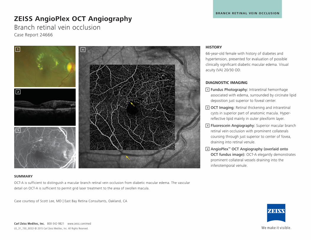
66-year-old female with history of diabetes and hypertension, presented for evaluation of possible clinically significant diabetic macular edema. Visual acuity (VA) 20/30 OD.
DIAGNOSTIC IMAGING
Fundus Photography: Intraretinal hemorrhage associated with edema, surrounded by circinate lipid deposition just superior to foveal center.
OCT Imaging: Retinal thickening and intraretinal cysts in superior part of anatomic macula. Hyper-reflective lipid mainly in outer plexiform layer.
Fluorescein Angiography: Superior macular branch retinal vein occlusion with prominent collaterals coursing through just superior to center of fovea, draining into retinal venule.
AngioPlex™ OCT Angiography (overlaid onto OCT fundus image): OCT-A elegantly demonstrates prominent collateral vessels draining into the inferotemporal venule.
Carl Zeiss Meditec, Inc. 800-342-9821 www.zeiss.com/med
US_31_150_0032I © 2015 Carl Zeiss Meditec, Inc. All Rights Reserved.
B R A N C H R E T I N A L V E I N O C C LU S I O N
SUMMARY
OCT-A is sufficient to distinguish a macular branch retinal vein occlusion from diabetic macular edema. The vascular
detail on OCT-A is sufficient to permit grid laser treatment to the area of swollen macula.
Case courtesy of Scott Lee, MD | East Bay Retina Consultants, Oakland, CA
1
2
4
3
1
2
3
4
ZEISS AngioPlex OCT Angiography Proliferative diabetic retinopathyCase Report 52918
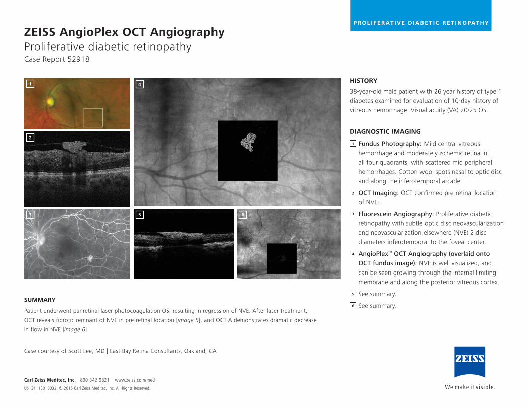
HISTORY
38-year-old male patient with 26 year history of type 1 diabetes examined for evaluation of 10-day history of vitreous hemorrhage. Visual acuity (VA) 20/25 OS.
DIAGNOSTIC IMAGING
Fundus Photography: Mild central vitreous hemorrhage and moderately ischemic retina in all four quadrants, with scattered mid peripheral hemorrhages. Cotton wool spots nasal to optic disc and along the inferotemporal arcade.
OCT Imaging: OCT confirmed pre-retinal location of NVE.
Fluorescein Angiography: Proliferative diabetic retinopathy with subtle optic disc neovascularization and neovascularization elsewhere (NVE) 2 disc diameters inferotemporal to the foveal center.
AngioPlex™ OCT Angiography (overlaid onto OCT fundus image): NVE is well visualized, and can be seen growing through the internal limiting membrane and along the posterior vitreous cortex.
See summary.
See summary.
Carl Zeiss Meditec, Inc. 800-342-9821 www.zeiss.com/med
US_31_150_0032I © 2015 Carl Zeiss Meditec, Inc. All Rights Reserved.
SUMMARY
Patient underwent panretinal laser photocoagulation OS, resulting in regression of NVE. After laser treatment,
OCT reveals fibrotic remnant of NVE in pre-retinal location [image 5], and OCT-A demonstrates dramatic decrease
in flow in NVE [image 6].
Case courtesy of Scott Lee, MD | East Bay Retina Consultants, Oakland, CA
1
2
4
3 5 6
1
2
4
3
P R O L I F E R AT I V E D I A B E T I C R E T I N O PAT H Y
5
6
0297
Carl Zeiss Meditec, Inc. 5160 Hacienda DriveDublin, CA 94568USAwww.zeiss.com/med
Carl Zeiss Meditec AG Goeschwitzer Strasse 51–5207745 JenaGermanywww.zeiss.com/med
US_
31_1
50_0
032I
Pr
inte
d in
USA
CZ
-XI/2
015
The
cont
ents
of t
his
case
repo
rt do
cum
ent m
ay d
iffer
from
the
curre
nt s
tatu
s of
app
rova
l of t
he p
rodu
ct in
you
r cou
ntry
. Ple
ase
cont
act y
our r
egio
nal r
epre
sent
ativ
e fo
r mor
e in
form
atio
n.
Subj
ect t
o ch
ange
in d
esig
n an
d sc
ope
of d
eliv
ery
and
as a
resu
lt of
ong
oing
tech
nica
l dev
elop
men
t. An
gioP
lex
is ei
ther
a tr
adem
ark
or re
gist
ered
trad
emar
k of
Car
l Zei
ss M
edite
c, In
c. in
the
Unite
d St
ates
and
/or o
ther
cou
ntrie
s. ©
201
5 by
Car
l Zei
ss M
edite
c, In
c. A
ll co
pyrig
hts
rese
rved
.