Guidelines for the Management of Diabetic Macular Edema · PDF fileGuidelines for the...
38
E-Mail [email protected] Review Ophthalmologica 2017;237:185–222 DOI: 10.1159/000458539 Guidelines for the Management of Diabetic Macular Edema by the European Society of Retina Specialists (EURETINA) Ursula Schmidt-Erfurth a Jose Garcia-Arumi b Francesco Bandello c Karina Berg d Usha Chakravarthy e Bianca S. Gerendas a Jost Jonas f Michael Larsen g Ramin Tadayoni h Anat Loewenstein i a Department of Ophthalmology and Optometry, Medical University of Vienna, Vienna, Austria; b Ophthalmology Department, Universidad Autónoma de Barcelona, Barcelona, Spain; c Department of Ophthalmology, Ospedale San Raffaele, Milan, Italy; d Department of Ophthalmology, Oslo University Hospital, Oslo, Norway; e School of Medicine, Dentistry, and Biomedical Sciences, The Queen’s University of Belfast, Belfast, UK; f Department of Ophthalmology, Medical Faculty Mannheim of the Ruprecht-Karls University Heidelberg, Mannheim, Germany; g Faculty of Health and Medical Sciences, University of Copenhagen, Copenhagen, Denmark; h Department of Ophthalmology, Hôpital Lariboisière, AP-HP, Université Paris 7 – Sorbonne Paris Cité, Paris, France; i Department of Ophthalmology, Tel Aviv Medical Center, Tel Aviv University, Tel Aviv, Israel tics. The following guidance for the management of DME has been composed from the best updated knowledge of lead- ing experts in Europe and represents another volume in the series of EURETINA recommendations for the management of retinal disease. © 2017 S. Karger AG, Basel Introduction The prevalence of diabetic macular edema (DME) is continuously rising worldwide and has become one of the major causes of vision loss in the working-age popula- tion. Clinical parameters and new diagnostic parameters from imaging with optical coherence tomography (OCT) as well as the overall advances of OCT technology have been identified to stage the disease. However, a large va- riety of therapeutic strategies are available to the ophthal- mologist: laser photocoagulation, anti-vascular endothe- Keywords Diabetic macular edema · Anti-VEGF therapy · Steroids Abstract Diabetic retinal disease is envisioned to become the plague of the coming decades with a steep increase of worldwide diabetes incidence followed by a substantial rise in retinal disease. Improvements in diagnostic and therapeutic care have to cope with this dilemma in a clinically and socioeco- nomically efficient manner. Laser treatment has found a less destructive competitor in pharmacological treatments. As a consequence of recent rigorous clinical trials, laser photoco- agulation is no longer recommended for the treatment of diabetic macular edema (DME), and anti-vascular endothe- lial growth factor therapy has emerged as first-line therapy. Steroids have maintained a role in the management of chronically persistent DME. The paradigm shifts in therapy are accompanied by a substantial break-through in diagnos- Received: December 21, 2016 Accepted: January 30, 2017 Published online: April 20, 2017 Ophthalmologica Univ.-Prof. Dr. Ursula Schmidt-Erfurth Department of Ophthalmology and Optometry Medical University of Vienna Waehringer Guertel 18–20, AT–1090 Vienna (Austria) E-Mail ursula.schmidt-erfurth @ meduniwien.ac.at © 2017 S. Karger AG, Basel www.karger.com/oph Downloaded by: 195.178.83.127 - 10/1/2017 2:33:30 PM
Transcript of Guidelines for the Management of Diabetic Macular Edema · PDF fileGuidelines for the...
E-Mail [email protected]
Review
Ophthalmologica 2017;237:185–222 DOI: 10.1159/000458539
Guidelines for the Management of Diabetic Macular Edema by the European Society of Retina Specialists (EURETINA)
Ursula Schmidt-Erfurth a Jose Garcia-Arumi b Francesco Bandello c
Karina Berg d Usha Chakravarthy e Bianca S. Gerendas a Jost Jonas f
Michael Larsen g Ramin Tadayoni h Anat Loewenstein i
a Department of Ophthalmology and Optometry, Medical University of Vienna, Vienna , Austria; b Ophthalmology Department, Universidad Autónoma de Barcelona, Barcelona , Spain; c Department of Ophthalmology, Ospedale San Raffaele, Milan , Italy; d Department of Ophthalmology, Oslo University Hospital, Oslo , Norway; e School of Medicine, Dentistry, and Biomedical Sciences, The Queen’s University of Belfast, Belfast , UK; f Department of Ophthalmology, Medical Faculty Mannheim of the Ruprecht-Karls University Heidelberg, Mannheim , Germany; g Faculty of Health and Medical Sciences, University of Copenhagen, Copenhagen , Denmark; h Department of Ophthalmology, Hôpital Lariboisière, AP-HP, Université Paris 7 – Sorbonne Paris Cité, Paris , France; i Department of Ophthalmology, Tel Aviv Medical Center, Tel Aviv University, Tel Aviv , Israel
tics. The following guidance for the management of DME has been composed from the best updated knowledge of lead-ing experts in Europe and represents another volume in the series of EURETINA recommendations for the management of retinal disease. © 2017 S. Karger AG, Basel
Introduction
The prevalence of diabetic macular edema (DME) is continuously rising worldwide and has become one of the major causes of vision loss in the working-age popula-tion. Clinical parameters and new diagnostic parameters from imaging with optical coherence tomography (OCT) as well as the overall advances of OCT technology have been identified to stage the disease. However, a large va-riety of therapeutic strategies are available to the ophthal-mologist: laser photocoagulation, anti-vascular endothe-
Keywords
Diabetic macular edema · Anti-VEGF therapy · Steroids
Abstract
Diabetic retinal disease is envisioned to become the plague of the coming decades with a steep increase of worldwide diabetes incidence followed by a substantial rise in retinal disease. Improvements in diagnostic and therapeutic care have to cope with this dilemma in a clinically and socioeco-nomically efficient manner. Laser treatment has found a less destructive competitor in pharmacological treatments. As a consequence of recent rigorous clinical trials, laser photoco-agulation is no longer recommended for the treatment of diabetic macular edema (DME), and anti-vascular endothe-lial growth factor therapy has emerged as first-line therapy. Steroids have maintained a role in the management of chronically persistent DME. The paradigm shifts in therapy are accompanied by a substantial break-through in diagnos-
Received: December 21, 2016 Accepted: January 30, 2017 Published online: April 20, 2017
Ophthalmologica
Univ.-Prof. Dr. Ursula Schmidt-Erfurth Department of Ophthalmology and Optometry Medical University of Vienna Waehringer Guertel 18–20, AT–1090 Vienna (Austria) E-Mail ursula.schmidt-erfurth @ meduniwien.ac.at
© 2017 S. Karger AG, Basel
www.karger.com/oph
Dow
nloa
ded
by:
195.
178.
83.1
27 -
10/
1/20
17 2
:33:
30 P
M
Schmidt-Erfurth et al.
Ophthalmologica 2017;237:185–222DOI: 10.1159/000458539
186
lial growth factor (VEGF), steroid and surgical therapy are applied with different procedures and their own com-plications. A novel era of DME therapy has started with these diverse approaches. These guidelines shall give an overview on the current available diagnostic and thera-peutic procedures and recommend their application.
Clinical Features of Diabetic Retinopathy
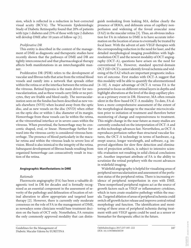
Diabetic retinopathy (DR) is the term applied to de-scribe the microvascular abnormalities that are seen in the fundus of persons with diabetes on clinical examina-tion or on color fundus photography. The earliest and the least severe manifestation is the dot-like microaneurysms (MA) which are localized saccular outpouchings of the capillary wall and appear as tiny red dots with sharp mar-gins. MA occur frequently in relation to areas of capillary nonperfusion, but the latter is an angiographic diagnosis. Retinal hemorrhages are another important manifesta-tion of DR and are found throughout the fundus ( Fig. 1 ) They may be flame-shaped, if located in the nerve fiber layer, or dot or blot-like, if located in the middle layersof the retina. Intraretinal microvascular abnormalities (IRMA) are tortuous and dilated intraretinal microvascu-
lar segments. Venous dilation and beading are typical DR-related changes, and the latter in particular represent focal increases in venous caliber, resembling a string of beads. Hard exudates are another manifestation of DR, arising as a consequence of chronic localized leakage from the retinal vessels ( Fig. 2 ). Hard exudates are main-ly composed of lipid and appear as yellowish white lesions usually with distinct margins and occur close to clusters of MAs. Soft exudates, otherwise termed cotton wool spots, arise when there is a block in the flow of axoplasm in the retinal nerve fiber layer which occurs due to focal ischemia. They represent infarcted inner retina and ap-pear as greyish-white round or oval areas with ill-defined feathery edges.
Diabetic Macular Edema DME represents an accumulation of fluid within the
central portion of the retina, which arises as a consequence of failure of the blood-retinal barrier (BRB). Diffuse ede-ma is caused by extensive capillary leakage, whereas local-ized edema is caused by focal leakage from grouped MAs. DME can occur in isolation without other signs of micro-angiopathy in the fundus; therefore, it merits being classi-fied as a separate entity. It is often associated with hard exudates and causes blurring and distortion of central vi-
Fig. 1. Standard photograph depicting MA and hemorrhages from diabetic retinopathy study. Report number 6 and 7. Courtesy of Investigative Ophthalmology & Visual Science [166] .
Fig. 2. Severe lipid exudation with hard exudates from grouped MA from diabetic retinopathy study. Report number 6 and 7. Courtesy of Investigative Ophthalmology & Visual Science [166] .
Dow
nloa
ded
by:
195.
178.
83.1
27 -
10/
1/20
17 2
:33:
30 P
M
Guidelines for the Management of Diabetic Macular Edema by EURETINA
Ophthalmologica 2017;237:185–222DOI: 10.1159/000458539
187
sion, which is reflected in a reduction in best-corrected visual acuity (BCVA). The Wisconsin Epidemiologic Study of Diabetic Retinopathy found that 20% of patients with type 1 diabetes and 25% of those with type 2 diabetes will develop DME after 10 years of follow-up [1] .
Proliferative DR This entity is described in the context of the manage-
ment of DME as diagnostic and therapeutic studies have recently shown that the pathways of DME and DR are tightly interconnected and that pharmacological therapy affects both manifestations in an interchangeable man-ner.
Proliferative DR (PDR) refers to the development of vascular and fibrous tufts that arise from the retinal blood vessels and ramify into a network that spreads either within the retina or at the interface between the retina and the vitreous. Retinal hypoxia is the main driver for neo-vascularization, and as these vessels carry little or no peri-cytes, they are friable and bleed easily. The neovascular-ization seen on the fundus has been described as new ves-sels elsewhere (NVE) when located away from the optic disc, and as new vessels on the disc (NVD) when either on the optic disc or within 1 disc diameter of its margin. Hemorrhage from these vessels can lie within the retina, at the vitreoretinal interface or in severe cases within the vitreous. When preretinal, the hemorrhage may be cres-centic shaped, oval, or linear. Hemorrhage further for-ward into the vitreous cavity is considered vitreous hem-orrhage. The presence of blood particularly in the macu-lar retina and within the vitreous leads to severe loss of vision. Blood is also inimical to the integrity of the retina. Subsequent development of fibrous bands resulting from organized hemorrhage can consecutively result in trac-tion of the retina.
Angiographic Manifestations in DME
Rationale Fluorescein angiography (FA) has been a valuable di-
agnostic tool in DR for decades and is formally recog-nized as an essential component in the assessment of se-verity of the pathology and delineation of location of ret-inal alteration to allow appropriate and targeted laser therapy [2] . However, there is currently only moderate consensus on the role of FA in the management of DME, as nowadays some clinicians would base treatment deci-sion on the basis of OCT only. Nonetheless, FA remains the only commonly approved modality that can distin-
guish nonleaking from leaking MA, define clearly the presence of IRMA, and delineate areas of capillary non-perfusion and widening of the foveal avascular zone (FAZ) in the macular retina [3] . Thus, an obvious indica-tion for FA in relation to DME is to have accurate infor-mation on the location of areas to eventually be treated by focal laser. With the advent of anti-VEGF therapies with the corresponding reduction in the need for laser, and the detailed morphological imaging possibilities with high-resolution OCT and the newest modality of OCT angiog-raphy (OCT-A), questions have arisen on the need for conventional FA. However, standard spectral-domain OCT (SD-OCT) cannot identify foveal ischemia and wid-ening of the FAZ which are important prognostic indica-tors of outcome. First studies with OCT-A suggest that this modality will be able to quantify the aforementioned [4–10] . A major advantage of OCT-A versus FA is the potential to focus on different retinal layers in depths and highlight alterations at the level of the deep capillary plex-us as a primary event in DR. However, MA often remain silent in the flow-based OCT-A modality. To date, FA al-lows a more comprehensive assessment of the extent of the morphological damage to the macular microcircula-tion prior to initiation of treatment and therefore better monitoring of change and responsiveness to treatment. This might change in the near future as many studies are currently conducted on the interpretation of OCT-A and as this technology advances fast. Nevertheless, as OCT-A reproduces perfusion rather than structural vascular fea-tures, the OCT-A technology in terms of hardware, e.g. swept source, longer wavelength, and software, e.g. im-proved algorithms for slow flow detection and elimina-tion of projection artifacts, is subject to intensive scien-tific evaluation not resulting in solid clinical conclusions yet. Another important attribute of FA is the ability to scrutinize the retinal periphery with the recent advances in widefield imaging.
Widefield angiography is helpful for visualizing areas of peripheral neovascularization and assessment of the perfu-sion status of the peripheral retina. There is increasing ev-idence of peripheral nonperfusion in eyes with DME. These nonperfused peripheral regions act as the source of growth factors such as VEGF or inflammatory cytokines, which in turn create exudative pathology within the mac-ula. Targeted ablation of areas of peripheral ischemia could switch off growth factor release and improve central retinal morphology and function. The identification and moni-toring of these areas of peripheral ischemia during treat-ment with anti-VEGF agents could be used as a sensor or biomarker for therapeutic effect in the future.
Dow
nloa
ded
by:
195.
178.
83.1
27 -
10/
1/20
17 2
:33:
30 P
M
Schmidt-Erfurth et al.
Ophthalmologica 2017;237:185–222DOI: 10.1159/000458539
188
Evidence In DME, the appearance of the macula on FA is highly
characteristic, with MA and IRMA interspersed between areas of focal or diffuse hyperfluorescence representing leakage from the incompetent macular microcirculation [11] . In addition, regions of capillary loss and dilation, arteriolar abnormalities and cystoid changes can be ob-served. When DME leakage is focal from groups of MA, there is usually surrounding accumulation of lipid that assumes a circinate distribution [3] . Ectatic dilated leak-ing perifoveal capillaries are seen and can be difficult to distinguish from other conditions such as perifoveal tel-angiectasia [12] , thus emphasizing the importance of ob-taining an FA prior to DME therapy initiation. The Early Treatment Diabetic Retinopathy Study (ETDRS) relied on the use of FA as a guide for focal treatment of indi-vidual leaking MA and macular grid laser to areas of dif-fuse leakage and capillary nonperfusion with therapeutic benefit [11] . Furthermore, in one study when using abla-tive therapies such as laser, the use of FA to delineate the areas requiring treatment resulted in improved accuracy [2] . More recently, the relationships between OCT and FA findings have been explored and the various charac-teristics related to each other indicating that both overlap and the complementarity of findings [13, 14] . Simultane-ous FA and SD-OCT allow improved characterization of DME features and allow distinction of perfused MA from nonperfused MA as well as the ability to localize fluid to different retinal layers [15] . In a recent study, abnormali-ties of choroidal perfusion were detected by indocyanine green angiography, and the combination of this imaging modality along with enhanced-depth OCT imaging and FA were reported as superior indicators of ocular perfu-sion status in diabetic eyes [16] . Emerging data also indi-cate that regular OCT cannot replace FA as it is not pos-sible to predict FAZ outline and size based on the metrics of retinal thickness or through the evaluation of the reti-nal structure using the former [17] . This might change as OCT-A is further evaluated in ongoing studies.
Another important consideration is the advent of widefield imaging combined with FA which has vastly improved our understanding of the role of peripheral vas-cular changes in driving central macular pathology [18] . Various studies have shown that peripheral ischemia is strongly related to presence and severity of DME as well as recalcitrance to therapy. When widefield imaging is used to calculate the ischemic index, the mean decrease in central macular thickness is highest in the lowest isch-emic index group and least in the in the worst ischemic index group [19] . In summary, the information that is
obtained from FA on central macular and peripheral ret-inal changes is critically important for the evaluation of the severity of the disease, aids in staging purposes, and is helpful in the monitoring of outcomes following treat-ment.
Recommendation FA is an important diagnostic tool for assessment of
the central and peripheral retina. It is recommended that FA is performed prior to the initiation of therapy to de-lineate and stage the DME and DR pathology. FA may be repeated as needed in the event of nonresponsiveness to therapy and/or for monitoring patients in the long term. OCT-A may be used to accompany FA imaging for its potential to offer additional insight into capillary loss and attribution to the superficial or deep capillary plexus mainly because of its non-invasive nature.
Features in Optical Coherence Tomography
Rationale Since its first introduction, OCT has become the most
frequently used diagnostic tool in ophthalmology and has revolutionized clinical imaging for diagnosis and disease management in most retinal diseases including DME. OCT is a fast, noninvasive technology that produces in vivo images of the retina. The most recent third-genera-tion OCT technology uses a swept-source (SS) light source that allows very fast imaging and provides three-dimensional raster images of high microstructural reso-lution, also referred to as optical histology [20] . The most commonly used second-generation OCT nowadays is SD-OCT allowing three-dimensional raster scans of up to a few hundred B-scans, also creating high-resolution im-ages, but working slower than SS-OCT. It supersedes time-domain (TD)-OCT that allowed imaging of 6 radial cuts only.
All OCT generations are able to generate central reti-nal thickness (CRT) values, but TD-OCT is less accurate due to its more likely decentration from the fovea and less available B scans. Nevertheless, CRT has been used as a quantitative feature to evaluate disease activity, progres-sion, and treatment response ever since OCT was avail-able in ophthalmology. Although there is a correlation between best-corrected visual acuity (BCVA) letter score and CRT in DME under therapy with anti-VEGF agents, this correlation is weak during the first year of therapy( r = 0.34–0.41) and largely lost in the long term [21] . The new OCT generations of SD- and SS-OCT are able to vi-
Dow
nloa
ded
by:
195.
178.
83.1
27 -
10/
1/20
17 2
:33:
30 P
M
Guidelines for the Management of Diabetic Macular Edema by EURETINA
Ophthalmologica 2017;237:185–222DOI: 10.1159/000458539
189
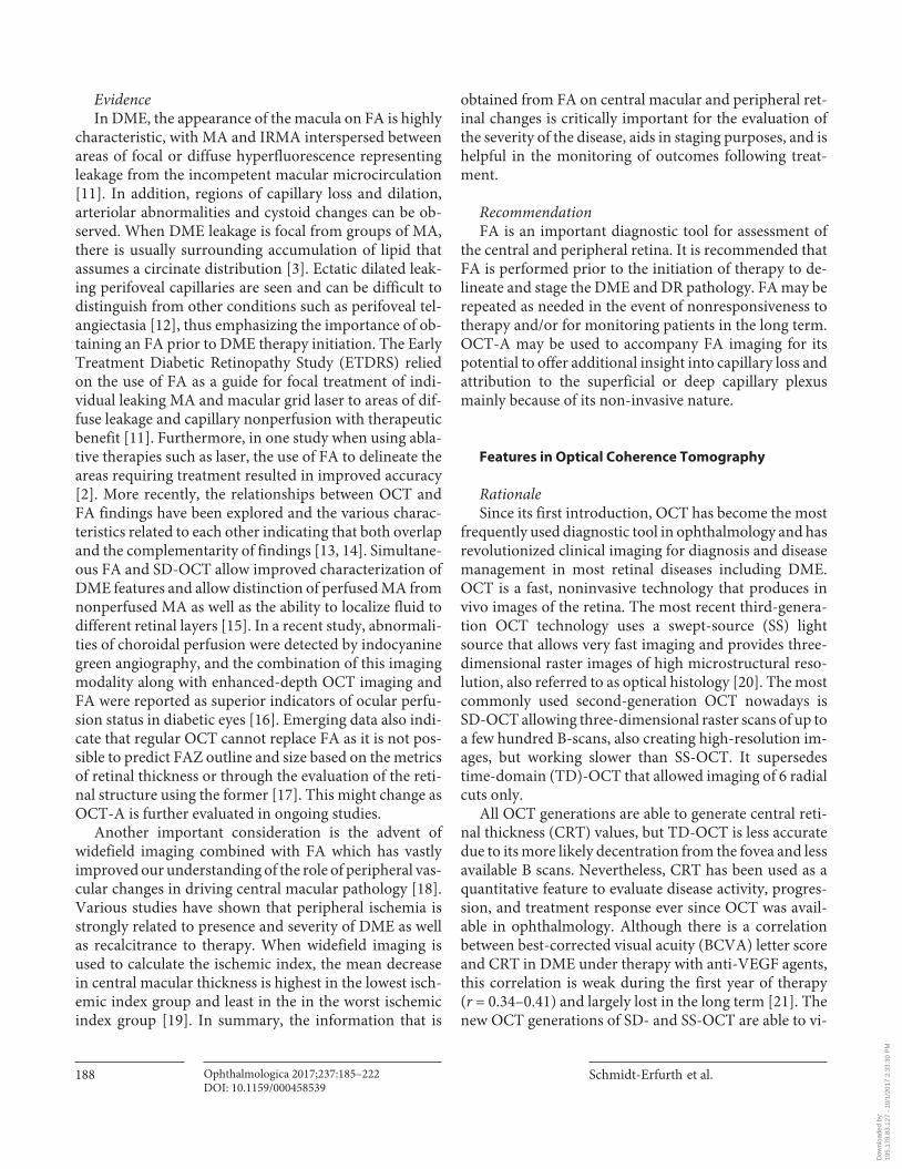
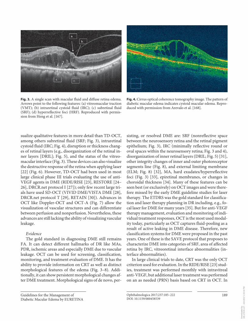
sualize qualitative features in more detail than TD-OCT, among others subretinal fluid (SRF; Fig. 3 ), intraretinal cystoid fluid (IRC; Fig. 4 ), disruption or thickness chang-es of retinal layers (e.g., disorganization of the retinal in-ner layers [DRIL]; Fig. 5 ), and the status of the vitreo-macular interface ( Fig. 3 ). These devices can also visualize the destructive response of the retina when applying laser [22] ( Fig. 6 ). However, TD-OCT had been used in most large clinical phase III trials evaluating the use of anti-VEGF agents in DME (RIDE/RISE [23] , RESTORE [24–26] , DRCR.net protocol I [27] ); only few recent large tri-als have used SD-OCT (VIVID DME/VISTA DME [28] , DRCR.net protocol T [29] , RETAIN [30] ). Advances in OCT like Doppler-OCT and OCT-A ( Fig. 7 ) allow the visualization of vascular structures and can differentiate between perfusion and nonperfusion. Nevertheless, these advances are still lacking the ability of visualizing vascular leakage.
Evidence The gold standard in diagnosing DME still remains
FA. It can detect different hallmarks of DR like MAs, PDR, ischemic areas and especially DME due to vascular leakage. OCT can be used for screening, classification, monitoring, and treatment evaluation of DME. It has the ability to provide information on CRT as well as distinct morphological features of the edema ( Fig. 3–8 ). Addi-tionally, it can show persistent morphological changes af-ter DME treatment. Morphological signs of de novo, per-
sisting, or resolved DME are: SRF (nonreflective space between the neurosensory retina and the retinal pigment epithelium; Fig. 3 ), IRC (minimally reflective round or oval spaces within the neurosensory retina; Fig. 3 and 4 ), disorganization of inner retinal layers (DRIL; Fig. 5 ) [31] , other integrity changes of inner and outer photoreceptor segments line ( Fig. 8 ), and external limiting membrane (ELM; Fig. 8 ) [32] , MA, hard exudates/hyperreflective foci ( Fig. 3 ) [33] , epiretinal membranes, or changes in choroidal thickness [34] . Many of these features can be seen best (or exclusively) on OCT images and were there-fore missed by the early DME guideline studies for laser therapy. The ETDRS was the gold standard for classifica-tion and laser therapy planning in DR including, e.g., fo-cal laser for DME for many years [35] . But for anti-VEGF therapy management, evaluation and monitoring of indi-vidual treatment responses, OCT is the most used modal-ity today, particularly as OCT captures fluid-pooling as a result of active leaking in DME disease. Therefore, new classification systems for DME were proposed in the past years. One of these is the SAVE protocol that proposes to characterize DME into categories of SRF, area of affected retina by IRC, vitreoretinal interface abnormalities (in-terface abnormalities).
In large clinical trials to date, CRT was the only OCT criterion used for evaluation. In the RIDE/RISE [23] stud-ies, treatment was performed monthly with intravitreal anti-VEGF, but additional laser treatment was performed on an as needed (PRN) basis based on CRT in OCT. In
Fig. 3. A single scan with macular fluid and diffuse retina edema. Arrows point to the following features: (a) vitreomacular traction (VMT); (b) intraretinal cystoid fluid (IRC); (c) subretinal fluid (SRF); (d) hyperreflective foci (HRF). Reproduced with permis-sion from Heng et al. [167] .
Fig. 4. Cirrus optical coherence tomography image. The pattern of diabetic macular edema indicates cystoid macular edema. Repro-duced with permission from Arevalo et al. [168] .
Dow
nloa
ded
by:
195.
178.
83.1
27 -
10/
1/20
17 2
:33:
30 P
M
Schmidt-Erfurth et al.
Ophthalmologica 2017;237:185–222DOI: 10.1159/000458539
190
the RESTORE [24] study, where intravitreal anti-VEGF monotherapy and laser were compared with a combina-tion therapy of both for DME, retreatment was based on BCVA letter score and the investigator’s decision which could include OCT changes and clinical examination. In the DRCR.net protocol I [27] intravitreal anti-VEGF in-jections were compared with laser treatment and with in-travitreal corticosteroid injections. In this study, retreat-ment was also based on the investigator’s discretion with-out predefined OCT retreatment criteria. In the VIVID DME/VISTA DME [28] studies with SD-OCT, a fixed treatment regimen was used for intravitreal anti-VEGF therapy, and laser retreatment was allowed as indicated
by ETDRS guidelines. CRT was used as the secondary ef-ficacy endpoint. In the DRCR.net protocol T [29] , CRT from OCT was one parameter for retreatment decisions. In conclusion, all these trials show that BCVA played the most important role with regard to retreatment; there-fore, CRT as an OCT parameter has played little role in the study design and analyses of most large clinical phase III trials on DME. However, ophthalmologists to date agree that treatment regimens must be individualized be-cause OCT biomarkers are key to identify the best PRN treatment scheme for each individual patient. A PRN reg-imen is the most frequently used treatment regimen for DME in anti-VEGF therapy in real life today, but the PRN
1 mm 1 mm
570 μm DRIL extentNo DRIL
1 mm 1 mm
GCL-IPL-INL interface
INL-OPL interface
OPL-ONL interface
a b
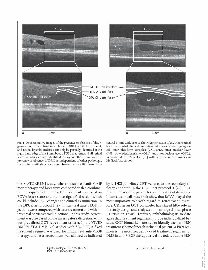
Fig. 5. Representative images of the presence or absence of disor-ganization of the retinal inner layers (DRIL). a DRIL is present, and retinal layer boundaries can only be partially identified at the right-hand edge of the 1-mm box. b DRIL is absent, and all retinal layer boundaries can be identified throughout the 1-mm box. The presence or absence of DRIL is independent of other pathology, such as intraretinal cystic changes. Insets are magnifications of the
central 1-mm-wide area to show segmentation of the inner retinal layers, with white lines demarcating interfaces between ganglion cell-inner plexiform complex (GCL-IPL), inner nuclear layer (INL), outer plexiform layer (OPL), and outer nuclear layer (ONL). Reproduced from Sun et al. [31] with permission from American Medical Association.
Dow
nloa
ded
by:
195.
178.
83.1
27 -
10/
1/20
17 2
:33:
30 P
M
Guidelines for the Management of Diabetic Macular Edema by EURETINA
Ophthalmologica 2017;237:185–222DOI: 10.1159/000458539
191
criteria are not standardized. The individual patient’s morphology has a close relationship with the treatment response and needs to serve as the basis for these criteria. Focal edema classified with the earlier described system, for example, may benefit most from anti-VEGF therapy with regard to BCVA letter score gain [36] . Some indi-vidual OCT biomarkers have already been identified from post hoc analyses of the multicenter studies and some smaller monocentric studies.
In the RESTORE study, patients have been treated af-ter an initial loading phase of 3 consecutive monthly in-jections on a PRN basis. Here, it could be shown in a post hoc analysis that patients with SRF at baseline had higher BCVA gains at the end of the first study year than patients without SRF at baseline, although no difference in their baseline BCVA letter score was detected [21] . This pro-tective role of SRF was reconfirmed by an OCT post hoc study analysis of the RIDE/RISE trials [37] . When looking at IRC, in RESTORE about 80% of patients had IRC at baseline. Patients with less IRC at baseline had a better baseline BCVA letter score and remained at a better score throughout the entire study duration [21] . This finding is supported by an analysis of another study of preserved tissue in 129 DME patients. Here, baseline vision corre-lated with the volume of preserved neurosensory tissue is less IRC between inner and outer retina [38] . Another finding from RESTORE indicated that throughout the entire study duration, patients with vitreomacular adhe-sion showed higher BCVA scores at baseline and main-tained these better scores under any therapy, whereas pa-tients with a posterior vitreous detachment started with lower BCVA letter scores and improved less than patients with vitreomacular adhesion [21] .
In an academic study with 120 patients, the impor-tance of DRIL was shown. DRIL that constituted more than 50% of the central millimeter on OCT was associ-
Baseline
Day 1ba
a b
ELM
INLOPLONLPRL
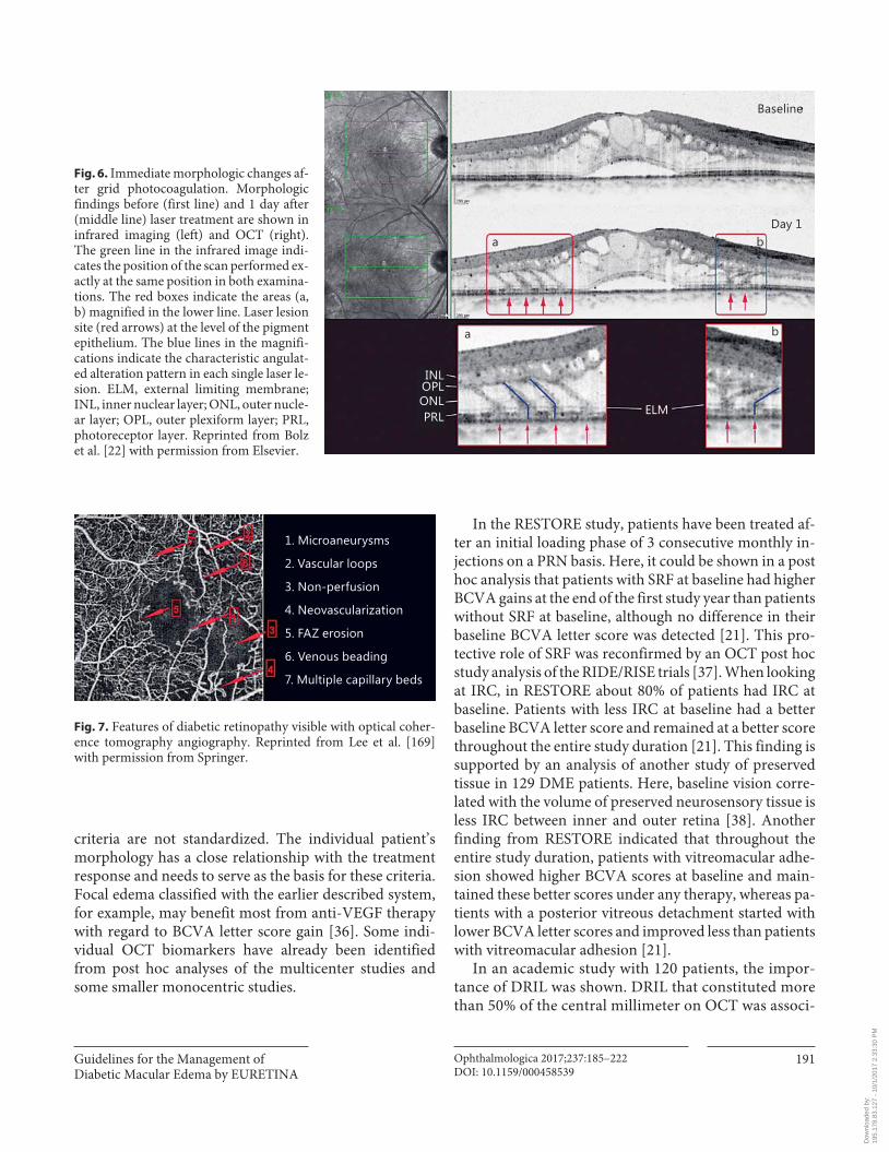
Fig. 6. Immediate morphologic changes af-ter grid photocoagulation. Morphologic findings before (first line) and 1 day after (middle line) laser treatment are shown in infrared imaging (left) and OCT (right). The green line in the infrared image indi-cates the position of the scan performed ex-actly at the same position in both examina-tions. The red boxes indicate the areas (a, b) magnified in the lower line. Laser lesion site (red arrows) at the level of the pigment epithelium. The blue lines in the magnifi-cations indicate the characteristic angulat-ed alteration pattern in each single laser le-sion. ELM, external limiting membrane; INL, inner nuclear layer; ONL, outer nucle-ar layer; OPL, outer plexiform layer; PRL, photoreceptor layer. Reprinted from Bolz et al. [22] with permission from Elsevier.
1. Microaneurysms
2. Vascular loops
3. Non-perfusion
4. Neovascularization
5. FAZ erosion
6. Venous beading
7. Multiple capillary beds
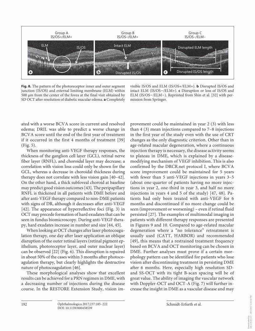
Fig. 7. Features of diabetic retinopathy visible with optical coher-ence tomography angiography. Reprinted from Lee et al. [169] with permission from Springer.
Dow
nloa
ded
by:
195.
178.
83.1
27 -
10/
1/20
17 2
:33:
30 P
M
Schmidt-Erfurth et al.
Ophthalmologica 2017;237:185–222DOI: 10.1159/000458539
192
ated with a worse BCVA score in current and resolved edema; DRIL was able to predict a worse change in BCVA score until the end of the first year of treatment if it occurred in the first 4 months of treatment [39] ( Fig. 5 ).
When monitoring anti-VEGF therapy responses, the thickness of the ganglion cell layer (GCL), retinal nerve fiber layer (RNFL), and choroidal layer may decrease; a correlation with vision loss could only be shown for the GCL, whereas a decrease in choroidal thickness during therapy does not correlate with less vision gain [40–42] . On the other hand, a thick subfoveal choroid at baseline may predict good vision outcomes [43] . The peripapillary RNFL is thickened in all patients with DME before and after anti-VEGF therapy compared to non-DME patients with signs of DR, although it decreases after anti-VEGF [42] . The appearance of hyperreflective foci ( Fig. 3 ) in OCT may precede formation of hard exudates that can be seen in fundus biomicroscopy. During anti-VEGF thera-py, hard exudates increase in number and size [44, 45] .
When looking at OCT changes after laser photocoagu-lation therapy, one day after laser application an oblique disruption of the outer retinal layers (retinal pigment ep-ithelium, photoreceptor layer, and outer nuclear layer) can be observed [22] ( Fig. 6 ). This disruption is repaired in about 50% of the cases within 3 months after photoco-agulation therapy, but clearly highlights the destructive nature of photocoagulation [46] .
These morphological analyses show that excellent results can be achieved for a PRN regimen in DME, with a decreasing number of injections during the disease course. In the RESTORE Extension Study, vision im-
provement could be maintained in year 2 (3) with less than 4 (3) mean injections compared to 7–8 injections in the first year of the study even with the use of CRT changes as the only diagnostic criterion. Other than in age-related macular degeneration, where a continuous injection therapy is necessary, the disease activity seems to plateau in DME, which is explained by a disease-modifying mechanism of VEGF inhibition. This is also confirmed by the DRCR.net protocol I, where BCVA score improvement could be maintained for 5 years with fewer than 5 anti-VEGF injections in years 3–5 (about one-quarter of patients having no more injec-tions in year 2, one-third in year 3, and half no more injections in years 4 and 5 of the study) [47, 48] . Pa-tients had only been treated with anti-VEGF for 6 months and discontinued if no more change could be seen (improvement or worsening) – even if retinal fluid persisted [27] . The examples of multimodal imaging in patients with different therapy responses are presented in Figures 9 and 10 . Compared to age-related macular degeneration where a “no tolerance” retreatment is usually used (CATT, HARBOR) and recommended [49] , this means that a restrained treatment frequency based on BCVA and OCT monitoring can be chosen in DME. Further analyses must prove if a certain mor-phology pattern can be identified for patients who lose vision after discontinuing treatment in persisting DME after 6 months. Here, especially high resolution SD- and SS-OCT with its tight B-scan spacing will be of great value. The ability of imaging the vascular network with Doppler-OCT and OCT-A ( Fig. 7 ) will further in-crease the insight in DME as a vascular disease and may
Group AIS/OS+/ELM+
Group BIS/OS–/ELM+
Group CIS/OS–/ELM–
ELM IS/OS Intact ELM
Disrupted IS/OS
Disrupted ELM length
Disrupted IS/OS length
500 μm
a b c
Fig. 8. The pattern of the photoreceptor inner and outer segment junction (IS/OS) and external limiting membrane (ELM) within 500 μm from the center of the fovea at the final visit obtained by SD OCT after resolution of diabetic macular edema. a Completely
visible IS/OS and ELM (IS/OS+/ELM+). b Disrupted IS/OS and intact ELM (IS/OS − /ELM+). c Disruption or loss of IS/OS and ELM (IS/OS − /ELM − ). Reprinted from Shin et al. [32] with per-mission from Springer.
Dow
nloa
ded
by:
195.
178.
83.1
27 -
10/
1/20
17 2
:33:
30 P
M
Guidelines for the Management of Diabetic Macular Edema by EURETINA
Ophthalmologica 2017;237:185–222DOI: 10.1159/000458539
193
deliver new hypotheses on imaging biomarkers. Indi-vidualized retreatment criteria based on OCT biomark-ers may be established in the future.
Recommendation In order to diagnose DME, OCT should be accompa-
nied by baseline FA and continued fundus biomicros-copy. When diagnosed and treated, a recurrence of ede-ma can usually be monitored solely by OCT in combina-tion with visual acuity measurements. Robust OCT biomarkers need to be reassured in large-scale clinical studies, but evidence indicates that good baseline predic-tors for a good treatment response with regard to high vision gains and/or good final visual acuity are SRF and/or small IRC and/or vitreomacular adhesion at baseline [21, 50] . The disorganization or disruption of the inner retinal layers [31] , disruption of the inner and outer pho-toreceptor segments and/or ELM [32] , and a thin subfo-veal choroid at baseline [43] may predict bad visual acu-ity after therapy.
Findings in OCT include qualitative and quantitative measures. CRT is an established quantitative measure-
ment. As a CRT increase consists of SRF and/or IRC and/or diffuse thickening, distinguishing between the different qualities may be useful. PRN treatment based solely on CRT changes is a practical approach for the management of DME; other promising more robust in-dividual OCT features have to be confirmed before they can feed into retreatment decisions. The next develop-ment will likely allow the quantification of these quali-tative biomarkers. Segmentation of retinal layers, IRC and SRF and volumetric computation for these features may have an impact on individualized PRN regimens [51, 52] .
Once criteria are validated, most recent OCT technol-ogy with high resolution and dense scanning will help three-dimensional image analysis to be automated and to detect disease activity as smallest morphological changes early on. Therefore, the optimal recommenda-tion is to monitor disease activity on a monthly basis with OCT even if no treatment is needed or intended in order to identify morphological changes as early as possible. These recommendations are based on evidence levels I and II.
a b c d
e f
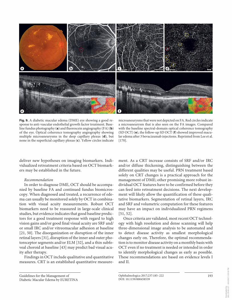
Fig. 9. A diabetic macular edema (DME) eye showing a good re-sponse to anti-vascular endothelial growth factor treatment. Base-line fundus photography ( a ) and fluorescein angiography (FA) ( b ) of the eye. Optical coherence tomography angiography showing multiple microaneurysms in the deep capillary plexus ( d ), but none in the superficial capillary plexus ( c ). Yellow circles indicate
microaneurysms that were not depicted on FA. Red circles indicate a microaneurysm that is also seen on the FA images. Compared with the baseline spectral-domain optical coherence tomography (SD OCT) ( e ), the follow-up SD OCT ( f ) showed improved macu-lar edema after 3 bevacizumab injections. Reprinted from Lee et al. [170] .
Dow
nloa
ded
by:
195.
178.
83.1
27 -
10/
1/20
17 2
:33:
30 P
M
Schmidt-Erfurth et al.
Ophthalmologica 2017;237:185–222DOI: 10.1159/000458539
194
Therapeutic Strategies
Laser Therapy Rationale Laser photocoagulation has represented the standard
of care for the treatment of DME prior to the advent of the intravitreal injection approach. The efficacy of focal laser treatment has been related to the occlusion of leak-ing vessels, especially MA, but the exact mechanism by which focal photocoagulation reduces DME is unknown. The mechanism of action of laser is mainly based on the destruction of ischemic retina, leading to improved oxy-genation of neighboring retinal areas, reduced produc-tion of proangiogenetic factors, and also release of cyto-kines from the retinal pigment epithelium and Muller cells. Different types of laser can be used, including Argon green (514 nm), dye yellow (577 nm), Krypton red (647 nm), and diode (810 nm) laser.
Evidence The ETDRS specifically explored the benefits of laser
treatment in DME. Laser photocoagulation was pre-scribed for all lesions located within 2 disc diameters of the macular center. The results at 3 years demonstrated that patients who were treated with focal photocoagula-tion for clinically significant macular edema achieved a 50% reduction in the risk of moderate visual loss (losing >15 letters of BCVA) compared to controls (12–24%) [11, 53] . Unfortunately, only 3% of patients showed a visual acuity improvement of 3 or more lines. Focal laser energy application should be addressed to leaking MA in combi-nation with grid laser treatment of areas of diffuse macu-lar leakage and nonperfusion in thickened retinas. Com-plications associated with conventional lasers include color vision, night vision, and contrast sensitivity impair-ment, along with enlargement of laser scars, secondary choroidal neovascularization, subretinal fibrosis, and vi-sual-field sensitivity deterioration.
Another randomized clinical trial, Protocol B of the DRCR.net, confirmed the positive effects of focal/grid
a b c d
e f
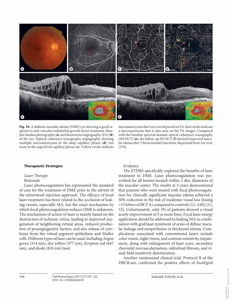
Fig. 10. A diabetic macular edema (DME) eye showing a good re-sponse to anti-vascular endothelial growth factor treatment. Base-line fundus photography ( a ) and fluorescein angiography (FA) ( b ) of the eye. Optical coherence tomography angiography showing multiple microaneurysms in the deep capillary plexus ( d ), but none in the superficial capillary plexus ( c ). Yellow circles indicate
microaneurysms that were not depicted on FA. Red circles indicate a microaneurysm that is also seen on the FA images. Compared with the baseline spectral-domain optical coherence tomography (SD OCT) ( e ), the follow-up SD OCT ( f ) showed improved macu-lar edema after 3 bevacizumab injections. Reprinted from Lee et al. [170] .
Dow
nloa
ded
by:
195.
178.
83.1
27 -
10/
1/20
17 2
:33:
30 P
M
Guidelines for the Management of Diabetic Macular Edema by EURETINA
Ophthalmologica 2017;237:185–222DOI: 10.1159/000458539
195
photocoagulation, demonstrating that in phakic patients laser was more effective than intravitreal injection of tri-amcinolone acetonide in DME patients at both 2 and 3 years of follow-up [54, 55] .
A more recent laser application is subthreshold grid laser treatment, which has been proposed in an attempt to minimize the destructive aspects of conventional grid laser photocoagulation, obtaining encouraging results [56] . The fundamental concept of subthreshold grid laser treatment is to reduce the laser damage to neurosensory layers by reducing the duration of light exposure and by using a subvisible clinical endpoint. In particular, the mi-cropulse diode laser allows the release of micropulses with low energy per pulse in order to confine the energy to the cells of the retinal pigment epithelium (RPE), avoiding lateral thermal spreading. A few randomized clinical trials have demonstrated that subthreshold grid laser treatment is as effective as conventional focal/grid laser photocoagulation, even though slower in terms of resolution of DME, in achieving the same functional and anatomical effects [57, 58] .
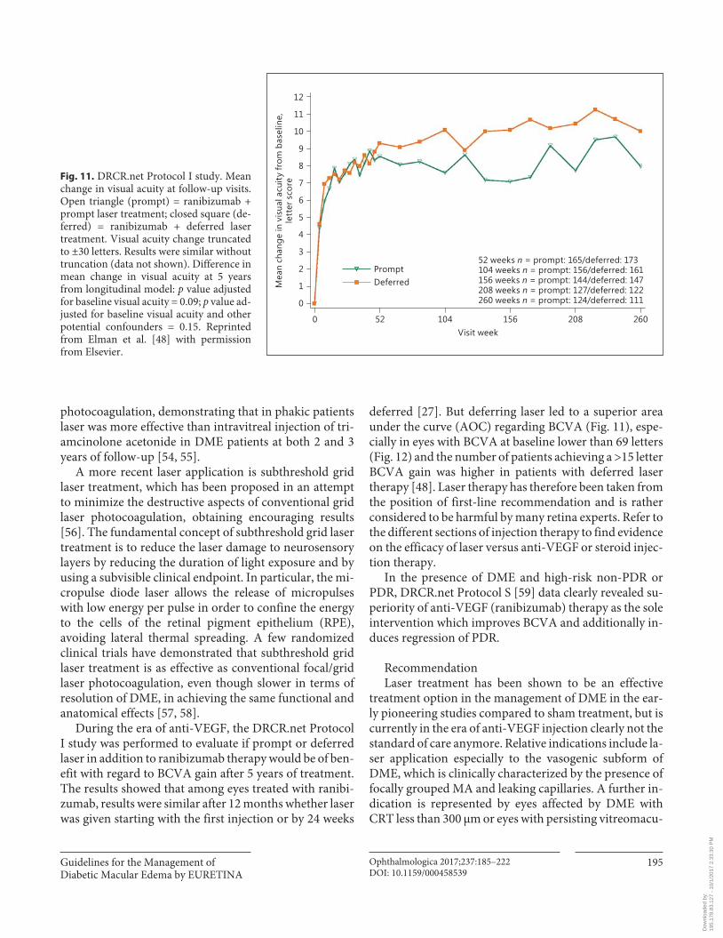
During the era of anti-VEGF, the DRCR.net Protocol I study was performed to evaluate if prompt or deferred laser in addition to ranibizumab therapy would be of ben-efit with regard to BCVA gain after 5 years of treatment. The results showed that among eyes treated with ranibi-zumab, results were similar after 12 months whether laser was given starting with the first injection or by 24 weeks
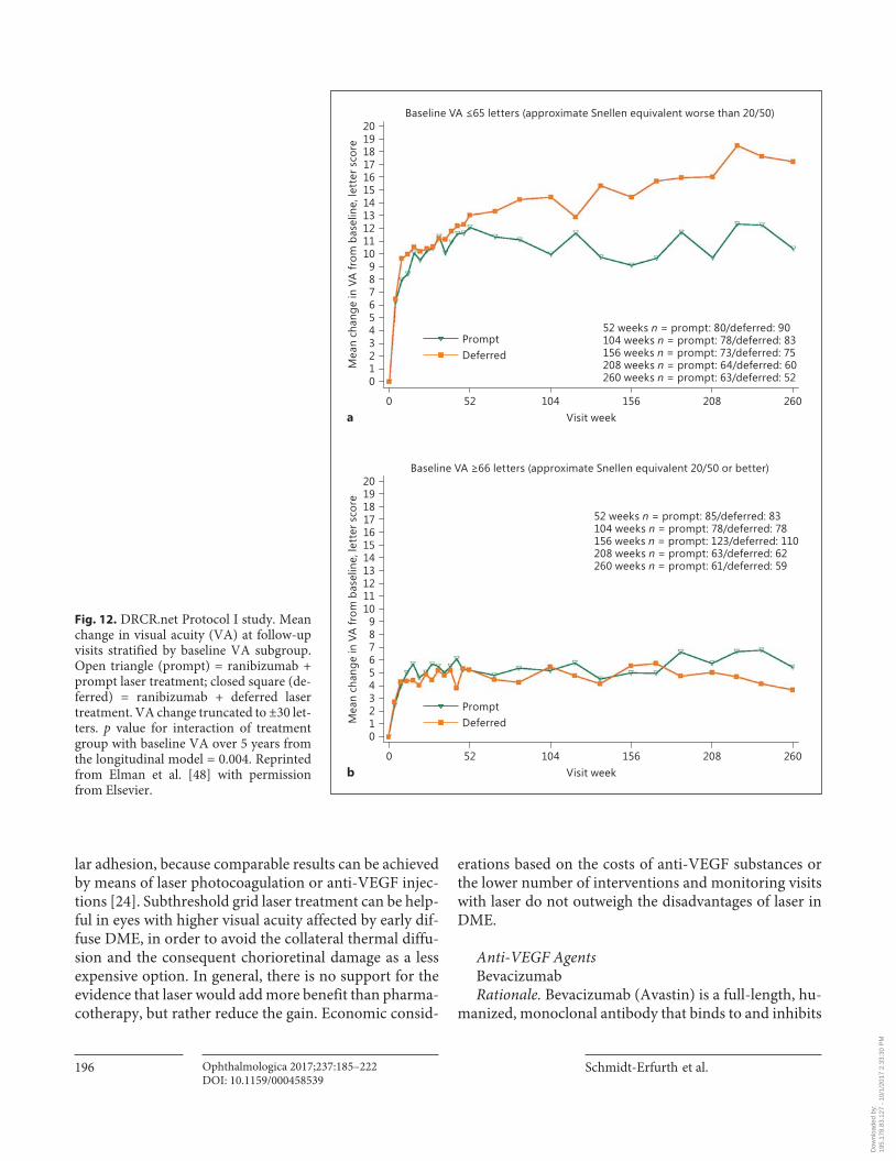
deferred [27] . But deferring laser led to a superior area under the curve (AOC) regarding BCVA ( Fig. 11 ), espe-cially in eyes with BCVA at baseline lower than 69 letters ( Fig. 12 ) and the number of patients achieving a >15 letter BCVA gain was higher in patients with deferred laser therapy [48] . Laser therapy has therefore been taken from the position of first-line recommendation and is rather considered to be harmful by many retina experts. Refer to the different sections of injection therapy to find evidence on the efficacy of laser versus anti-VEGF or steroid injec-tion therapy.
In the presence of DME and high-risk non-PDR or PDR, DRCR.net Protocol S [59] data clearly revealed su-periority of anti-VEGF (ranibizumab) therapy as the sole intervention which improves BCVA and additionally in-duces regression of PDR.
Recommendation Laser treatment has been shown to be an effective
treatment option in the management of DME in the ear-ly pioneering studies compared to sham treatment, but is currently in the era of anti-VEGF injection clearly not the standard of care anymore. Relative indications include la-ser application especially to the vasogenic subform of DME, which is clinically characterized by the presence of focally grouped MA and leaking capillaries. A further in-dication is represented by eyes affected by DME with CRT less than 300 μm or eyes with persisting vitreomacu-
11
10
9
8
7
6
5
4
3
2
1
12
0
260208156104520Visit week
Mea
n ch
ange
in v
isua
l acu
ity fr
om b
asel
ine,
lett
er s
core
PromptDeferred
52 weeks n = prompt: 165/deferred: 173104 weeks n = prompt: 156/deferred: 161156 weeks n = prompt: 144/deferred: 147208 weeks n = prompt: 127/deferred: 122260 weeks n = prompt: 124/deferred: 111
Fig. 11. DRCR.net Protocol I study. Mean change in visual acuity at follow-up visits. Open triangle (prompt) = ranibizumab + prompt laser treatment; closed square (de-ferred) = ranibizumab + deferred laser treatment. Visual acuity change truncated to ±30 letters. Results were similar without truncation (data not shown). Difference in mean change in visual acuity at 5 years from longitudinal model: p value adjusted for baseline visual acuity = 0.09; p value ad-justed for baseline visual acuity and other potential confounders = 0.15. Reprinted from Elman et al. [48] with permission from Elsevier.
Dow
nloa
ded
by:
195.
178.
83.1
27 -
10/
1/20
17 2
:33:
30 P
M
Schmidt-Erfurth et al.
Ophthalmologica 2017;237:185–222DOI: 10.1159/000458539
196
lar adhesion, because comparable results can be achieved by means of laser photocoagulation or anti-VEGF injec-tions [24] . Subthreshold grid laser treatment can be help-ful in eyes with higher visual acuity affected by early dif-fuse DME, in order to avoid the collateral thermal diffu-sion and the consequent chorioretinal damage as a less expensive option. In general, there is no support for the evidence that laser would add more benefit than pharma-cotherapy, but rather reduce the gain. Economic consid-
erations based on the costs of anti-VEGF substances or the lower number of interventions and monitoring visits with laser do not outweigh the disadvantages of laser in DME.
Anti-VEGF Agents Bevacizumab Rationale. Bevacizumab (Avastin) is a full-length, hu-
manized, monoclonal antibody that binds to and inhibits
19181716151413121110987654321
20
0
260208156104520Visit week
Mea
n ch
ange
in V
A fr
om b
asel
ine,
lett
er s
core
PromptDeferred
52 weeks n = prompt: 80/deferred: 90104 weeks n = prompt: 78/deferred: 83156 weeks n = prompt: 73/deferred: 75208 weeks n = prompt: 64/deferred: 60260 weeks n = prompt: 63/deferred: 52
Baseline VA 65 letters (approximate Snellen equivalent worse than 20/50)
19181716151413121110987654321
20
0
260208156104520Visit week
Mea
n ch
ange
in V
A fr
om b
asel
ine,
lett
er s
core
PromptDeferred
52 weeks n = prompt: 85/deferred: 83104 weeks n = prompt: 78/deferred: 78156 weeks n = prompt: 123/deferred: 110208 weeks n = prompt: 63/deferred: 62260 weeks n = prompt: 61/deferred: 59
Baseline VA 66 letters (approximate Snellen equivalent 20/50 or better)
a
b
Fig. 12. DRCR.net Protocol I study. Mean change in visual acuity (VA) at follow-up visits stratified by baseline VA subgroup. Open triangle (prompt) = ranibizumab + prompt laser treatment; closed square (de-ferred) = ranibizumab + deferred laser treatment. VA change truncated to ±30 let-ters. p value for interaction of treatment group with baseline VA over 5 years from the longitudinal model = 0.004. Reprinted from Elman et al. [48] with permission from Elsevier.
Dow
nloa
ded
by:
195.
178.
83.1
27 -
10/
1/20
17 2
:33:
30 P
M
Guidelines for the Management of Diabetic Macular Edema by EURETINA
Ophthalmologica 2017;237:185–222DOI: 10.1159/000458539
197
all VEGF isoforms. The molecule was developed to re-duce tumor growth by inhibiting pathological tumor ves-sel formation in metastatic colon cancer [60] . The medi-cation has received approval by the Food and Drug Ad-ministration (FDA) and the European Medicines Agency (EMA) for systemic treatment of several cancer forms. In oncology, treatment is administered as systemic infusions at doses of 5–15 mg/kg every 2 or 3 weeks with known increased risk of thromboembolic events, hypertension,
hemorrhages, and gastrointestinal perforation. Bevaci-zumab was designed for a prolonged retention in serum. The Fc region of the bevacizumab antibody binds to FcRn receptors that are highly expressed in vascular endothe-lial cells. The molecule is thereby protected from proteo-lytic catabolism and recycled systemically [61] . Based on a pharmacokinetic analysis of 491 patients who received 1–20 mg/kg of bevacizumab weekly, every 2 or every 3 weeks, the estimated half-life of bevacizumab in serum
20
15
10
5
0
25
4844403632282420161284 520Week
Mea
n ch
ange
from
bas
elin
e in
vis
ual a
cuity
,le
tter
sco
re
RanibizumabBevacizumabAflibercept
According to baseline visual acuity
20
15
10
5
0
25
4844403632282420161284 520Week
Mea
n ch
ange
from
bas
elin
e in
vis
ual a
cuity
,le
tter
sco
re
RanibizumabBevacizumabAflibercept
Overall
b
a
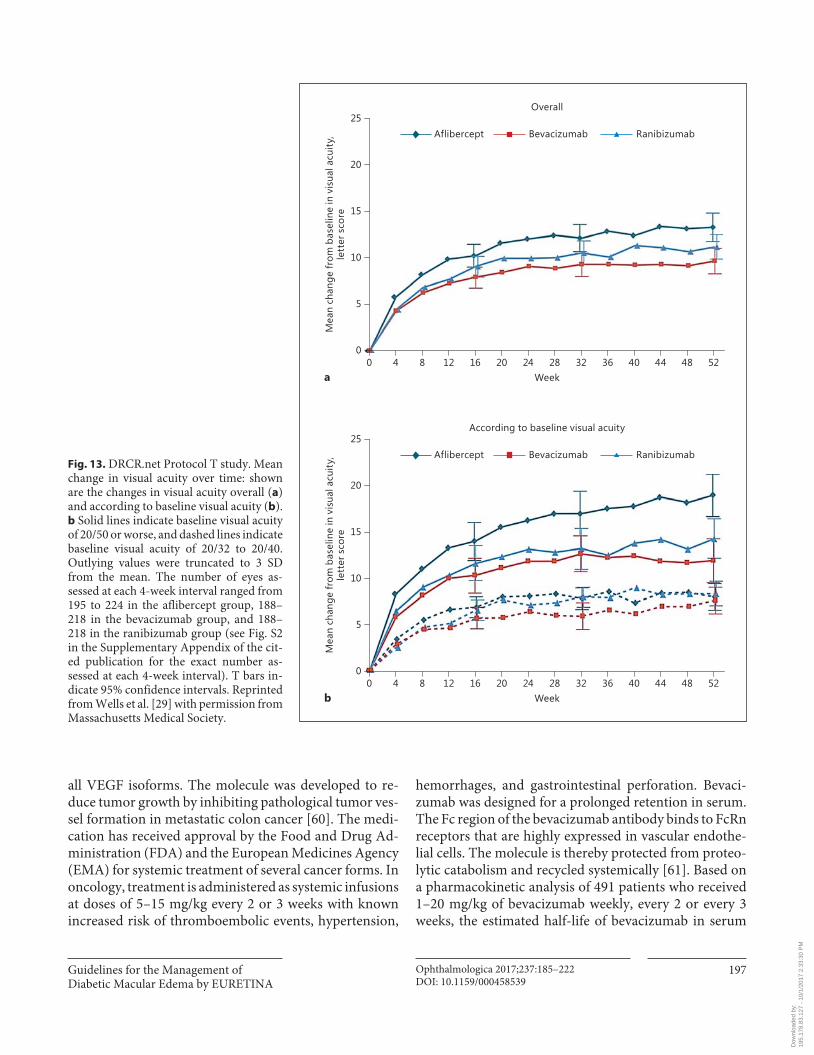
Fig. 13. DRCR.net Protocol T study. Mean change in visual acuity over time: shown are the changes in visual acuity overall ( a ) and according to baseline visual acuity ( b ). b Solid lines indicate baseline visual acuity of 20/50 or worse, and dashed lines indicate baseline visual acuity of 20/32 to 20/40. Outlying values were truncated to 3 SD from the mean. The number of eyes as-sessed at each 4-week interval ranged from 195 to 224 in the aflibercept group, 188–218 in the bevacizumab group, and 188–218 in the ranibizumab group (see Fig. S2 in the Supplementary Appendix of the cit-ed publication for the exact number as-sessed at each 4-week interval). T bars in-dicate 95% confidence intervals. Reprinted from Wells et al. [29] with permission from Massachusetts Medical Society.
Dow
nloa
ded
by:
195.
178.
83.1
27 -
10/
1/20
17 2
:33:
30 P
M
Schmidt-Erfurth et al.
Ophthalmologica 2017;237:185–222DOI: 10.1159/000458539
198
was approximately 20 days, ranging from 11 to 50 days. When used off-label for ocular disease, the standard dose of intraocular bevacizumab is 1.25 mg in 0.05 mL. Intra-vitreal half-life of bevacizumab in human vitreous sam-ples after one single injection ranged from 3 to 6.7 days [62] . Systemic retention after intravitreal treatment with bevacizumab was measured in patients with DME, with significantly reduced systemic plasma levels of VEGF up to 4 weeks after a single bevacizumab injection [63, 64] .
Intraocular bevacizumab is widely used as an off-label treatment for neovascular age-related macular degenera-tion (nAMD) and DME. Bevacizumab vials can be divid-ed into multiple dosages and the cost is manifold lower compared to ranibizumab and aflibercept. Since bevaci-zumab is not approved for the treatment of ocular disease, there has been a pressing need for robust comparative data of efficacy and safety.
Evidence. In March 2015, the DRCR.net published the first-year results of the Protocol T study. Protocol T is a randomized clinical trial, sponsored by the National In-stitutes of Health (NIH) [29] . The purpose was to com-pare efficacy and safety of intravitreal aflibercept, bevaci-zumab, and ranibizumab for the treatment of DME in-volving the center of the macula with vision loss. The study included 660 patients at a mean age of 61 ± 10 years, of which 90% had diabetes type 2. The mean visual acuity at baseline was 64.8 ± 11.3 letters (Snellen equivalent, ap-proximately 20/50), and the mean CRT was 412 ± 130 μm. The completion of the 1-year visit was 96%.
At 1 year, the mean BCVA letter score improved by 13.3 letters when treated with aflibercept, 9.7 letters with bevacizumab, and 11.2 letters with ranibizumab. The overall better results for aflibercept in the general popula-tion were, however, not considered relevant due to inter-action between baseline BCVA letter score and treatment results. When the initial BCVA score was 69 letters or more (Snellen equivalent, 20/40 or better), the mean BCVA letter score improved by 8.0 ± 7.6 for aflibercept, 7.5 ± 7.4 for bevacizumab and 8.3 ± 6.8 for ranibizumab, with no statistically significant difference between the treatment groups. For study eyes with an initial BCVA score of less than 69 letters (Snellen equivalent 20/50 or worse), the mean improvement was 18.9 ± 11.5 for afliber-cept, 11.8 ± 12.0 for bevacizumab, and 14.2 ± 10.6 for ra-nibizumab. Between the treatment groups with this lower baseline BCVA, there was a clear significant difference in favor of aflibercept, compared to bevacizumab and ran-ibizumab ( p = 0.0001 for aflibercept vs. bevacizumab, p = 0.0003 for aflibercept vs. ranibizumab, p = 0.21 for rani-bizumab vs. bevacizumab; Fig. 13 ). The median number
of intravitreal injections needed to achieve the BCVA gain was 9 (interquartile range 8–11) in the aflibercept group, 10 (8–12) in the bevacizumab group and 10 (8–11) in the ranibizumab group. The greatest mean reduction in CRT was 169 ± 139 μm for eyes treated with aflibercept, while the CRT was reduced by 101 ± 121 μm for bevaci-zumab and 147 ± 134 μm for ranibizumab (intravitreal injections needed to achieve the BCVA gain was 9 [inter-quartile range 8–11] in the aflibercept group, 10 [8–12] in the bevacizumab group, and 10 [8–11] in the ranibizum-ab group) supporting a superior efficacy of aflibercept.
For the 2-year results of Protocol T [65] , the findings slightly changed. BCVA gain at 2 years was 12.8 letters in the aflibercept, 10.0 letters in the bevacizumab, and 12.3 letters in the ranibizumab group (pairwise comparisons: p = 0.02 for aflibercept vs. bevacizumab, p = 0.47 for aflibercept vs. ranibizumab, and p = 0.11 for ranibizumab vs. bevacizumab), suggesting that ranibizumab caught up its overall difference in BCVA gain compared to afliber-cept, but bevacizumab did not. The median number of intravitreal injections needed in the second year to main-tain the BCVA gain was 5 (2–7) in the aflibercept group, 6 (2–9) in the bevacizumab group, and 6 (2–9) in the ra-nibizumab group. The greatest mean reduction in CRT was 171 ± 141 μm for eyes treated with aflibercept, while the CRT was reduced by 126 ± 143 μm for bevacizumab and 149 ± 141 μm for ranibizumab, with a statistically significant difference between bevacizumab and the other two drugs. The percentages of eyes undergoing at least one session of laser photocoagulation during the two years were 41, 64, and 52% in the aflibercept, bevacizu-mab, and ranibizumab groups.
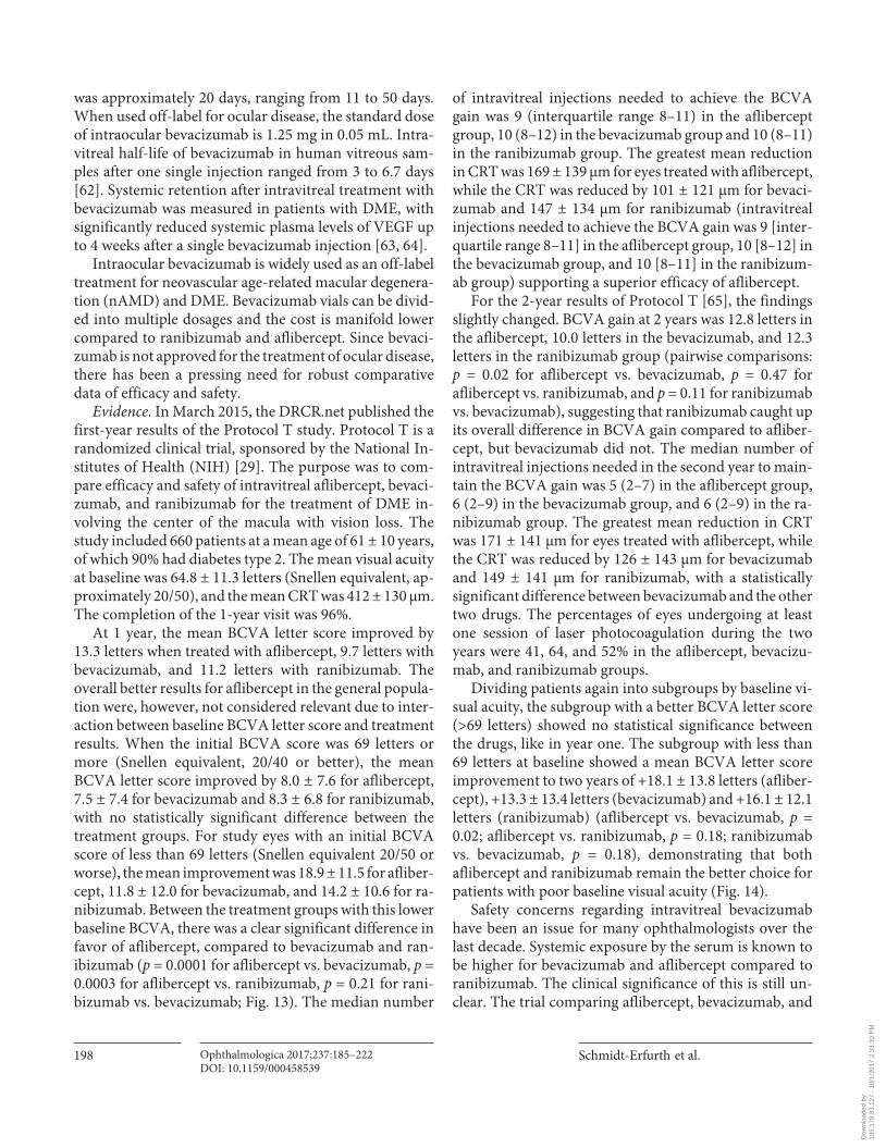
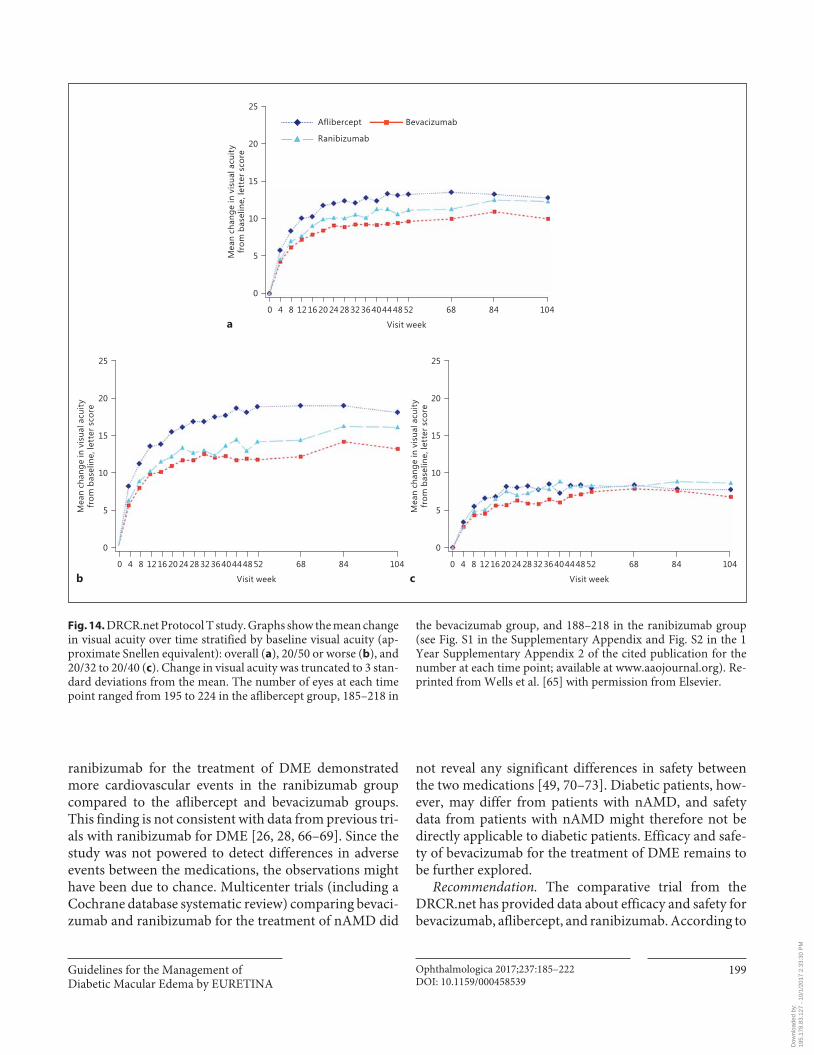
Dividing patients again into subgroups by baseline vi-sual acuity, the subgroup with a better BCVA letter score (>69 letters) showed no statistical significance between the drugs, like in year one. The subgroup with less than 69 letters at baseline showed a mean BCVA letter score improvement to two years of +18.1 ± 13.8 letters (afliber-cept), +13.3 ± 13.4 letters (bevacizumab) and +16.1 ± 12.1 letters (ranibizumab) (aflibercept vs. bevacizumab, p = 0.02; aflibercept vs. ranibizumab, p = 0.18; ranibizumab vs. bevacizumab, p = 0.18), demonstrating that both aflibercept and ranibizumab remain the better choice for patients with poor baseline visual acuity ( Fig. 14 ).
Safety concerns regarding intravitreal bevacizumab have been an issue for many ophthalmologists over the last decade. Systemic exposure by the serum is known to be higher for bevacizumab and aflibercept compared to ranibizumab. The clinical significance of this is still un-clear. The trial comparing aflibercept, bevacizumab, and
Dow
nloa
ded
by:
195.
178.
83.1
27 -
10/
1/20
17 2
:33:
30 P
M
Guidelines for the Management of Diabetic Macular Edema by EURETINA
Ophthalmologica 2017;237:185–222DOI: 10.1159/000458539
199
ranibizumab for the treatment of DME demonstrated more cardiovascular events in the ranibizumab group compared to the aflibercept and bevacizumab groups. This finding is not consistent with data from previous tri-als with ranibizumab for DME [26, 28, 66–69] . Since the study was not powered to detect differences in adverse events between the medications, the observations might have been due to chance. Multicenter trials (including a Cochrane database systematic review) comparing bevaci-zumab and ranibizumab for the treatment of nAMD did
not reveal any significant differences in safety between the two medications [49, 70–73] . Diabetic patients, how-ever, may differ from patients with nAMD, and safety data from patients with nAMD might therefore not be directly applicable to diabetic patients. Efficacy and safe-ty of bevacizumab for the treatment of DME remains to be further explored.
Recommendation. The comparative trial from the DRCR.net has provided data about efficacy and safety for bevacizumab, aflibercept, and ranibizumab. According to
20
15
10
5
25
0
104846848444036322824201612840 52
Visit week
Aflibercept Bevacizumab
Ranibizumab
20
15
10
5
25
0
104846848444036322824201612840 52
Visit week
Mea
n ch
ange
in v
isua
l acu
ity
from
bas
elin
e, le
tter
sco
re
20
15
10
5
25
0
104846848444036322824201612840 52
Visit week
Mea
n ch
ange
in v
isua
l acu
ity
from
bas
elin
e, le
tter
sco
re
Mea
n ch
ange
in v
isua
l acu
ity
from
bas
elin
e, le
tter
sco
re
a
b c
Fig. 14. DRCR.net Protocol T study. Graphs show the mean change in visual acuity over time stratified by baseline visual acuity (ap-proximate Snellen equivalent): overall ( a ), 20/50 or worse ( b ), and 20/32 to 20/40 ( c ). Change in visual acuity was truncated to 3 stan-dard deviations from the mean. The number of eyes at each time point ranged from 195 to 224 in the aflibercept group, 185–218 in
the bevacizumab group, and 188–218 in the ranibizumab group (see Fig. S1 in the Supplementary Appendix and Fig. S2 in the 1 Year Supplementary Appendix 2 of the cited publication for the number at each time point; available at www.aaojournal.org). Re-printed from Wells et al. [65] with permission from Elsevier.
Dow
nloa
ded
by:
195.
178.
83.1
27 -
10/
1/20
17 2
:33:
30 P
M
Schmidt-Erfurth et al.
Ophthalmologica 2017;237:185–222DOI: 10.1159/000458539
200
these results, the choice of treatment for DME depends on the baseline BCVA letter score. While aflibercept and ranibizumab are the drugs of choice for BCVA letter score of less than 69, all three medications are equivalent in improving vision in eyes with a baseline BCVA letter score of 69 or more. The numbers of serious adverse events were altogether small, but the follow-up was short. The much lower cost of off-label use of intravitreal beva-cizumab is indisputable, but all three medications should be available to ophthalmologists who are responsible for tailoring the treatment for each patient with DMA.
Ranibizumab Rationale. Ranibizumab (Lucentis) is a recombinant
humanized Fab fragment of a monoclonal antibody, de-signed for intraocular use, which binds and inactivates all isoforms of VEGF-A. The rationale for anti-VEGF ther-apy for DME is based on the observation that VEGF levels are increased in the retina and vitreous of eyes with DR [74] . VEGF has been demonstrated to increase the vessel permeability in vivo by increasing the phosphorylation of tight junction proteins and thus is an important mediator of the BRB breakdown [75] . Therefore, therapy that in-hibits VEGF represents an effective therapeutic modality targeting the underlying pathogenesis of DME. The intra-vitreal injection of ranibizumab has been shown to be
beneficial and relatively safe for the treatment of DME in several randomized clinical trials and has become a stan-dard for treatment of DME.
Evidence. Several studies reported the efficacy and su-periority of ranibizumab to laser photocoagulation in the treatment of DME and long-term data recently became available, allowing the evaluation of continued therapy effects. The RESTORE study was the first study to dem-onstrate that ranibizumab monotherapy provides signifi-cantly superior benefit over laser in patients with visual impairment due to DME [24] .
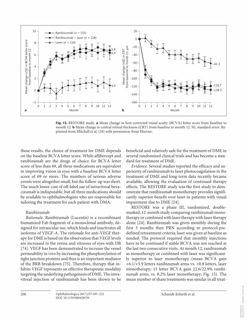
RESTORE was a phase III, randomized, double-masked, 12-month study comparing ranibizumab mono-therapy or combined with laser therapy with laser therapy alone [24] . Ranibizumab was given monthly during the first 3 months then PRN according to protocol-pre-defined retreatment criteria; laser was given at baseline as needed. The protocol required that monthly injections have to be continued if stable BCVA was not reached at the last two consecutive visits. At month 12, ranibizumab as monotherapy or combined with laser was significant-ly superior to laser monotherapy (mean BCVA gain +6.1/+5.9 letters ranibizumab arms vs. +0.8 letters, laser monotherapy; 15 letter BCVA gain 22.6/22.9% ranibi-zumab arms, vs. 8.2% laser monotherapy; Fig. 15 ). The mean number of sham treatments was similar in all treat-
8
6
4
2
0
10
–21110987654321 12
6.86.4
0.9
0Month
Mea
n ch
ange
(±SE
) in
BCVA
lett
er s
core
from
bas
elin
e to
mon
th 1
2
0
–20
–40
–60
–80
–100
–120
–140
20
–1601110987654321 120
Month
Mea
n ch
ange
(±SE
) in
CRT
from
bas
elin
e to
mon
th 1
2, μ
m
–61.3
–118.7–128.3
Ranibizumab (n = 115)Ranibizumab + laser (n = 118)Laser (n = 110)
a b
Fig. 15. RESTORE study. a Mean change in best-corrected visual acuity (BCVA) letter score from baseline to month 12. b Mean change in central retinal thickness (CRT) from baseline to month 12. SE, standard error. Re-printed from Mitchell et al. [24] with permission from Elsevier.
Dow
nloa
ded
by:
195.
178.
83.1
27 -
10/
1/20
17 2
:33:
30 P
M
Guidelines for the Management of Diabetic Macular Edema by EURETINA
Ophthalmologica 2017;237:185–222DOI: 10.1159/000458539
201
ment groups. The results of the RESTORE study demon-strate that treatment with ranibizumab is superior to laser treatment alone in improving BCVA in patients with vi-sual impairment due to DME and that laser does not add
any benefit. During this 1-year study period, combining laser with ranibizumab did not seem to provide any ad-vantage compared with ranibizumab monotherapy in terms of improving BCVA and treatment exposure.
10
8
6
4
2
0
12
–2Core study Extension study, ranibizumab 0.5 mg PRN
Mea
n of
the
chan
ge (±
SE) i
n BC
VAfr
om b
asel
ine,
ETD
RS le
tter
s
3432302826242220181614121086420 36Months
0
–20
–40
–60
–80
–100
–120
–140
–160
20
–180Core study Extension study, ranibizumab 0.5 mg PRN
Mea
n of
the
chan
ge (±
SE) i
n CR
STfr
om b
asel
ine,
μm
333027242118151296 3630Months
Core study assessment Interim analysis Full analysis/studycompletion
+8.0
+6.7
+6.0
+7.9+7.9
+7.1
+2.3
+6.7
+5.4
Prior ranibizumab 0.5 mg + laser (n = 83)Prior ranibizumab 0.5 mg (n = 83)
Prior laser (n = 74)
–63.3
–127.8
–139.7
–126.6
–142.7–142.1–145.9
–129.1
–140.6
a
b
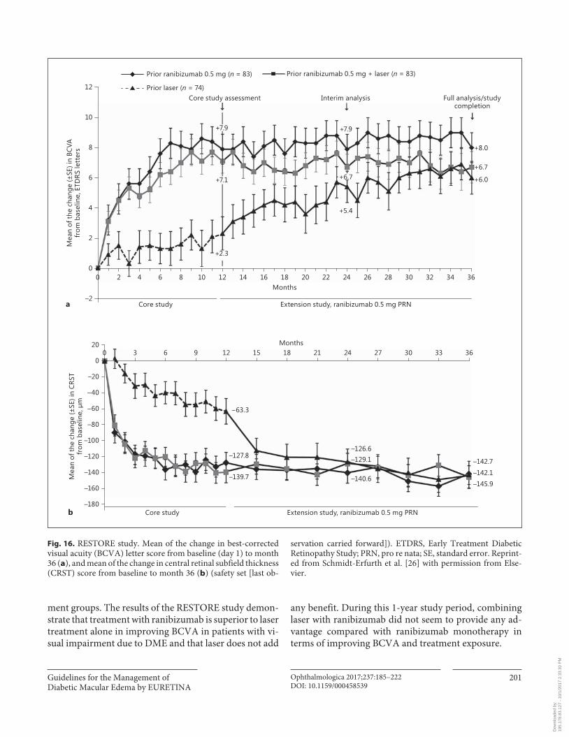
Fig. 16. RESTORE study. Mean of the change in best-corrected visual acuity (BCVA) letter score from baseline (day 1) to month 36 ( a ), and mean of the change in central retinal subfield thickness (CRST) score from baseline to month 36 ( b ) (safety set [last ob-
servation carried forward]). ETDRS, Early Treatment Diabetic Retinopathy Study; PRN, pro re nata; SE, standard error. Reprint-ed from Schmidt-Erfurth et al. [26] with permission from Else-vier.
Dow
nloa
ded
by:
195.
178.
83.1
27 -
10/
1/20
17 2
:33:
30 P
M
Schmidt-Erfurth et al.
Ophthalmologica 2017;237:185–222DOI: 10.1159/000458539
202
Based on the results of the RESTORE study, ranibi-zumab was approved in Europe by the EMA for treatment of visual impairment due to DME in 2011. This approval was reviewed in 2014. The approved dose in Europe is 0.5 mg per injection compared to 0.3 mg in the US. The med-ication is approved for monthly injections continued un-til the maximum visual acuity is achieved and/or the ab-sence of activity of the disease.
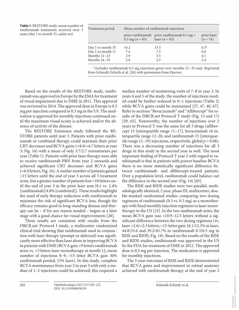
The RESTORE Extension study followed the RE-STORE patients until year 3. Patients with prior ranibi-zumab or combined therapy could maintain their prior CRT decreases and BCVA gains (+8.0/+6.7 letters at year 3; Fig. 16 ) with a mean of only 3.7/2.7 retreatments per year ( Table 1 ). Patients with prior laser therapy were able to receive ranibizumab PRN from year 2 onwards and achieved significant CRT decreases and BCVA gains (+6.0 letters; Fig. 16 ). A similar number of patients gained >15 letters until the end of year 3 across all 3 treatment arms, but a greater number of patients lost >10 letters un-til the end of year 3 in the prior laser arm (8.1 vs. 2.4% [ranibizumab]/4.8% [combined]). These results highlight the need of early therapy induction with ranibizumab to minimize the risk of significant BCVA loss, though the efficacy remains good in long-standing disease and ther-apy can be – if for any reason needed – begun at a later stage with a good chance for visual improvements [26] .
These results are consistent with results from the DRCR.net Protocol I study, a multicenter randomized clinical trial showing that ranibizumab used in conjunc-tion with laser therapy (prompt or deferred) was signifi-cantly more effective than laser alone in improving BCVA in patients with DME (BCVA gain +9 letters ranibizumab arms vs. +3 letters laser monotherapy at month 12, mean number of injections 8–9; >15 letter BCVA gain 30%ranibizumab pooled, 15% laser). In this study, complete BCVA maintenance from year 2 to year 3 with only a me-dian of 1–2 injections could be achieved; this required a
median number of monitoring visits of 7–8 in year 3. In years 4 and 5 of the study, the number of injections need-ed could be further reduced to 0–1 injections ( Table 2 ) while BCVA gains could be maintained [27, 47, 48, 67] . Refer to sections “Bevacizumab” and “Aflibercept” for re-sults of the DRCR.net Protocol T study ( Fig. 13 and 17 ) [29, 65] . Noteworthy, the number of injections over 2 years in Protocol T was the same for all 3 drugs (afliber-cept 15 [interquartile range 11–17], bevacizumab 16 in-terquartile range 12–20, and ranibizumab 15 [interquar-tile range 11–19] injections, respectively, global p = 0.08). There was a decreasing number of injections for all 3 drugs in this study in the second year as well. The most important finding of Protocol T year 2 with regard to ra-nibizumab is that in patients with poorer baseline BCVA there is no more statistically significant difference be-tween ranibizumab- and aflibercept-treated patients. Over a population level, ranibizumab could balance out the difference in the second year ( Fig. 14 ) [65] .
The RISE and RIDE studies were two parallel, meth-odologically identical, 2 year, phase III, multicenter, dou-ble-masked randomized studies comparing two dosing regimens of ranibizumab (0.3 vs. 0.5 mg) as a monother-apy with fixed monthly injection regimens to laser mono-therapy in the US [23] . In the two ranibizumab arms, the mean BCVA gain was +10.9–12.5 letters without a sig-nificant difference between the two dosing regimens (vs. laser +2.6/+2.3 letters; >15-letter gain 18.1/12.3% in laser, 44.8/33.6 and 39.2/45.7% in ranibizumab 0.3/0.5 mg in RISE and RIDE; Fig. 18 ). Based on the results of the RISE and RIDE studies, ranibizumab was approved in the US by the FDA for treatment of DME in 2012. The approved dose is 0.3 mg per injection. The medication is approved for monthly injections.
The 3-year outcomes of RISE and RIDE demonstrated that BCVA gains and improvement in retinal anatomy achieved with ranibizumab therapy at the end of year 2
Treatment period Mean number of ranibizumab injections
pri or ranibizumab 0.5 mg (n = 83)
prior ranibizumab 0.5 mg + laser (n = 83)
prior laser (n = 74)
Day 1 to month 35 14.2 13.5 6.5a
Day 1 to month 11 7.4 7.5 0.0Months 12 – 23 3.9 3.5 4.1Months 24 – 35 2.9 2.5 2.4
a Includes ranibizumab 0.5 mg injections given over months 12 – 35 only. Reprinted from Schmidt-Erfurth et al. [26] with permission from Elsevier.
Table 1. RESTORE study: mean number of ranibizumab treatments received over 3 years (day 1 to month 35, safety set)
Dow
nloa
ded
by:
195.
178.
83.1
27 -
10/
1/20
17 2
:33:
30 P
M
Guidelines for the Management of Diabetic Macular Edema by EURETINA
Ophthalmologica 2017;237:185–222DOI: 10.1159/000458539
203
were sustained through year 3 without any significant dif-ference in the two dosing regimens ( Fig. 19 ). The study design allowed patients in the laser group to cross over and receive monthly 0.5 mg ranibizumab in the third year. This delayed ranibizumab treatment did not result in the same extent of BCVA improvement observed in patients originally randomized to ranibizumab (BCVA gain >15 letters was 22.0/19.2% in the laser + delayedranibizumab compared to 36.8–51.2% in the 0.3/0.5 mg ranibizumab RISE/RIDE study arms). The mean BCVA gain from baseline to the end of year 3 was +4.7/4.3 letters in the laser + delayed ranibizumab group versus +10.6–14.2 in the ranibizumab arms of both studies, which indi-cates a permanent loss of BCVA if DME is treated too late with anti-VEGF [69] . An open-label extension of the studies allowed patients to be followed for another 14.1 months of ranibizumab PRN treatment. While maintain-ing 3-year BCVA, a mean of 4.5 injections were adminis-tered, and 25% of patients required no further injections [76] .
In none of the studies were overall significant differ-ences observed in BCVA gains between ranibizumab monotherapy and ranibizumab combined with laser treatment. The combined therapy was not superior to drug monotherapy and is therefore not recommended [77] . The RETAIN study, a multicenter controlled phase IIIB trial, evaluated the efficacy and safety of 0.5 mg ra-nibizumab in 2 “treat and extend” treatment algorithms compared with a PRN regimen over 2 years. In “treat and extend,” patients treated with a gradually prolonged time interval between injections after BCVA stabilization with monthly injections has been reached. The RETAIN study confirmed that this regimen is noninferior to PRN in mean average BCVA gain after 2 years (+8.3/+6.5 vs. +8.1 letters [PRN]; Fig. 20 ) with a mean of 12.4/12.8 versus 10.7 injections but with a large reduction in patient visits (9.0/8.9 vs. 16.6 visits) [30] .
In all these studies as well as in a meta-analysis [77] of 1,500 patients treated with ranibizumab in randomized clinical trials, the safety profile of ranibizumab was excel-
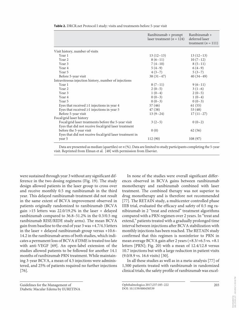
Table 2. DRCR.net Protocol I study: visits and treatments before 5-year visit
Ranibizumab + prompt laser treatment (n = 124)
Ranibizumab + deferred laser treatment (n = 111)
Visit history, number of visitsYear 1 13 (12 – 13) 13 (12 – 13)Year 2 8 (6 – 11) 10 (7 – 12)Year 3 7 (4 – 10) 8 (5 – 11)Year 4 5 (4 – 9) 6 (4 – 9)Year 5 4 (3 – 7) 5 (3 – 7)Before 5-year visit 38 (31 – 47) 40 (34 – 49)
Intravitreous injection history, number of injectionsYear 1 8 (7 – 11) 9 (6 – 11)Year 2 2 (0 – 5) 3 (1 – 6)Year 3 1 (0 – 4) 2 (0 – 5)Year 4 0 (0 – 3) 1 (0 – 4)Year 5 0 (0 – 3) 0 (0 – 3)Eyes that received ≥1 injections in year 4 57 (46) 61 (55)Eyes that received ≥1 injections in year 5 47 (38) 53 (48)Before 5-year visit 13 (9 – 24) 17 (11 – 27)
Focal/grid laser historyFocal/grid laser treatments before the 5-year visit 3 (2 – 5) 0 (0 – 2)Eyes that did not receive focal/grid laser treatmentbefore the 5-year visit 0 (0) 62 (56)Eyes that did not receive focal/grid laser treatment inyear 5 112 (90) 108 (97)
Data are presented as median (quartiles) or n (%). Data are limited to study participants completing the 5-year visit. Reprinted from Elman et al. [48] with permission from Elsevier.
Dow
nloa
ded
by:
195.
178.
83.1
27 -
10/
1/20
17 2
:33:
30 P
M
Schmidt-Erfurth et al.
Ophthalmologica 2017;237:185–222DOI: 10.1159/000458539
204
lent, without an increase of cardiovascular events com-pared to sham. No systemic safety concern emerged with 0.3/0.5 mg ranibizumab administered monthly compared with control through the RISE/RIDE extension study to 3 years [69] . In Protocol I, patients in the sham group ex-perienced higher rates of Anti-Platelet Trialists’ Collabo-ration (APTC)-classified systemic events than patients receiving ranibizumab [27] . In RESTORE, no meaningful differences in the number of arterial thromboembolic events or other systemic events were observed between the ranibizumab and the laser groups, although patients
with a history of stroke or transient ischemic attack were primarily excluded from this study [24] .
When looking at the different treatment regimens available for ranibizumab injection, studies are available for a fixed monthly injection [23] , a PRN regimen [24] , or a treat and extend regimen. Monthly injections seem to be feasible in the beginning of therapy, but are not re-quired over a period of more than 3–6 months as con-firmed by the decreasing numbers of injections needed after month 6 in several studies ( Tables 1 and 2 ) [25, 26, 48, 69] , even starting with a PRN regimen from baseline
–260
–20–40–60–80
–100–120–140–160–180–200–220–240
0
4844403632282420161284 520Week
Mea
n ch
ange
from
bas
elin
e in
cen
tral
subf
ield
thic
knes
s, μ
m
RanibizumabBevacizumabAflibercept
According to baseline visual acuity
–260
–20
–40
–60
–80
–100
–120
–140
–160
–180
–200
–220
–240
0
4844403632282420161284 520Week
Mea
n ch
ange
from
bas
elin
e in
cen
tral
subf
ield
thic
knes
s, μ
m
RanibizumabBevacizumabAflibercept
Overall
b
a
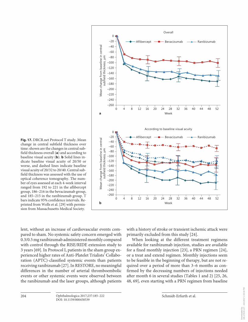
Fig. 17. DRCR.net Protocol T study. Mean change in central subfield thickness over time: shown are the changes in central sub-field thickness overall ( a ) and according to baseline visual acuity ( b ). b Solid lines in-dicate baseline visual acuity of 20/50 or worse, and dashed lines indicate baseline visual acuity of 20/32 to 20/40. Central sub-field thickness was assessed with the use of optical coherence tomography. The num-ber of eyes assessed at each 4-week interval ranged from 192 to 221 in the aflibercept group, 186–216 in the bevacizumab group, and 185–215 in the ranibizumab group. T bars indicate 95% confidence intervals. Re-printed from Wells et al. [29] with permis-sion from Massachusetts Medical Society.
Dow
nloa
ded
by:
195.
178.
83.1
27 -
10/
1/20
17 2
:33:
30 P
M

Guidelines for the Management of Diabetic Macular Edema by EURETINA
Ophthalmologica 2017;237:185–222DOI: 10.1159/000458539
205
results in comparable BCVA gains [29] . Retreatment and monitoring intervals can be adjusted to visual acuity or anatomic response to ranibizumab. Most studies propose monthly monitoring, but in certain cases bimonthly monitoring, as done in the RELIGHT study [78] , can be applied to reduce patient burden and monitoring costs. None of the proposed retreatment criteria in a PRN regi-men can be considered to be superior to the others as no comparability study has been performed. An alternative approach is the “treat and extend regimen.” The largest
criticism about this regimen in the long-term is the fact that each time treatment has to be administered even if disease and vision stability is already achieved, even if there are no concerns about efficacy and safety in 2 years of this retreatment regimen, there might be overtreat-ment, especially as DME seems to plateau after the first treatment period and very little or no injections are need-ed, which has been referred to as a disease-modifying ef-fect [48] . Bimonthly monitoring and “treat and extend” suggest that a flexible retreatment regimen of ranibizum-
Sham Ranibizumab 0.3 mg
10
5
15
024201612840
Mea
n ch
ange
in v
isua
l acu
ity,
ETD
RS le
tter
s
MonthDay 7
RISE
12.5*11.9*
2.6 2.3
–50
–100
–150
–200
–250
0
–30024181263210
Mea
n ch
ange
in C
FT, μ
m
MonthDay 7
–133.4
–250.6*–253.1*
–50
–100
–150
–200
–250
0
–30024181263210
MonthDay 7
–125.8
–259.8*–270.7*
10
5
15
024201612840
MonthDay 7
RIDE
12.0*10.9*
Ranibizumab 0.5 mg
a
b
Fig. 18. RIDE/RISE studies. Changes in visual acuity ( a ) and cen-tral foveal thickness (CFT) ( b ) from baseline through 24 months. Number of patients: 127, 125, and 125 (RISE), and 130, 125, and 127 (RIDE) in the sham, 0.3 mg, and 0.5 mg groups, respectively. Vertical bars are ±1 standard error of the mean. The last observa-tion carried forward imputation method was used. * p < 0.0001 vs.
sham (analysis of variance t test [stratified]). Differences were sta-tistically significant starting at the first posttreatment observation (day 7) and at each point thereafter; a hierarchical testing strategy controlled for multiple comparisons. ETDRS, Early Treatment Di-abetic Retinopathy Study. Reprinted from Nguyen et al. [23] with permission from Elsevier.
Dow
nloa
ded
by:
195.
178.
83.1
27 -
10/
1/20
17 2
:33:
30 P
M
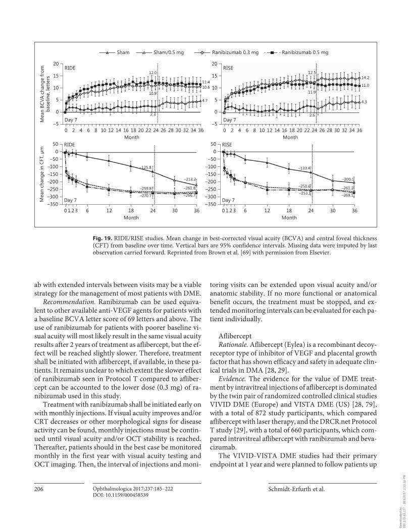
Schmidt-Erfurth et al.
Ophthalmologica 2017;237:185–222DOI: 10.1159/000458539
206
ab with extended intervals between visits may be a viable strategy for the management of most patients with DME.
Recommendation. Ranibizumab can be used equiva-lent to other available anti-VEGF agents for patients with a baseline BCVA letter score of 69 letters and above. The use of ranibizumab for patients with poorer baseline vi-sual acuity will most likely result in the same visual acuity results after 2 years of treatment as aflibercept, but the ef-fect will be reached slightly slower. Therefore, treatment shall be initiated with aflibercept, if available, in these pa-tients. It remains unclear to which extent the slower effect of ranibizumab seen in Protocol T compared to afliber-cept can be accounted to the lower dose (0.3 mg) of ra-nibizumab used in this study.
Treatment with ranibizumab shall be initiated early on with monthly injections. If visual acuity improves and/or CRT decreases or other morphological signs for disease activity can be found, monthly injections must be contin-ued until visual acuity and/or OCT stability is reached. Thereafter, patients should in the best case be monitored monthly in the first year with visual acuity testing and OCT imaging. Then, the interval of injections and moni-
toring visits can be extended upon visual acuity and/or anatomic stability. If no more functional or anatomical benefit occurs, the treatment must be stopped, and ex-tended monitoring intervals can be evaluated for each pa-tient individually.
Aflibercept Rationale. Aflibercept (Eylea) is a recombinant decoy-
receptor type of inhibitor of VEGF and placental growth factor that has shown efficacy and safety in adequate clin-ical trials in DMA [28, 29] .
Evidence. The evidence for the value of DME treat-ment by intravitreal injections of aflibercept is dominated by the twin pair of randomized controlled clinical studies VIVID DME (Europe) and VISTA DME (US) [28, 79] , with a total of 872 study participants, which compared aflibercept with laser therapy, and the DRCR.net Protocol T study [29] , with a total of 660 participants, which com-pared intravitreal aflibercept with ranibizumab and beva-cizumab.
The VIVID-VISTA DME studies had their primary endpoint at 1 year and were planned to follow patients up
Sham
15
10
5
0
20
–5343230282624222018161412108642 360
Mea
n BC
VA c
hang
e fr
omba
selin
e, le
tter
s
Month
RIDE
Day 7 Day 7
12.0
11.410.6
4.7
10.9
2.3
0–50
–100–150–200–250–300
50
–3506321 30241812 360
Mea
n ch
ange
in C
FT, μ
m
Month
RIDE
Day 7
–125.8
–213.2
–261.8
–266.7
–259.8
–270.7
0–50
–100–150–200–250–300
50
–3506321 30241812 360
Month
RISE
Day 7
–133.4
–200.1
–261.2
–269.1
–250.6
–253.1
15
10
5
0
20
–5343230282624222018161412108642 360
Month
RISE12.5
11.9
2.6
14.2
11.0
4.3
Sham/0.5 mg Ranibizumab 0.3 mg Ranibizumab 0.5 mg
Fig. 19. RIDE/RISE studies. Mean change in best-corrected visual acuity (BCVA) and central foveal thickness (CFT) from baseline over time. Vertical bars are 95% confidence intervals. Missing data were imputed by last observation carried forward. Reprinted from Brown et al. [69] with permission from Elsevier.
Dow
nloa
ded
by:
195.
178.
83.1
27 -
10/
1/20
17 2
:33:
30 P
M
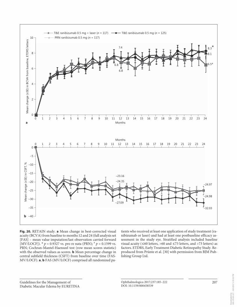
Guidelines for the Management of Diabetic Macular Edema by EURETINA
Ophthalmologica 2017;237:185–222DOI: 10.1159/000458539
207
T&E ranibizumab 0.5 mg (n = 125)
8
6
4
2
10
02322212019181716151413121110987654321 240
Months
Mea
n ch
ange
(±SE
) in
BCVA
from
bas
elin
e, E
TDRS
lett
ers
–5
–10
–15
–20
–25
–30
–35
0
–40
2322212019181716151413121110987654321 240Months
Mea
n ch
ange
(±SE
) in
CSFT
, %
7.4
6.8
6.8
8.3*8.1
6.5#
–23.16
–24.35
–27.09
–24.97
–24.98
–32.03
PRN ranibizumab 0.5 mg (n = 117)T&E ranibizumab 0.5 mg + laser (n = 117)
a
b
Fig. 20. RETAIN study. a Mean change in best-corrected visual acuity (BCVA) from baseline to months 12 and 24 (full analysis set [FAS] – mean value imputation/last observation carried forward [MV/LOCF]). * p = 0.9327 vs. pro re nata (PRN); # p = 0.1599 vs. PRN; Cochran-Mantel-Haenszel test (row mean scores statistic) with the observed values as scores. b Mean percentage change in central subfield thickness (CSFT) from baseline over time (FAS-MV/LOCF). a , b FAS (MV/LOCF) comprised all randomized pa-
tients who received at least one application of study treatment (ra-nibizumab or laser) and had at least one postbaseline efficacy as-sessment in the study eye. Stratified analysis included baseline visual acuity ( ≤ 60 letters, >60 and ≤ 73 letters, and >73 letters) as factors. ETDRS, Early Treatment Diabetic Retinopathy Study. Re-produced from Prünte et al. [30] with permission from BJM Pub-lishing Group Ltd.
Dow
nloa
ded
by:
195.
178.
83.1
27 -
10/
1/20
17 2
:33:
30 P
M
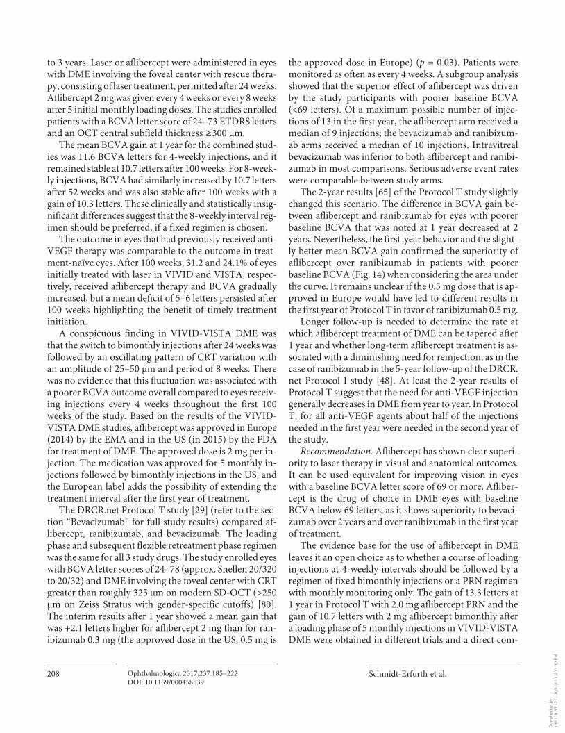
Schmidt-Erfurth et al.
Ophthalmologica 2017;237:185–222DOI: 10.1159/000458539
208
to 3 years. Laser or aflibercept were administered in eyes with DME involving the foveal center with rescue thera-py, consisting of laser treatment, permitted after 24 weeks. Aflibercept 2 mg was given every 4 weeks or every 8 weeks after 5 initial monthly loading doses. The studies enrolled patients with a BCVA letter score of 24–73 ETDRS letters and an OCT central subfield thickness ≥ 300 μm.
The mean BCVA gain at 1 year for the combined stud-ies was 11.6 BCVA letters for 4-weekly injections, and it remained stable at 10.7 letters after 100 weeks. For 8-week-ly injections, BCVA had similarly increased by 10.7 letters after 52 weeks and was also stable after 100 weeks with a gain of 10.3 letters. These clinically and statistically insig-nificant differences suggest that the 8-weekly interval reg-imen should be preferred, if a fixed regimen is chosen.
The outcome in eyes that had previously received anti-VEGF therapy was comparable to the outcome in treat-ment-naïve eyes. After 100 weeks, 31.2 and 24.1% of eyes initially treated with laser in VIVID and VISTA, respec-tively, received aflibercept therapy and BCVA gradually increased, but a mean deficit of 5–6 letters persisted after 100 weeks highlighting the benefit of timely treatment initiation.
A conspicuous finding in VIVID-VISTA DME was that the switch to bimonthly injections after 24 weeks was followed by an oscillating pattern of CRT variation with an amplitude of 25–50 μm and period of 8 weeks. There was no evidence that this fluctuation was associated with a poorer BCVA outcome overall compared to eyes receiv-ing injections every 4 weeks throughout the first 100 weeks of the study. Based on the results of the VIVID-VISTA DME studies, aflibercept was approved in Europe (2014) by the EMA and in the US (in 2015) by the FDA for treatment of DME. The approved dose is 2 mg per in-jection. The medication was approved for 5 monthly in-jections followed by bimonthly injections in the US, and the European label adds the possibility of extending the treatment interval after the first year of treatment.
The DRCR.net Protocol T study [29] (refer to the sec-tion “Bevacizumab” for full study results) compared af-libercept, ranibizumab, and bevacizumab. The loading phase and subsequent flexible retreatment phase regimen was the same for all 3 study drugs. The study enrolled eyes with BCVA letter scores of 24–78 (approx. Snellen 20/320 to 20/32) and DME involving the foveal center with CRT greater than roughly 325 μm on modern SD-OCT (>250 μm on Zeiss Stratus with gender-specific cutoffs) [80] . The interim results after 1 year showed a mean gain that was +2.1 letters higher for aflibercept 2 mg than for ran-ibizumab 0.3 mg (the approved dose in the US, 0.5 mg is
the approved dose in Europe) ( p = 0.03). Patients were monitored as often as every 4 weeks. A subgroup analysis showed that the superior effect of aflibercept was driven by the study participants with poorer baseline BCVA (<69 letters). Of a maximum possible number of injec-tions of 13 in the first year, the aflibercept arm received a median of 9 injections; the bevacizumab and ranibizum-ab arms received a median of 10 injections. Intravitreal bevacizumab was inferior to both aflibercept and ranibi-zumab in most comparisons. Serious adverse event rates were comparable between study arms.
The 2-year results [65] of the Protocol T study slightly changed this scenario. The difference in BCVA gain be-tween aflibercept and ranibizumab for eyes with poorer baseline BCVA that was noted at 1 year decreased at 2 years. Nevertheless, the first-year behavior and the slight-ly better mean BCVA gain confirmed the superiority of aflibercept over ranibizumab in patients with poorer baseline BCVA ( Fig. 14 ) when considering the area under the curve. It remains unclear if the 0.5 mg dose that is ap-proved in Europe would have led to different results in the first year of Protocol T in favor of ranibizumab 0.5 mg.
Longer follow-up is needed to determine the rate at which aflibercept treatment of DME can be tapered after 1 year and whether long-term aflibercept treatment is as-sociated with a diminishing need for reinjection, as in the case of ranibizumab in the 5-year follow-up of the DRCR.net Protocol I study [48] . At least the 2-year results of Protocol T suggest that the need for anti-VEGF injection generally decreases in DME from year to year. In Protocol T, for all anti-VEGF agents about half of the injections needed in the first year were needed in the second year of the study.
Recommendation. Aflibercept has shown clear superi-ority to laser therapy in visual and anatomical outcomes. It can be used equivalent for improving vision in eyes with a baseline BCVA letter score of 69 or more. Afliber-cept is the drug of choice in DME eyes with baseline BCVA below 69 letters, as it shows superiority to bevaci-zumab over 2 years and over ranibizumab in the first year of treatment.
The evidence base for the use of aflibercept in DME leaves it an open choice as to whether a course of loading injections at 4-weekly intervals should be followed by a regimen of fixed bimonthly injections or a PRN regimen with monthly monitoring only. The gain of 13.3 letters at 1 year in Protocol T with 2.0 mg aflibercept PRN and the gain of 10.7 letters with 2 mg aflibercept bimonthly after a loading phase of 5 monthly injections in VIVID-VISTA DME were obtained in different trials and a direct com-
Dow
nloa
ded
by:
195.
178.
83.1
27 -
10/
1/20
17 2
:33:
30 P
M
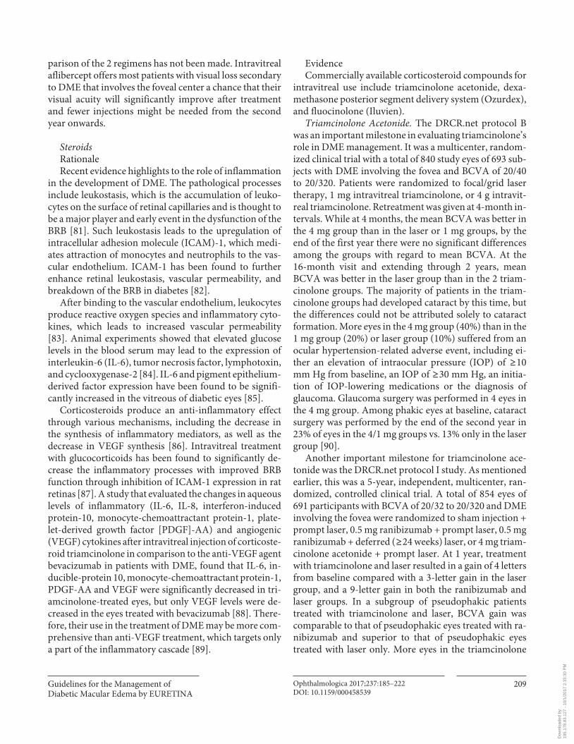
Guidelines for the Management of Diabetic Macular Edema by EURETINA
Ophthalmologica 2017;237:185–222DOI: 10.1159/000458539
209
parison of the 2 regimens has not been made. Intravitreal aflibercept offers most patients with visual loss secondary to DME that involves the foveal center a chance that their visual acuity will significantly improve after treatment and fewer injections might be needed from the second year onwards.
Steroids Rationale Recent evidence highlights to the role of inflammation
in the development of DME. The pathological processes include leukostasis, which is the accumulation of leuko-cytes on the surface of retinal capillaries and is thought to be a major player and early event in the dysfunction of the BRB [81] . Such leukostasis leads to the upregulation of intracellular adhesion molecule (ICAM)-1, which medi-ates attraction of monocytes and neutrophils to the vas-cular endothelium. ICAM-1 has been found to further enhance retinal leukostasis, vascular permeability, and breakdown of the BRB in diabetes [82] .
After binding to the vascular endothelium, leukocytes produce reactive oxygen species and inflammatory cyto-kines, which leads to increased vascular permeability [83] . Animal experiments showed that elevated glucose levels in the blood serum may lead to the expression of interleukin-6 (IL-6), tumor necrosis factor, lymphotoxin, and cyclooxygenase-2 [84] . IL-6 and pigment epithelium-derived factor expression have been found to be signifi-cantly increased in the vitreous of diabetic eyes [85] .
Corticosteroids produce an anti-inflammatory effect through various mechanisms, including the decrease in the synthesis of inflammatory mediators, as well as the decrease in VEGF synthesis [86] . Intravitreal treatment with glucocorticoids has been found to significantly de-crease the inflammatory processes with improved BRB function through inhibition of ICAM-1 expression in rat retinas [87] . A study that evaluated the changes in aqueous levels of inflammatory (IL-6, IL-8, interferon-induced protein-10, monocyte-chemoattractant protein-1, plate-let-derived growth factor [PDGF]-AA) and angiogenic (VEGF) cytokines after intravitreal injection of corticoste-roid triamcinolone in comparison to the anti-VEGF agent bevacizumab in patients with DME, found that IL-6, in-ducible-protein 10, monocyte-chemoattractant protein-1, PDGF-AA and VEGF were significantly decreased in tri-amcinolone-treated eyes, but only VEGF levels were de-creased in the eyes treated with bevacizumab [88] . There-fore, their use in the treatment of DME may be more com-prehensive than anti-VEGF treatment, which targets only a part of the inflammatory cascade [89] .
Evidence Commercially available corticosteroid compounds for
intravitreal use include triamcinolone acetonide, dexa-methasone posterior segment delivery system (Ozurdex), and fluocinolone (Iluvien).
Triamcinolone Acetonide. The DRCR.net protocol B was an important milestone in evaluating triamcinolone’s role in DME management. It was a multicenter, random-ized clinical trial with a total of 840 study eyes of 693 sub-jects with DME involving the fovea and BCVA of 20/40 to 20/320. Patients were randomized to focal/grid laser therapy, 1 mg intravitreal triamcinolone, or 4 g intravit-real triamcinolone. Retreatment was given at 4-month in-tervals. While at 4 months, the mean BCVA was better in the 4 mg group than in the laser or 1 mg groups, by the end of the first year there were no significant differences among the groups with regard to mean BCVA. At the 16-month visit and extending through 2 years, mean BCVA was better in the laser group than in the 2 triam-cinolone groups. The majority of patients in the triam-cinolone groups had developed cataract by this time, but the differences could not be attributed solely to cataract formation. More eyes in the 4 mg group (40%) than in the 1 mg group (20%) or laser group (10%) suffered from an ocular hypertension-related adverse event, including ei-ther an elevation of intraocular pressure (IOP) of ≥ 10 mm Hg from baseline, an IOP of ≥ 30 mm Hg, an initia-tion of IOP-lowering medications or the diagnosis of glaucoma. Glaucoma surgery was performed in 4 eyes in the 4 mg group. Among phakic eyes at baseline, cataract surgery was performed by the end of the second year in 23% of eyes in the 4/1 mg groups vs. 13% only in the laser group [90] .
Another important milestone for triamcinolone ace-tonide was the DRCR.net protocol I study. As mentioned earlier, this was a 5-year, independent, multicenter, ran-domized, controlled clinical trial. A total of 854 eyes of 691 participants with BCVA of 20/32 to 20/320 and DME involving the fovea were randomized to sham injection + prompt laser, 0.5 mg ranibizumab + prompt laser, 0.5 mg ranibizumab + deferred ( ≥ 24 weeks) laser, or 4 mg triam-cinolone acetonide + prompt laser. At 1 year, treatment with triamcinolone and laser resulted in a gain of 4 letters from baseline compared with a 3-letter gain in the laser group, and a 9-letter gain in both the ranibizumab and laser groups. In a subgroup of pseudophakic patients treated with triamcinolone and laser, BCVA gain was comparable to that of pseudophakic eyes treated with ra-nibizumab and superior to that of pseudophakic eyes treated with laser only. More eyes in the triamcinolone
Dow
nloa
ded
by:
195.
178.
83.1
27 -
10/
1/20
17 2
:33:
30 P
M

Schmidt-Erfurth et al.
Ophthalmologica 2017;237:185–222DOI: 10.1159/000458539
210
group (50%) than in the ranibizumab groups (9%) or the laser group (11%) had an IOP elevation ≥ 10 mm Hg, IOP >30 mm Hg or initiation of IOP-lowering medications during 2 years of follow-up. Glaucoma surgery was per-formed in 4 eyes (one eye in the laser group, one eye in the ranibizumab + prompt laser group, and two eyes in the triamcinolone group). More eyes in the triamcinolone group (59%) underwent cataract surgery over two years of follow-up than in the laser group (14%) or the ranibi-zumab groups (14%) [27] .
Dexamethasone. The first study that evaluated dexa-methasone for DME was the PLACID trial. The study was a randomized, controlled, multicenter clinical trial. A to-tal of 253 patients with center involving DME, with a BCVA letter score of 34–70 letters and CRT ≥ 275 μm were randomized to 0.7 mg Ozurdex implant and laser therapy at month 1 or laser monotherapy with a sham implant injection. Subjects could receive up to 3 addi-tional laser treatments and 1 additional Ozurdex injec-tion. While at 1 year there was no difference between the groups, the percentage of patients gaining at least 10 let-ters was significantly higher in the Ozurdex plus laser group vs. the laser monotherapy group at month 1 (31.7 vs. 11%, p < 0.001) and month 9 (31.7 vs. 17.3%, p = 0.007). Increased IOP occurred more frequently in the Ozurdex plus laser group than in the laser monotherapy group, with 15.2% of patients experiencing an increase of ≥ 10 mm Hg from baseline, 16.8% experiencing an IOP of ≥ 25 mm Hg and 4% an IOP of ≥ 35 mm Hg. In 15.9% of patients in the Ozurdex plus laser group vs. 1.6% patients in the laser monotherapy group treatment with IOP-low-ering medication was begun. However, no eyes required surgery to control IOP. Cataract-related adverse events were more common among phakic patients in the Ozur-dex plus laser group (22.2%) versus the laser monothera-py group (9.5%). Cataract surgery was performed in 4 eyes in the Ozurdex plus laser group in comparison to 5 eyes in the laser monotherapy group [91] .
The most important trial evaluating Ozurdex for DME were the MEAD trials. MEAD were 2 randomized, mul-ticenter, masked, sham-controlled, phase III trials with identical protocols. A total of 1,048 participants with cen-tral DME, BCVA of 20/50 to 20/200 and CRT of ≥ 300 μm were randomized to treatment with Ozurdex 0.7 mg, Ozurdex 0.35 mg or a sham procedure with a follow-up of 3 years. Retreatment was allowed according to pre-defined retreatment criteria, not more frequently than ev-ery 6 months. At the end of 3 years, the percentage of pa-tients with a ≥ 15-letter gain of BCVA from baseline was 22.2/18.4% in the Ozurdex 0.7/0.35 mg group versus 12%
in the sham group. An IOP increase of ≥ 10 mm Hg oc-curred in 27.7/24.8% of patients in the 0.7/0.35 mg Ozur-dex groups versus 13% in the sham group. An IOP of ≥ 35 mm Hg occurred in 6.6/5.2% of patients in the Ozurdex 0.7/0.35 mg groups versus 0.9% in the sham group. 41.5/37.6% of patients in the 0.7/0.35 mg Ozurdex groups needed IOP-lowering medications versus 9.1% in the sham group. One patient in each Ozurdex treatment group underwent glaucoma incisional surgery. The rate of cataract-related adverse events was 67.9/64.1% in the 0.7/0.35 mg Ozurdex groups, respectively, versus 20.4% in the sham group [92] .
The CHAMPLAIN study was a prospective, multi-center, open-label, 26-week study. Fifty-five patients with treatment-refractory DME and a history of previous pars plana vitrectomy (PPV) in the study eye received a single intravitreal injection of 0.7 mg Ozurdex. The mean BCVA gain from baseline was 6 letters after 8 weeks and 3 letters after 26 weeks. At 8 weeks, 30.4% of patients gained ≥ 10 letters. Increased IOP was reported in 16% of study eyes, with no patients requiring a surgical procedure to control IOP. Cataract progression was reported in 17% of phakic eyes, with 1 patient undergoing cataract surgery [93] .
Ozurdex 0.7 mg was approved by the FDA in Septem-ber 2014 for the treatment of adult patients with DME. It was approved by the EMA in July 2014 for the treatment of adult patients with visual impairment due to DME who are pseudophakic or who are considered insufficiently re-sponsive to or unsuitable for noncorticosteroid therapy. The implant releases the corticosteroid into the vitreous over a period of ≤ 6 months [94] . Therefore, the official product label in Europe recommends retreatment after approximately 6 months, and does not recommend ad-ministration to both eyes concurrently. Newer data sup-port the reinjection of the dexamethasone implant ear-lier than the recommended retreatment interval. The CHROME study, a retrospective real-world study, in-cluded patients with DME, retinal vein occlusion, and uveitis. The mean reinjection interval in this study was 2.3–4.9 months [95] .
Fluocinolone Acetonide. The FAME trials were 2 paral-lel, prospective, randomized, phase III, multicenter trials. A total of 956 patients with central DME, BCVA of 20/50 to 20/200 and CRT ≥ 250 μm were randomized to receive an intravitreal insert releasing 0.2 or 0.5 μg fluocinolone acetonide per day or sham injection. Based on predefined retreatment criteria, additional treatment could be given after 1 year. The percentage of patients with a BCVA gain of ≥ 15 letters after 2 years was 28% in each of the fluo-cinolone groups versus 16% in the sham group [96] .
Dow
nloa
ded
by:
195.
178.
83.1
27 -
10/
1/20
17 2
:33:
30 P
M
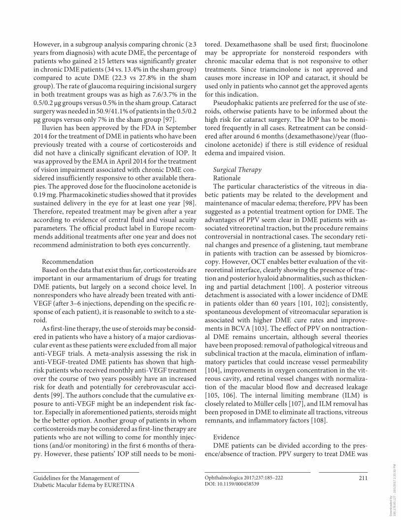
Guidelines for the Management of Diabetic Macular Edema by EURETINA
Ophthalmologica 2017;237:185–222DOI: 10.1159/000458539
211
However, in a subgroup analysis comparing chronic ( ≥ 3 years from diagnosis) with acute DME, the percentage of patients who gained ≥ 15 letters was significantly greater in chronic DME patients (34 vs. 13.4% in the sham group) compared to acute DME (22.3 vs 27.8% in the sham group). The rate of glaucoma requiring incisional surgery in both treatment groups was as high as 7.6/3.7% in the 0.5/0.2 μg groups versus 0.5% in the sham group. Cataract surgery was needed in 50.9/41.1% of patients in the 0.5/0.2 μg groups versus only 7% in the sham group [97] .
Iluvien has been approved by the FDA in September 2014 for the treatment of DME in patients who have been previously treated with a course of corticosteroids and did not have a clinically significant elevation of IOP. It was approved by the EMA in April 2014 for the treatment of vision impairment associated with chronic DME con-sidered insufficiently responsive to other available thera-pies. The approved dose for the fluocinolone acetonide is 0.19 mg. Pharmacokinetic studies showed that it provides sustained delivery in the eye for at least one year [98] . Therefore, repeated treatment may be given after a year according to evidence of central fluid and visual acuity parameters. The official product label in Europe recom-mends additional treatments after one year and does not recommend administration to both eyes concurrently.
Recommendation Based on the data that exist thus far, corticosteroids are
important in our armamentarium of drugs for treating DME patients, but largely on a second choice level. In nonresponders who have already been treated with anti-VEGF (after 3–6 injections, depending on the specific re-sponse of each patient), it is reasonable to switch to a ste-roid.
As first-line therapy, the use of steroids may be consid-ered in patients who have a history of a major cardiovas-cular event as these patients were excluded from all major anti-VEGF trials. A meta-analysis assessing the risk in anti-VEGF-treated DME patients has shown that high-risk patients who received monthly anti-VEGF treatment over the course of two years possibly have an increased risk for death and potentially for cerebrovascular acci-dents [99] . The authors conclude that the cumulative ex-posure to anti-VEGF might be an independent risk fac-tor. Especially in aforementioned patients, steroids might be the better option. Another group of patients in whom corticosteroids may be considered as first-line therapy are patients who are not willing to come for monthly injec-tions (and/or monitoring) in the first 6 months of thera-py. However, these patients’ IOP still needs to be moni-
tored. Dexamethasone shall be used first; fluocinolone may be appropriate for nonsteroid responders with chronic macular edema that is not responsive to other treatments. Since triamcinolone is not approved and causes more increase in IOP and cataract, it should be used only in patients who cannot get the approved agents for this indication.
Pseudophakic patients are preferred for the use of ste-roids, otherwise patients have to be informed about the high risk for cataract surgery. The IOP has to be moni-tored frequently in all cases. Retreatment can be consid-ered after around 6 months (dexamethasone)/year (fluo-cinolone acetonide) if there is still evidence of residual edema and impaired vision.
Surgical Therapy Rationale The particular characteristics of the vitreous in dia-
betic patients may be related to the development and maintenance of macular edema; therefore, PPV has been suggested as a potential treatment option for DME. The advantages of PPV seem clear in DME patients with as-sociated vitreoretinal traction, but the procedure remains controversial in nontractional cases. The secondary reti-nal changes and presence of a glistening, taut membrane in patients with traction can be assessed by biomicros-copy. However, OCT enables better evaluation of the vit-reoretinal interface, clearly showing the presence of trac-tion and posterior hyaloid abnormalities, such as thicken-ing and partial detachment [100] . A posterior vitreous detachment is associated with a lower incidence of DME in patients older than 60 years [101, 102] ; consistently, spontaneous development of vitreomacular separation is associated with higher DME cure rates and improve-ments in BCVA [103] . The effect of PPV on nontraction-al DME remains uncertain, although several theories have been proposed: removal of pathological vitreous and subclinical traction at the macula, elimination of inflam-matory particles that could increase vessel permeability [104] , improvements in oxygen concentration in the vit-reous cavity, and retinal vessel changes with normaliza-tion of the macular blood flow and decreased leakage [105, 106] . The internal limiting membrane (ILM) is closely related to Müller cells [107] , and ILM removal has been proposed in DME to eliminate all tractions, vitreous remnants, and inflammatory factors [108] .
Evidence DME patients can be divided according to the pres-
ence/absence of traction. PPV surgery to treat DME was
Dow
nloa
ded
by:
195.
178.
83.1
27 -
10/
1/20
17 2
:33:
30 P
M
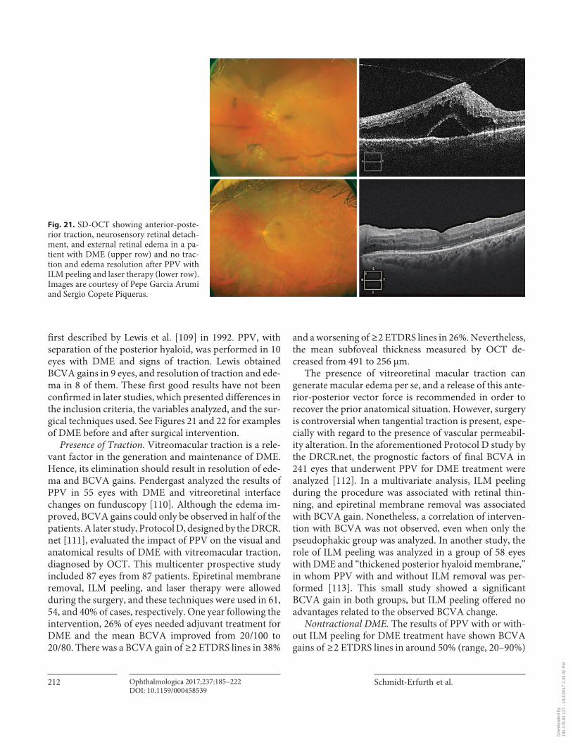
Schmidt-Erfurth et al.
Ophthalmologica 2017;237:185–222DOI: 10.1159/000458539
212
first described by Lewis et al. [109] in 1992. PPV, with separation of the posterior hyaloid, was performed in 10 eyes with DME and signs of traction. Lewis obtained BCVA gains in 9 eyes, and resolution of traction and ede-ma in 8 of them. These first good results have not been confirmed in later studies, which presented differences in the inclusion criteria, the variables analyzed, and the sur-gical techniques used. See Figures 21 and 22 for examples of DME before and after surgical intervention.
Presence of Traction. Vitreomacular traction is a rele-vant factor in the generation and maintenance of DME. Hence, its elimination should result in resolution of ede-ma and BCVA gains. Pendergast analyzed the results of PPV in 55 eyes with DME and vitreoretinal interface changes on funduscopy [110] . Although the edema im-proved, BCVA gains could only be observed in half of the patients. A later study, Protocol D, designed by the DRCR.net [111] , evaluated the impact of PPV on the visual and anatomical results of DME with vitreomacular traction, diagnosed by OCT. This multicenter prospective study included 87 eyes from 87 patients. Epiretinal membrane removal, ILM peeling, and laser therapy were allowed during the surgery, and these techniques were used in 61, 54, and 40% of cases, respectively. One year following the intervention, 26% of eyes needed adjuvant treatment for DME and the mean BCVA improved from 20/100 to 20/80. There was a BCVA gain of ≥ 2 ETDRS lines in 38%
and a worsening of ≥ 2 ETDRS lines in 26%. Nevertheless, the mean subfoveal thickness measured by OCT de-creased from 491 to 256 μm.
The presence of vitreoretinal macular traction can generate macular edema per se, and a release of this ante-rior-posterior vector force is recommended in order to recover the prior anatomical situation. However, surgery is controversial when tangential traction is present, espe-cially with regard to the presence of vascular permeabil-ity alteration. In the aforementioned Protocol D study by the DRCR.net, the prognostic factors of final BCVA in 241 eyes that underwent PPV for DME treatment were analyzed [112] . In a multivariate analysis, ILM peeling during the procedure was associated with retinal thin-ning, and epiretinal membrane removal was associated with BCVA gain. Nonetheless, a correlation of interven-tion with BCVA was not observed, even when only the pseudophakic group was analyzed. In another study, the role of ILM peeling was analyzed in a group of 58 eyes with DME and “thickened posterior hyaloid membrane,” in whom PPV with and without ILM removal was per-formed [113] . This small study showed a significant BCVA gain in both groups, but ILM peeling offered no advantages related to the observed BCVA change.
Nontractional DME. The results of PPV with or with-out ILM peeling for DME treatment have shown BCVA gains of ≥ 2 ETDRS lines in around 50% (range, 20–90%)
Fig. 21. SD-OCT showing anterior-poste-rior traction, neurosensory retinal detach-ment, and external retinal edema in a pa-tient with DME (upper row) and no trac-tion and edema resolution after PPV with ILM peeling and laser therapy (lower row). Images are courtesy of Pepe Garcia Arumi and Sergio Copete Piqueras.
Dow
nloa
ded
by:
195.
178.
83.1
27 -
10/
1/20
17 2
:33:
30 P
M
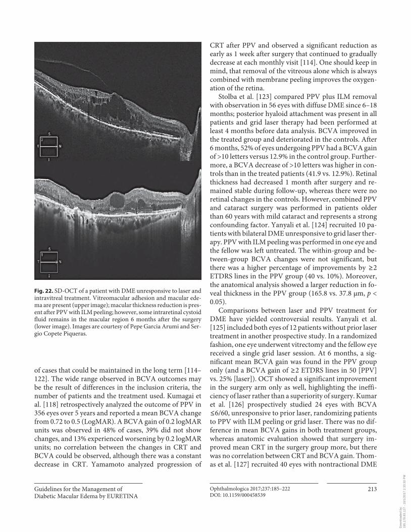
Guidelines for the Management of Diabetic Macular Edema by EURETINA
Ophthalmologica 2017;237:185–222DOI: 10.1159/000458539
213
of cases that could be maintained in the long term [114–122] . The wide range observed in BCVA outcomes may be the result of differences in the inclusion criteria, the number of patients and the treatment used. Kumagai et al. [118] retrospectively analyzed the outcome of PPV in 356 eyes over 5 years and reported a mean BCVA change from 0.72 to 0.5 (LogMAR). A BCVA gain of 0.2 logMAR units was observed in 48% of cases, 39% did not show changes, and 13% experienced worsening by 0.2 logMAR units; no correlation between the changes in CRT and BCVA could be observed, although there was a constant decrease in CRT. Yamamoto analyzed progression of
CRT after PPV and observed a significant reduction as early as 1 week after surgery that continued to gradually decrease at each monthly visit [114] . One should keep in mind, that removal of the vitreous alone which is always combined with membrane peeling improves the oxygen-ation of the retina.
Stolba et al. [123] compared PPV plus ILM removal with observation in 56 eyes with diffuse DME since 6–18 months; posterior hyaloid attachment was present in all patients and grid laser therapy had been performed at least 4 months before data analysis. BCVA improved in the treated group and deteriorated in the controls. After 6 months, 52% of eyes undergoing PPV had a BCVA gain of >10 letters versus 12.9% in the control group. Further-more, a BCVA decrease of >10 letters was higher in con-trols than in the treated patients (41.9 vs. 12.9%). Retinal thickness had decreased 1 month after surgery and re-mained stable during follow-up, whereas there were no retinal changes in the controls. However, combined PPV and cataract surgery was performed in patients older than 60 years with mild cataract and represents a strong confounding factor. Yanyali et al. [124] recruited 10 pa-tients with bilateral DME unresponsive to grid laser ther-apy. PPV with ILM peeling was performed in one eye and the fellow was left untreated. The within-group and be-tween-group BCVA changes were not significant, but there was a higher percentage of improvements by ≥ 2 ETDRS lines in the PPV group (40 vs. 10%). Moreover, the anatomical analysis showed a larger reduction in fo-veal thickness in the PPV group (165.8 vs. 37.8 μm, p < 0.05).
Comparisons between laser and PPV treatment for DME have yielded controversial results. Yanyali et al. [125] included both eyes of 12 patients without prior laser treatment in another prospective study. In a randomized fashion, one eye underwent vitrectomy and the fellow eye received a single grid laser session. At 6 months, a sig-nificant mean BCVA gain was found in the PPV group only (and a BCVA gain of ≥ 2 ETDRS lines in 50 [PPV] vs. 25% [laser]). OCT showed a significant improvement in the surgery arm only as well, highlighting the ineffi-ciency of laser rather than a superiority of surgery. Kumar et al. [126] prospectively studied 24 eyes with BCVA ≤ 6/60, unresponsive to prior laser, randomizing patients to PPV with ILM peeling or grid laser. There was no dif-ference in mean BCVA gains in both treatment groups, whereas anatomic evaluation showed that surgery im-proved mean CRT in the surgery group more, but there was no correlation between CRT and BCVA gain. Thom-as et al. [127] recruited 40 eyes with nontractional DME
Fig. 22. SD-OCT of a patient with DME unresponsive to laser and intravitreal treatment. Vitreomacular adhesion and macular ede-ma are present (upper image); macular thickness reduction is pres-ent after PPV with ILM peeling; however, some intraretinal cystoid fluid remains in the macular region 6 months after the surgery (lower image). Images are courtesy of Pepe Garcia Arumi and Ser-gio Copete Piqueras.
Dow
nloa
ded
by:
195.
178.
83.1
27 -
10/
1/20
17 2
:33:
30 P
M
Schmidt-Erfurth et al.
Ophthalmologica 2017;237:185–222DOI: 10.1159/000458539
214
and prior macular laser treatment, randomizing eyes to PPV or further macular laser. After 1 year, no functional or anatomical advantages were observed in the PPV arm. There were some differences in the mean duration of dia-betes, mean hemoglobin A1c (HbA1c) and the use of in-sulin between the groups, which may have been con-founding factors. Patel et al. [128] randomized 15 eyes to PPV without ILM peeling or to macular laser treatment. Although BCVA after 1 year was slightly better in the la-ser arm, anatomical and cone function results (measured by fine matrix mapping) were better in the PPV group. All of these interventional studies fail to show anything but the inefficacy of both laser and surgery as compared to anti-VEGF benefits.
Intravitreal injection of triamcinolone acetonide has also been compared with PPV in DME patients. In a pro-spective study of 40 eyes from 20 patients with bilateral DME, Doi et al. [129] compared PPV treatment in one eye and a single intravitreal injection of 4 mg triamcino-lone acetonide in the fellow eye. After 1 year, there was no significant BCVA gain from baseline and no significant difference in BCVA change between the groups. Mean CRT showed differences between the groups: in the tri-amcinolone arm it decreased in the first month, but after-wards increased to the pretreatment level, while in the surgery arm CRT decreased progressively, being signifi-cantly thinner after 1 year. In a retrospective study of 40 eyes treated with a combination of laser, triamcinolone, and PPV, Kim et al. [130] reported BCVA gains and CRT decrease after 3 years, with an increase of ≥ 2 ETDRS lines in 65% of cases.
Stefaniotou et al. [131] retrospectively analyzed the re-sults obtained in 73 eyes treated with PPV with or without ILM peeling. BCVA gains could be observed in both groups, but a greater improvement could be seen in pa-tients undergoing ILM peeling. These results are in con-trast with those of Hoerauf et al. [132] and Bardak et al. [133] who reported no significantly greater BCVA gain when ILM peeling was added to PPV. The study with the longest follow-up was conducted by Kumagai et al. [134] . These authors analyzed 116 eyes from 58 patients in whom PPV was undertaken in both eyes, with the addi-tion of ILM peeling in one of them only. In both groups, 75% of eyes showed BCVA gains after 5 years of follow-up, with no significant differences between the 2 groups. In studies by Figueroa et al. [135] and Shiba et al. [136] , no advantages of ILM peeling over conventional or tri-gon-assisted vitrectomy were reported. All of these stud-ies were small and not stratified regarding their baseline characteristics.
Presence of Traction and Nontractional DME in Com-parison. The presence of traction has been considered an indication for surgery, and the results are thought to be better than those of DME without signs of traction. In a prospective study, Shah et al. [137] analyzed factors pre-dicting the outcome of PPV for DME after 1 year and reported that eyes with vitreoretinal traction had higher BCVA gains than nontractional cases. In contrast, the retrospective study by Bonnin et al. [138] of 73 unre-sponsive eyes with DME with or without traction fol-lowed for a mean of 5.3 years reported improvements in BCVA and CRT after 3 years with no significant differ-ences between the 2 groups. The percentage of patients that gained ≥ 2 ETDRS lines at the final visit was also similar (55 vs. 56%). Hence, no solid recommendation favoring surgical intervention can be deducted from these studies.
Predictive Factors for Successful Surgical Therapy. Sev-eral factors have been associated with the final BCVA fol-lowing PPV. A poorer preoperative BCVA has been as-sociated with higher BCVA gains [112] . Greater retinal thickness, poorer glycemic control [139] , presence of SRF [137] , lack of ELM integrity [140] and disruption of the ellipsoid zone [141] have been associated with a poorer final absolute BCVA.
Recommendation DME treatment has vastly evolved in the last decade
with the development of intravitreal anti-VEGF injection therapy and dexamethasone implants, which has put sur-gery into a second-line position. DME surgery should be considered based on the vitreous and retinal status. The presence of anterior-posterior traction may be an indica-tion for PPV in eyes with DME. Tangential traction due to an epiretinal or hyaloid membrane should be consid-ered only when the response to anti-VEGF or dexameth-asone implants is incomplete. There is no consensus re-garding the advantages of PPV when there is no evidence of traction. Nonetheless, PPV has been proven to main-tain BCVA, and it should be considered for cases unre-sponsive to intravitreal treatments and no posterior vitre-ous detachment, keeping in mind that the anatomical outcome will be better than the functional results. Further studies are needed to evaluate the effectiveness of surgery in eyes that have previously been treated with the drugs available on the market. The recommendation for PPV is given when traction is present based on evidence levels II–III.
Dow
nloa
ded
by:
195.
178.
83.1
27 -
10/
1/20
17 2
:33:
30 P
M
Guidelines for the Management of Diabetic Macular Edema by EURETINA
Ophthalmologica 2017;237:185–222DOI: 10.1159/000458539
215
Systemic Diabetic Disease and Management
The Role of Metabolic Control Rationale Diabetes mellitus as a systemic disease can have vari-
ous macrovascular as well as microvascular complica-tions. Among these, DR and particularly DME play a ma-jor role as they are the major cause of blindness, and the number of people suffering from diabetes mellitus is in-creasing worldwide [142–144] . Therefore, the role of metabolic control needs to be evaluated whenever talking about the treatment of DR and particularly DME, a “side effect” of a systemic disease.
Evidence The role of metabolic control includes the question
about the relevant target levels of glycemia and blood pressure for the prevention of the development or pro-gression of DR, whether some hypoglycemic drugs and blood pressure-lowering agents are more effective than others for the prevention of DR, and the role of lipid-lowering agents [145] . The ETDRS study identified im-portant risk factors for progression to high-risk PDR in-cluding more severe stages of DR, decreased BCVA or increased DME, younger age or type 1 versus type 2 dia-betes mellitus and high levels of HbA1c [146, 147] . The list of risk factors for the progression to PDR also includ-ed a low hematocrit and increased serum lipids as surro-gates of a low quality of metabolic control. The long-term benefit of improving glycemic control was evaluated by large prospective studies such as the Diabetes Control and Complications Trial (DCCT) and the United King-dom Prospective Diabetes Study (UKPDS) [84, 148–154] . They provided strong evidence that tight control of gly-cemia (with a glycosylated hemoglobin [HbA1c] of 7% or less) reduced the risk of development and progression of DR in both type 1 and type 2 diabetes mellitus.
The DCCT addressed the questions whether in pa-tients with insulin-dependent diabetes mellitus an inten-sive therapy completely prevented the development of DR, whether the potential effect of therapy was depen-dent on the preexisting stage of DR, and which other fac-tors, besides the intensity of treatment, influenced theeffectiveness of therapy [148, 150–152] . The DCCT in-cluded 1,441 patients with insulin-dependent diabetes mellitus. The total study population was divided into a primary prevention cohort with a diabetes duration rang-ing between 1 and 5 years and without DR at baseline and into a secondary intervention cohort with 1–15 years’ du-ration and minimal to moderate nonproliferative DR.
The patients were randomly assigned to either intensive treatment, aiming at achieving glycemic levels as close to the normal range as possible or to conventional diabetes therapy. The DCCT revealed that an intensive treatment reduced the risks of DR, nephropathy, and neuropathy by 35–90% when compared with the conventional treat-ment. The risks of DR and nephropathy increased with higher mean HbA1c level and higher body mass index. The earlier an intensive treatment was started, the more effective it was, in particular if complications had not de-veloped yet. The risk reduction was maintained through 7 years of follow-up, although the difference between the former intensive treatment group and the conventional treatment group decreased and eventually became statis-tically nonsignificant by 5 years. The further rate of pro-gression of complications from their levels at the end of the DCCT remained less in the former intensive treat-ment group. The conclusions from the DCCT were that the benefits of 6.5 years of intensive treatment extended well beyond the period of its most intensive implementa-tion, that intensive treatment should be started as soon as it is safely possible after the onset of type 1 diabetes mel-litus and maintained thereafter and that one should aim for a practicable target HbA1c level of 7.0% or less.
The UKPDS addressed the question whether improved blood-glucose control had an effect on macrovascular complications, while it simultaneously decreased the pro-gression of diabetic microvascular disease such as DR [149] . It included more than 3,800 newly diagnosed pa-tients with type 2 diabetes with a median age of 54 years, who showed a mean of 2 fasting plasma glucose concen-trations of 6.1–15.0 mmol/L after initiation of diet of 3 months and who were randomly assigned to (1) an inten-sive treatment arm with (1a) sulfonylurea or (1b) insulin or to (2) a conventional treatment arm with diet. Over 10 years, HbA1c was 7.0% in the intensive care group com-pared with 7.9% in the conventional treatment group, with no significant difference in HbA1c between the 2 subgroups within the intensive care arm. Compared with the conventional group, the risk in the intensive care group was 12% lower for any diabetes-related endpoint, with the most marked impact with a 25% risk reduction on the microvascular endpoints, including the need for retinal laser therapy for DR. It was concluded that inten-sive blood-glucose control by either sulfonylureas or in-sulin substantially decreased the risk of microvascular complications including DR in patients with type 2 dia-betes, while none of the drugs had an adverse effect on cardiovascular outcomes, and while all intensive treat-ment increased the risk of hypoglycemia [149] .
Dow
nloa
ded
by:
195.
178.
83.1
27 -
10/
1/20
17 2
:33:
30 P
M
Schmidt-Erfurth et al.
Ophthalmologica 2017;237:185–222DOI: 10.1159/000458539
216
In a post-trial monitoring of the participants of the UKPDS, 4,209 out of 5,102 patients with newly diagnosed type 2 diabetes were randomly assigned to receive either conventional therapy (dietary restriction) or intensive therapy (either sulfonylurea or insulin or, in overweight patients, metformin) for glucose control [155] . Although differences between the 2 groups in HbA1c levels were lost after the first year of the study, the intensive care sul-fonylurea-insulin group showed a relative reduction in risk also after 10 years for any diabetes-related endpoint (9%, p = 0.04) and microvascular disease (24%, p = 0.001). One inferred that despite an early loss of glycemic differ-ences, a continued reduction in microvascular risk and emergent risk reductions for myocardial infarction and death from any cause were observed during 10 years of posttrial follow-up of the UKPDS. Another study of the UKPDS was conducted to examine whether intensive glucose control with metformin had any specific advan-tage or disadvantage as compared with insulin or sulfo-nylurea therapy in patients with type 2 diabetes to de-crease progression of microvascular disease and to reduce the risk of heart attacks [156] . Among patients allocated to intensive blood-glucose control, metformin showed a greater effect than chlorpropamide, glibenclamide, or in-sulin for any diabetes-related endpoint ( p = 0.003) in-cluding DR, all-cause mortality ( p = 0.02), and stroke( p = 0.03). The authors concluded that since intensive glucose control with metformin decreased the risk of di-abetes-related endpoints in overweight diabetic patients and since it was associated with less weight gain and few-er hypoglycemic attacks than it was with insulin and sul-fonylureas, metformin might be the first-line pharmaco-logical therapy of choice in these patients [155] .
In the Fenofibrate Intervention and Event Lowering in Diabetes (FIELD) Study, 9,795 patients aged 50–75 years with type 2 diabetes mellitus were randomly assigned to receive fenofibrate 200 mg/day ( n = 4,895) or matching placebo ( n = 4,900) [157] . At each clinic visit, information concerning laser treatment for DR – a prespecified ter-tiary endpoint of the main study – was gathered. It re-vealed that laser treatment was needed more frequently in participants with poorer glycemic or blood pressure control than in those with good control of these factors and in those with a greater burden of clinical microvas-cular disease, but the need for such treatment was not af-fected by plasma lipid concentrations. The FIELD Study also showed that the requirement for the first laser treat-ment for any retinopathy was significantly lower in study participants taking fenofibrate than in study participants without fenofibrate therapy.
Several other smaller-scaled studies also evaluated the relationship between the glycemic control and DR and arrived at similar conclusions [145, 147, 154] . Intensive glycemic control reduced the risk of any retinopathy by approximately 27%. Intensive therapy was most effective when initiated early in the course of diabetes, demon-strating a beneficial effect over the course and progres-sion of retinopathy. The long-term benefits of the inten-sive glycemic control greatly outweighed the risk of an early worsening of the retinopathy in the early phase of the therapy [158] . Every percent reduction in HbA1c (e.g., from 9 to 8%) lowered the risk of retinopathy by 30–40% and the effect appeared long lasting (metabolic memory) [153] . To avoid any waning of the beneficial ef-fect of intensive care therapy of diabetes mellitus, HbA1c should be maintained at target values as long as possible [84] . Correspondingly, a meta-analysis of 3 population-based studies showed a graded relation between the level of glycemia and frequency of DR signs, even below the diagnostic criterion for diabetes (fasting plasma glucose of 7.0 mmol/L) [159] .
In contrast to the findings obtained in the studies just described, the Action in Diabetes and Vascular Disease (ADVANCE) trial reported that aggressive glycemic con-trol with an HbA1c of <6.5% did not substantially affect the development or progression of DR in patients with type 2 diabetes [160, 161] . The Action to Control Cardio-vascular Risk in Diabetes (ACCORD) trial showed that such aggressive glycemic control could be associated with increased mortality, although the cause of unexpected ex-cess deaths remains unclear [162] . The Veterans Affairs Diabetes Trial revealed no significant benefits of intensive glycemic control with an HbA1c of <6.9% on DR out-comes after 5 years of follow-up [163] . The reason for the difference between these studies and the UKPDS could be related to the differences in the study populations, the length of follow-up, the timing of the therapy and the in-fluence of other contributing factors such as the control of arterial hypertension.
Lowering elevated serum lipid levels has been shown to decrease the risk of cardiovascular morbidity. TheETDRS data suggested that lipid lowering may also de-crease the risk of hard exudate formation and associated vision loss in patients with DR [146, 147] . The DCCT study revealed that the severity of DR was correlated with increasing blood concentrations of triglycerides and neg-atively associated with the concentration of high-density lipoprotein cholesterol [164] . In the FIELD study, fenofi-brate, a lipid-modifying drug, reduced the need for laser treatment of vision-threatening DR by 31% in patients
Dow
nloa
ded
by:
195.
178.
83.1
27 -
10/
1/20
17 2
:33:
30 P
M
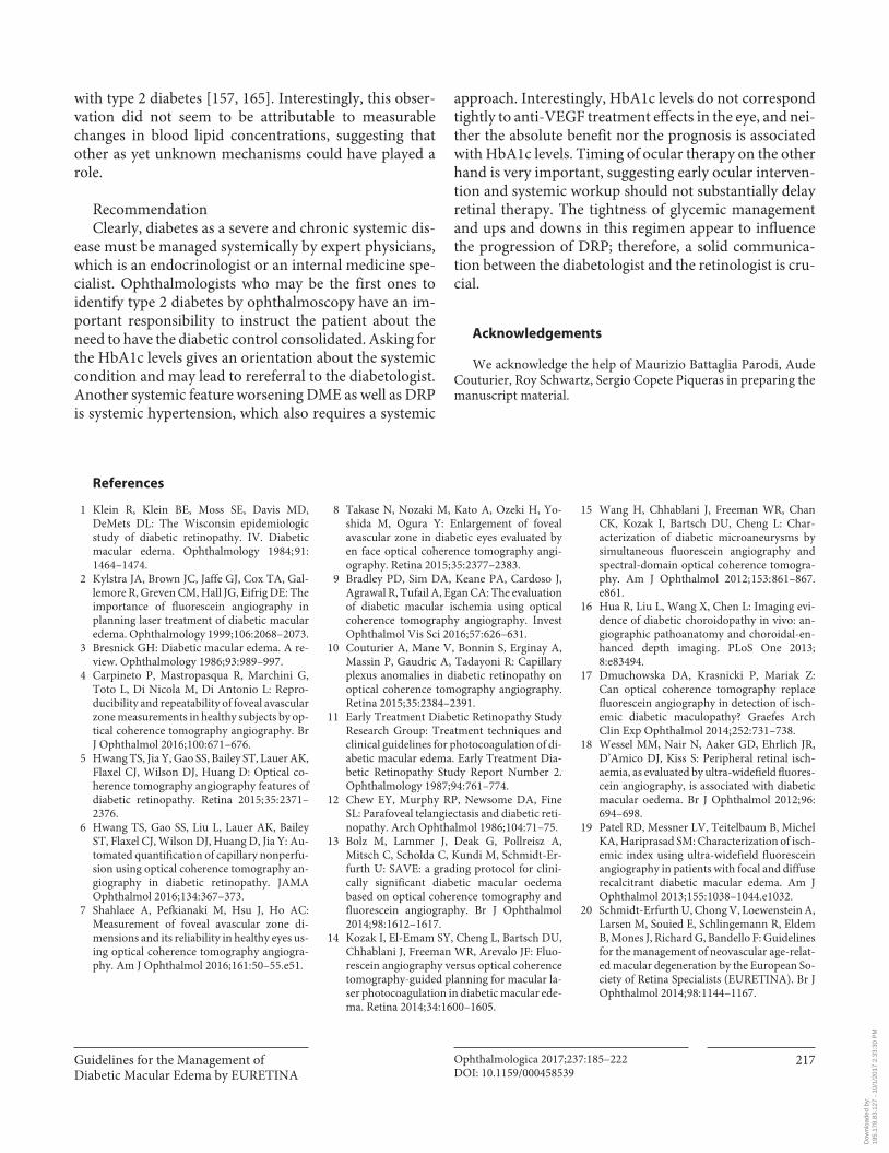
Guidelines for the Management of Diabetic Macular Edema by EURETINA
Ophthalmologica 2017;237:185–222DOI: 10.1159/000458539
217
with type 2 diabetes [157, 165] . Interestingly, this obser-vation did not seem to be attributable to measurable changes in blood lipid concentrations, suggesting that other as yet unknown mechanisms could have played a role.
Recommendation Clearly, diabetes as a severe and chronic systemic dis-
ease must be managed systemically by expert physicians, which is an endocrinologist or an internal medicine spe-cialist. Ophthalmologists who may be the first ones to identify type 2 diabetes by ophthalmoscopy have an im-portant responsibility to instruct the patient about the need to have the diabetic control consolidated. Asking for the HbA1c levels gives an orientation about the systemic condition and may lead to rereferral to the diabetologist. Another systemic feature worsening DME as well as DRP is systemic hypertension, which also requires a systemic
approach. Interestingly, HbA1c levels do not correspond tightly to anti-VEGF treatment effects in the eye, and nei-ther the absolute benefit nor the prognosis is associated with HbA1c levels. Timing of ocular therapy on the other hand is very important, suggesting early ocular interven-tion and systemic workup should not substantially delay retinal therapy. The tightness of glycemic management and ups and downs in this regimen appear to influence the progression of DRP; therefore, a solid communica-tion between the diabetologist and the retinologist is cru-cial.
Acknowledgements
We acknowledge the help of Maurizio Battaglia Parodi, Aude Couturier, Roy Schwartz, Sergio Copete Piqueras in preparing the manuscript material.
References
1 Klein R, Klein BE, Moss SE, Davis MD, DeMets DL: The Wisconsin epidemiologic study of diabetic retinopathy. IV. Diabetic macular edema. Ophthalmology 1984; 91: 1464–1474.
2 Kylstra JA, Brown JC, Jaffe GJ, Cox TA, Gal-lemore R, Greven CM, Hall JG, Eifrig DE: The importance of fluorescein angiography in planning laser treatment of diabetic macular edema. Ophthalmology 1999; 106: 2068–2073.
3 Bresnick GH: Diabetic macular edema. A re-view. Ophthalmology 1986; 93: 989–997.
4 Carpineto P, Mastropasqua R, Marchini G, Toto L, Di Nicola M, Di Antonio L: Repro-ducibility and repeatability of foveal avascular zone measurements in healthy subjects by op-tical coherence tomography angiography. Br J Ophthalmol 2016; 100: 671–676.
5 Hwang TS, Jia Y, Gao SS, Bailey ST, Lauer AK, Flaxel CJ, Wilson DJ, Huang D: Optical co-herence tomography angiography features of diabetic retinopathy. Retina 2015; 35: 2371–2376.
6 Hwang TS, Gao SS, Liu L, Lauer AK, Bailey ST, Flaxel CJ, Wilson DJ, Huang D, Jia Y: Au-tomated quantification of capillary nonperfu-sion using optical coherence tomography an-giography in diabetic retinopathy. JAMA Ophthalmol 2016; 134: 367–373.
7 Shahlaee A, Pefkianaki M, Hsu J, Ho AC: Measurement of foveal avascular zone di-mensions and its reliability in healthy eyes us-ing optical coherence tomography angiogra-phy. Am J Ophthalmol 2016; 161: 50–55.e51.
8 Takase N, Nozaki M, Kato A, Ozeki H, Yo-shida M, Ogura Y: Enlargement of foveal avascular zone in diabetic eyes evaluated by en face optical coherence tomography angi-ography. Retina 2015; 35: 2377–2383.
9 Bradley PD, Sim DA, Keane PA, Cardoso J, Agrawal R, Tufail A, Egan CA: The evaluation of diabetic macular ischemia using opticalcoherence tomography angiography. Invest Ophthalmol Vis Sci 2016; 57: 626–631.
10 Couturier A, Mane V, Bonnin S, Erginay A, Massin P, Gaudric A, Tadayoni R: Capillary plexus anomalies in diabetic retinopathy on optical coherence tomography angiography. Retina 2015; 35: 2384–2391.
11 Early Treatment Diabetic Retinopathy Study Research Group: Treatment techniques and clinical guidelines for photocoagulation of di-abetic macular edema. Early Treatment Dia-betic Retinopathy Study Report Number 2. Ophthalmology 1987; 94: 761–774.
12 Chew EY, Murphy RP, Newsome DA, Fine SL: Parafoveal telangiectasis and diabetic reti-nopathy. Arch Ophthalmol 1986; 104: 71–75.
13 Bolz M, Lammer J, Deak G, Pollreisz A, Mitsch C, Scholda C, Kundi M, Schmidt-Er-furth U: SAVE: a grading protocol for clini-cally significant diabetic macular oedema based on optical coherence tomography and fluorescein angiography. Br J Ophthalmol 2014; 98: 1612–1617.
14 Kozak I, El-Emam SY, Cheng L, Bartsch DU, Chhablani J, Freeman WR, Arevalo JF: Fluo-rescein angiography versus optical coherence tomography-guided planning for macular la-ser photocoagulation in diabetic macular ede-ma. Retina 2014; 34: 1600–1605.
15 Wang H, Chhablani J, Freeman WR, Chan CK, Kozak I, Bartsch DU, Cheng L: Char-acterization of diabetic microaneurysms by simultaneous fluorescein angiography and spectral-domain optical coherence tomogra-phy. Am J Ophthalmol 2012; 153: 861–867.e861.
16 Hua R, Liu L, Wang X, Chen L: Imaging evi-dence of diabetic choroidopathy in vivo: an-giographic pathoanatomy and choroidal-en-hanced depth imaging. PLoS One 2013; 8:e83494.
17 Dmuchowska DA, Krasnicki P, Mariak Z: Can optical coherence tomography replace fluorescein angiography in detection of isch-emic diabetic maculopathy? Graefes Arch Clin Exp Ophthalmol 2014; 252: 731–738.
18 Wessel MM, Nair N, Aaker GD, Ehrlich JR, D’Amico DJ, Kiss S: Peripheral retinal isch-aemia, as evaluated by ultra-widefield fluores-cein angiography, is associated with diabetic macular oedema. Br J Ophthalmol 2012; 96: 694–698.
19 Patel RD, Messner LV, Teitelbaum B, Michel KA, Hariprasad SM: Characterization of isch-emic index using ultra-widefield fluorescein angiography in patients with focal and diffuse recalcitrant diabetic macular edema. Am J Ophthalmol 2013; 155: 1038–1044.e1032.
20 Schmidt-Erfurth U, Chong V, Loewenstein A, Larsen M, Souied E, Schlingemann R, Eldem B, Mones J, Richard G, Bandello F: Guidelines for the management of neovascular age-relat-ed macular degeneration by the European So-ciety of Retina Specialists (EURETINA). Br J Ophthalmol 2014; 98: 1144–1167.
Dow
nloa
ded
by:
195.
178.
83.1
27 -
10/
1/20
17 2
:33:
30 P
M
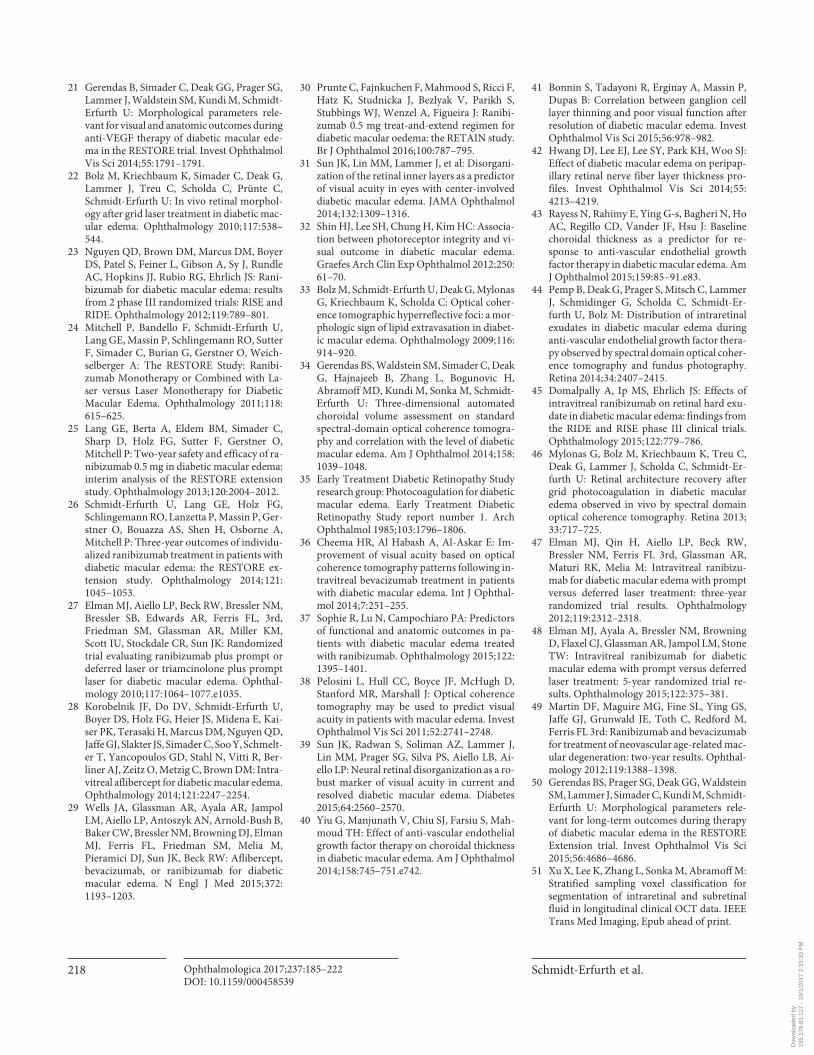
Schmidt-Erfurth et al.
Ophthalmologica 2017;237:185–222DOI: 10.1159/000458539
218
21 Gerendas B, Simader C, Deak GG, Prager SG, Lammer J, Waldstein SM, Kundi M, Schmidt-Erfurth U: Morphological parameters rele-vant for visual and anatomic outcomes during anti-VEGF therapy of diabetic macular ede-ma in the RESTORE trial. Invest Ophthalmol Vis Sci 2014; 55: 1791–1791.
22 Bolz M, Kriechbaum K, Simader C, Deak G, Lammer J, Treu C, Scholda C, Prünte C, Schmidt-Erfurth U: In vivo retinal morphol-ogy after grid laser treatment in diabetic mac-ular edema. Ophthalmology 2010; 117: 538–544.
23 Nguyen QD, Brown DM, Marcus DM, Boyer DS, Patel S, Feiner L, Gibson A, Sy J, Rundle AC, Hopkins JJ, Rubio RG, Ehrlich JS: Rani-bizumab for diabetic macular edema: results from 2 phase III randomized trials: RISE and RIDE. Ophthalmology 2012; 119: 789–801.
24 Mitchell P, Bandello F, Schmidt-Erfurth U, Lang GE, Massin P, Schlingemann RO, Sutter F, Simader C, Burian G, Gerstner O, Weich-selberger A: The RESTORE Study: Ranibi-zumab Monotherapy or Combined with La-ser versus Laser Monotherapy for Diabetic Macular Edema. Ophthalmology 2011; 118: 615–625.
25 Lang GE, Berta A, Eldem BM, Simader C, Sharp D, Holz FG, Sutter F, Gerstner O, Mitchell P: Two-year safety and efficacy of ra-nibizumab 0.5 mg in diabetic macular edema: interim analysis of the RESTORE extension study. Ophthalmology 2013; 120: 2004–2012.
26 Schmidt-Erfurth U, Lang GE, Holz FG,Schlingemann RO, Lanzetta P, Massin P, Ger-stner O, Bouazza AS, Shen H, Osborne A, Mitchell P: Three-year outcomes of individu-alized ranibizumab treatment in patients with diabetic macular edema: the RESTORE ex-tension study. Ophthalmology 2014; 121: 1045–1053.
27 Elman MJ, Aiello LP, Beck RW, Bressler NM, Bressler SB, Edwards AR, Ferris FL, 3rd, Friedman SM, Glassman AR, Miller KM, Scott IU, Stockdale CR, Sun JK: Randomized trial evaluating ranibizumab plus prompt or deferred laser or triamcinolone plus prompt laser for diabetic macular edema. Ophthal-mology 2010; 117: 1064–1077.e1035.
28 Korobelnik JF, Do DV, Schmidt-Erfurth U, Boyer DS, Holz FG, Heier JS, Midena E, Kai-ser PK, Terasaki H, Marcus DM, Nguyen QD, Jaffe GJ, Slakter JS, Simader C, Soo Y, Schmelt-er T, Yancopoulos GD, Stahl N, Vitti R, Ber-liner AJ, Zeitz O, Metzig C, Brown DM: Intra-vitreal aflibercept for diabetic macular edema. Ophthalmology 2014; 121: 2247–2254.
29 Wells JA, Glassman AR, Ayala AR, Jampol LM, Aiello LP, Antoszyk AN, Arnold-Bush B, Baker CW, Bressler NM, Browning DJ, Elman MJ, Ferris FL, Friedman SM, Melia M, Pieramici DJ, Sun JK, Beck RW: Aflibercept, bevacizumab, or ranibizumab for diabetic macular edema. N Engl J Med 2015; 372: 1193–1203.
30 Prunte C, Fajnkuchen F, Mahmood S, Ricci F, Hatz K, Studnicka J, Bezlyak V, Parikh S, Stubbings WJ, Wenzel A, Figueira J: Ranibi-zumab 0.5 mg treat-and-extend regimen for diabetic macular oedema: the RETAIN study. Br J Ophthalmol 2016; 100: 787–795.
31 Sun JK, Lin MM, Lammer J, et al: Disorgani-zation of the retinal inner layers as a predictor of visual acuity in eyes with center-involved diabetic macular edema. JAMA Ophthalmol 2014; 132: 1309–1316.
32 Shin HJ, Lee SH, Chung H, Kim HC: Associa-tion between photoreceptor integrity and vi-sual outcome in diabetic macular edema. Graefes Arch Clin Exp Ophthalmol 2012; 250: 61–70.
33 Bolz M, Schmidt-Erfurth U, Deak G, Mylonas G, Kriechbaum K, Scholda C: Optical coher-ence tomographic hyperreflective foci: a mor-phologic sign of lipid extravasation in diabet-ic macular edema. Ophthalmology 2009; 116: 914–920.
34 Gerendas BS, Waldstein SM, Simader C, Deak G, Hajnajeeb B, Zhang L, Bogunovic H, Abramoff MD, Kundi M, Sonka M, Schmidt-Erfurth U: Three-dimensional automated choroidal volume assessment on standard spectral-domain optical coherence tomogra-phy and correlation with the level of diabetic macular edema. Am J Ophthalmol 2014; 158: 1039–1048.
35 Early Treatment Diabetic Retinopathy Study research group: Photocoagulation for diabetic macular edema. Early Treatment Diabetic Retinopathy Study report number 1. Arch Ophthalmol 1985; 103: 1796–1806.
36 Cheema HR, Al Habash A, Al-Askar E: Im-provement of visual acuity based on optical coherence tomography patterns following in-travitreal bevacizumab treatment in patients with diabetic macular edema. Int J Ophthal-mol 2014; 7: 251–255.
37 Sophie R, Lu N, Campochiaro PA: Predictors of functional and anatomic outcomes in pa-tients with diabetic macular edema treated with ranibizumab. Ophthalmology 2015; 122: 1395–1401.
38 Pelosini L, Hull CC, Boyce JF, McHugh D, Stanford MR, Marshall J: Optical coherence tomography may be used to predict visual acuity in patients with macular edema. Invest Ophthalmol Vis Sci 2011; 52: 2741–2748.
39 Sun JK, Radwan S, Soliman AZ, Lammer J, Lin MM, Prager SG, Silva PS, Aiello LB, Ai-ello LP: Neural retinal disorganization as a ro-bust marker of visual acuity in current and resolved diabetic macular edema. Diabetes 2015; 64: 2560–2570.
40 Yiu G, Manjunath V, Chiu SJ, Farsiu S, Mah-moud TH: Effect of anti-vascular endothelial growth factor therapy on choroidal thickness in diabetic macular edema. Am J Ophthalmol 2014; 158: 745–751.e742.
41 Bonnin S, Tadayoni R, Erginay A, Massin P, Dupas B: Correlation between ganglion cell layer thinning and poor visual function after resolution of diabetic macular edema. Invest Ophthalmol Vis Sci 2015; 56: 978–982.
42 Hwang DJ, Lee EJ, Lee SY, Park KH, Woo SJ: Effect of diabetic macular edema on peripap-illary retinal nerve fiber layer thickness pro-files. Invest Ophthalmol Vis Sci 2014; 55: 4213–4219.
43 Rayess N, Rahimy E, Ying G-s, Bagheri N, Ho AC, Regillo CD, Vander JF, Hsu J: Baseline choroidal thickness as a predictor for re-sponse to anti-vascular endothelial growth factor therapy in diabetic macular edema. Am J Ophthalmol 2015; 159: 85–91.e83.
44 Pemp B, Deak G, Prager S, Mitsch C, Lammer J, Schmidinger G, Scholda C, Schmidt-Er-furth U, Bolz M: Distribution of intraretinal exudates in diabetic macular edema during anti-vascular endothelial growth factor thera-py observed by spectral domain optical coher-ence tomography and fundus photography. Retina 2014; 34: 2407–2415.
45 Domalpally A, Ip MS, Ehrlich JS: Effects of intravitreal ranibizumab on retinal hard exu-date in diabetic macular edema: findings from the RIDE and RISE phase III clinical trials. Ophthalmology 2015; 122: 779–786.
46 Mylonas G, Bolz M, Kriechbaum K, Treu C, Deak G, Lammer J, Scholda C, Schmidt-Er-furth U: Retinal architecture recovery after grid photocoagulation in diabetic macular edema observed in vivo by spectral domain optical coherence tomography. Retina 2013; 33: 717–725.
47 Elman MJ, Qin H, Aiello LP, Beck RW, Bressler NM, Ferris FL 3rd, Glassman AR, Maturi RK, Melia M: Intravitreal ranibizu-mab for diabetic macular edema with prompt versus deferred laser treatment: three-year randomized trial results. Ophthalmology 2012; 119: 2312–2318.
48 Elman MJ, Ayala A, Bressler NM, Browning D, Flaxel CJ, Glassman AR, Jampol LM, Stone TW: Intravitreal ranibizumab for diabetic macular edema with prompt versus deferred laser treatment: 5-year randomized trial re-sults. Ophthalmology 2015; 122: 375–381.
49 Martin DF, Maguire MG, Fine SL, Ying GS, Jaffe GJ, Grunwald JE, Toth C, Redford M, Ferris FL 3rd: Ranibizumab and bevacizumab for treatment of neovascular age-related mac-ular degeneration: two-year results. Ophthal-mology 2012; 119: 1388–1398.
50 Gerendas BS, Prager SG, Deak GG, Waldstein SM, Lammer J, Simader C, Kundi M, Schmidt-Erfurth U: Morphological parameters rele-vant for long-term outcomes during therapy of diabetic macular edema in the RESTORE Extension trial. Invest Ophthalmol Vis Sci 2015; 56: 4686–4686.
51 Xu X, Lee K, Zhang L, Sonka M, Abramoff M: Stratified sampling voxel classification for segmentation of intraretinal and subretinal fluid in longitudinal clinical OCT data. IEEE Trans Med Imaging, Epub ahead of print.
Dow
nloa
ded
by:
195.
178.
83.1
27 -
10/
1/20
17 2
:33:
30 P
M
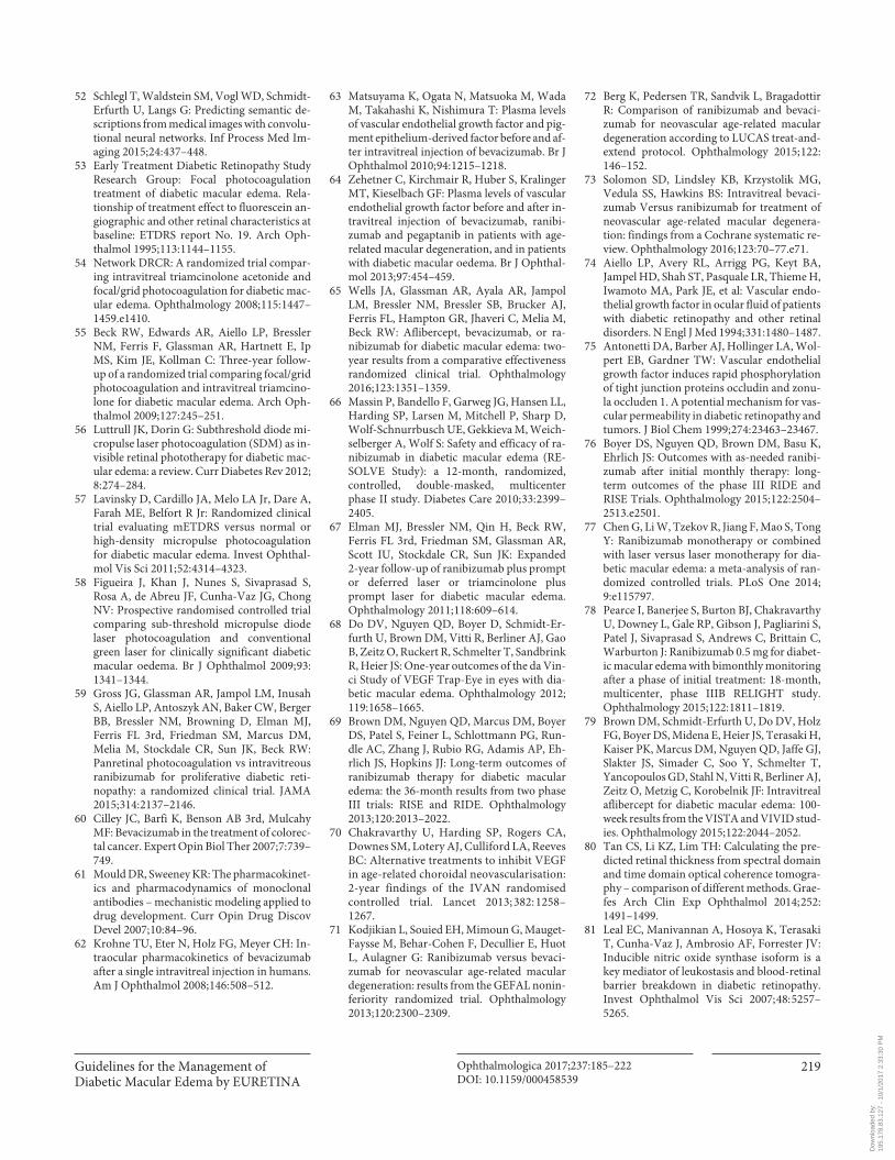
Guidelines for the Management of Diabetic Macular Edema by EURETINA
Ophthalmologica 2017;237:185–222DOI: 10.1159/000458539
219
52 Schlegl T, Waldstein SM, Vogl WD, Schmidt-Erfurth U, Langs G: Predicting semantic de-scriptions from medical images with convolu-tional neural networks. Inf Process Med Im-aging 2015; 24: 437–448.
53 Early Treatment Diabetic Retinopathy Study Research Group: Focal photocoagulation treatment of diabetic macular edema. Rela-tionship of treatment effect to fluorescein an-giographic and other retinal characteristics at baseline: ETDRS report No. 19. Arch Oph-thalmol 1995; 113: 1144–1155.
54 Network DRCR: A randomized trial compar-ing intravitreal triamcinolone acetonide and focal/grid photocoagulation for diabetic mac-ular edema. Ophthalmology 2008; 115: 1447–1459.e1410.
55 Beck RW, Edwards AR, Aiello LP, Bressler NM, Ferris F, Glassman AR, Hartnett E, Ip MS, Kim JE, Kollman C: Three-year follow-up of a randomized trial comparing focal/grid photocoagulation and intravitreal triamcino-lone for diabetic macular edema. Arch Oph-thalmol 2009; 127: 245–251.
56 Luttrull JK, Dorin G: Subthreshold diode mi-cropulse laser photocoagulation (SDM) as in-visible retinal phototherapy for diabetic mac-ular edema: a review. Curr Diabetes Rev 2012; 8: 274–284.
57 Lavinsky D, Cardillo JA, Melo LA Jr, Dare A, Farah ME, Belfort R Jr: Randomized clinical trial evaluating mETDRS versus normal or high-density micropulse photocoagulation for diabetic macular edema. Invest Ophthal-mol Vis Sci 2011; 52: 4314–4323.
58 Figueira J, Khan J, Nunes S, Sivaprasad S, Rosa A, de Abreu JF, Cunha-Vaz JG, Chong NV: Prospective randomised controlled trial comparing sub-threshold micropulse diode laser photocoagulation and conventional green laser for clinically significant diabetic macular oedema. Br J Ophthalmol 2009; 93: 1341–1344.
59 Gross JG, Glassman AR, Jampol LM, Inusah S, Aiello LP, Antoszyk AN, Baker CW, Berger BB, Bressler NM, Browning D, Elman MJ, Ferris FL 3rd, Friedman SM, Marcus DM, Melia M, Stockdale CR, Sun JK, Beck RW: Panretinal photocoagulation vs intravitreous ranibizumab for proliferative diabetic reti-nopathy: a randomized clinical trial. JAMA 2015; 314: 2137–2146.
60 Cilley JC, Barfi K, Benson AB 3rd, Mulcahy MF: Bevacizumab in the treatment of colorec-tal cancer. Expert Opin Biol Ther 2007; 7: 739–749.
61 Mould DR, Sweeney KR: The pharmacokinet-ics and pharmacodynamics of monoclonal antibodies – mechanistic modeling applied to drug development. Curr Opin Drug Discov Devel 2007; 10: 84–96.
62 Krohne TU, Eter N, Holz FG, Meyer CH: In-traocular pharmacokinetics of bevacizumab after a single intravitreal injection in humans. Am J Ophthalmol 2008; 146: 508–512.
63 Matsuyama K, Ogata N, Matsuoka M, Wada M, Takahashi K, Nishimura T: Plasma levels of vascular endothelial growth factor and pig-ment epithelium-derived factor before and af-ter intravitreal injection of bevacizumab. Br J Ophthalmol 2010; 94: 1215–1218.
64 Zehetner C, Kirchmair R, Huber S, Kralinger MT, Kieselbach GF: Plasma levels of vascular endothelial growth factor before and after in-travitreal injection of bevacizumab, ranibi-zumab and pegaptanib in patients with age-related macular degeneration, and in patients with diabetic macular oedema. Br J Ophthal-mol 2013; 97: 454–459.
65 Wells JA, Glassman AR, Ayala AR, Jampol LM, Bressler NM, Bressler SB, Brucker AJ, Ferris FL, Hampton GR, Jhaveri C, Melia M, Beck RW: Aflibercept, bevacizumab, or ra-nibizumab for diabetic macular edema: two-year results from a comparative effectiveness randomized clinical trial. Ophthalmology 2016; 123: 1351–1359.
66 Massin P, Bandello F, Garweg JG, Hansen LL, Harding SP, Larsen M, Mitchell P, Sharp D, Wolf-Schnurrbusch UE, Gekkieva M, Weich-selberger A, Wolf S: Safety and efficacy of ra-nibizumab in diabetic macular edema (RE-SOLVE Study): a 12-month, randomized, controlled, double-masked, multicenter phase II study. Diabetes Care 2010; 33: 2399–2405.
67 Elman MJ, Bressler NM, Qin H, Beck RW, Ferris FL 3rd, Friedman SM, Glassman AR, Scott IU, Stockdale CR, Sun JK: Expanded 2-year follow-up of ranibizumab plus prompt or deferred laser or triamcinolone plus prompt laser for diabetic macular edema. Ophthalmology 2011; 118: 609–614.
68 Do DV, Nguyen QD, Boyer D, Schmidt-Er-furth U, Brown DM, Vitti R, Berliner AJ, Gao B, Zeitz O, Ruckert R, Schmelter T, Sandbrink R, Heier JS: One-year outcomes of the da Vin-ci Study of VEGF Trap-Eye in eyes with dia-betic macular edema. Ophthalmology 2012; 119: 1658–1665.
69 Brown DM, Nguyen QD, Marcus DM, Boyer DS, Patel S, Feiner L, Schlottmann PG, Run-dle AC, Zhang J, Rubio RG, Adamis AP, Eh-rlich JS, Hopkins JJ: Long-term outcomes of ranibizumab therapy for diabetic macular edema: the 36-month results from two phase III trials: RISE and RIDE. Ophthalmology 2013; 120: 2013–2022.
70 Chakravarthy U, Harding SP, Rogers CA, Downes SM, Lotery AJ, Culliford LA, Reeves BC: Alternative treatments to inhibit VEGF in age-related choroidal neovascularisation: 2-year findings of the IVAN randomised controlled trial. Lancet 2013; 382: 1258–1267.
71 Kodjikian L, Souied EH, Mimoun G, Mauget-Faysse M, Behar-Cohen F, Decullier E, Huot L, Aulagner G: Ranibizumab versus bevaci-zumab for neovascular age-related macular degeneration: results from the GEFAL nonin-feriority randomized trial. Ophthalmology 2013; 120: 2300–2309.
72 Berg K, Pedersen TR, Sandvik L, Bragadottir R: Comparison of ranibizumab and bevaci-zumab for neovascular age-related macular degeneration according to LUCAS treat-and-extend protocol. Ophthalmology 2015; 122: 146–152.
73 Solomon SD, Lindsley KB, Krzystolik MG, Vedula SS, Hawkins BS: Intravitreal bevaci-zumab Versus ranibizumab for treatment of neovascular age-related macular degenera-tion: findings from a Cochrane systematic re-view. Ophthalmology 2016; 123: 70–77.e71.
74 Aiello LP, Avery RL, Arrigg PG, Keyt BA, Jampel HD, Shah ST, Pasquale LR, Thieme H, Iwamoto MA, Park JE, et al: Vascular endo-thelial growth factor in ocular fluid of patients with diabetic retinopathy and other retinal disorders. N Engl J Med 1994; 331: 1480–1487.
75 Antonetti DA, Barber AJ, Hollinger LA, Wol-pert EB, Gardner TW: Vascular endothelial growth factor induces rapid phosphorylation of tight junction proteins occludin and zonu-la occluden 1. A potential mechanism for vas-cular permeability in diabetic retinopathy and tumors. J Biol Chem 1999; 274: 23463–23467.
76 Boyer DS, Nguyen QD, Brown DM, Basu K, Ehrlich JS: Outcomes with as-needed ranibi-zumab after initial monthly therapy: long-term outcomes of the phase III RIDE and RISE Trials. Ophthalmology 2015; 122: 2504–2513.e2501.
77 Chen G, Li W, Tzekov R, Jiang F, Mao S, Tong Y: Ranibizumab monotherapy or combined with laser versus laser monotherapy for dia-betic macular edema: a meta-analysis of ran-domized controlled trials. PLoS One 2014; 9:e115797.
78 Pearce I, Banerjee S, Burton BJ, Chakravarthy U, Downey L, Gale RP, Gibson J, Pagliarini S, Patel J, Sivaprasad S, Andrews C, Brittain C, Warburton J: Ranibizumab 0.5 mg for diabet-ic macular edema with bimonthly monitoring after a phase of initial treatment: 18-month, multicenter, phase IIIB RELIGHT study. Ophthalmology 2015; 122: 1811–1819.
79 Brown DM, Schmidt-Erfurth U, Do DV, Holz FG, Boyer DS, Midena E, Heier JS, Terasaki H, Kaiser PK, Marcus DM, Nguyen QD, Jaffe GJ, Slakter JS, Simader C, Soo Y, Schmelter T, Yancopoulos GD, Stahl N, Vitti R, Berliner AJ, Zeitz O, Metzig C, Korobelnik JF: Intravitreal aflibercept for diabetic macular edema: 100-week results from the VISTA and VIVID stud-ies. Ophthalmology 2015; 122: 2044–2052.
80 Tan CS, Li KZ, Lim TH: Calculating the pre-dicted retinal thickness from spectral domain and time domain optical coherence tomogra-phy – comparison of different methods. Grae-fes Arch Clin Exp Ophthalmol 2014; 252: 1491–1499.
81 Leal EC, Manivannan A, Hosoya K, Terasaki T, Cunha-Vaz J, Ambrosio AF, Forrester JV: Inducible nitric oxide synthase isoform is a key mediator of leukostasis and blood-retinal barrier breakdown in diabetic retinopathy. Invest Ophthalmol Vis Sci 2007; 48: 5257–5265.
Dow
nloa
ded
by:
195.
178.
83.1
27 -
10/
1/20
17 2
:33:
30 P
M
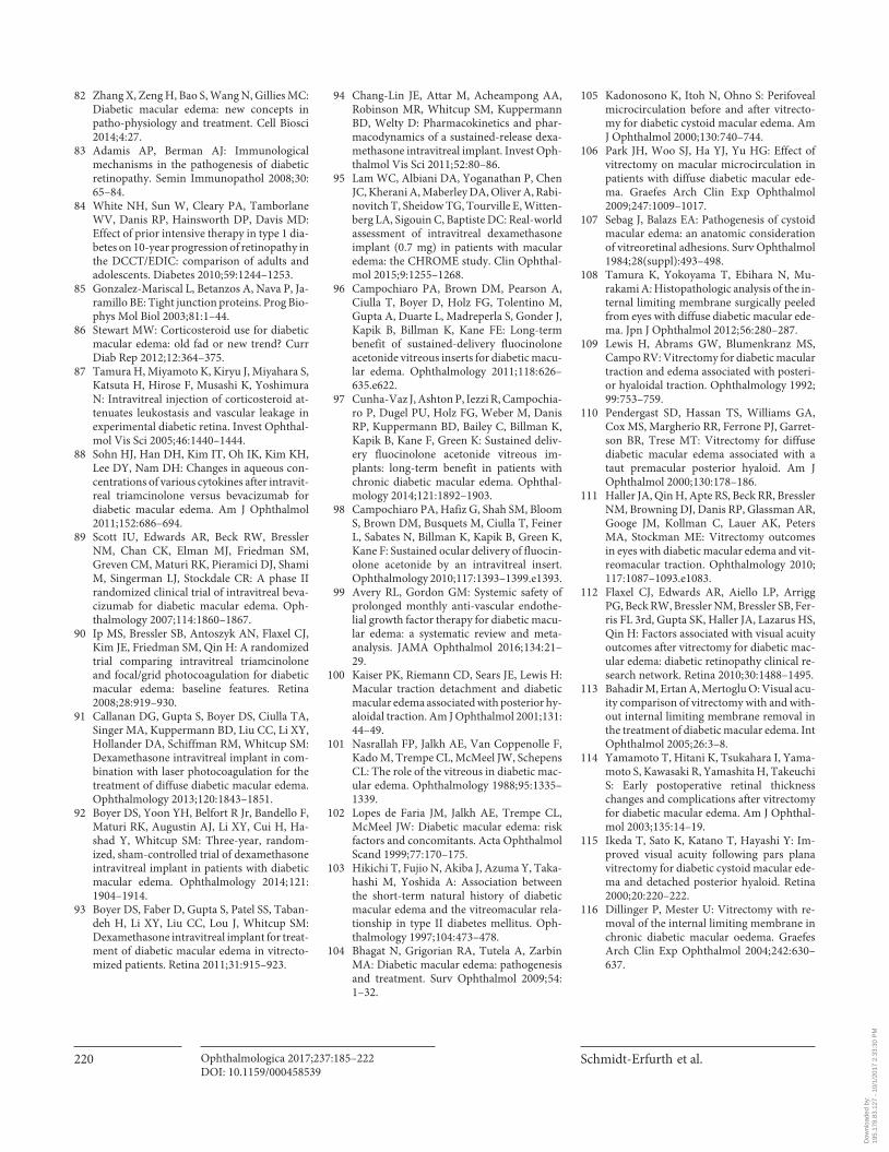
Schmidt-Erfurth et al.
Ophthalmologica 2017;237:185–222DOI: 10.1159/000458539
220
82 Zhang X, Zeng H, Bao S, Wang N, Gillies MC: Diabetic macular edema: new concepts in patho-physiology and treatment. Cell Biosci 2014; 4: 27.
83 Adamis AP, Berman AJ: Immunological mechanisms in the pathogenesis of diabetic retinopathy. Semin Immunopathol 2008; 30: 65–84.
84 White NH, Sun W, Cleary PA, Tamborlane WV, Danis RP, Hainsworth DP, Davis MD: Effect of prior intensive therapy in type 1 dia-betes on 10-year progression of retinopathy in the DCCT/EDIC: comparison of adults and adolescents. Diabetes 2010; 59: 1244–1253.
85 Gonzalez-Mariscal L, Betanzos A, Nava P, Ja-ramillo BE: Tight junction proteins. Prog Bio-phys Mol Biol 2003; 81: 1–44.
86 Stewart MW: Corticosteroid use for diabetic macular edema: old fad or new trend? Curr Diab Rep 2012; 12: 364–375.
87 Tamura H, Miyamoto K, Kiryu J, Miyahara S, Katsuta H, Hirose F, Musashi K, Yoshimura N: Intravitreal injection of corticosteroid at-tenuates leukostasis and vascular leakage in experimental diabetic retina. Invest Ophthal-mol Vis Sci 2005; 46: 1440–1444.
88 Sohn HJ, Han DH, Kim IT, Oh IK, Kim KH, Lee DY, Nam DH: Changes in aqueous con-centrations of various cytokines after intravit-real triamcinolone versus bevacizumab for diabetic macular edema. Am J Ophthalmol 2011; 152: 686–694.
89 Scott IU, Edwards AR, Beck RW, Bressler NM, Chan CK, Elman MJ, Friedman SM, Greven CM, Maturi RK, Pieramici DJ, Shami M, Singerman LJ, Stockdale CR: A phase II randomized clinical trial of intravitreal beva-cizumab for diabetic macular edema. Oph-thalmology 2007; 114: 1860–1867.
90 Ip MS, Bressler SB, Antoszyk AN, Flaxel CJ, Kim JE, Friedman SM, Qin H: A randomized trial comparing intravitreal triamcinolone and focal/grid photocoagulation for diabetic macular edema: baseline features. Retina 2008; 28: 919–930.
91 Callanan DG, Gupta S, Boyer DS, Ciulla TA, Singer MA, Kuppermann BD, Liu CC, Li XY, Hollander DA, Schiffman RM, Whitcup SM: Dexamethasone intravitreal implant in com-bination with laser photocoagulation for the treatment of diffuse diabetic macular edema. Ophthalmology 2013; 120: 1843–1851.
92 Boyer DS, Yoon YH, Belfort R Jr, Bandello F, Maturi RK, Augustin AJ, Li XY, Cui H, Ha-shad Y, Whitcup SM: Three-year, random-ized, sham-controlled trial of dexamethasone intravitreal implant in patients with diabetic macular edema. Ophthalmology 2014; 121: 1904–1914.
93 Boyer DS, Faber D, Gupta S, Patel SS, Taban-deh H, Li XY, Liu CC, Lou J, Whitcup SM: Dexamethasone intravitreal implant for treat-ment of diabetic macular edema in vitrecto-mized patients. Retina 2011; 31: 915–923.
94 Chang-Lin JE, Attar M, Acheampong AA, Robinson MR, Whitcup SM, Kuppermann BD, Welty D: Pharmacokinetics and phar-macodynamics of a sustained-release dexa-methasone intravitreal implant. Invest Oph-thalmol Vis Sci 2011; 52: 80–86.
95 Lam WC, Albiani DA, Yoganathan P, Chen JC, Kherani A, Maberley DA, Oliver A, Rabi-novitch T, Sheidow TG, Tourville E, Witten-berg LA, Sigouin C, Baptiste DC: Real-world assessment of intravitreal dexamethasone implant (0.7 mg) in patients with macular edema: the CHROME study. Clin Ophthal-mol 2015; 9: 1255–1268.
96 Campochiaro PA, Brown DM, Pearson A, Ciulla T, Boyer D, Holz FG, Tolentino M, Gupta A, Duarte L, Madreperla S, Gonder J, Kapik B, Billman K, Kane FE: Long-term benefit of sustained-delivery fluocinolone acetonide vitreous inserts for diabetic macu-lar edema. Ophthalmology 2011; 118: 626–635.e622.
97 Cunha-Vaz J, Ashton P, Iezzi R, Campochia-ro P, Dugel PU, Holz FG, Weber M, Danis RP, Kuppermann BD, Bailey C, Billman K, Kapik B, Kane F, Green K: Sustained deliv-ery fluocinolone acetonide vitreous im-plants: long-term benefit in patients with chronic diabetic macular edema. Ophthal-mology 2014; 121: 1892–1903.
98 Campochiaro PA, Hafiz G, Shah SM, Bloom S, Brown DM, Busquets M, Ciulla T, Feiner L, Sabates N, Billman K, Kapik B, Green K, Kane F: Sustained ocular delivery of fluocin-olone acetonide by an intravitreal insert. Ophthalmology 2010; 117: 1393–1399.e1393.
99 Avery RL, Gordon GM: Systemic safety of prolonged monthly anti-vascular endothe-lial growth factor therapy for diabetic macu-lar edema: a systematic review and meta-analysis. JAMA Ophthalmol 2016; 134: 21–29.
100 Kaiser PK, Riemann CD, Sears JE, Lewis H: Macular traction detachment and diabetic macular edema associated with posterior hy-aloidal traction. Am J Ophthalmol 2001; 131: 44–49.
101 Nasrallah FP, Jalkh AE, Van Coppenolle F, Kado M, Trempe CL, McMeel JW, Schepens CL: The role of the vitreous in diabetic mac-ular edema. Ophthalmology 1988; 95: 1335–1339.
102 Lopes de Faria JM, Jalkh AE, Trempe CL, McMeel JW: Diabetic macular edema: risk factors and concomitants. Acta Ophthalmol Scand 1999; 77: 170–175.
103 Hikichi T, Fujio N, Akiba J, Azuma Y, Taka-hashi M, Yoshida A: Association between the short-term natural history of diabetic macular edema and the vitreomacular rela-tionship in type II diabetes mellitus. Oph-thalmology 1997; 104: 473–478.
104 Bhagat N, Grigorian RA, Tutela A, Zarbin MA: Diabetic macular edema: pathogenesis and treatment. Surv Ophthalmol 2009; 54: 1–32.
105 Kadonosono K, Itoh N, Ohno S: Perifoveal microcirculation before and after vitrecto-my for diabetic cystoid macular edema. Am J Ophthalmol 2000; 130: 740–744.
106 Park JH, Woo SJ, Ha YJ, Yu HG: Effect of vitrectomy on macular microcirculation in patients with diffuse diabetic macular ede-ma. Graefes Arch Clin Exp Ophthalmol 2009; 247: 1009–1017.
107 Sebag J, Balazs EA: Pathogenesis of cystoid macular edema: an anatomic consideration of vitreoretinal adhesions. Surv Ophthalmol 1984; 28(suppl):493–498.
108 Tamura K, Yokoyama T, Ebihara N, Mu-rakami A: Histopathologic analysis of the in-ternal limiting membrane surgically peeled from eyes with diffuse diabetic macular ede-ma. Jpn J Ophthalmol 2012; 56: 280–287.
109 Lewis H, Abrams GW, Blumenkranz MS, Campo RV: Vitrectomy for diabetic macular traction and edema associated with posteri-or hyaloidal traction. Ophthalmology 1992; 99: 753–759.
110 Pendergast SD, Hassan TS, Williams GA, Cox MS, Margherio RR, Ferrone PJ, Garret-son BR, Trese MT: Vitrectomy for diffuse diabetic macular edema associated with a taut premacular posterior hyaloid. Am J Ophthalmol 2000; 130: 178–186.
111 Haller JA, Qin H, Apte RS, Beck RR, Bressler NM, Browning DJ, Danis RP, Glassman AR, Googe JM, Kollman C, Lauer AK, Peters MA, Stockman ME: Vitrectomy outcomes in eyes with diabetic macular edema and vit-reomacular traction. Ophthalmology 2010; 117: 1087–1093.e1083.
112 Flaxel CJ, Edwards AR, Aiello LP, Arrigg PG, Beck RW, Bressler NM, Bressler SB, Fer-ris FL 3rd, Gupta SK, Haller JA, Lazarus HS, Qin H: Factors associated with visual acuity outcomes after vitrectomy for diabetic mac-ular edema: diabetic retinopathy clinical re-search network. Retina 2010; 30: 1488–1495.
113 Bahadir M, Ertan A, Mertoglu O: Visual acu-ity comparison of vitrectomy with and with-out internal limiting membrane removal in the treatment of diabetic macular edema. Int Ophthalmol 2005; 26: 3–8.
114 Yamamoto T, Hitani K, Tsukahara I, Yama-moto S, Kawasaki R, Yamashita H, Takeuchi S: Early postoperative retinal thickness changes and complications after vitrectomy for diabetic macular edema. Am J Ophthal-mol 2003; 135: 14–19.
115 Ikeda T, Sato K, Katano T, Hayashi Y: Im-proved visual acuity following pars plana vitrectomy for diabetic cystoid macular ede-ma and detached posterior hyaloid. Retina 2000; 20: 220–222.
116 Dillinger P, Mester U: Vitrectomy with re-moval of the internal limiting membrane in chronic diabetic macular oedema. Graefes Arch Clin Exp Ophthalmol 2004; 242: 630–637.
Dow
nloa
ded
by:
195.
178.
83.1
27 -
10/
1/20
17 2
:33:
30 P
M
Guidelines for the Management of Diabetic Macular Edema by EURETINA
Ophthalmologica 2017;237:185–222DOI: 10.1159/000458539
221
117 Gandorfer A, Messmer EM, Ulbig MW, Kampik A: Resolution of diabetic macular edema after surgical removal of the posterior hyaloid and the inner limiting membrane. Retina 2000; 20: 126–133.
118 Kumagai K, Furukawa M, Ogino N, Larson E, Iwaki M, Tachi N: Long-term follow-up of vitrectomy for diffuse nontractional dia-betic macular edema. Retina 2009; 29: 464–472.
119 La Heij EC, Hendrikse F, Kessels AG, Der-haag PJ: Vitrectomy results in diabetic mac-ular oedema without evident vitreomacular traction. Graefes Arch Clin Exp Ophthalmol 2001; 239: 264–270.
120 Otani T, Kishi S: A controlled study of vit-rectomy for diabetic macular edema. Am J Ophthalmol 2002; 134: 214–219.
121 Tachi N, Ogino N: Vitrectomy for diffuse macular edema in cases of diabetic retinopa-thy. Am J Ophthalmol 1996; 122: 258–260.
122 Yanyali A, Horozoglu F, Celik E, Nohutcu AF: Long-term outcomes of pars plana vit-rectomy with internal limiting membrane removal in diabetic macular edema. Retina 2007; 27: 557–566.
123 Stolba U, Binder S, Gruber D, Krebs I, Ag-germann T, Neumaier B: Vitrectomy for persistent diffuse diabetic macular edema. Am J Ophthalmol 2005; 140: 295–301.
124 Yanyali A, Horozoglu F, Celik E, Ercalik Y, Nohutcu AF: Pars plana vitrectomy and re-moval of the internal limiting membrane in diabetic macular edema unresponsive to grid laser photocoagulation. Eur J Ophthal-mol 2006; 16: 573–581.
125 Yanyali A, Nohutcu AF, Horozoglu F, Celik E: Modified grid laser photocoagulation ver-sus pars plana vitrectomy with internal lim-iting membrane removal in diabetic macular edema. Am J Ophthalmol 2005; 139: 795–801.
126 Kumar A, Sinha S, Azad R, Sharma YR, Vohra R: Comparative evaluation of vitrec-tomy and dye-enhanced ILM peel with grid laser in diffuse diabetic macular edema. Graefes Arch Clin Exp Ophthalmol 2007; 245: 360–368.
127 Thomas D, Bunce C, Moorman C, Laidlaw DA: A randomised controlled feasibility tri-al of vitrectomy versus laser for diabetic macular oedema. Br J Ophthalmol 2005; 89: 81–86.
128 Patel JI, Hykin PG, Schadt M, Luong V, Bunce C, Fitzke F, Gregor ZJ: Diabetic mac-ular oedema: pilot randomised trial of pars plana vitrectomy vs macular argon photoco-agulation. Eye (Lond) 2006; 20: 873–881.
129 Doi N, Sakamoto T, Sonoda Y, Yasuda M, Yonemoto K, Arimura N, Uchino E, Ishi-bashi T: Comparative study of vitrectomy versus intravitreous triamcinolone for dia-betic macular edema on randomized paired-eyes. Graefes Arch Clin Exp Ophthalmol 2012; 250: 71–78.
130 Kim YT, Kang SW, Kim SJ, Kim SM, Chung SE: Combination of vitrectomy, IVTA, and laser photocoagulation for diabetic macular edema unresponsive to prior treatments; 3-year results. Graefes Arch Clin Exp Oph-thalmol 2012; 250: 679–684.
131 Stefaniotou M, Aspiotis M, Kalogeropoulos C, Christodoulou A, Psylla M, Ioachim E, Alamanos I, Psilas K: Vitrectomy results for diffuse diabetic macular edema with and without inner limiting membrane removal. Eur J Ophthalmol 2004; 14: 137–143.
132 Hoerauf H, Bruggemann A, Muecke M, Luke J, Muller M, Stefansson E, Hammes HP, Weiss C: Pars plana vitrectomy for dia-betic macular edema. Internal limiting membrane delamination vs posterior hya-loid removal. A prospective randomized tri-al. Graefes Arch Clin Exp Ophthalmol 2011; 249: 997–1008.
133 Bardak Y, Cekic O, Tig SU: Comparison of ICG-assisted ILM peeling and triamcino-lone-assisted posterior vitreous removal in diffuse diabetic macular oedema. Eye (Lond) 2006; 20: 1357–1359.
134 Kumagai K, Hangai M, Ogino N, Larson E: Effect of internal limiting membrane peel-ing on long-term visual outcomes for dia-betic macular edema. Retina 2015; 35: 1422–1428.
135 Figueroa MS, Contreras I, Noval S: Surgical and anatomical outcomes of pars plana vit-rectomy for diffuse nontractional diabetic macular edema. Retina 2008; 28: 420–426.
136 Shiba T, Kamura Y, Yagi F, Sato Y: Com-parison of surgical procedures for vitreous surgery in diabetic macular edema. Jpn J Ophthalmol 2009; 53: 120–124.
137 Shah SP, Patel M, Thomas D, Aldington S, Laidlaw DA: Factors predicting outcome of vitrectomy for diabetic macular oedema: re-sults of a prospective study. Br J Ophthalmol 2006; 90: 33–36.
138 Bonnin S, Sandali O, Bonnel S, Monin C, El Sanharawi M: Vitrectomy with internal lim-iting membrane peeling for tractional and nontractional diabetic macular edema: Long-term Results of a Comparative Study. Retina 2015; 35: 921–928.
139 Yamada Y, Suzuma K, Ryu M, Tsuiki E, Fu-jikawa A, Kitaoka T: Systemic factors influ-ence the prognosis of diabetic macular ede-ma after pars plana vitrectomy with internal limiting membrane peeling. Curr Eye Res 2013; 38: 1261–1265.
140 Chhablani JK, Kim JS, Cheng L, Kozak I, Freeman W: External limiting membrane as a predictor of visual improvement in diabet-ic macular edema after pars plana vitrecto-my. Graefes Arch Clin Exp Ophthalmol 2012; 250: 1415–1420.
141 Murakami T, Nishijima K, Akagi T, Uji A, Horii T, Ueda-Arakawa N, Muraoka Y, Yo-shimura N: Segmentational analysis of reti-nal thickness after vitrectomy in diabetic macular edema. Invest Ophthalmol Vis Sci 2012; 53: 6668–6674.
142 Litwak L, Goh SY, Hussein Z, Malek R, Prusty V, Khamseh ME: Prevalence of dia-betes complications in people with type 2 diabetes mellitus and its association with baseline characteristics in the multinational A1chieve study. Diabetol Metab Syndr 2013; 5: 57.
143 Zimmet P, Alberti KG, Magliano DJ, Ben-nett PH: Diabetes mellitus statistics on prev-alence and mortality: facts and fallacies. Nat Rev Endocrinol 2016; 12: 616–622.
144 Zimmet PZ, Magliano DJ, Herman WH, Shaw JE: Diabetes: a 21st century challenge. Lancet Diabetes Endocrinol 2014; 2: 56–64.
145 Cheung N, Mitchell P, Wong TY: Diabetic retinopathy. Lancet 2010; 376: 124–136.
146 Davis MD, Fisher MR, Gangnon RE, Barton F, Aiello LM, Chew EY, Ferris FL 3rd, Knat-terud GL: Risk factors for high-risk prolif-erative diabetic retinopathy and severe vi-sual loss: Early Treatment Diabetic Retinop-athy Study Report #18. Invest Ophthalmol Vis Sci 1998; 39: 233–252.
147 Rodriguez-Fontal M, Kerrison JB, Alfaro DV, Jablon EP: Metabolic control and dia-betic retinopathy. Curr Diabetes Rev 2009; 5: 3–7.
148 Progression of retinopathy with intensive versus conventional treatment in the Diabe-tes Control and Complications Trial. Diabe-tes Control and Complications Trial Re-search Group. Ophthalmology 1995; 102: 647–661.
149 Intensive blood-glucose control with sul-phonylureas or insulin compared with con-ventional treatment and risk of complica-tions in patients with type 2 diabetes (UKP-DS 33). UK Prospective Diabetes Study (UKPDS) Group. Lancet 1998; 352: 837–853.
150 Retinopathy and nephropathy in patients with type 1 diabetes four years after a trial of intensive therapy. The Diabetes Control and Complications Trial/Epidemiology of Dia-betes Interventions and Complications Re-search Group. N Engl J Med 2000; 342: 381–389.
151 Zhang L, Krzentowski G, Albert A, Lefebvre PJ: Risk of developing retinopathy in Diabe-tes Control and Complications Trial type 1 diabetic patients with good or poor metabol-ic control. Diabetes Care 2001; 24: 1275–1279.
152 Writing Team for the Diabetes Control and Complications Trial/Epidemiology of Dia-betes Interventions and Complications Re-search Group: Effect of intensive therapy on the microvascular complications of type 1 diabetes mellitus. JAMA 2002; 287: 2563–2569.
153 White NH, Sun W, Cleary PA, Danis RP, Davis MD, Hainsworth DP, Hubbard LD, Lachin JM, Nathan DM: Prolonged effect of intensive therapy on the risk of retinopathy complications in patients with type 1 diabe-tes mellitus: 10 years after the Diabetes Con-trol and Complications Trial. Arch Ophthal-mol 2008; 126: 1707–1715.
Dow
nloa
ded
by:
195.
178.
83.1
27 -
10/
1/20
17 2
:33:
30 P
M
Schmidt-Erfurth et al.
Ophthalmologica 2017;237:185–222DOI: 10.1159/000458539
222
154 Fullerton B, Jeitler K, Seitz M, Horvath K, Berghold A, Siebenhofer A: Intensive glu-cose control versus conventional glucose control for type 1 diabetes mellitus. Co-chrane Database Syst Rev 2014:CD009122.
155 Holman RR, Paul SK, Bethel MA, Matthews DR, Neil HA: 10-year follow-up of intensive glucose control in type 2 diabetes. N Engl J Med 2008; 359: 1577–1589.
156 Effect of intensive blood-glucose control with metformin on complications in over-weight patients with type 2 diabetes (UK-PDS 34). UK Prospective Diabetes Study (UKPDS) Group. Lancet 1998; 352: 854–865.
157 Keech AC, Mitchell P, Summanen PA, O’Day J, Davis TM, Moffitt MS, Taskinen MR, Simes RJ, Tse D, Williamson E, Merri-field A, Laatikainen LT, d’Emden MC, Cri-met DC, O’Connell RL, Colman PG: Effect of fenofibrate on the need for laser treatment for diabetic retinopathy (FIELD study): a randomised controlled trial. Lancet 2007; 370: 1687–1697.
158 Early worsening of diabetic retinopathy in the Diabetes Control and Complications Trial. Arch Ophthalmol 1998; 116: 874–886.
159 Wong TY, Liew G, Tapp RJ, Schmidt MI, Wang JJ, Mitchell P, Klein R, Klein BE, Zim-met P, Shaw J: Relation between fasting glu-cose and retinopathy for diagnosis of diabe-tes: three population-based cross-sectional studies. Lancet 2008; 371: 736–743.
160 Patel A, MacMahon S, Chalmers J, Neal B, Billot L, Woodward M, Marre M, Cooper M, Glasziou P, Grobbee D, Hamet P, Harrap S, Heller S, Liu L, Mancia G, Mogensen CE, Pan C, Poulter N, Rodgers A, Williams B, Bompoint S, de Galan BE, Joshi R, Travert F: Intensive blood glucose control and vascular outcomes in patients with type 2 diabetes. N Engl J Med 2008; 358: 2560–2572.
161 Gerstein HC, Miller ME, Byington RP, Goff DC Jr, Bigger JT, Buse JB, Cushman WC, Genuth S, Ismail-Beigi F, Grimm RH Jr, Probstfield JL, Simons-Morton DG, Friede-wald WT: Effects of intensive glucose lower-ing in type 2 diabetes. N Engl J Med 2008; 358: 2545–2559.
162 Liew G, Mitchell P, Wong TY: Systemic management of diabetic retinopathy. BMJ 2009; 338:b441.
163 Duckworth W, Abraira C, Moritz T, Reda D, Emanuele N, Reaven PD, Zieve FJ, Marks J, Davis SN, Hayward R, Warren SR, Goldman S, McCarren M, Vitek ME, Henderson WG, Huang GD: Glucose control and vascular complications in veterans with type 2 diabe-tes. N Engl J Med 2009; 360: 129–139.
164 Lyons TJ, Jenkins AJ, Zheng D, Lackland DT, McGee D, Garvey WT, Klein RL: Dia-betic retinopathy and serum lipoprotein subclasses in the DCCT/EDIC cohort. Invest Ophthalmol Vis Sci 2004; 45: 910–918.
165 Cheung N, Wong TY: Fenofibrate and dia-betic retinopathy. Lancet 2008; 371: 721–722; author reply 722.
166 Diabetic retinopathy study. Report Number 6. Design, methods, and baseline results. Re-port Number 7. A modification of the Airlie House classification of diabetic retinopathy. Prepared by the Diabetic Retinopathy. In-vest Ophthalmol Vis Sci 1981; 21: 1–226.
167 Heng LZ, Pefianaki M, Hykin P, Patel PJ: In-terobserver agreement in detecting spectral-domain optical coherence tomography fea-tures of diabetic macular edema. PLoS One 2015; 10:e0126557.
168 Arevalo JF, Lasave AF, Arias JD, Serrano MA, Arevalo FA: Clinical applications of op-tical coherence tomography in the posteri -or pole: the 2011 José Manuel Espino Lec-ture – Part II. Clin Ophthalmol 2013; 7: 2181–2206.
169 Lee J, Rosen R: Optical coherence tomogra-phy angiography in diabetes. Curr Diabetes Rep 2016; 16: 123.
170 Lee J, Moon BG, Cho AR, Yoon YH: Opti-cal coherence tomography angiography of DME and its association with anti-VEGF treatment response. Ophthalmology 2016; 123: 2368–2375.
Dow
nloa
ded
by:
195.
178.
83.1
27 -
10/
1/20
17 2
:33:
30 P
M