VOL 4 No 1 JANUARY 2017 DCMC Em gency Dep Radi ogy · PDF fileVOL 4 No 1 JANUARY 2017 Page 1...
7
VOL 4 No 1 JANUARY 2017 “DOCENDO DECIMUS” DCMC Emgency Deptment Radiogy Case of the Mth These cases have been removed of identifying information. These cases are intended for peer review and educational purposes only. Welcome to the DCMC Emergency Department Radiology Case of the Month! In conjunction with our pediatric radiology specialists from ARA, we hope you enjoy these monthly radiological highlights from the case files of the Emergency Department at DCMC. These cases are meant to highlight important chief complaints, cases, and radiology findings that we all encounter every day. If you enjoy these reviews, we invite you to check out Pediatric Emergency Medicine Fellowship Radiology rounds, which are offered quarterly and are held with the outstanding support of the pediatric radiology specialists at Austin Radiologic Association. If you have and questions or feedback regarding the Case of the Month format, feel free to email Robert Vezzetti, MD at [email protected]. This Month: Hand & finger injuries are very common in pediatrics. Most of these injuries require nothing more than symptomatic care and sometimes a splint or two. Sometimes the physical exam doesn’t reveal the extent of an injury. Let’s start the new year off with some pediatric hand cases….. Conference Schedule: January 2017 4th - 9:15 Toxicology: Environmental Toxins………………Drs Vaidya and Earp 10:15 Hyperbaric/Dive Medicine…………………..Drs Hill and Higginbotham 11:15 Urologic Surgical Emergencies……………Drs Berg and Gorn 11th - 9:15 Biostatistics 4………………….………..Dr Wilkinson 11:15 Grand Rounds: Pediatric Drownings…….Dr Emily Rose 18th - 9:15 Fever in the immunocompromised Child………….Dr Allen 10:15 Grand Rounds: Teaching/Medical Education…………..Dr Gonzalez del Rey 11:15 Fever and Neutropenia……………………..….Dr Schwartz 12:00 ED Staff Meeting 24th - Journal Club 25th - 9:15 M&M………………………………Drs Vaidya and Tabarrock 10:15 Board Review: HEENT…………………………Dr Floyed 11:15 TBD Guest Speakers: Dr Emily Rose (Univ of Southern California) Dr Javier Gonzalez del Rey (Cincinnati Children’s) Simulations are held at the CEC at UMC Brackenridge. Lectures are held at DCMC Command Rooms 3&4. Locations subject to change. All are welcome!
Transcript of VOL 4 No 1 JANUARY 2017 DCMC Em gency Dep Radi ogy · PDF fileVOL 4 No 1 JANUARY 2017 Page 1...

VOL 4 No 1 JANUARY 2017
Page �1
“DOCENDO DECIMUS”
DCMC Emergency Department Radiology Case of the Month
These cases have been removed of identifying information. These cases are intended for peer review and educational purposes only.
Welcome to the DCMC Emergency Department Radiology Case of the Month!
In conjunction with our pediatric radiology specialists from ARA, we hope you enjoy these monthly radiological highlights from the case files of the Emergency Department at DCMC. These cases are meant to highlight important chief complaints, cases, and radiology findings that we all encounter every day.
If you enjoy these reviews, we invite you to check out Pediatric Emergency Medicine Fellowship Radiology rounds, which are offered quarterly and are held with the outstanding support of the pediatric radiology specialists at Austin Radiologic Association.
If you have and questions or feedback regarding the Case of the Month format, feel free to email Robert Vezzetti, MD at [email protected].
This Month: Hand & finger injuries are very common in pediatrics. Most of these injuries require nothing more than symptomatic care and sometimes a splint or two. Sometimes the physical exam doesn’t reveal the extent of an injury. Let’s start the new year off with some pediatric hand cases…..
Conference Schedule: January 2017
4th - 9:15 Toxicology: Environmental Toxins………………Drs Vaidya and Earp 10:15 Hyperbaric/Dive Medicine…………………..Drs Hill and Higginbotham 11:15 Urologic Surgical Emergencies……………Drs Berg and Gorn
11th - 9:15 Biostatistics 4………………….………..Dr Wilkinson 11:15 Grand Rounds: Pediatric Drownings…….Dr Emily Rose 18th - 9:15 Fever in the immunocompromised Child………….Dr Allen 10:15 Grand Rounds: Teaching/Medical Education…………..Dr Gonzalez del Rey 11:15 Fever and Neutropenia……………………..….Dr Schwartz 12:00 ED Staff Meeting
24th - Journal Club
25th - 9:15 M&M………………………………Drs Vaidya and Tabarrock 10:15 Board Review: HEENT…………………………Dr Floyed 11:15 TBD
Guest Speakers: Dr Emily Rose (Univ of Southern California) Dr Javier Gonzalez del Rey (Cincinnati Children’s)
Simulations are held at the CEC at UMC Brackenridge.
Lectures are held at DCMC Command Rooms 3&4.Locations subject to change.
All are welcome!

VOL 4 No 1 JANUARY 2017
Page �2
Case Histories
Case 1: Nice Springtime day in the ED! That also means injury time, since the children of Central Texas are quiet active during this time of year. A screaming child is brought back from the Triage area. As you enter the room, the first thing you notice is tat the child’s father, who is carrying her, has her right hand wrapped in a blood towel. The mother is sobbing and the child’s older sibling, who is 4, is trying to run out of the room into the ED. Great. You obtain a brief history. The father tells you that the child was trying to go outside when her older cousin, who is seven and not present, closed a sliding door on her right pinky finger. They noticed that not only was there a lot of blood, but the finger tip is “just barely hanging on”. You ask the nurse to give the child some intranasal Fentanyl and let the child get comfortable. A few minutes later, she is still tearful, but she lets you examine the injured digit while her dad is holding her in his lap. You noticed there is an almost complete amputation of the right distal little finger fingertip. The nail is intact. You can’t see the phalanx itself, but there is a lot of dried blood. There does not appear to be any other finger or hand injury. The family wants to know if the fingertip will fall off and what the prognosis is. You ask yourself: Is there a fracture? Does it matter? Should I obtain imaging?
Case 2: A 17 year old male implying basketball and he states that he was pushed, causing him to “land on his hand”. He complains of left hand pain and swelling, without numbness or weakness. He denies other injury, including head or neck pain; he denies wrist or elbow pain. On exam you noticed that the child has mild edema and tenderness over the 4th and 5th metacarpal area. He can move his digits and there does not appear to be any phalanx injury. He has full range of motion of his wrist and elbow. He is neurovascularly intact. Just fell, huh? His father is in the room and thinks this is just a bruise. Do you need X-rays or is it really just bruised?
Case 3: A 7 year old male comes in next with , you guessed it, another finger injury. This time he was at school when his friend slammed a heavy metal door on his index finger. There was bleeding. He also does not want to let you examine the finger, but after intranasal Fentanyl, he lets you do so. You noticed that there is a 2 cm laceration over the palmar aspect of the right index finger. The nail appears intact. There is no other injury. Again, do you need films to rule out a fracture. Again, does it matter if he has a fracture?
Imaging the Pediatric Hand/Fingers Imaging the entire hand versus imaging the injured part is a matter of debate. Sometimes it is not practical to image just an injured finger (for example, in an infant or a toddler it can be difficult just to get a finger picture). Generally, imaging the injured part of the hand is appropriate. However, it is often easy to obtain a complete hand view, and if there is uncertainty as to where an injury may be or if there is suspicion of other injuries, then a complete hand series is the way to go.
Describing Hand Fractures Location - where’s it at? Orientation - transverse, spiral, longitudinal, oblique. Comminution - more than two fracture pieces. Open vs Closed - if a laceration or crush injury is associated with the fracture, it’s potentially an open fracture.
A Handy Guide to Ordering Hand Imaging: Suspected Metacarpal Fracture - AP, Lateral, Oblique views Suspected Scaphoid Fracture - Scaphoid series (AP, Oblique, Lateral, Posterioanterior Views). Suspected Phalanx Fracture - AP and true lateral views. Unknown Where the Injury Is - Complete Hand Series. Thumb Injury - Thumb Series w/specialized (hyperpronated) views.
January is thought to be named (as you know if you’ve been reading past issues) for the Roman deity Janus, who had two faces: one looking into the past and on into the future. Creepy…
He was in charge of transitions, gates, time, and doorways. During wartime, the doors to his temple were open and during peacetime, they were closed.

VOL 4 No 1 JANUARY 2017
Page �3
Hand and finger injuries are among the most common injuries encountered in the acute care setting. Often, all that is needed is symptomatic care, primarily in the form of pain control.
Fractures of the phalanx are more common in children, as opposed to adults, in which metacarpal fractures predominate. A significant portion of these fractures involve the intra-articular portions of the bones.
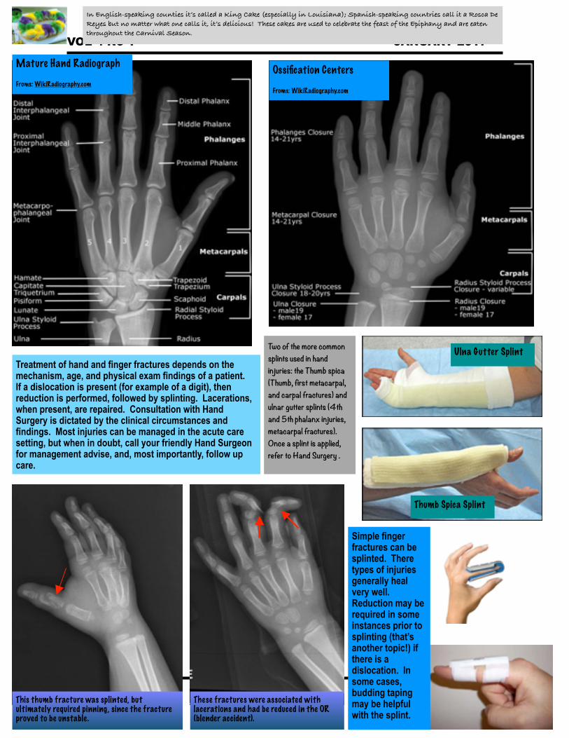
Remember hand and finger anatomy? I don’t. But lucky for us there are lots of references that can help refresh our memories of 1 year Gross Anatomy (those of us who want to remember, anyway). To the right is a nice reminder of the bones and the joint spaces of the hand and fingers. This a mature hand, remember that in pediatrics the bones develop in a predictable pattern that is demonstrated on radiographs, so consider the age of your patient when reading hand/finger films!
Just to be complete, here is a refresher on the insertion points of the various muscles that move the digits. It is a good idea to have a rough idea of what inserts where, because as you perform a physical examination you can get an idea of there is muscle injury or not with a particular injury. If a fracture is detected, then this will help you determine stability as well.
Open fractures - this fracture has an overlying open wound. Open fractures should be carefully explored and cleaned/irrigated, making sure there are no foreign bodies present. Antibiotic prophylaxis is given (often first generation Cephalosporins, but this depends on how contaminated a wound might be and the circumstances of the contamination). Some open fractures can be treated in the ED and followed up (phalangeal fractures) but most require exploration by Hand Surgery, either in the ED or in the operating room.
Displaced fractures - if clinically significant, require reduction before splinting. Sometimes a fracture will require operative repair at a later date; an attempt is made to reduce the fracture as such as possible prior to repair.
Non displaced fractures - can generally be managed with splinting.
Unstable fractures - if a fracture is unstable, then splitting is appropriate with urgent Hand Surgery followup for definitive operative repair.
January 5th or 6th, depending on where you live, marks 12th night. This is the beginning of the Carnival Season in New Orleans, leading up to my personal favorite, Mardi Gras!
The ABC’s of Reading Hand/Finger Films Alignment Bone (mineralization, joint spaces, fractures, lesions) Cartilage (older patients) And…..Soft tissues (edema, foreign bodies) As always, make sure it is the right patient!
Be sure to test each finger individually, allowing the patient to flex and extend the digits. This assess tendon integrity and can pick up a tendon rupture. Additionally, a good neurologic exam is warranted. The diagram above shows a nice way to remember the nerve innervation of both sides of the hand.
VOL 4 No 1 JANUARY 2017
Page �4
Mature Hand Radiograph Froms: WikiRadiography.com
Ossification Centers Froms: WikiRadiography.com
Treatment of hand and finger fractures depends on the mechanism, age, and physical exam findings of a patient. If a dislocation is present (for example of a digit), then reduction is performed, followed by splinting. Lacerations, when present, are repaired. Consultation with Hand Surgery is dictated by the clinical circumstances and findings. Most injuries can be managed in the acute care setting, but when in doubt, call your friendly Hand Surgeon for management advise, and, most importantly, follow up care.
This thumb fracture was splinted, but ultimately required pinning, since the fracture proved to be unstable.
These fractures were associated with lacerations and had be reduced in the OR (blender accident).
Two of the more common splints used in hand injuries: the Thumb spica (Thumb, first metacarpal, and carpal fractures) and ulnar gutter splints (4th and 5th phalanx injuries, metacarpal fractures). Once a splint is applied, refer to Hand Surgery .
Thumb Spica Splint
Ulna Gutter Splint
Simple finger fractures can be splinted. There types of injuries generally heal very well. Reduction may be required in some instances prior to splinting (that’s another topic!) if there is a dislocation. In some cases, budding taping may be helpful with the splint.
In English-speaking counties it’s called a King Cake (especially in Louisiana); Spanish-speaking countries call it a Rosca De Reyes but no matter what one calls it, it’s delicious! These cakes are used to celebrate the feast of the Epiphany and are eaten throughout the Carnival Season.
VOL 4 No 1 JANUARY 2017
Page �5
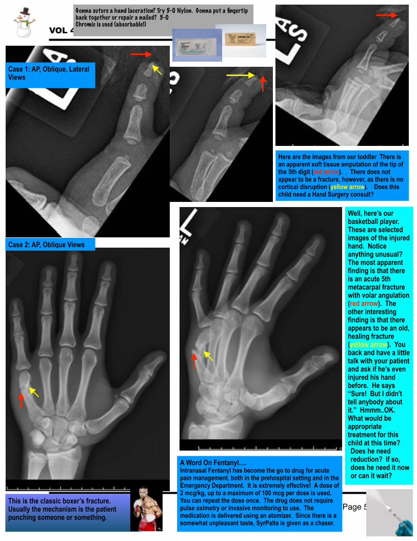
Case 1: AP, Oblique, Lateral Views
Here are the images from our toddler There is an apparent soft tissue amputation of the tip of the 5th digit (red arrow). . There does not appear to be a fracture, however, as there is no cortical disruption (yellow arrow). Does this child need a Hand Surgery consult?
Well, here’s our basketball player. These are selected images of the injured hand. Notice anything unusual? The most apparent finding is that there is an acute 5th metacarpal fracture with volar angulation (red arrow). The other interesting finding is that there appears to be an old, healing fracture (yellow arrow). You back and have a little talk with your patient and ask if he’s even injured his hand before. He says “Sure! But I didn't tell anybody about it.” Hmmm..OK. What would be appropriate treatment for this child at this time? Does he need reduction? If so, does he need it now or can it wait?
A Word On Fentanyl…. Intranasal Fentanyl has become the go to drug for acute pain management, both in the prehospital setting and in the Emergency Department. It is extremely effective! A dose of 2 mcg/kg, up to a maximum of 100 mcg per dose is used. You can repeat the dose once. The drug does not require pulse oximetry or invasive monitoring to use. The medication is delivered using an atomizer. Since there is a somewhat unpleasant taste, SyrPalta is given as a chaser.
Case 2: AP, Oblique Views
This is the classic boxer’s fracture. Usually the mechanism is the patient punching someone or something.
Gonna suture a hand laceration? Try 5-0 Nylon. Gonna put a fingertip back together or repair a nailed? 5-0 Chromic is used (absorbable!)

VOL 4 No 1 JANUARY 2017
Page �6
Case 3: Lateral, Oblique, and AP views
These images demonstrate an acute transverse middle to moderately displaced fracture of the tip of the digit (red arrow). This is consistent with a Tuft fracture. The interesting aspect with this child, though, is that there is an associated laceration with the fracture. This makes this an open fracture and calls for careful cleaning, irrigation, and exploration, in addition to antibiotics.
Nailbed Injuries - sometimes it is obvious and sometimes not. Tendons may displace the fracture fragment and the nailbed may be interrupted. It’s always a good idea to tell parents that the nail may not grow back normally, although a large percentage do. The bed is repaired and the finger splinted.
Antibiotics are indicated in open fractures and contaminated wounds. Keflex is used for simple open fractures that are not contaminated.
Disruption of the extensor mechanism of the finger at the DIP and may involve the bone (as in this case) or just the tendon. The resulting injury is called a Mallet finger. Treatment is usually done by splinting. If treatment is not initiated then a swan-neck deformity may result. The X-ray to the left demonstrates an avulsion fracture and resulting Mallet finger (red arrow). A Boutonniere deformity results in flexion at the PIP (red arrow) and hyperextension at the DIP (yellow arrow). Treatment of this injury is also generally conservative, with splinting. Sometimes operative repair may be needed.
Mallet Deformity: Avulsion fracture at the Distal Interphalangeal Joint (DIP)
“What started out roughly 300 years ago as a dry French bread–type dough with sugar on top and a bean inside now comes in many varieties depending on the country. Some king cakes are made of a sweet brioche dough in the shape of a hollow circle with a glazed topping sprinkled with colored sugar. Hundreds of thousands of King Cakes are eaten in New Orleans during the Carnival season. In other countries, king cakes are made with a puff pastry, filled with one of several fillings (e.g., almond, apple, chocolate/pear, etc.), and have a small figurine hidden inside. The figurine changes from bakery to bakery and often represents a hit movie or other cultural icon.” FROM: Wikipedia.
VOL 4 No 1 JANUARY 2017
Page �7
Teaching Points1. Hand and finger injuries require a thorough history and physical exam. Imaging is often indicated. Common views include the AP,
Lateral, and Oblique views. Which image view are obtained depends on history and physical exam.2. Open wounds should be thoroughly cleaned, irrgated, and explored. Lacerations are typically closed with 5-0 Nylon (hand and
finger lacerations) or 5-0 Chromic (naibed injuries and amputation injuries). Open fractures and grossly contaminated wounds are also treated with oral antibiotics.
3. Fingertip injuries often involve the nailbed. The usual mechanism is a crush injury (door slam, for example).4. Tendon injuries often accompany both blunt trauma and fingertip amputations. A good tendon examination is important, as is a
neurologic examination. Don’t forget IN Fentanyl as an initial analgesia step!5. Don’t forget Tetanus status; not everyone is fully immunized.6. When in doubt, refer to the Pediatric Emergency Department or consult Hand Surgery for treatment recommendations.
REFERENCES 1. Lee DH, Magnesia ME, Crosby SN. Fingertip injuries: an update on management. J Am Acad Orthop Surg. 2013; 21:756. 2. Altergott C, Garcia FJ, Nager AL. Pediatric fingertip injuries: do prophylactic antibiotics alter infection rates? Pediatr Emerg Care. 2008; 24:148. 3. Rubin G, Orbach H, Rinott, et al. The use of prophylactic antibiotics in treatment of fingertip amputation: a randomized prospective trial. Am J Emerg Med. 2015; 33:645. 4. Fetter-Zarzeka A, Joseph MM. Hand and fingertip injuries in children. Pediatr Emerg Care. 2002; 18:341. 5. Gillian H. Fingertip-nail bed injuries in children: current concepts and controversies of treatment. J Craniofac Surg. 2009; 20:1033. 6. Okie K, Bhattacharyya T. Trend in the management of open fractures: a critical analysis. J Bone Joint Surg Am. 2006; 88:2739. 7. Leggit JC, Meko CJ. Acute finger injuries: part II. Fractuyres, dislocations, and thumb injuries. Am Fam Physician. 2006; 73:827. 8. Boyd AS, Benjamin HJ and Ashland C. Splints and casts: Indications and methods. Am Fam Physician. 2009; 80:491. 9. Oetgen ME, Dodds SD. Non-operative treatment of common finger injuries. Curr Rev Musculoskelet Med. 2008; 1:97. 10. Henry MH. Fractures of the proximal phalanx and metacarpals in the hand: preferred methods of stabilization. J Am Acad Orthop Surg. 2008; 16:586.
Case Resolutions Case 1: The Hand Surgery Service at DCMC was consulted and felt that repair was appropriate in the ED by the ED physician. Under sedation with Ketamine, the fingertip near amputation was repaired, using Chromic 5-0 sutures. During the repair, the wound was completely explored and the nailed was found not to be involved. A dressing and splint was placed and the child was followed with Hand Surgery. After the edema subsided, the repair was viable and she did well.
Case 2: The Hand Surgery Service was also consulted and felt that, while operative repair may be needed in the future, it was appropriate to place an ulnar guttar splint and then allow any edema to subside. This was done and the patient was followed up in the Hand Clinic. There, a cast was placed and, once the edema subsided, plans were made to continue to follow the child. As of this writing, he did not require operative repair.
Case 3: This child had his repair with ketamine sedation as well. Because of the presence of the fracture and a laceration, he was treated as an open fracture, splinted, and sent home on Keflex with Hand Clinic followup. The wound healed well.
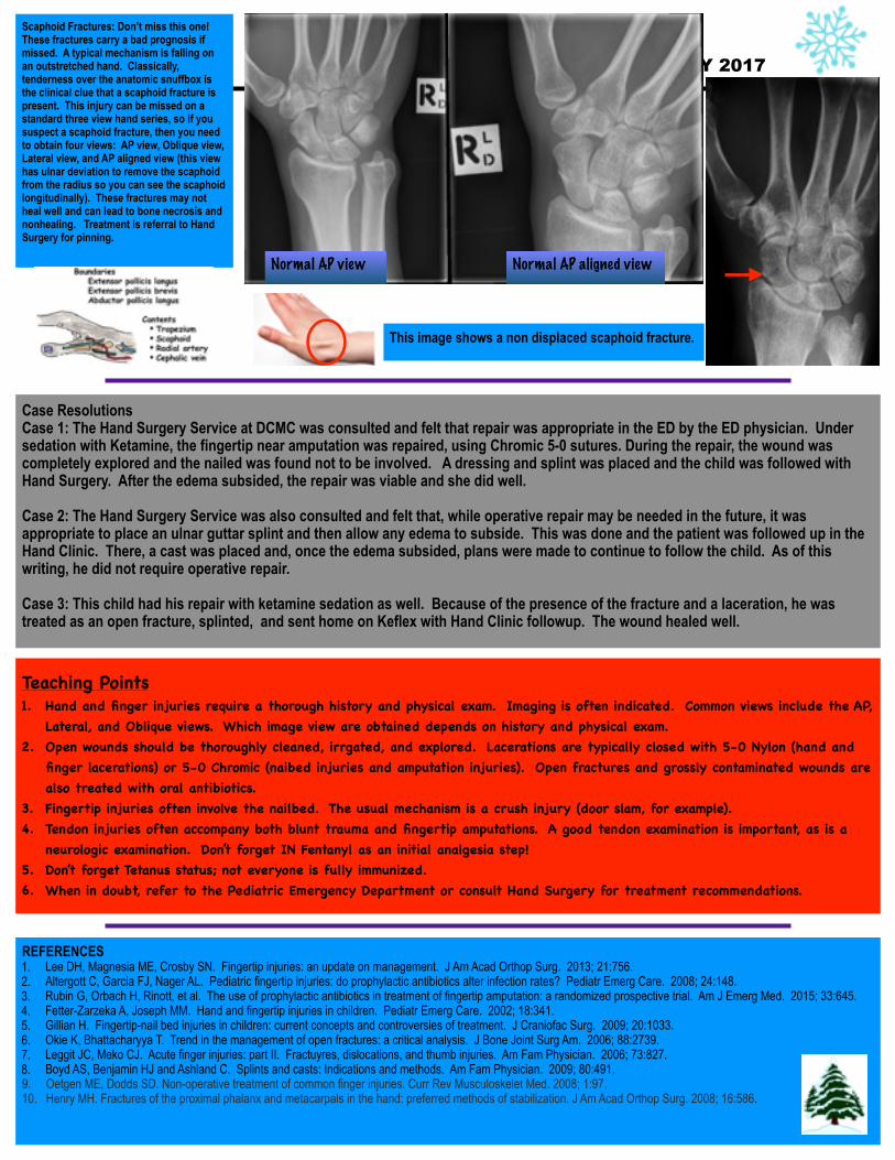
Scaphoid Fractures: Don’t miss this one! These fractures carry a bad prognosis if missed. A typical mechanism is falling on an outstretched hand. Classically, tenderness over the anatomic snuffbox is the clinical clue that a scaphoid fracture is present. This injury can be missed on a standard three view hand series, so if you suspect a scaphoid fracture, then you need to obtain four views: AP view, Oblique view, Lateral view, and AP aligned view (this view has ulnar deviation to remove the scaphoid from the radius so you can see the scaphoid longitudinally). These fractures may not heal well and can lead to bone necrosis and nonhealing. Treatment is referral to Hand Surgery for pinning.
Normal AP view Normal AP aligned view
This image shows a non displaced scaphoid fracture.