Jur Kista Radi,Ular
6
Contemp Clin Dent. 2011 Jan-Mar; 2(1): 59–62. doi: 10.410309!6-23!".!9295 #MC$D: #MC32201!9 An unusual case report of bilateral mandibular radicular cysts %i&anta '. Joi* '. +. ',an* and M. M. a/appa ,tor inormation Cop&ri t and i/en e inorma tion i arti/le a een /ited & oter arti/le in #MC. Abstract +o to: Introduction Radicular cysts are the most common inflammatory cysts arising from the epithelial residues in the periodont al ligament as a result of periapical periodon titis following necrosis of the pulp, remains asymptomatic and left unnoticed until detected during routine periapical radiography. These cysts comprise about 52% to 68% of all the cysts affecting the human jaw. !" Their incidence is highest in third and fourth decade of life with male predominance. 2" #natomically the periapical cysts occur in all tooth$bearing sites of the jaw but are more freuent in the ma&illary than the mandibular region. !,'" Radicular cysts can heal spontaneousl y after endodontic treatment or e&traction. (owe)er, some authors propose that suspected radicular cysts must be totally enucleated surgically to remo)e all epithelial remnants. *" +o to: Case Report # !'$year$old girl repoted to the +ept. of edodontics and re)enti)e +entistry with a complaint of dull pain and swelling in the right posterior mandibular region since past !5 days. The patient had pain in the same region ! year before and had ta-en medication for that. he had underg one e&traction of the carious mandibular right first molar ' days before. /&traoral e&aminatio n re)ealed a s mooth superfacial swelli ng of about ' 0 2.5 cm e&tending from the corner of mouth to the angle of mandible and from the infraorbital margin to the lower border of mandible 1igure !". The swelling was tender and firmwith egg shell crac-ling on palpation. The inferior border of mandible was intact. The right submandibular lymph nodes were palpable and tender. ntraorally, the e&traction soc-et of
-
Upload
ricco-ardes -
Category
Documents
-
view
227 -
download
0
Transcript of Jur Kista Radi,Ular
7/24/2019 Jur Kista Radi,Ular
http://slidepdf.com/reader/full/jur-kista-radiular 1/6
Contemp Clin Dent. 2011 Jan-Mar; 2(1): 59–62.
doi: 10.410309!6-23!".!9295
#MC$D: #MC32201!9
An unusual case report of bilateral mandibular radicular cysts
%i&anta '. Joi* '. +. ',an* and M. M. a/appa
,tor inormation Cop&rit and i/ene inormation
i arti/le a een /ited & oter arti/le in #MC.
Abstract
+o to:
Introduction
Radicular cysts are the most common inflammatory cysts arising from the epithelial
residues in the periodontal ligament as a result of periapical periodontitis following
necrosis of the pulp, remains asymptomatic and left unnoticed until detected during
routine periapical radiography.
These cysts comprise about 52% to 68% of all the cysts affecting the human jaw. !" Their
incidence is highest in third and fourth decade of life with male predominance.2"
#natomically the periapical cysts occur in all tooth$bearing sites of the jaw but are more
freuent in the ma&illary than the mandibular region.!,'"
Radicular cysts can heal spontaneously after endodontic treatment or e&traction.
(owe)er, some authors propose that suspected radicular cysts must be totally enucleated
surgically to remo)e all epithelial remnants.*"
+o to:
Case Report
# !'$year$old girl repoted to the +ept. of edodontics and re)enti)e +entistry with a
complaint of dull pain and swelling in the right posterior mandibular region since past !5
days. The patient had pain in the same region ! year before and had ta-en medication for
that. he had undergone e&traction of the carious mandibular right first molar ' days
before.
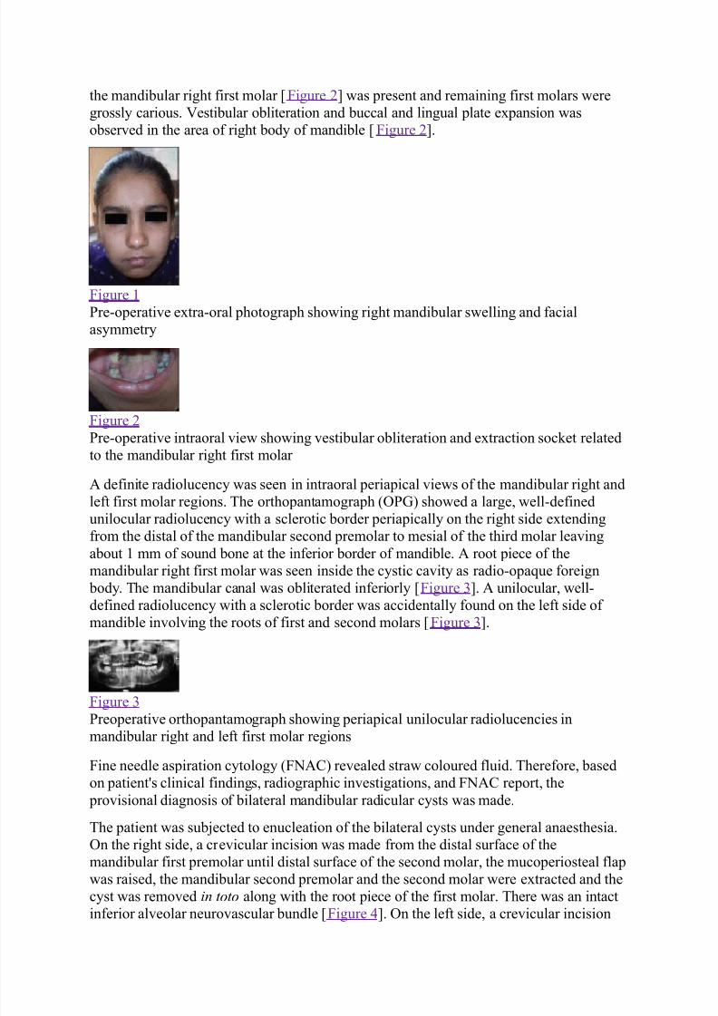
/&traoral e&amination re)ealed a smooth superfacial swelling of about ' 0 2.5 cm
e&tending from the corner of mouth to the angle of mandible and from the infraorbital
margin to the lower border of mandible 1igure !". The swelling was tender and firmwith
egg shell crac-ling on palpation. The inferior border of mandible was intact. The rightsubmandibular lymph nodes were palpable and tender. ntraorally, the e&traction soc-et of
7/24/2019 Jur Kista Radi,Ular
http://slidepdf.com/reader/full/jur-kista-radiular 2/6
the mandibular right first molar 1igure 2" was present and remaining first molars were
grossly carious. 3estibular obliteration and buccal and lingual plate e&pansion was
obser)ed in the area of right body of mandible 1igure 2".
1igure !
re$operati)e e&tra$oral photograph showing right mandibular swelling and facial
asymmetry
1igure 2
re$operati)e intraoral )iew showing )estibular obliteration and e&traction soc-et related
to the mandibular right first molar
# definite radiolucency was seen in intraoral periapical )iews of the mandibular right and
left first molar regions. The orthopantamograph 47 showed a large, well$defined
unilocular radiolucency with a sclerotic border periapically on the right side e&tending
from the distal of the mandibular second premolar to mesial of the third molar lea)ingabout ! mm of sound bone at the inferior border of mandible. # root piece of the
mandibular right first molar was seen inside the cystic ca)ity as radio$opaue foreign
body. The mandibular canal was obliterated inferiorly 1igure '". # unilocular, well$
defined radiolucency with a sclerotic border was accidentally found on the left side of
mandible in)ol)ing the roots of first and second molars 1igure '".
1igure '
reoperati)e orthopantamograph showing periapical unilocular radiolucencies in
mandibular right and left first molar regions
1ine needle aspiration cytology 41#97 re)ealed straw coloured fluid. Therefore, based
on patient:s clinical findings, radiographic in)estigations, and 1#9 report, the
pro)isional diagnosis of bilateral mandibular radicular cysts was made.
The patient was subjected to enucleation of the bilateral cysts under general anaesthesia.
n the right side, a cre)icular incision was made from the distal surface of the
mandibular first premolar until distal surface of the second molar, the mucoperiosteal flap
was raised, the mandibular second premolar and the second molar were e&tracted and the
cyst was remo)ed in toto along with the root piece of the first molar. There was an intact
inferior al)eolar neuro)ascular bundle 1igure *". n the left side, a cre)icular incision
7/24/2019 Jur Kista Radi,Ular
http://slidepdf.com/reader/full/jur-kista-radiular 3/6
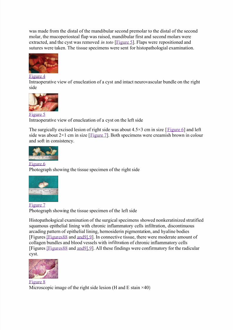
was made from the distal of the mandibular second premolar to the distal of the second
molar, the mucoperiosteal flap was raised, mandibular first and second molars were
e&tracted, and the cyst was remo)ed in toto 1igure 5". 1laps were repositioned and
sutures were ta-en. The tissue specimens were sent for histopathologial e&amination.
1igure *
ntraoperati)e )iew of enucleation of a cyst and intact neuro)ascular bundle on the right
side
1igure 5ntraoperati)e )iew of enucleation of a cyst on the left side
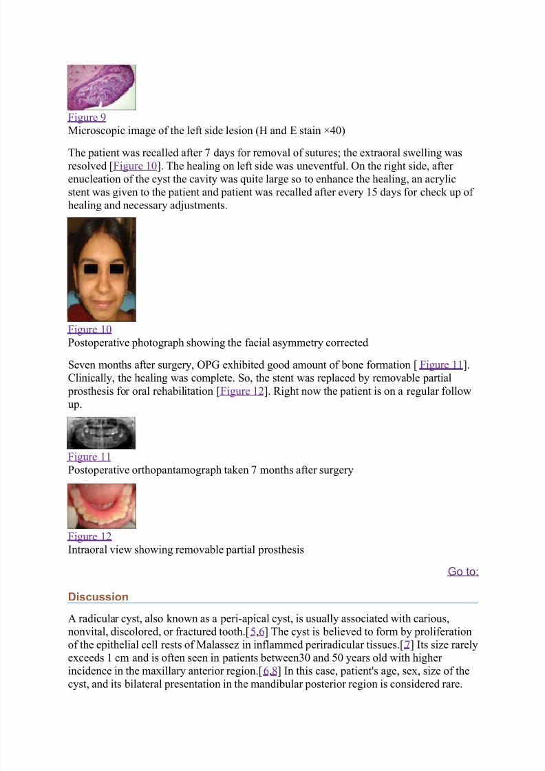
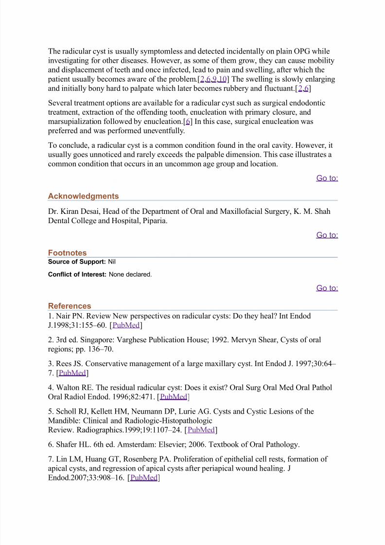
The surgically e&cised lesion of right side was about *.50' cm in si;e 1igure 6" and left
side was about 20! cm in si;e 1igure <". =oth specimens were creamish brown in colour
and soft in consistency.
1igure 6
hotograph showing the tissue specimen of the right side
1igure <
hotograph showing the tissue specimen of the left side
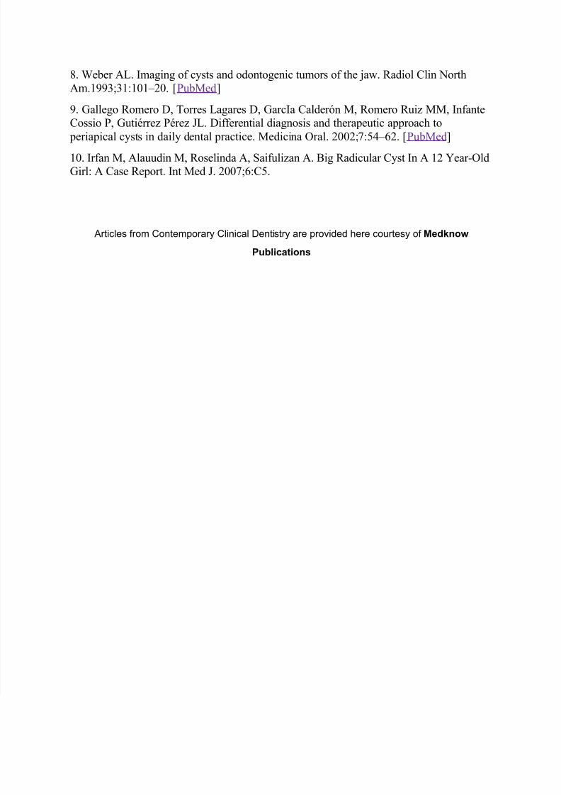
(istopathological e&amination of the surgical specimens showed non-eratini;ed stratified
suamous epithelial lining with chronic inflammatory cells infiltration, discontinuous
arcading pattern of epithelial lining, hemosiderin pigmentation, and hyaline bodies1igures 1igures8 8 and and>". >". n connecti)e tissue, there were moderate amount of
collagen bundles and blood )essels with infiltration of chronic inflammatory cells
1igures 1igures8 8 and and>". >". #ll these findings were confirmatory for the radicular
cyst.
1igure 8
?icroscopic image of the right side lesion 4( and / stain 0*@7
7/24/2019 Jur Kista Radi,Ular
http://slidepdf.com/reader/full/jur-kista-radiular 4/6
1igure >
?icroscopic image of the left side lesion 4( and / stain 0*@7
The patient was recalled after < days for remo)al of suturesA the e&traoral swelling was
resol)ed 1igure !@". The healing on left side was une)entful. n the right side, after
enucleation of the cyst the ca)ity was uite large so to enhance the healing, an acrylic
stent was gi)en to the patient and patient was recalled after e)ery !5 days for chec- up of
healing and necessary adjustments.
1igure !@
ostoperati)e photograph showing the facial asymmetry corrected
e)en months after surgery, e&hibited good amount of bone formation 1igure !!".
9linically, the healing was complete. o, the stent was replaced by remo)able partial
prosthesis for oral rehabilitation 1igure !2". Right now the patient is on a regular follow
up.
1igure !!
ostoperati)e orthopantamograph ta-en < months after surgery
1igure !2ntraoral )iew showing remo)able partial prosthesis
+o to:
Discussion
# radicular cyst, also -nown as a peri$apical cyst, is usually associated with carious,
non)ital, discolored, or fractured tooth.5,6" The cyst is belie)ed to form by proliferation
of the epithelial cell rests of ?alasse; in inflammed periradicular tissues.<" ts si;e rarely
e&ceeds ! cm and is often seen in patients between'@ and 5@ years old with higher
incidence in the ma&illary anterior region.6,8" n this case, patient:s age, se&, si;e of thecyst, and its bilateral presentation in the mandibular posterior region is considered rare.
7/24/2019 Jur Kista Radi,Ular
http://slidepdf.com/reader/full/jur-kista-radiular 5/6
The radicular cyst is usually symptomless and detected incidentally on plain while
in)estigating for other diseases. (owe)er, as some of them grow, they can cause mobility
and displacement of teeth and once infected, lead to pain and swelling, after which the
patient usually becomes aware of the problem.2,6,>,!@" The swelling is slowly enlarging
and initially bony hard to palpate which later becomes rubbery and fluctuant.2,6"
e)eral treatment options are a)ailable for a radicular cyst such as surgical endodontic
treatment, e&traction of the offending tooth, enucleation with primary closure, and
marsupiali;ation followed by enucleation.6" n this case, surgical enucleation was
preferred and was performed une)entfully.
To conclude, a radicular cyst is a common condition found in the oral ca)ity. (owe)er, it
usually goes unnoticed and rarely e&ceeds the palpable dimension. This case illustrates a
common condition that occurs in an uncommon age group and location.
+o to:
Acknowledgments
+r. Biran +esai, (ead of the +epartment of ral and ?a&illofacial urgery, B. ?. hah
+ental 9ollege and (ospital, iparia.
+o to:
FootnotesSource of Support: %il
Conflict of Interest: %one de/lared.
+o to:
References
!. air . Re)iew ew perspecti)es on radicular cystsC +o they healD nt /ndod
E.!>>8A'!C!55F6@. ub?ed"
2. 'rd ed. ingaporeC 3arghese ublication (ouseA !>>2. ?er)yn hear, 9ysts of oral
regionsA pp. !'6F<@.
'. Rees E. 9onser)ati)e management of a large ma&illary cyst. nt /ndod E. !>><A'@C6*F
<. ub?ed"
*. Galton R/. The residual radicular cystC +oes it e&istD ral urg ral ?ed ral athol
ral Radiol /ndod. !>>6A82C*<!. ub?ed"
5. choll RE, Bellett (?, eumann +, Hurie #. 9ysts and 9ystic Hesions of the
?andibleC 9linical and Radiologic$(istopathologic
Re)iew. Radiographics.!>>>A!>C!!@<F2*. ub?ed"
6. hafer (H. 6th ed. #msterdamC /lse)ierA 2@@6. Te&tboo- of ral athology.
<. Hin H?, (uang T, Rosenberg #. roliferation of epithelial cell rests, formation of
apical cysts, and regression of apical cysts after periapical wound healing. E
/ndod.2@@<A''C>@8F!6. ub?ed"
7/24/2019 Jur Kista Radi,Ular
http://slidepdf.com/reader/full/jur-kista-radiular 6/6
8. Geber #H. maging of cysts and odontogenic tumors of the jaw. Radiol 9lin orth
#m.!>>'A'!C!@!F2@. ub?ed"
>. allego Romero +, Torres Hagares +, arca 9alderIn ?, Romero Rui; ??, nfante
9ossio , utiJrre; Jre; EH. +ifferential diagnosis and therapeutic approach to
periapical cysts in daily dental practice. ?edicina ral. 2@@2A<C5*F62. ub?ed"
!@. rfan ?, #lauudin ?, Roselinda #, aifuli;an #. =ig Radicular 9yst n # !2 Kear$ld
irlC # 9ase Report. nt ?ed E. 2@@<A6C95.
rti/le rom Contemporar& Clini/al Dentitr& are pro7ided ere /o,rte& o Medknow
Publications