UNIVERSITI PUTRA MALAYSIA OPTICAL PROPERTIES AND … · Sampel cecair seperti klorin, sakarida, air...
25
UNIVERSITI PUTRA MALAYSIA OPTICAL PROPERTIES AND KINETIC BEHAVIOUR OF SOME CHEMICAL AND BIOLOGICAL SPECIES USING SURFACE PLASMON RESONANCE OPTICAL SENSOR WAN YUSMA WATI BINTI WAN YUSOFF FS 2005 42
Transcript of UNIVERSITI PUTRA MALAYSIA OPTICAL PROPERTIES AND … · Sampel cecair seperti klorin, sakarida, air...
UNIVERSITI PUTRA MALAYSIA
OPTICAL PROPERTIES AND KINETIC BEHAVIOUR OF SOME CHEMICAL AND BIOLOGICAL SPECIES USING SURFACE PLASMON
RESONANCE OPTICAL SENSOR
WAN YUSMA WATI BINTI WAN YUSOFF
FS 2005 42
OPTICAL PROPERTIES AND KINETIC BEHAVIOUR OF SOME CHEMICAL AND BIOLOGICAL SPECIES USING SURFACE PLASMON
RESONANCE OPTICAL SENSOR
WAN YUSMA WATI BINTI WAN YUSOFF
MASTER OF SCIENCE UNIVERSITI PUTRA MALAYSIA
2005
OPTICAL PROPERTIES AND KINETIC BERA VIOUR OF SOME CHEMICAL AND BIOLOGICAL SPECIES USING SURFACE PLASMON
RESONANCE OPTICAL SENSOR
By
WAN YUSMA WATI BINTI WAN YUSOFF
Thesis Submitted to the School of Graduate Studies, Universiti Putra Malaysia, in Fulfilment of the Requirements for the Degree of Master of Science
July 2005
Abstract of thesis presented to the Senate of Universiti Putra Malaysia in fulfilment of the requirements for the degree of Master of Science
OPTICAL PROPERTIES AND KINETIC BEHAVIOUR OF SOME CHEMICAL AND BIOLOGICAL SPECIES USING SURFACE PLASMON
RESONANCE OPTICAL SENSOR
Chairman
Faculty
By
WANYUSMAWATI WAN YUSOFF
July 2005
: Professor W. Mahmood Mat Yuuus, PhD
: Science
Surface plasmon resonance (SPR) spectroscopy is a surface-sensitive technique that
has been used to characterize the thickness and index of refraction of dielectric
medium at noble metal interface. Nowadays surface plasmon resonance technique has
emerged as a powerful technique for a variety of chemical and biological sensor
applications.
In this study, gold and silver with purity of 99.99% were used to fabricate thin metal
films. The thin film was deposited onto a glass cover slip and attached onto the
surface of a 60° prism using index matching oil . Liquid samples, such as chlorine,
saccharide, swimming pool water, pesticide, virus and DNA were studied using
Kretschmann Surface Plasmon Resonance technique. All the measurements were
carried out at room temperature. The experiment was carried out by measuring the
intensity of the optical reflectivity as a function of incident angle.
ii
It found that the shift of resonance angle (.dB) increased linearly with the sample
concentration. The detection limit of the sensor was estimated better than 0.0 1 pM for
the sample of DNA (Olig02-Bio). Larger sensor sensitivity of 9.42°/(mollL) is
obtained for the sucrose sample.
The kinetic behaviour of the system was also examined to monitor the self
assembling process on the metal surface in real time. The shift in resonance angle
increased greatly with time during the increment of the molecules deposited on the
gold surface. In contrast it was found decrease with time during self-assembling
process.
This work also studied the molecule-dielectric interaction for a thin Fatty
Hydroxamic Acid (FHA) film (extract from crude palm oil), which the FHA layer
was coated using spin coating on the top of metal film. When the medium outside the
surface of Au film was changed from air to FHA layer, the resonance angle shifted to
the higher value. The shift of resonance angle increased linearly with the increasing
concentration FHA layer. When the metal ion was attached to the FHA film, the
resonance angle was changed to the maximum value.
The experimental results reveal that the technique that based on surface plasmon
resonance phenomenon can be used to determine the optical properties and the kinetic
behaviour. It also suitable to study the molecule-dielectric interaction for the polymer
film. This technique can become an effective chemical optical sensor. Saccharide,
pesticide and chlorine concentration in water can be detected using this sensor.
Furthermore it also can be used to detect DNA and viruses solution.

Abstrak tesis yang dikemukakan kepada Senat Universiti Putra Malaysia sebagai memenuhi keperluan untuk ijazah Master Sains
SIFAT OPTIK DAN PERLAKUAN KINETIK TERHADAP BEBERAPA SPESIS KIMIA DAN BIOLOGI MENGGUNAKAN SENSOR OPTIK
RESONANS PLASMON PERMUKAAN
Oleh
WAN YUSMA WATI BINTI WAN YUSOFF
Julai 2005
Pengerusi : Profesor W. Mahmood Mat Yunus, PhD
Fakulti : Sains
iii
Spektroskopi Resonans Plasmon Permukaan (SPR) ialah satu teknik sensitif-
permukaan yang digunakan untuk mengenalpasti ketebalan dan indeks biasan
medium dielektrik pada antaramuka logam. Kini, teknik resonans plasmon
permukaan telah muncul sebagai teknik yang berguna dalam pelbagai penggunaan
pengesan kimia dan biologi.
Dalam kaj ian ini, emas dan perak dengan ketulenan 99.99% digunakan untuk
membuat filem tipis logam. Fi lem tipis logam tersebut telah disaputkan kepada slip
kaca dan dilekatkan kepada satu permukaan prisma 60° dengan menggunakan
minyak indeks sepadan. Sampel cecair seperti klorin, sakarida, air kolam renang,
pestisid, virus dan DNA dikaji dengan menggunakan teknik Plasmon Resonans
Permukaan Kretschmann. Semua pengukuran telah di lakukan pada suhu bi l ik.
Eksperimen telah dilakukan dengan mengukur keamatan keterpantulan optik sebagai
satu fungsi kepada sudut tuju.
iv
Keputusan menunjukkan anjakan sudut resonans (..dO) meningkat secara linear dengan
kepekatan larutan sam pel . Had pengesanan bagi pengesan dianggarkan lebih baik
daripada 0.01 pM untuk sampel DNA (Oligo2-Bio) dan kepekaan pengesan tertinggi
ialah 9.42°/(mollL) untuk sampel sukrosa.
Perlakuan kinetik sistem juga telah diperiksa untuk memerhati proses berkumpul
sendiri pada permukaan logam dalam masa nyata. Anjakan sudut resonans bertambah
secara mendadak dengan masa ketika peningkatan endapan molekul pada permukaan
emas. Anjakan ini didapati menurun dengan masa ketika proses perhimpunan
sendiri.
Kajian ini juga mengkaji interaksi antara molekul-dielektrik untuk filem tipis Fatty
Hydroxamic Acid, FHA (ekstrak dari minyak kelapa sawit mentah), dengan lapisan
FHA telah disaput menggunakan 'spin coating' atas filem logam. Apabila medium
perrnukaan luar filern tipis logarn ditukarkan daripada udara kepada filern polirner
FHA, sudut resonans berganjak kepada nilai yang lebih tinggi. Anjakan sudut
resonans telah rneningkat secara linear dengan kepekatan lapisan FHA. Apabila ion
logarn dilekatkan pada lapisan FHA, sudut resonans telah berubah kepada nilai
rnaksimurn.
Keputusan eksperirnen menunjukkan teknik yang berdasarkan fenomena resonans
plasmon permukaan boleh digunakan untuk menentukan sifat-sifat optik dan
kelakuan kinetik. Ia juga sesuai untuk mengkaj i interaksi molekul-dielektrik bagi
filem pol imer. Teknik ini boleh menjadi pengesan pengesan optik kimia yang
berkesan. Sakarida, pestisid dan kepekatan klorin dalam air boleh dikesan
menggunakan teknik ini. Malahan pengesan ini boleh Juga digunakan untuk
mengesan DNA dan virus dalam larutan.

VI
ACKNOWLEDGEMENTS
In the name of Allah, Most Gracious, Most Merciful ...
Alhamdulillah, I gratefully thank God, Almighty Allah S.W.T., for giving me health
and strength for successfully completing my project and writing this thesis. I also
extend greet to Prophet, Muhammad S.A.W.
I wish to express my sincere gratitude and thanks to my research supervisor, Prof. Dr.
W. Mahmood Mat Yunus for his kind advice, proper guidance and support
throughout the project. Without his interest and continuous support, this academic
research project will not be possible to finish.
Deep from my heart, I would like to convey special thank to my co-supervisors Prof.
Dr. Mohd Maarof Moksin and Assoc. Prof. Dr. Zainal Abidin Talib and all the staff
in Department of Physics, Faculty of Science, Universiti Putra Malaysia in assisting
and helping me in order to accomplish this research project successfully.
Last but no least, I would like to thank my ever supportive parents who never fail to
be there for their love, support and prayers and also I want to thanked for their
understanding and encouragement through the duration of this research project. Also,
I am really proud of all my friends for their never ending support.
Thank you very much.
May Allah Ta'ala bless you all.
VII
I certify that an Examination Committee met on 13th July 2005 to conduct the final examination of Wan Yusmawati Wan Yusoff on her Master of Science thesis entitled "Optical Properties and Kinetic Behaviour of Some Chemicals and Biological Species Using Surface Plasmon Resonance Optical Sensor" in accordance with Universiti Pertanian Malaysia (Higher Degree) Act 1980 and U niversiti Pertanian Malaysia (Higher Degree) Regulations 1981. The Committee recommends that the candidate be awarded the relevant degree. Members of the Examination Committee are as follows:
Zaidan Abd. Wahab, PhD Associate Professor Faculty of Science Universiti Putra Malaysia (Chairman)
Abdul Halim Shaari, PhD Professor Faculty of Science Universiti Putra Malaysia (Internal Examiner)
Azmi Zakaria, PhD Associate Professor Faculty of Science Universiti Putra Malaysia (Internal Examiner)
Noriah Bidin, PhD Associate Professor Faculty of Science Universiti Teknologi Malaysia (External Examiner)
HMAT ALI, PhD ProfessorlDepu Dean School of Graduate Studies Universiti Putra Malaysia
Date: 22 AUG 2005
viii
This thesis submitted to the Senate of Universiti Putra Malaysia has been accepted as fulfilment of the requirements for the degree of Master of Science. The members of the Supervisory Committee are as follows:
W. Mahmood Mat Yunus, PhD Professor Faculty of Science Universiti Putra Malaysia (Member)
Mohd. Maarof Moksin, PhD Professor Faculty of Science Universiti Putra Malaysia (Member)
Zainal Abidin Talib, PhD Associate Professor Faculty of Science Universiti Putra Malaysia (Member)
AINI ID ERIS, PhD ProfessorlDean School of Graduate Studies Universiti Putra Malaysia
Date: 08 SEP 2005
ix
DECLARATION
I hereby declare that the thesis is based on my original work except for quotations and citations which have been duly acknowledged. I also declare that it has not been previously or concurrently submitted for any degree at UPM or other institutions.
WAN YUSMAWATI WAN YUSOFF
Date: \ q aQ-h� :Jr;O�
TABLE OF CONTENTS
Page
ABSTRACT ABSTRAK iii ACKNOWLEDGEMENTS vi APPRO V AL vii
DECLARATION ix LIST OF TABLES xii LIST OF FIGURES xiv LIST OF ABBREVIATIONSINOTATION/GLOSSARY OF TERM xviii
CHAPTER 1
2
INTRODUCTION 1.1 Surface Plasmon Resonance
1 .1.1 The architecture of the measurement setup -why using a prism?
1 .1.2 How the surface plasmon excited? 1 .2 Sample Background
1 .2. 1 Saccharide 1 .2 .2 Chlorine 1 .2.3 Pesticide 1 .2.4 DNA Solution 1 .2.5 Virus 1 .2.6 Heavy Metal
1 .3 Benefit From The Surface Plasmon Resonance Study
1 .4 The Objective of the Study 1 .5 Chapter Organization
LITERATURE REVIEW 2 . 1 Review on Surface Plasmon Resonance 2 .2 Review on Metal Surface 2.3 Literature Review on the Dielectric Constant
1 .1
1 .4 1 .6 1 .7 1 .7 1 .8
l. 1 O 1 . 12 1 . 1 2 l. 1 3
1 . 1 4 l. 1 5 1 . 1 5
2. 1 2.2 2.3
3 THEORY 3. 1 Surface Plasmon Resonance 3 . 1
3. 1 . 1 Surface Electromagnetic Waves at Two Media Interface 3 .2
3 . 1 .2 Surface Plasmon Resonance Scattering 3.5 3 . 1 .3 Angle Dependence of the Reflectivity of
Surface Plasmon Resonance 3.8 3 . 1 .4 Surface Plasmon Resonance Coupling 3. 10
3 .2 Real Time Interaction Analysis 3 . 11
4 METHODOLOGY 4. 1 Sample Preparation
4. 1 . 1 Substrate Cleaning 4. 1 4. 1
x
4. 1 .2 Thin Film Preparation 4.2 4. 1 .3 Preparation of Dielectric Medium 4.3
4.2 Experimental Setup 4.7 4.2. 1 Modulated Beam Systems 4.8 4.2.2 Sample Cell 4.9 4.2.3 Data Acquisition 4.9
4.3 Experimental Procedure 4. 1 0 4.4 Fitting Experimental Data to the Theoretical Data 4. 1 1 4.5 X-Ray Diffraction Analysis 4. 1 1 4.6 Spin Coating Technique 4. 1 3
5 RESULTS AND DISCUSSION
6
5 . 1 Introduction 5 . 1 5 .2 Preliminary Study of Metal Surface 5 . 1
5 .2 . 1 X-Ray Diffraction Analysis of Metal Surface 5.2 5 .2.2 Environment Effect on the Metal Surface
Stability 5 .4 5 .3 Optical Properties Measurement of Dielectric
Medium 5 . 1 2 5 .3 . 1 Preliminary Experiment 5 . 1 2 5 .3.2 Saccharide 5 . 1 4 5 .3 .3 Chlorine 5 . 1 8 5 .3 .4 Swimming Pool 5 .20 5.3 .5 Pesticide 5.22 5.3 .6 DNA Solution 5 .25 5 .3 .7 Virus Solution 5.28
5 .4 Surface Plasmon Optical Sensor 5 .3 1 5 .4. 1 Saccharide 5 .3 1 5 .4.2 Chlorine 5 .34 5 .4.3 Pesticide 5.36 5 .4.4 DNA Solution 5 .38 5 .4.5 Virus Solution 5 .4 1
5 .5 Kinetic Behaviour 5.43 5 .5 . 1 Saccharide 5 .44 5 .5 .2 Chlorine 5 .47 5 .5 .3 Pesticide 5 .53 5 .5 .4 DNA Solution 5 .55
5 .6 Polymer Film Study 5 .58
CONCLUSION 6. 1 Conclusion 6.2 Suggestion
6. 1 6.4
REFERRENCES APPENDICES
R.l A.l B.l BIODATA OF THE AUTHOR
xi
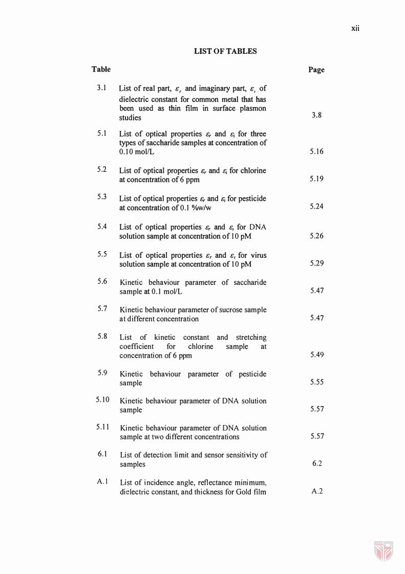
LIST OF TABLES
Table
3 . 1 List of real part, G, and imaginary part, G, of
dielectric constant for common metal that has been used as thin film in surface plasmon studies
5 . 1 List of optical properties Gr and £j for three types of saccharide samples at concentration of 0. 1 0 mollL
5 .2 List of optical properties Gr and £, for chlorine at concentration of 6 ppm
5 .3 List of optical properties Gr and G/ for pesticide at concentration of 0. 1 %w/w
5.4 List of optical properties Gr and G/ for DNA solution sample at concentration of 1 0 pM
5.5 List of optical properties G r and G / for virus solution sample at concentration of 1 0 pM
5 .6 Kinetic behaviour parameter of saccharide sample at 0. 1 mollL
5.7 Kinetic behaviour parameter of sucrose sample at different concentration
5 .8 List of kinetic constant and stretching coefficient for chlorine sample at concentration of 6 ppm
5.9 Kinetic behaviour parameter of pesticide sample
5 . 1 0 Kinetic behaviour parameter of DNA solution sample
5 . 1 1 Kinetic behaviour parameter of DNA solution sample at two different concentrations
6. 1 List of detection limit and sensor sensitivity of samples
A.I List of incidence angle, reflectance minimum, dielectric constant, and thickness for Gold film
xii
Page
3.8
5 . 1 6
5 . 1 9
5 .24
5 .26
5 .29
5 .47
5 .47
5 .49
5 .55
5 .57
5 .57
6 .2
A.2
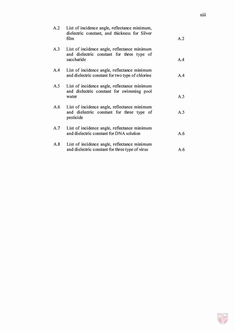
A.2 List of incidence angle, reflectance minimum, dielectric constant, and thickness for Silver film
A.3 List of incidence angle, reflectance minimum and dielectric constant for three type of saccharide
AA List of incidence angle, reflectance minimum and dielectric constant for two type of chlorine
A.5 List of incidence angle, reflectance minimum and dielectric constant for swimming pool water
A.6 List of incidence angle, reflectance minimum and dielectric constant for three type of pesticide
A.7 List of incidence angle, reflectance minimum and dielectric constant for DNA solution
A.8 List of incidence angle, reflectance minimum and dielectric constant for three type of virus
xiii
A.2
AA
AA
A.5
A.5
A.6
A.6
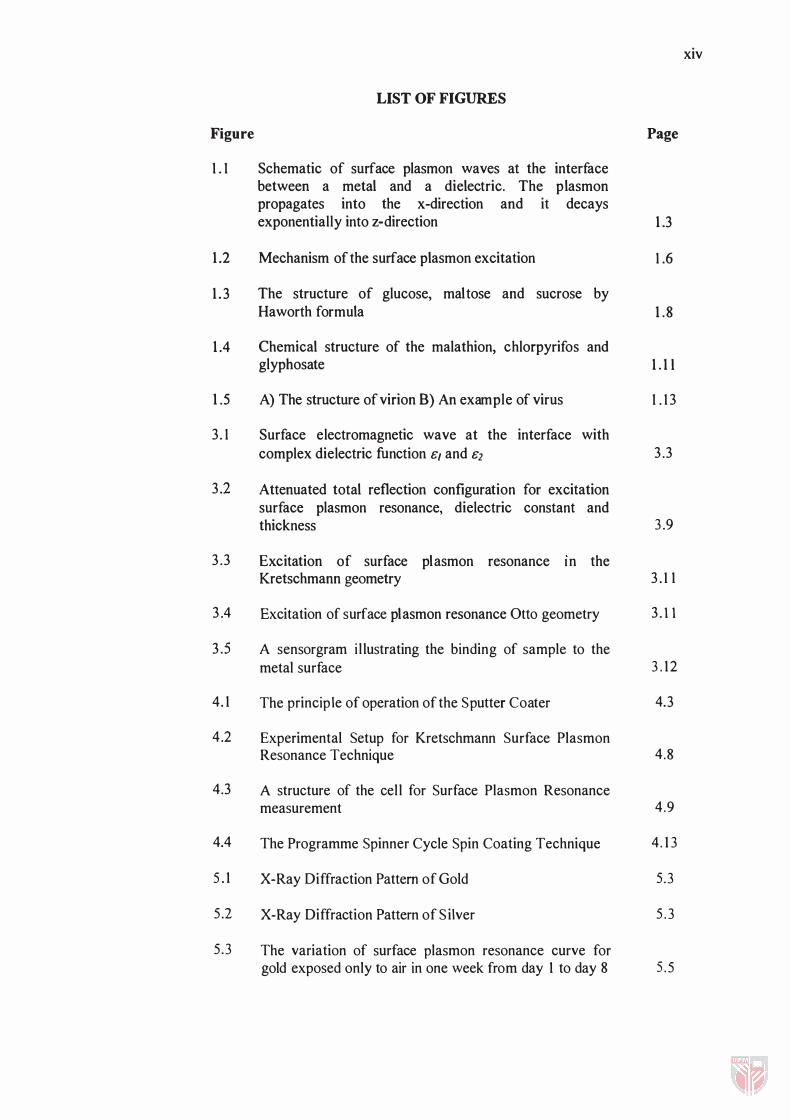
Figure
1 . 1
1.2
1.3
1.4
1.5
3. 1
3.2
LIST OF FIGURES
Schematic of surface plasmon waves at the interface between a metal and a dielectric. The plasmon propagates into the x-direction and it decays exponentially into z-direction
Mechanism of the surface plasmon excitation
The structure of glucose, maltose and sucrose by Haworth formula
Chemical structure of the malathion, chlorpyrifos and glyphosate
A) The structure of virion B) An example of virus
Surface electromagnetic wave at the interface with complex dielectric function 8[ and 82
Attenuated total reflection configuration for excitation surface plasmon resonance, dielectric constant and thickness
3.3 Excitation of surface plasmon resonance In the Kretschmann geometry
3.4
3.5
4. 1
4.2
Excitation of surface plasmon resonance Otto geometry
A sensorgram i l lustrating the binding of sample to the metal surface
The principle of operation of the Sputter Coater
Experimental Setup for Kretschmann Surface Plasmon Resonance Technique
4.3 A structure of the cel l for Surface Plasmon Resonance measurement
4.4
5 . 1
5.2
5 .3
The Programme Spinner Cycle Spin Coating Technique
X-Ray Diffraction Pattern of Gold
X-Ray Diffraction Pattern of S ilver
The variation of surface plasmon resonance curve for gold exposed only to air in one week from day 1 to day 8
xiv
Page
1.3
1 .6
1.8
1.1l
1 . 13
3.3
3.9
3. 1 1
3.11
3 . 12
4.3
4.8
4.9
4. 1 3
5.3
5 . 3
5 . 5

xv
5 .4 The fitting graph from experimental (dot) as compare to the theoretical (line) 5.6
5 .5 The d ielectric constant as a function of time for gold sample 5.6
5 .6 The gradual movement of a surface plasmon resonance curve for an Ag film expose to only air in one week from day 1 to day 8 5 .9
5 .7 The incidence angle as a function of time 5.9
5 .8 The reflectance minimum as a function of time 5 .10
5.9 The dielectric constant as a function of time for Ag film 5 .10
5.10 The graph of thickness for Ag film as a function of time 5 .11
5.11 Fitting experimental data to the theoretical data for distilled water (n=1.330) 5. 13
5 .12 Reflectance curve for distilled water, glucose, maltose and sucrose at 0 . 10 mollL 5 . 1 5
5. 13 The real part of dielectric constant, er as a function of saccharide concentrat ion 5 . 1 7
5. 1 4 The imaginary part of dielectric constant, e, as a function of saccharide concentration 5 . 1 7
5.15 The real part of dielectric constant, er as a function of chlorine concentrat ion 5 . 1 9
5. 1 6 The imaginary part of dielectric constant, e, a s a function of chlorine concentration 5 .20
5. 17 Optical reflectance as a function of incidence angle for swimming pool sample at different time collected 5 .2 1
5 . 1 8 The real and imaginary part of dielectric constant, er and e, as a function of time-collected sample 5 .22
5. 19 The real part of dielectric constant, er as a function of pesticide concentrat ion 5 .24
5 .20 The imaginary part of dielectric constant, e, as a function of pestic ide concentration 5.25
5.2 1 The real part of dielectric constant, er as a function of DNA solution concentrat ion 5.27
5 .22 The imaginary part of dielectric constant, 8j as a function of DNA solution concentration 5.27
5.23 The real part of dielectric constant, 8j as a function of virus concentration 5.29
5.24 The imaginary part of dielectric constant, 8j as a function of virus concentration 5.30
5.25 Optical reflectance as a function of incidence angle for maltose sample at different concentration 5.33
5 .26 The shift of resonance angle versus glucose, maltose and sucrose concentration (mol/L) 5.33
5 .27 Optical reflectance as a function of incidence angle for Calcium Hypochlorite at different concentration 5.35
5.28 The shift of resonance angle versus chlorine concentration (pM) 5.35
5.29 Optical reflectance as a function of incidence angle for malathion at different concentration 5.37
5.30 The shift of resonance angle as a function of %w/w concentration 5.3 8
5 .3 1 Optical reflectance as a function of incident angle for DNA solution at different concentration 5 .40
5 .32 The shift of resonance angle versus Oligol -Dig and Oligo2-Bio concentration (pM) 5 .40
5 .33 Optical reflectance as a function of incident angle for Viruses l -N solution at different concentration 5.42
5.34 The shift of resonance angle versus viruses concentration (pM) 5 .42
5 .35 The kinetic behaviour of distilled water 5 .43
5 .36 Shift of resonance angle versus time for glucose, maltose and sucrose at concentration of 0. 1 mollL 5 .44
5 .37 The shift of resonance angle versus time at different concentration (moIlL) of sucrose solution 5 .46
5 . 38 The resonance angle shift versus time for two types of chlorine sample at 6 ppm 5.48
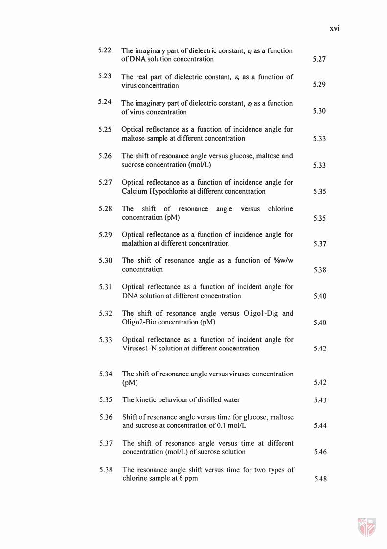
xvi
xvii
5.39 The SPR shift of resonance angle as a function of time collected 5.50
5.40 The pH value as a function of time collected 5.5 1
5.41 The comparison of resonance angle versus time for samples 5.52
5.42 The resonance angle shift versus time for pesticide measurement at concentration of 0. 1 %w/w 5.54
5.43 The time dependence of the shift in resonance angle for Oligo 1-Dig and Oligo2-Bio interfaces at 10 pM solution 5.56
5.44 The kinetic behaviour for Oligo I-Dig and Oligo2-Bio interfaces at 10 and 5 pM solution 5.57
5.45 The typical reflectance of FHA at different concentration 5.60
5.46 The shift of resonance angle as a function of FHA layer concentration 5.60
5.47 The SPR curve measured for 1: a bare Au film; 2: Au film with an over layer of FHA; 3: AulFHAIV (V); 4: AuIFHAICu (II) and 5: Aul FHAlFe (III) 5.6 1
A.l X-Ray Diffraction Pattern of Ag film for day 1 A.3
A.2 X-Ray Diffraction Pattern of Ag film for day 8 A.3
xviii
LIST OF ABBREVIATIONSINOTATION/GLOSSARY OF TERM
E;
EO
e
a
DNA
RNA
PC
FHA
HCI
EDTA
UV
XRD
MIP
The initial shift of resonance angle respect to distilled water
Real part of dielectric constant
Imaginary part of dielectric constant
Dielectric constant of medium prism
Incidence angle
Resonant angle of incidence
External angle
Surface plasmon resonance angle
The shift of resonance angle
Internal angle of prism
Magnetic permeability
Wavevector component along surface electromagnetic wave propagation
Wavevector of a plasmon
Wavevector media 1
Wave vector media 2
Wavelength
Refractive index of prism
Refractive index of metal
Refractive index of dielectric
Dielectric constant of medium metal film
Dielectric constant of medium dielectric
Oligodeoxyribonucleic acid
Ribonucleid Acid
Personal Computer
Fatty Hydroxamic Acid
Hydrochloric Acid
Ethylene Diamine Tetra Aceticacid
Ultra Violet
X-ray Diffractometer
Molecular Imprinted Polymer
BaP benzo[ a ]pyrene
DOP dioctyl phthalate
ssDNA single-stranded oligonucleotides
HDT hexanedithiol
TM Transverse Magnetic Field
TE Transverse Electric Field
M
V
MWR
LFS
SPR
ATR
SP
R
Rp Rr r A
10 1r d fJ
c
k
K
T
Au
Ag
G70
G90
Cu (II)
Concentration solution
Volume of concentration
Molecular weight relative
Low Square Fitting
Surface Plasmon Resonance
Attenuated total reflection
Surface Plasmon
Reflection coefficient
Reflectance minimum
Reflectance as a function of incidence angle
Actual optical reflectance that loss factor has been considered
Loss factor
Angle of prism
Incidence light
Reflected light
Thickness
' stretching coefficient'
Time constant
Concentration
Adsorption constant
Kinetic constant
Absolute temperature
Gold
Si lver
Calcium Hypochlorite
Trichloroisocyanuric Acid
Copper (II)
xix

xx
Fe (III) Ferum (III)
V (V) Vanadium (V)
wtJwt Weight per weight
ppm Part per million
pM PicoMolar
rpm Round per minutes
CHAPTER 1
INTRODUCTION
1.1 Surface Plasmon Resonance
Surface plasmon resonance (SPR) is well known as a powerful and expensive optical
method for the study of interface phenomena. SPR is an optical phenomenon arising
in thin metal films under condition of total internal reflection. This phenomenon
produces a sharp dip in the intensity of the reflected light at a specific angle (called
the resonant angle). This resonant angle depends on several factors, including the
refractive index of the medium (refractive index is directly correlated to the
concentration of dissolved material in the medium) close to the non-illuminated side
of the metal film. By keeping other factors constant, SPR is used to measure the
change in the concentration of molecules in the surface layer of solution in contact
with the sensor surface.
Surface plasmon resonance is a collective oscil lation of the free electron charges, at a
metal-dielectric boundary, which propagates along interface. These charge
fluctuations are accompanied by an electromagnetic field having a maximum at the
metal-dielectric interface and decaying exponential ly with distance from either side
of it [Sadowski et ai., 1 99 1 ; Kitaj ima et ai., 1 98 1 ] . The resonance excitation of the
surface plasmon resonance occurs at a characteristic angle of incidence, which
depends on the thickness as well as on the dielectric permittivity of the layers and of
adjacent medium. S ince the permittivity depend on the frequency of the exciting
laser l ight, so does too the resonance angle. When the frequency is fixed, SPR
1.2
permits the measurement of changes in the refractive index in the medium adjacent
to the metal film as well as changes in the absorption layer on the metal surface. The
plasmon wave can be excited at the interface between a thin metal film and air or
other non-metal medium with a positive dielectric sign. The wave can be thought of
as having a section inside the thin film and a section outside of the film in the air /
metal interface, much like an ocean wave has part of the wave unseen inside the
ocean, while another part of the wave is seen in the ocean / horizon interface. Under
normal circumstances, a laser light source incident upon a thin film is reflected or
scattered, and there would be an insignificant surface plasmon wave, which would
absorb very little of the incident energy [Kolomenskii et at., 1997].
Surface plasmon resonance occurs when the energy from incident light is of just the
right frequency and angle of incidence to couple its energy with the surface plasmon,
so that no light is reflected from a normally reflective surface. Thickness of a thin
film can be determined through surface plasmon resonance due to the fact that as the
thickness of the thin film increases, less of the surface plasmon is in the air / metal
interface and more of it is contained within the metal itself. We can use the
characteristic peak and shape of the resonance curve to characterize different thin
film thickness of different materials.
Typically, SPR technique employs the principle of attenuated total reflection (ATR)
using either Kretschmann or Otto geometries. Surface plasmon are collective
oscillations of free charge of metal, which under appropriate conditions, may be
coupled to by incident optical radiation resulting in the absorption of light. Coupling
is accomplished in two ways either angular or spectral. In the former, the incident


![Cambridge International Examinations Cambridge International … · 2019-02-07 · C kolam renang. [1] 12 Asma menyuruh ... Saya terpaksa batalkan rancangan kita untuk hujung minggu](https://static.fdocuments.in/doc/165x107/5c84639109d3f2a3488d1d61/cambridge-international-examinations-cambridge-international-2019-02-07.jpg)