Universitat Autònoma de Barcelona Departament de ... · Unitat de Biofísica-CEB, Facultat de...
165
1
Transcript of Universitat Autònoma de Barcelona Departament de ... · Unitat de Biofísica-CEB, Facultat de...

1

2
3
Universitat Autònoma de Barcelona Departament de Bioquímica i Biologia Molecular
Unitat de Biofísica-CEB, Facultat de Medicina
A thesis submitted as part of the requirements for the degree of
Doctor in Biochemistry, Molecular Biology and Biomedicine
Thesis Director PhD candidate
Dr. David Garcia Quintana Asrar Ahmad Malik

4

5
Dedicated to
Abuji, Didi, Tathi, Khan bhai, Enkay, Noor,
And my beautiful wife
Insha

6

7
CONTENTS

8

9
ABSTRACT 15
INTRODUCTION 19
1. Eukaryotic cell cycle 21
2. Why Saccharomyces cerevisiae 23
3. Cyclin Dependent Kinases(CDKs) 24
4. Cyclins 26
4.1. G1-phase cyclins 27
4.2. S-phase cyclins 27
4.3. G2- phase cyclins 28
4.4. M-phase cyclins 28
5. Regulation of Cyclin Dependent Kinase activity (CDK-activity) 29
5.1. CDK activating kinase (Cak1) 29
5.2. Cyclin binding and degradation 30
• SCF (Skp1, Cullin, F-box) 32
• Anaphase promoting complex (APC) 33
5.3. Cyclin dependent kinase inhibitors (CKIs). Sic1 35
5.4. Cyclin dependent kinase inhibitory tyrosine phosphorylation. Swe1 37
6. Transcription factors specific to the different phases of the cell cycle 38
6.1. Transcription factor SBF (Swi4 / 6 Cell-Cycle Box Binding Factor) 39
6.2. Transcription factor MBF (MLU and Cell-cycle Box Binding Factor) 40

10
6.3. SFF transcription factor 40
6.4. Inactivation of Transcription Factors 41
7. Cell cycle progression 42
7.1. G1 Phase 42
7.2. S-phase 45
7.3. M-phase 49
8. Cyclin dependent kinase mediated control of cell cycle 51
8.1. Substrate Specificity 52
8.2. Quantitative Model 57
OBJECTIVE 57
MATERIALS AND METHODS 61
9. Model organism 63
10. Yeast Genetic Background 64
10.1 Culture media 65
10.2 Plasmids used in this study 66
• pFa6a family 66
• pRS family 66
• PCM family 66
10.3 Generating Saccharomyces cerevisiae strains 67
10.3.1 Transformation of yeast 67

11
10.3.2 Deletion of genes in strains 68
10.3.3 Tagging of genes 69
10.3.4 Sectors for second selection and functional assays 71
10.3.5 Over-expression of Proteins 71
11. Molecular Biology Techniques 72
11.1 Polymerase chain reaction (PCR) 72
11.2 Electrophoresis of DNA in agarose gel 73
11.3 Cloning 73
11.4 Ligation 74
11.5 Escherichia coli Transformation 74
11.6 Extraction of Plasmid 75
11.7 Preparation E. coli DH5α competent cells using rubidium chloride 76
11.8 Yeast Genomic DNA Extraction 77
12. Cell Biology Techniques 79
12.1 Cell density of culture 79
12.2 Experiments in unperturbed cell cycle 79
12.3 Experiments involved with over-expression of proteins 81
12.4 Experiments involved with temperature sensitive mutant proteins 83
13. Biochemical Techniques 84

12
13.1 Total protein extraction using trichloroacetic acid (TCA) 84
13.2 Polyacrylamide gel electrophoresis (SDS-PAGE) 85
13.3 Western Blotting 87
13.4 Flow cytometry (FACS, Fluorescent Activated Cell Sorting) 89
13.5 Microscopy 91
13.6 Immunoprecipitation (IP) 91
13.7 In-vitro kinase assay 92
Oligonucleotides used as primers in this study 94
Constructs used in this study 98
Strains used in this study 99
RESULTS 102
14. Triggering of later events by early cylins 104
15. Hyperstable Cln2 triggers replication in the absence of S and M-CDK
activity 106
16. Nuclear localization is essential for Cln2(7A) function in triggering
replication 111
17. Sic1ΔN remains associated with CDK complex 113
18. Nuclear Cln2(7A) mediated replication in Sic1(9A) and Sic1C70
background 114

13
19. S-phase cyclins play no role in replication mediated by nuclear Cln2(7A) 118
20. G1 cyclin Cln2 triggers later event of SFF activation 120
21. M-CDK activity plays no role in nuclear Cln2(7A) mediated replication 1122
Discussions 127
Conclusions 135
Abbreviations 139
Chemicals used 145
References 151
Acknowledgements 163

14

15
ABSTRACT

16

17
Progression through the cell cycle is directed by cyclin dependent kinases (CDKs), which are
essential for normal and cancer cell proliferation. CDKs are activated by association with
cyclins specific to each cell cycle phase (G1, S, G2, and M). Thus, CDK associated with G1
phase cyclins promote the expression of proteins necessary for DNA replication, but is
unable to activate it. CDK associated with S phase cyclins, in turn, triggers the activation of
chromosomal origins of replication, but is unable to promote chromosome condensation and
segregation of sister chromatids, which is carried out by CDK associated with mitotic cyclins.
Despite the essential role of CDKs in cell cycle progression, how the different cyclins
promote specifically the various processes of the cell cycle was still an open question by the
time this thesis was initiated. Since the discovery of cyclins by Nobel laureate Tim Hunt
[Evans et al. (1983) Cell 33:389] it was assumed that cyclins confer substrate specificity.
However, later on, fellow Nobel laureate Paul Nurse proposed an alternative quantitative
model, [Stern & Nurse (1996) Trends Genet 12:345], based on the observation that
successive waves of cyclins result in increased levels of CDK activity. While low levels of
activity (CDK associated with G1 cyclins) are sufficient promote progression through the G1
phase and start the pro-S phase transcription program, would be unable to trigger replication.
Moderate levels of activity (CDK associated with S phase cyclins) would be able to activate
replication but not mitosic events, which would require the high CDK activity associated
with M phase cyclins.
If correct, the quantitative model should fulfill two predictions, but only one was
demonstrated by the Nurse lab. Eukaryotic unicellular yeast cells are able to survive with a
single mitotic cyclin, which is notwithstanding able to orderly drive the cell through the
different cell cycle phases [Fisher & Nurse (1996) EMBO J 15:850]. Therefore M-CDK
activity is able to promote the previous phases of the cycle in the fission yeast
Schizosaccharomyces pombe However, if a purely quantitative mechanism applies, the

18
complementary prediction should be true as well: an early cyclin to be able to trigger later
events if expressed at levels high enough.
To test such prediction we generated a budding yeast Saccharomyces cerevisiae strain
carrying a G1 cyclin G1 resistant to degradation, under a strong inducible promoter, and
fused to a nuclear localization signal. Our results show that one such cyclin is capable of
firing chromosome replication conditions in which the S phase cyclins, G2 and M are
suppressed. Therefore, our results support the quantitative model against the requirement of
substrate specificity. How eukaryotic cells prevent premature activation of the critical cell
cycle processes that lead to genomic instability, seems therefore trust in the regulation of
activity levels and limiting the presence of cyclin at specific time and space.
19
INTRODUCTION

20

21
1 Eukaryotic Cell Cycle
Every organism, however simple or complex needs to self sustain and the only way to make
new cell is to duplicate already existing cell. In order to survive, these basic building blocks
of the organism, cells, require to proliferate through a series of coordinated events were each
individual cells duplicate there genomic data and divide themselves to complete a single
cycle of life commonly referred as a cell cycle (Norbury and Nurse 1992).
The concept of cell division has been studied for over a century, however, the real hallmark
was achieved in early 1950´s when Howard and Pelc's work on, Vicia faba, revealed that the
cell goes through many discrete phases before and after cell division. It was shown that cell
carries out DNA replication in specific phase of cell cycle and this phase being clearly
separated from mitosis. It was later in 1970´s when Leland Hartwell, Paul Nurse and Timothy
Hunt laid the ground breaking discoveries in cell cycle. Discovery of Cdc proteins by
Hartwell using temperature sensitive techniques, discovery of cyclins by Tim Hunt and
regulation of cell cycle by Paul Nurse opened the doorways for the most important findings
in the field of cell cycle (explained in detail in latter segments).
Eukaryotic cell cycle basically consists of four distinct phases: a single round of chromosome
duplication where entire genome is replicated taking place during the synthesis phase thus
called as S-phase, followed by the segregation of replicated chromosomes into two daughter
cells during mitosis phase also known as M-phase (Pardee 1989, Collins, Jacks et al. 1997,
Johnson and Walker 1999). Both pivotal phases are preceded by gap phases known as Gap1
(G1) and Gap2 (G2) respectively (Figure 1). G1 occurs between M-phase and S-phase while
as G2 occurs between S-phase and M-phase. During G1 phase, cells grow in size and ensure
the presence of all proteins required for the process of replication before deciding for the
entry into the synthesis phase (S-phase). During G2 phase, cells continue to grow while also

22
making sure of presence of all proteins required for mitosis and ultimate division of cell
(Pardee 1989, Johnson and Walker 1999). The final M-phase is itself coupled into two
processes, a major part consists of the dividing of two identical chromosomes into two
daughter nuclei and cytokinesis, a process were cells divide their cytoplasm into two identical
daughter cells.
Figure 1. Schematic mitotic cell cycle of budding yeast Saccharomyces cerevisiae. After passing the START, cells are irreversibly committed to a new round of cell division. DNA replication takes place in the S-phase, while as cells segregate the replicated DNA into two sister chromatids followed by the nuclear and cytoplasmic division.

23
2. Why Saccharomyces cerevisiae
The details of cell cycle vary from one organism to other, including the time required to
complete certain events, even in the same organism. However, organization of the cell cycle
and its control system are essentially the same in all eukaryotic cells. The proteins required
for the control system first appeared over a billion years ago. Remarkably, these proteins are
so well conserved over the course of evolution that many of them function perfectly when
transferred from a human cell to a yeast cell. Therefore, cell cycle and its regulation in a
variety of organisms can be studied and findings pooled from all of them to assemble a
unified picture of how eukaryotic cells proliferate.
The budding yeast Saccharomyces cerevisiae is the simplest and most powerful model
systems for studying the genetics of cell cycle control among several other organisms like,
Xenopus, sea urchins, Drosophila melanogaster and cultured mammalian cells. It is
considered as a better model organism due to its numerous advantages over other model
organisms, for example, S. cerevisiae is a small single celled eukaryotic organism with a
short generation time (doubling time 1.25–2 hours at 30 °C) and can be easily cultured. These
are all positive characteristics in that they allow for the swift production and maintenance of
multiple specimen lines at low cost.
Also, in S. cerevisiae, upon transformation, new genes can either be added or deleted
through homologous recombination. Furthermore, its ability to grow as a haploid
has simplified the creation of gene knockout strains, which apparently is more intricate in
other model organisms. And most importantly, as a eukaryote, S. cerevisiae shares the
complex internal cell structure of plants and animals without the high percentage of non-

24
coding DNA that can confound research in higher eukaryotes. Therefore making S. cerevisiae
a perfect tool for better understanding of cell cycle and its intricate mechanisms.
3. Cyclin Dependent Kinase (CDK)
As mentioned above S. cerevisiae has a same cell cycle as other eukaryotic organisms, gap1
phase followed by DNA replication, followed by gap2 phase and finally mitosis and division
of cell. Cell cycle is a self sustaining process, however, in its essence; cell cannot drive itself
without the help of well coordinated machinery. The core protein responsible for driving the
cell cycle is a protein kinase known as Cyclin-dependent kinase (CDK).
Cyclin-dependent kinases are serine/threonine kinases that specifically phosphorylate
proteins on their serine and threonine amino acid residues thus rendering the target protein
either active or inactive. In budding yeast S. cerevisiae, this highly conserved protein is
encoded by gene CDC28 (Cdk1 being the equivalent of cyclin dependent kinase in humans
and Cdc2 in S. pombe) which regulates the progression of cell cycle in different phases
(Hartwell 1976).
As the name of this protein suggests, this kinase protein is dependent on another set of
proteins called cyclins. Cyclin dependent kinase proteins are the catalytic subunit of activated
hetrodimer, where as the cyclins are regulatory subunits. The kinase activity of cyclin
dependent kinase protein remains inactive unless they bind to its specific regulatory subunit,
cyclins. While as cyclins alone confer no kinase activity (Morgan 1995, Pines 1995). Once
activated by binding of cyclin, oscillating level of cyclin-cyclin dependent kinase complex
orchestrates the regulation and progression of cell cycle through a series of biochemical
reaction called phosphorylation that activates or inactivates target proteins in order to

25
coordinate the entry into the next phase of the cell cycle. Figure 2 (Pardee 1989, Norbury and
Nurse 1991).
Even though there are multiple CDK´s present in budding yeast S. cerevisiae like Cdc28,
Kin28, Ctk1, Pho85 and Ssn3. Cdc28 however is alone sufficient and essential for the
progression of cell cycle (Hartwell, Mortimer et al. 1973, Jallepalli and Kelly 1997).
Moreover, early study of cdc mutants by Lee Hartwell showed that gene CDC28, encoding
for Cdc28 protein is essential and temperature sensitive mutations of Cdc28 block the
progression of cells at the Start (Hartwell 1974, Reed 1980, Mendenhall, Richardson et al.
1988).
The levels of cyclin dependent kinase (CDK) proteins relatively remains constant throughout
the cycle, however, the cellular levels of cyclins increase and decrease as cell progresses
Figure 2. Shows the canonical representation of cyclin-CDK activity. 1. Cyclins bind to the CDK, thus, activating the complex. 2. The complex binds to the target protein and finally, 3. CDK transfers a ᵧ phosphaste to the target protein.
26
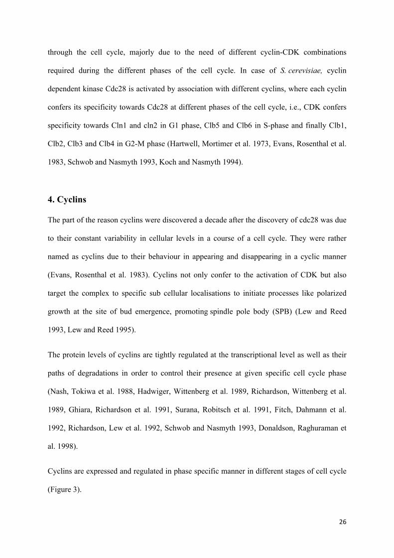
through the cell cycle, majorly due to the need of different cyclin-CDK combinations
required during the different phases of the cell cycle. In case of S. cerevisiae, cyclin
dependent kinase Cdc28 is activated by association with different cyclins, where each cyclin
confers its specificity towards Cdc28 at different phases of the cell cycle, i.e., CDK confers
specificity towards Cln1 and cln2 in G1 phase, Clb5 and Clb6 in S-phase and finally Clb1,
Clb2, Clb3 and Clb4 in G2-M phase (Hartwell, Mortimer et al. 1973, Evans, Rosenthal et al.
1983, Schwob and Nasmyth 1993, Koch and Nasmyth 1994).
4. Cyclins
The part of the reason cyclins were discovered a decade after the discovery of cdc28 was due
to their constant variability in cellular levels in a course of a cell cycle. They were rather
named as cyclins due to their behaviour in appearing and disappearing in a cyclic manner
(Evans, Rosenthal et al. 1983). Cyclins not only confer to the activation of CDK but also
target the complex to specific sub cellular localisations to initiate processes like polarized
growth at the site of bud emergence, promoting spindle pole body (SPB) (Lew and Reed
1993, Lew and Reed 1995).
The protein levels of cyclins are tightly regulated at the transcriptional level as well as their
paths of degradations in order to control their presence at given specific cell cycle phase
(Nash, Tokiwa et al. 1988, Hadwiger, Wittenberg et al. 1989, Richardson, Wittenberg et al.
1989, Ghiara, Richardson et al. 1991, Surana, Robitsch et al. 1991, Fitch, Dahmann et al.
1992, Richardson, Lew et al. 1992, Schwob and Nasmyth 1993, Donaldson, Raghuraman et
al. 1998).
Cyclins are expressed and regulated in phase specific manner in different stages of cell cycle
(Figure 3).

27
4.1 G1 cyclins (Cln1, Cln2 and Cln3): These cyclins associated with cyclin dependent
kinase drive cell cycle from G1 to S-phase and also promote activities like spindle pole body
duplication and budding (Lew and Reed 1995). Cyclin dependent kinase activity associated
with Cln1 and Cln2 (which are closely related proteins with overlapping functions) are
expressed in late G1 phase which upon association with Cdc28 activates its kinase activity
(Wittenberg, Sugimoto et al. 1990, Nasmyth 1996). There mRNA accumulation in late G1 is
dependent on two transcription factor complexes, MBF (Swi6p-Mbp1p) and SBF (Swi6p-
Swi4p), which bind to MCB and SCB promoter elements, respectively (Cross, Hoek et al.
1994, Stuart and Wittenberg 1994). However, Cln3 on the other hand is not regulated under
cell cycle transcription but instead as post-transcriptional modification (Tyers, Tokiwa et al.
1992, Cross and Blake 1993). Albeit, it rather plays a role in regulating the transcriptional
activation of other G1 cyclins, Cln1 and Cln2 (Dirick, Bohm et al. 1995, Stuart and
Wittenberg 1995).
4.2 S-phase cyclins (Clb5 and Clb6): Cyclin dependent kinase activity associated with S-
phase cyclins brings about DNA replication. The levels of S-phase cyclins not only remain
high in S-phase but remain such through G2 and early M phase, thus helping in promoting
early events of mitosis and finally falling at onset of anaphase. Clb5 and Clb6 are expressed
periodically throughout the cell cycle and are most abundant during late G1 (Schwob and
Nasmyth 1993, Spellman, Sherlock et al. 1998). Promoters of CLB5 and CLB6 contain MCB
(MluI cell cycle box) motifs, which are elements found in several DNA synthesis genes. The
transcriptional activator MBF (MCB-binding factor), which is comprised of
the Mbp1 and Swi6 proteins, bind to the MCB elements to activate transcription of CLB5 and
CLB6 (Lew D.J et al 1997).
28
4.3 G2 cyclins (Clb3 and Clb4): These cyclins accumulate during S-phase and G2 phase.
Cyclin dependent kinase activity associated with G2 cyclins promotes the transition of G2/M
phase, and are also presumed to be essential of spindle assembly (Surana, Robitsch et al.
1991, Fitch, Dahmann et al. 1992).
4.4 M-phase cyclins (Clb1 and Clb2): As cells begin to enter mitosis, the levels of M-phase
cyclins start to increase and reach to its peak when cells are in metaphase. Cyclin dependent
kinase activity associated with M-phase cyclins promotes transition from G2 to M phase and
through the process of mitosis, M-phase cyclins are degraded before the exit of mitosis
(Schwob and Nasmyth 1993, Donaldson, Raghuraman et al. 1998).
Figure 3. This diagram shows the orderly expression of different cyclins in S. cerevisiae in their respective phases. In budding yeast cell cycle, a single catalytic subunit (cdc28) drives the whole cell cycle by associating with specific regulatory subunits, cyclins, from different cell cycle phases.

29
5. Regulation of Cyclin Dependent Kinase activity (CDK-activity)
Since the progression of the cell cycle is coordinated and driven by the cyclin-CDK activities,
it is more apparent to have the regulation of CDK activities rather tightly regulated in order to
ensure the execution of cell cycle division with precision (Morgan 1997). Cell regulates the
levels of cyclin-CDK activity by four critical mechanisms, CAK (CDK activation kinase)
phosphorylation, binding of cyclins and there destruction, binding of cyclin dependent kinase
inhibitor subunits (CKIs) and by regulatory inhibitory phosphorylation (Morgan 1995,
Morgan 1997).
5.1 CDK activation kinases (CAKs): In budding yeast S. cerevisiae, cyclin dependent
kinase proteins are phosphorylated by CDK activation kinase proteins at threonine 169
adjacent to the active site before cyclin-CDK complex is formed. Timing of this
phosphorylation varies in different model organisms and moreover, CAK kinases are not
regulated in cell cycle pathway manner, however, studies have shown that phosphorylation of
CDK by CAK is pivotal for viability of cells (Ross, Kaldis et al. 2000). Figure 4.
Figure 4. This diagram shows phosphorylation of threonine 169 by Cak1 which is followed by binding of cyclins.

30
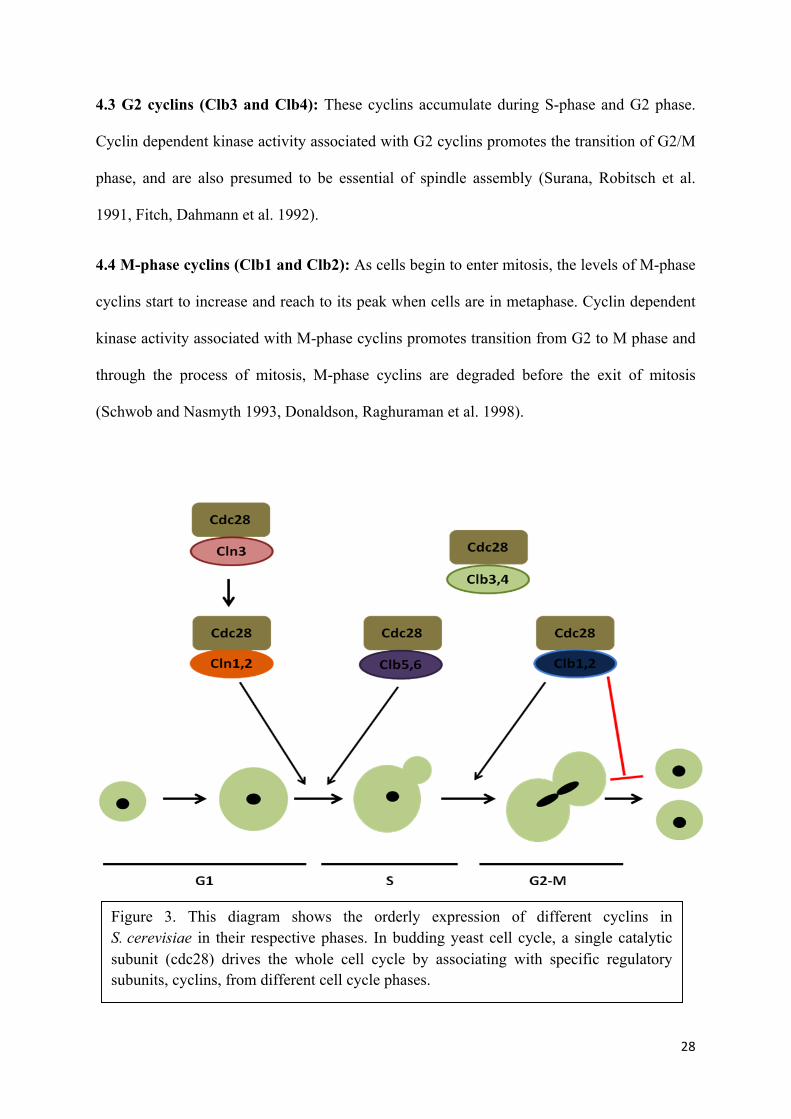
5.2 Cyclin binding and degradation: Cyclin binding is a process where cyclins bind to an
inactive CDK in order to expose the active site, or ATP-binding site, which is a cleft between
a small amino-terminal lobe and a larger carboxy-terminal lobe. In its inactive form, Cyclin
dependent kinase proteins have a flexible loop called the activation loop or T-loop which
blocks the cleft. Due to the barrier of T loop and in absence of cyclin binding, the position of
several key amino acid residues is not optimal for ATP-binding, thus rendering CDK inactive
(Figure 5). CAKs phophorylate T loop at Thr 169 to increase the complex activity towards
the cyclin binding (Ross, Kaldis et al. 2000).
With cyclin binding, the conformational change in two alpha helices permit the appropriate
ATP binding. L12 helix that comes just before the T-loop in the primary sequence becomes a
beta strand and helps rearrange the T-loop in a manner to prevent its blocking of active site.
While as the other alpha helix called the PSTAIRE helix rearranges and helps change the
position of the key amino acid residues in the active site, eventually leading to the activation
of cyclin-cyclin dependent kinase complex activity (De Bondt, Rosenblatt et al. 1993,
Morgan
31
1997).
Firgure 5. Shows the structure of cyclin dependent kinase. The smallerN-terminal lobe, containing a beta sheet and the PSTAIRE helix. Below N terminal lobe is larger C-terminal lobe. The active site cleft between the lobes contains ATP-binding site (ball and stick representation). The T-loop (residues 146–170), which blocks the cleft (dark black). And, the small L12 helix disrupts active site residues involved in ATP orientation (Morgan 1997).

32
Furthermore, the rising levels of cyclin-CDK activities are regulated by degrading the cyclins
at the specific stages of cell cycle once the role of their functional properties are met. Cells
have evolved with complex mechanisms and machineries to help degrade the cyclins via
proteasome regulation to avoid the repetition of processes as well as eliminating the proteins
to allow passage from one phase to the next. The two major proteasome complexes are Skp,
Cullin, F-box containing complex (or SCF complex) and Anaphase Promoting Complex
(APC).
SCF (Skp1, Cullin, F-box): G1 cyclins are highly unstable and are self regulated, which
upon being phosphorylated are subjected to degradation under Skp, Cullin, F-box containing
complex (or SCF complex) (Schneider, Patton et al. 1998). SCF complex is a multi-protein
E3 ubiquitin ligase complex which helps in catalyzing the ubiquitination of proteins destined
for proteasomal degradation.SCF is active throughout the cell cycle, and the stability of
individual substrates is regulated by their phosphorylation and depending on different
phosphate binding proteins (F box proteins) guiding different sets of substrates to
destruction.e.g., Cdc4, Grr1 (Figure 6) (Murray 2004).
Figure 6. Schematic representation of SCF (Skp/cullin/F-box) complex. Composed of a core organization, the association of a cullin-like protein Cdc53, a protein containing a particular zinc finger domain Rbx1 which recruits E2-ubiquitin conjugate and phosphate binding F box proteins which guides different sets of substrates to destruction (Teixeira and Reed 2013).
33
Apart from degrading G1 cyclins, SCF complex has important roles in the ubiquitination of
other proteins involved in the cell cycle. For example, the essential step to enter into the S-
phase is achieved by the degradation of S and M-CDK inhibitor (also known as all CLB
inhibitor) Sic1 by SCF complex coupled with the F-box protein Cdc4 (SCFcdc4) (Feldman,
Correll et al. 1997, Skowyra, Craig et al. 1997, Verma, Annan et al. 1997, Visintin, Craig et
al. 1998, Yoshida, Asakawa et al. 2002, Queralt and Igual 2003, D'Amours, Stegmeier et al.
2004, Mayor, Lipford et al. 2005). While as Clns on the other hand are degraded by SCF
coupled with F-box Grr1 (Barral, Jentsch et al. 1995, Skowyra, Craig et al. 1997, Patton,
Willems et al. 1998, Schneider, Patton et al. 1998).
Cdc6, an essential protein required for Pre-RC assembely at the origin of replication is also
degraded by SCFcdc4 mediated ubiquitination after first round of replication in order to
prevent re-replication (Donovan, Harwood et al. 1997)
Among all Clbs (S phase and M phase cyclins), only Clb6, the paralog of Clb5 is degraded by
SCFcdc4 because Clb6 lacks the destruction box motif responsible for the anaphase promoting
complex-mediated destruction (which will be explained momentarily) however, contains
putative Cdc4 degron motifs in the N terminus, therefore targeted to the SCF(Cdc4)
ubiquitin ligase complex (Jackson, Reed et al. 2006).
Anaphase promoting complex (APC): All mitotic cyclins included S phase cyclin Clb5 are
degraded by Anaphase promoting complex (APC), causing the inactivation of M-phase
cyclin dependent kinase activity which is essential for exit from mitosis (Jaspersen, Charles
et al. 1998, Visintin, Craig et al. 1998, Wasch and Cross 2002).
APC/C is a multi-protein E3 ubiquitin ligase complex like SCF complex which helps in
catalyzing the ubiquitination of proteins destined for proteasomal degradation. APC core
consists of 14 different proteins, including Apc2 cullin-like subunit that serves as a scaffold.

34
Apc11 RING-finger protein interacts with E2 enzyme responsible for elongating ubiquitin
chains.
Unlike SCF complex, APC/C is not active throughout the cell cycle, albeit, its activation is
achieved via further association with one of the two coactivator subunits: Cdc20 and Cdh1.
Both the adapters recognise destruction motifs also known as degrons on C-terminal domains
of target protein. The motifs present on target protein recognised by APC/C are the consensus
sequence of RXXLXXXXN also known as D-box and KEN-box with consensus sequence of
KENXXXN (Glotzer, Murray et al. 1991, Peters 2006, Barford 2011) (Figure 7).
Anaphase promoting complex (APC) regulates the mitosis at two different steps, first at
metaphase-anaphase transition followed by at the exit of the mitosis. At the metaphase-
anaphase transition, APC associates with the co-activator Cdc20 (APCCdc20) and is
responsible for the Securin/Pds1 proteasome pathway (Cohen-Fix, Peters et al. 1996,
Visintin, Prinz et al. 1997). The protein Securin inhibits the separation of sister chromatids
preventing the action of Separase/Esp1, which is to cleave protein Cohesin. Cohesin/Scc1 is a
Figure 7. Schematic representation of APC (Anaphase promoting complex). Composed of a core organization, the association of a cullin-like protein Apc2, a protein containing a particular zinc finger domain Apc11 which recruits E2-ubiquitin conjugate and adapters Cdc20 and Cdh1, which recognise destruction motifs. Cdc26 is important for assembly of the complex (Teixeira and Reed 2013).

35
protein that keeps two sister chromatids attached together, and upon activation of Separase,
Cohesion is cleaved, thus, allowing chromosome segregation (Shirayama, Toth et al. 1999,
Salah and Nasmyth 2000, Wang, Liu et al. 2001, Agarwal, Tang et al. 2003, Rahal and Amon
2008).
The second association of APC with Cdh1 (APCCdh1) is involved in the exit of mitosis, where
all Clb cyclins are completely eliminated. Unlike APCcdc20, APCcdh1 is inactive at the
beginning of mitosis (Harper, Burton et al. 2002) due to phosphorylation of Cdh1 by cyclin
dependent kinase activity associated with Clbs (Schwab, Lutum et al. 1997, Visintin, Prinz et
al. 1997, Zachariae, Schwab et al. 1998, Kramer, Scheuringer et al. 2000). However, release
of phosphatase Cdc14 from nucleus via FEAR and MEN (Mitotic Exit Network) pathways at
the end of anaphase dephosphorylates Cdh1, causing the subsequent activation of APCCdh1
(Jaspersen, Charles et al. 1998, Yoshida, Asakawa et al. 2002, D'Amours, Stegmeier et al.
2004). Upon activation of APCCdh1, APCCdh1 promotes degradation of mitotic cyclins, leading
to the inactivation of M-phase CDK activity and eventual exit from mitosis (Schwab, Lutum
et al. 1997, Jaspersen, Charles et al. 1998, Visintin, Craig et al. 1998, Wasch and Cross
2002).
5.3 Cyclin dependent kinase inhibitors (CKIs): In order to prevent cell cycle events to run
out of order, cyclin-dependent kinase inhibitor proteins (CKIs) interact with the cyclin-CDK
complex and block its activity till its right time for cells to enter next stage. In budding yeast,
CKIs are strong inhibitors of S-phase and M-phase CDK activities. CKIs inhibit the activity
of S-phase-CDK activity at G1 to prevent untimely initiating of DNA replication. Once the
levels of G1/S cyclin-CDK levels increase, the CKIs are phosphorylated and degraded by
ubiquitin-mediated proteolysis pathway (Schwob, Bohm et al. 1994, Sheaff and Roberts
1996, Verma, Annan et al. 1997). In budding yeast S. cerevisiae , cyclin-dependent kinase
inhibitor (CKI) Sic1 regulates the cell cycle at the G1 to S transition by inhibiting the activity

36
of the cyclin-dependent kinase (CDK) Cdc28. p27KIP1 is the structural homologue of
inhibitory domain of Sic1 in mammals, however, they lack the sequence homology (Lew
2003).
Sic1 is expressed at M/G1 transition and is dependent on the transcription factor Swi5
(Knapp, Bhoite et al. 1996, Toyn, Johnson et al. 1997, Aerne, Johnson et al. 1998). Sic1 is an
inhibitor of the cyclin-CDK complexes containing S phase and M phase cyclins (Clbs) but
not G1 (Cln) cyclin (Mendenhall 1993, Mendenhall, al-Jumaily et al. 1995). However, cells
overcome the inhibition of Sic1 protein and enter S phase by phosphorylating and targeting it
for degradation via ubiquitin-mediated proteolysis pathway (Sheaff and Roberts 1996).
Cyclin dependent kinase Cdc28 associated with Clns phosphorylates Sic1 (Mendenhall, Jones
et al. 1987, Wittenberg and Reed 1988, Schneider, Yang et al. 1996). Cyclin dependent
kinase inhibitor protein Sic1 needs to be phosphorylated on a minimum of six of its nine
potential phosphorylation sites and it becomes targeted for degradation (Verma, Annan et al.
1997, Nash, Tang et al. 2001, Mittag, Marsh et al. 2010). Phosphorylated Sic1 is bound
by Cdc4, which is the substrate recognition subunit of the E3 ligase SCF-Cdc4 (Skowyra,
Craig et al. 1997, Deshaies and Ferrell 2001, Nash, Tang et al. 2001). In conjunction with the
E2 enzyme Cdc34, SCFCdc4 polyubiquitinates Sic1 on N-terminal residues and once
ubiquitinated, the polyubiquitin-binding protein Rpn10p targets Sic1 to proteasome for
degradation (Feldman, Correll et al. 1997, Skowyra, Craig et al. 1997, Verma, McDonald et
al. 2001, Petroski and Deshaies 2003, Kus, Caldon et al. 2004, Mayor, Lipford et al. 2005,
Petroski and Deshaies 2005, Sadowski, Suryadinata et al. 2010).
The other cyclin dependent kinase inhibitor present in budding yeast is, Far1, Far1 inhibits
Cln-Cdk1 complexes at Start, especially in presence of pheromone. Unlike Sic1, only one site
on Far1 requires to be phosphorylated rather than six to target it for degradation (Peter and
Herskowitz 1994, Gartner, Jovanovic et al. 1998).
37
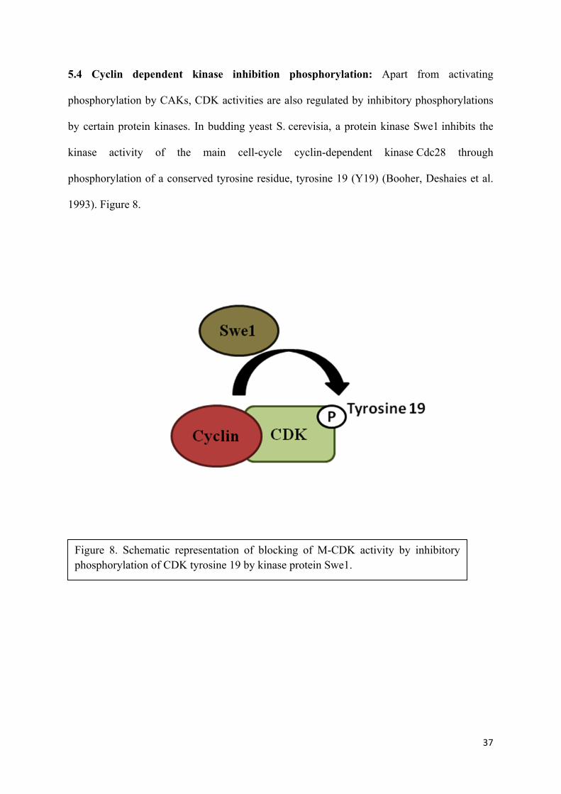
5.4 Cyclin dependent kinase inhibition phosphorylation: Apart from activating
phosphorylation by CAKs, CDK activities are also regulated by inhibitory phosphorylations
by certain protein kinases. In budding yeast S. cerevisia, a protein kinase Swe1 inhibits the
kinase activity of the main cell-cycle cyclin-dependent kinase Cdc28 through
phosphorylation of a conserved tyrosine residue, tyrosine 19 (Y19) (Booher, Deshaies et al.
1993). Figure 8.
Figure 8. Schematic representation of blocking of M-CDK activity by inhibitory phosphorylation of CDK tyrosine 19 by kinase protein Swe1.

38
6. Transcription factors specific to the different phases of the cell cycle
While cell cycle progression is controlled by regulating levels of cyclin dependent kinase
activity as explained in previous segments, other set of cell cycle control is carried out by
transcriptional factors activated sequentially which regulates the transcription program
periodically (Orlando, Lin et al. 2008).
These transcription factors essentially encode the expression of different specific cyclins of
each phase of the cell cycle among other proteins. Cyclins themselves being under the
category of periodically expressed genes and while as cyclin dependent kinase playing a role
in the regulation of cell cycle transcription, however, are not exclusively responsible for
establishing the periodic transcription program. This suggests that transcription program
periodically works as an oscillator, independent of CDK activity (Orlando, Lin et al. 2008).
Nonetheless, the timely scheduled expression of genes required for cell cycle regulated
processes such as DNA replication and mitosis is not alone sufficient for triggering these
events, but the accurate execution of cell cycle events is more likely to require both properly
timed transcription and post-transcriptional modifications mediated by cyclin dependent
kinase (Orlando, Lin et al. 2008).
G1 transcription factors present in budding yeast S. cerevisiae, are known as SBF (Swi4 / 6
Cell-Cycle Box Binding Factor) and MBF (MLU And Cell-cycle Box Binding Factor) whose
functional analogs to human is E2F (Cooper 2006). Upon activation of SBF and MBF, these
transcription factors promote the entry into the S-phase from G1 phase, since cells cross the
limiting step of cell cycle known as Start and thus committing the cell for the round of DNA
replication and mitosis.
39
SBF (Swi4 / 6 Cell-Cycle Box Binding Factor) and MBF (MLU And Cell-cycle Box Binding
Factor) bind to the promoters of 235 genes (Iyer, Horak et al. 2001, Simon, Barnett et al.
2001). SBF (Swi4 / 6 Cell-Cycle Box Binding Factor) binds the promoters of several other
transcription factors, including HCM1, PLM2, POG1, TOS4, TOS8, TYE7, YAP5, YHP1,
and YOX1 and ChIp-ChIp analysis has shown that the promoters of these genes with
consistent roles in G1/S events including DNA replication, bud growth, and spindle pole
complex formation (Horak, Luscombe et al. 2002).
6.1 Transcription factor SBF (Swi4 / 6 Cell-Cycle Box Binding Factor): This transcription
factor consists of two proteins: a protein with transactivator activity, known as Swi6 and a
protein with DNA binding activity, Swi4 (Nasmyth and Dirick 1991). Unlike Swi6, the
expression of Swi4 varies throughout the cell cycle, peaking in G1 (MacKay, Mai et al.
2001).
Along with gene expression of critical G1 cyclins CLN1, CLN2, budding and biosynthesis of
membranes (Breeden and Nasmyth 1987, Andrews and Herskowitz 1989, Taba, Muroff et al.
1991, Iyer, Horak et al. 2001, Simon, Barnett et al. 2001), SBF also controls the expression of
OH which is a site-specific endonuclease required for gene conversion at the MAT locus
(homothallic switching) at Start. Deletions in the promoter helped in the identification of a
consensus sequence (CACGAAAA) that confers to Start-specific transcription (Nasmyth
1985). This specific sequence is recognized by Swi4 protein and is called as SCB (Swi4 / 6
Cell-Cycle Box) (Ogas, Andrews et al. 1991).
G1 transcription repressor, Whi5 binds to the SCB binding factor (SBF) at SCB target
promoters in early G1. The phosphorylation of Whi5 by the cyclin dependent kinase activity
associated with Cln3 relieves its repression and promoter binding (Costanzo, Nishikawa et al.
2004). pRB is the functional analog of Whi5 in human´s (de Bruin, McDonald et al. 2004).

40
6.2 Transcription factor MBF (MLU and Cell-cycle Box Binding Factor): MBF also
consists of two proteins, one with transactivator activity same as in SBF, Swi6 and a DNA
binging protein Mbp1 (Wijnen, Landman et al. 2002). MCB box (MLU and Cell-Cycle Box)
which is recognized by Mbp1 has a consensus sequence (ACGCGT) of specific promoters
that confers to Start-specific transcription (Verma, Patapoutian et al. 1991). MBF is
responsible for the transcription of genes involved in DNA replication such as S phase cyclin
CLB5 and CLB6. It is also involved in transcription of several other essential proteins
required for DNA replication and DNA repair such as Pol1, Pol12, Rfa1, Cdc45, subunits of
ribonucleotide reductase (RNR), and the cyclin dependent kinase activity inhibitor in M
phase, Swe1 (Feldman, Correll et al. 1997, Iyer, Horak et al. 2001, Simon, Barnett et al.
2001)
6.3 Transcription Factor SFF: Timely transcription of cell cycle-regulated genes is
organized into clusters, exhibiting similar patterns of regulation. In previous sections, we
understood that in most cases periodic transcription is achieved by both repressive and
activating mechanisms. Studies have shown the group of at least 35 yeast genes that are
transcribed roughly from the end of S phase until nuclear division (Cho, Campbell et al.
1998, Spellman, Sherlock et al. 1998). Based on the CLB2 mitotic cyclin gene, this set of
genes has been termed as the Clb2 cluster (Ghiara, Richardson et al. 1991, Surana, Robitsch
et al. 1991). Other members of this family include CLB1, CDC5, CDC20, SWI5 and ACE2.
Through the analysis of the SWI5 promoter, the insight into regulation of this gene cluster
was first obtained. A protein complex was shown to be capable of binding to specific
elements in the SWI5 promoter, thus coining the term Swi5 factor (SFF) (Taba, Muroff et al.
1991). Recent studies on SWI5 and CLB2, has revealed that SFF sites are binding sites for
members of the forkhead family of transcription factors, especially two forkhead protein
members, Fkh1 and Fkh2. (Kumar, Reynolds et al. 2000, Hollenhorst, Pietz et al. 2001) .

41
Although, activating pathway of SFF transcription factors is not fully understood, but
proteins expressed under it are pivotal for the processes of M-phase and M/G1 phase.
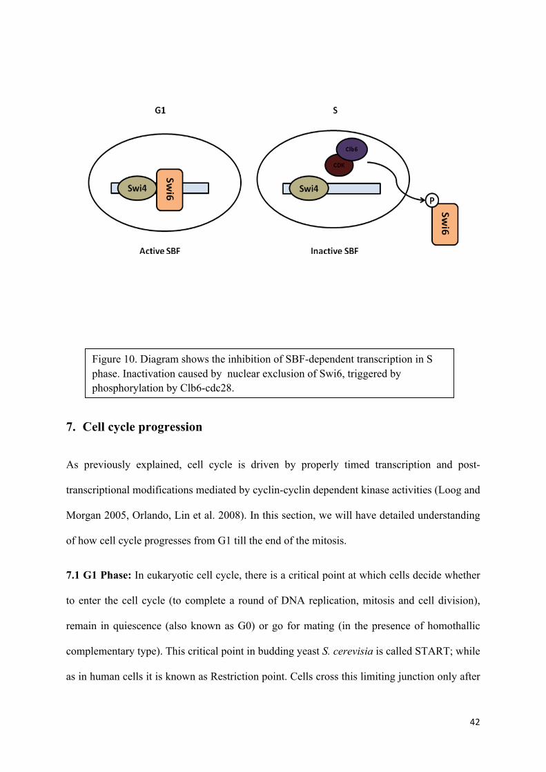
6.4 Inactivation of Transcription Factors: In order to have limited and controlled
transcription of MBF and SBF promoters, they are inactivated in different ways. MBF
becomes inactive when repressor protein, Nrm1 binds to the MBF, with help of protein
Mbp1, whose presence for recruitment is necessary (Koch, Moll et al. 1993, de Bruin,
Kalashnikova et al. 2006). While as SBF inactivation is achieved by nuclear expulsion of
transactivator protein Swi6 by cyclin dependent kinase activity associated with Clb6 and not
Clb5 (Taba, Muroff et al. 1991, Sidorova, Mikesell et al. 1995, Queralt and Igual 2003,
Geymonat, Spanos et al. 2004) (Figure 9 and Figure 10).
Figure 9. Diagram shows the inhibition of MBF-dependent transcription in S phase. Inactivation caused by binding of MBF repressor Nrm1.
42
7. Cell cycle progression
As previously explained, cell cycle is driven by properly timed transcription and post-
transcriptional modifications mediated by cyclin-cyclin dependent kinase activities (Loog and
Morgan 2005, Orlando, Lin et al. 2008). In this section, we will have detailed understanding
of how cell cycle progresses from G1 till the end of the mitosis.
7.1 G1 Phase: In eukaryotic cell cycle, there is a critical point at which cells decide whether
to enter the cell cycle (to complete a round of DNA replication, mitosis and cell division),
remain in quiescence (also known as G0) or go for mating (in the presence of homothallic
complementary type). This critical point in budding yeast S. cerevisia is called START; while
as in human cells it is known as Restriction point. Cells cross this limiting junction only after
Figure 10. Diagram shows the inhibition of SBF-dependent transcription in S phase. Inactivation caused by nuclear exclusion of Swi6, triggered by phosphorylation by Clb6-cdc28.

43
it has reached to an appropriate size and environmental conditions are feasible (Johnston,
Pringle et al. 1977) (Hicks and Herskowitz 1976, Nasmyth 1983, Fitch, Dahmann et al.
1992).
Crossing Start is highly regulated till cells acquire the right size and proteins required for the
process is properly accumulated. The process involves the activation of previously explained
transcription factors (SBF, MBF), and G1/S phase promoters. The cyclin dependent kinase,
Cdc28, activity associated with G1 cyclin Cln3 is the responsible for crossing the Start, Cln3-
Cdc28 activity is tightly regulated in G1 phase with corresponding to cell cycle progression
and cell growth (Hartwell and Unger 1977) (Johnston, Pringle et al. 1977).
As mentioned previously, transcription of Cln3 is not regulated by the cell cycle, rather by
post-translational modifications, (Tyers, Tokiwa et al. 1992) (Cross and Blake 1993)
moreover, prior to its function, Cln3 is inhibited at two levels. At first, a RNA binding
protein, Whi3, involved in cell size, binds to the mRNA of Cln3 which leads to its translation
in endoplasmic reticulum (Gari, Volpe et al. 2001, Wang, Gari et al. 2004), and in order to
retain Cln3 in endoplasmic reticulum, N-terminal Cdc28 binding domain of Whi3 interacts
with cyclin dependent kinase Cdc28 and Cln3-Cdc28 complex including members of HSP70
family, Ssa1 and Ssa2, thus restricting the nuclear accumulation of Cln3-Cdc28 complexes
(Wang, Gari et al. 2004, Verges, Colomina et al. 2007). As G1 progresses and cell reaches to
the optimal size, a J chaperone, Ydj1, interacts with the C-terminal regions of Cln3 and
facilitates its phosphorylation by Cdc28 as a signal for degradation also plays a positive role
in releasing the Cln3-Cdc28 complex from endoplasmic reticulum and allowing its nuclear
accumulation (Verges, Colomina et al. 2007).
The accumulation of Cln3-Cdc28 complex in nucleus raises its kinase activity, which leads to
the phosphorylation of Whi5. Whi5 is the inhibitor of SBF (Swi4 / 6 Cell-Cycle Box Binding

44
Factor) complex and upon being phosphorylated is excluded from the nucleus (Costanzo,
Nishikawa et al. 2004, de Bruin, McDonald et al. 2004). Once SBF is activated, it is
responsible for transcription of proteins involved in budding, biosynthesis of membranes and
cell wall as well as G1 cyclin Cln1 and Cln2. On the other hand, pathway responsible for the
activation of MBF is still unknown, however, it is responsible for transcribing pivotal
proteins involved in DNA replication especially polymerase alpha subunits Pol1 and Pol12 as
well as S-phase cyclins Clb5 and Clb6 (Figure 11) (Johnston, Morgan et al. 1996, Simon,
Barnett et al. 2001).
With this sequential transcription of proteins and cyclin dependent kinase, cdc28 kinase
activity associated with Cln3 in G1 progresses cell cycle past Start, once cells past Start, they
enter and are committed into the cell cycle for a single round of DNA replication followed by
mitosis and cell division with no possible point of return.

45
7.2 S-phase: Once cells cross the Start, proteins required for replication are accumulated and
recruited at specific positions. Before the beginning of S-phase, a protein known as Origins
of Complex Recognition (ORC), recognizes and binds to a DNA sequence called the
Autonomous Replication Sequences (ARS) which is considered as the origin of replication,
Figure 11. Shows the schematic diagram of G1 phase and G1-S transition. Cln3-Cdc28 is activated once cells reach the sufficient size, Whi5 involved in inactivation of transcriptional promoter of S phase SBF (Swi4-Swi6). The activation of SBF and MBF (Swi6-Mbp1) marks the crossing of Start. SBF transcribes, the G1 cyclins Cln1, 2, while MBF transcribes, the S phase cyclin Clb5, 6. The S-CDK activity, however, is inhibited by Sic1. The accumulation of CDK activity associated Cln1, 2 phosphorylates Sic1, marking it for destruction with subsequent activation of S-CDK and S phase.

46
(Stinchcomb, Struhl et al. 1979, Bell and Stillman 1992). In budding yeast, ORC remains
attached to origins throughout the cell cycle (Diffley and Cocker 1992). The protein ORC on
the ARS acts as the platform for the other proteins that constitute the pre-RC (pre-
Replication Complex).
The beginning of replication is carried out in two step mechanism, first step, licensing of
origins, the recruitment of proteins in pre-RC are achieved by the sequential binding of Cdc6,
which is required for the binding of another DNA replication licensing factor, Cdt1 (Devault,
Vallen et al. 2002) followed by the recruitment of helicase MCM complex (Cocker, Piatti et
al. 1996, Santocanale and Diffley 1996, Aparicio, Weinstein et al. 1997, Detweiler and Li
1997, Donovan, Harwood et al. 1997, Tanaka and Diffley 2002, Tanaka, Umemori et al.
2007) This Mcm2-7, cdc6, cdt1 containing complex bound to origin are called as pre-
replicative complex. Pre-replication complex can be formed only during G1 phase due to low
CDK activities (Zegerman and Diffley 2007).
In the meantime, MBF has transcribed the S-phase cyclins (as explained previously) Clb5
and Clb6. Clb5 and Clb6 are associated with cyclin dependent kinase, Cdc28, however, its
kinetic activity is being inhibited by an all Clb inhibitor protein, Sic1 (explained in section
5.3). Sic1 inhibits Clb-Cdc28 kinase activity by blocking the active site of Cdc28 (Donovan,
Toyn et al. 1994, Schwob, Bohm et al. 1994, Schneider, Yang et al. 1996). The accumulation
Cdc28 kinase activity associated with Cln1 and 2 promotes the phosphorylation of Sic1,
marking it for degradation by the ubiquitin-ligase complex SCFCdc4 (Schneider, Yang et al.
1996, Tyers 1996, Skowyra, Craig et al. 1997, Nash, Tang et al. 2001). The destruction of
Sic1 causes the release of S phase cyclin dependent kinase activity (Clb5-Cdc28 and Clb6-
Cdc28), thus marking second step in the beginning of DNA replication (S phase).

47
Rise in cyclin dependent kinase activity associated with Clb5, Clb6, commonly known as S-
phase CDK activity and S-phase kinase Cdc7-Dbf4 (DDK) activity, both are essential for
triggering the replication. Sld2 and Sld3 are the two essential constituents of the pre-RC and
key targets of S-CDK (Tanaka, Umemori et al. 2007, Zegerman and Diffley 2007). While as
DDK (Cdc7-Dbf4) phosphorylates Mcm4 subunit of the replicative helicase MCM, which
promotes the formation of a stable complex between MCM and Cdc45 (Sheu and Stillman
2006).
After phosphorylation of two essential proteins, Sld2 and Sld3 by S-CDK activity, the pre-
RC complex is converted into pre-IC (pre- Initiation Complex) upon binding of several other
additional proteins at origins, including, Dbp11 (DNA replication initiation protein which
helps in loading of DNA pol epsilon onto the pre-replication complexes at origins), DNA
replication initiation factors, Cdc45 (recruits elongation machinery) and GINS (Sld5p, Psf1p,
Psf2p, Psf3p) (Araki, Leem et al. 1995, Masumoto, Sugino et al. 2000, Kamimura, Tak et al.
2001, Takayama, Kamimura et al. 2003, Tanaka, Umemori et al. 2007).
Loading of Dbp11 and GINS, result in the activation of helicase and the opening of double-
stranded DNA with the recruitment of DNA polymerases, processes finally commencing into
DNA replication (Figure.12) (Aparicio, Weinstein et al. 1997, Aparicio, Stout et al. 1999,
Labib, Tercero et al. 2000, Gambus, Jones et al. 2006, Kanemaki and Labib 2006, Moyer,
Lewis et al. 2006). At the beginning of replication, not all origins fire at once, instead origins
fire throughout the S-phase (Zegerman and Diffley 2009). While whole genome is being
replicated throughout the S-phase, levels of M-phase cyclins start to increase towards the end
of S-phase. Since levels of cyclin dependent kinase activity associated with Clb5 and Clb6
are not enough to trigger the mitosis, the timely scheduled transcription of proteins required
for mitosis start to accumulate.
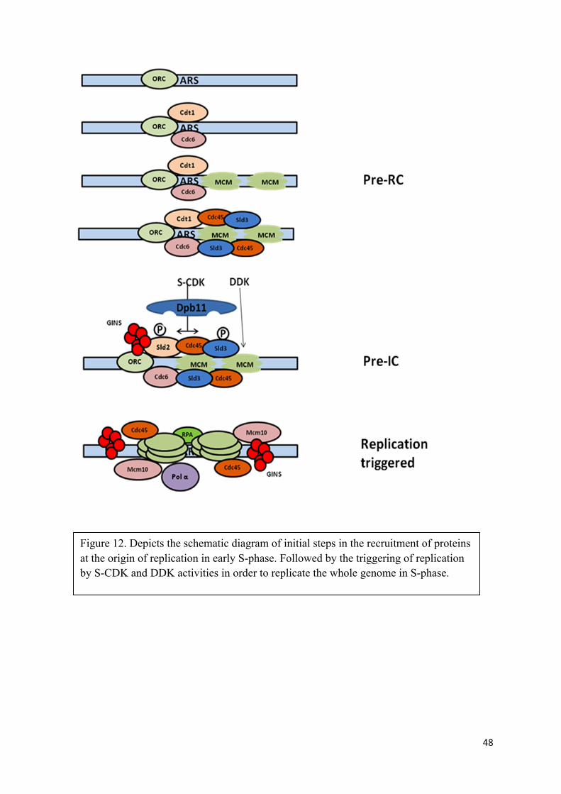
48
Figure 12. Depicts the schematic diagram of initial steps in the recruitment of proteins at the origin of replication in early S-phase. Followed by the triggering of replication by S-CDK and DDK activities in order to replicate the whole genome in S-phase.

49
7.3 M-phase: M-phase is brought about by cyclin dependent kinase activity associated with
mitotic cyclins (Clb 1, 2, 3, 4). Though not much is known about the mitosis compared to S-
phase, however, studies have shown that mitotic Clbs play roles involved in entry into
mitosis, chromosome segregation and mitotic exit (Fitch, Dahmann et al. 1992, Richardson,
Lew et al. 1992, Amon, Tyers et al. 1993, Miele 2004, Rahal and Amon 2008). High levels of
M-CDK activity promotes nuclear envelope breakdown, spindle assembly and organization,
chromosome condensation, and Golgi fragmentation, and also contributes towards the APC/C
regulation (Nigg 2001, Miele 2004).
Chromosome segregation takes place during anaphase and is triggered by the disintegration
of the protein linkages that hold sister chromatids together (Nasmyth 2002). After the
completion of replication, chromosomes condense and align on the metaphase plate where
spindle pole bodies attach to the kinetechores of each sister chromatid. However, chromatids
are linked together, which is mediated by cohesin complexes (Scc1/Mcd1, Scc3, Smc1, and
Smc3) (Nasmyth 2002). At the metaphase–anaphase transition, the Anaphase-Promoting
Complex/Cyclosome (APC/C) associated with the co-activator Cdc20 (APCCdc20) targets
Securin/Pds1 for proteosomal degradation. Securin/Pds1 is a protein that inhibits anaphase by
binding separin/Esp1p, thence, blocking cyclin destruction and mitotic exit. Clb1, 2–CDK
activity is required for the timely activation of Anaphase-Promoting Complex/Cyclosome
(APCCdc20) (Rahal and Amon 2008). After degradation of Pds1, separin/Esp1p ( a caspase-
like cysteine protease) is free and cleaves the cohesin subunit Scc1/Mcd1 is, leading to the
loss of cohesion between sister chromatids and subsequent chromosome segregation (Cohen-
Fix, Peters et al. 1996) (Yamamoto, Guacci et al. 1996, Ciosk, Zachariae et al. 1998,
Shirayama, Toth et al. 1999, Salah and Nasmyth 2000, Uhlmann, Wernic et al. 2000, Wang,
Liu et al. 2001, Nasmyth 2002) (Agarwal, Tang et al. 2003, Rahal and Amon 2008).

50
The second part, exit from mitosis requires complete elimination of the Clbs and levels of
cyclin dependent kinase activity. This is achieved by APC/C associated with Cdh1, since
APCcdh1 is inactive at the beginning of mitosis (Harper, Burton et al. 2002) due to
phosphorylation of Cdh1 by Clb1, 2 CDK activity (Schwab, Lutum et al. 1997, Visintin,
Prinz et al. 1997, Zachariae, Schwab et al. 1998, Kramer, Scheuringer et al. 2000), its
activation involves the release of phosphatase Cdc14 from nucleus via FEAR and MEN
(Mitotic Exit Network) pathways at the end of anaphase, which dephosphorylates Cdh1,
causing the subsequent activation of APCCdh1 (Jaspersen, Charles et al. 1998, Yoshida,
Asakawa et al. 2002, D'Amours, Stegmeier et al. 2004). Upon activation of APCCdh1,
APCCdh1 promotes degradation of mitotic cyclins, leading to the inactivation of M- CDK
activity and eventual exit from mitosis (Schwab, Lutum et al. 1997, Jaspersen, Charles et al.
1998, Visintin, Craig et al. 1998, Wasch and Cross 2002).

51
8. Cyclin Dependent kinase control of cell cycle
So far we understood that cell cycle encompasses the ordered series of events for the purpose
of survival of cell, where there chromosomal DNA gets duplicated and then distributed
among two daughter cells. We also established that cells maintain to carry out the stages of
cell cycle in an orderly fashion by regulating levels of cyclin dependent kinase activity, the
oscillating activity of cyclin dependent kinase acts as a major regulator for cell cycle
progression (Orlando, Lin et al. 2008).
However, since the discovery of cylins and cyclin dependent kinases over four decades ago,
scientist are still not very clear about how these oscillating levels of cyclin dependent kinases
are translated into ordered series of cellular events. The question how cyclin dependent
kinase regulates cell cycle progression was raised for several reasons; 1) both pivotal phases,
S-phase and M-phase are promoted by same catalytic subunit Cdk1 (Cdc2 in case of fission
yeast and Cdc28 in case of budding yeast) (Nurse 1981). 2) cyclin dependent kinase activity
associated with specific set of cyclins of a given phase can perform the functions of phase
preceding it, while as cannot do the functions of phase following it. For example, Cdc28
activity associated with mitotic cyclins can trigger replication but cannot do the function of
G1 cyclins (Fangfang Hu and Oscar M. Aparicio 2004). Similarly, Cdc28 activity associated
with S-phase cyclins cannot trigger the events of mitosis but can rescue the all Cln null strain
(Epstein and Cross 1992, Schwob, Bohm et al. 1994, Lopez-Girona, Mondesert et al. 1998).
In order to address this question, that, how one protein can fulfil two different roles in a cell
cycle at two different times, several explanations were put forward by scientists. Two of them
are of major importance. One of them, known as substrate specificity, put forward by Nobel
laureate Timothy Hunt.
52
8.1 Substrate Specificity
Substrate specificity may refer to the specificity of cyclin dependent kinase activity towards a
certain specific set of substrates when recruited by a cyclin in a given cell cycle phase. This
concept originated due to the fact that three distinct cyclins, Cln1, Cln2, and Cln3 are
required for transition of G1/S and entry into the S-phase (Nash, Tokiwa et al. 1988,
Hadwiger, Wittenberg et al. 1989, Richardson, Wittenberg et al. 1989, Tyers, Tokiwa et al.
1992). While as different set of cyclins, Clb1, Clb2, Clb3 and Clb4 are required for the
process of mitosis (Minshull, Blow et al. 1989, Ghiara, Richardson et al. 1991, Surana,
Robitsch et al. 1991). Moreover, third set of cyclins (Clb5 and Clb6) are required for the
timely onset of DNA replication (Epstein and Cross 1992, Schwob and Nasmyth 1993).
In addition to that, crystal structure study of S-phase cyclins showed the presence of Cdk
consensus S/T-P recognition sites, and also identified an RxL peptide motif on CDK
substrates which are readily recognized by the hydrophobic patch present on the S-phase
cyclins (Adams, Sellers et al. 1996). The Similar patch is identified in G1 cyclins , however,
not in mitotic cyclins (Petri, Errico et al. 2007, Day, Cleasby et al. 2009). The substrate
recognition function of S-phase CDK provided by hydrophobic path of cyclin was confirmed
in several Cdk substrates, suggesting RxL motif as a means by which S-phase cyclin
recognizes certain specific substrates (Loog and Morgan 2005).
These findings, the timely transcriptional control of cyclins, and how each cyclin of each
phase promotes the particular function of that phase might suggest that different cyclins act at
different times with respect to their specificity towards target proteins to promote ordered cell
cycle events. That is, G1 cyclins associated with cyclin dependent kinase might only
phosphorylate targets required for G1/S transition, while as S-phase cyclins associated CDK

53
activity would only phosphorylate proteins responsible for DNA replication and finally, M-
phase CDK activity provide specificity towards the entry and exit of mitosis.
Although, S-phase cyclins with cyclin-specific substrate recognition ability are required to
trigger the replication by phosphorylating the two essential proteins, Sld2 and Sld3
(Zegerman and Diffley 2007) (Tanaka, Umemori et al. 2007), albeit, the rigorous test to
check the requirement of S-phase cyclins showed that, cells can trigger replication on time
and are viable in the absence of both S-phase cyclins (Clb5 and Clb6) (Fangfang Hu and
Oscar M. Aparicio 2004). This suggests that the ability of triggering the replication is not
limited to the S-phase cyclins alone. Similarly, cells with triple mutant of cln1, 2, and 3 are
not viable since, it is important to have at least one G1 cyclin, which is required for
proliferation and to trigger the Start (Richardson, Wittenberg et al. 1989), however, ectopic
expression of S-phase cyclin Clb5 rescues the non-viability of triple mutant cells (Epstein and
Cross 1992, Schwob, Bohm et al. 1994, Lopez-Girona, Mondesert et al. 1998). These
findings suggest that G1 and S-phase cyclins are dispensable and are not pivotal for the
sequential cell cycle progression; while as mitotic cyclins are essential.

54
8.2 Quantitative Model
As explained earlier, the question raised by observation, that, how can a single protein (Cdk1)
be responsible for two apparently different process (DNA replication and Mitosis) and
maintain the ordered cell cycle progression, Nobel Laureate Paul Nurse proposed a model
known as, Quantitative model. According to this model, different levels of cyclin dependent
kinase activity regulates the cell cycle progression. In simple terms, the low levels of cyclin
dependent kinase activity associated with G1 cyclins trigger the events of G1-phase, however
are not sufficient to trigger the DNA replication. While as, intermediate levels of cyclin
dependent kinase activity associated with S-phase cyclins (Clb5 and Clb6) are enough to
trigger the events of DNA replication but this amount of kinase activity is not enough to
trigger the events of mitosis. Finally the higher level of cyclin dependent kinase activity
associated with mitotic cyclins (Clb 1 and Clb2) bring about entry and exit from mitosis,
where the cyclin dependent kinase activity falls and cells start with another cycle. Figure 13.
Figure 13. A quantitative model suggesting a single source of Cdk activity is sufficient for ordering sequential S phase and mitosis. S phase is triggered by an intermediate level of Cdk activity, while as mitosis depends on a higher kinase activity level. 1996)

55
The levels of CDK activity, one at the stage of triggering the DNA replication, the
intermediate one and the other, high level of CDK activity at the time of mitosis are kept at
two levels by inhibitory Cdk tyrosine phosphorylation by Swe1 (wee1 in case of fission
yeast). Once CKI is inhibited, the CDK activity is boosted and rises to high levels to trigger
the mitosis. However, recent study has shown the function of cyclin dependent kinase activity
works in both the stages of replication as well as mitosis, without two distinct levels of cyclin
dependent kinase activation (Coudreuse and Nurse 2010). Also, the regulation of single
mitotic cyclin associated with Cdk activity can be achieved at the constant level of a fusion
protein, where low, intermediate and high levels of CDK activity by chemical inhibition, and
the concentrations are sufficient to drive orderly S-phase and M-phase (Coudreuse and Nurse
2010). These findings rationalize the existence of quantitative model, even though there is
presence of other cyclins in cell, which could be just due to improvement in intricacy and
delicacy of cell system over the course of evolution.
Although, quantitative model answers several impeding questions about regulation of cell
cycle, it also raises some. Quantitative model to be utterly the reason for cell cycle regulation
is far from being solved yet, some of the questions and challenges raised towards quantitative
model are about understanding the timings of entry into the mitosis upon the completion of
DNA replication and or exit from the mitosis after faithful segregation of replicated
chromosomes, since it is basically dependent on synthesis of cylins and there timely
destruction, because, if cyclin dependent kinase activity raises quickly before due time, the
ordering of S-phase and M-phase will be hampered (Moore, Kirk et al. 2003, Coudreuse and
Nurse 2010). Also, if basic requirement for cell cycle progression is not the substrate
specificity, rather rising levels of cyclin dependent kinase activity, which dictates the
progression of cell cycle, then cyclin dependent kinase activity associated with early cyclins

56
should be able to trigger latter events if expressed in higher level, at the right place and the
right time. Addressing this question will be the major focus of this thesis.

57
OBJECTIVES

58

59
Objectives
The objective of this thesis is to validate the Quantitative model to explain how Cyclin
Dependent Kinases regulate the orderly progression of cell cycle.
- We will explore the prediction required to validate a purely quantitative control: Whether
an early cyclin bears the power to trigger a later cell cycle event if accumulated at high
enough levels, at the right time, at the right place.
- In case a purely quantitative model applies, we will investigate the molecular barriers
that are in place to avoid premature, deleterious events to take place.

60

61
MATERIALS AND METHODS

62

63
Materials and Methods:
9. Model organism:
In our study, we used budding yeast, S. cerevisiae as the model organism for the purpose of
experiments. As previously mentioned in section 2, budding yeast is the perfect model
organism for several reasons including small generation time (doubling time 1.25–2 hours at
30 °C), synchrony of the population, visual surveillance on progression of cell cycle
according to the presence and size of the bud. Moreover, S. cerevisiae can be genetically
modified to relative ease due to its prioritized repair of DNA double strand breaks by
homologous recombination. With that context, if linear DNA is introduced into a cell of yeast
with unprotected ends with homology to the genome sequence, integration would be of high
frequency, thus replacing the corresponding genomic material. With the help of this
technique we could mutant genes, or integrate them into the area of the genome that are of
interest, by a simple process of transformation.

64
10. Yeast Genetic Background:
In order to have controlled access and technical advantages over the model organism, strains
with modified genetic backgrounds were used, which made experimental procedures and
results more accountable and simplified. Budding yeast Saccharomyces cerevisiae with the
genetic background of W303 was used in this study (Thomas and Rothstein 1989) W303 has
been modified with respect to the strains found in nature to stay in a haploid, the HO
endonuclease mutant, which is the site-specific endonuclease required for gene conversion at
the MAT locus (homothallic switching) (Schwob and Nasmyth 1993
Moreover, W303 is a mutant gene for each of the synthetic pathways of five essential
nutrients: ADE2 (adenine, ade2-1), HIS3 (histidine, his3-11, 15), LEU2 (leucine, leu2-3,
112), TRP1 (synthesis of amino acid marker auxòtrofic trp1-1), and URA3 (synthesis of the
nucleobase uracil, ura3-1). With the help of these auxotrophic markers available in strain, the
selection of transformants was made highly probable, since the introduction of the wild type
gene restores the ability of “auxotrophic marker gene” missing strain to live in a selective
medium in the absence of the corresponding amino acid or nucleobase. Our work was based
upon haploid strains, W303-1a, which allows synchronization of cells in G1-phase with
pheromone α-factor. The α-factor pheromone is a natural peptide released by strains of type
homothallic Mat α. The pheromone α-factor blocks Mat a type of haploid cells in G1 phase,
in expectation of entering into the reproduction phase. The peptide Trp-His-Trp-Leu-Gln-
Leu-Lys-Pro-Gly-Gln-Pro-Met-Tyr was used to synchronize cultures of Mat a in G1-phase in
the absence of Mat α cells. Which acted as a pivotal tool, since, it makes it possible to release
population of cells into S-phase synchronously.
65
The strains used in this study were bar1Δ. The BAR1 gene encodes a protease that removes
the α-factor. bar1Δ mutant cells are sensitive towards the low concentrations of the peptide,
being sufficient to synchronize cells at the concentrations of 100 times lower than in cells
BAR1 which is 50 mg / ml.
10.1 Culture media:
YPD (1% (w / v) yeast extract, 2% (w / v) peptone, 2% (w / v) glucose) was used as rich
medium while as other carbon source (sucrose, raffinose or galactose.) were used according
to the needs of the experiment.
The minimal medium also known as SD media (Synthetic minimal medium Dextrose)
containing 0.67% (w / v) yeast nitrogen base (YNB), 2% (w / v) glucose, and supplemented
amino acids and/or nuclobase according to the selection required for the experiment, the
following are the components required for desired supplementation: 40 mg / ml adenine,
uracil, leucine, tryptophan, histidine.
The sporulation medium, RSM (Rich Sporulation Medium), consisting of 0.5% (w / v) yeast
extract, 3% (w / v) potassium acetate, 0.001% glucose (w / v), supplemented with 0.16 mg /
ml adenine and uracil, 0.08 mg / ml histidine, leucine, lysine, tryptophan, methionine and
arginine 32 mg / ml tyrosine and 0.4 mg / ml phenylalanine.
Stocks of concentrated sugars, YNB, and amino nitrogen bases were sterilized by
microfiltration (pore size 0.20 microns). Most of the sugars where sterilised this way rather
than autoclaving, in order to prevent caramelization.
The medium used for counter selection of URA3 consists of 1 mg / ml 5-fluoroorotic (5-
FOA) medium in SC (Synthetic Complete): 2 mg / ml Drop-out mix without Ura (Sigma),
0.67% (w / v) basis yeast nitrogen (YNB), 2% (w / v) glucose, 50 mg / ml uracil.

66
The medium LB (Lysogeny broth) consists of 1% (w / v) Bacto-Tryptone, 0.5% (w / v) yeast
extract and 0.5% (w / v) NaCl.
The medium to prepare cells E. coli DH5α competent Psi-broth, contains 2% (w / v)
Tryptone, 0.5% (w / v) yeast extract, 0.5% (w / v), MgSO4 · 7H2O, pH 7.6.
The solid media have the same composition as that described for liquid media plus 2% (w / v)
agar.
10.2 Plasmids used in this study:
pFa6a family:
Plasmids family pFA6-KanMX6 (Bahler, Wu et al. 1998) were used to generate strains
expressing proteins fused to a C-terminal tag. The pFA6a cassette used as a template for PCR
amplification consists of the sequence of tag of interest and six copies of the gene KanMX
geneticin antibiotic resistance (G418). The tags used in our case were 13myc (pFA6a-
KanMX6-13myc) and 3HA (pFA6a-KanMX6-3HA).
pRS family:
Family of pRS series of plasmids (Sikorski and Hieter 1989) were used as the PCR template
to amplify the genes of wild type auxotrophic markers corresponding to W303, in the process
of generating strains of S. cerevisiae deletion mutants and as well as integrative plasmids.
PCM family:
The plasmid from PCM family were used in several experiments, these plasmids have
activator/repressor expression system for budding yeast in which tetracyclines control in
opposite ways the ability of tetR-based activator and repressor molecules to
bind tetO promoters (Gari, Piedrafita et al. 1997).
67
10.3 Generating Saccharomyces cerevisiae strains:
10.3.1 Transformation of yeast
This method is used to transform the yeast cells upon weakening of the cell wall and
transformation of genetic material of interest by heat shock. To perform the transformation,
12 ml culture of the parent strain in exponential growth (1. 107 cells / ml) were used. The
pellets of cells were acquired by centrifuging at the speed of 3000 RPM for 3 minutes
(Eppindorf centrifuge 5418, Fisher Bioblock Scientific 1-159, Sigma, centrifuge). The cells
were washed twice with sterile water, and then washed once, later re-suspended in 0.1 M
lithium acetate (pH 7.5), 10 mM Tris-HCl (pH 7.5), 1 mM EDTA, at a concentration of 2. 109
cells / ml. To 50 µL of this cell suspension, 1-2 mg of DNA to be transformed was added,
followed by 5 µL of ssDNA (10 mg / ml) (single-stranded DNA from salmon sperm, which
helps transporter function by increasing the probability of passing the DNA into the cell).
This mixture is allowed to sit on ice for some time (from 5 minutes to 10 minutes) followed
by addition of 300 µL of 50% (v / v) polyethylene glycol 3350, 0.1 M acetate lithium (pH
7.5), 10 mM Tris-HCl (pH 7.5), 1 mM EDTA. The role of high weight polyethylene
molecules is to occupy the space in the mixture, making the encounter between DNA and
cells more probable. The mixture was vortexted at low speed to ensure the uniform
solubilisation and then incubated at 30 ° C for 30 minutes, however, at 24 ° C in case of
temperature sensitive mutants. After 30 minutes of incubation, DMSO was added to a final
concentration of 10% (v / v) and cells were subjected to a heat shock of 15 minutes at 42 ° C,
followed by 60 seconds on ice.
Cells were centrifuged (Eppindorf centrifuge 5418, Fisher Bioblock Scientific 1-159, Sigma,
centrifuge) for 2 minutes, and pellets were re-suspended in 200 µL of TE pH 7.5 (10 mM
Tris-HCl (pH 7.5), 1 mM EDTA) and seeded onto appropriate selective medium plates

68
(Schiestl and Gietz 1989, Gietz, St Jean et al. 1992, Bartel, Chien et al. 1993). The colonies
were allowed to grow at 30 ° C (24 ° C in case of temperature sensitive mutants) for three to
four days followed by second selection on fresh selective medium plates to ensure the purity
of clones. Finally, modifications made in the strain were checked by either genomic PCR,
functional assay or by Immunoblot of total cellular extracts (TCA extraction).
10.3.2 Deletion of genes in strains
Deletions of certain specific genes were achieved by transforming the cassette obtained from
PCR amplification of the wild type auxotrophic markers available in vectors using the PRS
and or pFA6a as a template. For the PCR amplification, primers were designed in a way that
there 5´ tails had 60 nucleotide over hangs which were present in the sequences flanking the
gene of interest to be deleted (Figure 14).
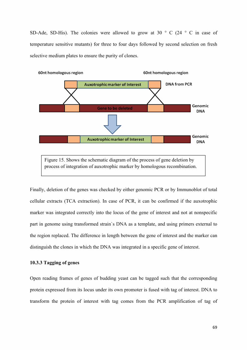
Upon transformation of deletion PCR product, gene deletion occurred through homologous
recombination, which happens by repair system activated by fragment of double strand DNA
unprotected ends with a homologous region of the genome (Figure 15). Cell transformants
were selected on minimal medium plates without supplemented amino acids and/or nuclobase
according to the selection required for the deletion. For example, (SD-Ura, SD-Trp, SD-Leu,
Figure 14. Cassette of PCR product used for the deletion of the gene.
69
SD-Ade, SD-His). The colonies were allowed to grow at 30 ° C (24 ° C in case of
temperature sensitive mutants) for three to four days followed by second selection on fresh
selective medium plates to ensure the purity of clones.
Finally, deletion of the genes was checked by either genomic PCR or by Immunoblot of total
cellular extracts (TCA extraction). In case of PCR, it can be confirmed if the auxotrophic
marker was integrated correctly into the locus of the gene of interest and not at nonspecific
part in genome using transformed strain`s DNA as a template, and using primers external to
the region replaced. The difference in length between the gene of interest and the marker can
distinguish the clones in which the DNA was integrated in a specific gene of interest.
10.3.3 Tagging of genes
Open reading frames of genes of budding yeast can be tagged such that the corresponding
protein expressed from its locus under its own promoter is fused with tag of interest. DNA to
transform the protein of interest with tag comes from the PCR amplification of tag of
Figure 15. Shows the schematic diagram of the process of gene deletion by process of integration of auxotrophic marker by homologous recombination.
70
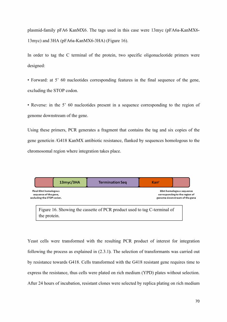
plasmid-family pFA6 KanMX6. The tags used in this case were 13myc (pFA6a-KanMX6-
13myc) and 3HA (pFA6a-KanMX6-3HA) (Figure 16).
In order to tag the C terminal of the protein, two specific oligonucleotide primers were
designed:
• Forward: at 5’ 60 nucleotides corresponding features in the final sequence of the gene,
excluding the STOP codon.
• Reverse: in the 5’ 60 nucleotides present in a sequence corresponding to the region of
genome downstream of the gene.
Using these primers, PCR generates a fragment that contains the tag and six copies of the
gene geneticin /G418 KanMX antibiotic resistance, flanked by sequences homologous to the
chromosomal region where integration takes place.
Yeast cells were transformed with the resulting PCR product of interest for integration
following the process as explained in (2.3.1). The selection of transformants was carried out
by resistance towards G418. Cells transformed with the G418 resistant gene requires time to
express the resistance, thus cells were plated on rich medium (YPD) plates without selection.
After 24 hours of incubation, resistant clones were selected by replica plating on rich medium
Figure 16. Showing the cassette of PCR product used to tag C-terminal of the protein.

71
with 200 µg / ml G418. Positive colonies for correct tagging in the strain at the desired gene
locus were checked by Immunoblot of total cellular extracts (TCA extraction) monoclonal
antibodies against the tag (9E10 anti-myc or anti-HA 12CA5) while as using the
corresponding parent strain as a negative control.
10.3.4 Sectors for second selection and functional assays
Upon transformation of a PCR product or integrative plasmid, colonies were passed through
second selection by streaking the colony onto corresponding plates in form of sectors. Each
colony was streaked on a sector and then re-spread from the end of the line in a zigzag form,
such that individual colonies were produced.
Functional assay was also carried out in a same way as second selection. In functional assay,
cells are grown on plates with specific carbon source or a drug, where specific gene of
interest weather deleted or over-expressed renders cells unviable. For the sake of robustness,
each plate was accompanied by streaking of a positive and negative control colonies.
10.3.5 Over-expression of Proteins
In the course of this thesis, several proteins were over-expressed to achieve desired results
towards the cell cycle progression. Over-expression of proteins was obtained by cloning
(cloning will be explained in next segment) gene of interest under the galactose-inducible,
GAL1, 10. promoter in pRS family plasmids and transformed into the yeast strain.
Transcription of gene was induced by adding the carbon source galactose to the culture media
at will for the time period of one hour. The induction of this promoter was also turned off by
adding rich source of carbon, dextrose, which yeast cells prefer over other sources of carbons,
thus helping in the shut off of the Gal promoter.

72
11. Molecular Biology Techniques
11.1 Polymerase chain reaction (PCR)
Polymerase chain reaction (PCR) was used to amplify the sequences of genes of interest;
which included, cloning of genes into desired plasmids with different family backgrounds
(pRS, pCM, pFA6a), cloning of gens into generating deletion cassettes, integration of
sequences leading to proteins fused to tags, replacement of promoters, inserting mutations in
a wild type gene sequence.
In order to carry out amplification of genes, each specific gene required specific primers,
primers were designed according to the need of an experiment (Shown in Table 3). In case of
cloning plasmids, the oligonucleotides incorporating the restriction enzyme sequences at the
5 'end of target sequences were used to clone the PCR product into the appropriate vector.
Furthermore oligonucleotides, incorporating the 60 nucleotides sequence flanking the region
to be replaced at 5 'end where used in case of integration, deletion and tagging. Amplified
products were checked by running a gel electrophoreses, stained with ethidium bromide.
The PCR reactions in the thermal cycler GeneAmp PCR System 2400 (Applied Biosystems)
consisting of:
Step <500bp 500-1000bp 1000-3000bp >3000bp
1.Initial denature 95° C for 2 min 95° C for 2 min 95° C for 2 min 95° C for 2 min
2.Denature 95° C for 20 sec 95° C for 20 sec 95° C for 20 sec 95° C for 20 sec
3.Annealing 55° C for 10 sec 55° C for 10 sec 55° C for 10 sec 55° C for 10 sec
4.Extension 70° C for 10 70° C for 15 70° C for 20 70° C for 25

73
s/kb s/kb s/kb s/kb
Repeat Step 2-4 25 to 32 cycles, according to the need of the product.
11.2 Electrophoresis of DNA in agarose gel
The agarose gel electrophoresis is a technique used to separate DNA molecules according to
their linear size. This technique was used to verify all steps of cloning, as well as deletion and
integration of genes in a yeast genome from PCR.
Gels using 1% (w / v) agarose (agarose D-1 Low EEO-TQM, Pronadisa) buffer TAE (50 mM
Tris-acetic acid pH 8.5, 2 mM EDTA) with 0.5 µg / ml ethidium bromide. The ethidium
bromide is a fluorescent agent which intercalates into the DNA that was retained during
electrophoresis allowing the display to be illuminated with UV light (365 nm).
11.3 Cloning
The open reading frames to be cloned were amplified by using PCR with pure genomic DNA
as a template. PCR products were purified using commercially available kits (QIAquick PCR
purification kit, Qiagen) as well as homemade purification kits (Epoch). The quality and the
concentration of DNA purified was analyzed by running gel electrophoresis stained with
ethidium bromide.
100 ɳg of purified PCR product was digested with restriction enzyme or enzymes (New
England Biolabs) chosen to perform cloning, buffers and conditions required for the optimal
function of enzymes were carried out as specified by the manufacturer. Like PCR products,
the vectors where DNA was to be cloned were also digested with the same enzymes and
conditions.

74
In order to make cloning more efficient, the vectors alone were dephosphorylated to prevent
recirculation favoured ligation of the vector with itself. To achieve dephosphorylation, 5 units
of Antarctic phosphatase (New England Biolabs) were added and incubated at 37 ° C for 15
minutes. Phosphatase was inactivated after incubation at 65 ° C for 5 minutes. The digested
PCR product and vector both were purified (QIAquick PCR purification kit Qiagen) and
ligated.
11.4 Ligation
The ligations were usually carried out at the ratio of 7:1 (vector : insert), which facilitated the
meeting of the insert with the vector at close proximity. Ligation was performed using 1 unit
of T4 ligase (Roche) in a final volume of 10 µL in the 10 x buffer supplied by the
manufacturer. The mixture was incubated at 16 ° C overnight or at 25 ° C for two hours
(quick ligation). The ligation mixture was transformed in E. coli, for the purpose of
amplification of the plasmid.
11.5 Escherichia coli Transformation
E. coli DH5α competent cells (preparation of competent cells will be explained shortly) were
used as the strain for the transformation process. 50 µL of competent cells and 10 µL ligation
mixtures were mixed and incubated on ice for 30 minutes followed by a heat shock for 90
seconds at 42 ° C. After heat shock, cells were immediately cooled in ice-water for 60
seconds to reduce mortality and closing of cell membranes. 1 ml of LB media was added to
the cells and incubated at 37 ° C for 1 hour in order to express the gene for resistance towards
ampicillin before plating cells onto selective medium plates.
Finally, the cells were centrifuged at maximum speed (14000 RPM) (Eppindorf centrifuge
5418, Fisher Bioblock Scientific 1-159, Sigma, centrifuge) and pellets were re-suspended in
75
fresh 200 µl of LB and seeded on LB plates supplemented with 100 µg / ml ampicillin
(LBA). Only those transformed cells containing vector with an ampicillin resistant gene (in
principle only possible by ligation of the insert) resulted in colonies. Since the clones
included in our thesis did not allow distinguishing of false positives for blue-white selection
(Liu, Cashion et al. 1997), control plate was included for the efficiency of the ligation
reaction with vector but without insert. The number of colonies obtained in the control would
indicate the rate of false positives. An adequate number of individual colonies were
inoculated into 5 ml of LBA at 37 ° C for overnight incubation. On the following day plasmid
DNA was extracted and the presence of insert was checked by either digestion with
restriction enzymes and/or by PCR. Results were checked by running gel electrophoresis of
digested/amplified DNA stained with ethidium bromide.
11.6 Extraction of Plasmid
To extract plasmid DNA, 1.5 ml of saturated culture was taken from the overnight culture.
Cells were centrifuged at maximum speed (14000 RPM) (Eppindorf centrifuge 5418, Fisher
Bioblock Scientific 1-159, Sigma, centrifuge) for 90 seconds and supernatant was discarded.
Pellets were resuspened in 200 µl of MX1 (50 mM TrisHCl, 10 mM EDTA pH 8.0, 100
µg/ml RNase A) suspenstion buffer by either vortexing or by pipette tip to avoid clumps and
uniform solubilisation. 250 µl of buffer MX2 (0.2 M NaOH, 1 % SDS) (lysis solution) was
added to the solution and mixed gently by inverting tubes 8-10 times. This lysis step was
crucial and vortexing was avoided to prevent plasmid contamination with genomic DNA.
Mixture was allowed to incubate at room temperature for 1-5 minutes followed by addition of
350 µl of neutralising buffer MX3 (4 M Guanidine hydrochloride, 0.5 M Potassium acetate,
pH 4.2). Upon adding of neutralising buffer, solution was mixed quickly by inverting tubes 8-
10 times to avoid partial neutralisation of lysate. Adding neutralising buffer turns solution
into white precipitation form, this precipitate was centrifuged for 10 minutes at the speed of

76
13000 RPM (Eppindorf centrifuge 5418, Fisher Bioblock Scientific 1-159, Sigma,
centrifuge). The supernatant was carefully placed in GenCatchTM plus columns onto a
collection tube and centrifuged at 5000 RPM for 60 seconds. Flow through was discarded and
the column was washed once with 500 µl of WN (5 M Guanidine hydrochloride, 20 mM
Tris-HCl, pH 6.6) wash buffer at 9000 RPM for 60 seconds. Flow through was again
discarded and column was washed once with 700 µl WS (10 mM Tris-HCl pH 7.5, 80%
ethanol) wash buffer at 9000 RPM for 60 seconds. Residual ethanol from WS buffer was
removed by centrifuging column at 13000 RPM for two minutes. Finally, 50 µl of elution
buffer (10 mM Tris-HCl, pH 8.5) was added to the column and incubated at room
temperature for 5 minutes and centrifuged at 13000 RPM for 2 minutes collecting the eluted
plasmid in a 1.5 ml eppindorf. The quality and quantification of plasmid was checked by
running gel electrophoresis. Purified plasmid was either stored at -20 °C or carried on with
the experiment.
11.7 Preparation E. coli DH5α competent cells using rubidium chloride
The competent cells for the purposes of E. coli transformation were made by chemical
method using rubidium chloride which weakens the cell wall of bacteria in order to allow the
transfer of genetic material into the cell at the time of heat shock.
A fresh single colony of E. coli DH5α was inoculated in 20 ml of LB (12 and incubated
overnight at 37 ° C in an incubator with orbital agitation at 200 RPM. On the next day, 1 ml
of this culture was used to inoculate 100 ml of Psi-broth medium (2% (w / v) Tryptone, 0.5%
(w / v) yeast extract, 0.5% (w / v), MgSO4 · 7H2O, pH 7.6). The cells were allowed to grow
at 37 ° C till they reached an optical density of range 0.48 to 0.6 at 550 nm absorbance;
moreover, optical density not to exceed 0.7 was critical point in making competent cells. It
was imperative after this point to perform rest of the steps at 4 ° C in cold room or on ice.

77
After obtaining desired OD, cells were allowed to cool down in ice-cold water for 15 minutes
followed by harvesting cells by centrifuging for 5 minutes at 4 ° C at 3000 g (Beckman
CoulterTM, Rotor ID JLA-10,500). After removing the supernatant cells were gently
resuspended in cold 40 ml of buffer TfbI (30 mM potassium acetate, 100 mM rubidium
chloride, 10 mM calcium chloride, 50 mM manganese chloride, 15% (v / v) glycerol). Upon
resuspension in cold room, cells were centrifuged again following the same conditions as
above. Supernatant was discarded and pellets were resuspended in 4 ml of cold buffer TbfII
(10 mM MOPS, 75 mM calcium chloride, 10 mM rubidium chloride, 15% (v / v) glycerol).
After cells are resuspended in TbfII, they were left on ice for 15 minutes. At this point
competent cells were either directly used for the transformation processes or aliquoted into
1.5 ml cold eppindorfs and stored at -80 ° C by rapid freezing in liquid nitrogen.
11.8 Yeast Genomic DNA Extraction
For genomic DNA extraction, single colony was inoculated into 5 ml of media, followed by
centrifugation of saturated culture next day. The pellet was resuspended into lysis buffer (100
mM NaCl, 10 mM Tris-HCl pH 8.0, 1 mM EDTA, 0.1% (v / v) SDS) using Gilson pipette.
After resuspention, 400 µl by volume glass beads were added to the solution and 400 µl of
phenol / chloroform / isoamyl alcohol (25:24:1). The cells were vortexed at high speed for 90
seconds to lyse the cells mechanically. Mixture was then set for centrifugation in order to
separate the organic phase and the aqueous phase for 3 minutes at 14000 RPM, at room
temperature. Second extraction was done by adding same volume of phenol / chloroform /
isoamyl alcohol (25:24:1) to the aqueous solution, followed by vortexing and centrifuge.
DNA precipitates were present in the aqueous phase due to alcohol precipitation by adding a
volume of isopropanol equal to aqueous phase. After mixing the mixture quickly, samples
were incubated on ice for at least 15 minutes to allow precipitation of DNA. The DNA was
recovered by centrifugation in an Eppendorf 5418 centrifuge at 14,000 RPM, for 10 minutes

78
at room temperature. The DNA pellet was washed twice, each with 1 ml of ethanol 70% (v /
v). Finally dissolved in 50 µL of 10 mM Tris-HCl (pH 8.0), 1 mM EDTA + 0.01 mg / ml
RNase. The samples were incubated at 37 ° C for 3 hours in order to solubilise DNA before
using it as a template for PCR.

79
12. Cell Biology Techniques
12.1 Cell density of culture
The cell density of the cultures was determined by counting individual cells in a Neubauer
chamber under an optical microscope. Culture samples were diluted by 1:10, 1:20, and 1:50
in water according to the density of parent culture. In order to have individual cells for
counting, samples were sonicated for 9 seconds at 40W (sonicator Sonic Dismembrator
(Dynatech)) prior to counting.
12.2 Experiments in unperturbed cell cycle
To study the behaviour of cell cycle progression in mutant strains under the unperturbed cell
cycle conditions, we conducted experiments in rich medium (YPD) at 30 ° C as shown in
(Figure 17).
A culture was inoculated from a fresh single colony in an incubator with orbital shaking at
200 RPM for overnight. The next morning density of culture was determined by counting the
cells and diluted in rich medium (YPD) to 5.106 cells / ml in a volume appropriate to the size
and number of aliquots required for the experiment.
The cells were allowed to grow until the cell density was doubled (one generation time),
about 1.107 cells / ml. At this junction, first time point aliquot of growing asynchronous cells
was taken and the rest of the cell culture was synchronized in G1 phase by adding 50 ng / ml
α-factor (for bar1Δ strains) to the medium. After allowing cells to grow in presence of α-
factor for 2 hours, cells were checked for proper synchronization in G1 phase. This was
achieved by making 1:10 dilution of some of the culture with water and counted using
Neubauer chamber, determining the budding index (BI). The budding index is number of
budded cells divided by total number of cells multiplied by 100 which indicates the level of
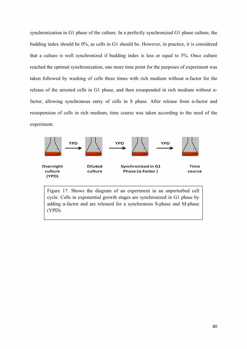
80
synchronization in G1 phase of the culture. In a perfectly synchronized G1 phase culture, the
budding index should be 0%, as cells in G1 should be. However, in practice, it is considered
that a culture is well synchronized if budding index is less or equal to 5%. Once culture
reached the optimal synchronization, one more time point for the purposes of experiment was
taken followed by washing of cells three times with rich medium without α-factor for the
release of the arrested cells in G1 phase, and then resuspended in rich medium without α-
factor, allowing synchronous entry of cells in S phase. After release from α-factor and
resuspension of cells in rich medium, time course was taken according to the need of the
experiment.
Figure 17. Shows the diagram of an experiment in an unperturbed cell cycle. Cells in exponential growth stages are synchronized in G1 phase by adding α-factor and are released for a synchronous S-phase and M-phase (YPD).

81
12.3 Experiments involved with over-expression of proteins
To study the behaviour of cell cycle progression when specific proteins were over-expressed
during the cell cycle, we carried out experiments in rich medium, however poor source of
carbon was used (Raffinose) such that, induction by better source of carbon would induce the
protein of interest under galactose-inducible, GAL1, 10. promoter, as shown in (Figure 18).
A culture was inoculated from a fresh single colony in an incubator with orbital shaking at
200 RPM for overnight. The next morning density of culture was determined by counting the
cells and diluted in rich medium (YPRaf) to 5.106 cells / ml in a volume appropriate to the
size and number of aliquots required for the experiment. However, instead of YPD whose
main source of carbon is dextrose, YPRaf was used in these experiments, having raffinose as
the main source of carbon.
The cells were allowed to grow until the cell density was doubled (one generation time),
about 1.107 cells / ml. At this junction, first time point aliquot of growing asynchronous cells
was taken and the rest of the cell culture was synchronized in G1 phase by adding 50 ng / ml
α-factor ( for bar1Δ strains) to the medium. After allowing cells to grow in presence of α-
factor for 2.5 hours, cells were checked for proper synchronization in G1 phase. This was
achieved by making 1:10 dilution of some of the culture with water and counted using
Neubauer chamber, determining the budding index (BI). The budding index is number of
budded cells divided by total number of cells multiplied by 100 which indicates the level of
synchronization in G1 phase of the culture. In a perfectly synchronized G1 phase culture, the
budding index should be 0%, as cells in G1 should be. However, in practice, it is considered
that a culture is well synchronized if budding index is less or equal to 5%. Once culture
reached the optimal synchronization, one more time point for the purposes of experiment was
taken. In order to induce the gene of interest at will, such that the protein of interest was over-

82
expressed, powdered Galactose was added to the culture to the final 2% sugar concentration.
This step counted for induction and cells were allowed to grow in galactose media for 1 hour.
After finishing the induction of cells for 1 hour, one more time point was taken for
experimental purposes. Finally cells were washed three times with rich media without α-
factor containing galactose for the release of the arrested cells in G1 phase. Once cells were
released, they were resuspended in YPGal and time course was taken.
Figure 18. Shows the diagram of an experiment with galactose induction of genes in cell cycle. Cells in exponential growth stages are synchronized in G1 phase by adding α-factor, followed by induction by adding galactose and then released for a synchronous S-phase and M-phase (YPGal).

83
12.4 Experiments involved with temperature sensitive mutant proteins
To study the behaviour of cell cycle progression when specific proteins with temperature
sensitive mutations were used during the cell cycle, we carried out experiments in rich
medium YPRaf at 24 ° C (permissive temperature) as shown in (Figure 19).
A culture was inoculated from a fresh single colony in an incubator with orbital shaking at
200 RPM for overnight at 24 ° C. The next morning density of culture was determined by
counting the cells and diluted in rich medium (YPRaf) to 5.106 cells / ml in a volume
appropriate to the size and number of aliquots required for the experiment.
The cells were allowed to grow until the cell density was doubled (one generation time),
about 1.107 cells / ml. At this junction, first time point aliquot of growing asynchronous cells
was taken and the rest of the cell culture was synchronized in G1 phase by adding 50 ng / ml
α-factor ( for bar1Δ strains) to the medium. After allowing cells to grow in presence of α-
factor for 3 hours, cells were checked for proper synchronization in G1 phase. This was
achieved by making 1:10 dilution of some of the culture with water and counted using
Neubauer chamber, determining the budding index (BI). In a perfectly synchronized G1
phase culture, the budding index should be 0%, as cells in G1 should be. However, in
practice, it is considered that a culture is well synchronized if budding index is less or equal
to 5%. Once culture reached the optimal synchronization, one more time point for the
purposes of experiment was taken. Before induction, cell cultures were shifted from
permissive temperature of 24 ° C to non permissive temperature of 38 ° C. Cell cultures were
kept in non permissive temperature for 1 hour, enough time to bring about conformational
change into the thermo-sensitive proteins that renders them non-functional. In order to induce
the gene of interest at will, such that the protein of interest was over-expressed, powdered
Galactose was added to the culture to the final 2% sugar concentration while keeping cells at

84
non-permissive temperature. This step counted for induction and cells were allowed to grow
in galactose media for 1 hour. After finishing the induction of cells for 1 hour, one more time
point was taken for experimental purposes. Finally cells were washed three times with pre-
warmed rich media without α-factor containing galactose for the release of the arrested cells
in G1 phase. Once cells were released, they were resuspended in pre-warmed YPGal and
time course was taken while keeping cells in non-permissive temperature.
13. Biochemical Techniques
13.1 Total protein extraction from yeast cells using trichloroacetic acid (TCA)
Total protein extraction is an excellent method of extracting proteins from yeast cells
effectively, while inactivating proteases, kinases and phosphotases immediately in the
process.
The aliquots taken from experiments, either fresh or ones taken from -80 °C (thawed at first)
were resuspended in 200 µl of 20% (w / v) TCA. After resuspension, 200 µl of sand glass by
volume were added to cells which helped in breaking of cell wall by mechanical agitation
Figure 19. Shows the diagram of an experiment with temperature sensitive proteins and galactose induction of genes in cell cycle. Cells in exponential growth stages are synchronized in G1 phase by adding α-factor, followed by heat inactivation of thermo-sensitive proteins and induction by adding galactose and then released for a synchronous S-phase and M-phase (YPGal).

85
with sand glass. Cells were vortexed for 1 minute at maximum speed. Lysates were recovered
and washed twice with sand glass vortexing and fresh 200 µl of 5% (w / v) trichloroacetic
acid. Each volume of lysate added to the wash was recovered in order to maximize the
amount of protein present in the lysate and in between dead spaces of the sand glass.
The denatured proteins which were insoluble in trichloroacetic acid were easily recovered by
centrifuging the lysates at low speed of 3,000 RPM (Eppendorf centrifuge 5418) for 10
minutes at room temperature. Supernatant was discarded and pellets were dissolved in an
appropriate volume of buffer implementation for electrophoretic samples (50 mM Tris-HCl
pH 6.8, 8% (v / v) glycerol, 4% (v / v) 2 - mercaptoethanol, 1.6% (w / v) SDS, 0.008% (w /
v) bromophenol blue). The residual remains of TCA exceeding the buffering capacity of the
volumes of buffer commonly used was neutralized by balancing the pH of the samples by
adding 0.2 volumes of 1 M Tris-base (This step helps in preventing protiolysis of samples in
following step). Finally the samples were boiled for 5 minutes and then centrifuged at 3,000
RPM (Eppendorf 5418, centrifuge) for 5 minutes at room temperature, which helped in
pelleting the insoluble material. The supernatants were either loaded immediately into a
PAGE gel electrophoresis or stored at -20 °C for future use.
13.2 Polyacrylamide gel electrophoresis (SDS-PAGE)
Sodium dodecyl sulfate -Polyacrylamide gel electrophoresis (SDS-PAGE), is a technique
widely used to separate the proteins according to their electrophoretic mobility (where
mobility is a function of the length, conformation and charge of the molecule) and there
molecular mass (Laemmli 1970).
Sodium dodecyl sulfate (SDS), an anionic detergent was applied to protein samples in order
to linearize and impart a negative charge to linearized proteins. Binding of SDS to the

86
proteins imparted an even distribution of charge per unit mass, thereby resulted in a
fractionation by approximate size during electrophoresis.
Gels of 1.5 mm thickness were used in this thesis while as different concentrations of
acrylamide 7.5%, 10%, 12%, and 15% were used depending upon the molecular weight of
the protein of interest. The ratio between acrylamide-bisacrylamide was always 37,5:1
(BioRad). Samples from TCA (explained in 5.1) were used to run in these gels. The proteins
were scored according to their position on a gel with respect to the subsequent analysis of
molecular weight ladders, Broad Range Prestained Starndar (Fermentas) and Broad Range
Standard (BioRad).
Gels were run in electrophoresis buffer (25 mM Tris-glycine pH 8.3, 0.1% (w / v) SDS) using
a generator-Power Pac Basic (BioRad). Following are the conditions of voltage and time used
for different proteins:
• For the analysis of Sic1ΔN, 10% acrylamide, 15 minutes of stacking at 90 V and 60
minutes of 200 V.
• For the analysis of Cln2, 10% acrylamide, 15 minutes of stacking at 90 V and 75
minutes of 220 V.
• For the analysis of Clb5, 10% acrylamide, 15 minutes of stacking at 90 V and 65
minutes of 200 V.
• For the analysis of Clb2, 10% acrylamide, 15 minutes of stacking at 90 V and 60
minutes of 200 V.
• For the analysis of Swe1-13myc, 7.5 % acrylamide, 35 minutes of stacking at 120 V
and 65 minutes of 195 V.
• For the analysis of the two forms of phosphorylated Pol12 (86 and 91 kDa), 7.5%
acrylamide, 45 minutes at 120 V followed by 65 minutes 190 V.
87
• For the analysis of Cdc28 (34 kDa) 10% acrylamide, 15 minutes of stacking at 90 V
and 50 minutes at 200V.
13.3 Western Blotting
The proteins separated by SDS-PAGE were transferred onto nitrocellulose membranes
(Protran BA85) by semi-dry transfer (Panther Semidry Electrobotter, Owl Scientific). The
transfer machine used constant amperage, adjusting according to the percentage of gel and
the molecular mass of the protein to be transferred (2 to 3.2 mA/cm2) for 60 minutes. The
transfer buffer was modified, containing SDS to increase the efficiency of transfer and
alkaline pH, which minimized the number of cargo proteins that had zero to coincide with its
isoelectric point (50 mM Tris-glycine pH 9.1, 0.0373% (w / v) SDS, 20% (v / v) methanol).
Once proteins were transferred onto a membrane, the membrane was stained with Ponceau S
(0.2% (w / v) Ponceau S, 3% (w / v) TCA). Staining with Ponceau allowed the visualization
of the most abundant proteins transferred to the membrane suggesting the control for correct
transfer proteins, moreover, scanned images were used as a loading control. Removal of
staining was imperative for the efficient immunobloting and was achieved by washing the
membrane with TBST (100 mM Tris-HCl pH 7.4, 150 mM NaCl, 0.1% (v / v) Tween-20) till
membrane was clean for Ponceau staining.
The membranes were blocked by incubating them in TBST + 5% (w / v) skimmed milk
powder, for 20 minutes on a shaker. Blocking of membrane by milk prevented the binding of
an antibody to nonspecific parts of the membrane. Blocking agent (milk) was decanted and
membranes were incubated in a dilution of primary antibody against specific protein or
against the tag fused to the specific protein to be detected for a 120 minutes with gentle
agitation on a shaker to promote uniform interaction of antibody with proteins on membrane

88
The primary antibodies and dilutions used in this study:
• Anti-Cln2 (Santacruz y-115, # sc-33624), 1:1000 dilution in TBST + 5% milk.
• Anti-Sic1 (Santacruz FL-284, # sc-50441), 1:1,000 dilution in TBST+5% milk.
• Anti-Clb5 (Santacruz yN-19, # sc-6704), 1:1,000 dilution in TBST+5% milk.
• Anti-Clb2 (Santacruz y-180, # sc-9071), 1:2,000 dilution in TBST+1% milk.
• 9E10 α-myc (mouse monoclonal, Roche), 1:2,000 dilution in TBST + 1% milk.
• 9E10 α-myc (monoclonal mouse hybridoma supernatant) diluted 1:500 in TBST.
• 12CA5 α-HA (mouse monoclonal, Roche) diluted 1:1000 in TBST + 1% milk.
• 12CA5 α-HA (monoclonal mouse hybridoma supernatant) diluted 1:100 in TBST.
• 6D2 Pol12-α (mouse monoclonal, given by Dr. Foiana Marco), dilution 1:2000 in
TBST + 1% milk.
• Anti-pY15-Cdk1 (Cell Signaling #9111, anti human pY15-Cdk1, cross-reacts nicely
with budding yeast pY19-Cdc28), 1:1,000 dilution in TBST + BSA.
After incubation of membrane in primary antibody for 2 hours, primary antibody was
recovered and reused several times in the course of this study. Residual primary antibody was
cleaned by washing the membrane three times with TBST. Membranes were then incubated
in secondary antibody for 35 minutes. Secondary antibodies used in this study were as:
• For mouse primary antibodies, anti-mouse IgG conjugated to peroxidise Radish
Rustica (HRP) (Dako P0161) 1:5.000 dilution in TBST + 5% milk.
• For Goat primary antibodies, anti-goat IgG conjugated to HRP (Santa Cruz
Biotechnology sc-2020), diluted 1:2500 in TBST + 5% milk.
• For Rabbit primary antibodies, anti-Rabbit IgG-HRP (goat polyclonal, purified IgG,
Dako P0448), dilution in 1:2,000 in TBST + 1% milk

89
13.4 Flow cytometry (FACS, Fluorescent Activated Cell Sorting)
Flow cytometry or simply known as FACS is a technique which allowed us to quantify the
amount of DNA present in each cells in a sample. The results were achieved by staining the
DNA with a fluorescent dye propidium iodide which intercalates in between the DNA
molecules. With the help of this technique we determined the stage of the cell cycle cultures
were in as well as the degree of synchrony and the rate of progression of the replication and
cell division.
Before running the samples for cell sorting, cells were fixed, treated and stained. The sample
aliquots taken from the experiments at around 1.107 cells per sample were centrifuged at the
speed of 10000 RPM in an Eppendorf centrifuge 5418 and pellets were fixed with 1 ml of
70% (v / v) ethanol and incubated overnight at 4 ° C. On the next day, cells were rehydrated
by washing them twice with 1 ml of 50 mM Tris-HCl pH 7.8, while giving a pause for two
minutes between the washes to allow proper dissolving of pellet. Cells were incubated in the
same buffer in the presence of 200 mg / ml RNase A, overnight at 24 ° C. In order to make
propidium iodide seep into the cells, cell wall was partially made permeable by incubating
them in 5 mg/ml pepsin dissolved in 50 mN HCl for 30 minutes at 37 ° C. Pepsin solution
was activated prior to use by incubating at 37 ° C for 30 minutes. And same was inactivated
by a single wash in 1 ml of FACS buffer (200 mM Tris-HCl pH 7.8, 211 mM NaCl, 78 mM
MgCl2) to bring back to pH 7.8, in order to prevent the further degradation. All spins were
carried out at 10000 RPM in an Eppendorf centrifuge 5418, and the pellets with 1.107
cells/ml were dissolved in 250 µl of FACS buffer with 16 µg/ml propidum iodide. This cell
suspension was either used directly or stored at 4 ° C away from light.
For the analysis of flow cytometry, 50 µl of the cell suspension were added to the 1 ml of 50
mM Tris-HCl pH 7.8. Samples were sonicated for 9 seconds at 40W (sonicator Sonic

90
Dismembrator, Dynatech) to make sure that each reading corresponded to a single cell and
prevented for example two cells containing 1C DNA to be counted as a single particle with a
2C DNA content. Samples were analyzed on a FACScan apparatus (Becton-Dickinson),
reading the fluorescence of propidium iodide (FL2-H). The data represented graphically as
histograms of the number of events (Y) front-FL2 fluorescence H (X) for 20,000 events.
Graphs were plotted to see the progression of cell cycle were each peak represented the
corresponding time point in the experiment and content of DNA present, as shown in (Figure
20).
Figure 20. Shows the schematic diagram of a plot constructed on an experiment. 1C peaks represent the one content of DNA prior to replication, as cells pass through the cell cycle, upon replication DNA content is doubled and represented as two contents, 2C.

91
13.5 Microscopy
Microscopy was used to check the phenotype of cells as well as the behaviour of
chromosomes upon the course of experiments. Stained samples from flow cytometry were
checked under the microscope Nikkon eclipse 90i (CFI60 infinity optical system, i-Series
Support Tools consisting of EZSetup, iSetup, and iControl; Nikon NIS-Elements Software.)
which gave bright red flourescence of DNA material.
13.6 Immunoprecipitation (IP)
Immunoprecipitation is the method which helped in studying of interaction between two or
more proteins in a complex in-vivo of the cell. Instead of using harsh acid, TCA, mild lysis
buffer were used to extract the whole protein extract in its native condition.
Cell cultures with the number of cells at around 2.109 were centrifuged at 3000 RPM for 5
minutes in an (). Cell pellets were washed once with the lysis buffer (50 mM Tris-HCl pH
7.7, 75 mM NaCl, 1 mM EDTA, and 10 % (v / v) glycerol) and snap frozen using liquid
nitrogen and stored at -80 °C. Prior to the extraction of the proteins, cells were thawed on ice
cold water, and resuspended in 2 ml of cold lysis buffer , supplemented with protease
inhibitors (1 mM AEBSF, 0.15 mM aprotinin, 1 mM leupeptin, 1 mM pepstatin),
phosphatase inhibitors (0.5 mM sodium pyrophosphate, 2 mM NaF, 2 mM β-
glycerophosphate), 1 mM DTT and 0.1% (v / v) nonidet-P40.
1.5 ml of glass beads by volume were added to the solution and vortexed 12 times for 60
seconds each with a gap of 60 seconds incubation on ice, in cold room (4 °C). The 60
seconds of gap in between the agitation allowed cells to cool down and prevent denaturation

92
of proteins due to heat produced during agitation. The lysates were allowed to sit on ice in the
cold room for 60 minutes in order to maximize the extraction process. After incubation on
ice, in order to extract the proteins and get rid of insoluble debris, cells were transferred to
cold eppindorfs, and centrifuged at 14000 RPM for 30 minutes. The supernatant containing
the total cell extract in its native conditions were transferred to a fresh cold eppindorf.
The immunoprecipitated proteins were incubated with primary antibody 3F10 anti-3HA
Affinity Matrix (Roche). 3F10 is a monoclonal rat, with high affinity towards the HA
epitope, which gives better performance in immunoprecipitacions compared to 12CA5 that
were used in Immunoblotting. 20 µl of beads were incubated with the extract, where 10 µl
were the sephearose beads with antibody attached and 10 µl was the storing solution of the
antibody. The cell lysates were incubated with 3F10 for 1 h at 4 ° C on the rotation block.
After this time, the mixture was centrifuged at 500 g, and extracts were removed, the
immunoprecipitates were washed five times each with 1 ml of cold lysis buffer to remove
excess proteins which did not get attached to the antibody. Finally, the proteins were released
by boiling the samples in presence of Lammeli buffer. Protein-protein interactions were
checked by running SDS- PAGE followed by immunoblotting.
13.7 In-vitro kinase assay
In-vitro kinase assay was used to measure the kinase activity of given proteins. In order to
carry out In-vitro kinase assay, samples were collected, processed and immunoprecipitated as
explained in previous segment.
Before carrying out kinase assay, last wash of cell samples with protein of interest bound to
the beads is washed once with 25 mM MOPS buffer. Moreover, instead of incubating into 20
µl of antibody 3F10 anti-3HA Affinity Matrix (Roche), cell lysates were incubated with 40 µl
of antibody. Such that, half of the samples were kept for western blotting while as other half

93
were used for kinase assay. 6 µl of HBII buffer (5 mM EGTA, 15 mM MgCl2, 15 mM
MOPS, 1 mM Na2VO3, 1 mM AEBSF, 0.15 mM aprotinin, 1 mM leupeptin, , 7 mM β-
glycerophosphate, 1 mM DTT, ) was added to the samples and incubated at room
temperature for 15 minutes, followed by addition of 10 µl of reaction buffer. Reaction buffer
is the 1:1 ration of two solutions, S1 and S2. S1: 100 mM ATP and 5 µCi of γ-[32P] - ATP
(Amsersham, GE Healthcare), S2: 4 mg/ml Histone H1 in 50 mM mops as a substrate.
Samples were incubated at 30 °C for 15 minutes. Finally adding 5 µl of sample buffer,
samples were boiled for 3 minutes. Kinase activity was checked by running SDS-PAGE
followed by staining/destaining of the gel. At last, autoradiograph gel was exposed to a
photographic film sensitive towards β radiation emitted by 32P incorporated into protein
phosphorylation.

94
Table 1. Strains used in this study
Name Genotype
W303-1a MATa ade2-1 ura3-1 his3-11,15 leu2-3,112 can1-100
YAM18 W303-1a MATa ade2-1 ura3-1 his3-11,15 leu2-3,112 can1-100 bar1::URA3
GAL-Cln2-(7A)-NlsMycNls (TRP1)
YAM19 W303-1a MATa ade2-1 ura3-1 leu2-3,112 can1-100 bar1::URA3 GAL-Sic1C70
(HIS3)
YAM20 W303-1a MATa ade2-1 ura3-1 leu2-3,112 can1-100 bar1::URA3 Cln2(7A)-
NlsMycNls (TRP1) GAL-Sic1C70 (HIS3)
YAM22 W303-1a MATa ade2-1 ura3-1 his3-11,15 trp1-1 leu2-3,112 can1-100 Clb6-del
(KANMX6) bar1-del GAL-Cln2-(7A)-NlsMycNls (TRP1)
YAM24 W303-1a MATa ade2-1 ura3-1 trp1-1 leu2-3,112 can1-100 bar1-del
his3::clb5::HIS3
YAM27 W303-1a MATa ade2-1 ura3-1 trp1-1 leu2-3,112 can1-100 bar1-del
his3::clb5::HIS3 GAL-Sic1DelN (URA3)
YAM29 W303-1a MATa ade2-1 ura3-1 his3-11,15 trp1-1 leu2-3,112 can1-100 Clb6-del
(KANMX6) bar1-del GAL-Sic1DelN (URA3)
YAM33 W303-1a MATa ade2-1 ura3-1 trp1-1 leu2-3,112 can1-100 bar1-del clb5::HIS3
GAL-Sic1DelN (URA3) GAL-Cln2-(7A)-NlsMycNls (TRP1)
YAM35 W303-1a MATa ade2-1 ura3-1 his3-11,15 trp1-1 leu2-3,112 can1-100 Clb6-del
(KANMX6) bar1-del GAL-Sic1DelN (URA3) GAL-Cln2-(7A)-NlsMycNls
(TRP1)
YAM36 W303-1a MATa ade2-1 ura3-1 his3-11,15 trp1-1 leu2-3,112 can1-100 bar1-del

95
GAL-Sic1delN (URA3)
YAM42 W303-1a MATa ade2-1 ura3-1 his3-11,15 leu2-3,112 can1-100 bar1-del GAL-
Cln2-(7A)-NlsMycNls (TRP1)
YAM43 W303-1a MATa ade2-1 ura3-1 his3-11,15 leu2-3,112 can1-100 bar1-del. GAL-
Cln2-(7A)-NlsMycNls (TRP1) Gal-Sic1delN (URA3)
YAM44 W303-1a MATa ade2-1 ura3-1 trp1-1 leu2-3,112 can1-100 bar1-del
his3::clb5::HIS3 clb6-del (kanmx) GAL-Sic1DelN (URA3)
YAM46 15Dau Mata ura3Dns clb1::URA3 clb2::LEU1 clb3::KanMx6 clb4::HIS2
clb5::ARG4 clb6::ADE1 GAL-CLB1:LEU2
YAM50 W303-1a MATa ade2-1 ura3-1 his3-11,15 leu2-3,112 can1-100 bar1-del GAL-
Cln2-(7A)-NlsMycNls (TRP1) GAL-Sic1delN (URA3)
YAM52 W303-1a MATa ade2-1 ura3-1 his3-11,15 leu2-3,112 can1-100 bar1-del. GAL-
Sic1delN (URA3), Swe1-13Myc (KanMX)
YAM53 W303-1a MATa ade2-1 ura3-1 his3-11,15 leu2-3,112 can1-100 bar1-del. GAL-
Cln2-(7A)-NlsMycNls (TRP1) GAL-Sic1delN (URA3) Swe1-13Myc (KanMX)
YAM54 W303-1a MATa ade2-1 ura3-1 his3-11,15 leu2-3,112 can1-100 bar1-del. GAL-
Sic1delN (URA3) Cdc28-3HA (KanMX)
YAM55 W303-1a MATa ade2-1 ura3-1 his3-11,15 leu2-3,112 can1-100 bar1-del GAL-
Cln2-(7A)-NlsMycNls (TRP1) GAL-Sic1delN (URA3) Cdc28-3HA (KanMX)
YAM56 W303-1a MATa ade2-1 ura3-1 his3-11,15 leu2-3,112 can1-100 bar1-del. GAL-
Sic1delN (URA3) Clb2-del::Leu2
YAM57 W303-1a MATa ade2-1 ura3-1 his3-11,15 leu2-3,112 can1-100 bar1-del. GAL-
Cln2-(7A)-NlsMycNls (TRP1) Sic1delN (URA3) Clb2-del::Leu2
YAM58 W303-1a MATa ade2-1 ura3-1 his3-11,15 leu2-3,112 can1-100 bar1-del GAL-
Sic1delN (URA3) Clb6-13Myc (KanMX)

96
YAM59 W303-1a MATa ade2-1 ura3-1 his3-11,15 leu2-3,112 can1-100 bar1-del. GAL-
Cln2- (7A)-NlsMycNls (TRP1) Gal-Sic1delN (URA3) Clb6-13Myc (KanMX)
YAM60 15Dau Mata ura3Dns clb1::URA3 clb2::LEU1 clb3::KanMx6 clb4::HIS2
clb5::ARG4 clb6::ADE1 GAL-CLB1:LEU2
YAM61 W303-1a MATa ade2-1 ura3-1 his3-11,15 leu2-3,112 can1-100 bar1-del. Gal
Sic1delN (URA3) Swe1-13Myc (KanMX) Hsl1-del::Leu2
YAM62 W303-1a MATa ade2-1 ura3-1 his3-11,15 leu2-3,112 can1-100 bar1-del. . GAL-
Cln2-(7A)-NlsMycNls (TRP1) Gal-Sic1delN (URA3), Swe1-13Myc (KanMX),
Hsl1-del::Leu2
YAM64 W303-1a MATa ade2-1 ura3-1 trp1-1 leu2-3,112 can1-100 bar1-del
his3::clb5::HIS3 . GAL-Cln2-(7A)NlsMycNls (TRP1) GAL-Sic1DelN (URA3)
YAM65 W303-1a MATa ade2-1 ura3-1 trp1-1 leu2-3,112 can1-100 bar1-del
his3::clb5::HIS3 GAL-Cln2-(7A)-NlsMycNls (TRP1) clb6-del (kanmx) GAL-
Sic1DelN (URA3)
YAM66 W303-1a MATa ade2-1 ura3-1 his3-11,15 leu2-3,112 can1-100 bar1-del. GAL-
Sic1delN (URA3) Clb2-3HA (KanMX)
YAM67 W303-1a MATa ade2-1 ura3-1 his3-11,15 leu2-3,112 can1-100 bar1-del. GAL-
Sic1delN (URA3) Clb5-3HA (KanMX)
YAM68 W303-1a MATa ade2-1 ura3-1 his3-11,15 leu2-3,112 can1-100 bar1-del. GAL-
Cln2-(7A)-NlsMycNls (TRP1) GAL-Sic1delN (URA3) Clb2-3HA (KanMX)
YAM69 W303-1a MATa ade2-1 ura3-1 his3-11,15 leu2-3,112 can1-100 bar1-del. GAL-
Cln2-(7A)-NlsMycNls (TRP1) GAL-Sic1delN (URA3) Clb5-3HA (KanMX)
YAM70 15Dau Mata ura3Dns clb1::URA3 clb2::LEU1 clb3::KanMx6 clb4::HIS2
clb5::ARG4 clb6::ADE1 GAL-CLB1:LEU2, tetO7-Cln2-(7A)-NlsMycNls
(TRP1)

97
YAM71 W303-1a MATa ade2-1 ura3-1 his3-11,15 trp1-1 leu2-3,112 can1-100 clb1-del
(without marker) clb2ts (allele VI) CDC14-3HA ura3::bar1::URA3 (Counter
selected)
YAM72 W303-1a MATa ade2-1 ura3-1 his3-11,15 leu2-3,112 can1-100 bar1-del. tetO7-
Cln2-(7A)-NlsMycNls (TRP1) Gal-Sic1delN (URA3)
YAM73 W303-1a MATa ade2-1 ura3-1 his3-11,15 leu2-3,112 can1-100 bar1-del. GAL-
Cln2-(7A)-NlsMycNls (TRP1) GAL-Sic1 (9A)
YAM74 W303-1a MATa ade2-1 ura3-1 his3-11,15 trp1-1 leu2-3,112 can1-100 clb1-del
(without marker) clb2ts (allele VI) CDC14-3HA ura3::bar1::URA3 (Counter
selected), GAL-Cln2-(7A)-NlsMycNls (TRP1)
YAM75 W303-1a MATa ade2-1 ura3-1 his3-11,15 leu2-3,112 can1-100 bar1-del GAL-
Cln2-(7A)(WITHOUT, NLS) (TRP1) GAL-Sic1delN (URA3)
YAM76 W303-1a MATa ade2-1 ura3-1 his3-11,15 trp1-1 leu2-3,112 can1-100 bar1-del,
GAL-Sic1(9A) (URA3)
YAM77 W303-1a MATa ade2-1 ura3-1 his3-11,15 trp1-1 leu2-3,112 can1-100 clb1-del
(without marker) clb2ts (allele VI) CDC14-3HA ura3::bar1::URA3 (Counter
selected), GAL-Sic1delN (URA3)
YAM78 W303-1a MATa ade2-1 ura3-1 his3-11,15 trp1-1 leu2-3,112 can1-100 clb1-del
(without marker) clb2ts (allele VI) CDC14-3HA ura3::bar1::URA3 (Counter
selected) GAL-Cln2-(7A)-NlsMycNls (TRP1)

98
Table 2. Constructs used in this study
Name Construct description
pAM1 pRS306-GAL-MCS-13Myc Vector to clone ORFs under Gal promoter
(to be expressed as C-ter 13 myc tag fusion).
pAM2 pRS306-GAL-Sic1delN
pAM3 pRS304-GAL-CLN2-NLS-1xMyc-NLS
pAM9 pRS304GAL-CLN2 (7A)-NLSmycNLS
pAM10 pCM252- CLN2 (7A)-NLSmycNLS
pAM11 pRS306- GAL Sic1 (9A)
pAM12 pRS304-GAL Clb2
pAM13 pRS304-GAL-CLN2(7A) without NLS

99
Table 3. Oligonucleotides used as primers in this study
Name Sequence Strain/Plasmid
Sic1ΔN-F CGGATCCATGTCTTTTAAAAATGCGCCATT
pRS306-GAL-Sic1ΔN Sic1ΔN-R TCTCGCGGCCGCTCACAAGTCTTCCTCGGAGATTAGCTTTTGTTCATGC
TCTTGATCCCTAGATTG
Cln2-NlsmycNls- F
CGGGATCC ATGGCTAGTGCTGAACCAAG
pRS304-GAL-Cln2NLSmycNLS Cln2-
NlsmycNls- R1 CTCGGAGATTAGCTTTTGTTCGCCTTCCACCTTGCGCTTTTTCTTAGGTGGACCACTGCGTATTACTTGGGTATTGCCCATACC
Cln2-NlsmycNls- R2
TCTCGCGGCCGCTCACCTCGATCCACCCTCAACTTTTCTTTTCTTTTTGGGCGGACCCAAGTCTTCC
TCGGAGATTAGCTTTTGTTC
Cln2-NlsmycNls- F
CGGGATCCATGGCTAGTGCTGAACCAAG
pRS304-GAL-Cln2-(7A) NLSmycNLS
Cln2-NlsmycNls- R1
CTCGGAGATTAGCTTTTGTTCGCCTTCCACCTTGCGCTTTTTCTTAGGTGGACCACTGCGTATTACTTGGGTATTGCCCATACC
Cln2-NlsmycNls- R2
TCTCGCGGCCGCTCACCTCGATCCACCCTCAACTTTTCTTTTCTTTTTGGGCGGACCCAAGTCTTCCTCGGAGATTAGCTTTTGTTC
Sic1C70- F CGGATCCATGGGTAAAAATCCCTTTGCATC
pRS303-GAL- Sic1C70 Sic1C70-R TCTCGCGGCCGCTCAATGCTCTTGATCCCTAGATTG
Clb3 Del-F (KNMX)
GCAACTGGCTCACCTACCCAC
YAM46 Clb3 Del-R (KNMX)
CCTCCAGAAAAGTCAATACAC
Hsl1 del-F CCAACTATATTCACTCCTTTATTATTTGTTATTAATATTTTTTATTTTTA
CCACACGACGCGGTATTTTCTCCTTACGC
YAM61/62
YAM61/62
Hsl1 del-R CTCGTACGTTAAATTTTTCAAATTATTGTTGTATAATTATATAACATCTATATAGAATAGATTGTACTGAGAGTGCAC
Hsl1 CHK- F GACAAGGTAAAAAGAACGAC
Hsl1 CHK- R CTTCGTGTCTCATGTCTCTCC
Cln2- R TCTCGCGGCCGCCTATATTACTTGGGTATTGC YAM 75
Clb2-F CGGATCCATGTCCAACCCAATAGAAAACAC
pRS304-GAL- Clb2 Clb2-R TCTCGCGGCCGCTCATTCATGCAAGGTCATTAT
Clb2p-Fa6a-F (3HA)
TCCGTCCAATGGGCTTTAAAGGTTAGAAAAAACGGCTATGATATAATGACCTTGCATGAACGGATCCCCGGGTTAATTAA
YAM 66/68 Clb2-pFa6a-R (3HA)
GGTGTGAAATTTGTAAGAATAAATGGAAATGTGAAAAGAAATGATTAAAAAGCAGCCATAGAATTCGAGCTCGTTTAAAC
Clb2-del-F TATTTCCTTCCAAGAAGCCTTTTATTGATTACCCCCTCTCTCTCTTCATTGATCTTATAGGCGGTATTTTCTCCTTACGC

100
Clb2-del-R CATTTTATATGGACATTTATCGATTATCGTTTTAGATATTTTAAGCATCTGCCCCTCTTCAGATTGTACTGAGAGTGCAC
YAM56/57 chk-clb2-del-F AACCTACTGCGAGCTATTCA
chk-clb2-del-R CGGAAACGGTTGAACTGAAA
CLB5pfa6aF (3HA)
ACTTTCAAGTGGTGTACATCCGAAATGCATAGCAACTTTCAAAATCTATTTAATCTTAAGCGGATCCCCGGGTTAATTAA
YAM67/69 CLB5pfa6aR (3HA)
ACATATATATAGATAGATAGATAGATGCAAAAAATTTTTTCCTTTTAGTTCAGCAAAAAGGAATTCGAGCTCGTTTAAAC
chk-clb5-del-F) CTTCAAGAGACTCAATCACC
YAM27 (chk-clb5-del-R) GAAGTGAACACGCATATCAC
Clb5-del-F GCTTAATAATTAGCAGTAACGCGCTTTTCCCTGTATTTAAAGCCGCTGAACACCTTTACGCGGTATTTTCTCCTTACGC
Clb5-del-R GATGATAATAGTAGTAATACTGGTGGTATTATATATGTTGAAGTGAACACGCATATCACAGATTGTACTGAGAGTGCAC
Cdc28-pfa6a-F (3HA)
ATTAGCGCCAGAAGAGCAGCCATCCACCCCTACTTCCAAGAATCACGGATCCCCGGGTTAATTAA
YAM54/55 Cdc28-pfa6a-R (3HA)
GCAGTAGCATTTGTAATATAATAGCGAAATAGATTATAATGCTTAGAATTCGAGCTCGTTTAAAC
Clb3 Del-F (NAT1)
CATGAAGCGCGCCATCGACATATACAACAGCGAAGGCCAGCAACTGGCTCACCTACCCACCGTACGCTGCAGGTCGAC
YAM82 Clb3 Del-R (NAT1)
AGAACACCCGAGTTTTCCTCCAGAAAAGTCAATACACATTACAATCTCCTCGTCAAAATCATCGATGAATTCGAGCTCG
Swe1-13Myc-F CGCAATGCAGGTGCTATTATCCAGGAAGACGACTTTGGACCTAAGCCAAAATTTTTTATA
YAM52/53 Swe1-13Myc-R TAGAGCGGCCGCCACCGCGGTGGAGCTCCAATTCGCCCTATAGTGAGT
CGTATTACAATTATGCTTGAAGCGGCTGTACT
Clb6-del-F GCTTCACTAATCTAGCGTTC
YAM 45/65 Clb6-del-R AAGATGCAGGGGGTTAGCTGG
Clb6-Chk-F GCTAGGGTTAAGTTTTTGCC
Clb6-Chk-R CACGGTATCAGTTTTGTAGG

101

102
RESULTS

103

104
Results:
14. Triggering of later events by early cylins
Each cyclin associates with its catalytic subunit cyclin dependent kinase to perform its
function in phase specific manner, however, it is still uncertain, why early cyclins cannot
trigger later events in cell cycle progression. Theory that suggests that increasing levels of
cyclin dependent kinase activity is responsible for driving the cell cycle, then, prediction, that
over-expressing early cyclin at the right time, to the right sub cellular location should bring
about later event. To test this theory, we explored if G1 cyclin would be able to trigger
replication which is a latter event with respect to the G1 phase when over-expressed and
localised into the nucleus.
In order to study if G1 cyclin might trigger the replication, experiments were carried out in
strains where G1 cyclin Cln2 tagged with two nuclear localization signal (NLS) sequences at
the C-terminal and hyperstable form of all Clbs inhibitor Sic1ΔN, whose 50 amino acid long
N-terminal sequence was deleted (explained in detail in section 5), were expressed under
galactose-inducible promoter, GAL1, 10. Since Sic1ΔN is the strong inhibitor of S-CDK and
M-CDK activity, strain consisting of only Sic1ΔN under galactose-inducible promoter
blocked the cell cycle progression at the G1-S transition and thus acted as the control strain.
While as strains consisting of both Sic1ΔN and Cln2- NlsmycNls under the galactose-
inducible promoter acted as the experimental strain. Strains were checked in unperturbed
conditions for proper cell cycle progression, and for integrated Sic1ΔN by functional assay
prior to experiments (Figure 21).

105
However, flow cytometry results showed that over-expressing nuclear Cln2 in experimental
strain could not trigger the replication in presence of an all Clb inhibitor. The reason behind
the failure of Cln2 to trigger replication was lack of effective accumulation of protein due to
its quick degradation by self regulating properties (Figure 22).
Figure 21. A. Flow cytometry shows the proper cell cycle progression in unperturbed conditions. B. Over-expresion of Sic1ΔN blocks cell cycle progression in both control and experimental strains.

106
15. Hyperstable Cln2 triggers replication in absence of S and M-CDK activity
Since Cln2 was being self regulated at faster speed compared to its expression, the desired
accumulation required for the purpose of increasing cyclin dependent kinase activity was not
achieved. Hence, hyperstable form of Cln2 was used whose all seven S-P/T (Serine
threonine) consensus sites for CDK phosphorylation were mutated to non-phosphorylatable
form, alanine. With help of these mutations, protein was still functional, however, could not
be identified and degraded by SCF ubiquitin ligase proteasome due to absence of
phosphorylation of given sites required for identification of PEST site on Cln2 by F-box
Grr1, thus rendering protein hyperstable.
Experiment was carried out in strains where hyperstable Cln2 (henceforth known as
Cln2(7A)) tagged with two nuclear localization signal (NLS) sequences at the C-terminal and
hyperstable form of all Clbs inhibitor Sic1ΔN, whose N-terminal degron sequence was
deleted, were expressed under galactose-inducible promoter, GAL1, 10. Since Sic1ΔN blocks
the S-CDK and M-CDK activity, strain consisting of only Sic1ΔN under galactose-inducible
Figure 22. A. Flow cytometry results show, Cln2 was unable to trigger replication when over-expressed and localised to nucleus. B. Western blots shows accumulation of Sic1ΔN, however, no sizeable accumulation of nuclear Cln2 protein. Ponceau staining was used as loading control.

107
promoter blocked the cell cycle progression at the G1-S transition and thus acted as the
control strain. While as strains consisting of both Sic1ΔN and Cln2(7A)-NlsmycNls under
the galactose-inducible promoter acted as the experimental strain. Strains were checked in
unperturbed conditions for proper cell cycle progression, and for integrated Sic1ΔN by
functional assay prior to experiments. Functional assay suggested that both strains were not
viable upon over-expression of strong Clb inhibitor, while cells progressed in wild type form
under unperturbed conditions (Figure 23).
Figure 23. A. Flow cytometry shows the proper cell cycle progression in unperturbed conditions. B. Over-expresion of Sic1ΔN blocks cell cycle progression in both control and experimental strains.

108
Results showed the proper accumulation of hyperstable all Clbs inhibitor Sic1ΔN as well as
hyperstable form of nuclear Cln2(7A) upon the induction of galactose-inducible promoter.
Flow cytometry results showed that nuclear, hyperstable Cln2(7A) triggered the replication in
absence of S and M-CDK activity as showen in (Figure 24).
Microscopic results showed the phenotype of cells compatible with effects of over-expression
of strong Clb inhibitor Sic1ΔN. Cells released from α- factor arrest in G1 phase and upon
induction into the medium containing galactose showed the phenotype with elongated buds
due to their polar growth and failure to switch to isotropic growth because of absence of S
and M-CDK activity. On the other hand, the phenotype of replicating cells was not as drastic
as shown by control cells (Figure 25).
Figure 24 A. Western blots shows accumulation of Sic1ΔN, however, no sizeable accumulation of nuclear Cln2 protein. Ponceau staining was used as loading control. B.. Flow cytometry results show, Cln2 (7A) triggered replication when over-expressed and localised to nucleus in presence of strong Clb inhibitor Sic1ΔN.
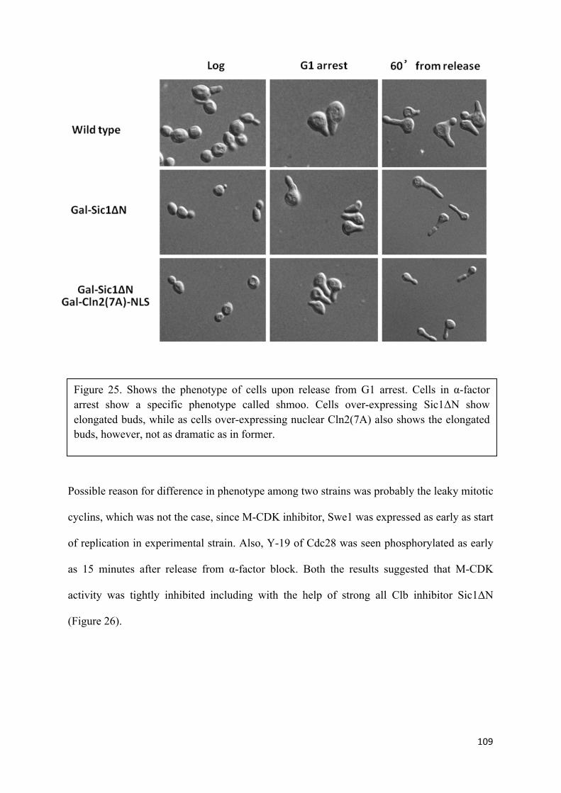
109
Possible reason for difference in phenotype among two strains was probably the leaky mitotic
cyclins, which was not the case, since M-CDK inhibitor, Swe1 was expressed as early as start
of replication in experimental strain. Also, Y-19 of Cdc28 was seen phosphorylated as early
as 15 minutes after release from α-factor block. Both the results suggested that M-CDK
activity was tightly inhibited including with the help of strong all Clb inhibitor Sic1ΔN
(Figure 26).
Figure 25. Shows the phenotype of cells upon release from G1 arrest. Cells in α-factor arrest show a specific phenotype called shmoo. Cells over-expressing Sic1ΔN show elongated buds, while as cells over-expressing nuclear Cln2(7A) also shows the elongated buds, however, not as dramatic as in former.

110
Due to the result of elongated buds by Sic1ΔN over-expression, it apparently was impossible
to count buds and give representation of budding index in these experiments. Moreover,
DNA staining by propidium iodide showed the early entrance into anaphase as well as
disintegration of chromosomes in control cells after the release from α- factor arrest, while as
replicating cells which over-express nuclear Cln2(7A) showed delayed or no anaphases with
intact DNA material. However, the reason behind this behaviour is not well understood yet,
as shown in (Figure 27).
Figure 26. Shows that tyrosine 19 of Cdc28 in replicating cells is phosphorylated early compared to non-replicating cells. Also Swe1, that phosphorylates the tyrosine19 was expressed in accord to phosphorylation seen in both replicating and non replicating cells, confirming, block of M-CDK activity.

111
16. Nuclear localization is essential for Cln2(7A) function in triggering replication
As shown in previous results, nuclear Cln2(7A) was properly accumulated compared to
wildtype Cln2 protein and culminated into triggering replication in absence of S and M-CDK
activity, however, it was imperative to explore if Cln2(7A) was required to be localized into
the nucleus.
Figure 27. Shows the stained DNA of control and experimental strain upon release from G1 arrest. Cells over-expressing Sic1ΔN show disintegrated chromosomes and early anaphases, while as cells over-expressing both, Sic1ΔN and nuclear Cln2(7A) show delayed anaphase and compact DNA material.

112
In order to check that, strains with background of Sic1ΔN were transformed with a copy of
CLN2(7A) gene under the galactose inducible promoter lacking nuclear localization signal
(NLS). Like previous experiments, strain with Sic1ΔN alone under the galactose inducible
promoter acted as control strain, while as, strain with Sic1ΔN and Cln2(7A) without NLS
under the galactose inducible promoter acted as experimental strain. Results showed that,
indeed, nuclear localization signal was pivotal for the Cln2(7A) to trigger replication. Even
though hyperstable protein Cln2(7A) was accumulated, at the phase level, almost equally as
in previous experiment, however, flow cytometry results showed no progression of
replication from one content DNA to two content. These results suggested that for Cln2(7A)
to do its function, it must be localized into the nucleus rather than cytoplasm (Figure 28).
Figure 28. A. Accumulation of hyperstable Cln2 without nuclear localization signal and Clb inhibitor Sic1∆N. B. Flow cytometry shows no replication in strains over-expressing hyperstable Cln2 lacking nuclear localization signal.

113
17. Sic1ΔN remains associated with CDK complex
Previous result showed that over-expressing nuclear Cln2(7A) in presence of strong Clb
inhibitor Sic1ΔN resulted in triggering of replication. However, it also raised the question of
possible override of Sic1ΔN by Cln2(7A), since Cln2 associated with Cdc28 in normal cell
cycle phosphorylates Sic1 and leads it for destruction by SCFCdc4 ubiquitin-mediated
proteolysis pathway.
In order to explore whether Sic1ΔN in experimental strain was associated with Cdc28
complex rather than been disassociated due to over-expression of nuclear hyperstable Cln2,
the CDK complex, Cdc28 was immunoprecipitated after one hour induction by galactose and
release from α-factor block in both control and experimental strains. Upon pooling Cdc28
and complex associated with it from the cells, IP western was carried out against S-phase
cyclin Clb5 and S and M- CDK inhibitor Sic1ΔN. Results showed the equal amounts of Clb5
and Sic1ΔN associated with Cdc28 in both control as well as experimental cells.
These results suggested that the replication was triggered by nuclear Cln2(7A) rather than
leaky S-phase cyclins which might have created an artifactual result due to disassociation of
Sic1ΔN from the complex (Figure 29).

114
18. Nuclear Cln2(7A) mediated replication in Sic1(9A) and Sic1C70 background
Sic1ΔN has profoundly been used as S and M-CDK inhibitor in several studies, however, in
our study, in order to reproduce our results, we used two other forms of hyperstable all Clb
inhibitor Sic1 to achieve same results as in Sic1ΔN.
Sic1 has nine potential phosphorylation sites, out of which minimum of six require to be
phophorylated in order to be targeted for degradation. In case of Sic1ΔN, first 50 amino acids
were truncated, such that first four T/P sites were lost, thus left with only 5 possible sites to
be phosphorylated which were not enough to target it for degradation. Therefore making
Sic1ΔN hyperstable. In case of Sic1(9A), all nine S-T/P sites were mutated into alanine, thus
rendering the protein hyperstable, since it could not be phosphorylated and targeted for
degradation by SCFCdc4 ubiquitin-mediated proteolysis pathway. Similarly, in case of
Sic1C70, only final C-terminal 70 amino acids were used as all Clb inhibitor while as rest of
the 216 amino acids were deleted (Figure 30).
Figure 29. Shows the equal amounts of Clb5 and Sic1ΔN associated with Cdc28 in both strains containing Gal- Sic1ΔN (control strain) as well as strains containing Gal- Sic1ΔN and Gal- Cln2(7A)-NLS (experimental cells)

115
Functional assay showed that both the inhibitors worked as efficiently as Sic1ΔN by blocking
cell cycle progression once over-expressed under galactose-inducible promoter on rich media
with galactose as a sugar source (Figure 31).
Figure 30. Shows the schematic diagram of different forms of all Clbs’s inhibitor Sic1. a) full length, wild type Sic1. b) Sic1∆N, hyperstable form of Sic1 made by truncating first 50 aminoacids, losing 4 T/P sites. c) Sic1(9A), hyperstable form of Sic1 having all S-T/P consensus sequence for CDK phosphorylation mutated to non-phosphorylating form. d) Sic1C70, hyperstable form of Sic1 having first 216 amino acids deleted losing all 9 consensus sequence for CDK phosphorylation.

116
Upon carrying experiments in strains containing Sic1(9A) and Sic1C70 alone as the control
strains and Sic1(9A) and Sic1C70 including nuclear Cln2(7A) as experimental strains, results
showed that, hyperstable nuclear Cln2 was able to trigger the replication in both the
backgrounds. Thus making our previous result more concrete and robust. (Figure 32 and 33).
Figure 31. A. shows the functional assay of strains with Sic1(9A) on YPGAL plates. B. Shows the functional assay of strains with Sic1C70 on YPGAL plates.

117
Figure 33. A. Shows the accumulation of nuclear hyperstable Cln2 B. Cytometry results showed that nuclear Cln2(7A) triggered replication in presence of all clb inhibitor Sic1C70.
Figure 32. A. Shows the accumulation of nuclear hyperstable Cln2 B. Cytometry results showed that nuclear Cln2(7A) triggered replication in presence of all clb inhibitor Sic1(9A).

118
19. S-phase cyclins play no role in replication mediated by nuclear Cln2(7A)
Although it is not very clear yet how MBF transcription factors are activated, however,
several studies have shown positive loop of Cln2-Cdc28, which helps in further transcription
of S-phase cyclins. In our study, due to presence of nuclear, hyperstable Cln2, it was
imperative to explore the probability of high expression of major S-phase cyclin Clb5, which
inadvertently, might override Sic1ΔN and be reason for replication. In order to investigate
such possibility, levels of Clb5 protein were checked in both control and experimental strains.
Immunoblotting results suggested that levels of S-phase cyclin, Clb5, were less if not more in
experimental strain compared to control strain (Figure 34 A). Moreover, to make such
observation rather robust, Clb5 associated with Cdc28 was immunoprecipitated and checked
for its kinase activity towards histone H1 as its substrate. In-vitro results showed that Clb5-
CDK complex had no kinase activity whatsoever in both replicating and non-replicating cells
(Figure 34 B).
Figure 34. A. Shows that expression of Clb5 in replicating cells is less compared to amount of Clb5 present in non-replicating cells. B. In vitro kinase assay showing no kinase activity by Clb5-CDK complex. absence of S-phase cylins.

119
Furthermore, since Clb5 was only one of the two S-phase cyclins, Clb6 was still available,
which is shown to have capability of triggering the replication in absence of Clb5 function. In
order to address this question, both, CLB5 and CLB6 genes were deleted from the strains
with back ground of Sic1ΔN (control strain) and Sic1ΔN; Cln2(7A)-NlsMycNls
(experimental strain). Functional assay and experiment in unperturbed conditions showed
block of cell cycle progression in cells overexpressing Sic1ΔN and late triggering of
replication due to S-phase null cyclins respectively (Figure 35). Experiments were carried on
both the strains and cytometry results showed that nuclear Cln2(7A) was still able to trigger
replication in experimental strain (Figure 36).
Figure 35. A. Flow cytometry shows cells trigger replication late in absence of S-phase cylins.
B. Functional assay of strains.

120
20. G1 cyclin Cln2 triggers later event of SFF activation
Like MBF, very less is known about the process of activation of SFF transcription factors.
However, several researchers have speculated the role of S-phase associated cyclin dependent
kinase activity play some role in it. According to our theory of quantity model, we presumed,
if nuclear Cln2(7A) triggered replication which is a later event, it must also trigger the
transcription of certain proteins which are expressed in later stages from G1-phase.
In order to explore this, major mitotic cyclin, Clb2, which is expressed from SFF
transcription factor was checked by immunoblotting in both control and experimental strains.
Results showed that indeed, Clb2 was expressed in replicating cells as early as at 15 minutes
from release from α-factor block compared to non replicating cells, which was at around 60
minutes from α-factor block (Figure 37 A). This result showed that early phase cyclin could
Figure 36. A. Accumulation of hyperstable Cln2(7A) and Sic1∆N in cells with back ground of S-phase null cyclins. B. Flow cytometry shows cells overexpressing Cln2(7A)-NLS trigger replication in presence of Sic1∆N and absence of S-phase cylins.
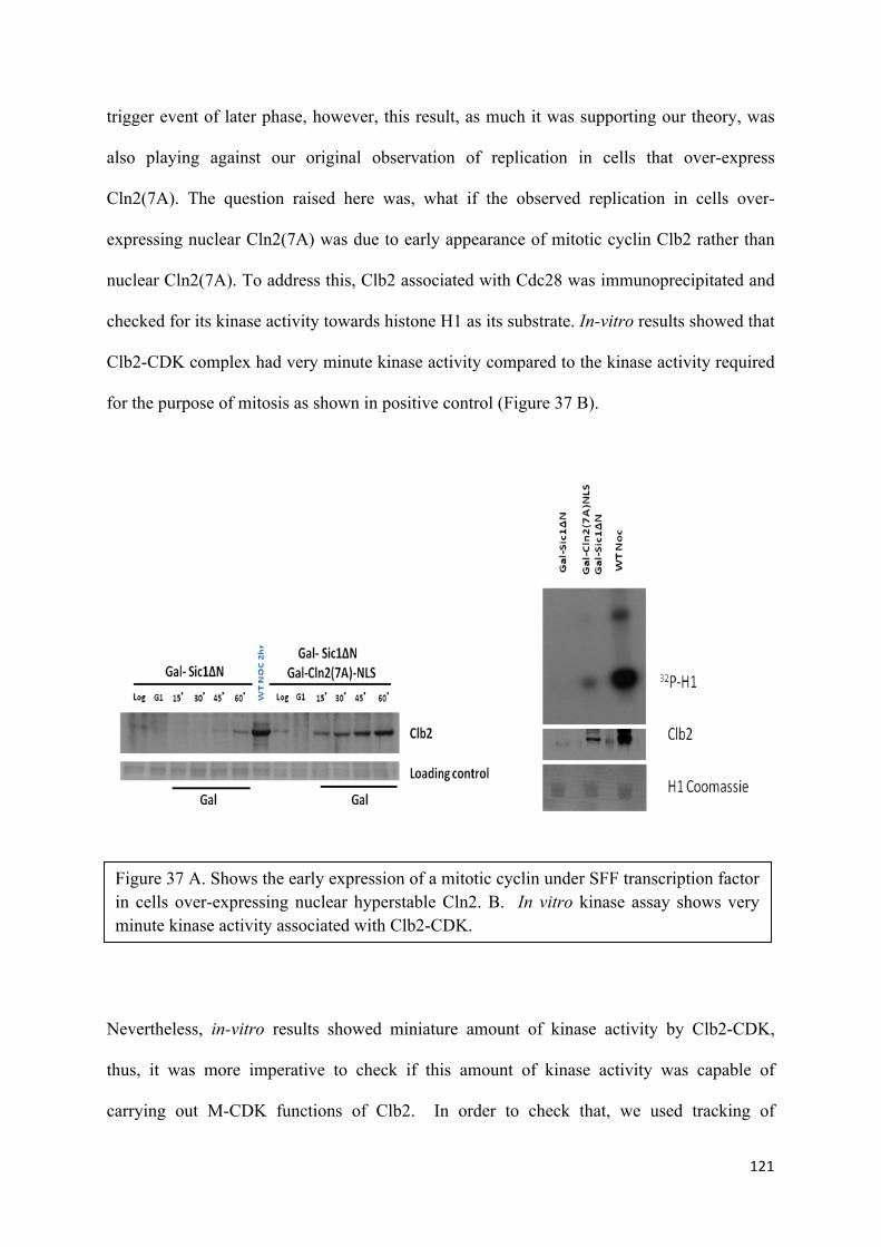
121
trigger event of later phase, however, this result, as much it was supporting our theory, was
also playing against our original observation of replication in cells that over-express
Cln2(7A). The question raised here was, what if the observed replication in cells over-
expressing nuclear Cln2(7A) was due to early appearance of mitotic cyclin Clb2 rather than
nuclear Cln2(7A). To address this, Clb2 associated with Cdc28 was immunoprecipitated and
checked for its kinase activity towards histone H1 as its substrate. In-vitro results showed that
Clb2-CDK complex had very minute kinase activity compared to the kinase activity required
for the purpose of mitosis as shown in positive control (Figure 37 B).
Nevertheless, in-vitro results showed miniature amount of kinase activity by Clb2-CDK,
thus, it was more imperative to check if this amount of kinase activity was capable of
carrying out M-CDK functions of Clb2. In order to check that, we used tracking of
Figure 37 A. Shows the early expression of a mitotic cyclin under SFF transcription factor in cells over-expressing nuclear hyperstable Cln2. B. In vitro kinase assay shows very minute kinase activity associated with Clb2-CDK.

122
phosphorylation of Pol12. Pol12 is a bona fide mitotic cyclin dependent kinase activity
substrate as tested in our lab (Gloria Palou, Roger Palou, and David G. Quintana, submitted).
The phosphorylation was checked by running western blot against Pol12 in several
experimental and control strains. All the cells, replicating and non-replicating cells showed
no phosphorylation of M-CDK substare whatsoever, suggesting, whatever residual or basal
mitotic activity, which was seen in kinase assay was not enough to bring about replication
(Figure 38).
21. M-CDK activity plays no role in nuclear Cln2(7A) mediated replication
Previous results have shown that M-CDK activity (Clb2-Cdc28) was very low, and by
checking M-CDK target phosphorylation (Pol12), it was apparent that this low amount of
kinase activity wasn’t enough to trigger the replication. However, to make results rather
Figure 38. No phosphorylation of M-CDK substrate Pol12 in cells over expressing Cln2(7A)NLS in back ground of Sic1∆N alone as well as Sic1∆N and clb5,clb6 deletion.

123
convincing and more robust we carried out same experiment in two different ways to achieve
convincing results, one in absence of major mitotic cyclin Clb2 expecting to see replication
and second more importantly in presence of over-expressed Clb2 in back ground of Sic1ΔN.
In first case, Clb2 was deleted from both the strains, control (Sic1ΔN alone) and experimental
strain (Sic1ΔN and Cln2(7A)NlS). Cytometry results showed that, cells over-expressing
nuclear Cln2(7A) triggered replication in presence of all Clb inhibitor Sic1N as well as
absence of Clb2 cyclin (Figure 39).
While in case of over-expressing Clb2 under the galactose-inducible promoter, over-
expressed Clb2, which was higher than levels of Cln2(7A)Nls at the phase value couldn over-
ride the Sic1ΔN and failed to trigger replication. These results reinforced the observation
Figure 39. . Shows cells replicate when hyperstable nuclear Cln2 is over-expressed in back ground of Sic1∆N and absence of major mitotic cyclin Clb2.

124
made in previous section that Clb2-Cdc28 activity was not responsible for the triggering of
replication in cells over expressing nuclear Cln2(7A) (Figure 40).
Needless to say, Clb2 is not the only mitotic cyclin, Clb1 could also play role in triggering
replication in absence of S-phase cyclins. However, we could not delete both the mitotic
cyclins to check our strains for replication as cells are not viable in absence of both mitotic
cyclins. To explore that possibility, we worked with the strain where Clb1 was deleted and
Clb2 was temperature sensitive and can be rendered non-functional at will when required in
the course of experiment. Cytometry results showed that cells still carried out replication in
presence of all Clb inhibitor Sic1ΔN, absence of Clb1 and non-functional temperature
sensitive Clb2 (Figure 41).
Figure 40. A. Accumulation of Clb2 under the galactose inducible promoter. B. Clb2 is unable to override the Sic1∆N block of cell cycle progression.

125
Finally, because these experiments were carried out separately in different strains, we
attempted to construct a strain lacking S phase cyclins Clb5 and Clb6, and M phase cyclin
Clb2, together with Sic1ΔN under the galactose inducible promoter. Unfortunately, it was not
possible to work with this strain, since cells were barely viable.
In any case, the results shown in this section indicate that replication occurs when Cln2(7A)-
NLS is induced in the presence of a strong Clb-CDK inhibitor and the absence of S-phase
cyclins Clb5 and Clb6 or M-phase cyclins Clb1 and Clb2, supporting that replication can be
triggered by a G1 cyclin when made hyperstable and accumulated in the nucleus.
Figure 41. A. Accumulation of Cln2(7A)-NLS and Sic1∆N in back ground of clb1∆ clb2-ts. B. Replication triggered by Cln2(7A)-NLS in presence of Sic1∆N and absence of mitotic cyclins.

126

127
DISCUSSION
C
C
A

128
129
It has been well understood that during the cell growth, cellular components double in
number before they get divided in between two daughter cells. In eukaryotic cells, due to
increased genome size, the S-phase which usually takes up about 1/3 of the cell cycle
duration is separated from mitosis unlike prokaryotes (where two phases are thought to be
inherently linked). This increased level of intricacy in cells has evolved the accurate cell
cycle control which guards the duplication of chromosomes and there faithful distribution
among daughter cells. As abundantly mentioned before, this replication of genetic material
done in S-phase and condensing into chromosomes for the purposes of segregation into two
newly forming daughter cells in mitosis is mediated by the oscillating levels of cyclin
dependent kinase activities. However, it is not very clear yet, how these oscillating CDK
activities bring about ordered series of cellular events.
Nevertheless, at present, there are several theories put forward by esteemed researchers,
which mend to the enigma behind the cell cycle ordering. First among all is the surveillance
mechanism of cell cycle also known by a term check point system, a system which
coherently prevents later events to happen until earlier events are not faithfully completed
yet. Since this surveillance mechanism prevents cyclin dependent kinase activities to trigger
mitosis until replication of whole genome is completed without any insult to its genomic
integrity, which by default is the ordering of S-phase and M-phase, it is considered to be a
valid candidate for cause of cell cycle progression (Hartwell 1978, Hartwell and Weinert
1989). Studies with evidential results have shown that, even though surveillance mechanism
do recognize the DNA damage and double strand breaks which apparently makes
checkpoints to block the later events (Lisby, Rothstein et al. 2001, Bartek, Lukas et al. 2004)
but on the other hand, ongoing replication tends to be invisible to surveillance mechanism.
One of the studies has shown that in cells where DNA replication in S-phase is at very slow
pace due to inefficient replication origin licensing has early onset of mitosis which results

130
into the mis-segregation, as well as DNA breaks at the time chromosomal segregation. The
other checkpoint system, which decides and controls the transition into anaphase from
metaphase, is known as mitotic checkpoint. However, progression into mitosis is largely
independent of mitotic checkpoint, because when cells enter the mitosis, checkpoint
components are only assembled at that time and once cells enter the anaphase, there is no
longer monitoring of kinetochores attachment tension and checkpoint has already passed
(Palframan, Meehl et al. 2006, Mirchenko and Uhlmann 2010). In spite of importance of un-
adulterated DNA replication, and faithful segregation of chromosomes, the ordering of cell
cycle progression seems to be checkpoint independent.
Since the discovery of cyclins and their unique cyclic behavior of appearing and disappearing
at the specific phases of cell cycle made a compelling argument about their role in regulating
the cell cycle progression. One of the relating concept originated due to the fact that three
distinctive cyclins, Cln1, Cln2, and Cln3 are required for transition of G1/S and entry into the
S-phase (Nash, Tokiwa et al. 1988, Hadwiger, Wittenberg et al. 1989, Richardson,
Wittenberg et al. 1989, Tyers, Tokiwa et al. 1992). On the other hand, another set of cyclins
(Clb5 and Clb6) are required for the timely onset of DNA replication. (Epstein and Cross
1992, Schwob and Nasmyth 1993), and last but not the least, third set of cyclins, Clb1, Clb2,
Clb3 and Clb4 are required for the process of mitosis.(Minshull, Blow et al. 1989, Ghiara,
Richardson et al. 1991, Surana, Robitsch et al. 1991). This discovery of cyclins and there
phase specific function gave rise to the concept known as cyclin conferred substrate
specificity model.
Further studies into the crystal structure of S-phase cyclins revealed the presence of Cdk
consensus S/T-P recognition sites, and in addition to that also identified an RxL peptide motif
on CDK substrates which are readily recognized by the hydrophobic patch present on the S-
phase cyclins (Adams, Sellers et al. 1996). However mitotic cyclins lack this patch but is
131
identified in G1 cyclins. (Petri, Errico et al. 2007, Day, Cleasby et al. 2009). The substrate
recognition function of S-phase CDK provided by hydrophobic path of cyclin was confirmed
in several Cdk substrates, suggesting RxL motif as a means by which S-phase cyclin
recognizes certain specific substrates (Loog and Morgan 2005). This observation, how each
cyclin of each phase promotes the particular function of that phase might suggest that
different cyclins act at different times with respect to their specificity towards target proteins
to promote ordered cell cycle events and sequential cell cycle progression. However, on a
closer look, studies have shown that, cells can trigger replication on time and are viable in the
absence of both S-phase cyclins (Clb5 and Clb6). (Fangfang Hu and Oscar M. Aparicio
2004). Also triple mutant cells, cln1, 2, and 3 are not viable (Richardson, Wittenberg et al.
1989), however, ectopic expression of S-phase cyclin Clb5 rescues the non-viability of triple
mutant cells. (Epstein and Cross 1992, Schwob, Bohm et al. 1994, Lopez-Girona, Mondesert
et al. 1998). These findings suggest the ability of triggering the replication is not limited to
the S-phase cyclins alone or a later cyclin can trigger the events and perform the function of
early phase cyclin. The inference taken from these studies suggest that substrate specificity,
as important as it is for the smooth functioning of intricate physiology of cell cycle, it is not
the ultimate cause or reason for sequential and orderly cell cycle progression.
Recent studies have shown that minimal control network based on a single monomolecular
CDK module drives the cell cycle. CDK oscillator associated with single cyclin, provides the
timing and directionality to a circuit of two activity thresholds which defines two different
phases of cell cycle prevailing over surveillance mechanism of S-phase. (Coudreuse and
Nurse 2010). The oscillating activities are based upon the concept of quantitative model,
where, different levels of cyclin dependent kinase activity regulate the cell cycle progression.
These results not only suggest that, attainment of specific ratios of CDK to cyclins, substrate
specificity mediated by cylins upon binding CDK and check point presence are not essential

132
for sequential cell cycle progression, but also advocates the quantitative model as the core
control of cell cycle progression. Even though there is strong evidence towards the
quantitative model to be the sole essential concept for orderly cell cycle progression, the
question is yet far from being solved. For quantitative model to be the ultimate basal
processes followed by cells to promote cell cycle progression, a prediction that, a cyclin from
an early phase, if expressed in higher enough levels and located to the specific sub cellular
localization at the right time, should bring about the events of latter phase, was the sole
purpose of this study. Results in this study showed that early cyclins cannot do the function
of latter cyclins even if localized to the right place, if not accumulated in higher amount.
However, further experiments revealed that, early cyclin, a G1 cyclin, Cln2 can indeed
trigger replication which is a latter event with respect to G1 phase. Replication was triggered
by Cln2 when accumulated in higher levels using non degradable mutant protein as well as
located into the nucleus in absence of S-phase and M-phase activity. Not only the replication,
early cyclin also caused the early expression of later transcription factor SFF. Results also
suggested that it was necessary for Cln2 to be localized to the nucleus to perform this
function. These results highly complemented the already existing concept of quantitative
model, where high levels of cyclin dependent kinase activity associated with a single cyclin
could bring about the events of other phases of cell cycle when merely located at the right
place on the right time. Now the question arises about the integrity of cell and it physiology, a
cell is a clever creature on its own, and has evolved over a billion of years for its
sustainability and survival. To address that, as we always spoke in terms of right place at the
right time, perhaps cell takes it as wrong place at the wrong time. For instance, in a normal
cell cycle, cyclin D1 (Which corresponds to Clns in yeast) gets relocalized from nucleus to
cytoplasm for the purposes of degradation at the beginning of S-phase, however, studies have
shown that some tumors contain several cells that have retained cyclin D1 in nucleus

133
throughout the cell cycle. In prostate cancer, 14% of primary tumors and all metastatic
tumors had constant presence of cyclin D1 in there nucleus. Likewise, in esophageal cancer
and mantle cell lymphomas showed 55% and 40% of high proportions of cells with nuclear
cyclin D1 respectively. Such observation was not only found in the localization of cyclin but
also in its levels as well (Lu, Gladden et al. 2003, Benzeno, Lu et al. 2006, Barbash and Diehl
2008) Studies have shown, 14% of esophageal cancer had mutations in F-box protein 4
(FBXO4), which renders it to loss of function and cannot code for SCF ubiquitin ligase
component required for degradation of cyclin D1. These mutations not only rendered the
cyclin hyperstable, but also lead to constitutive nuclear localization of cyclin D1(Moore
2013). Our results are in accord with the observation made from other studies in mammalian
cells. In yeast, like any other cell, cells control their environment at a very intricate level,
even though our results support the quantitative model and also boosts its accuracy few
notches, we were able to fathom how quantitative model works and why an early cyclin
should not be allowed to perform functions of latter event. As observed in our results, when
cyclin Cln2 is not accumulated in higher amount, it was not able to trigger the replication, but
could only trigger replication when accumulated in high amount in its hyperstable form,
which is not in the benefit of cell since at the time of Cln2 expression, cells have not grown
enough to enter S-phase neither are all proteins and complements fully expressed required for
healthy and unadulterated genomic replication. To prevent such catastrophes as explained
earlier by having constant Cyclin D1 in nucleus, cells self regulate the Cln2 such that it is not
accumulated to high enough levels to cause pre-mature replication. Apart from the levels of
cyclin, sub cellular localization also plays essential role in meditating the fate of cell. In our
study, we proved that, even hyperstable form of Cln2 was not able to trigger the replication
when devoid of nuclear localization signal. This also play in accord with the healthy cell
cycle, since in normal cell cycle, cln2 in majorly cytosolic, thus preventing its early

134
devastating triggering of replication before due time, however, as mentioned previously,
mutation in F-box 4, not only makes it hyperstable but also lead to constitutive nuclear
localization of cyclin D1(Moore 2013).
Results shown in this study could have evolutionary implications, where a single cyclin
associated with cyclin dependent kinase could drive cell cycle, while as other cyclins were
introduced with course of time just in case for making intricate cell cycle processes run glib.
However, in our study, we did not see triggering of mitosis by over-expressing early G1
cyclin. Partly it is presumed due to use of hyperstable Cln2 to trigger the replication, which
cannot be degraded, where as we already know, for mitosis to occur, CDK activity should
reduce to zero. To adjust and figure out if same could happen with the events of mitosis,
whether G1 cyclin and or S-phase cylins can trigger events of mitosis would be the future
focus of this study.

135
Conclusions

136

137
1- Cells regulate levels of cyclins by avoiding the accumulation at the wrong time.
2- Cell prevent deleterious effects by localising of cyclins and cyclin dependent kinase
complex to sub- cellular localizations where there function imparts no risk to the
integrity of cell.
3- G1 cyclin has potential of triggering the replication.
4- G1cyclin has potential of triggering latter events, e.g., SFF transcription.
5- In summary, early cyclins can trigger later events, if expressed in higher levels, at the
right time, and at the right location.
6- Raising levels of G1cyclin dependent kinase activity triggered events of later phases,
thus supporting the Quantitative model of cell cycle progression.

138

139
ABBREVIATIONS

140

141
Abbreviations Full form
1C 1 content of DNA
2C 2 contents of DNA
AEBSF 4-(2-Aminoethyl) benzenesulfonyl fluoride HCL
BSA Bovine serum albumin
CDK Cyclin dependent kinase
M-CDK M phase-Cyclin dependent kinase
S/CDK S phase-Cyclin dependent kinase
DDK Dbf4 dependent kinase
DMSO Dimethyl sulfoxide
DNA Deoxyribonucleic acid
dNTP Deoxyribonucleotide triphosphate
DSB Double strand break
DTT Dithiothreitol
EDTA Ethylenediaminetetraacetic acid
EGTA ethylene glycol tetraacetic acid
FACS Fluorescence Activated Cell Sorting
Gal Galactose

142
IPTG Isopropyl β-D-1-thiogalactopyranoside
Kb Kilobase
LB Lysogeny Broth
LBA Lysogeny Broth with Ampicillin
MCM minichromosome maintenance
NP-40 Nonidet P-40
ORC origin recognition complex
PAGE Polyacrylamide gel electrophoresis
PCR Polymerase Chain Reaction
Raf Rafinose
RNA Ribonucleic acid
SDS Sodium dodecyl sulfate
ssDNA Single stranded DNA
TAE Tris base, acetic acid and EDTA.
TBS Tris base, Salt.
TCA Trichloroacetic acid
TE Tris/EDTA buffer
TEMED N, N, N’, N’- Tetramethylethylenediamine
Tris tris(hydroxymethyl)aminomethane

143
WT Wild type
YP Yeast extract/Peptone
YPD Rich media with Glucose (Dextrose)
YPGal Rich media with galactose (Galactose)
YPRaff Rich media with rafinose (Raffinose)
YPSucr Rich media with sucrose (Sucrose)

144

145
CHEMICALS USED

146

147
Chemical used Company- Reference
Acrylamide:Bisacrylamide37,5:1) BioRad (ref. 161-0148)
Agarose Laboratoris Conda (D-1 low EEQ-GQT, ref. 8017)
Agar Laboratoris Conda (ref. 1800.00)
AEBSF Melford (ref. MB2003)
Ampicillin Melford ( ref- A0104)
Anti- c-myc (clone 9E10) Roche (ref. 11667149001)
Anti-3HA (clone 12CA5) Roche (ref. 11583816001)
Anti-IgG-HRP (anti goat) Santacruz (ref. SC-2020)
Anti-IgG-HRP (anti rat) Dako (ref. P0161)
Anti-HA High affinity Matrix (clone 3F10) Roche (ref. 11815016001)
Aprotinina Roche (ref. 1583794)
APS Melford (ref. A1512-100G)
Coomassie blue Sigma (Brilliant Blue 250 R)
Ethidium bromide Bio-Rad (ref. 161-0433)
BSA certified free of immunoglobulins Sigma ( ref. A7030)
Magnesium Chloride Sigma (ref. M-2670)

148
Manganese Chloride Merck (ref. 105927)
Methanol SCHARLAB ref (ME03022500)
DMSO Sigma (ref. D2650-100ML)
DNA polymerase (PCR) KOD hot polymerase Novagen ref- 71086-3 Taq Polymerase ref- NEB- M0273S
dNTPs (A,T,G,C) Roche
Restriction Enzymes New England Biolabs
Yeast extract BD (BactoTM Yeast Extract, ref. 212750) Laboratoris Conda (ref. 1702.00)
EDTA Sigma (ref. E-6758)
EGTA Sigma (ref. E-8145)
Phenol-Chloroform:Isoamyl Alcohol Sigma (ref. P3803-250ML)
Antarctic phosphatase New England Biolabs (Ref. M02895)
Glycine Melford (ref. G0709)
Galactose Sigma (ref. G0750-100G)
Glucose Merck (ref. 36922074)
G418 Melford (ref. G0175)
Immunoblot detection- Amersham ECL GE ref- (RPN2209)
Propidium iodide Sigma (ref. P4170-1G)
IPTG Melford (ref. MB1008)
Leupeptin Roche (ref. 253286)

149
Lysozyme Sigma (ref. L6876-1G)
Skim milk powder Nestle
Marker DNA New England Biolabs (1 Kb ladder, ref. N3232S)
Marker protein Fermentas (ref. SM0441)
Pepsin Sigma (ref. P7012-1G)
Pepstatin Roche (ref. 11524488001)
Peptone BD (BactoTM Peptone, ref. 211677) OXOID (Bacteriological Peptone, ref. 736279)
Ponceau-S Sigma (ref. P-3504)
Raffinose Sigma (ref. R0250-100G)
Ribonuclease A (RNase A) Sigma (ref. P5125-100MG)
SDS Melford (ref. S1030)
Sucrose Sigma (ref.S7903-250MG)
T4 DNA ligase– Roche (ref. 12759622)
TCA Sigma (ref. T0699-250ML)
TEMED BioRad (ref. 101-0800)
Triptone Laboratoris Conda (ref. 1612.00)
Tris base Sigma (ref. T-1503)
Tween-20 Bio-Rad (ref. 170-6531)
Uracil Sigma (ref. U-0750)

150
Yeast nitrogen base Difco (ref. 291940)
2-Propanol Fluka (ref. 59304-100ML)
5-FOA Melford (ref. 703-95-7)

151
REFERENCES

152

153
Adams, P. D., W. R. Sellers, S. K. Sharma, A. D. Wu, C. M. Nalin and W. G. Kaelin, Jr. (1996). "Identification of a cyclin-‐cdk2 recognition motif present in substrates and p21-‐like cyclin-‐dependent kinase inhibitors." Mol Cell Biol 16(12): 6623-‐6633.
Aerne, B. L., A. L. Johnson, J. H. Toyn and L. H. Johnston (1998). "Swi5 controls a novel wave of cyclin synthesis in late mitosis." Mol Biol Cell 9(4): 945-‐956.
Agarwal, R., Z. Tang, H. Yu and O. Cohen-‐Fix (2003). "Two distinct pathways for inhibiting pds1 ubiquitination in response to DNA damage." J Biol Chem 278(45): 45027-‐45033.
Amon, A., M. Tyers, B. Futcher and K. Nasmyth (1993). "Mechanisms that help the yeast cell cycle clock tick: G2 cyclins transcriptionally activate G2 cyclins and repress G1 cyclins." Cell 74(6): 993-‐1007.
Andrews, B. J. and I. Herskowitz (1989). "The yeast SWI4 protein contains a motif present in developmental regulators and is part of a complex involved in cell-‐cycle-‐dependent transcription." Nature 342(6251): 830-‐833.
Aparicio, O. M., A. M. Stout and S. P. Bell (1999). "Differential assembly of Cdc45p and DNA polymerases at early and late origins of DNA replication." Proc Natl Acad Sci U S A 96(16): 9130-‐9135.
Aparicio, O. M., D. M. Weinstein and S. P. Bell (1997). "Components and dynamics of DNA replication complexes in S. cerevisiae: redistribution of MCM proteins and Cdc45p during S phase." Cell 91(1): 59-‐69.
Araki, H., S. H. Leem, A. Phongdara and A. Sugino (1995). "Dpb11, which interacts with DNA polymerase II(epsilon) in Saccharomyces cerevisiae, has a dual role in S-‐phase progression and at a cell cycle checkpoint." Proc Natl Acad Sci U S A 92(25): 11791-‐11795.
Bahler, J., J. Q. Wu, M. S. Longtine, N. G. Shah, A. McKenzie, 3rd, A. B. Steever, A. Wach, P. Philippsen and J. R. Pringle (1998). "Heterologous modules for efficient and versatile PCR-‐based gene targeting in Schizosaccharomyces pombe." Yeast 14(10): 943-‐951.
Barbash, O. and J. A. Diehl (2008). "SCF(Fbx4/alphaB-‐crystallin) E3 ligase: when one is not enough." Cell Cycle 7(19): 2983-‐2986.
Barford, D. (2011). "Structural insights into anaphase-‐promoting complex function and mechanism." Philos Trans R Soc Lond B Biol Sci 366(1584): 3605-‐3624.
Barral, Y., S. Jentsch and C. Mann (1995). "G1 cyclin turnover and nutrient uptake are controlled by a common pathway in yeast." Genes Dev 9(4): 399-‐409.
Bartek, J., C. Lukas and J. Lukas (2004). "Checking on DNA damage in S phase." Nat Rev Mol Cell Biol 5(10): 792-‐804.
Bartel, P., C. T. Chien, R. Sternglanz and S. Fields (1993). "Elimination of false positives that arise in using the two-‐hybrid system." Biotechniques 14(6): 920-‐924.
Bell, S. P. and B. Stillman (1992). "ATP-‐dependent recognition of eukaryotic origins of DNA replication by a multiprotein complex." Nature 357(6374): 128-‐134.
Benzeno, S., F. Lu, M. Guo, O. Barbash, F. Zhang, J. G. Herman, P. S. Klein, A. Rustgi and J. A. Diehl (2006). "Identification of mutations that disrupt phosphorylation-‐dependent nuclear export of cyclin D1." Oncogene 25(47): 6291-‐6303.
Booher, R. N., R. J. Deshaies and M. W. Kirschner (1993). "Properties of Saccharomyces cerevisiae wee1 and its differential regulation of p34CDC28 in response to G1 and G2 cyclins." EMBO J 12(9): 3417-‐3426.
Breeden, L. and K. Nasmyth (1987). "Cell cycle control of the yeast HO gene: cis-‐ and trans-‐acting regulators." Cell 48(3): 389-‐397.
Cho, R. J., M. J. Campbell, E. A. Winzeler, L. Steinmetz, A. Conway, L. Wodicka, T. G. Wolfsberg, A. E. Gabrielian, D. Landsman, D. J. Lockhart and R. W. Davis (1998). "A genome-‐wide transcriptional analysis of the mitotic cell cycle." Mol Cell 2(1): 65-‐73.

154
Ciosk, R., W. Zachariae, C. Michaelis, A. Shevchenko, M. Mann and K. Nasmyth (1998). "An ESP1/PDS1 complex regulates loss of sister chromatid cohesion at the metaphase to anaphase transition in yeast." Cell 93(6): 1067-‐1076.
Cocker, J. H., S. Piatti, C. Santocanale, K. Nasmyth and J. F. Diffley (1996). "An essential role for the Cdc6 protein in forming the pre-‐replicative complexes of budding yeast." Nature 379(6561): 180-‐182.
Cohen-‐Fix, O., J. M. Peters, M. W. Kirschner and D. Koshland (1996). "Anaphase initiation in Saccharomyces cerevisiae is controlled by the APC-‐dependent degradation of the anaphase inhibitor Pds1p." Genes Dev 10(24): 3081-‐3093.
Collins, K., T. Jacks and N. P. Pavletich (1997). "The cell cycle and cancer." Proc Natl Acad Sci U S A 94(7): 2776-‐2778.
Cooper, K. (2006). "Rb, whi it's not just for metazoans anymore." Oncogene 25(38): 5228-‐5232.
Costanzo, M., J. L. Nishikawa, X. Tang, J. S. Millman, O. Schub, K. Breitkreuz, D. Dewar, I. Rupes, B. Andrews and M. Tyers (2004). "CDK activity antagonizes Whi5, an inhibitor of G1/S transcription in yeast." Cell 117(7): 899-‐913.
Coudreuse, D. and P. Nurse (2010). "Driving the cell cycle with a minimal CDK control network." Nature 468(7327): 1074-‐1079.
Cross, F. R. and C. M. Blake (1993). "The yeast Cln3 protein is an unstable activator of Cdc28." Mol Cell Biol 13(6): 3266-‐3271.
Cross, F. R., M. Hoek, J. D. McKinney and A. H. Tinkelenberg (1994). "Role of Swi4 in cell cycle regulation of CLN2 expression." Mol Cell Biol 14(7): 4779-‐4787.
D'Amours, D., F. Stegmeier and A. Amon (2004). "Cdc14 and condensin control the dissolution of cohesin-‐independent chromosome linkages at repeated DNA." Cell 117(4): 455-‐469.
Day, P. J., A. Cleasby, I. J. Tickle, M. O'Reilly, J. E. Coyle, F. P. Holding, R. L. McMenamin, J. Yon, R. Chopra, C. Lengauer and H. Jhoti (2009). "Crystal structure of human CDK4 in complex with a D-‐type cyclin." Proc Natl Acad Sci U S A 106(11): 4166-‐4170.
De Bondt, H. L., J. Rosenblatt, J. Jancarik, H. D. Jones, D. O. Morgan and S. H. Kim (1993). "Crystal structure of cyclin-‐dependent kinase 2." Nature 363(6430): 595-‐602.
de Bruin, R. A., T. I. Kalashnikova, C. Chahwan, W. H. McDonald, J. Wohlschlegel, J. Yates, 3rd, P. Russell and C. Wittenberg (2006). "Constraining G1-‐specific transcription to late G1 phase: the MBF-‐associated corepressor Nrm1 acts via negative feedback." Mol Cell 23(4): 483-‐496.
de Bruin, R. A., W. H. McDonald, T. I. Kalashnikova, J. Yates, 3rd and C. Wittenberg (2004). "Cln3 activates G1-‐specific transcription via phosphorylation of the SBF bound repressor Whi5." Cell 117(7): 887-‐898.
Deshaies, R. J. and J. E. Ferrell, Jr. (2001). "Multisite phosphorylation and the countdown to S phase." Cell 107(7): 819-‐822.
Detweiler, C. S. and J. J. Li (1997). "Cdc6p establishes and maintains a state of replication competence during G1 phase." J Cell Sci 110 ( Pt 6): 753-‐763.
Devault, A., E. A. Vallen, T. Yuan, S. Green, A. Bensimon and E. Schwob (2002). "Identification of Tah11/Sid2 as the ortholog of the replication licensing factor Cdt1 in Saccharomyces cerevisiae." Curr Biol 12(8): 689-‐694.
Diffley, J. F. and J. H. Cocker (1992). "Protein-‐DNA interactions at a yeast replication origin." Nature 357(6374): 169-‐172.
Dirick, L., T. Bohm and K. Nasmyth (1995). "Roles and regulation of Cln-‐Cdc28 kinases at the start of the cell cycle of Saccharomyces cerevisiae." EMBO J 14(19): 4803-‐4813.
Donaldson, A. D., M. K. Raghuraman, K. L. Friedman, F. R. Cross, B. J. Brewer and W. L. Fangman (1998). "CLB5-‐dependent activation of late replication origins in S. cerevisiae." Mol Cell 2(2): 173-‐182.

155
Donovan, J. D., J. H. Toyn, A. L. Johnson and L. H. Johnston (1994). "P40SDB25, a putative CDK inhibitor, has a role in the M/G1 transition in Saccharomyces cerevisiae." Genes Dev 8(14): 1640-‐1653.
Donovan, S., J. Harwood, L. S. Drury and J. F. Diffley (1997). "Cdc6p-‐dependent loading of Mcm proteins onto pre-‐replicative chromatin in budding yeast." Proc Natl Acad Sci U S A 94(11): 5611-‐5616.
Epstein, C. B. and F. R. Cross (1992). "CLB5: a novel B cyclin from budding yeast with a role in S phase." Genes Dev 6(9): 1695-‐1706.
Evans, T., E. T. Rosenthal, J. Youngblom, D. Distel and T. Hunt (1983). "Cyclin: a protein specified by maternal mRNA in sea urchin eggs that is destroyed at each cleavage division." Cell 33(2): 389-‐396.
Feldman, R. M., C. C. Correll, K. B. Kaplan and R. J. Deshaies (1997). "A complex of Cdc4p, Skp1p, and Cdc53p/cullin catalyzes ubiquitination of the phosphorylated CDK inhibitor Sic1p." Cell 91(2): 221-‐230.
Fitch, I., C. Dahmann, U. Surana, A. Amon, K. Nasmyth, L. Goetsch, B. Byers and B. Futcher (1992). "Characterization of four B-‐type cyclin genes of the budding yeast Saccharomyces cerevisiae." Mol Biol Cell 3(7): 805-‐818.
Gambus, A., R. C. Jones, A. Sanchez-‐Diaz, M. Kanemaki, F. van Deursen, R. D. Edmondson and K. Labib (2006). "GINS maintains association of Cdc45 with MCM in replisome progression complexes at eukaryotic DNA replication forks." Nat Cell Biol 8(4): 358-‐366.
Gari, E., L. Piedrafita, M. Aldea and E. Herrero (1997). "A set of vectors with a tetracycline-‐regulatable promoter system for modulated gene expression in Saccharomyces cerevisiae." Yeast 13(9): 837-‐848.
Gari, E., T. Volpe, H. Wang, C. Gallego, B. Futcher and M. Aldea (2001). "Whi3 binds the mRNA of the G1 cyclin CLN3 to modulate cell fate in budding yeast." Genes Dev 15(21): 2803-‐2808.
Gartner, A., A. Jovanovic, D. I. Jeoung, S. Bourlat, F. R. Cross and G. Ammerer (1998). "Pheromone-‐dependent G1 cell cycle arrest requires Far1 phosphorylation, but may not involve inhibition of Cdc28-‐Cln2 kinase, in vivo." Mol Cell Biol 18(7): 3681-‐3691.
Geymonat, M., A. Spanos, G. P. Wells, S. J. Smerdon and S. G. Sedgwick (2004). "Clb6/Cdc28 and Cdc14 regulate phosphorylation status and cellular localization of Swi6." Mol Cell Biol 24(6): 2277-‐2285.
Ghiara, J. B., H. E. Richardson, K. Sugimoto, M. Henze, D. J. Lew, C. Wittenberg and S. I. Reed (1991). "A cyclin B homolog in S. cerevisiae: chronic activation of the Cdc28 protein kinase by cyclin prevents exit from mitosis." Cell 65(1): 163-‐174.
Gietz, D., A. St Jean, R. A. Woods and R. H. Schiestl (1992). "Improved method for high efficiency transformation of intact yeast cells." Nucleic Acids Res 20(6): 1425.
Glotzer, M., A. W. Murray and M. W. Kirschner (1991). "Cyclin is degraded by the ubiquitin pathway." Nature 349(6305): 132-‐138.
Hadwiger, J. A., C. Wittenberg, M. D. Mendenhall and S. I. Reed (1989). "The Saccharomyces cerevisiae CKS1 gene, a homolog of the Schizosaccharomyces pombe suc1+ gene, encodes a subunit of the Cdc28 protein kinase complex." Mol Cell Biol 9(5): 2034-‐2041.
Harper, J. W., J. L. Burton and M. J. Solomon (2002). "The anaphase-‐promoting complex: it's not just for mitosis any more." Genes Dev 16(17): 2179-‐2206.
Hartwell, L. H. (1974). "Saccharomyces cerevisiae cell cycle." Bacteriol Rev 38(2): 164-‐198.
Hartwell, L. H. (1976). "Sequential function of gene products relative to DNA synthesis in the yeast cell cycle." J Mol Biol 104(4): 803-‐817.
Hartwell, L. H. (1978). "Cell division from a genetic perspective." J Cell Biol 77(3): 627-‐637.
Hartwell, L. H., R. K. Mortimer, J. Culotti and M. Culotti (1973). "Genetic Control of the Cell Division Cycle in Yeast: V. Genetic Analysis of cdc Mutants." Genetics 74(2): 267-‐286.

156
Hartwell, L. H. and M. W. Unger (1977). "Unequal division in Saccharomyces cerevisiae and its implications for the control of cell division." J Cell Biol 75(2 Pt 1): 422-‐435.
Hartwell, L. H. and T. A. Weinert (1989). "Checkpoints: controls that ensure the order of cell cycle events." Science 246(4930): 629-‐634.
Hicks, J. B. and I. Herskowitz (1976). "Interconversion of Yeast Mating Types I. Direct Observations of the Action of the Homothallism (HO) Gene." Genetics 83(2): 245-‐258.
Hollenhorst, P. C., G. Pietz and C. A. Fox (2001). "Mechanisms controlling differential promoter-‐occupancy by the yeast forkhead proteins Fkh1p and Fkh2p: implications for regulating the cell cycle and differentiation." Genes Dev 15(18): 2445-‐2456.
Horak, C. E., N. M. Luscombe, J. Qian, P. Bertone, S. Piccirrillo, M. Gerstein and M. Snyder (2002). "Complex transcriptional circuitry at the G1/S transition in Saccharomyces cerevisiae." Genes Dev 16(23): 3017-‐3033.
Iyer, V. R., C. E. Horak, C. S. Scafe, D. Botstein, M. Snyder and P. O. Brown (2001). "Genomic binding sites of the yeast cell-‐cycle transcription factors SBF and MBF." Nature 409(6819): 533-‐538.
Jackson, L. P., S. I. Reed and S. B. Haase (2006). "Distinct mechanisms control the stability of the related S-‐phase cyclins Clb5 and Clb6." Mol Cell Biol 26(6): 2456-‐2466.
Jallepalli, P. V. and T. J. Kelly (1997). "Cyclin-‐dependent kinase and initiation at eukaryotic origins: a replication switch?" Curr Opin Cell Biol 9(3): 358-‐363.
Jaspersen, S. L., J. F. Charles, R. L. Tinker-‐Kulberg and D. O. Morgan (1998). "A late mitotic regulatory network controlling cyclin destruction in Saccharomyces cerevisiae." Mol Biol Cell 9(10): 2803-‐2817.
Johnson, D. G. and C. L. Walker (1999). "Cyclins and cell cycle checkpoints." Annu Rev Pharmacol Toxicol 39: 295-‐312.
Johnston, G. C., J. R. Pringle and L. H. Hartwell (1977). "Coordination of growth with cell division in the yeast Saccharomyces cerevisiae." Exp Cell Res 105(1): 79-‐98.
Johnston, L. H., B. A. Morgan and N. Bouquin (1996). "A yeast transcription factor activating G1 cyclin expression has similarity to bacterial signal transduction proteins." Biochem Soc Trans 24(1): 49-‐54.
Kamimura, Y., Y. S. Tak, A. Sugino and H. Araki (2001). "Sld3, which interacts with Cdc45 (Sld4), functions for chromosomal DNA replication in Saccharomyces cerevisiae." EMBO J 20(8): 2097-‐2107.
Kanemaki, M. and K. Labib (2006). "Distinct roles for Sld3 and GINS during establishment and progression of eukaryotic DNA replication forks." EMBO J 25(8): 1753-‐1763.
Knapp, D., L. Bhoite, D. J. Stillman and K. Nasmyth (1996). "The transcription factor Swi5 regulates expression of the cyclin kinase inhibitor p40SIC1." Mol Cell Biol 16(10): 5701-‐5707.
Koch, C., T. Moll, M. Neuberg, H. Ahorn and K. Nasmyth (1993). "A role for the transcription factors Mbp1 and Swi4 in progression from G1 to S phase." Science 261(5128): 1551-‐1557.
Koch, C. and K. Nasmyth (1994). "Cell cycle regulated transcription in yeast." Curr Opin Cell Biol 6(3): 451-‐459.
Kramer, E. R., N. Scheuringer, A. V. Podtelejnikov, M. Mann and J. M. Peters (2000). "Mitotic regulation of the APC activator proteins CDC20 and CDH1." Mol Biol Cell 11(5): 1555-‐1569.
Kumar, R., D. M. Reynolds, A. Shevchenko, A. Shevchenko, S. D. Goldstone and S. Dalton (2000). "Forkhead transcription factors, Fkh1p and Fkh2p, collaborate with Mcm1p to control transcription required for M-‐phase." Curr Biol 10(15): 896-‐906.
Kus, B. M., C. E. Caldon, R. Andorn-‐Broza and A. M. Edwards (2004). "Functional interaction of 13 yeast SCF complexes with a set of yeast E2 enzymes in vitro." Proteins 54(3): 455-‐467.

157
Labib, K., J. A. Tercero and J. F. Diffley (2000). "Uninterrupted MCM2-‐7 function required for DNA replication fork progression." Science 288(5471): 1643-‐1647.
Laemmli, U. K. (1970). "Cleavage of structural proteins during the assembly of the head of bacteriophage T4." Nature 227(5259): 680-‐685.
Lew, D. J. (2003). "The morphogenesis checkpoint: how yeast cells watch their figures." Curr Opin Cell Biol 15(6): 648-‐653.
Lew, D. J. and S. I. Reed (1993). "Morphogenesis in the yeast cell cycle: regulation by Cdc28 and cyclins." J Cell Biol 120(6): 1305-‐1320.
Lew, D. J. and S. I. Reed (1995). "Cell cycle control of morphogenesis in budding yeast." Curr Opin Genet Dev 5(1): 17-‐23.
Lisby, M., R. Rothstein and U. H. Mortensen (2001). "Rad52 forms DNA repair and recombination centers during S phase." Proc Natl Acad Sci U S A 98(15): 8276-‐8282.
Liu, Z., L. M. Cashion, L. Zhu and Y. Luo (1997). "A mammalian gene expression vector with blue-‐white selection for efficient subcloning in Escherichia coli." Anal Biochem 246(2): 264-‐267.
Loog, M. and D. O. Morgan (2005). "Cyclin specificity in the phosphorylation of cyclin-‐dependent kinase substrates." Nature 434(7029): 104-‐108.
Lopez-‐Girona, A., O. Mondesert, J. Leatherwood and P. Russell (1998). "Negative regulation of Cdc18 DNA replication protein by Cdc2." Mol Biol Cell 9(1): 63-‐73.
Lu, F., A. B. Gladden and J. A. Diehl (2003). "An alternatively spliced cyclin D1 isoform, cyclin D1b, is a nuclear oncogene." Cancer Res 63(21): 7056-‐7061.
MacKay, V. L., B. Mai, L. Waters and L. L. Breeden (2001). "Early cell cycle box-‐mediated transcription of CLN3 and SWI4 contributes to the proper timing of the G(1)-‐to-‐S transition in budding yeast." Mol Cell Biol 21(13): 4140-‐4148.
Masumoto, H., A. Sugino and H. Araki (2000). "Dpb11 controls the association between DNA polymerases alpha and epsilon and the autonomously replicating sequence region of budding yeast." Mol Cell Biol 20(8): 2809-‐2817.
Mayor, T., J. R. Lipford, J. Graumann, G. T. Smith and R. J. Deshaies (2005). "Analysis of polyubiquitin conjugates reveals that the Rpn10 substrate receptor contributes to the turnover of multiple proteasome targets." Mol Cell Proteomics 4(6): 741-‐751.
Mendenhall, M. D. (1993). "An inhibitor of p34CDC28 protein kinase activity from Saccharomyces cerevisiae." Science 259(5092): 216-‐219.
Mendenhall, M. D., W. al-‐Jumaily and T. T. Nugroho (1995). "The Cdc28 inhibitor p40SIC1." Prog Cell Cycle Res 1: 173-‐185.
Mendenhall, M. D., C. A. Jones and S. I. Reed (1987). "Dual regulation of the yeast CDC28-‐p40 protein kinase complex: cell cycle, pheromone, and nutrient limitation effects." Cell 50(6): 927-‐935.
Mendenhall, M. D., H. E. Richardson and S. I. Reed (1988). "Dominant negative protein kinase mutations that confer a G1 arrest phenotype." Proc Natl Acad Sci U S A 85(12): 4426-‐4430.
Miele, L. (2004). "The biology of cyclins and cyclin-‐dependent protein kinases: an introduction." Methods Mol Biol 285: 3-‐21.
Minshull, J., J. J. Blow and T. Hunt (1989). "Translation of cyclin mRNA is necessary for extracts of activated xenopus eggs to enter mitosis." Cell 56(6): 947-‐956.
Mirchenko, L. and F. Uhlmann (2010). "Sli15(INCENP) dephosphorylation prevents mitotic checkpoint reengagement due to loss of tension at anaphase onset." Curr Biol 20(15): 1396-‐1401.
Mittag, T., J. Marsh, A. Grishaev, S. Orlicky, H. Lin, F. Sicheri, M. Tyers and J. D. Forman-‐Kay (2010). "Structure/function implications in a dynamic complex of the intrinsically disordered Sic1 with the Cdc4 subunit of an SCF ubiquitin ligase." Structure 18(4): 494-‐506.
158
Moore, J. D. (2013). "In the wrong place at the wrong time: does cyclin mislocalization drive oncogenic transformation?" Nat Rev Cancer 13(3): 201-‐208.
Moore, J. D., J. A. Kirk and T. Hunt (2003). "Unmasking the S-‐phase-‐promoting potential of cyclin B1." Science 300(5621): 987-‐990.
Morgan, D. O. (1995). "Principles of CDK regulation." Nature 374(6518): 131-‐134.
Morgan, D. O. (1997). "Cyclin-‐dependent kinases: engines, clocks, and microprocessors." Annu Rev Cell Dev Biol 13: 261-‐291.
Moyer, S. E., P. W. Lewis and M. R. Botchan (2006). "Isolation of the Cdc45/Mcm2-‐7/GINS (CMG) complex, a candidate for the eukaryotic DNA replication fork helicase." Proc Natl Acad Sci U S A 103(27): 10236-‐10241.
Murray, A. W. (2004). "Recycling the cell cycle: cyclins revisited." Cell 116(2): 221-‐234.
Nash, P., X. Tang, S. Orlicky, Q. Chen, F. B. Gertler, M. D. Mendenhall, F. Sicheri, T. Pawson and M. Tyers (2001). "Multisite phosphorylation of a CDK inhibitor sets a threshold for the onset of DNA replication." Nature 414(6863): 514-‐521.
Nash, R., G. Tokiwa, S. Anand, K. Erickson and A. B. Futcher (1988). "The WHI1+ gene of Saccharomyces cerevisiae tethers cell division to cell size and is a cyclin homolog." EMBO J 7(13): 4335-‐4346.
Nasmyth, K. (1983). "Molecular analysis of a cell lineage." Nature 302(5910): 670-‐676.
Nasmyth, K. (1985). "A repetitive DNA sequence that confers cell-‐cycle START (CDC28)-‐dependent transcription of the HO gene in yeast." Cell 42(1): 225-‐235.
Nasmyth, K. (1996). "At the heart of the budding yeast cell cycle." Trends Genet 12(10): 405-‐412.
Nasmyth, K. (2002). "Segregating sister genomes: the molecular biology of chromosome separation." Science 297(5581): 559-‐565.
Nasmyth, K. and L. Dirick (1991). "The role of SWI4 and SWI6 in the activity of G1 cyclins in yeast." Cell 66(5): 995-‐1013.
Nigg, E. A. (2001). "Mitotic kinases as regulators of cell division and its checkpoints." Nat Rev Mol Cell Biol 2(1): 21-‐32.
Norbury, C. and P. Nurse (1991). "Cyclins and cell cycle control." Curr Biol 1(1): 23-‐24.
Norbury, C. and P. Nurse (1992). "Animal cell cycles and their control." Annu Rev Biochem 61: 441-‐470.
Nurse, G. T. (1981). "Malaria prophylaxis in areas of chloroquine resistance." Lancet 2(8249): 749.
Ogas, J., B. J. Andrews and I. Herskowitz (1991). "Transcriptional activation of CLN1, CLN2, and a putative new G1 cyclin (HCS26) by SWI4, a positive regulator of G1-‐specific transcription." Cell 66(5): 1015-‐1026.
Orlando, D. A., C. Y. Lin, A. Bernard, J. Y. Wang, J. E. Socolar, E. S. Iversen, A. J. Hartemink and S. B. Haase (2008). "Global control of cell-‐cycle transcription by coupled CDK and network oscillators." Nature 453(7197): 944-‐947.
Palframan, W. J., J. B. Meehl, S. L. Jaspersen, M. Winey and A. W. Murray (2006). "Anaphase inactivation of the spindle checkpoint." Science 313(5787): 680-‐684.
Pardee, A. B. (1989). "G1 events and regulation of cell proliferation." Science 246(4930): 603-‐608.
Patton, E. E., A. R. Willems, D. Sa, L. Kuras, D. Thomas, K. L. Craig and M. Tyers (1998). "Cdc53 is a scaffold protein for multiple Cdc34/Skp1/F-‐box proteincomplexes that regulate cell division and methionine biosynthesis in yeast." Genes Dev 12(5): 692-‐705.
Peter, M. and I. Herskowitz (1994). "Direct inhibition of the yeast cyclin-‐dependent kinase Cdc28-‐Cln by Far1." Science 265(5176): 1228-‐1231.

159
Peters, J. M. (2006). "The anaphase promoting complex/cyclosome: a machine designed to destroy." Nat Rev Mol Cell Biol 7(9): 644-‐656.
Petri, E. T., A. Errico, L. Escobedo, T. Hunt and R. Basavappa (2007). "The crystal structure of human cyclin B." Cell Cycle 6(11): 1342-‐1349.
Petroski, M. D. and R. J. Deshaies (2003). "Context of multiubiquitin chain attachment influences the rate of Sic1 degradation." Mol Cell 11(6): 1435-‐1444.
Petroski, M. D. and R. J. Deshaies (2005). "Mechanism of lysine 48-‐linked ubiquitin-‐chain synthesis by the cullin-‐RING ubiquitin-‐ligase complex SCF-‐Cdc34." Cell 123(6): 1107-‐1120.
Pines, J. (1995). "Cyclins and cyclin-‐dependent kinases: theme and variations." Adv Cancer Res 66: 181-‐212.
Queralt, E. and J. C. Igual (2003). "Cell cycle activation of the Swi6p transcription factor is linked to nucleocytoplasmic shuttling." Mol Cell Biol 23(9): 3126-‐3140.
Rahal, R. and A. Amon (2008). "Mitotic CDKs control the metaphase-‐anaphase transition and trigger spindle elongation." Genes Dev 22(11): 1534-‐1548.
Reed, S. I. (1980). "The selection of amber mutations in genes required for completion of start, the controlling event of the cell division cycle of S. cerevisiae." Genetics 95(3): 579-‐588.
Richardson, H., D. J. Lew, M. Henze, K. Sugimoto and S. I. Reed (1992). "Cyclin-‐B homologs in Saccharomyces cerevisiae function in S phase and in G2." Genes Dev 6(11): 2021-‐2034.
Richardson, H. E., C. Wittenberg, F. Cross and S. I. Reed (1989). "An essential G1 function for cyclin-‐like proteins in yeast." Cell 59(6): 1127-‐1133.
Ross, K. E., P. Kaldis and M. J. Solomon (2000). "Activating phosphorylation of the Saccharomyces cerevisiae cyclin-‐dependent kinase, cdc28p, precedes cyclin binding." Mol Biol Cell 11(5): 1597-‐1609.
Sadowski, M., R. Suryadinata, X. Lai, J. Heierhorst and B. Sarcevic (2010). "Molecular basis for lysine specificity in the yeast ubiquitin-‐conjugating enzyme Cdc34." Mol Cell Biol 30(10): 2316-‐2329.
Salah, S. M. and K. Nasmyth (2000). "Destruction of the securin Pds1p occurs at the onset of anaphase during both meiotic divisions in yeast." Chromosoma 109(1-‐2): 27-‐34.
Santocanale, C. and J. F. Diffley (1996). "ORC-‐ and Cdc6-‐dependent complexes at active and inactive chromosomal replication origins in Saccharomyces cerevisiae." EMBO J 15(23): 6671-‐6679.
Schiestl, R. H. and R. D. Gietz (1989). "High efficiency transformation of intact yeast cells using single stranded nucleic acids as a carrier." Curr Genet 16(5-‐6): 339-‐346.
Schneider, B. L., E. E. Patton, S. Lanker, M. D. Mendenhall, C. Wittenberg, B. Futcher and M. Tyers (1998). "Yeast G1 cyclins are unstable in G1 phase." Nature 395(6697): 86-‐89.
Schneider, B. L., Q. H. Yang and A. B. Futcher (1996). "Linkage of replication to start by the Cdk inhibitor Sic1." Science 272(5261): 560-‐562.
Schwab, M., A. S. Lutum and W. Seufert (1997). "Yeast Hct1 is a regulator of Clb2 cyclin proteolysis." Cell 90(4): 683-‐693.
Schwob, E., T. Bohm, M. D. Mendenhall and K. Nasmyth (1994). "The B-‐type cyclin kinase inhibitor p40SIC1 controls the G1 to S transition in S. cerevisiae." Cell 79(2): 233-‐244.
Schwob, E. and K. Nasmyth (1993). "CLB5 and CLB6, a new pair of B cyclins involved in DNA replication in Saccharomyces cerevisiae." Genes Dev 7(7A): 1160-‐1175.
Sheaff, R. J. and J. M. Roberts (1996). "End of the line: proteolytic degradation of cyclin-‐dependent kinase inhibitors." Chem Biol 3(11): 869-‐873.
Sheu, Y. J. and B. Stillman (2006). "Cdc7-‐Dbf4 phosphorylates MCM proteins via a docking site-‐mediated mechanism to promote S phase progression." Mol Cell 24(1): 101-‐113.

160
Shirayama, M., A. Toth, M. Galova and K. Nasmyth (1999). "APC(Cdc20) promotes exit from mitosis by destroying the anaphase inhibitor Pds1 and cyclin Clb5." Nature 402(6758): 203-‐207.
Sidorova, J. M., G. E. Mikesell and L. L. Breeden (1995). "Cell cycle-‐regulated phosphorylation of Swi6 controls its nuclear localization." Mol Biol Cell 6(12): 1641-‐1658.
Sikorski, R. S. and P. Hieter (1989). "A system of shuttle vectors and yeast host strains designed for efficient manipulation of DNA in Saccharomyces cerevisiae." Genetics 122(1): 19-‐27.
Simon, I., J. Barnett, N. Hannett, C. T. Harbison, N. J. Rinaldi, T. L. Volkert, J. J. Wyrick, J. Zeitlinger, D. K. Gifford, T. S. Jaakkola and R. A. Young (2001). "Serial regulation of transcriptional regulators in the yeast cell cycle." Cell 106(6): 697-‐708.
Skowyra, D., K. L. Craig, M. Tyers, S. J. Elledge and J. W. Harper (1997). "F-‐box proteins are receptors that recruit phosphorylated substrates to the SCF ubiquitin-‐ligase complex." Cell 91(2): 209-‐219.
Spellman, P. T., G. Sherlock, M. Q. Zhang, V. R. Iyer, K. Anders, M. B. Eisen, P. O. Brown, D. Botstein and B. Futcher (1998). "Comprehensive identification of cell cycle-‐regulated genes of the yeast Saccharomyces cerevisiae by microarray hybridization." Mol Biol Cell 9(12): 3273-‐3297.
Stinchcomb, D. T., K. Struhl and R. W. Davis (1979). "Isolation and characterisation of a yeast chromosomal replicator." Nature 282(5734): 39-‐43.
Stuart, D. and C. Wittenberg (1994). "Cell cycle-‐dependent transcription of CLN2 is conferred by multiple distinct cis-‐acting regulatory elements." Mol Cell Biol 14(7): 4788-‐4801.
Stuart, D. and C. Wittenberg (1995). "CLN3, not positive feedback, determines the timing of CLN2 transcription in cycling cells." Genes Dev 9(22): 2780-‐2794.
Surana, U., H. Robitsch, C. Price, T. Schuster, I. Fitch, A. B. Futcher and K. Nasmyth (1991). "The role of CDC28 and cyclins during mitosis in the budding yeast S. cerevisiae." Cell 65(1): 145-‐161.
Taba, M. R., I. Muroff, D. Lydall, G. Tebb and K. Nasmyth (1991). "Changes in a SWI4,6-‐DNA-‐binding complex occur at the time of HO gene activation in yeast." Genes Dev 5(11): 2000-‐2013.
Takayama, Y., Y. Kamimura, M. Okawa, S. Muramatsu, A. Sugino and H. Araki (2003). "GINS, a novel multiprotein complex required for chromosomal DNA replication in budding yeast." Genes Dev 17(9): 1153-‐1165.
Tanaka, S. and J. F. Diffley (2002). "Interdependent nuclear accumulation of budding yeast Cdt1 and Mcm2-‐7 during G1 phase." Nat Cell Biol 4(3): 198-‐207.
Tanaka, S., T. Umemori, K. Hirai, S. Muramatsu, Y. Kamimura and H. Araki (2007). "CDK-‐dependent phosphorylation of Sld2 and Sld3 initiates DNA replication in budding yeast." Nature 445(7125): 328-‐332.
Teixeira, L. K. and S. I. Reed (2013). "Ubiquitin ligases and cell cycle control." Annu Rev Biochem 82: 387-‐414.
Thomas, B. J. and R. Rothstein (1989). "Elevated recombination rates in transcriptionally active DNA." Cell 56(4): 619-‐630.
Toyn, J. H., A. L. Johnson, J. D. Donovan, W. M. Toone and L. H. Johnston (1997). "The Swi5 transcription factor of Saccharomyces cerevisiae has a role in exit from mitosis through induction of the cdk-‐inhibitor Sic1 in telophase." Genetics 145(1): 85-‐96.
Tyers, M. (1996). "The cyclin-‐dependent kinase inhibitor p40SIC1 imposes the requirement for Cln G1 cyclin function at Start." Proc Natl Acad Sci U S A 93(15): 7772-‐7776.
Tyers, M., G. Tokiwa, R. Nash and B. Futcher (1992). "The Cln3-‐Cdc28 kinase complex of S. cerevisiae is regulated by proteolysis and phosphorylation." EMBO J 11(5): 1773-‐1784.
Uhlmann, F., D. Wernic, M. A. Poupart, E. V. Koonin and K. Nasmyth (2000). "Cleavage of cohesin by the CD clan protease separin triggers anaphase in yeast." Cell 103(3): 375-‐386.
Verges, E., N. Colomina, E. Gari, C. Gallego and M. Aldea (2007). "Cyclin Cln3 is retained at the ER and released by the J chaperone Ydj1 in late G1 to trigger cell cycle entry." Mol Cell 26(5): 649-‐662.

161
Verma, R., R. S. Annan, M. J. Huddleston, S. A. Carr, G. Reynard and R. J. Deshaies (1997). "Phosphorylation of Sic1p by G1 Cdk required for its degradation and entry into S phase." Science 278(5337): 455-‐460.
Verma, R., H. McDonald, J. R. Yates, 3rd and R. J. Deshaies (2001). "Selective degradation of ubiquitinated Sic1 by purified 26S proteasome yields active S phase cyclin-‐Cdk." Mol Cell 8(2): 439-‐448.
Verma, R., A. Patapoutian, C. B. Gordon and J. L. Campbell (1991). "Identification and purification of a factor that binds to the Mlu I cell cycle box of yeast DNA replication genes." Proc Natl Acad Sci U S A 88(16): 7155-‐7159.
Visintin, R., K. Craig, E. S. Hwang, S. Prinz, M. Tyers and A. Amon (1998). "The phosphatase Cdc14 triggers mitotic exit by reversal of Cdk-‐dependent phosphorylation." Mol Cell 2(6): 709-‐718.
Visintin, R., S. Prinz and A. Amon (1997). "CDC20 and CDH1: a family of substrate-‐specific activators of APC-‐dependent proteolysis." Science 278(5337): 460-‐463.
Wang, H., E. Gari, E. Verges, C. Gallego and M. Aldea (2004). "Recruitment of Cdc28 by Whi3 restricts nuclear accumulation of the G1 cyclin-‐Cdk complex to late G1." EMBO J 23(1): 180-‐190.
Wang, H., D. Liu, Y. Wang, J. Qin and S. J. Elledge (2001). "Pds1 phosphorylation in response to DNA damage is essential for its DNA damage checkpoint function." Genes Dev 15(11): 1361-‐1372.
Wasch, R. and F. R. Cross (2002). "APC-‐dependent proteolysis of the mitotic cyclin Clb2 is essential for mitotic exit." Nature 418(6897): 556-‐562.
Wijnen, H., A. Landman and B. Futcher (2002). "The G(1) cyclin Cln3 promotes cell cycle entry via the transcription factor Swi6." Mol Cell Biol 22(12): 4402-‐4418.
Wittenberg, C. and S. I. Reed (1988). "Control of the yeast cell cycle is associated with assembly/disassembly of the Cdc28 protein kinase complex." Cell 54(7): 1061-‐1072.
Wittenberg, C., K. Sugimoto and S. I. Reed (1990). "G1-‐specific cyclins of S. cerevisiae: cell cycle periodicity, regulation by mating pheromone, and association with the p34CDC28 protein kinase." Cell 62(2): 225-‐237.
Yamamoto, A., V. Guacci and D. Koshland (1996). "Pds1p is required for faithful execution of anaphase in the yeast, Saccharomyces cerevisiae." J Cell Biol 133(1): 85-‐97.
Yoshida, S., K. Asakawa and A. Toh-‐e (2002). "Mitotic exit network controls the localization of Cdc14 to the spindle pole body in Saccharomyces cerevisiae." Curr Biol 12(11): 944-‐950.
Zachariae, W., M. Schwab, K. Nasmyth and W. Seufert (1998). "Control of cyclin ubiquitination by CDK-‐regulated binding of Hct1 to the anaphase promoting complex." Science 282(5394): 1721-‐1724.
Zegerman, P. and J. F. Diffley (2007). "Phosphorylation of Sld2 and Sld3 by cyclin-‐dependent kinases promotes DNA replication in budding yeast." Nature 445(7125): 281-‐285.
Zegerman, P. and J. F. Diffley (2009). "DNA replication as a target of the DNA damage checkpoint." DNA Repair (Amst) 8(9): 1077-‐1088.

162

163
ACKNOWLEDGEMENTS

164
165
My most humble and sincere thanks to;
As always, first and foremost, I thank my supervisor, Dr. David Garcia Quintana for his
dedication and endless help in the process of achieving this degree. His undeterred trust on
me, his relentless support on daily basis, and his intellectual contributions made this thesis
possible. I have my undying respect and gratitude towards him.
I would also like to thank my lab mates, Gloria Palou, Roger Palou, Ping Ren and David
Montpeyó.
I am thankful to Fanli Zeng, for his friendship which helped me in dire times of need, and as
well as his help and support during my time on bench.
Apart from members of my lab, I am thankful to members of other groups in our department,
who made my stay in lab happy and glib, Oliver, Montse, Ero, Arash, Yibin, Gabriella, Bea,
Txell, Josu, Neus, Albert, Adrian, Maribel, Guillem, Vidhya, Liying, Marco, Pablo, Alex,
Ateh, Daniel, Elodia, Mateu, and other esteemed bosses of the unit, Dr. Toni Morros, Dr.
Joan Manyosa, Prof. Mireia Duñach, Prof. Esteve Padrós, Dr. Manel Sabés, Dr. Josep
Cladera, and Dr. Tzvetana Lazarova.