Ulvan from Ulva armoricana (Chlorophyta) activates the ... · Green tide algae Ulva sp. were...
9
Contents lists available at ScienceDirect Algal Research journal homepage: www.elsevier.com/locate/algal Ulvan from Ulva armoricana (Chlorophyta) activates the PI3K/Akt signalling pathway via TLR4 to induce intestinal cytokine production Mustapha Berri a,⁎ , Michel Olivier a , Sébastien Holbert a , Joëlle Dupont b , Hervé Demais c , Matthieu Le Goff d , Pi Nyvall Collen d a ISP, INRA, Université Tours, 37380, Nouzilly, France b UMR85, Physiologie de la Reproduction et des Comportements, Nouzilly, France c Biovet Conseil, 1 Route du Linès, F-56700, Merlevenez, France d Amadéite SAS “Pôle biotechnologique” du Haut du Bois, F-56580, Bréhan, France ARTICLE INFO Keywords: Algae Ulvan Intestinal Epithelial cells Immunostimulatory Cytokines ABSTRACT The biological activities of water-soluble sulfated polysaccharides of green algae (ulvans) have been explored for use as bioactive molecules for the benefit of human and animal health. A purified ulvan fraction was prepared from the green algae Ulva armoricana harvested in the Brittany (France) and tested for its capacity to stimulate the immune response of the gut using an in vitro system of porcine intestinal epithelial (IPEC-1) cells. RT-qPCR and ELISA analyses showed a significant increase in the mRNA and protein expression of cytokines such as CCL20, IL8, and TNFα. Using human embryonic kidney (HEK) 293 reporter cell lines for pattern recognition receptors, ulvan was found to primarily stimulate TLR4. We also examined the effect of the ulvan fraction on different signalling pathways involved in the activation of cytokine gene expression. Western blot analyses of ulvan-treated HEK293-TLR4 cells showed an increase in the phosphorylation of Akt and the p65 subunit of nuclear factor-κB. Inhibition of Akt phosphorylation with the specific inhibitor abrogated the ulvan-mediated enhancement of IL-8 secretion. The overall results showed that ulvan is an immunostimulatory compound by itself, and furthermore, it could be used to effectively complex and deliver TLR ligands to relevant immune cells in vaccination strategies. 1. Introduction Marine algal-originated compounds exhibit various biological ac- tivities with relevant applications in areas spanning the food, cosmetic and pharmaceutical industries to microbiology and biotechnology [1,2]. The biologically active metabolites produced by marine algae are mainly peptides, polyunsaturated fatty acids, pigments, polyphenols and polysaccharides [3,4]. Highly complex sulfated polysaccharides are commonly found in the cell wall matrix of three major marine algae including green, brown and red algae, which range from 4% to 76% of the dry weight [5–7]. The sulfated polysaccharides extracted from green, brown and red algae are referred to as ulvans, fucoidans and carrageenans respectively [5–9]. Ulvans from green algae such as Ulva rigida and Enteromorpha sp. are mainly composed of sulfated rhamnose residues linked to uronic acids, resulting in a repeated disaccharide unit β-D-glucuronosyl-(1,4)-α-L-rhamnose 3-sulfate, called aldobiuronic acid [10]. In vitro and in vivo studies have shown that ulvans, similarly fu- cans and carrageenans, exhibit a wide range of biological activities such as anticoagulant, antiviral, antibacterial, anti-tumoural, anti-pro- liferative and immuno-modulatory activities [11–14]. Detailed screen- ings of the functions of low molecular weight ulvans have demonstrated that their interaction with immune cells triggers cellular and molecular events and leads to the activation of the immune system or the control of the inflammation process [15,16]. Recent studies showed that ulvan is able to enhance phagocytosis and other macrophage functions, such as the production of reactive oxygen species (ROS) and nitric oxide (NO) and the secretion of pro-inflammatory cytokines such as tumour necrosis factor (TNFα), the interleukins IL-1, IL-6, IL-8, IL-12, and in- terferon (IFN) [17–20]. In addition, ulvans have been explored for their anti-inflammatory effects using a macrophage cell line activated by li- popolysaccharide (LPS) and showed a significant reduction in the LPS- triggered secretion of pro-inflammatory cytokines, including IL-1β, IL-6 and TNF-α, and NO production [21,22]. These data highlight that low molecular weight ulvans possess immunomodulatory activities, but the underlying molecular mechanisms involved in the immune response regulation by the ulvan have not been clearly elucidated. Nevertheless, http://dx.doi.org/10.1016/j.algal.2017.10.008 Received 5 April 2017; Received in revised form 24 August 2017; Accepted 11 October 2017 ⁎ Corresponding author. E-mail address: [email protected] (M. Berri). Algal Research 28 (2017) 39–47 Available online 21 October 2017 2211-9264/ © 2017 The Authors. Published by Elsevier B.V. This is an open access article under the CC BY license (http://creativecommons.org/licenses/BY/4.0/). MARK
Transcript of Ulvan from Ulva armoricana (Chlorophyta) activates the ... · Green tide algae Ulva sp. were...

Contents lists available at ScienceDirect
Algal Research
journal homepage: www.elsevier.com/locate/algal
Ulvan from Ulva armoricana (Chlorophyta) activates the PI3K/Akt signallingpathway via TLR4 to induce intestinal cytokine production
Mustapha Berria,⁎, Michel Oliviera, Sébastien Holberta, Joëlle Dupontb, Hervé Demaisc,Matthieu Le Goffd, Pi Nyvall Collend
a ISP, INRA, Université Tours, 37380, Nouzilly, Franceb UMR85, Physiologie de la Reproduction et des Comportements, Nouzilly, Francec Biovet Conseil, 1 Route du Linès, F-56700, Merlevenez, Franced Amadéite SAS “Pôle biotechnologique” du Haut du Bois, F-56580, Bréhan, France
A R T I C L E I N F O
Keywords:AlgaeUlvanIntestinalEpithelial cellsImmunostimulatoryCytokines
A B S T R A C T
The biological activities of water-soluble sulfated polysaccharides of green algae (ulvans) have been explored foruse as bioactive molecules for the benefit of human and animal health. A purified ulvan fraction was preparedfrom the green algae Ulva armoricana harvested in the Brittany (France) and tested for its capacity to stimulatethe immune response of the gut using an in vitro system of porcine intestinal epithelial (IPEC-1) cells. RT-qPCRand ELISA analyses showed a significant increase in the mRNA and protein expression of cytokines such asCCL20, IL8, and TNFα. Using human embryonic kidney (HEK) 293 reporter cell lines for pattern recognitionreceptors, ulvan was found to primarily stimulate TLR4. We also examined the effect of the ulvan fraction ondifferent signalling pathways involved in the activation of cytokine gene expression. Western blot analyses ofulvan-treated HEK293-TLR4 cells showed an increase in the phosphorylation of Akt and the p65 subunit ofnuclear factor-κB. Inhibition of Akt phosphorylation with the specific inhibitor abrogated the ulvan-mediatedenhancement of IL-8 secretion. The overall results showed that ulvan is an immunostimulatory compound byitself, and furthermore, it could be used to effectively complex and deliver TLR ligands to relevant immune cellsin vaccination strategies.
1. Introduction
Marine algal-originated compounds exhibit various biological ac-tivities with relevant applications in areas spanning the food, cosmeticand pharmaceutical industries to microbiology and biotechnology[1,2]. The biologically active metabolites produced by marine algae aremainly peptides, polyunsaturated fatty acids, pigments, polyphenolsand polysaccharides [3,4]. Highly complex sulfated polysaccharides arecommonly found in the cell wall matrix of three major marine algaeincluding green, brown and red algae, which range from 4% to 76% ofthe dry weight [5–7]. The sulfated polysaccharides extracted fromgreen, brown and red algae are referred to as ulvans, fucoidans andcarrageenans respectively [5–9]. Ulvans from green algae such as Ulvarigida and Enteromorpha sp. are mainly composed of sulfated rhamnoseresidues linked to uronic acids, resulting in a repeated disaccharide unitβ-D-glucuronosyl-(1,4)-α-L-rhamnose 3-sulfate, called aldobiuronic acid[10]. In vitro and in vivo studies have shown that ulvans, similarly fu-cans and carrageenans, exhibit a wide range of biological activities such
as anticoagulant, antiviral, antibacterial, anti-tumoural, anti-pro-liferative and immuno-modulatory activities [11–14]. Detailed screen-ings of the functions of low molecular weight ulvans have demonstratedthat their interaction with immune cells triggers cellular and molecularevents and leads to the activation of the immune system or the controlof the inflammation process [15,16]. Recent studies showed that ulvanis able to enhance phagocytosis and other macrophage functions, suchas the production of reactive oxygen species (ROS) and nitric oxide(NO) and the secretion of pro-inflammatory cytokines such as tumournecrosis factor (TNFα), the interleukins IL-1, IL-6, IL-8, IL-12, and in-terferon (IFN) [17–20]. In addition, ulvans have been explored for theiranti-inflammatory effects using a macrophage cell line activated by li-popolysaccharide (LPS) and showed a significant reduction in the LPS-triggered secretion of pro-inflammatory cytokines, including IL-1β, IL-6and TNF-α, and NO production [21,22]. These data highlight that lowmolecular weight ulvans possess immunomodulatory activities, but theunderlying molecular mechanisms involved in the immune responseregulation by the ulvan have not been clearly elucidated. Nevertheless,
http://dx.doi.org/10.1016/j.algal.2017.10.008Received 5 April 2017; Received in revised form 24 August 2017; Accepted 11 October 2017
⁎ Corresponding author.E-mail address: [email protected] (M. Berri).
Algal Research 28 (2017) 39–47
Available online 21 October 20172211-9264/ © 2017 The Authors. Published by Elsevier B.V. This is an open access article under the CC BY license (http://creativecommons.org/licenses/BY/4.0/).
MARK

marine immunomodulatory carrageenan and fucoidan polysaccharides,like those derived from plants, fungi or yeast, are able to regulate theinnate immune response directly by binding to Toll-like receptors, suchas TLR4, expressed on target cells and inducing signalling pathwaysthat lead to the activation of the transcription factor NF-kappa B[23–25].
For example, red algae λ-carrageenan stimulated mouse T cell cul-tures in a Toll-like receptor-4 (TLR4)-dependent manner, generating a Thelper 1 (Th1)-patterned cytokine response. However, splenocytesprepared from TLR4-deficient mice still retained some ability to pro-duce interferon-γ in response to λ-carrageenan, suggesting that PatternRecognition Receptors (PRRs) other than TLR4 were also involved [26].Carrageenan has also been used as an adjuvant and has generated highantigen-specific immune responses via the TLR4 pathway in mice vac-cinated with the human papillomavirus E7 peptide [27]. Brown algaefucoidan induced macrophage activation through the membrane re-ceptors TLR4, CD14, and scavenger receptor class A and through MAPKsignalling pathways [28]. Fucoidan also has immunostimulatory andmaturing effects on bone marrow-derived dendritic cells via a pathwayinvolving the transcription factor NF-kB. However, the receptor in-volved has not been identified [29].
The potential of marine algae harvested on France's shores tomodulate the immune response has rarely been investigated. Recently,and for the first time, an aqueous extract of marine sulfated poly-saccharides (MSPs) prepared from the green macroalgae Ulva armor-icana harvested in 2012 from the shores of northern Brittany wasevaluated for its antibacterial and immunostimulatory activities. Weshowed that this MSP extract was able to inhibit the bacterial growth ofpathogenic bacteria and stimulate the mRNA expression of immuneresponse mediators such as IL1α, IL1β, L-6, IL-8, TNFα, TGFβ andCCL20 using an in vitro system of the porcine intestinal epithelial cellline IPEC-1 [30].
In the present study, a new MSP batch was prepared from algae thatwere harvested in 2013 in the same area and used for the purification ofa low molecular weight ulvan fraction. We attempted first to evaluatethe immunostimulatory activity of this ulvan fraction in comparisonwith the MSP extract and second to elucidate the molecular mechan-isms underlying this biological activity. Thus, we investigated whetherthis ulvan fraction was able to stimulate cytokine expression using anIPEC-1 in vitro model and evaluated its interaction with HEK293 celllines expressing a panel of PRRs to identify the target receptor. We havealso undertaken the identification of the signalling pathway involved incytokine expression after TLR stimulation. Understanding the me-chanism of the ulvan-mediated immunostimulatory activity is veryimportant for the design of bioactive polysaccharides as health-im-proving molecules for future preventive/therapeutic strategies to en-hance the immune response of the host.
2. Materials and methods
2.1. Ulvan fraction preparation
Green tide algae Ulva sp. were collected on the beach at Plestin lesGrèves (Bretagne, France, 48°39′28″N 3°37′47″W) in June 2013, Algae
were then washed with tap fresh water, drained, and deep-frozen. Thealgae were thawed, wet ground, and both liquid and solid phases wereseparated as part of an industrial process. The liquid was fractionatedby tangential filtration (Tami Industries, Nyons, France) and con-centrated on a single effect concentrator (EVA 1000, Pignat, Genas,France). The filtrate was dried by freeze-drying (Christ alpha 1–4 LSC,Fisher Scientific, France), and the freeze-dried material was ground to apowder using a bead-mill (MiniMill, Philips, France) with two zirco-nium bowls and four zirconium beads per bowl.
The desalting process of the MSP extract and the ulvan fractionpurification were performed as described before [30,31]. Biochemicalanalyses, protein and sugar composition as well as the molecular weightof the ulvan fraction were also given in these two references [30,31].
The contamination of different fractions with LPS was evaluatedusing a commercially available kit (E-toxate Kit, Sigma). No LPS wasdetected by this assay in either preparation.
2.2. IPEC-1 culture, differentiation and stimulation with ulvan
The IPEC-1 cell line is a non-transformed epithelial cell line derivedfrom the small intestine of a newborn unsuckled piglet [32]. They weregrown, differentiated and stimulated with either the crude extract orthe purified ulvan fraction as previously described [30]. Briefly, thecells were cultured in DMEM/HAMF-12 supplemented with 5% foetalcalf serum, 1% insulin-transferrin‑selenium, 2 mM L-glutamine, 5 ng/mL epidermal growth factor, 50 U/mL penicillin and 50 mg/mL strep-tomycin. IPEC-1 cells were grown for 2 days to reach total confluence,after which the cells were allowed to differentiate for 12 days in culturemedium containing 10−7 M dexamethasone and lacking foetal calfserum. Before performing the stimulation assay, the cytotoxic effects ofthe MSP extracts (0–10 mg/mL) or ulvan (0–1 mg/mL) were examinedusing trypan blue staining and microscopic observation/counting asdescribed previously [30]. The cell proliferation was not affected at aconcentration of 500 μg/mL even after 72 h of treatment. Therefore, theIPEC-1 cells were seeded in triplicate onto 4.2 cm2 cell culture inserts(pore size of 0.4 μm) at a density of 3.5 × 10⁵ cells per insert anddifferentiated as described above. After 14 days of cell culture, a con-centration of 500 μg/mL and two serial decimal dilutions (50 and 5 μg/mL) of the MSP extract and the ulvan fraction were used to stimulatethe epithelial cells for 4 h in a 37 °C-humidified incubator with 5% CO2.
2.3. Cytokine expression analysis upon IPEC-1 stimulation
The IL-8, TNFα and CCL20 expression in differentiated IPEC-1 cellswas investigated at both the mRNA and protein level using quantitativereal-time PCR (qPCR) and ELISA, respectively. The RNA extraction,cDNA synthesis and qPCR were conducted as previously described [30].Briefly, after 4 h of IPEC-1 stimulation, high-quality RNA was purifiedusing a Nucleospin RNA-L kit and 1 μg of total RNA was reverse-tran-scribed using MuMLV reverse transcriptase. After heat inactivation at93 °C for 5 min, the generated cDNA was subjected to qPCR using theMesa Green qPCR Mastermix Plus for SYBR Assay (Eurogentec, Angers,France). The primers used to amplify the cytokines and housekeepinggenes are shown in Table 1. The qPCR data were analysed using the
Table 1Characteristics of the primer pairs used for RT-PCR analysis. Primer sequences (5′ → 3′), annealing temperature of primer pairs (°C), expected PCR fragment sizes (bp), and PubMedaccession numbers for the genes used for constructing the primers. The housekeeping genes used in this study are underlined.
Primer name Sense sequence Antisense sequence Ann. temp. (°C) Length (bp) Accession no.
RPL-19 AACTCCCGTCAGCAGATCC AGTACCCTTCCGCTTACCG 60 147 AF435591HMBS2 AGGATGGGCAACTCTACCTG GATGGTGGCCTGCATAGTCT 58 83 DQ845174HPRT-1 GGACTTGAATCATGTTTGTG CAGATGTTTCCAAACTCAAC 60 91 DQ845175CCL-20 GCTCCTGGCTGCTTTGATGTC CATTGGCGAGCTGCTGTGTG 66 146 NM_001024589IL-8 TCCTGCTTTCTGCAGCTCTC GGGTGGAAAGGTGTGGAATG 62 100 NM_213867TNF α CCAATGGCAGAGTGGGTATG TGAAGAGGACCTGGGAGTAG 62 116 ×54859
M. Berri et al. Algal Research 28 (2017) 39–47
40

2ΔΔCt method, where the amount of the target gene, normalized to anendogenous reference and relative to an experimental control, is givenby 2-ΔΔCt [30]. The results were expressed as the relative fold change(Fc) in comparison with untreated control cells. For protein analysis,differentiated IPEC-1 cells were incubated only with 500 μg/mL ulvanovernight at 37 °C in a humidified incubator with 5% CO2. Both theapical and basolateral supernatants were then removed, and the IL-8and TNFα production was measured by ELISA using commercial kits(R & D Systems, Lille, France).
2.4. Identification of the putative ulvan receptor
Five different human embryonic kidney (HEK293) reporter cell lineswere purchased from Invivogen (Invivogen, Toulouse, France), eachwith a different inserted construct for human TLR4, TLR5, TLR9, NOD1,or NOD2. These cell lines are commonly used to determine TLR or NODreceptor activation upon ligand stimulation by assessing the IL-8 pro-duction in comparison with HEK293-null cells. HEK293-TLR4 cells alsoexpress the MD-2 and CD14 co-receptors needed for TLR activation. Thecells were cultured in DMEM (Life Technologies Europe B.V.) con-taining 10% heat-inactivated bovine foetal serum, 2 mM L-glutamine,4.5 g/L D-glucose, penicillin/streptomycin (50 U/mL and 50 mg/mL,respectively) and Normocin (100 mg/mL). The cells were maintained in75-cm2 tissue culture flasks (Greiner-BioOne, Germany) in incubators at37 °C with 5% CO2. For receptor identification experiments, the cellswere plated at a density of 1.5 × 10⁶ cells/well in six-well cultureplates (Becton Dickinson Labware, Le Pont De Claix, France), culturedfor 48 h and then stimulated for 16 h with the ulvan fraction at aconcentration of 500 μg/mL. The resulting supernatants were used toquantify the production of IL-8 by ELISA (Peprotech, Neuilly-sur-Seine,France). As positive controls, the recommended agonists, ultra-pure LPSO111B4 (100 ng/mL) for TLR4, flagellin (100 ng/mL) for TLR5, CPG-ODN 2395 (10 μg/mL) for TLR9, iE-DAP (1 μg/mL) for NOD1 and MDP(1 μg/mL) for NOD2, were used. All agonists were purchased fromInvivogen except LPS O111B4, which was obtained from Sigma.
2.5. Western blot analysis of the signalling pathways
The Erk1/2 (extracellular signal-regulated kinases), p38 MAPK, andAMPK (AMP-activated protein kinase) MAPK signalling pathways aswell as the PI3K/Akt1/2 (phosphatidylinositol 3-kinase) and NF-κBpathways were investigated. For the receptor identification experi-ments, HEK-TLR4/MD-2 cells were plated at a density of 1.5 × 10⁶
cells/well in six-well culture plates, cultured for 48 h and then stimu-lated with 500 μg/mL ulvan fraction for 5, 10, 30 or 60 min, and thesupernatants were harvested for IL-8 quantification by ELISA(Peprotech, USA). In addition, protein preparations and Western blotanalyses were performed as previously described [33]. Briefly, the cellswere disrupted using a lysis buffer containing protease and phosphataseinhibitors. Equal amounts of proteins were separated using SDS-PAGEand transferred onto nitrocellulose membranes. The membranes wereblocked and incubated with the appropriate primary antibodies at afinal dilution of 1:1000. After washing steps, the membranes were in-cubated for 2 h at room temperature with a horseradish peroxidase-conjugated secondary antibody (final dilution 1:10,000), and the pro-teins were detected by enhanced chemiluminescence (Western Light-ning Plus-ECL, Perkin Elmer, Courtaboeuf, France) using a SyngeneG:Box system (Ozyme, Saint-Quentin-en-Yvelines, France) with theGeneSnap software (Syngene UK, Cambridge, UK, release 7.09.17). Thedetected signals were quantified with the GeneTools software (SyngeneUK, release 4.01.02), and the results were expressed as the signal in-tensity in arbitrary units after normalization as indicated in the figurelegends.
2.6. Antibodies used in Western blot assays
Antibodies against p-Akt (Ser473), p-ERK1/2 (Thr202/Tyr204), p-p38 (Thr180/Tyr182) and p- NF-κB p65 (Ser536) were purchased fromOzyme (Saint quentin en Yvelines, France), while the p-AMPK (Thr172)antibody was obtained from Cell Signalling Technology, Netherlands.Monoclonal and polyclonal antibodies against total Akt, ERK1/2, p38,AMPK and NF-κB were obtained from Tebu Bio (Le Perray-en-Yvelines,France) and Ozyme. All antibodies were used at a 1:1000 dilution inassays.
2.7. Inhibition of the Akt1/2 signalling pathway
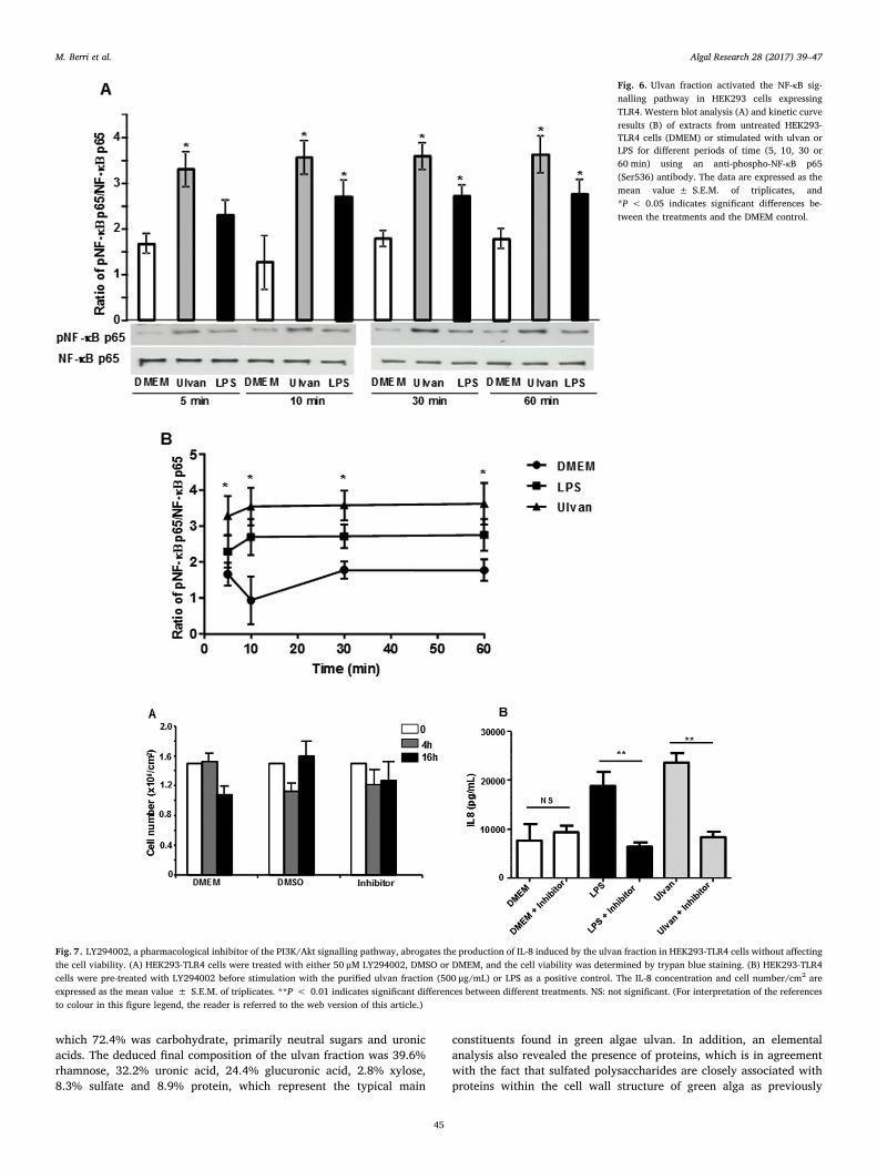
For studying the involvement of Akt in cytokine expression,HEK293-TLR4 cells were pre-treated with LY294002, a pharmacolo-gical inhibitor of phosphatidylinositol 3-kinase (PI3K) (Sigma). Thesolution was prepared as 1000× concentrated stocks in dimethylsulfoxide (DMSO) in order to ensure that the final concentration ofDMSO in the culture medium did not exceed 0.1%. 1 h before stimu-lation, the HEK293-TLR4 cells were treated with the LY294002 in-hibitor at a final concentration of 50 μM to block the signalling pathwayduring 4 and 16 h of stimulation with ulvan. After the incubation step,the media were collected, and the concentration of IL-8 was measuredby ELISA using a commercial kit (R & D) according to the manufac-turer's instructions. The control group was cultured in DMEM withDMSO. We also examined whether the inhibition of the PI3K signallingpathway by LY294002 has any cytotoxic effects on HEK293-TLR4 cells.The cells were treated with either 50 μM LY294002, DMSO or DMEM,and the cell viability was assessed using trypan blue staining and mi-croscopic observation/counting as described previously [30].
2.8. Statistical analysis
The data for the comparison of differences in mRNA expression andprotein production between treated and untreated cells were expressedas relative values. The data are expressed as the mean value oftriplicates± SEM. Because the data are independent and non-normallydistributed, they were analysed using the Kruskal-Wallis test followedby Bonferroni-Dunn post-test group comparison tests of means usingGraphPad (GraphPad Prism version 4.00 for Windows; GraphPadSoftware, San Diego, CA, USA). The statistical differences between thevarious treatments were considered significant either when the P-valueswere < 0.05 (*) or< 0.01 (**).
3. Results
3.1. Composition of the ulvan fraction
The chemical composition of the crude extract and the purifiedulvan fraction was determined by elemental and proximate analysis,monomeric sugar analysis and molecular weight. Table 2 shows that thepurification step removed all salts and minerals to reduce the ashcontent from 32.9 to 2.9%. As shown by the proximate analysis, thisprocedure also enhanced the organic matter content to 92.5%, whichwas composed mainly of carbohydrate and proteins. The sugars com-position analysis of the MSP extract yielded meaningless and unusableresults, with low levels of sugars detected with high standard errorvalues, which correlated to the low reproducibility of the method in thepresence of a high content of salts. After an ultra-filtration step, whichwas aimed at removing salts from the sample, the deduced organicmatter was 89.6%, of which 72.4% was carbohydrate, mostly neutralsugars and uronic acids. The yield of the ulvan fraction is calculated andshowed a value of 20.5%.
The composition of the sugar residues was determined with a lowstandard deviation and could account for 70.9% of the dry weight,slightly lower than indicated by the proximate analysis. The deduced
M. Berri et al. Algal Research 28 (2017) 39–47
41
final composition of the ulvan fraction consisted of 39.55% rhamnose,32.2% uronic acid, 24.4% glucuronic acid, 2.75% xylose and 8.3%sulfate. The weight-average (Mw) and the number-average (Mn) mo-lecular weight were estimated as 3.2 × 10³ and 2.9 × 10³ Da, re-spectively, and the polydispersity index was calculated to be 1.1.
3.2. Ulvan stimulated the mRNA expression of IL-8, TNF-α and CCL20 inIPEC-1 cells
Before investigating the stimulation effect of both extracts, the cy-totoxic effects were examined by incubating IPEC-1 cells with in-creasing amounts of MSP extract (Fig. 1A) and ulvan (Fig. 1B), and thecell viability was determined using a trypan blue exclusion test. Therewas no significant sign of cytotoxicity and the cell proliferation was notaffected at 0.5 mg/mL. Therefore, the capacity of the ulvan fraction tostimulate cytokine expression was first investigated at the mRNA levelin comparison with the MSP crude extract after the exposure of dif-ferentiated IPEC-1 cells to concentrations of 500, 50 and 5 μg/mL for4 h. The relative quantification of mRNA, expressed as a fold change,showed that the 500 μg/mL concentration of the MSP extract was ableto significantly increase (P < 0.01) the relative expression of CCL20(74.9), IL-8 (×85.4) and TNF-α (×29.9). The purification step did not
hamper the ulvan biological activity, and the activation of cytokineexpression in IPEC-1 cells was retained. In fact, the mRNA expressionanalysis showed that the purified ulvan fraction increased the expres-sion of CCL20 (×61.5), IL-8 (×79.7) and TNF-α (×36.4) to a similarextent as the MSP extract (Fig. 2A and B).
3.3. Ulvan induced the secretion of the IL-8 and TNF-α proteins indifferentiated IPEC-1 cells
Having shown that differentiated IPEC-1 cells were able to expresscytokines at the mRNA level in response to the purified ulvan fraction,we sought to confirm and extend this result by ELISA analysis. IPEC-1cells were cultured on transwell filters and stimulated overnight byadding ulvan at a concentration of 500 μg/mL, and the supernatants ofthe apical and basolateral compartments were analysed separately. Asseen in Fig. 3, the ulvan fraction was able to stimulate the secretion ofboth the IL-8 and TNFα proteins into the collected supernatants, but atdifferent levels depending on the compartment. Significantly increasedamounts of IL-8 (P < 0.01) were detected in both the apical(5446 ± 308 pg/mL) and basolateral (1980 ± 190 pg/mL) compart-ments compared to untreated controls (Fig. 3). Ulvan treatment alsostimulated TNF-α release from IPEC-1 cells in both compartments but atmuch lower level than IL-8 (125.5 ± 18.5 pg/mL versus5446 ± 308 pg/mL and 7.8 ± 1.2 pg/mL versus 1980 ± 190 pg/mL). For the following experiments, only the ulvan fraction was eval-uated.
3.4. Ulvan induced IL-8 protein expression in HEK293-TLR4 cells
To identify the receptor involved, the ulvan fraction was tested atthe concentration of 500 μg/mL in human embryonic kidney (HEK)293cell lines stably expressing the receptors TLR4, TLR5, TLR9, NOD1 andNOD2. The ulvan/receptor interaction was evaluated by monitoring IL-8 release into the supernatant by ELISA. Our results showed that thepurified ulvan fraction was able to activate TLR4 and induced the up-regulation of IL-8 production in the HEK293/TLR4-MD-2-CD14 cell linecompared to HEK293-null cells. This fraction did not significantly ac-tivate any of the remaining receptors (Fig. 4).
3.5. Ulvan interaction with TLR4 mediated the activation of the PI3K/Aktand NF-κB signalling pathway
We next examined the effect of the purified ulvan fraction on theactivation of different protein kinases that are involved in cytokine geneexpression. Thus, HEK293-TLR4 cells were incubated with ulvan(500 μg/mL) for different lengths of time (5, 10, 30 and 60 min), andthe phosphorylation status of MAPKs (ERK1/2 and p38), Akt, AMPK
Table 2Composition of the low molecular weight aqueous MSP extract and the purified ulvanfraction used in this study. The analysis of the composition of sugar units was carried outdirectly on the purified ulvan fraction. n.d. means not determined.
Composition (%) MSP extract Ulvan fraction
ElementalC 21.14 ± 0.06 28.80 ± 0.07H 4.73 ± 0.13 5.80 ± 0.003N 2.29 ± 0.01 2.00 ± 0.10O 33.1 ± 0.10 52.38 ± 0.09S 4.33 ± 0.04 5.12 ± 0.01
Proximate (on DM basis)Ash 32.9 ± 0.1 2.9 ± 0.1Fat < 0.1 < 0.1Proteins 10.2 ± 1.0 8.9 ± 0.3Neutral sugars 11.7 ± 1.8 40.2 ± 1.4Uronic acids 7.4 ± 2.4 32.2 ± 2.5Sulfate groups 4.0 ± 1.0 8.3 ± 0.6
Monomeric sugarsArabinose n.d. n.d.Galactose n.d. 0,9 ± 0,09Glucose n.d. 1.8 ± 0.81Xylose n.d. 2.75 ± 0.08Mannose n.d. 1.46 ± 0.02Rhamnose n.d. 39.55 ± 1.56Glucuronic acid n.d. 24.4 ± 1.01
Fig. 1. IPEC-1 cells were treated with various concentrations of MSP extract (0–10 mg/mL) (A) or purified ulvan fraction (0–1 mg/mL) (B), and cell viability was assessed after 3 days(D0, D1, D2 and D3) using trypan blue staining and microscopic observation/counting. The data are expressed as the mean value ± S.E.M. of triplicates. The results are compared withthe untreated control after 3 days (D3) of incubation. Different letters indicate statistically significant differences (a, b, c and d) with P < 0.05. (For interpretation of the references tocolour in this figure legend, the reader is referred to the web version of this article.)
M. Berri et al. Algal Research 28 (2017) 39–47
42

and NF-κB was evaluated using phospho-specific antibodies. Westernblot results and a kinetic curve analysis of pAkt/Akt showed that theinteraction of the ulvan fraction with TLR4 in HEK293 cells resulted ina significant increase in Akt phosphorylation that was detectable after a5 min incubation compared to untreated cells, similar to the LPS-treated cells (Fig. 5A and B). The phosphorylation status analysis ofMAPKs (Erk1/2 and p38) and AMP-activated kinase did not indicateany differences in the activation of these signalling pathways betweendifferent treatments (data not shown). Akt regulates the transcriptionalactivity of nuclear factor-κB (NF-κB) by inducing the phosphorylationand subsequent degradation of inhibitor of κB (IκB). We thereforeanalysed the phosphorylation status of p65 in HEK293-TLR4 cells and,as shown in Fig. 6 A and B, we observed that the ulvan fraction sig-nificantly increased the levels of phosphorylated p65.
3.6. Inhibition of the PI3K/Akt signalling pathway using the inhibitorLY294002
We first examined whether the pharmacological inhibitor LY294002has any cytotoxic effects on the proliferation of HEK293-TLR4 cells. Thetreatment of cells with either LY294002 or DMSO did not affect the cell
viability in comparison with DMEM alone even after 16 h of incubation(Fig. 7A). The involvement of the PI3K/Akt signalling pathway wasthen verified using the inhibitor LY294002. The inhibitor, used at afinal concentration of 50 μM for 16 h, was able to block this signallingpathway and induce a> 65% decrease in IL-8 secretion (Fig. 7B). Si-milar results were obtained in the cells treated with LPS, whileLY294002 did not affect IL-8 secretion in untreated cells (Fig. 7B).
4. Discussion
Green algae have emerged in recent years as a rich and importantsource of bioactive natural compounds that could be used as a newgeneration of growth enhancers and natural antibiotic alternatives topotentiate the immune function and limit the infections of farm animalsand therefore improve animal health [34,35]. However, further re-search is needed to identify the bioactive components, mechanisms ofaction, and in vivo biological effects to ultimately adapt the findings foragronomic use. In this context, we recently prepared an aqueous marinesulfated polysaccharides (MSP) crude extract from the green macro-algae Ulva armoricana and showed its capacity to stimulate the mRNAexpression of a broad range of cytokines and chemokines using an in
Fig. 2. Stimulation of the expression of IL-8, TNFα, and CCL20 in differentiated porcine epithelial (IPEC-1) cells using the MSP extract (A) and the purified ulvan fraction (B). The IPEC-1cells were treated for 4 h with different concentrations (500, 50 and 5 μg/mL) of the extracts, and the expression of target genes was determined as the fold change relative to controls.The data are expressed as the mean value ± S.E.M. of triplicates, and the results were considered statistically significant at P-value< 0.01 (P < 0.01).
Fig. 3. Purified ulvan stimulated differentiated IPEC-1 cells to produce the TNFα and IL-8 proteins. IPEC-1 cells were incubated with 500 μg/mL ulvan overnight at 37 °C, and the apicaland basolateral supernatants were harvested separately and used for protein analysis using commercial ELISA kits. The results were considered statistically significant for P-values below0.01 (P < 0.01).
M. Berri et al. Algal Research 28 (2017) 39–47
43

vitro system of the porcine intestinal epithelial cell line IPEC-1 [30]. Thechemical composition analysis indicated that the main component ofthe extract is ulvan. This component is regarded as the main candidatefor the immunostimulatory activity, although further purification of thesample is needed for final confirmation. In the current study, we pre-pared a new MSP batch from algae that were harvested in the same area
during the summer of 2013, and an ulvan fraction was purified toevaluate its immunomodulation potential in comparison with the crudeMSP extract. The composition analysis of this new MSP batch showedsimilar components to those of the MSP extract that was prepared andtested previously [30]. The purification process allowed us to obtain anulvan fraction with a high content of organic matter (up to 89.6%), of
Fig. 4. Purified ulvan fraction stimulates only HEK293-TLR4 cells to produce the IL-8 protein. HEK293-TLR4, TLR5, TLR9, NOD1 and NOD2 cells were plated at a density of1.5 × 106 cells/well in six-well culture plates and stimulated for 16 h either with the ulvan fraction at a concentration of 500 μg/mL or the recommended agonists as positive controls.The agonists ultra-pure LPS O111B4 (100 ng/mL) for TLR4, flagellin (100 ng/mL) for TLR5, CPG-ODN 2395 (10 μg/mL) for TLR9, iE-DAP (1 μg/mL) for NOD1 and MDP (1 μg/mL) forNOD2 were used. The data are expressed as the mean value± S.E.M. of triplicates, and the IL-8 production was considered statistically significant for P-values below 0.01 (P < 0.01).
Fig. 5. Ulvan fraction induced the phosphorylation of Akt in HEK293 cells expressing TLR4. HEK293-TLR4 cells were stimulated with 500 μg/mL of ulvan fraction for 5, 10, 30 or 60 min,and the cell lysates were analysed for the expression of both Akt and phospho-Akt (p-Akt). Western blot (A) and kinetic curve results of pAkt/Akt (B) are expressed as the meanvalue ± S.E.M. of triplicates, and *P < 0.05 indicates significant differences between the treatments and the DMEM control.
M. Berri et al. Algal Research 28 (2017) 39–47
44
which 72.4% was carbohydrate, primarily neutral sugars and uronicacids. The deduced final composition of the ulvan fraction was 39.6%rhamnose, 32.2% uronic acid, 24.4% glucuronic acid, 2.8% xylose,8.3% sulfate and 8.9% protein, which represent the typical main
constituents found in green algae ulvan. In addition, an elementalanalysis also revealed the presence of proteins, which is in agreementwith the fact that sulfated polysaccharides are closely associated withproteins within the cell wall structure of green alga as previously
Fig. 6. Ulvan fraction activated the NF-κB sig-nalling pathway in HEK293 cells expressingTLR4. Western blot analysis (A) and kinetic curveresults (B) of extracts from untreated HEK293-TLR4 cells (DMEM) or stimulated with ulvan orLPS for different periods of time (5, 10, 30 or60 min) using an anti-phospho-NF-κB p65(Ser536) antibody. The data are expressed as themean value ± S.E.M. of triplicates, and*P < 0.05 indicates significant differences be-tween the treatments and the DMEM control.
Fig. 7. LY294002, a pharmacological inhibitor of the PI3K/Akt signalling pathway, abrogates the production of IL-8 induced by the ulvan fraction in HEK293-TLR4 cells without affectingthe cell viability. (A) HEK293-TLR4 cells were treated with either 50 μM LY294002, DMSO or DMEM, and the cell viability was determined by trypan blue staining. (B) HEK293-TLR4cells were pre-treated with LY294002 before stimulation with the purified ulvan fraction (500 μg/mL) or LPS as a positive control. The IL-8 concentration and cell number/cm2 areexpressed as the mean value ± S.E.M. of triplicates. **P < 0.01 indicates significant differences between different treatments. NS: not significant. (For interpretation of the referencesto colour in this figure legend, the reader is referred to the web version of this article.)
M. Berri et al. Algal Research 28 (2017) 39–47
45

described [10,36].The treatment of differentiated IPEC-1 cells with this new MSP
batch as well as the purified ulvan fraction induced the upregulation ofCCL20, TNFα and IL-8 mRNA. In addition, the purified ulvan fractionwas able to stimulate cytokine secretion in both the basal and apicalcompartments as analysed by ELISA. These results are similar to thoseobtained previously, showing that the extraction process is suitable toprepare MSP extracts with reproducible analytical compositions andimmunostimulatory activities [30]. Moreover, the purification step wasparticularly useful for improving the immunostimulatory activity,which could be attributed to the ulvan fraction. The stimulation of theintestinal cytokines production of the MSP extract as well as the ulvanfraction could not be attributed to endotoxin contamination, because noendotoxin compounds were detected within the crude and fractionatedpolysaccharides by an E-Toxate kit analysis (data not shown). This in-vestigation is of practical significance for determining the optimal ex-traction processing in order to produce natural bioactive moleculeswith an effective immunostimulatory activity to be used in a livestockdiet to improve the immune response of animals and thereby enhancetheir resistance against infectious diseases.
Studies evaluating the immunological properties of algae-derivedpolysaccharides were mainly carried out using macrophage cell linessuch as murine RAW264.7, and rarely tested on intestinal epithelialcells. Like fucoidans and carageanans, ulvans were tested directly onmacrophages cell line or after challenging them with LPS to test theimmunostimulatory or the anti-inflammatory responses respectively[15,16]. Although macrophages are essential effector cells of innateimmunity, intestinal epithelial cells are also of interest since they ex-press pattern-recognition receptors (PRRs) that enable them to act asdynamic sensors of microbial environment and foreign antigens. Thus,differentiated IPEC-1 cells used in our study is a relevant and suitable invitro model that will allow to test and evaluate the effect of dietarybioactive compounds intake such as marine sulfated polysaccharides tostimulate the intestinal immune response. It has become clear that in-testinal epithelial cells are crucial mediators of intestinal homeostasis,reinforcing the barrier function and participating in the coordination ofappropriate mucosal immune responses by producing a broad range ofcytokines involved in the activation, trafficking and function of ad-jacent immune cells [37–39]. Because of their ability to recognizespecific carbohydrate moieties and elicit immune responses, we hy-pothesized that PRRs, and more specifically TLRs and NODs, are acti-vated by the ulvan fraction which leads, through signalling cascades, tothe stimulation of cytokine production. Thus, we used an HEK cellmodel to target a large panel of TLR and NOD receptors and showedthat the ulvan fraction was able to activate immune signalling in HEKcells expressing TLR4. This implies that ulvan can act directly on targetcells, including intestinal or immune cells, via TLR4 and differentiallyaffect the expression of immunological parameters. We also analysedthe activation of signalling pathways, and showed that the interactionof ulvan with TLR4 mediated the activation of the PI3K/Akt signallingpathway, which regulates the transcriptional activity of NF-κB. Theinvolvement of the PI3K/Akt pathway was confirmed using the specificpharmacological inhibitor LY294002. In a previous report, normalhuman colonic epithelial cell line NCM460 were exposed to sulfatedpolygalactose carrageenan (CGN) purified from red algae and showed astimulation of IL-8 production. Similarly to our study, this CGN wasable to recognize TLR4 receptor and induce B cell lymphoma-NFκBactivation to mediate cytokine production [40].
The activation of the PI3K/Akt pathway has been reported to beassociated with TLR2, TLR3, TLR4, and TLR5 in different cells [41,42]and plays both pro-inflammatory and anti-inflammatory roles in TLRsignalling [43,44]. Thus, this ulvan fraction may play dual roles andexhibit immunomodulatory activities that may be of potential appli-cation either in stimulating the immune response or in controlling in-flammation, as was reported for other algae-derived polysaccharideextracts [15,21].
Overall, these in vitro data provide novel insights into the molecularmechanism used by marine sulfated polysaccharides to stimulate theproduction of soluble cytokines and chemokines and suggest that thissignalling can be achieved upon the interaction with TLR4, inducing theactivation of the PI3K/Akt and NF-κB pathway. Although these in vitroresults help to explain the molecular mechanisms used by ulvan tostimulate the expression of cytokines, we cannot rule out the involve-ment of other receptors and alternative signalling pathways through amechanism that is likely more complex at the intestine surface. NaturalTLR ligands (TLRLs) and their mimetics are increasingly being appliedin immunotherapeutic strategies. As TLRLs are potent inducers of in-nate immune responses, they have been applied as adjuvants to sti-mulate adaptive immune responses [45]. The orientation of the phar-maceutical industry towards naturally derived polymers asmultifunctional materials in drug delivery applications has become asubject of increasing interest, driving the continuous exploitation ofsuch compounds [46]. Algal sulfated polysaccharides have recentlybeen used in applications in the production of nanoparticles and mi-croparticles, mainly owing to their ionic nature [47]. Thus, ligands forTLR can be encapsulated within biodegradable nanoparticles of ulvanto be delivered to relevant target immune cells in vaccination strategies.However, further studies are needed using ex vivo and in vivo models toconfirm the activity of marine sulfated polysaccharides on the stimu-lation of the immune response under physiological conditions andduring infectious or inflammatory processes.
Acknowledgements
This work was financed by a grant from the BPI-France/ISI Ulvansproject (n°: I1110001W) to Amadéite company, Bréhan (France). Theauthors wish to thank Christelle Gouin (Amadeite) for the production ofthe demineralized algal extracts and for her contribution to the prox-imate analyses and Dr. Nathalie Guriec (CHU Brest) for the LPS ana-lysis. We also gratefully acknowledge Dr. Isabelle Oswald (ToxalimUnit, INRA center of Toulouse, France) for kindly providing the IPEC-1cell line.
Author's contributions
M.B. conceived, coordinated the study and wrote the manuscript,M.O. performed all the experiments of IPEC-1 cells stimulation and RT-qPCR, S.H. performed the experiments related to the identification ofthe ulvan receptor, J.D. performed the western blot analysis and sig-nalling pathways identification, H.D. and M.L. participated to the ex-periments design, P.N.C. participated to the study design and su-pervised the MSP and the ulvan preparation.
All the authors read and approved the manuscript.
Competing financial interests
The authors declare no competing financial interests.
Statement of informed consent, human/animal rights
No conflicts, informed consent, human or animal rights applicable.
References
[1] L. O'Sullivan, B. Murphy, P. McLoughlin, P. Duggan, P.G. Lawlor, H. Hughes,G.E. Gardiner, Prebiotics from marine macroalgae for human and animal healthapplications, Mar. Drugs 88 (2010) 2038–2064.
[2] A. Jiménez-Escrig, E. Gómez-Ordóñez, P. Rupérez, Seaweed as a source of novelnutraceuticals: sulfated polysaccharides and peptides, Adv. Food Nutr. Res. 64(2011) 325–337.
[3] K. Chojnacka, A. Saeid, Z. Witkowska, L. Tuhy, Biologically active compounds inseaweed extracts-the prospects for the application, Open Conf. Proc. J. 3 (2012)20–28.
[4] M.F.J. Raposo, R.M.S.C. Morais, A.M.M.B. Morais, Health applications of bioactive
M. Berri et al. Algal Research 28 (2017) 39–47
46

compounds from marine microalgae, Life Sci. 93 (2013) 479–486.[5] S. Holdt, S. Kraan, Bioactive compounds in seaweed; functional food applications
and legislation, J. Appl. Phycol. 23 (2011) 543–597.[6] W.A.J.P. Wijesinghe, Y.J. Jeon, Biological activities and potential cosmeceutical
applications of bioactive components from brown seaweeds: a review, Phytochem.Rev. 10 (2011) 431–443.
[7] L.G. Ferreira, M.D. Noseda, A.G. Gonçalves, D.R. Ducatti, M.T. Fujii, M.E. Duarte,Chemical structure of the complex pyruvylated and sulfated agaran from the redseaweed Palisada flagellifera (Ceramiales, Rhodophyta), Carbohydr. Res. 347 (2012)83–94.
[8] L.E. Rioux, S.L. Turgeon, M. Beaulieu, Characterization of polysaccharides extractedfrom brown seaweeds, Carbohydr. Polym. 69 (2007) 530–537.
[9] P. Laurienzo, Marine polysaccharides in pharmaceutical applications: an overview,Mar. Drugs 8 (2010) 2435–2465.
[10] M. Lahaye, A. Robic, Structure and functional properties of Ulvan, a polysaccharidefrom green seaweeds, Biomacromolecules 8 (2007) 1765–1774.
[11] I. Wijesekara, R. Pangestuti, S.K. Kima, Biological activities and potential healthbenefits of sulfated polysaccharides derived from marine algae, Carbohydr. Polym.84 (2011) 14–21.
[12] J.C. Lee, M.F. Hou, H.W. Huang, F.R. Chang, C.C. Yeh, J.Y. Tang, H.-W. Chang,Marine algal natural products with anti-oxidative, anti-inflammatory, and anti-cancer properties, Cancer Cell Int. 13 (2013) 55 http://www.cancerci.com/content/13/1/5513.
[13] S.N. Fedorov, S.P. Ermakova, T.N. Zvyagintseva, V.A. Stonik, Anticancer and cancerpreventive properties of marine polysaccharides: some results and prospects, Mar.Drugs 11 (2013) 4876–4901.
[14] D.H. Ngoa, I. Wijesekara, T.S. Voa, Q.V. Ta, S.K. Kim, Marine food-derived func-tional ingredients as potential antioxidants in the food industry: an overview, FoodRes. Int. 44 (2011) 523–529.
[15] D. Chen, X.Z. Wu, Z.Y. Wen, Sulphated polysaccharides and immune response:promotors or inhibitors, Panminerva Med. 50 (2008) 177–183.
[16] I. Jaswir, H.A. Monsur, Anti-inflammatory compounds of macro algae origin: areview, J. Med. Plant Res. 5 (2011) 7146–7154.
[17] W.J. Na, W.J. Kim, S.M. Kim, J.K. Park, S.M. Lee, S.O. Kim, A. Synytsya, Y. Park,Purification, characterization and immunostimulating activity of water-solublepolysaccharide isolated from Capsosiphon fulvescens, Int. Immunopharmacol. 10(2010) 364–570.
[18] S. Karnjanapratum, M. Tabarsa, M. Chou, S.G. You, Characterization and im-munomodulatory activities of sulfated polysaccharides from Capsosiphon fulvescens,Int. J. Biol. Macromol. 51 (2012) 720–729.
[19] J.K. Kim, M.L. Cho, S. Karnjanapratum, I.S. Shin, S.G. You, In vitro and in vivoimmunomodulatory activity of sulfated polysaccharides from Enteromorpha pro-lifera, Int. J. Biol. Macromol. Vol. 49 (2011) 1051–1058.
[20] M. Tabarsa, M. Rezaei, Z. Ramezanpour, J.R. Waaland, Chemical compositions ofthe marine algae Gracilaria salicornia (Rhodophyta) and Ulva lactuca (Chlorophyta)as a potential food source, J. Sci. Food Agric. 92 (2012) 2500–2506.
[21] T.S. Vo, D. Ngo, S.K. Kim, Potential targets for anti-inflammatory and anti-allergicactivities of marine algae: an overview inflammation and allergy. Drug, Targets 11(2012) 90–101.
[22] P.A. Hwang, S.Y. Chien, Y.L. Chan, M.K. Lu, C.H. Wu, Z.L. Kong, C.J. Wu, Inhibitionof lipopolysaccharide (LPS)-induced inflammatory responses by Sargassum hemi-phyllum sulfated polysaccharide extract in RAW 264.7 macrophage cells, J. Agric.Food Chem. 59 (2011) 2062–2068.
[23] S. de Kivit, A.D. Kraneveld, J. Garssen, L.E. Willemsen, Glycan recognition at theinterface of the intestinal immune system: target for immune modulation viadietary components, Eur. Aust. J. Pharm. 668 (2011) 124–132.
[24] F. Peri, V. Calabrese, Toll-like receptor 4 (TLR4) modulation by synthetic andnatural compounds: an update, J. Med. Chem. 57 (2014) 3612–3622.
[25] X. Zhang, C. Qi, Y. Guo, W. Zhou, Y. Zhang, Toll-like receptor 4-related im-munostimulatory polysaccharides: primary structure, activity relationships, andpossible interaction models, Carbohydr. Polym. 149 (2016) 186–206.
[26] R.F. Tsuji, K. Hoshino, Y. Noro, N.M. Tsuji, T. Kurokawa, T. Masuda, S. Akira,B. Nowak, Suppression of allergic reaction by lambda-carrageenan: toll-like re-ceptor 4/MyD88-dependent and -independent modulation of immunity, Clin. Exp.
Allergy 33 (2003) 249–258.[27] Y.Q. Zhang, Y.C. Tsai, A. Monie, C.F. Hung, T.C. Wu, Carrageenan as an adjuvant to
enhance peptide-based vaccine potency, Vaccine 28 (2010) 5212–5219.[28] T. Teruya, H. Tatemoto, T. Konishi, T. Tako, Structural characteristics and in vitro
macrophage activation of acetyl fucoidan from Cladosiphon okamuranus, Glycoconj.J. 26 (2009) 1019–2028.
[29] M.H. Kim, H.G. Joo, Immunostimulatory effects of fucoidan on bone marrow-de-rived dendritic cells, Immunol. Lett. 115 (2008) 138–143.
[30] M. Berri, C. Slugocki, M. Olivier, E. Helloin, I. Jacques, H. Salmon, H. Demais, M. LeGoff, P. Nyvall Collen, Marine-sulfated polysaccharides extract of Ulva armoricanagreen algae exhibits an antimicrobial activity and stimulates cytokine expression byintestinal epithelial cells, J. Appl. Phycol. 28 (2016) 2999–3008.
[31] K. Hardouin, G. Bedoux, A.S. Burlot, C.D. Moreno, J.P. Bergé, P. Nyvall-Collen,N. Bourgougnon, Enzyme-assisted extraction (EAE) for the production of antiviraland antioxidant extracts from the green seaweed Ulva armoricana (Ulvales,Ulvophyceae), Algal Res. 16 (2016) 233–239.
[32] R. Gonzalez-Vallina, H.R. Wang, H.M. Zhan, R.M. Berschneider, N.O. Lee,D.D. Davidson, Black, lipoprotein and apolipoprotein secretion by a newborn pigletintestinal cell line (IPEC-1), Am. J. Phys. 27 (1996) 249–259.
[33] M. Delgado-Ortega, S. Melo, D. Punyadarsaniya, C. Ramé, M. Olivier, D. Soubieux,D. Marc, G. Simon, G. Herrler, M. Berri, J. Dupont, F. Meurens, Innate immuneresponse to a H3N2 subtype swine influenza virus in newborn porcine trachea cells,alveolar macrophages, and precision-cut lung slices, Vet. Res. 45 (2014) 42 http://www.veterinaryresearch.org/content/45/1/42.
[34] I. Michalak, K. Chojnacka, Algae as production systems of bioactive compounds,Eng. Life Sci. 15 (2015) 60–76.
[35] I. Hamed, F. Özogul, Y. Özogul, M. Joe, J.M. Regenstein, Marine bioactive com-pounds and their health benefits: a review, Compr. Rev. Food Sci. Food Saf. 14(2015) 446–465.
[36] A. Robic, D. Bertrand, J.F. Sassi, Y. Lerat, M. Lahaye, Determination of the chemicalcomposition of ulvan, a cell all polysaccharide from Ulva spp. (Ulvales, Chlorophyta)by FT-IR and chemometrics, J. Appl. Phycol. 21 (2009) 451–456.
[37] D. Artis, R.K. Grencis, The intestinal epithelium: sensors to effectors in nematodeinfection, Mucosal Immunol. 1 (2008) 252–264.
[38] N. Miron, V. Cristea, Enterocytes: active cells in tolerance to food and microbialantigens in the gut, Clin. Exp. Immunol. 167 (2012) 405–412.
[39] L.W. Peterson, D. Artis, Intestinal epithelial cells: regulators of barrier function andimmune homeostasis, Nat. Rev. Immunol. 14 (2014) 141–153.
[40] S. Bhattacharyya, R. Gill, M.L. Chen, F. Zhang, R.J. Linhardt, P.K. Dudeja,J.K. Tobacman, Toll-like receptor 4 mediates induction of the Bcl10-NFκB-Interleukin-8 inflammatory pathway by carrageenan in human intestinal epithelialcells, J. Biol. Chem. (2008) 10550–10558.
[41] M.H. Laird, S.H. Rhee, D.J. Perkins, A.E. Medvedev, W. Piao, M.J. Fenton,S.N. Vogel, TLR4/MYD88/PI3K interactions regulate TLR4 signalling, J. Leukoc.Biol. 85 (2009) 966–977.
[42] N. Molnarfi, L. Gruaz, J.M. Dayer, D. Burger, Opposite regulation of IL-1beta andsecreted IL-1 receptor antagonist production by phosphatidylinositide-3 kinases inhuman monocytes activated by lipopolysaccharides or contact with T cells, J.Immunol. 178 (2007) 446–454.
[43] E.A. Medina, I.R. Morri, M.T. Berton Phosphatidylinositol, 3-kinase activation at-tenuates the TLR2-mediated macrophage proinflammatory cytokine response toFrancisella tularensis live vaccine strain, J. Immunol. 185 (2010) 7562–7572.
[44] M.V. Rajaram, L.P. Ganesan, K.V. Parsa, J.P. Butchar, J.S. Gunn, S. Tridandapani,Akt/protein kinase B modulates macrophage inflammatory response to Francisellainfection and confers a survival advantage in mice, J. Immunol. 177 (2006)6317–6324.
[45] S. Manicassamy, B. Pulendran, Modulation of adaptive immunity with toll-like re-ceptors, Semin. Immunol. 21 (2009) 185–193.
[46] V.A. Arsul, S.R. Lahoti, Natural polysaccharides as pharmaceutical excipients,World J. Pharm. Res. 3 (2014) 3776–3790.
[47] L. Cunha, A. Grenha, Sulfated seaweed polysaccharides as multifunctional materialsin drug delivery applications, Mar. Drugs 14 (2016) 42, http://dx.doi.org/10.3390/md14030042.
M. Berri et al. Algal Research 28 (2017) 39–47
47