Tuberculous pericarditis: Long-term outcome in patients who received medical therapy alone
7
Tuberculous pericarditis: Long-term outcome in patients who received medical therapy alone A conservative approach to operative intervention in the treatment of active tuberculous pericarditis was adopted and only 4 of 16 consecutive patients underwent pericardiectomy, all within 2 months of diagnosfs. One patient died of constrictive pericarditis despite pericardiectomy, and one died of acute bronchopneumonia after 8 months of otherwise successful medical management. All 14 long-term survivors were reevaluated to exclude chronic constricttve pericardltis and other potential sequelae of tuberculous perlcardltls. Reevaluation included physical examination, chest radiograph, electrocardiogram, M-mode and two-dimensional echocardiogram, computed tomography (CT) scan, and in pabents 575 years of age, incremental cycle exercise to maximum oxygen consumption. None were found to have chronic constrictive pericarditis or convincing evidence of other recognized comulicatlons of tuberculous pericarditls. Our results suggest that when pericardiectomy is not required for the relief of cardiac compression during the acute phase of tuberculous pericardftis and patients are treated with medical therapy alone, an excellent long-term outcome may be anticipated. (AM HEART J 1989;117:1133.) Richard Long, MD, Magdy Younes, MD, PhD, Norman Patton, MD, and Earl Hershfield, MD. Winnipeg, Manitoba, Canada By evading early detection and stimulating host responses-effusion and pericardial inflammation- which may have critical effects on myocardial mechanics, tubercle bacilli infecting the pericardium can threaten life despite the availability of effective antituberculous drugs. Though many patients once diagnosed as having active tuberculous pericarditis appear to do well with chemotherapy alone,*r2 ‘the potential for cardiac compression has prompted some centers to recommend early pericardiectomy for all cases of tuberculous perica.rditis~4 an approach that is supported by a recent clinical decisionanalysis of the literature.5 While pericardi- ectomy is clearly indicated in cases where decom- pensated cardiac compression exists and is unre- lieved by pericardiocentesis, its use in other cases is based on the premisethat it offers long-term advan- tages, notably reduction in the incidence and sever- ity of chronic pericardial constriction. There are currently no systematic studies of the long-term outcomeof medical therapy for tuberculouspericar- From the Departments of Medicine and Surgery, the University of Manitoba. This study was supported in part by the Respiratory Research Fund, Health Sciences Centre, Winnipeg, Manitoba, Canada. Received for publication Aug. 24, 1988; accepted Dec. 26,1968. Reprint requests: Richard Long, MD, Room C5116, Department of Medi- cine, St. Boniface General Hospital, 409 Tache Ave., Winnipeg, Manitoba, Canada, R2H 2A6. ditis, yet this information is relevant to a decision regarding pericardiectomy during the active phase. Circumstances in the province of Manitoba, Can- ada, ahowed us to examinethis issue. A conservative approach had been adopted in the 1960s whereby only patients with cardiaccompression that was not relieved by pericardiocentesis, or that did not resolve, or became worse during 6 to 8 weeks of chemotherapy, underwent pericardiectomy. Of a total of 16 patients diagnosed with tuberculous pericarditis over the past 16 years, 12 received medical therapy alone with or without biopsy. This communication reports the outcome in these patients. METHODS Between January 1970 and March 1936, 16 patients were diagnosed as having tuberculous pericarditis (Table I). The diagnosis was based on the following factors. (1) Bacteriology from the pericardium, and/or perieardial fluid (n = 7). (2) Clinical evidence of pericarditis, a peri- cardial effusion on echocardiogram, active tuberculosis elsewhere, and a good response to chemotherapy (n = 6). (3) Clinical criteria alone (n = 3). The specific criteria judged to be adequate for the diagnosis of tuberculous pericarditis in these cases were: clinical evidence of peri- carditis with a pericardial effusion on echocardiogram, a positive tuberculin test, a negative work-up for other causes of pericarditis, and a good response to chemother- apy. In addition, case No. 3 had a left pleural exudate with a predominence of lymphocytes (71%); case No. 4 had 1133
-
Upload
richard-long -
Category
Documents
-
view
214 -
download
0
Transcript of Tuberculous pericarditis: Long-term outcome in patients who received medical therapy alone

Tuberculous pericarditis: Long-term outcome in patients who received medical therapy alone
A conservative approach to operative intervention in the treatment of active tuberculous pericarditis was adopted and only 4 of 16 consecutive patients underwent pericardiectomy, all within 2 months of diagnosfs. One patient died of constrictive pericarditis despite pericardiectomy, and one died of acute bronchopneumonia after 8 months of otherwise successful medical management. All 14 long-term survivors were reevaluated to exclude chronic constricttve pericardltis and other potential sequelae of tuberculous perlcardltls. Reevaluation included physical examination, chest radiograph, electrocardiogram, M-mode and two-dimensional echocardiogram, computed tomography (CT) scan, and in pabents 575 years of age, incremental cycle exercise to maximum oxygen consumption. None were found to have chronic constrictive pericarditis or convincing evidence of other recognized comulicatlons of tuberculous pericarditls. Our results suggest that when pericardiectomy is not required for the relief of cardiac compression during the acute phase of tuberculous pericardftis and patients are treated with medical therapy alone, an excellent long-term outcome may be anticipated. (AM HEART J 1989;117:1133.)
Richard Long, MD, Magdy Younes, MD, PhD, Norman Patton, MD, and Earl Hershfield, MD. Winnipeg, Manitoba, Canada
By evading early detection and stimulating host responses-effusion and pericardial inflammation- which may have critical effects on myocardial mechanics, tubercle bacilli infecting the pericardium can threaten life despite the availability of effective antituberculous drugs. Though many patients once diagnosed as having active tuberculous pericarditis appear to do well with chemotherapy alone,*r2 ‘the potential for cardiac compression has prompted some centers to recommend early pericardiectomy for all cases of tuberculous perica.rditis~4 an approach that is supported by a recent clinical decision analysis of the literature.5 While pericardi- ectomy is clearly indicated in cases where decom- pensated cardiac compression exists and is unre- lieved by pericardiocentesis, its use in other cases is based on the premise that it offers long-term advan- tages, notably reduction in the incidence and sever- ity of chronic pericardial constriction. There are currently no systematic studies of the long-term outcome of medical therapy for tuberculous pericar-
From the Departments of Medicine and Surgery, the University of Manitoba. This study was supported in part by the Respiratory Research Fund, Health Sciences Centre, Winnipeg, Manitoba, Canada. Received for publication Aug. 24, 1988; accepted Dec. 26,1968. Reprint requests: Richard Long, MD, Room C5116, Department of Medi- cine, St. Boniface General Hospital, 409 Tache Ave., Winnipeg, Manitoba, Canada, R2H 2A6.
ditis, yet this information is relevant to a decision regarding pericardiectomy during the active phase.
Circumstances in the province of Manitoba, Can- ada, ahowed us to examine this issue. A conservative approach had been adopted in the 1960s whereby only patients with cardiac compression that was not relieved by pericardiocentesis, or that did not resolve, or became worse during 6 to 8 weeks of chemotherapy, underwent pericardiectomy. Of a total of 16 patients diagnosed with tuberculous pericarditis over the past 16 years, 12 received medical therapy alone with or without biopsy. This communication reports the outcome in these patients. METHODS
Between January 1970 and March 1936, 16 patients were diagnosed as having tuberculous pericarditis (Table I). The diagnosis was based on the following factors. (1) Bacteriology from the pericardium, and/or perieardial fluid (n = 7). (2) Clinical evidence of pericarditis, a peri- cardial effusion on echocardiogram, active tuberculosis elsewhere, and a good response to chemotherapy (n = 6). (3) Clinical criteria alone (n = 3). The specific criteria judged to be adequate for the diagnosis of tuberculous pericarditis in these cases were: clinical evidence of peri- carditis with a pericardial effusion on echocardiogram, a positive tuberculin test, a negative work-up for other causes of pericarditis, and a good response to chemother- apy. In addition, case No. 3 had a left pleural exudate with a predominence of lymphocytes (71%); case No. 4 had
1133

1134 Long et al. may 1999
American Heart Journal
Table I. Vital statistics and basis for diagnosis in 16 patients with tuberculous pericarditis
Case No.
Age at diagnosis
(Yr) Sex Race
Months of pericardial symptoms Clinical
Basis for diagnosis ---.-~
+VE culture or Pericardial histology from fluidltissue
other site in ____- clinical case Bacteriologic Histologic
1 23 M N 2 66 F C 3 26 F C 4 80 M SEA 5 6 F N 6 35 M N 7 32 M N 8* 36 M N 9 21 M N
10 19 M N 11 74 M C 12 19 M N 13 31 F SEA 14 59 M C 15 80 F C 16* 79 F C
6 1 1 2 1 1 1.5 1 4.5 1
1 1 2.5 2 1.5
X X x
x X
X X X X
X X X X
X X x X X X X
X X
X X
X X X
X
C, Caucasian; N, native Indian; SEA, Southeast Asian; +VE, positive. * = Death.
bloody pericardial fluid with lymphocyte predominence (71% ) and no tumor cells as well as bilateral pleural effusions; and case No. 12 had bilateral upper lobe fibroproductive changes on chest radiograph.
The investigation of the 16 patients consisted of a detailed retrospective review of the records of each patient as well as a prospective reevaluation, beginning in 1936, of all surviving patients (n = 14, 11 without prior pericardi- ectomy). Reevaluation was designed to identify chronic constrictive pericarditis and other potential sequelae of tuberculous pericarditis, i.e., annular constriction,B arryth- mias, conduction disturbances, impaired ventricular func- tion, and coronary insufficiency. It consisted of the follow- ing six examinations.
1. History and physical examination. Specific symp- toms and signs were sought in each case. These included: dyspnea on exertion, chest pain, palpitations, weight loss/gain, pulsus paradoxus, jugular venous distension, Kussmaul’s sign, apical systolic retraction, pericardial friction rub, murmurs, extra sounds (knock), hepatomega- ly, ascites, and peripheral edema.
2. Posteroanterior and krteral chest radiogreph. A radiologist was asked to comment specifically on the presence or absence of cardiomegaly and pericardial calci- fication. In this and in studies No. 3, 4, and 5, the interpreter was aware of the patient’s previous diagnosis but was unaware of how it was treated.
3. TwWe-Md electrec8rdtogram. A cardiologist was asked to comment specifically on changes associated with constrictive pericarditis.’ Noted were the heart rate and rhythm, the presence or absence of low voltage (i.e., Q@S <5 mm in the limb leads and QRS <15 mm in the precordial leads), ST and T wave abnormalities, conduc-
tion disturbances and P mitrale (P wave in lead II >2.5 mm in width and notched).
4. M-made ahd two-dlmerWonai echocardiogram. Echocardiography was performed with commercially available scanners (Ultramark 3, mark 600, Advanced Technology Laboratories, Bsllevue, Wash.; and Toshiba SSL-53M, Tokyo, Japan). Echocardiograms were exam- ined for evidence of pericardial thickening or effusion and for wall motion abnormalities associated with constrictive pericarditis. Specifically, these were (1) flat endocardial motions; (2) abnormal interventricular septal diastolic motion*; (3) abnormal interventricular septal systolic motionlo; (4) presence of an “atria1 systolic notch.“” In addition, the ejection fraction was calculated from the two-dimensional echocardiogram1z and a record was made of any valvular abnormalities.
5. Compwtod torn-by scan. Computed tomogra- phy (CT) was performed on a GE 9800 B (n = lo), Siemens DRH (n = 3), and GE 9300 quick scanner (n = 1) whole body scanners (General Electric Medical Systems Group, Milwaukee, Wise.; and Siemens Corp., Iselin, N.J.) that used rotating gantries. Intravenous contrast was not administered. The radiologist reported specifically on: (1) the thickness of the ventral parietal pericardium13v’4 and thickening elsewhere; (2) calcification; and (3) the ratio of the widest diameter of the inferior vena cava at its junction with the right atrium to the widest diameter of the descending thoracic aorta in the same slice.15
6. Spiromdfy and increnrcr&W cycle exeroiae. These were carried out in all patients 575 years old, eight without prior pericar&c@my. Exercise ergometry was performed in a manner identical to that reported previ- 0us1y.‘~ Electrocardiographic (ECG) lead V, was moni-
volume 117 Number 5 Treatment of tuberculous pericarditis 1135
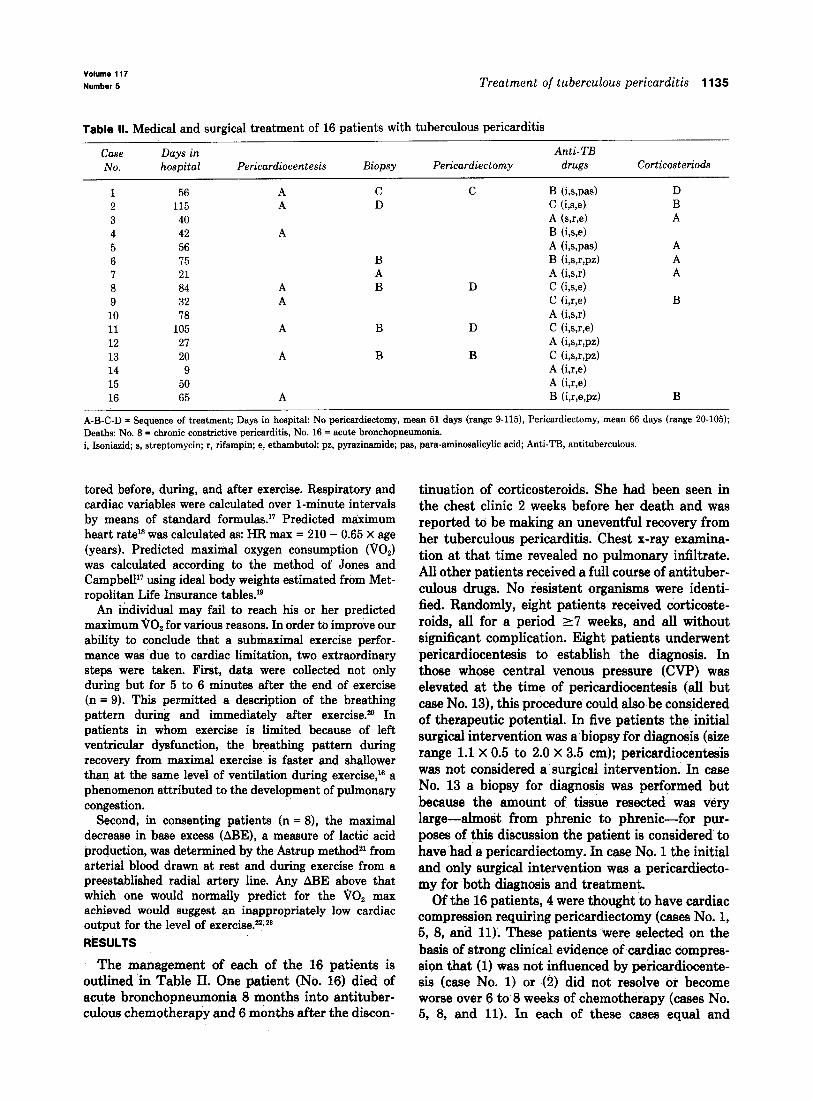
Table II. Medical and surgical treatment of 16 patients with tuberculous pericarditis
Case Days in A&i-TB
No. hospital Pericardiocentesis Biopsy Pericardiectomy drugs Corticosteriods
1 56 A C C B (i,s,w) D
2 115 A D C (i,s,e) B
3 40 A (s,r,e) A 4 42 A B (is,4 5 56 A (iwas) A
6 75 B B (i,s,r,pz) A I 21 A A (i,s,r) A 8 84 A B D C he) 9 32 A C (i,r,e) B
10 18 A (i,s,r) 11 105 A B D C (i,s,r,e) 12 27 A (i,s,wd 13 20 A B B C (ism4 14 9 A he) 15 50 A he) 16 65 A B (imsp-4 B
A-B-C-D = Sequence of treatment; Days in hospital: No pericardiectomy, mean 51 days (range g-115), Pericardiectomy, mean 66 days (range 20-105); Deaths: No. 8 = chronic constrictive pericarditis, No. 16 = acute bronchopneumonia. i, Isoniazid, s, streptomycin; r, rifampin; e, ethambutol; ps, pyrazinamide; pas, para-aminosahcylic acid; Anti-TB, antituberculous.
tored before, during, and after exercise. Respiratory and cardiac variables were calculated over l-minute intervals by means of standard formulas.17 Predicted maximum heart raW was calculated as: HR max = 210 - 0.65 X age (years). Predicted maximal oxygen consumption (00,) was calculated according to the method of Jones and Campbell” using ideal body weights estimated from Met- ropolitan Life Insurance tables.lg
An individual may fail to reach his or her predicted maximum 00, for various reasons. In order to improve our ability to conclude that a submaximal exercise perfor- mance was due to cardiac limitation, two extraordinary steps were taken. First, data were collected not only during but for 5 to 6 minutes after the end of exercise (n = 9). This permitted a description of the breathing pattern during and immediately after exercise.2o In patients in whom exercise is limited because of left ventricular dysfunction, the breathing pattern during recovery from maximal exercise is faster and shallower than at the same level of ventilation during exercise,16 a phenomenon attributed to the development of pulmonary congestion.
Second, in consenting patients (n = 8), the maximal decrease in base excess (ARE), a measure of 1actiC acid production, was determined by the Astrup methodzl from arterial blood drawn at rest and during exercise from a preestablished radial artery line. Any AEkE above that which one would normally predict for the 00, max achieved would suggest an inappropriately low cardiac output for the level of exercise.22*2a RESULTS
The management of each of the 16 patients is outlined in Table II. One patient (No. 16) died of acute bronchopneumonia 8 months into antituber- culous chemotherapy and 6 months after the discon-
tinuation of corticosteroids. She had been seen in the chest clinic 2 weeks before her death and was reported to be making an uneventful recovery from her tuberculous pericarditis. Chest x-ray examina- tion at that time revealed no pulmonary infiltrate. All other patients received a full course of antituber- culous drugs. No resistent organisms were identi- fied. Randomly, eight patients received corticoste- roids, all for a period 17 weeks, and all without significant complication. Eight patients underwent pericardiocentesis to establish the diagnosis. In those whose central venous pressure (WI?) was elevated at the time of pericardiocenteais (all but case No. 13)) this procedure could also be considered of therapeutic potential. In five patients the initial surgical intervention was a biopsy for diagnosis (size range 1.1 X 0.5 to 2.0 X 3.5 cm); pericardiocentesis was not considered a surgical intervention. In case No. 13 a biopsy for diagnosis was performed but because the amount of tissue resected was very large-almost from phrenic to phrenic-for pur- poses of this discussion the patient is considered to have had a pericardiectomy. In case No. 1 the initial and only surgical intervention was a pericardiecto- my for both diagnosis and treatment.
Of the 16 patients, 4 were thought to have cardiac compression requiring pericardiectomy (cases No. 1, 5, 8, and 11); These patients .were selected on the basis of strong clinical evidence of cardiac compres- sion that (1) was not influenced by pericardiocente- sis (case No. 1) or (2) did not resolve or become worse over 6 to 8 weeks of chemotherapy (cases No. 5, 8, and 11). In each of these cases equal and

1136 Long et al. hw 1989
American Heart Journal
Table III. Reevaluation of patients with tuberculous pericarditis treated medically: Length of follow-up, physical examination, and echocardiogram (n = 11)
Case No.
Length of follow-up
fyr) * Physical
examination
Echocardiogram --.--.___ ------.
FEM Valve EF Pericardium? (mm) ABN. (76)
2 14.75 Angina pectoris; pedal edema; hypertension X 6.0 MI 76 AI
3 13.00 Normal 3.0 52 4 11.00 Asthma; rheumatoid arthritis; pulsus 6.0 AS 63
paradoxus >lO mm Hg; CVP and liver span upper limit normal; crepitations in lungs; peripheral edema, grade 2-3/6 SEM at base
5 11.00 Normal 5.0 56 6 5.00 Alcoholic, liver 14 cm span X 8.0 74 7 3.00 Mild pretibial edema X 8.0 60 9 2.50 Normal 8.0 63
10 1.83 Normal 8.0 68 12 1.67 Apical systolic retraction 5.0 62 14 1.25 Occasional ectopic beat 2.5 75 15 1.00 Apex beat 13-14 cm from MSL; grade 2/6 5.0 AI 72
apical systolic murmur; hypertension Mean r SD 6.00 + 5.31 5.9 zk 2.0 66 + 8
CVP, Central venous pressure; MI, mitral incompetence; AI, aortic incompetance; abn, abnormality; SEM, systolic ejection murmur; MSL, midsternal line; FEM, flat endocardial motion; EF, ejection fraction; AS, aortic stenosis. *From the time of diagnosis to reevaluation. tPericardia1 effusion or thickening.
elevated atrial mean and ventricular end-diastolic pressures were confirmed at right heart catheteriza- tion. Where the effect of pericardiocentesis on CVP was recorded before and after tap (cases No. 1,2,11, 16 at or before diagnosis and case No. 8 six weeks after diagnosis), it fell into the normal range in cases not selected for pericardiectomy (Nos. 2 and 16) and did not fall into the normal range in cases selected for pericardiectomy (Nos. 1,8, and 11). In the three patients in whom cardiac compression did not resolve or became worse over 6 to 8 weeks of chemotherapy, CVP remained elevated ~8 cm H20, whereas in all remaining patients it fell into the normal range over this period of time (p < 0.005, Fischer’s exact test). In retrospect, one marker of advanced disease may have been of some assistance in the selection process; the average thickness of the fixed pericardial biopsy specimen as noted by the pathologist at the time of tissue trimming was 11.7 + 1.5 mm (range 10.0 to 13.0) in patients who required pericardiectomy (Nos. .l, 8, and ll), and 4.0 -+ 1.8 mm (range 2.0 to 6.0) in patients who did not (Nos. 2, 6, 7, and 13) (p < 0.01). Of the four patients thought to have cardiac compression requiring pericardiectomy, three actually underwent the procedure (Nos. 1, 8, and 11). In patient No. 5 surgery was delayed and over a period of weeks the
cardiac compression resolved with chemotherapy alone. This patient received steroids. One of the patients who had undergone pericardiectomy (No. 8) died of constrictive pericarditis (confirmed at heart catheterization on two separate occasions within 9 months of pericardiectomy) 33 months after the diagnosis of tuberculous pericarditis. This patient had not received steroids.
The results of the reevaluation of all patients not undergoing pericardiectomy are given in Tables III and IV. No patient gave a history of recurrent acute pericarditis. Only one patient (No. 4, age 92) had physical findings suggesting the possibility of con- strictive pericarditis and/or annular constricting pericarditis. His laboratory results were as follows: chest x-ray film was unremarkable; ECG showed nonspecific ST and T wave changes; the echocardio- gram confirmed the presence of aortic stenosis; and the CT scan was normd: Because he did not have symptoms that interfered with his activities of daily living (ambulant with a walker) and because of his age, we could not justify ‘further investigation. One patient (No. 2, age 80) had a history of angina pectoris. However, physical examination was not remarkable except for systolic hype&e&on and all laboratory investigatiens were normal. One patient (No. 15, age 81) had cardiomegaly on chest x-ray

Volume 117 Number 5 Treatment of tuberculous pericarditis 1137
Table IV. Reevaluation of patients with tuberculous pericarditis treated medically: Spirometry and exercise (n = 8)
VO, HR at ABE at Case FEV, FVC max VO, max rro, AV,* No. (X predi (% pred) FE V,IFVC % pred (% pred) mar %VC
3 3.59 (116) 5 3.69 (124) 6 3.17 (85) 7 2.82 (86) 9 4.87 (112)
10 5.14 (121) 12 5.20 (119) 14 3.10 (97)
Mean + SD (108 + 16)
4.51 (120) 80 3.99 (116) 92 4.86 (104) 65 3.47 (92) 78 5.76 (103) 85 5.57 (109) 92 5.64 (104) 92 4.61 (109) 67 (107 f 9) 81 f 11
96 180 (100) 55 134 (92) 75 188 (110)
122 155 (70) 66 138 (92) 85 150 (86) 86 175 (98) 95 140 (85)
85 zt 21 (92 2 12)
-1.0 -0.93 -2.5 -0.10 -3.4 +4.07 -1.6 -2.37 -1.7 +0.33 -5.7 -1.36
-4.19 -0.65
FEV,, Forced expiratory volume in 1 second; FVC, forced vital capacity; pred, predicted; HR, heart rate; BE, base excess; VT, tidal volume; VC, vital capacity. *Difference between exercise and recovery tidal volume as a percent of vital capacity.
examination, but this was associated with left ven- tricular prominence, a history of hypertension, and an otherwise unremarkable laboratory investigation. No patient had pericardial calcification on chest x-ray examination. All patients were in normal sinus rhythm and none had conduction disturbances. The ECG showed nonspecific ST and T wave abnormal- ities in two patients (Nos. 4 and 6), P-mitrale in one (No. 5), and one patient (No. 14) had the occasional ventricular ectopic beat. This last patient had a history of skipped beats prior to his pericarditis. Echocardiographic abnormalities were few: anterior pericardial thickening of uncertain significance in three (Nos. 2,6, and 7), a stenotic valve lesion in one (No. 4), and mild regurgitant valve lesions in two (Nos. 2 and 15). Wall motion and ejection fraction were normal in all patients. CT findings were also few: the ventral parietal pericardial thickness was normal (<2 mm) in all; one had thin patchy thicken- ing elsewhere (No. 6), and many (n = 8) had what appeared to be a small area of pericardial calcifica- tion that could not always be differentiated from streak artifact. Though variability in inferior vena cava (IVC) diameter is recognized in normals, in no patient was it markedly enlarged; mean f SD IVC/ A0 (aortic) ratio was 1.4 f 0.2; range was 1.0 to 1.8.
Spirometry was normal in all patients tested (n = 8) except for two (Nos. 6 and 14) who showed evidence of mild airway obstruction (FEVJFVC [forced expiratory volume in 1 second/forced vital capacity] = 65% and 67%) respectively). Both of these had a smoking history.
Six of the eight patients attained normal levels of maximal exercise (175 % predicted 00, max). In the other two (Nos. 5 & 9), the maximum level reached was well below normal (but see below). In all
patients heart rate at the highest level of exercise (mean 92 % , range 70 % to 110 % ) was not different from that predicted for normal subjects at compara- ble levels of exercise. Likewise, the degree of meta- bolic acidosis (decrease in base excess [BE]) was appropriate for the level of exercise reached in all subjects tested. In fact, there was a lesser degree of exercise-related metabolic acidosis than occurs on average in normal subjects at similar levels of work. Only one patient (No. 7) showed evidence of relative tachypnea during recovery. This patient, however, was young and reached a 00, max exceeding his predicted value (122%), so that there was no ques- tion of any exercise limitation. This degree of rapid shallow breathing during recovery is also observed in some young normal subjects after very heavy exer- cise.24 In the absence of any evidence of inappropri- ate tachycardia, metabolic acidosis, ventilatory limi- tation, or tachypnea, the poor maximal performance of patients Nos. 5 and 9 was attributed to inade- quate effort.
Survivors who had previously undergone pericar- diectomy (Nos. 1, 11, and 13) were also reevaluated. Patient No. 1 had parietal pericardial thickening at CT (6.3 mm) but no wall motion abnormality at echocardiogram, and appeared “superfit” at exer- cise. Except for two wall motion abnormalities on echocardiography in patient No. 11, reevaluation in cases No. 11 and 13 revealed no abnormalities.
DISCUSSION
Though many patients once diagnosed as having active tuberculous pericarditis appear to do well with chemotherapy alone,lpz no systematic studies of the long-term outcome in these patients has been reported. In this investigation we reasoned that if patients were able to exercise to normal maximal

1138 Long et al. May 1989
American Heart Journal
levels and display normal responses in the process, then any abnomalities undetected by our resting evaluation would be of questionable significance. The results suggest that when pericardiectomy is not required for the relief of cardiac compression in the acute phase of tuberculous pericarditis, the long-term outcome with medical therapy alone is excellent.
The records of all patients diagnosed with active tuberculous pericarditis over a 16-year period were carefully reviewed. Of the total of 16, fourteen (11 not having undergone pericardiectomy) were still alive. All survivors were reevaluated for the presence of constrictive pericarditis as well as other recog- nized sequelae of this disease. Of the resting studies, physical examination suggested the possibility of constrictive pericarditis in one patient (No. 4). However, his laboratory investigations did not sup- port this diagnosis, and when we took into account his age (92 years) and the fact that his activities of daily living were not limited by cardiac symptoms, we could not justify further investigation. Whereas the length of follow-up in this patient was >ll years, in most other cases it was shorter, raising the possibility that those with a short follow-up might have nonconstricting pericardial thickening that might later mature to produce constriction. Experi- ence in pleural and pulmonary tuberculosis, howev- er, provides no precedent for the argument that active fibrosis continues after patients have com- pleted a full course of chemotherapy. Moreover, only one survivor (No. 1, pericardiectomy) had evidence of pericardial thickening at CT. Thus a significant constricting process was not present nor likely to develop in any of the medically treated patients. An additional patient (No. 16, no pericardiectomy) died of bronchopneumonia but without evidence of peri- cardial constriction. Evidence at rest for other recog- nized sequelae of tuberculous pericarditis was also minimal. Annular constriction resulting in aortic stenosis could not be excluded in case No 4; and epicardial-myocardial fibrosis resulting in coronary insufficiency, or ventricular ectopic beats, could not be excluded in cases No. 2 and 14, respectively. These abnormalities could, however, be explained by more common pathophysiologic mechanisms. Finally, the results of our resting studies were supported by our failure to demonstrate any evidence of abnormal cardiac performance during the severe challenge of maximal exercise. Of the patients tested, none showed evidence of inappropriate cardiac out- put (inappropriate tachycardia or acidosis) or pulmo- nary congestion (tachypnea during recovery), two recognized complications of pericardial constric-
tionz5vz6 Thus the combined anatomic and physiologic evidence presented in this study essentially excludes any significant pericardial pathology in those patients who did not receive pericardiectomy.
In the past, the major reason for the failure of medical therapy was cardiac compression. In the series of Hageman et al.,l 35% of the patients who received medical therapy alone died, and in the series of Rooney et a1.,27 10% of the patients who received medical therapy alone died. In the majority of cases the cause of death was cardiac tamponade or constriction. Recognizing the presence of cardiac compression is therefore critical in these patients. Knowing when and how to intervene, however, is not always clear. Added to this vexing question has always been the knowledge that pericardiectomy in a medical failure is technically more difficult and presents a greater hazard to the patient the longer it is delayed.
In the present series the decision was made to perform pericardiectomy only in those patients in whom there was evidence of cardiac compression that was not relieved by pericardiocentesis or that did not resolve or became worse over 6 to 8 weeks of chemotherapy. Interpreted in the light of current understanding of the pathophysiology of pericardial disease, these patients had “effusive-constrictive” hemodynamics present at the time of diagnosis or becoming apparent in the weeks immediately fol- lowing diagnosis. 28 The latter refers to cardiac com- pression due to both a thickened pericardium as well as to excess pericardial fluid. It is defined by a persistently elevated right atria1 and CVP after the intrapericardial pressure has been returned to nor- mal by pericardiocentesis. To the extent that our population of patients is representative, the results suggest that this policy toward pericardiectomy is an adequate one, as no patient who was not so treated suffered any long-term ill effects. Finally, though it is not possible to conclude from this study that corticosteroids are beneficial, their demonstrated ability to hasten the resolution of tuberculous pleu- ral effusions2g suggests that they may allow a more rapid determination of the contribution of tampon- ade versus constriction.
The authors are very grateful to the many persons who contributed to the completion of this project. These include Drs. John Teskey and Helmut Unruh of the Cardiovascular and Thoracic Surgery Units, who reviewed the manuscript; Drs. D. O’Donnell, M. Kroeker, M. Rigby, D. Ostrow, S. Vedal, and C. Tomlinson, who assisted with the noninvasive tests; J. MacMor- ran, A. Maskell, G. Last, and S. Olien of the Manitoba Tubercu- losis Registry; and W. Eriksen and M. Poworoznyk of the pulmonary function and exercise laboratory, Respiratory Centre, Winnipeg.
Volume 117
Number 5
REFERENCES
1.
2.
3.
4.
5.
6.
7.
8.
9.
10.
11.
12.
13.
14.
Hageman JH, D’Esopo ND, Glenn WWL. Tuberculosis of the pericardium. A long-term analysis of forty-four proved cases. N Engl J Med 1964;270:327. Sagrist&Sauleda J, Permanyer-Miralda G, Soler-Soler J. Tuberculous pericarditis: ten-year experience with a prospec- tive protocol for diagnosis and treatment. J Am Co11 Cardiol 1988;11:724. Carson TJ, Murray GF, Wilcox BR, Starek PJK. The role of surgery in tuberculous pericarditis. Ann Thorac Surg 1974;17:163. Larrieu AJ, Tyers GFO, Williams EH, Derrick JR. Recent experience with tuberculous pericarditis. Ann Thorac Surg 1980;29:464. Sonnenberg FA, Pauker SG. Elective pericardiectomy for tuberculous pericarditis: should the snappers be snipped? Med Decis Making 1986;6:110. Mounsey P. Annular constrictive pericarditis. Br Heart J 1959;21:325. Dalton JC, Pearson RJ Jr, White PD. Constrictive pericardi- tis: a review and long-term follow-up of 78 cases. Am J Cardiol 1956;45:445. Voelkel AG, Pietro DA, Folland ED, et al. Echocardiograpbic features of constrictive pericarditis. Circulation 1978;58:871. Candell-Riera J. de1 Castillo G. Permanver-Miralda G. Soler- Soler J. Echocardiographic features of the interventricular septum in chronic constrictive pericarditis. Circulation 1978;57:1154. Sutton FJ, Whitley NO, Applefield MM. The role of echocar- diography and computed tomography in the evaluation of constrictive pericarditis. AM HEART J 1985;109:350. Tei C, Child JS, Tanaka H, Shah PM. Atria1 systolic notch on the interventricular septal echogram: an echocardiographic sign of constrictive pericarditis. J Am Co11 Cardiol 1983; 1:907. Quinones MA, Waggoner AD, Reduto LA, et al. A new, simplified and accurate method for determining ejection fraction with two-dimensional echocardiography. Circulation 1981;64:744. Moncada R, Baker M, Salinas M, et al. Diagnostic role of computed tomography in pericardial heart disease: congeni- tal defects, thickening, neoplasms and effusions. AM HEART J 1982;103:263. Isner JM, Carter BL, Bankoff MS, et al. Computed tomogra- phy in the diagnosis of pericardial heart disease. Ann Intern Med 1982;97:473.
15.
16.
17.
18.
19.
20.
21.
22.
23.
24.
25.
26.
27.
28.
Treatment of tuberculous pericarditis 1139
Doppman JL, Rienmuller R, Lissner J, et al. Computed tomography in constrictive pericardial disease. J Comput Assist Tomogr 1981$X. Gallagher CG, Younes M. Breathing pattern during and after maximal exercise in patients with chronic obstructive lung disease, interstitial lung disease, and cardiac disease and in normal subjects. Am Rev Respir Dis 1986;133:581. Jones NL, Campbell EJM. Clinical exercise testing. Philadel- phia: W.B. Saunders Co, 1982. Lange-Anderson K, Shephard RJ, Denolin H, et al. Funda- mentals of exercise testing. Geneva: World Health Organiza- tion, 1971. Halpern SL, Glenn MB, Goodhart RS. New height-weight tables: importance of new criteria. JAMA 1960;173:1576. Hey EN, Lloyd BB, Cunningham DJC, et al. Effects of various respiratory stimuli on the depth and frequency of breathing in man. Respir Physiol 1966;1:193. Goldberger E. The Astrup method of determining acid-base disturbances. In: Goldberger E, ed. A primer of water, electrolyte and acid-base syndromes. Philadelphia: Lea & Febiger, 1975:174-207. Riddle W, Younes M, Remmers J, De Groot W. Graphical analysis of patient performance in the pulmonary function laboratory. In: Proceedings of the Fourth Annual Symposium on Computer Application in Medical Care. November 2-5, 1980; Washington, D.C: 283-90. Wasserman K, Whipp BJ. Exercise physiology in health and disease. Am Rev Respir Dis 1975;112:219. Younes M, Burks J. Breathing pattern during and after exercise of different intensities. J Appl Physiol 1985; 59898. Shabetai R. Diseases of the pericardium. In: Hurst JW, ed. The heart vol. II. New York: McGraw-Hill Book Co, Inc, 1986:1249-75. Robbins MA, Pizzarello RA, Stechel RP, et al. Resting and exercise hemodynamics in constrictive pericarditis and a case of cardiac amyloidosis mimicking constriction. Cathet Car- diovasc Diagn 1983;9:463. Rooney JJ, Crocco JA, Lyons HA. Tuberculous pericarditis. Ann Intern Med 1972;72:73. Hancock EW. On the elastic and rigid forms of constrictive pericarditis. AM HEART J 1980;100:917.
29. Tani P, Poppius H, Makipaja J. Cortisone therapy for exudative tuherculous pleurisy in the light of a follow-up study. Acta Tuberc Stand 1964;44:303.