Constrictive Pericarditis
39
Constrictive Pericarditis Nisha I. Parikh, MD MPH July 21 st 2009 Echo Conference
description
Constrictive Pericarditis. Nisha I. Parikh, MD MPH July 21 st 2009 Echo Conference. Summary of Talk. Background Clinical features Echocardiographic diagnosis M-mode Doppler Constriction versus restriction Treatment and prognosis. Historical Perspective. - PowerPoint PPT Presentation
Transcript of Constrictive Pericarditis
Constrictive Pericarditis
Nisha I. Parikh, MD MPH
July 21st 2009
Echo Conference
Summary of Talk
Background Clinical features Echocardiographic diagnosis
M-modeDoppler
Constriction versus restriction Treatment and prognosis
Historical Perspective
The history of constrictive pericarditis is replete with famous names in medicine
Richard Lower described a patient with dyspnea and an intermittent pulse in 1669
Lancisi first reported on the constrictive syndrome in 1828
Corrigan described the pericardial knock in 1842
Kussmaul described his sign and the associated paradoxical pulse in 1873.
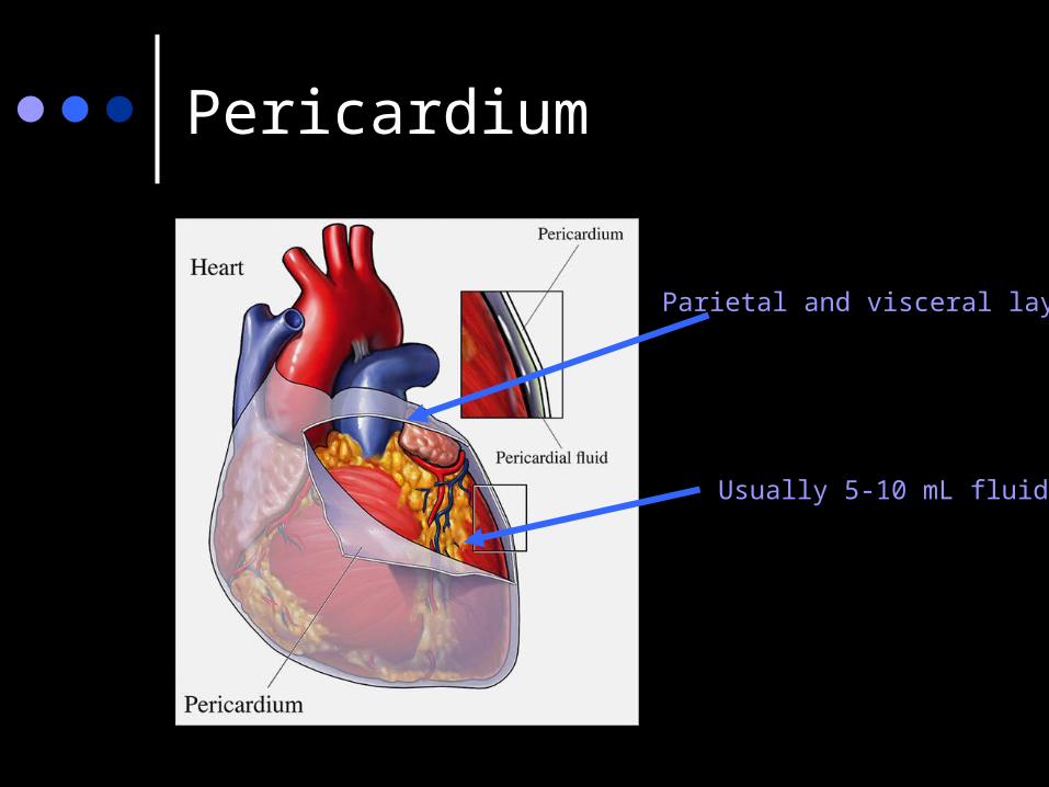
Pericardium
Usually 5-10 mL fluid
Parietal and visceral layers
Pericardium
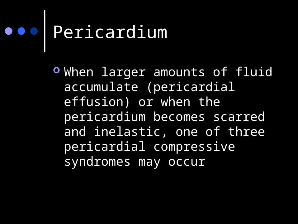
When larger amounts of fluid accumulate (pericardial effusion) or when the pericardium becomes scarred and inelastic, one of three pericardial compressive syndromes may occur
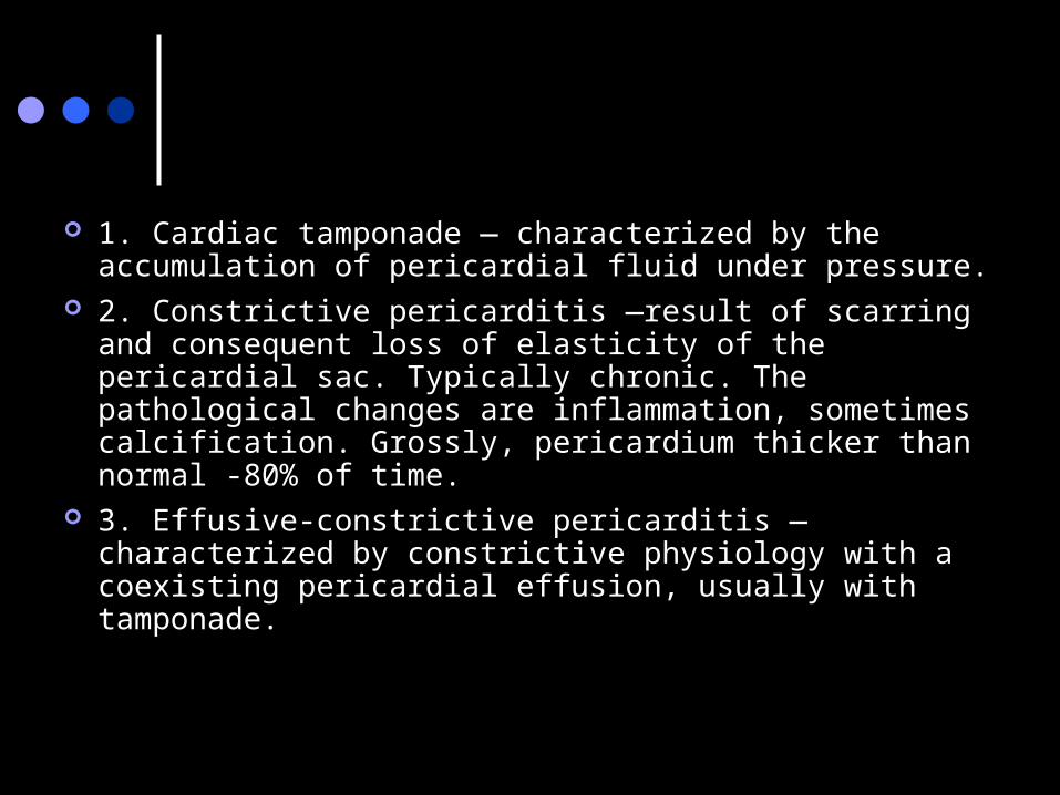
1. Cardiac tamponade — characterized by the accumulation of pericardial fluid under pressure.
2. Constrictive pericarditis —result of scarring and consequent loss of elasticity of the pericardial sac. Typically chronic. The pathological changes are inflammation, sometimes calcification. Grossly, pericardium thicker than normal -80% of time.
3. Effusive-constrictive pericarditis —characterized by constrictive physiology with a coexisting pericardial effusion, usually with tamponade.
Epidemiology
9% of patients with acute pericarditis for any reason go on to develop constrictive physiology.
Acute pericarditis is only clinically diagnosed in 1 in 1,000 hospital admissions
Frequency of a diagnosis of constrictive pericarditis is less than 1 in 10,000 hospital admissions.
Constrictive Pericarditis - HPI
67 % presented with symptoms of heart failure (HF)
8 % with chest pain 6 % with abdominal symptoms 4 % with atrial arrhythmia 5 % with symptoms of cardiac
tamponade
Constrictive Pericarditis - Etiology
Idiopathic or viral — 42 to 49 % Post cardiac surgery — 11 to 37 % Post radiation therapy — 9 to 31 % Connective tissue disorder — 3 to 7 % Postinfectious (tuberculous or purulent
pericarditis) — 3 to 6 % Miscellaneous causes (malignancy, trauma,
drug-induced, asbestosis, sarcoidosis, uremic pericarditis) — 1 to 10 %
Constricitve Pericarditis - PE
Elevated JVP Peripheral edema Ascites Hepatomegaly Pleural effusion S3 Pulsus paradoxus Kussmaul’s sign Cachexia- late stages
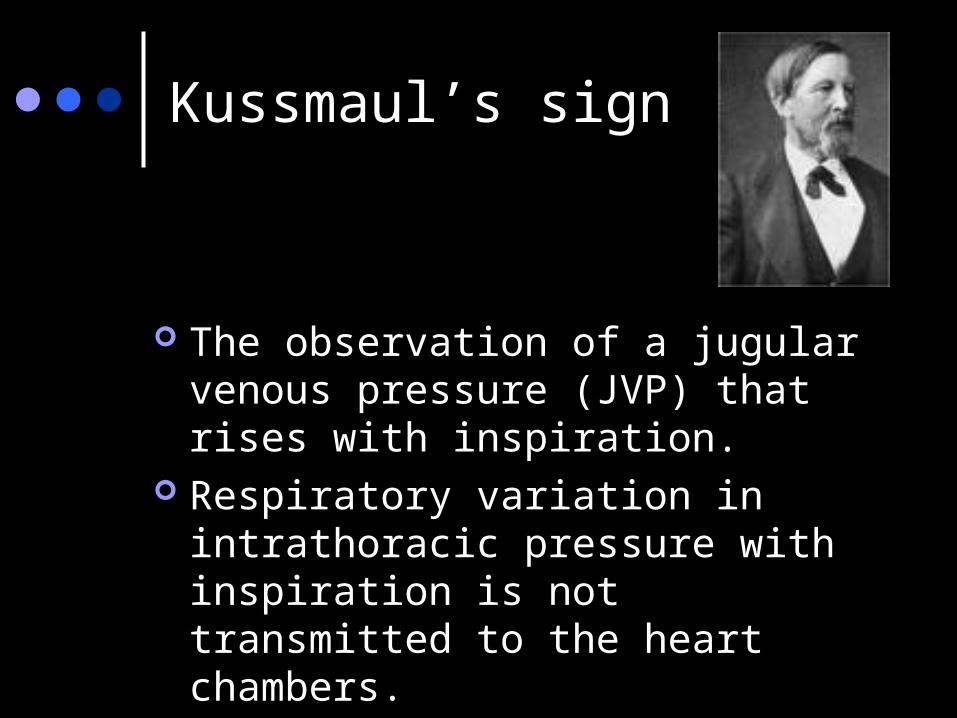
The observation of a jugular venous pressure (JVP) that rises with inspiration.
Respiratory variation in intrathoracic pressure with inspiration is not transmitted to the heart chambers.
Kussmaul’s sign
Physiology of constriction
In the pericardial compressive syndromes, the pericardium is inelastic and total cardiac volume cannot change
The result is enhanced ventricular interaction or…ventricular interdependence
Physiology of constriction
Pericardial constriction leads to impairment of ventricular filling, usually affecting all four cardiac chambers, preventing ventricular filling in mid and late diastole.
As a result, the majority of ventricular filling occurs rapidly in early diastole and the ventricular volume does not increase after the end of the early filling period.
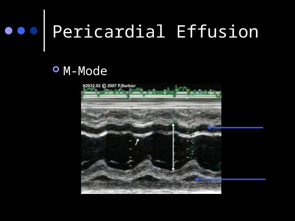
Pericardial Effusion
M-Mode
Pericardial effusion
M-mode Cannot determine volume of accumulated fluid accurately
Pericardial thickening
This can be visualized by transesophageal echo (often requiring multiple views), however, this is best seen using other imaging modalities such as CT or MRI.
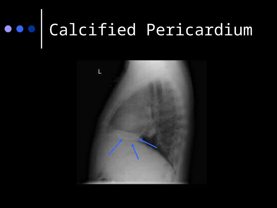
Calcified Pericardium

Pericardial calcifications CT
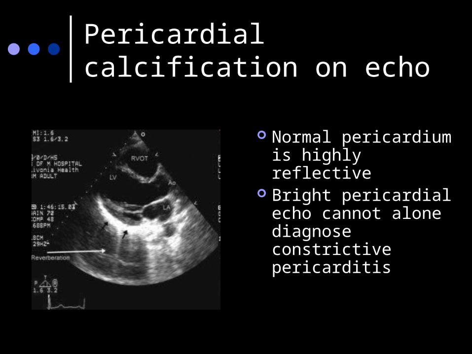
Pericardial calcification on echo
Normal pericardium is highly reflective
Bright pericardial echo cannot alone diagnose constrictive pericarditis
Specific echo exam for constriction
Neither sensitive nor specific Must diagnose via a combination of
physical exam/ history findings and echo findings

M-mode findings in constriction Abrupt relaxation of the posterior wall
with flattening of endocardial motion during diastole
Abnormal septal motion:Mimics conduction disturbancesMimics RV p/v overloadEarly diastolic notching followed by
paradoxical and then normal motion of the ventricular septum
diastolic septal bounce:
Thought to be due to the rapid filling during early diastole leading to asymmetrical filling of the right and left ventricals which creates a fluctuating pressure gradient that manifests as an abrupt shift of the septum.
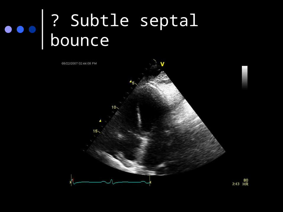
? Subtle septal bounce
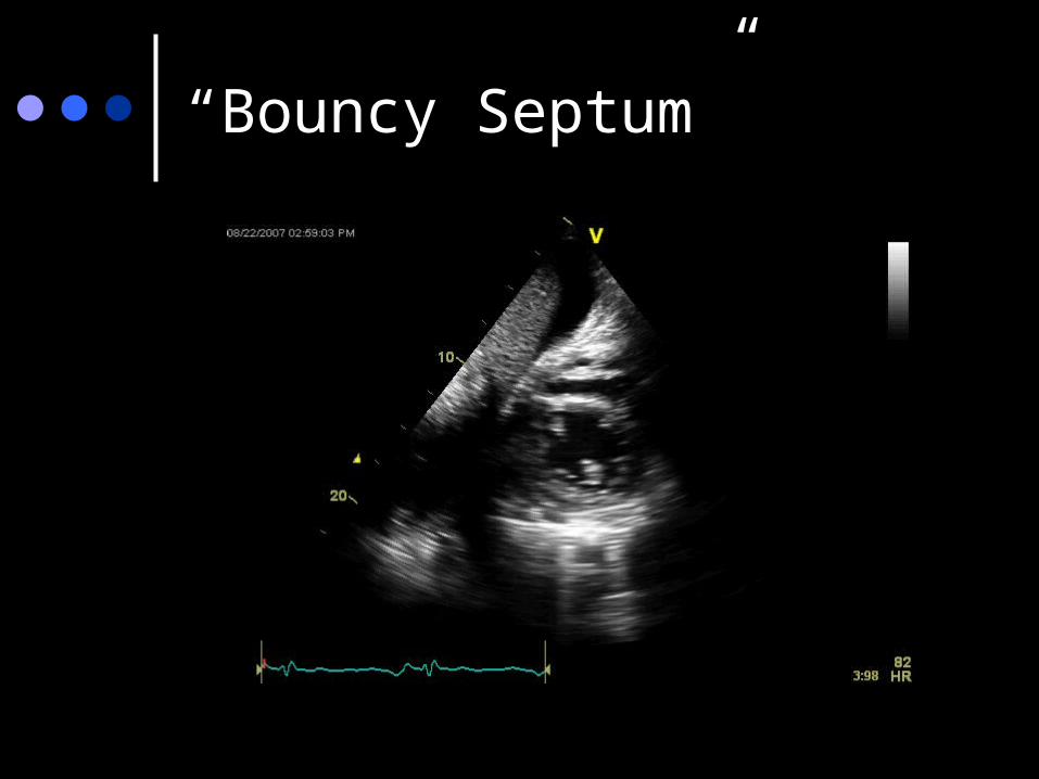
“Bouncy Septum”
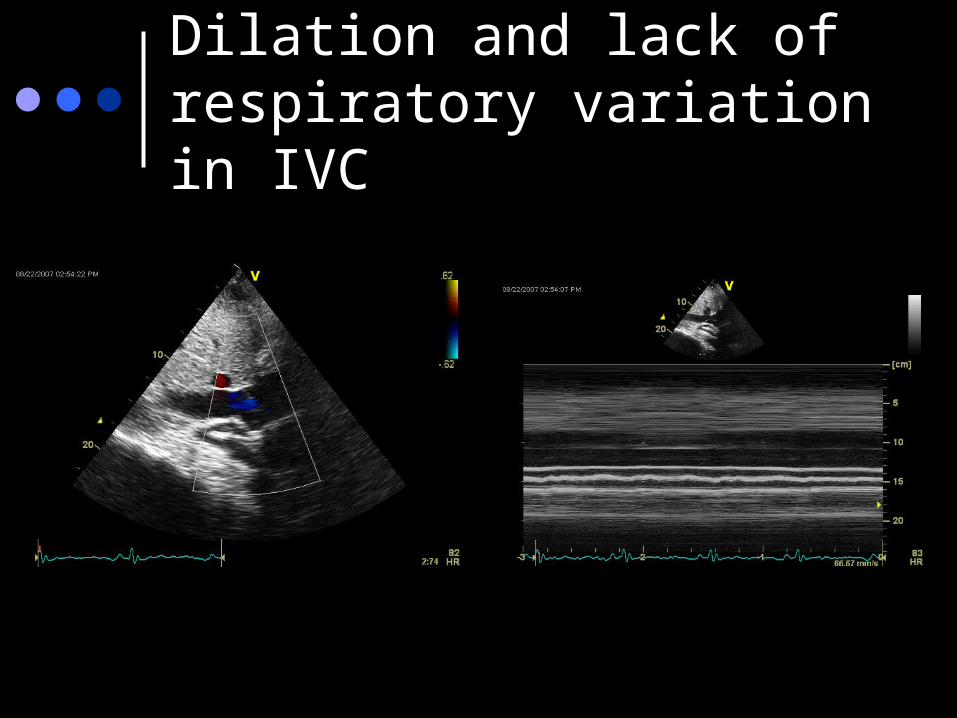
Dilation and lack of respiratory variation in IVC
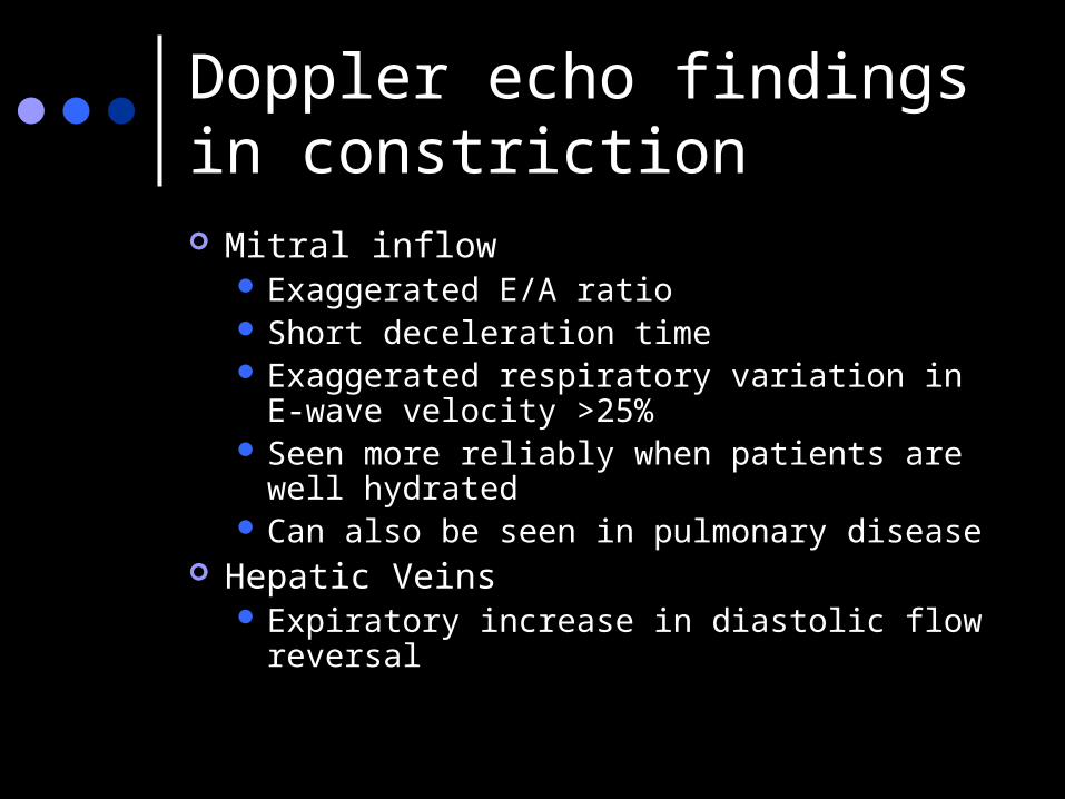
Doppler echo findings in constriction Mitral inflow
Exaggerated E/A ratio Short deceleration time Exaggerated respiratory variation in E-wave
velocity >25% Seen more reliably when patients are well
hydrated Can also be seen in pulmonary disease
Hepatic Veins Expiratory increase in diastolic flow reversal
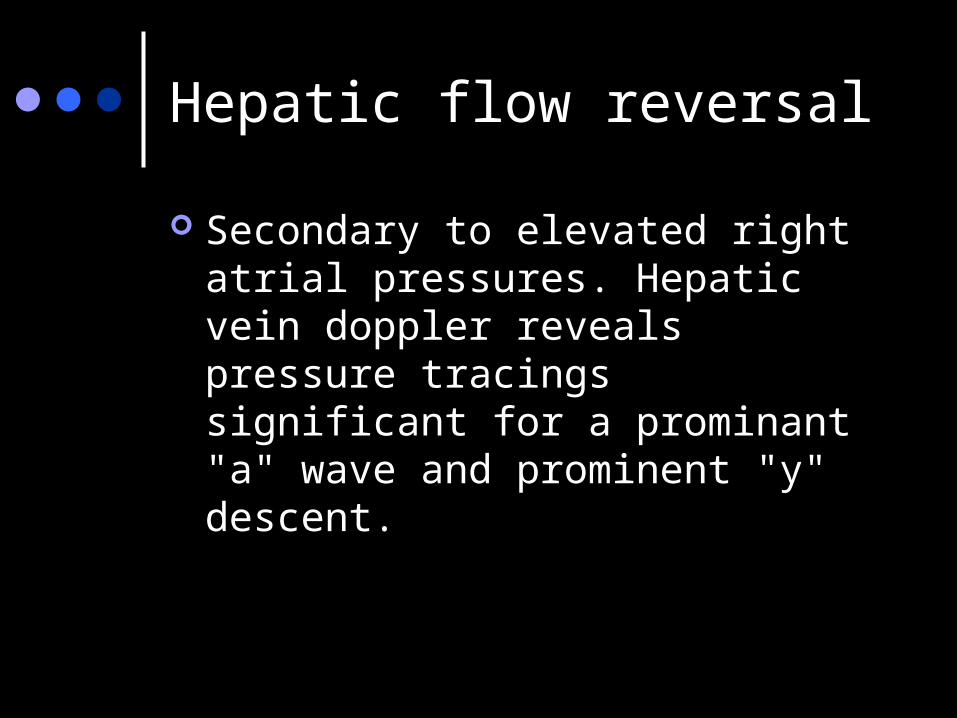
Hepatic flow reversal
Secondary to elevated right atrial pressures. Hepatic vein doppler reveals pressure tracings significant for a prominant "a" wave and prominent "y" descent.
Atrial dilation
Mild Secondary to elevated atrial
pressures More severe atrial dilatation seen in
restrictive cardiomyopathy.
Constrictive Pericarditis – other tests?
CT – not very sens/spec Cardiac MRI – growing in favor BNP – usually only a mild elevation
due to limited wall stretch Cath – GOLD STANDARD
Effusive constrictive pericarditis
Combination of tamponade and constriction
Common etiologies: malignancy and radiation therapy
Pericardial thickening may prevent RA collapse
Hemodynamic compromise and JVD persist even after tap
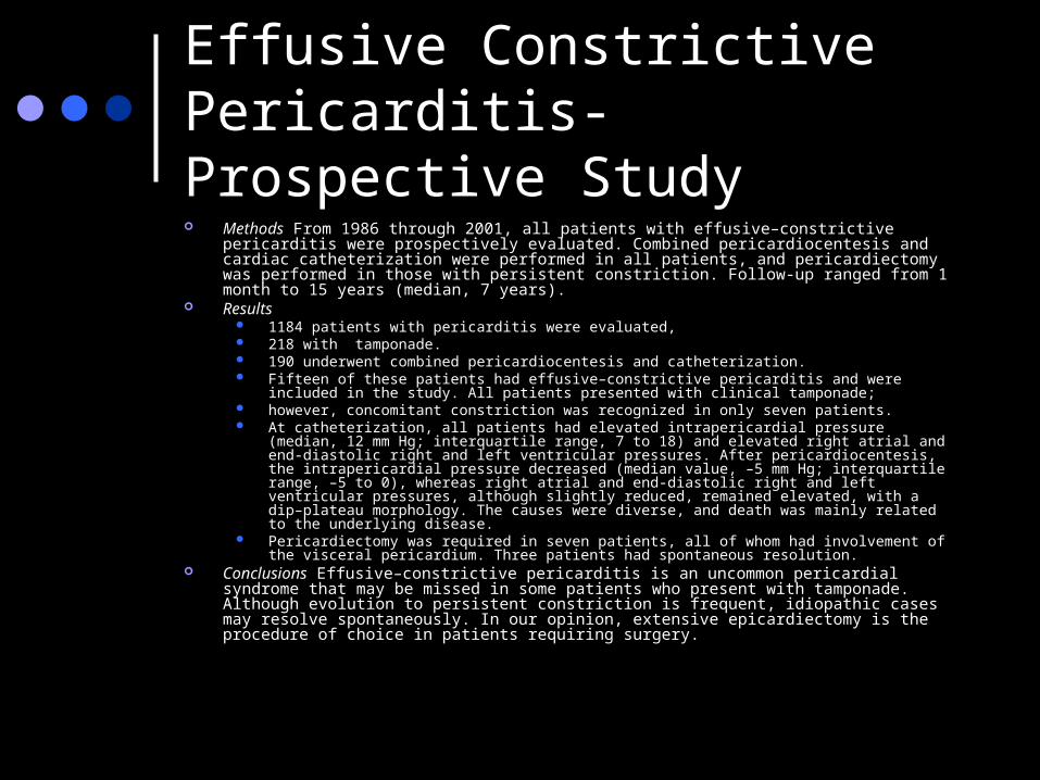
Effusive Constrictive Pericarditis- Prospective Study Methods From 1986 through 2001, all patients with effusive–constrictive pericarditis were prospectively
evaluated. Combined pericardiocentesis and cardiac catheterization were performed in all patients, and pericardiectomy was performed in those with persistent constriction. Follow-up ranged from 1 month to 15 years (median, 7 years).
Results 1184 patients with pericarditis were evaluated, 218 with tamponade. 190 underwent combined pericardiocentesis and catheterization. Fifteen of these patients had effusive–constrictive pericarditis and were included in the study. All patients
presented with clinical tamponade; however, concomitant constriction was recognized in only seven patients. At catheterization, all patients had elevated intrapericardial pressure (median, 12 mm Hg; interquartile range,
7 to 18) and elevated right atrial and end-diastolic right and left ventricular pressures. After pericardiocentesis, the intrapericardial pressure decreased (median value, –5 mm Hg; interquartile range, –5 to 0), whereas right atrial and end-diastolic right and left ventricular pressures, although slightly reduced, remained elevated, with a dip–plateau morphology. The causes were diverse, and death was mainly related to the underlying disease.
Pericardiectomy was required in seven patients, all of whom had involvement of the visceral pericardium. Three patients had spontaneous resolution.
Conclusions Effusive–constrictive pericarditis is an uncommon pericardial syndrome that may be missed in some patients who present with tamponade. Although evolution to persistent constriction is frequent, idiopathic cases may resolve spontaneously. In our opinion, extensive epicardiectomy is the procedure of choice in patients requiring surgery.
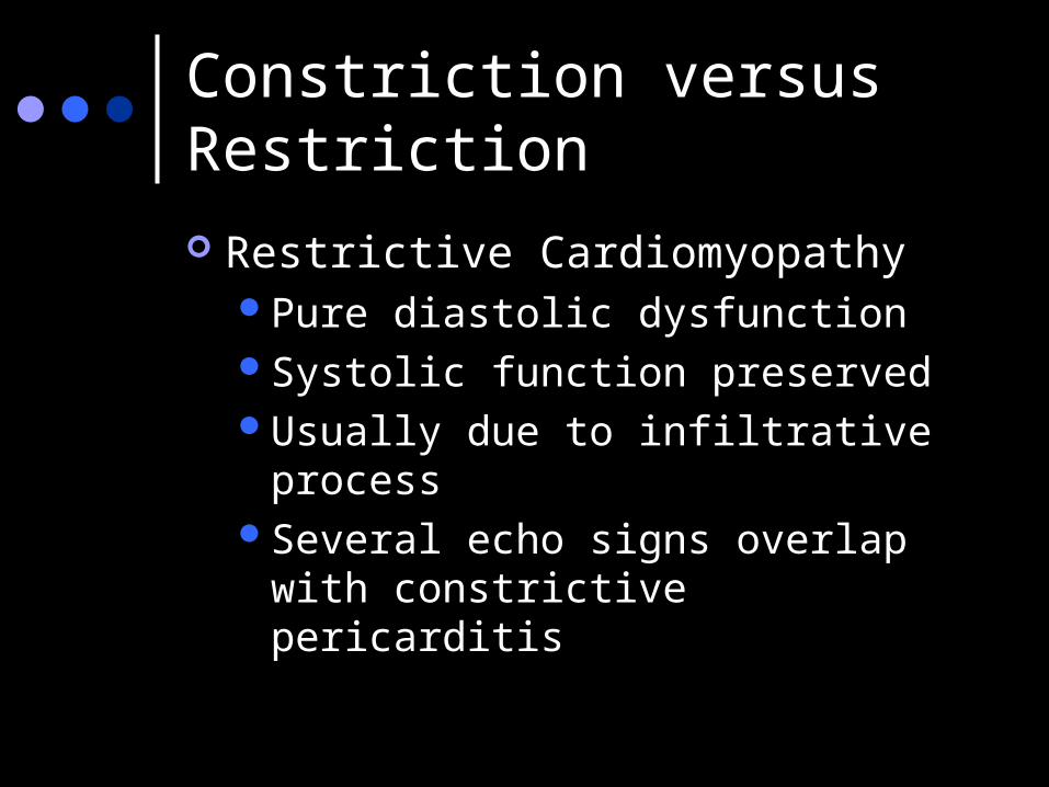
Constriction versus Restriction
Restrictive CardiomyopathyPure diastolic dysfunctionSystolic function preservedUsually due to infiltrative processSeveral echo signs overlap with
constrictive pericarditis
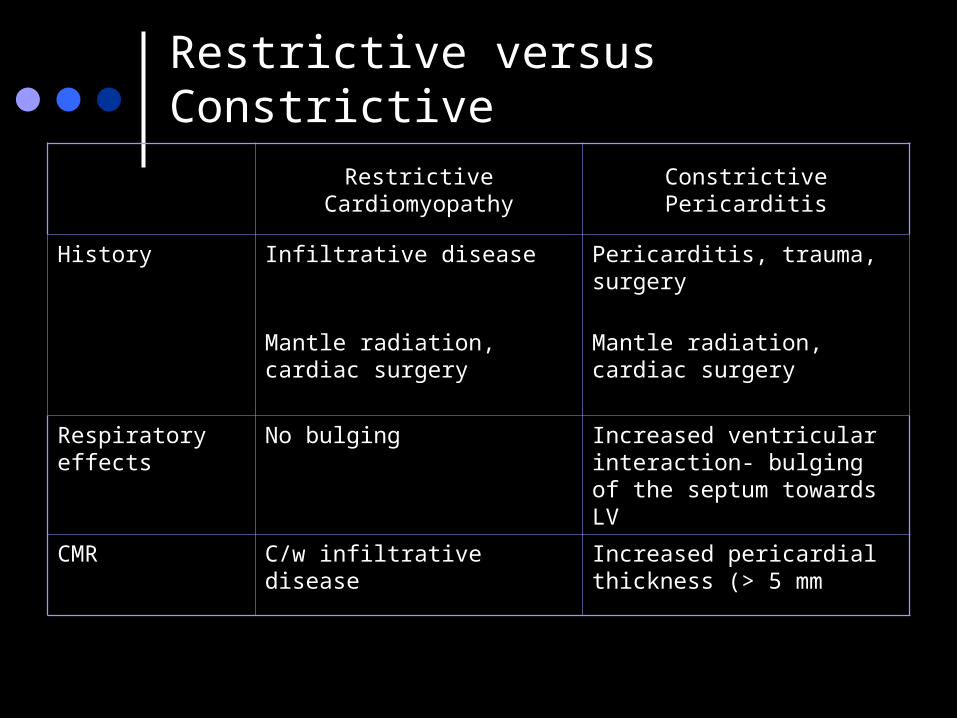
Restrictive versus Constrictive
Restrictive Cardiomyopathy Constrictive Pericarditis
History Infiltrative disease Pericarditis, trauma, surgery
Mantle radiation, cardiac surgery
Mantle radiation, cardiac surgery
Respiratory effects
No bulging Increased ventricular interaction- bulging of the septum towards LV
CMR C/w infiltrative disease Increased pericardial thickness (> 5 mm
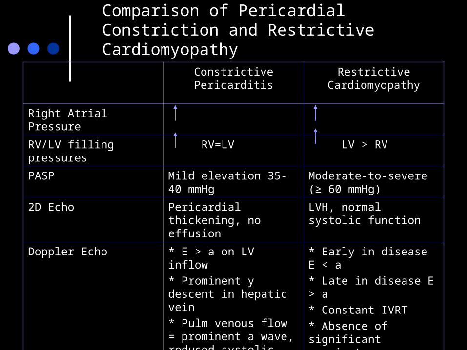
Comparison of Pericardial Constriction and Restrictive Cardiomyopathy
Constrictive Pericarditis Restrictive Cardiomyopathy
Right Atrial Pressure
RV/LV filling pressures RV=LV LV > RV
PASP Mild elevation 35-40 mmHg
Moderate-to-severe (≥ 60 mmHg)
2D Echo Pericardial thickening, no effusion
LVH, normal systolic function
Doppler Echo * E > a on LV inflow
* Prominent y descent in hepatic vein
* Pulm venous flow = prominent a wave, reduced systolic phase
* Resp variation in IVRT and E velocity
* Atria: mildly enlarged
* Early in disease E < a
* Late in disease E > a
* Constant IVRT
* Absence of significant respiratory variation
* Marked enlarged atria
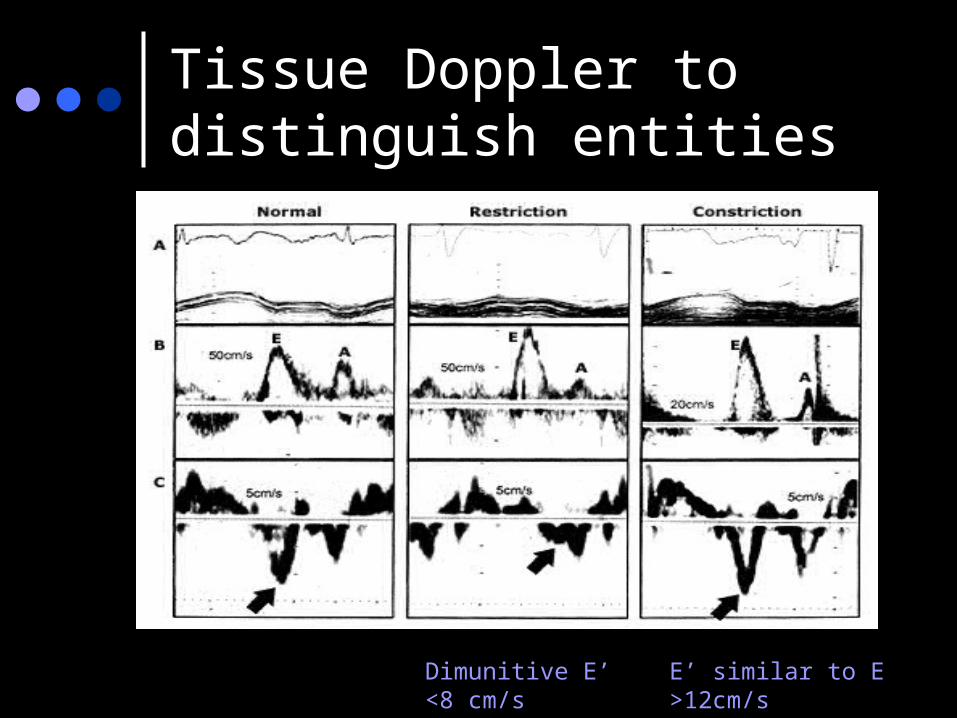
Tissue Doppler to distinguish entities
Dimunitive E’<8 cm/s
E’ similar to E>12cm/s
Treatment
Definitive treatment is surgical Earlier the better Extensive decortication favored, especially at the diaphragmatic-
ventricular contact regions. Complications
excessive bleeding atrial and ventricular arrhythmias ventricular wall ruptures.
Published surgical mortality 5-15%. Perioperative mortality rate (within 30 days) was found to be
6.1%. progressive heart failure Sepsis renal failure respiratory failure arrhythmia
Post-op course
80-90% achieve NYHA class I or II postoperatively.
Abnormal diastolic filling (which can be correlated with clinical status) often remains
Only 60% of patients have complete normalization of cardiac hemodynamics.
In 58 patients who underwent total pericardectomy for constriction, 30% still had some significant symptoms after 4 years.
These patients were more likely to have a persistent restrictive or constrictive pattern to their transmitral and transtricuspid Doppler signals as determined by respiratory recording.
Survival post pericardiectomy Long-term survival after pericardiectomy depends on
the underlying cause. Idiopathic with best prognosis (88% survival at 7 yrs), Constriction due to cardiac surgery (66% at 7 years). Worst prognosis occurs in postradiation constrictive
pericarditis (27% survival at 7 years). (likely represents confounding comorbidities).
Predictors of poor outcomes in patients who undergo pericardiectomy history of prior radiation worsening renal function pulmonary hypertension systolic heart failure Hyponatremia advanced age.

Thanks
For
Listening!!