TRAUMA IN RELATION TO CONDITIONS OF - …ilj.law.indiana.edu/articles/21_4_Altschule.pdf · TRAUMA...
54
TRAUMA IN RELATION TO CONDITIONS OF LUNG AND THORAX MARK D. ALTSCHULE* INTRODUCTION The lungs are particularly susceptible to injury because of their large size, their delicacy of structure and their con- stant contact with the air. Because of their complicated physiology and their importance in body economy, extensive damage to the lungs or interference with their function may result in serious, if not fatal, consequences. Disease or injury outside the lungs, i.e., in the brain', spinal cord 2 , kidney, heart' and blood, 5 may result in marked derangements in pulmonary function with consequent severe disability or death; the present discussion will, however, omit considera- tion of these causes of respiratory dysfunction and will con- cern itself onlkr with those forms of injury which cause or- ganic lesions in the lungs, respiratory passages and thoracic cage T which are known to occur or may occur in industry or through accident. Accordingly, discussion will be limited to diseases of the lung consequent to harmful stimuli which act directly on the lungs and in conformance with the follow- ing analysis: I. Irritant Dusts A. Those causing allergic 8 reactions in the lungs B. Those irritating the lungs directly * Harvard Medical School; Staff, Beth Israel Hospital, Boston. 1. For instance, cessation of respiration following injury to the brain. 2. For instance, paralysis of muscles of respiration following injury to the spinal cord. 3. For instance, severe shortness of breath in acidosis due to renal (kidney) disease. 4. For instance, severe congestion of the lungs in certain types of heart disease. See White, Paul Dudley and Smith, Hubert Winston: Scientific Proof in Respect to Injuries of the Heart (Medicolegal Aspects of the Heart), (1946) 24 N.C.L. Rev. 106, a study in this Symposium series. 5. For instance, shortness of breath in severe anemia. 6. For instance, the trachea (wind-pipe) and bronchi (main branch- es of the trachea). 7. For instance, the ribs, sternum (breast bone) and that portion of the spine in the region of the chest. 8. Allergic reactions: The hypersensitiveness of an individual to an antigen (i.e., a foreign protein.) (577)
Transcript of TRAUMA IN RELATION TO CONDITIONS OF - …ilj.law.indiana.edu/articles/21_4_Altschule.pdf · TRAUMA...

TRAUMA IN RELATION TO CONDITIONS
OF LUNG AND THORAX
MARK D. ALTSCHULE*
INTRODUCTION
The lungs are particularly susceptible to injury becauseof their large size, their delicacy of structure and their con-stant contact with the air. Because of their complicatedphysiology and their importance in body economy, extensivedamage to the lungs or interference with their function mayresult in serious, if not fatal, consequences. Disease or injuryoutside the lungs, i.e., in the brain', spinal cord2, kidney,heart' and blood,5 may result in marked derangements inpulmonary function with consequent severe disability ordeath; the present discussion will, however, omit considera-tion of these causes of respiratory dysfunction and will con-
cern itself onlkr with those forms of injury which cause or-ganic lesions in the lungs, respiratory passages and thoracic
cageT which are known to occur or may occur in industryor through accident. Accordingly, discussion will be limitedto diseases of the lung consequent to harmful stimuli which
act directly on the lungs and in conformance with the follow-
ing analysis:I. Irritant Dusts
A. Those causing allergic8 reactions in the lungsB. Those irritating the lungs directly
* Harvard Medical School; Staff, Beth Israel Hospital, Boston.
1. For instance, cessation of respiration following injury to the brain.2. For instance, paralysis of muscles of respiration following injury
to the spinal cord.3. For instance, severe shortness of breath in acidosis due to renal
(kidney) disease.4. For instance, severe congestion of the lungs in certain types of
heart disease. See White, Paul Dudley and Smith, Hubert Winston:Scientific Proof in Respect to Injuries of the Heart (MedicolegalAspects of the Heart), (1946) 24 N.C.L. Rev. 106, a study inthis Symposium series.
5. For instance, shortness of breath in severe anemia.6. For instance, the trachea (wind-pipe) and bronchi (main branch-
es of the trachea).7. For instance, the ribs, sternum (breast bone) and that portion
of the spine in the region of the chest.8. Allergic reactions: The hypersensitiveness of an individual to
an antigen (i.e., a foreign protein.)(577)

INDIANA LAW JOURNAL
II. Irritant GasesIII. Aspirated Liquids" (excluding drowning)
A. Irritating liquidsB. Oils
IV. Foreign BodyA AspiratedB. Forced in or blown in through the chest wall,
diaphragm or neckC. Entering through the oesophagus or stomach
V. Penetrating InjuryA. Fracture of ribsB. Destruction of lung tissueC. HemorrhageD. HemothoraxE. PneumothoraxF. Interstitial and mediastinal emphysema
IV. Non-Penetrating InjuryA. CrushingB. BlastC. A blow or fall
VII. Muscular StrainA. PneumothoraxB. Mediastinal emphysemaC. HemoptysisD. Activation of antecedent disease
VIII. Trauma ElsewhereA. Embolization (excluding septic embolization fol-
lowing abortion)1. Due to phlebitis
a. post-traumaticb. induced by treatment
2. Air embolus3. Fat embolus4. Amniotic fluid embolus
B. Post-traumatic pneumoniaIX. Pneumonia Following ExposureX. Thermal Change
A. BurningB. Freezing
Several of these groups, i.e., the injurious dusts, drown-
9. Aspirated liquids: Liquids drawn into the trachea (wind pipe)and lungs in course of breathing.
578 [Vol. 21

TRAUMA OF LUNGS AND THORAX
ing and septic embolization following abortion, have beenthe subject of so much study for so long a time, that a hugeliterature, ranging from brief reports to voluminous mono-graphs, on the occurence, pathogenesiso manifestations, treat-ment, prevention and medico-legal importance of injury con-sequent to the action of these factors is available. Indeed,these topics are already considered in an extensive and com-plicated body of law. Accordingly, and also because of thefact that some of these aspects of injury to the lungs arecovered in the companion articles by Dr. Norbert Enzer andDr. Leslie Silverman, no discussion will be undertaken hereof these factors. Similarly, consideration of burning andfreezing will not be included.
Although a large number of different conditions causedby many different factors will be touched on, this discussionshould not be considered encyclopedic; the intention is ratherto illustrate the wide variety of pulmonary lesions causedby trauma of one sort or another under different circum-stances. Reference will be made to current medical .literatureonly in those instances in which the subjects discussed maynot be covered adequately in standard medical texts.
ANATOMY AND PHYSIOLOGY OF THE LUNGS
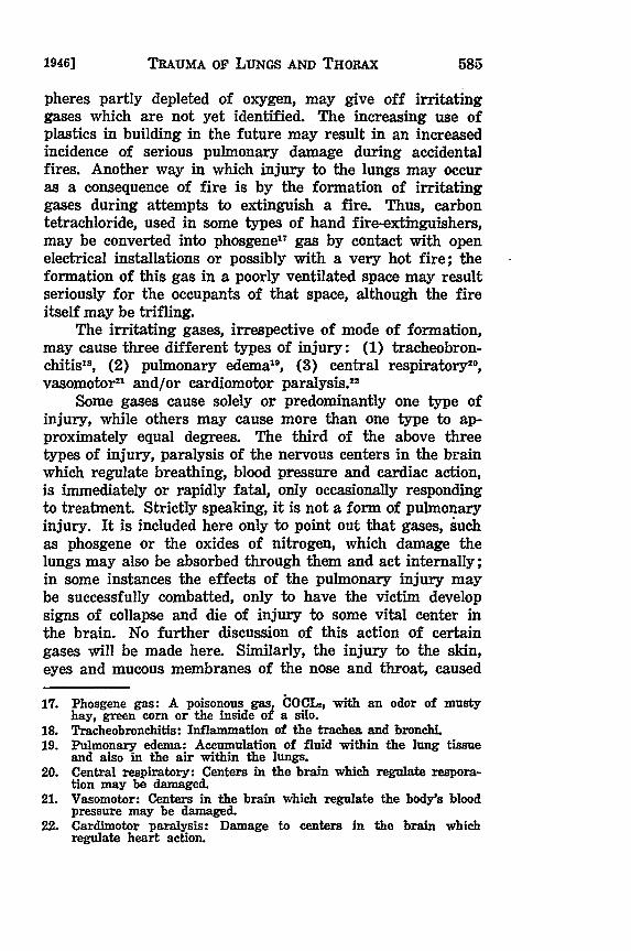
The lungs, two in number, lie to either side of the heartand occupy most of the chest. The right lung consists of threelobes and is larger than the left, which contains only two.
Right bronchus Tractms
Right pulmonary artery
Pulmonary trunk L eft bronchus
Upper lobe Left I 'oar
Upper lbe
Middle lobe
Lower lobeLowrer lobe
Fig. 1 Diagram of lungs with the branch; and blood vessels.
10. Pathogenesis: The development of morbid conditions or of disease.
1946]
INDIANA LAW JOURNAL
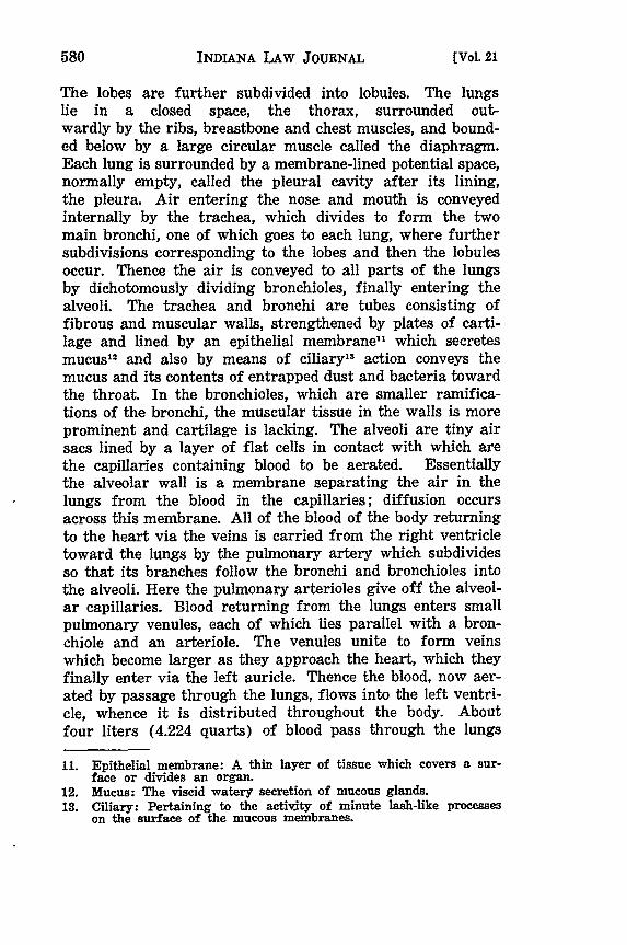
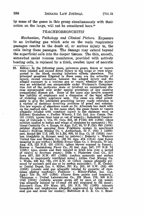
The lobes are further subdivided into lobules. The lungslie in a closed space, the thorax, surrounded out-wardly by the ribs, breastbone and chest muscles, and bound-ed below by a large circular muscle called the diaphragm.Each lung is surrounded by a membrane-lined potential space,normally empty, called the pleural cavity after its lining,the pleura. Air entering the nose and mouth is conveyedinternally by the trachea, which divides to form the twomain bronchi, one of which goes to each lung, where furthersubdivisions corresponding to the lobes and then the lobulesoccur. Thence the air is conveyed to all parts of the lungsby dichotomously dividing bronchioles, finally entering thealveoli. The trachea and bronchi are tubes consisting offibrous and muscular walls, strengthened by plates of carti-lage and lined by an epithelial membrane', which secretesmucus 12 and also by means of ciliary" action conveys themucus and its contents of entrapped dust and bacteria towardthe throat. In the bronchioles, which are smaller ramifica-tions of the bronchi, the muscular tissue in the walls is moreprominent and cartilage is lacking. The alveoli are tiny airsacs lined by a layer of flat cells in contact with which arethe capillaries containing blood to be aerated. Essentiallythe alveolar wall is a membrane separating the air in thelungs from the blood in the capillaries; diffusion occursacross this membrane. All of the blood of the body returningto the heart via the veins is carried from the right ventricletoward the lungs by the pulmonary artery which subdividesso that its branches follow the bronchi and bronchioles intothe alveoli. Here the pulmonary arterioles give off the alveol-ar capillaries. Blood returning from the lungs enters smallpulmonary venules, each of which lies parallel with a bron-chiole and an arteriole. The venules unite to form veinswhich become larger as they approach the heart, which theyfinally enter via the left auricle. Thence the blood, now aer-ated by passage through the lungs, flows into the left ventri-cle, whence it is distributed throughout the body. Aboutfour liters (4.224 quarts) of blood pass through the lungs
11. Epithelial membrane: A thin layer of tissue which covers a sur-face or divides an organ.
12. Mucus: The viscid watery secretion of mucous glands.13. Ciliary: Pertaining to the activity of minute lash-like processes
on the surface of the mucous membranes.
[Vol. 21

TRAUMA OF LUNGS AND THORAX
ITcrminal brand3oc
Tez minA broncbiia-4{
}AIhcalar ducks
Fig. 2. Arthetecture Structional units of the lungtissue.
every minute while the organism is at rest. Exercise in-creases the flow greatly.
Functions of the Lungs. The lungs have a numberof functions. Blood from all parts of the body, made de-ficient in oxygen and laden with carbon dioxide as a conse-quence of passage through the tissues, comes into contactwith the air in the lungs, discharging its excess of carbon diox-ide and taking up oxygen. Since carbon dioxide dissolved inthe blood acts as an acid,1' it is apparent that changes in res-piration which cause variations in carbon dioxide content ofthe blood are important in regulating the acidity of theblood. A further function of the lungs is in relation tomaintaining the body temperature at a normal level. Heat,
14. CO, + H.O = HzCO, which is an acid in that it has hydrogenatoms which are replaceable by positive atoms.
1946]
INDIANA LAW JOURNAL
constantly produced in the body by the metabolic processes,must be dissipated. Most of it is lost through the skin, somethrough the urine (water, largely drunk cold and execretedwarm) and about a quarter of the total via the lungs. Thisis accompanied by warming the air inhaled and, more par-ticularly, by the vaporization of water in the lungs. Air isinhaled containing a small amount of water vaporized in itand exhaled saturated with water derived from the blood.The vaporization of this water, about a quart a day, re-quires just as much heat as would be required for the evapor-ation of a quart of water in 24 hours from a pan.
The Respiratory Movements. All of these functionsare accomplished by the respiratory movements. The dia-phragm (the flat circular muscle which separates the thoraxfrom the abdominal cavity) arched when relaxed, contracts,becoming shortened and consequently flattened, thereby cre-ating a negative pressure within the thorax and sucking airinto the lungs via the trachea and bronchi. Contraction ofsome of the chest muscles elevates the ribs aiding in thecreation of the negative pressure. About 400 cc. of air aresucked in at each inspiration at rest; several times thatvolume is inhaled at each inspiration during exertion. Re-laxation of the diaphragm and the muscles of the chestinitiates expiration. Expiration is accomplished by the in-trinsic elasticity of the lungs, the muscles of respirationnormally remaining relaxed during this phase of respira-tion. Consequently these muscles must be drawn back intoposition for the next inspiration by the negative pressurecreated by the return of the lungs to a collapsed state, effect-ed by the elasticity of these organs. The rate and depth ofrespiration are regulated by a respiratory center in the brainwhose activity varies in response to nerve-borne impulseswhich bombard it, by changes in oxygen and carbon dioxidecontent and acidity of the blood which bathes it, by the needfor aiding in the dispersal of the body heat and by thestate of distension of the lungs. The respiratory center emitsnervous impulses which are carried down the spinal cord tothe neck where the phrenic nerve, one on each side, leavesthe spinal cord and passes down through the chest to thelungs. It is apparent that respiration is an extremely impor-tant and complex process. Increased respiratory activity canbe expected to occur when there is increased need for the
(Vol. 21

TRAUMA OF LUNGS AND THORAX
*.g 0 9
: Is
OQ a O
19461
INDIANA LAW JOURIAL
uptake of oxygen or the excretion of carbon dioxide (i.e.,in fever, thyroid disease, etc., and during exercise), whenthe uptake of oxygen and release of carbon dioxide are im-paired by pulmonary (lung) disease, when there is increasedneed for heat dispersal (i.e., in fever and thyroid diseaseand when the elimination of heat through the skin is dimin-ished) and when the blood becomes abnormally acid (i.e.,as a consequence of certain phases of diabetes, kidney diseaseor gastrointestinal disease).
IRRITATING GASES
Sources of Noxious Gases. Distinction must be made be-tween gases which injure the lungs and those which, likecarbon monoxide, enter through yet do not injure the lungsbut act on some other organ; only the former will be dis-cussed here. Although there is widespread recognition ofthe fact that pulmonary injury due to gases was a commoncause of severe and/or prolonged disability, or even death,during the War of 1914-1918, it is not generally appreciatedthat equally severe damage to the lungs may result fromgases used in industry (such as chlorine) or liberated nor-mally during industrial processes (such as nitrogen dioxideor tetroxide formed during etching, metal pickling, photo-engraving or oxy-acetylene welding). Some substances, nor-mally liquid, may give off irritating vapors which may act,at least in part, on the lungs; these include some refrigerantsand also liquid fuels such as gasoline and kerosene. Inaddition, irritating gases may be generated during certaintypes of accidents. For instance, the contact of brine or seawater with exposed electrical installations may under somecircumstances result in the liberation of chlorine 15 gas whichmay reach a sufficiently great concentration in a closed space,such as a compartment in a partially sunken ship, to be in-jurious. Fire is especially likely to result in the formationof irritating gases, either in the form of flame itself, or assome product of combustion. An example of the latter isthe nitrogen dioxide or tetroxidec formed when x-ray filmburned during the Cleveland Clinic fire of 1929. Complexorganic materials, when burned in ordinary air or in atmos-
15. Chlorine: A greenish-yellow gas with a characteristic sharp odor.16. Tetroxide: NO. or N20, a poisonous volatile liquid, giving off
brownish irritant fumes.
[Vol. 21
TRAUMA OF LUNGS AND THORAX
pheres partly depleted of oxygen, may give off irritatinggases which are not yet identified. The increasing use ofplastics in building in the future may result in an increasedincidence of serious pulmonary damage during accidentalfires. Another way in which injury to the lungs may occuras a consequence of fire is by the formation of irritatinggases during attempts to extinguish a fire. Thus, carbontetrachloride, used in some types of hand fire-extinguishers,may be converted into phosgene" gas by contact with openelectrical installations or possibly with a very hot fire; theformation of this gas in a poorly ventilated space may resultseriously for the occupants of that space, although the fireitself may be trifling.
The irritating gases, irrespective of mode of formation,may cause three different types of injury: (1) tracheobron-chitis 8, (2) pulmonary edema 9, (3) central respiratory"0 ,vasomotor 2' and/or cardiomotor paralysis 22
Some gases cause solely or predominantly one type ofinjury, while others may cause more than one type to ap-proximately equal degrees. The third of the above threetypes of injury, paralysis of the nervous centers in the brainwhich regulate breathing, blood pressure and cardiac action,is immediately or rapidly fatal, only occasionally respondingto treatment. Strictly speaking, it is not a form of pulmonaryinjury. It is included here only to point out that gases, suchas phosgene or the oxides of nitrogen, which damage thelungs may also be absorbed through them and act internally;in some instances the effects of the pulmonary injury maybe successfully combatted, only to have the victim developsigns of collapse and die of injury to some vital center inthe brain. No further discussion of this action of certaingases will be made here. Similarly, the injury to the skin,eyes and mucous membranes of the nose and throat, caused
17. Phosgene gas: A poisonous gas, COCI, with an odor of mustyhay, green corn or the inside of a silo.
18. Tracheobronchitis: Inflammation of the trachea and bronchi.19. Pulmonary edema: Accumulation of fluid within the lung tissue
and also in the air within the lungs.20. Central respiratory: Centers in the brain which regulate respora-
tion may be damaged.21. Vasomotor: Centers in the brain which regulate the body's blood
pressure may be damaged.22. Cardimotor paralysis: Damage to centers in the brain which
regulate heart action.
1946]
INDIANA LAW JOURNAL
by some of the gases in this group simultaneously with theiraction on the lungs, will not be considered here.23
TRACHEOBRONCHITIS
Mechanism, Pathology and Clinical Picture. Exposureto an irritating gas which acts on the main respiratorypassages results in the death of, or serious injury to, thecells lining these passages. The damage may extend beyondthe superficial cells into the deeper tissues. The thin, smooth,somewhat moist mucous membrane, provided with activelybeating cells, is replaced by a thick, swollen layer of necrotic
23. Editor: In the following cases, poisonous gases, fumes or vaporswere inhaled and caused direct injury to the lungs or were trans-ported in the blood, causing injurious effects elsewhere. Theprincipal questions litigated in these cases are the actuality ofinjury, causal connection between the alleged injury and thealleged exposure to a noxious gas or vapor, whether the injurywas an accidental one compensable under Workmen's Compensa-tion Act of the particular state or involved an occupational dis-ease compensable only under special provisions of any existingoccupational disease act. Some of the cases also involve commonlaw liability of employers and a discussion of the legal effectsof violating safety appliance acts. The cases are cited princi-pally to give the interested practicing lawyer ready reference toa variety of decisions involving problems of proof and substan-tive law aspects of situations much as Dr. Altschule has dealt withon the medical side. In the cases cited, the gases fumes or vaporsallegedly inhaled with resultant injury to the claimant were asfollows: Gustafson v. Parlier Winery, 2 Cal. Ind. Acc. Comm. Dec.101 (1915) (gases from tank or vat of winery): Industrial Commis-sion of Colorado V. Ule. 97. Colo. 253, 38 P (2d) 803 (1935) (dopesolution applied to bodies and wings of airplanes by spraygun) ; Na-tional Casualty Co. v. Hoage, 64 App. D.C. 33, 73 F. (2d) 850 (1934)sulphur dioxide escaping from Frigidaire system in apartmenthouse); Sullivan Mining Co. v. Aschenbach, 33 F. (2d) 1 (1929)cert. denied 280 U.S. 586, 74 L.Ed. 635, 50 Sup. Ct. 35 (1929) (car-bon bisulphide in thinner used by painter); Doherty v. WesternUnited Gas and Electric Co., 188 fI1. App. 494 (1914) (gas fumesescaping in gas works); McBeth-Evans Glass Co. v. Brunson, 70 Ind.App. 513, 122 N.E. 439 (1919) (glass blower exposed to fumes);Brewer v. Veedersburg Paver Co., 92 Ind. App. 547, 177 N.E. 74(1931) (gas, smoke and dust inhaled in brick kiln over a periodof twelve to fourteen years); Dille v. Plainview Coal Co., 217Iowa 827, 250 N.W. 607 (1933) (carbon monoxide and carbondioxide in improperly ventilated mine); Jellico Coal Mining Co.v. Walls, 160 Ky. 730, 170 S.W. 19 (1914) (miner allegedly poi-soned by carbonic acid gas or by carbon monoxide); T. M. Crutch-er Dental Depot, Inc. v. Miller, 251 Ky. 201, 64 S.W. (2d) 466(1933) (cromic acid gas allegedly inhaled by operator of chro-mium plating machine); Faulkner v. Milner-Fuller, Inc. (La.App.) 154 So. 507 (1934) (fumes from paints and lacquers);Thompson v. United Laboratories Co., 221 Mass. 276, 108 N.E.1042 (1915) (volatile arsenic given off by cans of rat poisonwas allegedly inhaled by 17-year old girl employed to fill cans);Johnson's Case, 279 Mass. 481, 181 N.E. 761 (1932) (chronicbronchitis and emphysema allegedly aggravated by inhalation ofcoal gas and dense tar smoke); Baltimore & 0. R. Co. v. Bran-
[Vol. 21
TRAUMA OF LUNGS AND THORAX
tissues2' exuding large amounts of plasma25 and later pus."The diameter of the airway is cohsequently narrowed as awhole and may be plugged locally by masses of debris. Themuscle cells which make up much of the walls of the respira-tory passages may be thrown into spasm, further narrowingthe airway. Accordingly, the patient experiences varyingdegrees of pain, and more particularly, shortness of breath
son, 128 Md. 678, 98A 225 (1916), reversed 242 U.S. 623, 37 S.Ct. 244, 61 L. Ed. 534 (1916) (inhalation of paint spray from"paint gun"); Tomlanovich v. American Boston Mining Co.,272 Mich. 493, 262 N.W. 293 (1935) (tuberculosis allegedly ac-tivated by breathing gas, smoke and dust produced by blastingin mine); Adler v. Interstate Power Co., 180 Minn. 192, 230 N.W.486 (1930) (coal and coke fumes in power plant); Jackson v.Euclid-Pine Inv. Co., 223 Mo. App. 805, 22 S.W. (2d) 849 (1930)(carbon monoxide poisoning of garage mechanic); Bender v. Mid-west Pipe and Supply Co. (Mo. App.) 57 S.W. (2d) 707 (1933)(sulphur dioxide from burning coke in welding department);Decker v. Raymond Concrete Pile Co., 336 Mo. 1116, 82 S.W. (2d)267 (1935) (Acetylene gas inhaled by user of acetylene torch);Thomson v. Amoskeag Mfg. Co., 86 N.H. 436, 170 Atl. 769 (1934)(Dormant tuberculosis allegedly aggravated by poisonous gases);Bove v. Donner-Hanna Coke Corp., 236 N.Y. App. Div. 37, 258N.Y. Supp. 229 (1929) (Coke oven gases); O'Connor v. Consolidat-ed Gas Co. of New York, 243 N.Y. App. Div. 661, 276 N.Y. Supp.998 (1935) (Gas inhaled by foreman of gang working on gaspipes as result of gas main bursting); Dixon v. Gaso Pump andBurner Mfg. Co. 167 Okla. 401, 29 P. (2d) 764 (1934) (Carbonmonoxide poisoning); Coca-Cola Bottling Co. v. Mowry, 167 Okla.644, 31 P. (2d) 562 (1934) (Inhalation of caustic soda fumes, longcontinued); Johnston v. E. E. Orcutt Garage, 103 Pa. Super. 507,157 Atl. 46 (1931) (Carbon monoxide gas); Sinkiewicz v. Susque-hanna Collieries Co., 115 Pa. Super. 377, 175 At. 757 (1934)(Gases in smoke-filled room of mine where dynamite had beenfired); Katora v. N.J. Zinc Co., 116 Pa. Super. 257, 176 Atl. 762(1935) (Carbon Monoxide poison in gas plant); Alston v. Vir-ginia-Carolina Chemical Co., 104 S.C. 410, 89 S.E. 497 (1916)(Fumes and gases in acid tower of fertilizer factory); Consol-idated Kansas City Smelting and Refining Co. v. Dill (Tex. Civ.App.), 188 S.W. 439 (1916) (Fumes in smelter inhaled by carpenter);Commercial Standard Ins. Co. v. Noack (Tex. Civ. App.), 45S.W. (2d) 798 (1931), Reversed, Com. App. 62 S.W. (2d) 72 (Car-bon monoxide poisoning); Associated Indemnity Corp. v. Baker(Tex. Civ. App.) 76 S.W. (2d) 153 (1934) (Sulphuric acid fumes al-legedly inhaled while cleaning and blowing out tubes of a steamcondenser in an electric light and power plant); Depre v. PacificCoast Forge Co. 151 Wash. 430, 276 Pac. 89 (1929) (Noxious gasesfrom mixture of. muriatic acid, sulphuric acid and water used toremove scale from metal preparatory to galvanizing it); Pellerinv. Washington Veneer Co., 163 Wash. 555, 2 P.(2d) 658 (1931)(Carbon bisulphide poisoning slowly contracted from exposure togases and vapors generated in mixing glue in veneer mill).
24. Necrotic tissue: A tissue is an aggregation of fibers and cellscomposing a structural element; necrotic tissue is dead tissue.
25. Plasma: The liquid component of blood.26. Pus: A liquid product of inflammation made up of white blood
cells, plasma and debris.
19461

INDIANA LAW JOURNAL
(dyspnea) and cough. The dyspnea may be severe enoughto require oxygen. In some instances attempts have beenmade to suck out the obstructing debris through a broncho-scope. 2
T The cough may be dry, or may be productive of amixture of mucus, serum and debris. If spasm of the bronchiis severe, asthmatic wheezing may be noted. Physical exam-ination should show the signs of bronchitis, but the x-raymay not reveal the abnormality.28
The denuded bronchi, filled with debris, are extremelysusceptible to infection by bacteria in the air or in the breathof neighboring individuals. A superimposed bacterial bron-chitis or bronchopneumonia may develop, making the damagemore extensive and the patient much more sick.
Within a period of hours or days, depending on the tox-icity of the gas and the amount of exposure to it and thepresence or absence of bacterial invasion, healing begins.The necrotic debris is sloughed off, to be replaced partly orwholly by normal tissue. Where damage to deeper layers ofthe trachea and bronchi has occurred, with consequent great-er degrees of disorganization of structure, complete healingmay be retarded or may never occur. A state of chronic in-flammation may persist. Muscle cells, in particular, regen-erate poorly and may be replaced by inelastic scar tissue.In some areas, due to the pressure changes within the bronchiduring respiration, a thin patch of scar tissue may balloonout, forming a sac; this state is called bronchitectasis. Itsoccurrence is favored by the development of strictures, con-sequent to contraction and puckering of scar tissue, higherin the bronchial tree. Examination may show a few signsof congestion in the bronchi or may be entirely negative.Similarly, ordinary x-ray studies may also fail to reveal thedisease; recourse must be had to the instillation of lipiodol1'into the bronchi to outline the dilated bronchi. These saccularareas retain secretion which becomes infected and the patientdevelops a chronic productive cough and a greatly increasedsusceptibility to respiratory infection; common colds, insteadof passing off in. three or four days, descend into the lungs,
27. Bronchoscope: A tubular instrument for inspecting the interiorof the bronchi; it is inserted via the mouth and trachea.
28. Schatzki, R.: Management of the Cocoanut Grove Burns at theMassachusetts General Hospital. Roentgenologic Report of thePulmonary Lesions, Ann. Surg. 117: 834, 1943.
29. Lipiodol: An oil which is opaque to x-rays.
[Vol. 21

TRAUMA OF LUNGS AND THORAX
causing severe coughs which may last for weeks. Additionalparts of the bronchial tree may thereby be successively in-volved. In some instances small patches of inflamed lungtissue (pneumonitis) develop about bronchiectatic areas. Theymay result in chest pain and irregular fever for prolongedperiods.
Complications. If the bronchitis and bronchiectasis areassociated with considerable obstruction to respiration, eitherbecause of extensive damage to the bronchi or because ofassociated asthma, the patient will develop a state of over-inflation of the lungs (emphysema). In a previous para-graph it was pointed out that inhalation is an active andexhalation a passive process; when obstruction is present,it is easier to inhale than to exhale, so that obstruction ul-timately leads to overdistension of the lungs. When thisoccurs, the entire respiratory mechanism is upset: the pa-tient has to work harder to breath and mixing is poor withinthe lungs, so that the blood is poorly oxygenated and does notgive off as much carbon dioxide as normally. The patientmay become short of breath, may become blue (cyanotic)and may develop a train of symptoms involving various or-gans which are now bathed in inadequately aerated blood.These symptoms include mental sluggishness, intolerance ofheat, and a variety of poorly defined gastrointestinal com-plaints.
In some cases the accumulation of secretion in a bron-chus, or the formation of a stricture across it, may causea lobule or lobe of lung to become completely functionlessand collapsed (atelectasis). This usually is readily detectedon examination and always by x-ray.
Another complication of bronchiectasis is sudden hemor-rhage (hemoptysis), which may occur in a previously appar-ently healthy individual. A large hemoptysis may be fatal.
In extreme instances, the heart muscle bathed in poorlyaerated blood and forced to work harder pumping the bloodthrough the abnormal lung may fail; cor pulmonale is said tohave developed. Patients with this condition are usually in-valids and have a short life expectancy.30
Some patients in whom none of the above sequelae to
30. See, in this Symposium series: White, Paul Dudley and Smith,Hubert Winston: Scientific Proof in Respect to Injuries of theHeart (Medicolegal Aspects of the Heart) (1946) 24 N.C.L. Rev.
106.
1946]

INDIANA LAW JOURNAL
gassing occur may suffer in another way, i.e., by the lightingup of a latent disease, usually tuberculosis, or by its exacer-bation if already overt.
PULMONARY EDEMA
Contact of a sufficiently great concentration of certaingases with the walls of the alveoli may so damage them thatthe capillaries they contain no longer retain blood and largeamounts, i.e., pints, of plasma, mixed with variable amountsof red blood cells, pour out into the air spaces. This mayoccur soon after gassing or several hours later; in the lattercase the patient may feel entirely well in the interim. Thepatient at once commences to drown in his own secretions.He becomes intensely dyspneic (breathless) and cyanotic(blue) and coughs up large amounts of colorless or pinkfrothy fluid- Unless treatment, including the administrationof oxygen, is instituted at once, the patient is almost certainto die; he may die in spite of treatment, either because thetreatment is ineffective in his case or because he goes intoshock following the loss of so much blood and plasma intoand through his lungs. He may remain in a state of pulmon-ary edema for hours or days; during this time his lungs areunusually susceptible to bacteria in the air or in the breathof nearby individuals and a fulminating extensive pneumoniamay supervene.
After the subsidence of the pulmonary edema, the lungsmay heal completely or else emphysema may develop. Itwas pointed out previously that exhalation is a passive pro-cess and is accomplished by the elasticity of the lungs. Thiselasticity may be lost following gassing either by the destruc-tion of the universally present fine elastic fibrils in the pul-monary tissue or because of the overgrowth of a fine diffusescar tissue in the alveolar walls. Whatever the mechanism,loss of elasticity results in a state of overinflation of thelungs (pulmonary emphysema). The consequences of pul-monary emphysema have already been discussed. If diffusescarring (fibrosis) occurs, the lungs become abnormally rigid,making breathing more difficult. In addition, the scarringmay interpose a thin wall of relatively impermeable materialbetween the blood in the capillaries and the air in the alveolarspaces, so that the normal gaseous exchange across the alve-olar walls cannot occur. In this case all the symptoms of
[Vol. 21

TRAUMA OF LUNGS AND THORAX
oxygen lack (anoxia) will occur. Physical examination ofthe lungs may be perfectly normal, and the x-ray may evenin the presence of extensive fine scarring reveal no abnor-mality.31 Cor pulmonale32 may develop later.
PROBLEMS OF IDENTIFICATIONFOLLOWING GASSING
Proof that claimant was gassed. In attempting to es-tablish beyond doubt the occurrence of gassing in civil life,the ideal situation would be one in which the supposed victimwas in contact for a known period of time with a gas ofknown composition present in a concentration known to betoxic. This is clearly an impossibility: gases are too evanes-cent to allow investigators td return to the scene hours ordays later to measure their concentration at the time of theaccident. Moreover, not all of the irritant gasses have beenidentified or can be identified.33 Accordingly, attempts toestablish the fact of gassing must be based upon indirect evi-dence. Some of this evidence may have a high order of valid-ity: if a gas known to be toxic is used in industry or isformed during an industrial process, its accidental escapeinto or its accumulation within a space occupied by men maybe detected by a characteristic smell or. color. This would betrue of chlorine certainly 34 and probably of many other gases.On the other hand, establishment of the fact of the presencein the air of the gas in concentrations sufficient to be detect-able does not necessarily in the case of all gases establishthe fact of gassing, i.e., the attainment in the lungs of a con-centration sufficient to be toxic. Also, some gases, such asphosgene, may be toxic when present in concentrations toosmall to be detected with certainty; this gas is fatal in con-
31. Altschule, M.D.; Linenthal, H. and Zamcheck, N.: Lung Volumeand Pulmonary Dynamics in R~ynaud's Disease. Effect of Ex-posure to Cold, Proc. Soc. Exper. Biol. and Med., 48: 503, 1941.
32. Cor pulmonale: Dilation of the right side of the heart from pul-monary embolism (obstruction of a pulmonary artery by a bloodclot brought to it in the blood stream) or obstructng pulmonarydisease. The condition is attended by cyanosis (bluish colorationof the skin.) See White, P.D. and HT.W. Smith, op. cit. supra,f.n. 30.
33. Aub. J.C.; Pittman, H., and Brues, A.M.; Management of theCocoanut Grove Burns at the Massachusetts General Hospital.Pulmonary Complications: A Clinical Description, Ann. Surg. 117;834, 1943.
34. See f.n. 11, supra.
1946]

INDIANA LAW JOURNAL
centrations of 25 parts in a million. Or because of panic orloss of consciousness, the victim may not have noted thepresence of the gas. It would appear, therefore, that medicaltestimony as to the occurrence of the symptoms of gassingis of the greatest importance in situations involving questionsof exposure to irritant gases. The physician's records shouldinclude a note as to odors persisting in the victim's clothesor hair; this may, however, be misleading in the case of anindividual gassed during a fire, since the clothes or hairmay themselves be charred, or may smell only of smoke.Note should be made of the condition of the victim's skin,eyes, nose and mouth. Some gases are extremely irritatingto these organs, particularly the eyes. Ordinary smoke maybe very irritating to the eyes, but its effects wear off morerapidly than those of most of the irritant gases. The pres-ence of charred tissue in the nose and throat establishes theinhalation of flame. The occurrence of the previously dis-cussed symptoms of tracheobronchitis, pulmonary edema 35
or central vasomotor or respiratory paralysis should be noted.It is to be remembered that they need not appear immediately,but may become overt only after hours or even a day or two.Medical testimony as to the presence of symptoms of gassingin a person who was in a situation in which a toxic gas mighthave been generated would appear to be as close as one cancome in civil life to establishing the fact of gassing. If the vic-tim is dead, postmortem examination of the lungs would bevery helpful.
Proof that Specific Lung Conditions Were Caused byGassing. Attempts to prove conclusively that the manifesta-tions of bronchitis, bronchiectasis, asthma, pulmonary emphy-sema, pulmonary fibrosis or cor pulmonale, which a patientmay show, are consequent to his having been gassed previous-ly may be very difficult. All of these disorders, when conse-quent to gassing, are indistinguishable from their manifesta-tions when due to some other cause, such as allergy or theordinary varieties of pneumonia, bronchitis and influenza.Statistical studies as to the incidence of these sequelae arenot very helpful, since statistics are of little help in evaluat-ing a single case. In addition, most of them are based on
35. Pulmonary edema: Congestion of the lungs resulting from aneffusion of serous fluid from the blood into the air vesicles andinterstitial tissue of the lungs.
[Vol. 21

TRAUMA OF LUNGS AND THORAX
data derived from the War of 1914-1918, and gassing inwarfare is likely to be quite different from gassing in civillife. Not only may the gases involved be different, althoughthis is not necessarily true, but the military are provided withsensitive detecting devices and effective protective mechan-isms, so that while the numbers involved may be much great-er, the average exposure would be milder. The latter was notof course true of the first gas attacks, such as that on afront of three and three-quarters miles at Ypres on April 22,1915, when a force of Canadians and others were gassed,five thousand dying outright and ten thousand others becom-ing casualties.2" Moreover, such studies as have been pub-lished have as their authors members of the military forceswho consciously or unconsciously attempted to minimize thefrequency of persistent pulmonary disorders following gassingin soldiers. Thus examination of the case reports of Gilchristand Matz3" makes it appear to the present author, as wellas to more distinguished medical authorities, i.e., the editorsof the British Medical Journal, 8 that Gilchrist and Matz7
may have been too conservative in their analysis of the nu-merical frequency of the sequelae to gassing. Similarly, thestudies of Berghof39 on 2000 men (one-third of them gassedwith chlorine) three or four months after gassing, led himto conclude that 50 per cent had no permanent pulmonarydamage, although he states that his "normals" frequentlyexhibited cough or dyspnea on exertion; his conclusions werebased solely on physical examination which, as pointed outabove,31 may be misleading. Meakins and Priestley' foundchanges in the blood indicative of marked impairment of aer-ation and therefore proving severe damage in the lungs,
36. Prentiss, A.M.: Chemicals in War, New York, McGraw-Hill BookCo., Inc., 1937.
37. Gilchrist, H. L., and Matz, P.B.: The Residual Effects of War-fare Gases: The Use of Clorine Gas, With Report of Cases, Med.Bull. Vet. Admin. 9:229, 1933. '
Gilchrist, H. L., and Matz, P.B.: The Use of Mustard Gas,With Report of Cases, id. at p. 339.
Gilchrist, H.L., and Matz, P.B.: The Use of Phosgene Gas,With Report of Cases, Med. Bull. Vet. Adminis. 10: 1, 1933.
Gilchrist, H.L., and Matz, P.B.: The Use of Arsenical Com-pounds, with Report of Cases, id. at p. 79.
38. Editorial: Residual Effects of Chlorine Gassing, Brit. Med. J. 1:196, 1933.
39. Berghoff, R.S.: The More Common Gases; Their Effects on theRespiratory Tract, Arch. Int. Med. 24: 678, 1919.
40. Official History of the War, Medical Services, British, vol. II, 1923.
1946]
INDIANA LAW JOURNAL
but concluded that these changes were a neurotic manifesta-tion,'4 1 a conclusion which evokes only amazement in modernphysiological and clinical students of pulmonary disease. Nev-ertheless, all of these studies, the data of earlier authorsanalyzed in those reports, and various discussions of civiliangassing such as the remarkably well annotated studies ofVon Oettingen42 on nitrous fumes, prove that sequelae togassing, in the form of bronchitis, bronchiectasis, pulmonaryemphysema, pulmonary fibrosis, cor pulmonale and activa-tion of latent tuberculosis, are of common occurrence.
The establishment of these disorders as sequelae togassing is very difficult without the previous establishmentof the fact of gassing as outlined above. Nevertheless it mustbe remembered that such antecedent evidence of gassing maybe lacking: the patient may have been unconscious or delir-ious because of other trauma (injury) following exposure tothe gas and unable to complain of the symptoms; he maynot have had competent medical attention or any attentionfollowing exposure to the gas, etc. If the fact of gassinghas been established and if a patient previously shown bycompetent medical testimony to have been without pulmonarydisease before gassing, shows a few weeks after the accidentevidence on physical or x-ray examination one of the sequelaelisted above, it can be safely concluded that the gassingcaused the sequela. That is as close as one can come toperfect proof as to the causal relation between exposure togas and the development of pulmonary disease thereafter.
Special Diagnostic Procedures. In the case of bronchitisand bronchiectasis physical examination and ordinary x-raystudies may reveal nothing abnormal and bronchoscopy andthe introduction of the opaque oil, lipiodol, may be necessaryto establish the diagnosis. It is doubtful if a patient can bemade to submit to these procedures; he might be persuaded todo so, if he is not neurotic or malingering, on the basis that theresults of such studies would be beneficial to him. In the ab-
41. Neurotic Manifestation: That is, a symtomatic expression of aneurosis, a disturbance believed to be due to psychological causesor maladjustment without any organic basis or lesion. Psycho-neurosis involves a mental disorder caused primarily by somepsychic conflict or maladjustment, without disturbance of in-tellectual functions, without distortion of reality, and withoutprimary mood pathology (abnormality).
42. von Oettingen, W.F.: The Toxicity and Potential Dangers ofNitrous Fumes, Pub. Health Bull., No. 272, 1941.
[Vol. 21

TRAUMA OF LUNGS AND THORAX
sence of such studies the diagnosis would have to rest on thehistory and an estimate of the patient's personality.43
Late Effects: Problem of Proving Causal Connection.Where a period of months or years elapses between exposureto a gas and the development of one of the conditions con-sidered to be sequelae to gassing, establishment of a causalrelation is very difficult, if not impossible. Nevertheless, suchsituations might arise. For instance, a patient may feel en-tirely well after gassing, but yet have within his lungs an areaof bronchiectasis which might make its presence known longafter the gassing only by a massive hemorrhage. This hem-orrhage might very well be a sequel of the exposure to gasseveral years before, or it might not in that he may have hadthe bronchiectasis before he was gassed, or it may have devel-oped consequent to a respiratory infection which occurredsome time after the episode of gassing. The same situationmight arise in the case of the other sequelae of exposure togases. Even the most meticulous medical study may not suf-fice to establish the origin of the manifestations in somepatients of this type, although in other instances carefulmedical history may be very helpful.
ASPIRATION OF LIQUIDS
Cause and Effects of Aspiration. The entry of large vol-umes of any liquid into the lungs results in drowning andsuffocation, if this material is not very soon expelled. Thispulmonary disorder will not be discussed here.4 ' Certainliquids are extremely irritating when aspirated even in smallamounts, while others cause no immediately detectable reac-tion but, after the passage of time, may cause extensive dis-ease of the lungs.
Substances which might with some frequency cause im-mediately detectable damage when aspirated include keroseneand gasoline'5,' 6 and gastric contents. In the case of thefirst two it is not clearly established that pulmonary lesions
43. The instillation of lipiodol has been performed thousands of timesin patients, with only mild discomfort and extremely rare reac-tions. The introduction of a bronchoscope is more discomforting,but, in good hands, is not a dangerous procedure.
44. See, in this Symposium series, Helpern, M.: The Medicolegal Ex-amination in Cases of Suspected Homicide, J. of Crim. L. &Crim. (March-April, 1946).
45. Machle, W.: Gasoline Intoxication, J.A.M.A. 117: 1965, 1941.46. Cope, C. L.: Aspiration of Petrol, Lancet 1: 469, 1942.
19461

INDIANA LAW JOURNAL
found after aspiration are due to the action of these liquidsdirectly on the lungs: their vapors may act like irritant gasesand these substances in addition are systemic poisons- andmay act on the lungs after having first entered the bloodstream. Whatever the preliminary mechanism, we can saywith certainty that following aspiration of gasoline or kero-sene, extensive destruction of the lining of the bronchi andof the alveolar spaces may occur, causing bronchopneumoniawith the production of a large amount of brownish sputum,4 8
smelling characteristically. In severe poisoning, this is notimportant, as the systemic intoxication is usually fatal; inmilder poisoning after aspiration, the pneumonia may bethe chief or only manifestation."6 In instances in which heal-ing is taking place, invasion by bacteria may occur, withresulting exacerbation or even fatality. There is no recordof the subsequent course of patients in the group with symp-toms which are predominantly or solely pulmonary, althoughthere is no reason to believe that all escape the occasionallyoccurring, more lasting, consequences of pneumonia, i.e.,bronchitis, bronchiectasis, emphysema and fibrosis.
Aspiration of gastric contents occurs in individuals whovomit while unconscious and the entry of the gastric acid,digestive juices and food particles into the lungs results ina fulminating type of pneumonia which is resistant to treat-ment ordinarily successful in pneumonia. Secondary bacterialinfection usually is part of the picture. The diagnosis ofaspiration pneumonia is made with certainty only at autopsyand, accordingly, there are no data available on the subse-quent course of patients who may recover from it.
A not uncommonly encountered lesion in hospital prac-tice is oil aspiration pneumonia. The oils implicated are usu-ally medicinal oils, 4
9 i.e., mineral oil, castor oil and cod liveroil, a smaller number of instances occurring after aspira-tion of food oils such as cream, olive oil, cottonseed oil, etc.It is possible that the large number of marine disasters inrecent years may give rise to the recognition of pulmonarycomplications following aspiration of fuel or lubricating oils.
47. Causing collapse, disorientation, jaundice, signs of renal irrita-tion, etc.
48. Sputum: Matter ejected from the mouth, having its origin inthe mouth, trachea, bronchi or lungs.
49. Kaplan, L.: Combined Cod Liver Oil and Liquid Petrolatum Pneu-monia in a Child, Am. J. Dis. Child. 62: 1217, 1941.
596 [Vol. 21

TRAUMA OF LUNGS AND THORAX
Aspiration of oils may occur in patients who are unconsciousor who, because of depression of the activity of parts ofthe brain, have lost the cough reflex. An important groupof cases of oil pneumonia occurs as the consequence of theprotracted use of oily medications in the nose, with a slowirregular trickle of the oil into the lungs. Oil in the lungssets up an irritative process which results in bronchitis andpatchy pneumonia associated with marked fibrosis. Onceestablished, the disease runs an irregular course of exacer-bations and remissions, apparently unrelated to subsequentaspirations of oil, with a good deal of disability because ofcough, fever and chest pain. The outlook appears to be un-favorable, although this is not conclusively true, since thediagnosis is with certainty made only at autopsy."0
Problems of Identification Following Aspiration of Li-quids. Proving that pulmonary injury is due to the aspira-tion of an irritating liquid involves many of the same prob-lems as in the case of inhalation of gases, with the importantdifference that liquids, being more stable and more easilyhandled, can be more readily identified. Here again, if signsof bronchitis and/or pneumonia develop within a day ortwo after the accidental aspiration of a liquid which isknown to be irritating, the pulmonary lesion should be re-garded as the consequence of such aspiration. Aspirationof any liquid is more reaf3ily appreciated by the victim thanthe inhalation of some gses, since the immediate responseto the former is coughing, choking and the production ofsputum, unless the patient is unconscious. In the latter in-stance, the sputum may have a characteristic and readilyidentifiable odor or the licuid may be detected in the hair,about the face or on the clothing. The establishment of the
50. Editor: For medicolegal capes involving aspiration of irritatingfluids (excluding drowning), see the following:Baltimore & O.R. Co. v. Branson, .128 Md. 678, 98 Atl. 225 (1916),reversed 242 U.S. 623, 37 Sup. Ct. 244 61 L. Ed. 534 (1916)(Spray from paint gun); Evans v. Chevrolet Motor Co., 232 Mo.
App. 927, 105 S.W. (2d) 1081 (1937) (Inhalation of spr-,y solu-tion containing soap, water and whale oil used to settle dust);Depre v. Pacific Coast Forge Co., 145 Wash. 263, 259 .7ac. 720(1927) (Fumes from sulphuric and muriatic acids); MacRae v.Unemployment Compensation Commission, 217 N.C. 769, 9 S.E.(2d) 595 (1940) (Sputum expectorated by tubercular employeeentered mouth of X, a fellow employee and two months later hevas found to be suffering from tuberculosis. Held: a compen-sable accidental injury arising out of and in the course off employ-ment.)
1946]

INDIANA LAW JOURNAL
fact that bronchitis, bronchiectasis, pulmonary emphysemaand fibrosis, or their exacerbation if previously present, arethe consequences of aspiration of an irritant liquid is subjectto the same difficulties as in the case of gassing (vide supra) ;the same is true in regard to the question of the lighting upof latent tuberculosis. Attempts to prove such a relationshipshould be based on the type of reasoning and study alreadyoutlined for gases.
The case of oil aspiration pneumonia offers many moredifficulties in that there may be a period of days, weeks orpossibly a longer time before any manifestation of pulmonarydisease becomes apparent. The property which the oils haveof inducing fibrosis", may make x-ray studies valuable inthis respect, since the appearance and progression of a fi-brotic process in the lungs, where none was present before,within a period of months after known inhalation of oil,would be most suggestive. Where the history of aspirationof oil is not obtainable because of unconsciousness of the vic-tim at the time of the accident, physician's data as to thepresence of oil in the hair, in the nose, about the face and onthe clothing may be helpful. If the disease is fatal, autopsyaffords conclusive proof as to the nature of the process.
FOREIGN BODY IN THE LUNG
Modes of Entrance. Foreign bodies may enter the lungsas missiles or debris blown in through the chest wall, down-ward through the neck or upward through the abdomen. For-eign bodies may also enter the lungs by aspiration (a) throughindustrial accident (pins, nails or screws held between thelips), (b) dental procedures or trauma to the mouth, (c)operations on the nose or throat (fragments of tonsil orother tissue) or (d) other accidents. In rare instances a for-eign body in the oesophagus or stomach, i.e., a fishbone,may work through into the lung.
Symptomatology. The symptomatology of foreign bodyin the lungs varies to some extent with the mode of entranceand with the nature of the body. Aspirated solid materialat first may cause pain and strangulation as it enters thelarynx52; proceeding downward into the trachea, it may cause
51. Fibrosis: The formation of fibrous tissue.52. Larynx: Air passage containing the vocal cords, situated between
the mouth and the trachea.
[Vol. 21

TRAUMA OF LUNGS AND THORAX
pain, cough and severe spasm resulting in wheezing, and thepatient may feel it bobbing about within the trachea. Thenceit passes into a bronchus, almost always a lower bronchusand usually in the right lung.53 Once past the trachea and ina bronchus, all symptoms cease for a greater or lesser periodof time. Accordingly, if the patient is excited or unconsciouswhile inhaling the body, he may be unaware of its passagethrough the larynx and trachea, and if, when he is quieteddown or restored to consciousness, it is in a bronchus, hemay not know that he aspirated it. Once lodged in a bron-chus, the body may cause symptoms after a period of a fewhours to over twenty years. If obstruction of a bronchusoccurs, loss of function of the lobe of the lung into whichit leads results. The air trapped in this lobe is absorbedinto the blood stream and the lobe collapses (atelectasis).This functionless lung tissue is very susceptible to infection.In other instances the local irritation may give rise to thecoughing of blood (hemoptysis), in larger or smaller volume.Vegetable matter, such as nuts and grains, is extremely ir-ritating and commonly causes a fulminating tracheobron-chitis5,4 and pneumonia, with severe prostration, high feverand the production of a large amount of thick tenacious sput-um; unless treated, this condition may be rapidly fatal. Lessirritating materials may give rise to a variety of other con-ditions. In some instances the foreign body may cause localdamage to a bronchus, giving rise to localized bronchitiswhich occasionally heals over without further incident; usu-ally, however, the patient is left with evidences of localizedbronchitis and/or bronchiectasis in the form of chronic coughwith localized evidences of congestion on physical examina-tion. The symptoms of bronchitis due to foreign body maynot become apparent for months after its aspiration. If thebronchitis is more severe, sputum may be profuse, fever highand prostration and wasting prominent. In some instancesthe surrounding lung is inflamed also (pneumonitis) and thesigns of inflammation on examination are more widespread;the x-ray shows a patch of inflammation in the lung sub-stance. The center of an area of pneumonitis may liquefy,
53. The right lower bronchus leads almost straight down from thetrachea instead of at a sharp angle, as in the case of the otherbronchi.
54. Fulminating tracheobronchitis: Inflamation of the trachea (windpipe) and bronchi which begins suddenly with an intense severity.
1946]
INDIANA LAw JOURNAL
becoming an abscess, and the patient shows high irregularfever, rapid wasting, profuse, extremely foul sputum (malo-dorous enough to be disgusting at a distance of ten feet ormore) and characteristic changes in the fingertips knownby the descriptive term of "clubbing".15 If untreated, a lungabscess may rupture into the pleural space, causing em-pyema 56 or through the diaphragm into the liver or into theabdominal cavity. The severe forms of bronchitis, pneumon-itis and lung abscess give symptoms usually within a fewdays to a few weeks after aspiration of the foreign body.Any suppurative57 pulmonary disease may give rise to ab-scess of the brain. In rare instances an aspirated foreignbody may cause no symptoms whatsoever, making its pres-ence manifest months or years later, only when it is coughedup. X-ray studies are frequently helpful, for almost invari-ably they reveal the foreign body if it is metallic or calcifer-ous (bones, teeth, some pebbles); unfortunately, vegetablematter is not detectable by this procedure.58
Problems associated with foreign material forced in orblown in through the chest wall or diaphragm may be dis-cussed under two heads, namely: (a) those associated withpenetrating wounds of the chest (vide infra) and (b) thoseassociated with damage which may occur if the foreign bodyremains in the lungs for any length of time after the accident.There are some resemblances and some differences in thenature of the processes set up when foreign bodies enterthe lungs through the chest wall as opposed to the trachea.In the former the picture may be colored by the occurrence of
55. "Clubbing": A bulbous enlargement of the ends of the fingers,with curvature of the nails.
56. Empyema: Pus within the pleural cavity.57. Suppurative: Giving rise to the formation of pus or associated
with its presence.58. Editor: Mediocolegal cases involving aspiration of a foreign
body seem most frequently to be malpractice actions againstdentists for alleged injury to the lungs caused by aspiration ofa tooth or other material during course of treatment, usually whilethe patient is under an anesthetic. See, for instance, the follow-ing: McGehee v. Schiffman, 4 Cal. App. 50, 87 Pac. 290 (1906);Nelson v. Parker, 104 Cal. App. 770, 286 Pac. 1078 (1930); Bol-lenbach v. Bloomenthal, 341 Ill. 539, 173 N.E. 670 (1930), re-versing 255 Ill. App. 305 (1930); Toy v. Mackintosh, 222 Mass.430, 110 N.E. 1034 (1916), Ann. Cas. 1918 C1188; Yarringtonv. Pittenger, 6 N.J. Misc. 710, 142 Atl. 565 (1928); 8 N.J. Misc.143, 149 Atl. 347 (1930); Schamoni v. Semler, 147 Ore. 353, 31P. (2d) 776 (1934); Goehring v. McDiarmid, 289 Pa. 193, 137Atl. 187 (1927).
[Vol. 21

TRAUMA OF LUNGS AND THORAX
manifestations of penetrating wounds of the chest (videinfra) and also by differences in localization. Foreign bodiesentering through the chest wall and coming to rest in thesubstance of the lung may give rise to pneumonitis or tolung abscess, as discussed above. However, foreign bodiesentering through the chest wall or diaphragm only very un-commonly come to rest in a bronchus and accordingly do notusually give rise to manifestations of localized bronchitisand/or bronchiectasis. Similarly, such a foreign body maynot enter the lung at all, remaining withinithe pleural space,causing pleurisy 9 with stabbing pain on coughing or deep in-spiration, and a friction rub60 audible over the area.
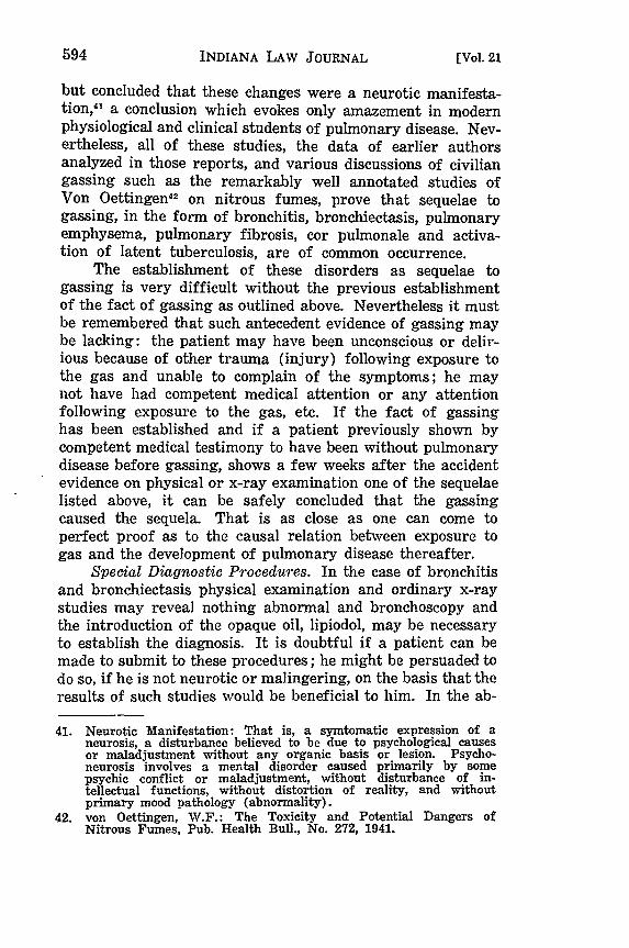
Foreign Material in Pleural Space: Clinical Manifesta-tions. The presence of foreign material in a pleural space
Fig. 4. Pleural Effusion, Note how collection offluid (solid black area) in pleural space has col-lapsed lung and pushed the thoracic (chest) con-tents toward the side opposite to the effusion.
59. Pleurisy: Inflammation or irritation of a pleural surface.60. Friction rub: A scratching or rubbing sound heard through the
stethoscope over an area of pleurisy.
19461

INDIANA LAW JOURNAL
causes an outpouring of fluid from the blood stream into thisspace, giving rise to typical manifestations. The patient com-
plains of a heavy sensation on that side, slight cough andvariable degrees of shortness of breath; there may be slightfever. Since the fluid collapses the lung on that side and may
push the entire thoracic contents toward the other side, thetrachea, heart and lungs are accordingly displaced. No breathsounds or other sounds are audible over the fluid when one lis-tens with the stethoscope, and the percussion note"' is duller
than over the portion of the chest containing aerated lung. In
extreme cases the fluid may accumulate to such a degree asto compress the blood vessels leading to the heart, thus im-
peding the return of blood to the heart, or it may interferewith cardiac action itself; this may result in fatality if the
condition is not treated. Treatment consists in the evacuationof the fluid through a hollow trocar.6 2 The fluid may recuruntil the foreign body is removed. Some care must be exer-cised in removing the fluid, since too rapid removal of toolarge an amount may so derange cardiac and/or pulmonaryfunction as to cause collapse or even death. If infection is
introduced with the foreign body, or subsequently, pus may
accumulate in the pleural space in large amount, i.e., a pint
to a quart or more, with all the manifestations of empyema,i.e., those of pleural fluid plus the fever, wasting and pros-
tration of severe, intractable, continued infection. This is
treated by drainage to remove the pus, as sulfonamides are
useless; now that penicillin has become freely available it
is likely that the use of that substance will make treatment
simpler and more effective. At present it is sometimes nec-
essary to do a number of operations, including removal of
ribs (thoracoplasty), for drainage and to collapse the empy-
ema cavity, so that is not uncommon for the patient to be
quite ill for many weeks. or months.In any case, the symptoms due to foreign body in the
lungs do not clear up until the object is removed. Occasion-
ally, the foreign body will be coughed up spontaneously, but
usually it must be removed either by bronchoscopy,6 3 if as-
61. Percussion note: Sound made by tapping the chest.62. Trocar: Sharp, pointed, hollow tube of metal.63. The insertion of a bronchoscope (see f.n. 27, supra) and ex-
amination of the visible lung through it.
602 [Vol. 21

TRAUMA OF LUNGS AND THORAX
pirated,64 or by operation if otherwise introduced into the
lungs.Problems of Identification which Arise From Foreign
Body in the Lung. The entry of a foreign body into the lungis usually a dramatic episode not often overlooked by a nor-mal individual. Accordingly, a history of the entry of sucha body into the chest, either by aspiration or by penetration,followed within a period of a few hours or days by the onsetof some of the symptoms of foreign body in the lung describedabove, suggests a causal relation which can be corroboratedby x-ray demonstration of the body and finally by its removal.However, as was pointed out above, an aspirated body in abronchus need cause no symptoms for a period of some hoursto twenty or more years, and unconsciousness, intoxicationor excessive excitement or distraction might result in un-awareness on the part of an individual that he has inhaledthe foreign body. In some instances the long latent periodbetween the accident when recognized and the first occur-rence of symptoms may confuse the diagnosis. In other in-stances where the aspiration of several bodies is recognizedseveral might be coughed up, leaving the victim with thebelief that he had brought up all he had inhaled, so thatwhen he developed pulmonary symptoms some months later,he might overlook the fact that foreign body might be atfault. The development of evidence of localized bronchitis 5
or bronchiectasis, 6 atelectasis, 67 pneumonitis6s or lung abscessshould cause inquiry as to possible aspirations, however re-mote in time, and should make the physician undertake x-raystudies which might reveal the foreign body. Indeed, manyphysicians feel that the occurrence of any of the above dis-orders, with or without a history of inhalation of a foreignbody, is an indication for bronchoscopy, to examine the re-gion involved and remove any body there detected. Where
64. If aspirated, the foreign body will be in the trachea (wind-pipe)or in the small branches (bronchi) which subdivide to carryair to and from the lung tissue.
65. Bronchitis: Inflamation of the bronchial tubes. See discussionsupra.
66. Bronchiectasis: Dilation of the bronchi or of a bronchus. Itmay affect the tube uniformly or it may occur in irregular pockets.It is marked by fetid breath and paroxysmal coughing, with theexpectoration of mucopurulent matter. See discussion supra.
67. Atelectasis: Partial collapse of the lung. See discussion supra.68. Pneumonitis: A form of pneumonia. See discussion supra.
19461

INDIANA LAw JOURNAL
a definite history of aspiration is not obtained, the natureof a foreign body found may throw some light on the circum-stances of its entry into the lungs. Thus a history of injuryto the mouth with the breaking of teeth or dentures, with thesubsequent finding of such fragments in the lung, should befairly conclusive evidence as to aspiration at the time ofaccident, even though the victim was unaware of it. Similar-ly, the finding in the lungs of tacks, screws or small partsof machinery which might have been held between the lipsof the victims while at work should be sufficient proof ofaspiration during an accident which occurred during work.Or the finding of a pebble in the lung of the victim of anaccident which resulted in the victim's being thrown on hisface to the ground in pebbly soil might be considered asproof of aspiration during the accident, even though thevictim was unaware of having inhaled the foreign body. Aforeign body which might disintegrate and be coughed up infragments or become merged into the pus of a lung abscess,i.e., fragments of tonsil or a piece of meat, might be undetect-able at the time of bronchoscopy several weeks after aspira-tion, when the complete clinical picture is present. Withouta history of the aspiration of such material within a periodof a few weeks or months previously, it is not possible todemonstrate that the lesion in the lung is due to a foreignbody.
In the case of foreign bodies blown in or forced throughthe chest wall, the immediate problem is that of a penetratingwound, and observations made at that time should demon-strate the presence of foreign bodies. In this connection itis to be remembered that a very small abdominal wound orneck wound may be the point of entry of a pulmonary for-eign body which may occasionally be overlooked; in mostsuch cases the pulmonary symptoms would call attention tothe entry of such material into the chest even though nowound is visible over the chest (vide infra.)
After the removal of a foreign body, the inflammatoryreaction in the bronchi, lungs or pleural space provoked byit should subside in a longer or shorter time. In the caseof an infected pleural space the course may be long andsequelae, in the form of pain, shortness of breath and sus-ceptibility to infection, important. Most patients with local-ized bronchitis, pneumonitis or lung abscess due to foreign
(Vol. 21

TRAUMA OF LUNGS AND THORAX
body experience recovery, but some remain with chronicchanges in the lungs in the form of bronchiectasis or fibrosis.When chronic symptoms develop immediately after an acuteepisode due to aspirated foreign body, they should be con-sidered a consequence of its aspiration. If a long latentperiod exists between the appearance of chronic manifesta-tions and the subsidence of the acute manifestations, a causalrelation may be impossible to prove unless x-ray evidence,based on ordinary x-ray studies or on films made after theintroduction of lipiodol,69 demonstrate that the chronic symp-toms are due to a lesion in the location of an antecedentlydemonstrated but subsequently removed foreign body.
Lighting up of a latent tuberculous process during re-action to a foreign body is theoretically possible but wouldbe difficult to prove, unless the exacerbation of the tuber-culosis occurred during or shortly after the acute illness dueto inhalation of the body. Pulmonary tuberculosis does notbecome manifest for several weeks at least after the infectionsets in and, accordingly, the finding of active tuberculosisa few days after aspiration of a foreign body indicates nocausal relation between the two. This does not, however,rule out exacerbation of a previously existing tuberculouslesion.
PENETRATING WOUNDS
Depending on its size, depth and location, a penetratingchest wound may have a number of different effects singlyor in combination. A common consequence of such woundsis fracture of one or more ribs. This may be so painfulas to limit respiratory movements and, in addition, the brokenends of the ribs may tear the lining of the pleural space70
giving rise to complications described below.7 1
69. See f.n. 29, supra.70. Pleural space: Each lung is surrounded by a serous membrane
called the pleura. This has two layers: the visceral pleura whichimmediately invests the lungs and an outer layer called theparietal pleura which lines the inner wall of the chest cavity orthorax. The pleural space is the space between the visceral andthe parietal pleura.
71. Editor: For medicolegal cases involving penetrating injuries ofthe thorax and lungs, see the following: Denver and R.G.R. Co.v. Mitchell, 42 Colo. 43, 94 Pac. 289 (1908) (Horseback riderthrown from horse frightened by train); H. L. Hunt, Inc., v.Frisby, 185 Ark. 1188, 51 S.W. (2d) 516 (1932) (Fracture ofribs and injury to lungs: verdict for $25,000 reduced to $20,000.) ;
INDIANA LAW JOURNAL
A certain amount of destruction of, or at least damageto, lung tissue occurs in patients with penetrating woundsof the lungs. If severe, death or severe disability may occurquickly. The cause of the death or disability may be therendering of a large part of the pulmonary tissue function-less, so that the victim dies of asphyxia, or it may be dueto shock consequent to extensive tissue damage. The damagedlung provides a favorable culture medium for bacterialgrowth, and secondary infection may become a problem.If recovery ensues under these circumstances, healing isusually associated with a severe degree of disability formany months, with some permanent residual disability insome cases. Specific treatment consists in operative removalof dead or non-viable lung, removal of foreign bodies andtaking of certain measures against complications.
The lung tissue is richly supplied by blood vessels andaccordingly, a good deal of hemorrhage usually occurs. Thismay occur into the damaged lung or into the remaining nor-
Mulcahay v. Larson, 130 Conn. 112, 32 A. (2d) 161 (1943) (Fivefractured ribs causing penetrating injury of lungs); Hannaher v.Blue Cab Co., 322 Ill. App. 277, 54 N.E. (2d) 257 (1944) (Frac-tured ribs punctured lungs); Niemeyer v. McCarty (Ind. App.) 48N.E. (2d) 829 (1943) (Fractured ribs allegedly resulting in in-jury to lungs); Birmingham v. Lehigh & W. Coal Co. (N.J.L.)95 Atl. 242 (1915) (Consolidation of lung tissues allegedly oc-curring after accidental injury); Schott v. Weiss, 92 N.J.L. 494,105 Atl. 192 (1918) (Passenger thrown from defendant's jitneybus while alighting-fractured rib and punctured lung); Kalo-gerakas v. Public Service Coordinated Transport, 10 N.J.Misc.,175, 158 Atl. 408 (1932) (Three fractured ribs resulting inpleurisy); Upton v. Bell Cabs, 154 So. 359 (La. App. 1934)(Fracture of five ribs on one side and two on the other resultedin traumatic pneumonia and pleurisy); Clemens v. Southern Ad-vance Bag and Paper Co. (La. App.) 20 So. (2d) 749 (1944) (15%pneumothorax of left lung and pleural effusion following injuries>;
ittin v. Sumner, 176 N.Y. App. Div. 617, 163 N.Y. Supp. 443(1917) (Fourteen year-old male pedestrian struck by automobilesustained fracture of three ribs and puncture of lung) ; Fagerile v.New York Life Ins. Co., 129 Ore. 485, 278 Pac. 104 (1929) (Gunshotwound through left lung; question of total and permanent dis-ability under insurance policy.); Poneitawcki v. Harres, 200 Wis.504, 228 N.W. 126 (1930) (Fracture of rib sustained by womanallegedly caused injury to lung and resultant tuberculosis.);Williams v. City of Spokane, 73 Wash. 237, 131 Pac. 833 (1913)(1400 pound cement form fell on bridge carpenter; resultantfracture of eight ribs allegedly caused pleural adhesions whichgreatly interfered with respiration); Lanahan v. Hydraulic-PressBrick Co. (Mo. App.) 55 S.W. (2d) 327 (1932) (Watchdog ran in-to night watchman and knocked him down; watchman fracturedninth, tenth and eleventh ribs and this caused him to developlobar pneumonia, from which he died, according to autopsy re-ports. Held: a compensable accident under the Workmen's Com-pensation Act.).
[Vol. 21

TRAUMA OF LUNGS AND THORAX
mal tissue, and the victim may drown in his own blood ordie of shock due to blood loss. A variable amount of bloodmay be lost when coughed up or as it runs from the externalwound. The treatment of this complication is to control thehemorrhage and restore the lost blood by transfusion.
Hemothorax. A small wound of the pleural surface mayresult in a good deal of bleeding into the pleural space (hemo-thorax). This is a serious complication in that the victim maylook deceptively well at first, with little or no externalbleeding, only to go into a rapid decline after a period ofone-half to several hours. This occurs after a considerableamount of blood has accumulated within the pleural space,causing embarrassment of circulation and respiration by com-pression, in the same manner as described above as due topleural fluid. In addition, since the potential capacity ofthe pleural space is several quarts, the patient may bleedto death internally, with no indication of outward hemor-rhage.72 The treatment is to control bleeding if possible,transfuse to make up for blood lost and to remove blood fromthe pleural space by means of a hollow needle, rinsing thespace with sterile salt solution. During the accident itselfor as a consequence of manipulation designed to relieve hemo-thorax, the pleural space may become infected (empyema),with the development, as described above, of the manifesta-tions of infection. Hemothorax is likely to be associatedwith severe prolonged disability which is made even longerby the development of empyema.
Pneumothorax. It was pointed out in the discussion ofpulmonary physiology (vide supra) that there normally existsin the pleural spaces a negative pressure which is necessaryfor effective respiration and which maintains the lungs intheir distended state. Penetrating wounds allow the entry ofair into the pleural space either through the perforated chestwall or by puncturing the lung; the lung in some cases maybe punctured by the sharp ends of broken ribs. The entryof .air into the pleural space destroys the negative intra-pleural. pressure, the pressure becoming that of the atmos-phere. This is known as pneumothorax. The lung on thatside accordingly collapses and shortness of breath (dyspnea)and cyanosis (bluish coloration of the skin due to insufficient
72. The total blood volume is 4.5 liters. Loss of a liter or more ofblood may have serious consequences, including death.
-1946]

INDIANA LAW JOURNAL
Fig. 5. Pneumothorax. Entry of air into the leftpleural space has restored the negative intrapleuralpressure normally present with the result that theleft lung has collapsed.
oxygenation) may develop. If the wounds are sealed off, theair is slowly absorbed and the lung re-expands. Pneumo-thorax of this type in not uncomfortable in a patient at rest,although it makes strenuous exertion impossible. Indeed, itmay act in a beneficent manner in that it is always associat-ed with a collapsed, motionless lung on the same side; abruised or oozing lung heals more readily when collapsedand at rest. On the other hand, if the pressure of the airin the pleural space increases, the function of the other lungand of the heart is impaired and death may result. Thisincrease in pressure in the pneumothorax is known as tensionpneumothorax and results from the check-valve action of cer-tain small wounds. Respiration was described in the sectionon physiology (vide supra) as consisting of two phases, anactive muscular phase during inhalation and a phase of re-laxation during exhalation. The forceful muscular action ofinhalation may draw such air through a narrow wound, air
(Vol. 21

TRAUMA OF LUNGS AND THORAX
fli
Fig. 6. Tension Pnemothorax. The flap value actionof the penetrating wound enables air to be drawn induring inspiration which cannot be expelled duringexpiration. The increasive pressure may impair func-tion of the heart and opposite lung, and in extremecases, cause death if not relieved.
which cannot be expelled during exhalation. Treatment con-sists of the introduction of a hollow needle into the chest,connected with a rubber tube leading below the surface ofwater in a bottle, so that air under pressure in the pneumo-thorax may escape while air from the outside is kept outby the water in the bottle. After 48 hours this may be re-moved, as by then the wounds giving rise to a check-valveaction will have closed over. A pneumothorax may becomeinfected, giving rise to empyema.73
Interstitial Emphysema. Air from a bruised lung maybe forced into the tissues of the thorax and spread up intothe neck (mediastinal emphysema) or spread through thetissues of the chest wall (interstitial emphysema). The latteris usually not troublesome, unless the air works its way up
73. Empyema: Accumulation of pus in a cavity of the body, especiallyin the chest.
1946]

INDIANA LAW JOURNAL
around the trachea, impairing respiration. It is readily de-tected by a crackling sensation on palpating the area and isrelieved by introducing needles into the skin. Mediastinalemphysema may cause more severe respiratory embarrass-ment, requiring operation to release the trapped air. It isdetected by characteristic crackling sounds drowning out theheart sounds on auscultation.7 4
Until relatively recently most patients with serious chestwounds died; recent advances in operative technique, anes-thesia, oxygen therapy and control of shock, hemorrhage andinfection have made the outlook much more favorable. Never-theless, patients with such wounds have long periods of disa-bility. The handling of chest wounds is at present undergoingmany changes and, accordingly, it is not possible to makeany conclusive statement regarding the amount of prolongedor permanent disability occurring after such wounds. In-fection, extensive removal of lung tissue and resection7 5 of
ribs to secure exposure for operation are factors that mightbe expected to leave the patient with a greater or lesserdegree of impairment of pulmonary function.
Problems of Identification in Penetrating Chest Wounds.Patients with penetrating chest wounds are almost alwaysso severely disabled as to require immediate medical atten-tion. Accordingly, the relation of the injury to the subsequentcourse is likely to be established by the close medical super-vision which these patients require. Moreover, the periodof observation is usually so long that sequelae will developunder the eyes of the attending physician and will be recog-nized as such. A small number of patients with chest woundsdue to the penetration downward of a very small missile ofhigh velocity which has entered the neck may die of hemo-thorax, but appear to die a non-traumatic death. The findingof a neck wound, however insignificant in size and appear-ance, should be an indication for postmortem examination.
NON-PENETRATING TRAUMA TO THE CHEST
Non-penetrating trauma to the chest may be discussedunder three heads: the effects of (a) crushing, (b) blast and(c) a simple blow or fall.
74. Auscultation: Listening for sounds within the body, usually bymeans of a stethoscope.
75. Resection: Excision of a part of an organ.
[Vol. 21

TRAUMA OF LUNGS AND THORAX
Crushing injuries. Crush injuries involving fracturesof many ribs in several parts of the chest are likely to beserious and are usually associated with various degrees ofhemorrhage, shock and the destruction or rendering func-tionless of portions of the lungs; the damaged lungs arevery susceptible to infection and accordingly bacterial inva-sion may be a complication. Hemothorax,76 pneumothorax 7
and mediastinal emphysema78 may also be part of the picture.The manifestations of shock, i.e., pallor, sweating, restless-ness changing to stupor, dyspnea, rising pulse rate and fallingblood pressure, indicate a serious prognosis; only energetictreatment, including transfusion of blood or plasma, maysave the patient. Hemothorax, pneumothorax and interstitialor mediastinal emphysema may aggravate the patient's con-dition (vida supra) and require immediate treatment. Sev-ere pain, exacerbated by breathing and involving several por-tions of the chest wall, is important not only because ofthe discomfort but also because of the tendency to makerespiration very shallow and therefore inefficient. Treat-ment should include strapping of the chest and the use of an-algesic drugs7 9 ; the latter must be used cautiously, sincethey depress respiration and may thereby cause fatality.The crushing of a good deal of the thoracic cage so thatit loses its rigidity in several places may produce a respira-tory abnormality which is fatal in itself; one side of thechest may be sufficiently strong to make possible relativelynormal respiration on that side, while the other side of thechest, badly crushed, makes it impossible to secure the pres-sure changes on that side necessary for respiration. Respira-tion may therefore occur back and forth from the relativelywell functioning lung into the poorly functioning lung. Withthe shallow respiration consequent to crush injuries, littleor no respiratory exchange through the trachea to the outsideair may occur. The patient in such cases uses up more and
76. Hemothorax: A collection of blood in the thoracic (chest) cavity.77. Pneumothorax: An accumulation of air or gas in the pleural
cavity. The air enters either by an external wound, a lung per-foration, burrowing abscesses, or from the rupture of a superficiallung cavity. Pneumothorax is attended with sudden and severepain and rapidly increasing dyspnea (breathlessness).
78. Mediastinal emphysema: The presence of air in the mediastinaltissues. The mediastinum is the space between the two pleuralsacs which invest the left and right lungs respectively.
79. Analgesic drugs: Used for relief of pain.
19461

INDIANA LAW JOURNAL
more of the oxygen in the air and adds more and morecarbon dioxide to it as it is passed back and forth betweenthe two lungs, until finally symptoms of oxygen lack and/orcarbon dioxide retention develop and the patient may die.A similar course of events may occur when one side of thediaphragm is paralyzed because of injury to it or to thephrenic nerve0 on that side. Treatment calls for the useof oxygen and a tight strapping; too tight a binder may beharmful and it may restrict respiration. Because of painand the difficulties involved in building up a high pressure inthe chest following crushing injuries, victims of such injuriesfind it impossible to cough satisfactorily, so that secretionsor blood accumulate in the lungs and respiratory passagesobstructing the entrance and exit of air. These materialsprovide an excellent culture medium for bacteria, favoringthe development of extensive bronchitis and pneumonia. Toprevent the accumulation of blood and secretion in the lungs,the patient must be made to cough every few hours, one ormore attendants supporting manually the broken parts ofthe chest. In any case, the onset of infection is associatedwith a high sustained fever, sweating and increased prostra-tion. The infection which follows a crush injury may bepneumonitis, pneumonia, abscess or empyema. Depending onthe severity of the damage and the development of infection,the victim will be seriously disabled for a variable period oftime; the disability is almost always prolonged and usuallyrequires many weeks of hospitalization. Late sequelae havenot been studied, since in former years fatality followingcrush injuries to the chest was the rule and only recently haveappreciable numbers of survivals been obtained. Nevertheless,it seems reasonable to assume that the sequelae of fibrosis,bronchitis and bronchiectasis and emphysema, and those ofempyema will be recognized in the future.
Blast Injuries. Although the effects on the lungs pro-duced by blast were recognized many years ago, there wasno widespread appreciation until recently of the severityof the injury .experienced by individuals exposed to explosion.Severe trauma to the brain and intestines, the latter especiallysevere in the case of individuals in the water during an under-water explosion, may dominate the clinical picture; these
80. Phrenic nerve: The nerve which regulates the activity of thediaphragm.
[Vol. 21

TRAUMA OF LUNGS AND THORAX
manifestations will not be discussed here. Injury to thelungs8s is apparently produced through the chest wall, sinceclosure of the mouth and nose does not prevent it. A num-ber of changes may occur in the lungs, consisting mainlyin rupture of alveolar walls and hemorrhage; hemorrhage ina given area may be so extensive as to replace an entire lobeor lobule with blood clot, while hemorrhage into the pleuramay give rise to massive hemothorax. Air contained in thealveoli may be forced into or through the tissues of thelungs by the explosion and with production of pneumothoraxor the formation of air-filled blebs on the surface of thelungs. The function of the lungs is greatly impaired, andin addition the blood in the air spaces impedes aeration ofthe blood, so that depletion of oxygen in the blood pluscarbon-dioxide retention occurs. The damage also acts toset up many foci of irritation in the lungs, so that coughand very rapid respiration result. The patients also havedyspnea (breathlessness) and appear cyanotic (bluish) ; signsof congestion in the lungs are detectable by physical and/orx-ray examination. If hemorrhage or tissue damage is severe,the manifestations of shock may appear and in some instancesthe signs and symptoms 8 2 of hemothorax or pneumothoraxmay develop. The lungs appear greatly ballooned out forreasons which are not clear. The injury caused by blastmay be immediately or rapidly fatal, although recovery, onthe other hand, may be rapid. Statistical studies of theafter-effects of blast are not available, but scattered reportsof pain and shortness of breath persisting for as long aperiod of 26 months after the injury have appeared. 3
Blow or Fall. Injury to the chest caused by a blow orfall may take a variety of forms. One complaint may bebruising of and pain in the muscles of the chest which maydisappear in a few days, never to recur and leaving nosequelae. In other instances, bruising of the lung may occur
81. Dean, D.M.: Thomas, A.R., and Allison, R.S.: Effects of High-Explosive Blast on the Lungs, Lancet. 2: 224, 1940.
82. Signs and symptoms: In medical diagnosis the term signs refersto physical evidences of abnormality, injury or disease perceptibleto the examining physician as, for instance, a swollen joint, afractured bone, cyanosis (bluish coloration of the skin). Symp-toms are subjective feeling states which the patient must usuallydescribe to his physician, as for instance pain, emotional disturb-ance and the like.
83. Correspondence, Lancet 1: 241, 1942.
19461

INDIANA LAW JOURNAL
and the patient exhibits stabbing pain on deep inspirationand/or coughing and a friction rub may be heard over the
injured area. After a few days to a week or two, all of
these manifestations disappear. There is no evidence thatpermanent damage to the lung occurs in this type of injury.
One or more ribs may be fractured as a consequence of
external trauma (injury) ; pain, exacerbated by deep breath-ing and/or coughing is the chief complaint and indicatesirritation of the pleural surfaces. There is tenderness topalpation (touch) over the area involved and the x-rayreveals the fracture. Fracture of a rib cartilage may be
overlooked during x-ray studies, although the symptoms maybe quite severe. A small area of interstitial emphysemaoverlying the fractured rib is not uncommon. The symptoms
of fractured rib are usually well controlled by adhesive strapp-ing and after a period of a few weeks cure results, with no
sequelae. Infrequently, extensive interstitial emphysema ex-
tending up into the neck, or pneumothorax or hemothoraxmay be a complication of fractured rib, prolonging the period
of disability. A condition known as "pathologic fracture"of a rib is not uncommon and is a consequence of antecedent
disease of the rib involved. The ribs may be the seat of a
variety of bone diseases, including cancer, which may have
spread from some other part of the body. Such ribs may be
fractured by very slight trauma, or even by a sudden move-
ment, and heal poorly. This may be the first indication
that a patient is suffering from a disease of the bones,including cancer.
Pneumothorax and/or mediastinal emphysema may occur
following trauma to the chest by a blow or fall even in the
absence of fracture of ribs. The onset of these conditions
is usually indicated by severe pain, more severe often than
is to be expected from the degree of trauma, and radiation
of pain to a shoulder. In the case of pneumothorax temporary
collapse may occur and dyspnea and cyanosis may be promin-
ent. The manner in which these may cause serious disability
(tension pneumothorax, etc.) has been discussed above, as
has their treatment. Disability may last for several weeks
and, in the absence of infection, should leave no residual.
Pneumothorax and mediastinal emphysema may be recurrent.
Antecedent disease of the lung (tuberculosis with cavi-
tation) or of the bronchi (bronchitis and bronchiectasis) is
[Vol. 21

TRAUMA OF LUNGS AND THORAX
usually associated with increased vascularity (blood supply)and frequently loss of elasticity of the part involved. Ac-cordingly, transmission of the force of a blow to such areasmay result in rupture of blood vessels and the coughing up ofblood in varying amount, i.e., hemoptysis. The hemorrhagemay be so severe as to be incapacitating in itself for severalweeks; even if smaller in amount, hospitalization for severalweeks is to be recommended. The process usually subsides,although scattered authenticated reports of activation oftuberculosis following non-penetrating trauma to the chesthave been recorded. As was pointed out above, the courseof tuberculosis is such that signs of activation of tuberculosis,with the exception of hemorrhage, take several weeks to de-velop
Problems of Identification in Non-Penetrating Traumato the Chest. The manifestations of crush injury are sostriking as to make it unlikely that they will be overlooked.These injuries are so severe and hospitalization accordinglyso prolonged that the development of sequelae, which maycause severe disability, occurs under observation. Causalrelationships are therefore established quite readily. Thesame is likely to be true of blast lung, although here, becauseof injury to the brain and/or rupture of intestines, thepulmonary lesion may be overlooked or underestimated atthe time of accident. However, the cerebral (brain) andintestinal lesions may ultimately, i.e., after several months,heal satisfactorily, leaving respiratory symptoms as the onlylasting disorder. The persistence of chest pain, cough anddyspnea (breathlessness) in a patient known to be the victimof blast should be enough to make one consider these pul-monary manifestations as a sequel of injury following theexplosion. As in the case of sequelae of war gas poisoning,the evaluation of disability first becoming manifest sometime after the accident may be difficult; this is particularlytrue where neurosis8 4 has also been precipitated by the trau-ma.
Bruising of the lung should not be followed by sequelae.In that condition and following fracture of ribs, the relation
84. Neurosis: See f.n. 41, supra. For a full discussion of medicolegalaspects of traumatic neuroses see: Smith, Hubert Winston andHarry C. Solomon: Traumatic Neuroses in Court, Va.L.Rev. 30:87, 1943; Annals Int. Med. 21: 367, 1944.
19461

INDIANA LAW JOURNAL
of the symptoms to the accident is readily established. Evenin the case of a patient unconscious for some time after theaccident, the diagnosis of fractured ribs can be made byx-ray. In the absence of infection, secondary to the complica-tions of pneumothorax, hemothorax or mediastinal emphy-sema, disability is usually of short duration and sequelaeshould not occur. In the case of fracture of ribs which arethe seat of a previously existing bone disease, the presenceof antecedent partial destruction of the rib is detectable byx-ray and the subsequent course of the patient will be thatof a victim of that bone disease.
The proof that hemorrhage" from the lungs, althoughassociated with antecedent tuberculosis or bronchiectasis,is a consequence of a blow or a fall is often inconclusive.The occurrence of the hemorrhage immediately or at leastwithin a few hours after the accident strongly suggests acausal relationship. It is well known that spontaneous hem-orrhage is common in those diseases and, accordingly, itsoccurrence some time after an accident makes it unlikelythat the latter caused it. The evidence regarding the hemor-rhage should be fortified by the testimony of competent wit-nesses, including preferably a physician. In the case of anunconscious patient, the finding of frothy blood, i.e., bloodmixed with air, on the face and clothing, is helpful. Insome instances following the initial massive hemorrhage,there is continued coughing up of blood-tinged sputum orof small amounts of blood; this phenomenon may serve tocorroborate, where witnesses to the accident are not available,the patient's story of having had a hemorrhage from thelungs.
The question of the lighting up of latent tuberculosisor of its aggravation where overt by a blow is always diffi-cult to settle because of the unpredictable course of thedisease. If the blow or fall, by rupturing a small cavity,causes spread of the disease, weeks may elapse before themanifestations become severe enough to make the patientseek medical attention. On the other hand, tuberculosis oc-casionally manifests rapid and unexpected spontaneous ex-acerbations. Absolute proof of the relation of a blow or fallto the appearance or aggravation of tuberculosis is, in the
85. Hemorrhage: Blood flowing from a ruptured vessel, in this casecoughed up.
[Vol. 21

TRAUMA OF LUNGS AND THORAX
absence of hemoptysis immediately after the accident, impos-sible of realization, except in the rare case where by chancex-ray studies of the chest were made before the accident andfound negative. In the absence of this rare coincidence, com-petent medical testimony as to the time relation between thetrauma and the appearance or exacerbation of symptoms,such as cough, fever, weight loss, weakness, a positive x-ray,etc., should be relied on to establish the fact that a shortperiod, i.e., several weeks, elapsed between the two. However,if too short a period, i.e., several days, elapsed between thetwo, a causal relation is unlikely.6
TRAUMA TO THE CHEST CAUSEDBY MUSCULAR STRAIN
A variety of disorders of the lungs may occur aftera muscular strain: these include spontaneous pneumothor-ax," spontaneous mediastinal emphysema, hemoptysis,"8 frac-
86. Editor: For cases involving alleged causation or aggravation oflung conditions, including tuberculosis, by a blow, see the follow-ing: Hayhurst v. Boyd Hospital, 43 Idaho 661, 254 Pac. 528 (1927)(Pneumonia and tuberculosis; verdict for $15,250, upheld); Mal-loy v. Chicago Great Western R. Co, 185 Iowa 846, 170 N.W.481 (1919) (Ruptare of blood vessels in lung); ConsolidatedKansas City Smelting and Refining Co. v. Tinchert, 5 Kan. App.130, 48 Pac. 889 (1897) (Crushing injuries of chest and lungs un-pairing respiratory functions); Manhattan Const. Co. v. Tot-tress, 161 Oka. 69, 17 P. (2d) 407 (1932) (Fall injuring back andallegedly aggravating pre-existing lung condition); Victor Gaso-line Co. v. Weatherman, 163 Okla. 113, 21 P.(2d) 36 (1988) (Em-
pyema resulting from sharp edge of barrel striking claimant'sside; held: evidence sustained award of Workmen's Comnsa-tion benefits); Graham v. Kinmmel Drilling Co., 113 Pa. Super.235, 173 Atl. 721 (1934) (Pleurisy attributed by doctor to oneof two falls sustained in course of employment, there being noother discoverable cause; award of Workmen's Compensation af-firmed.); Mitchell v. San Antonio Public Service Co. (Tex. Civ.App.), 15 S.W. (2d) 694 (1929), reversed in (Tex. Com. App.) 35S.W. (2d) 140 (1931) (Tuberculosis in fifteen year-old girl al-legedly caused by a blow when automobile in which she was rid-ing was struck by defendant's streetcar.).
87. Editor: For cases of pneumothorax following muscular strain,see: Pacific Employers' Ins. Co. v. Pillsbury, 61 Fed. (2d) 1011932); Delaune v. Young (La. App.) 166 So. 149 (1936); Quig-ley v. Industrial Accident Commission (Cal. App.), 35 P. (2d)-544 (1934) (Hemothorax) subsequent opinion; 3 Cal. (2d) 46;43 P. (2d) 289; Kelce v. Swift and Co., 115 Pa. Super, 570,176 Atl. 66 (1935)) (Hydrohemothorax). In all the casescited the precipitating cause was the lifting of a heavy weightin course of employment.
88. Editor: For cases involving hemoptysis (spitting of blood) al-legedly caused by muscular strain, see: Kangas' Case, 282 Mass.155, 184 N.E. 380 (1933) (Employee suffering from pulmonarytuberculosis had serious pulmonary hemorrhage immediately after
19461

INDIANA LAw JOURNAL
ture of ribs which are the seat of previously established dis-ease, and possibly activation of tuberculosis89
Spontaneous Pneumothorax. Spontaneous pneumothoraxmay occur in individuals with disease of the lung, i.e., activeor latent tuberculosis or pulmonary emphysema, and in nor-mal individuals. The mechanism which causes the ruptureof the pleura thus allowing air to enter the pleural space isnot clearly understood except in the case of pulmonaryemphysema. In this disease the lungs are overdistended withair and the pleura in a given area may become very thin, sothat an air-filled thin-walled bleb gradually forms; this mayrupture. In any case the manifestations are sudden onsetof chest pain, frequently referred to a shoulder, and suddendyspnea with cyanosis; collapse may occur. In the case oftension pneumothorax, the dyspnea and cyanosis increaserapidly, the visible veins in the neck become greatly dis-tended, the blood pressure may fall and the patient may die.The treatment has been discussed earlier in this paper. Dis-ability lasts for a few weeks usually. Spontaneous pneumo-thorax may be recurrent. The amount of strain necessary tocause this disorder cannot be stated, since not infrequentlyit occurs when the patient is not exerting himself or evenwhen he is at rest; it is not necessarily a consequence of,strain.
Spontaneous Mediastinat Emphysema. Spontaneous med-iastinal emphysema"° usually occurs in apparently normal in-dividuals and its mechanism is not known. It may be associ-ated with or followed by pneumothorax. It is ushered inby severe pain which may be referred to a shoulder andmay be made worse on lying down; it may be confused
overexerting herself in turning an overhead wheel while workingat her loom in a factory.); McCluskey v. Stock Exch. Bldg. Corp.,100 Pa, Super. 136 (1930); Southwestern Surety Ins. Co. v.Owens (Tex. Civ. App.) 198 S.W. 662 (1917).
89. Editor: For cases allegedly involving activation of pre-existingdormant tuberculosis by muscular strain, see: Hartford Accidentand Indemnity Co. v. Industrial Accident Commission, 140 Cal.App. 482, 35 P. (2d) 366 (1934); Bosdyck v. Rochester FoldingBox Co, 245 N.Y. App. Div. 880, 282 N.Y. Supp. 146 (1935);Industrial Commission of Ohio v. Lathrop, 52 Ohio App. 55, 2N.E. (2d) 828 (1935).
90. Mediastinal emphysema: See f.n. 78, supra.
618 [Vol. 21

TRAUMA OF LUNGS AND THORAX
with the pain of coronary thrombosis.9 1 On auscultation 92
the typical crackling sounds, drowning out the heart sounds,are heard. The treatment is bed-rest, the disability lastingfor a few weeks. The amount of strain necessary to causethis disorder cannot be stated, since it is not always theconsequence of strain, occurring in many instances whilethe patient is at rest or asleep.
Hemoptysis. Hemoptysis, i.e., hemorrhage from the lung,may occur during straining in patients with tuberculosiscavitation or bronchiectasis. During severe muscular strainthe glottis° 3 is usually closed, preventing egress of air fromthe lungs, while the abdominal musculature is tightened,pressing the diaphragm upward and compressing the lungs.This compression is, however, resisted by the air trapped inthe lungs and a high pressure may be built up, which mayrupture a cavity lined by inelastic scar tissue. A blood vesselin the wall of such a cavity may be torn and hemorrhagemay result. The treatment of hemoptysis is bed-rest for sev-eral weeks; occasionally, the loss of blood is so severe as tomake transfusion necessary. Here again the amount ofstrain necessary to produce the condition cannot be stated,since hemoptysis not infrequently occurs spontaneously, evenwhile the patient with tuberculosis or bronchiectasis is atrest of asleep.
Activation of Tuberculosis by Strain. Activation orexacerbation of tuberculosis consequent to a strain has beenthe subject of contention for years. It is theoretically possiblethat, because of changes in pressure within the lung associat-ed with straining while the breath is held, rupture of atuberculous cavity may occur with dissemination of the dis-ease.
Problems of Identification in Trauma Due to Strain.The establishment of causal relationships between a strain
91. Hamman, L.: Spontaneous Mediastinal Emphysema, Bull. JohnsHopkins Hosp. 64: 1, 1939. See, in this Symposium series, White,
Paul Dudley and Hubert Winston Smith, op. cit. supra., f.n. 30.92. Auscultation: The act of listening for sounds within the body,
chiefly for ascertaining the condition of the lungs, heart, pleura,and other organs, and for the detection of pregnancy. As arule, auscultation is performed by using a stethoscope appliedto the appropriate area of the body. The stethoscope makes thesound more audible to the examining physician.
93. Glottis: The aperture, or chink, between the vocal cords, togetherwith all that part of the larynx which is concerned in voiceproduction.
19461

INDIANA LAW JOURNAL
Fig. 7. Tuberculous cavity with fluid level
and the occurrence of the disorders discussed in this sectioncan never be considered as absolutely conclusive, since allof them may occur spontaneously. If pneumothorax, medi-astinal emphysema or hemoptysis occurs during an unusuallysevere straining activity, such as heavy lifting or pushing,it is reasonable to conclude that a relation between thestrain and the pulmonary disorder exists. Care must betaken to distinguish this type of physical strain from emo-tional or mental strain and from the strain of prolongedwork at a task which is not straining in the sense hereused. Evidence offered by reliable witnesses as to the de-velopment of the dramatic symptoms here described in as-sociation with an unusual strain, together with competentmedical testimony based on examinations, including x-raystudies, made shortly after the accident, combine to forma very strong case in support of the concept that the dis-order is related to the strain. The occurrence of one ofthese disorders some time after the strain, on the otherhand, makes it very unlikely that the strain caused it.
(Vol. 21

TRAUMA OF LUNGS AND THORAX
Fig. 8. Rupture of tuberculous cavity into brouchusduring strain with discharge of infected material.
Evaluation as to the role of a strain in the exacer-bation or activation of a tuberculous process is even moredifficult than in the case of a blow or fall. The latter arelikely to be witnessed by others, to be recorded and to beeasily identifiable as to day and hour of occurrence. Astrain, on the other hand, is usually not recognized as suchby onlookers or in many cases by the patient himself; thelatter may some time after the exacerbation or activation ofhis tuberculous process remember having experienced a strainshortly before. The value of such testimony from both themedical and legal point of view is small. No general rulecan be formulated in regard to the criteria to be used inassessing the part played by a strain in activation or exacer-bation of tuberculous processes. The time relation betweenthe alleged strain and the development of signs of tubercu-losis is important: too short or too long an elapsed periodbetween the two makes a causal relation unlikely.
19461

INDIANA LAW JOURNAL
LESIONS OF THE LUNGS SECONDARY TO TRAUMAELSEWHERE IN THE BODY
Pneumonia. Pulmonary complications may follow traumato other parts of the body. The development of pneumoniafollowing aspiration into the lungs of gastric contents inunconscious individuals has already been discussed. 94 In ad-dition any individual who is made to remain in bed for longperiods of time, i.e., weeks, may develop pneumonia spon-taneously. This is especially likely to occur if the individualis elderly, greatly debilitated or a cardiac (heart) patient,and more particularly if he is immobilized by a large castor some mechanical appliance used in the treatment of frac-tures. The pneumonia becomes manifest when fever, chestpain, cough and possibly dyspnea (breathlessness) are de-tected, together with the characteristic physical and x-rayfindings in the lungs on examination. The development ofpneumonia complicates and prolongs the course of the pri-mary illness, may result fatally and may be followed by thesequelae already discussed in relation to other forms ofpulmonary infection, i.e., bronchitis, bronchiectasis, fibrosisand emphysema.9 5
Embolization. Another form of pulmonary lesion occurr-ing after trauma (injury) is embolization, i.e., the pluggingby a blood clot or other material of one or more arteriesleading to the lung, and infarction, i.e., the death of thoseportions of the lung fed by such arteries. If embolizationis extensive, death of the patient may occur due to blockageof the circulation before infarction can be detected. Theemboli which give rise to the disorder may consist of avariety of different substances and may originate in dif-ferent ways. The most common cause of pulmonary emboli-zation is the formation of thrombi, i.e., clots, in the veinswhich ultimately drain into the right auricle and ventricleof the heart and then pass with the blood through the pul-
94. See discussion, supra.95. Editor: For cases involving pneumonia allegedly due to trauma,
usually to some other part of the body, see: Meyer v. KraussLaundry and Dry Cleaners 98 Ind. App. 388, 189 N.E. 431(1934); Black Mountain Corp. v. Black, 220 Ky. 85, 294 S.W. 830(1937); Ricci's Case, 294 Mass. 67, 200 N.E. 568 (1936); Andersonv. Fisher Body Corp., 239 Mich. 506, 214 N.W. 938 (1927); Zim-merman v. Goodfellow Lumber Co. (Mo. App.) 56 S.W. (2d) 608(1933).
[Vol. 21

TRAUMA OF LUNGS AND THORAX
4-
4. %.
Fig. 9. Mechanism of embolus formation: a part of a blood clot(thrombus) within a vessel breaks off and is carried in the bloodstream to a new site, as, for instance the lungs, where it lodges in andblocks a vessel, reducing or cutting off the blood supply to tissuesserved by that vessel
monary artery into the lungs. Following infected abortions,the veins in the pelvis become thrombosed and the breakingoff of portions of these clots may give rise to embolization;septic abortion will not be discussed here since an extensivebody of legal and medical writing on this subject alreadyexists. The veins in the legs may become thrombosed follow-ing relatively simple abdominal surgery even in patients ingood condition, while patients who have to remain in bedfor long periods of time, especially if debilitated, may developthe condition without having been operated upon. Thiscondition, thrombophlebitis of the leg veins, may make itselfmanifest by pain and/or tenderness in the calves or maynever be overt, suggesting its presence only by embolizationor by its accidental finding at autopsy. The manifestationsof pulmonary embolization are varied, including sudden death,collapse with the picture of shock, sudden dyspnea and/or cy-anosis, sudden onset of fever, hemoptysis and chest pain.If infarction occurs, the chest pain may be of the pleuritictype, i.e., made worse by deep breathing and coughing andreferred to the shoulder. The presence of infarction can bedetected by the typical signs on physical and x-ray examina-tion, and the electrocardiogram may be helpful" Hydro-
96. The electrocardiogram is a device based upon the string galvan-ometer which enables the action current generated by the func-tioning heart to be recorded on a moving tape. See Smith, HubertW. and J. E. F. Riseman: Applied Use of the Electrocardiogramin Legal Proceedings, 15 Rocky Mt. L. Rev. 251, 1943; Riseman,J.E.F. and Hubert W. Smith: Some Legal Aspects of Heart Dis-ease and the Electrocardiogram, Ann. Int. Med. 19: 81, 1943.
1946]
- 44r__ ,
qW00W_-'I%

INDIANA LAW JOURNAL
thorax or hemothorax may occur. Pulmonary embolizationmay be fatal, and if not, it may prolong and complicate thepatient's primary illness. Since it may occur after evenminor abdominal surgery, its occurrence may create legalproblems. Thus, a patient operated on for hernia regardedas occupational in origin may die of pulmonary emboliza-tion or at least have his hospital stay greatly prolonged. 7
The occurrence of thrombophlebitis in the legs giving riseto pulmonary embolization may require that he also be oper-ated on for ligation of the femoral veins. Pulmonary em-bolization is not commonly followed by sequelae; in veryrare instances enough of the circulation through the lungsmay be permanently blocked to cause increased work of theheart in forcing the blood through and the heart may fail(chronic cor pulmonale). 98
Another, much less common cause of pulmonary embol-ization is phlebitis which follows intravenous therapy ofvarious sorts. Thus, varicose veins are commonly treatedby injecting a solution into them which causes formationof clots, closing off the veins; in rare instances such a clotmay break off and be carried to theluns. Phlebitis may oc-casionally develop in the arm veins following injection ofsolutions of various sorts, particularly those containing highconcentrations of glucose, i.e., 25 to 50 percent. Very rarelypulmonary embolization may follow.
Fat Embolism. Another uncommon type of emboliza-tion is that due to free fat in the blood reaching the lungs.Fat in the cells in adipose tissue in various parts of the body,including the bone marrow, may be broken down by crush-ing trauma, including fracture of a bone, and the finelydivided fat forced into the veins where the particles agglut-inate. When this mass of fat is carried to the lungs, it mayblock off many of the small vessels and even the larger ones,causing shock or sudden death. The diagnosis is made with
97. Editor: If an original injury is compensable, the untoward ef-fects of medical treatment are also compensable. The courtsextend this doctrine to include even those injuries which are dueto medical malpractice, provided the plaintiff or claimant se-lected the attending physician without notice of his incompetency.
98. For medicolegal cases involving post-traumatic embolization, see:Black Mountain Corp. v. Black, 220 Ky. 85, 294 S.W. 820 (1927);Geagan's Case, 301 Mass. 319, 17 N.E. (2d) 172 (1938); Con-solidated Underwriters v. Langley, 141 Texas 78, 170 S.W. (2d)463 (1943).
[Vol. 21

TRAUMA OF LUNGS AND THORAX
certainty only at autopsy. An apparently uncomplicated brok-en leg or a simple crushing injury to a fatty part of thebody may therefore result fatally. In rare instances fat em-bolism may occur as a consequence of damage to the livercaused by a commercial solvent, carbon tetrachloride1
Air Embolism. Air embolism may occur during traumaor without trauma in situations where atmospheric pressureis rapidly lowered, i.e., in rapid ascent in fighter planes orin caisson disease;100 the embolic manifestations usually in-volve the brain and spinal cord. Following trauma (injury)to or operation on areas containing many large veins, airmay, however, enter and be carried in the form of smallbubbles to the lungs, blocking off small vessels and causingshock or sudden death. The condition is diagnosed with cer-tainty only at autopsy.
A recently described type of embolization is that ofmeconium'01 through the placentaW 0 2 into the lung, causingshock and death. It is diagnosed at autopsy. Its relation totrauma has not been established, although in one case itoccurred after an operation on the pregnant uterus.108
Problems of Identification in Lesions of the Lungs Fol-lowing Trauma Elsewhere. The occurrence of pneumonia orof pulmonary embolism due to phlebitis is easily recognized;
99. MacMahon, H. E., and Weiss, S.: Carbon Tetrachloride Poisoningwith Macroscopic Fat in the Pulmonary Artery, Am. J. Path. 5:623, 1929.
100. Caisson disease: A condition which occurs when individuals areexposed to a sudden lowering of the atmospheric pressure, as inworkers coming from high pressure caissons into the ordinary at-mosphere. The sudden change in pressure causes the release of gasesdissolved in the blood in the form of bubbles everywhere through-out the body. For medicolegal cases involving caisson disease,see: Knock v. Industrial Acc. Comm. of Calif., 200 Cal. 456, 253 Pac.712 (1927); Quail v. Ind. Acc. Comm., 138 Cal. App. 412, 32P(2d) 402 (1934); Maryland Casualty Co. v. Gerlaske, 68 F (2d)497 (1934); Taylor v. List and Weatherly Const. Co., (La. App.),146 So. 353 (1933); Williams v. Mo. Bridge and Iron Co., 212Mich. 150, 180 N.W. 357 (1920); Beaty v. Foundation Co., 245Mich 256, 222 N.W. 77 (1928); Doehler Die Castings Co. v. Mc-Neely, 21 Ohio App. 148, 152 N.E. 792 (1926); Missouri ValleyBridge & Iron Co. v. Ballard, 53 Tex. Civ. App. 110, 116 S.W.93 (1909).
101. Meconium: Fecal matter discharged into the amniotic cavityby the foetus.
102. Placenta: The very vascular organ in the uterus connecting themother and foetus.
103. Steiner, P. E., and Lushbaugh, C. C.: Maternal Pulmonary Em-bolism by Amniotic Fluid as a Cause of Obstetric Shock and Un-expected Deaths in Obstetrics, J.A.M.A. 117: 1245 and 1340, 1941.
19461

INDIANA LAW JOURNAL
its relation to the trauma which required the hospitalizationin the first place is clear. In the case of embolization, sequelaeare rare; cor pdnonale may occur rarely and should bereadily detected. The sequelae of pneumonia, i.e., bronchitis,bronchiectasis, fibrosis and empyema have already been dis-cussed elsewhere in this chapter. The more uncommon typesof embolization, those due to fat, air and meconium, are diag-nosed only at autopsy.
PULMONARY DISEASE DEVELOPING
AFTER EXPOSURE
Prolonged unusual exposure to cold and wet without ade-quate protection may be followed by an upper respiratoryinfectiono4 and may occasionally, especially in the elderly,debilitated, unconscious or intoxicated, result in bronchitisor pneumonia. There is usually a period of one to a fewdays between the start of the exposure and the develop-ment of bronchitis or pneumonia, so that a short period ofexposure might be followed by a latent period of a day or so,during which only a mild upper respiratory infection wasmanifest, before the onset of the more serious complications.After a longer period of exposure, however, the bronchitisor pneumonia might be found fully established when the in-dividual is brought in. Pneumonia of this type may be follow-ed by the usual complications and sequelae, as outlined pre-viously in this discussion, including exacerbation of tuber-culosis. 105
104. Editor: In a number of cases, student nurses, interns or phy-sicians have contracted tuberculosis or other diseases presumablyby inhaling bacteria while in attendance on sick patients and theinteresting problem has arisen whether or not the evidence jus-tifies a finding that this was an accidental injury sustained in thecourse and scope of employment and so compensable under theWorkmen's Compensation Law. See, for instance: Ayers v.Hoage, 61 App. D.C. 388, 63 F (2d) 364 (1933); Smith's Case,307 Mass. 516, 30 N.E. (2d) 536 (1940); Miller v. City of NewYork, 282 N.Y. 707, 26 N.E. (2d) 821 (1940), reversing 257 App.Div. 1092, 14 N.Y. Supp. (2d) 680 (1939), and 258 App. Div. 820,15 N.Y.S. (2d) 718 (1939); Vanore v. Mary Immaculate Hospital,260 App. Div. 820, 22 N.Y.S. (2d) 350 (1940); Milwaukee Countyv. Industrial Commission, 224 Wis. 302, 272 N.W. 46 (1937).
105. Editor: For medicolegal cases involving pneumonia allegedlydue to exposure to cold and/or wet, see the following: Goble v.Clark, 56 F. (2d) 170 (1931); Anderson v. Hoage, 63 App. D.C.169, 70 F. (2d) 773 (1934); Strauss National Bank & Trust Co.v. Marcus, 274 Ill. App. 597 (1934); Platisa v. Inland Steel Co.100 Ind. App. 278, 195 N.E. 294 (1935); Cincinnati Times Star
626 [Vol. 21

TRAUMA OF LUNGS AND THORAX
Problems of Identification in Pulmonary Disease De-veloping After Exposure. The presence of bronchitis orpneumonia is detected with relative ease and with a highdegree of accuracy. The cause of such an infection is notnearly so readily ascertained. The existence of a latent per-iod of upper respiratory infection in most cases before theonset of bronchitis or pneumonia is helpful in evaluatingcausal relationships. If a brief period of exposure werefollowed within an hour or two by full-blown pneumonia,the valid conclusion would be that the patient was alreadyon his way to having pneumonia before the exposure began.On the other hand, if the pulmonary complication developedmore than a few days after the end of the exposure, itwould usually not be considered a consequence of the expos-ure. Data as to the presence of an epidemic of pulmonary
Co. v. Clay's Admr., 195 Ky. 465, 243 S.W. 16 (1922) (Illegalemployment of minor, a ten year old newsboy, allegedly causedhis death from pneumonia. Damages were recovered in a suitbrought by an administrator but for the sole benefit of the boy'sfather. Held, on appeal: recovery of damages for the father'sbenefit could not be allowed if he knew of or suffered the em-ployment of the minor in the work in question.); Warfield Na-tural Gas Co. v. Clark's Adm'x., 257 Ky. 724, 79 S.W. (2d) 2197 A.L.R. 971 (1934) (On February 6 defendant gas companycut off widow's gas for non-payment of bill nine days before ithad a right to do so. Widow's son, a newsboy, as a result ofstaying all night in the icy house, developed a chill and pneu-monia from which he died. In an action against the gas com-pany, the mother recovered a judgment of $11,500, for her son'sdeath: held, on appeal: affirmed.); Carter v. Priebe and Sons(Mo. App.), 77 S.W. (2d) 171 (1934); Morgan v. Simplot (Idaho)155 P. (2d) 917 (1945); Kardos v. American Smelting and Re-fining Co., 133 N.J.L. 39, 42 AtI. (2d) 271 (1945), affirming 132N.J.L. 579, 39 A(2d) 509 (1944); Kern v. Premier Coal SavingDevice Corp. 246 N.Y. App. Div. 661, 283 N.Y. Supp. 281 (1935);Lacey v. Washburn and Williams Co. 105 Pa. Super. Ct. 43, 160Atl. 455 (1932); Senlock v. Phila. and Reading Coal and Iron Co.,104 Pa. Super Ct. 156, 158 At. 663 (1932); Poklembo v. HazleBrook Coal Co. 116 Pa. Super. Ct. 532, 176 Atl. 850 (1935);Waleski v. Susquehanna Collieries Co. 108 Pa. Super. Ct 342, 164Atl. 355 (1935); Wilkins v. McSorely 119 Pa. Super. Ct. 442, 179Atl. 759 (1935); Mussman v. Steele, 126 Neb. 353, 253 N.W. 347(1934) (X, negligently driving ambulance, overturned it whileconveying Y, a person suffering from septicemia, to a hospital.There was evidence offered that Y contracted pneumonia as aresult of exposure following the accident. Held: whether X'snegligence caused or hastened Y's death was a question for thejury.); Packard v. Industrial Commission, 213 Wis. 1, 250 N.W.768 (1933). And see, Carson v. Fort Smith Light and TractionCo., 108 Ark. 452, 158 S.W. 129, Ann. Cas. 1915B 92 (1913)(Asthma allegedly caused or aggravated by exposure to cold asresult of defendant's wrongfully turning off gas supply to plain-tiff's house.).
19461

INDIANA LAW JOURNAL
infections in the community, or of an antecedent cases inthe patient's own household, should be secured in attemptingto establish the cause of bronchitis or pneumonia in a patientclaiming compensation after exposure.
PROBLEMS OF EFFECTS FOLLOWING TRAUMATO THE LUNGS
All of the conditions listed above as sequelae of traumaare, with the exception of tuberculosis, incurable. Their symp-toms may be ameliorated and their progression retarded orstopped by adequate medical treatment, but the damage doneremains. Accordingly, problems of disability and of com-pensation are important.
Disability Resulting From Tuberculosis. In the case oftuberculosis, the patient may require sanatorium care or itsequivalent for a period of six or eight months to. a few yearsbefore he is discharged as cured or arrested; during thistime he must be regarded as completely disabled. Followingthis, if a good deal of scarring has occurred in his lungs asthe tuberculosis healed, he may suffer varying degrees ofdisability, as will be discussed below in relation to fibrosisin general. If such scarring is minimal (the more commonoccurrence), he may appear to be entirely well, the only ab-normal finding being a small patch of fibrosis in the lungson x-ray examination. However, the patient will thereafter,probably for the rest of his life, have to follow the wellknown regime for cured tuberculars in regard to food, rest,hygienic conditions, etc.. The imposition of this regime mayconstitute partial disability in the case of a busy sales-man-ager, for instance, whose business activities normally in-volved long hours, great tension, irregular eating and sleep-ing, ingestion of large amounts of alcohol, etc., and whois therefore forced to take another job with a considerablysmaller income after recovery from tuberculosis. On the otherhand, if the patient had previously held a quiet or sedentaryjob as watchman or time-keeper, for instance, he could readilyresume his previous work without hazard to himself andtherefore need not be considered, from the occupational pointof view at least, as having any disability.
Disability Resulting from Fibrosis, Emphysema, Asthma.Individual Variations. In the case of pulmonary fibrosis 0 6
106. See fa.n 31, supra.
[Vol. 21
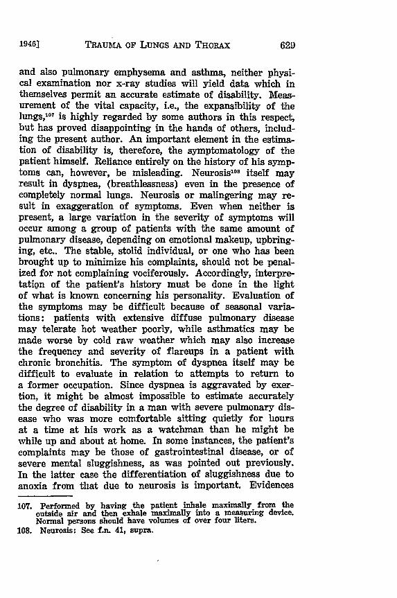
TRAUMA OF LUNGS AND THORAX
and also pulmonary emphysema and asthma, neither physi-cal examination nor x-ray studies will yield data which inthemselves permit an accurate estimate of disability. Meas-urement of the vital capacity, i.e., the expansibility of thelungs,207 is highly regarded by some authors in this respect,but has proved disappointing in the hands of others, includ-ing the present author. An important element in the estima-tion of disability is, therefore, the symptomatology of thepatient himself. Reliance entirely on the history of his symp-toms can, however, be misleading. Neurosisos itself mayresult in dyspnea, (breathlessness) even in the presence ofcompletely normal lungs. Neurosis or malingering may re-sult in exaggeration of symptoms. Even when neither ispresent, a large variation in the severity of symptoms willoccur among a group of patients with the same amount ofpulmonary disease, depending on emotional makeup, upbring-ing, etc.. The stable, stolid individual, or one who has beenbrought up to minimize his complaints, should not be penal-ized for not complaining vociferously. Accordingly, interpre-tation of the patient's history must be done in the lightof what is known concerning his personality. Evaluation ofthe symptoms may be difficult because of seasonal varia-tions: patients with extensive diffuse pulmonary diseasemay tolerate hot weather poorly, while asthmatics may bemade worse by cold raw weather which may also increasethe frequency and severity of flareups in a patient withchronic bronchitis. The symptom of dyspnea itself may bedifficult to evaluate in relation to attempts to return toa former occupation. Since dyspnea is aggravated by exer-tion, it might be almost impossible to estimate accuratelythe degree of disability in a man with severe pulmonary dis-ease who was more comfortable sitting quietly for hoursat a time at his work as a watchman than he might bewhile up and about at home. In some instances, the patient'scomplaints may be those of gastrointestinal disease, or ofsevere mental sluggishness, as was pointed out previously.In the latter case the differentiation of sluggishness due toanoxia from that due to neurosis is important. Evidences
107. Performed by having the patient inhale maximally from theoutside air and then exhale maximally into a measuring device.Normal persons should have volumes of over four liters.
108. Neurosis: See f.n. 41, supra.

INDIANA LAw JOURNAL
of deficient oxygenation of the blood are helpful in this dif-ferentiation; these evidences consist in cyanosis (bluish color-ation of the skin) visible on examination and a decrease inoxygen saturation of the arterial blood and an increase inthe number of circulating red blood cells, detectable in thelaboratory.
Reactivation or Exaberbation of Antecedent Disease. Theproblem of reactivation or exacerbation of antecedent diseaseis also important. Medical testimony as to the degree ofdisability before the trauma (injury) must serve as thebasis for any evaluation of disability after trauma. Evenso, great difficulty may be encountered. For instance, anindividual with a given degree of pulmonary disease mightfeel entirely well and be able to live a normal life and yet,after experiencing minimal additional damage to his lungsduring gassing, might then have enough pulmonary damageto push him over the line which separates the asymptomaticfrom the partly disabled patient. This is an extreme case,but the problem might very well arise.
It is apparent that no generalization can be made con-cerning the occurrence of disability or its degree after trau-ma. Accuracy of evaluation in a given case can be approach-ed only by a cooperative study of the circumstances on thepart of honest and competent medical and legal authorities.
[Vol. 21