
Common Eye Conditions in Horses - Avondale Vets · Common Eye Conditions in Horses Superficial...
5
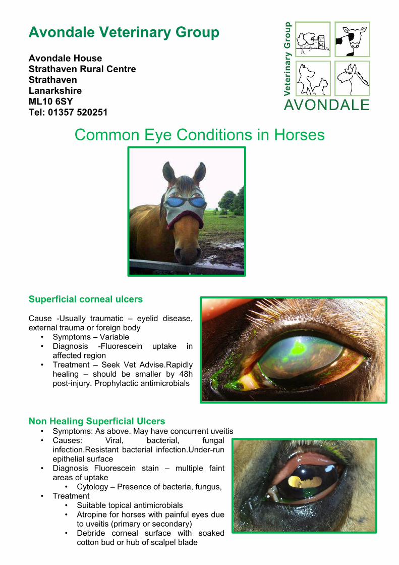
Avondale Veterinary Group Avondale House Strathaven Rural Centre Strathaven Lanarkshire ML10 6SY Tel: 01357 520251 Common Eye Conditions in Horses Superficial corneal ulcers Cause -Usually traumatic – eyelid disease, external trauma or foreign body • Symptoms – Variable • Diagnosis -Fluorescein uptake in affected region • Treatment – Seek Vet Advise.Rapidly healing – should be smaller by 48h post-injury. Prophylactic antimicrobials Non Healing Superficial Ulcers • Symptoms: As above. May have concurrent uveitis • Causes: Viral, bacterial, fungal infection.Resistant bacterial infection.Under-run epithelial surface • Diagnosis Fluorescein stain – multiple faint areas of uptake • Cytology – Presence of bacteria, fungus, • Treatment • Suitable topical antimicrobials • Atropine for horses with painful eyes due to uveitis (primary or secondary) • Debride corneal surface with soaked cotton bud or hub of scalpel blade
Transcript of Common Eye Conditions in Horses - Avondale Vets · Common Eye Conditions in Horses Superficial...
Avondale Veterinary Group Avondale House Strathaven Rural Centre Strathaven Lanarkshire ML10 6SY Tel: 01357 520251
Common Eye Conditions in Horses
Superficial corneal ulcers Cause -Usually traumatic – eyelid disease, external trauma or foreign body
• Symptoms – Variable • Diagnosis -Fluorescein uptake in
affected region • Treatment – Seek Vet Advise.Rapidly
healing – should be smaller by 48h post-injury. Prophylactic antimicrobials
Non Healing Superficial Ulcers • Symptoms: As above. May have concurrent uveitis • Causes: Viral, bacterial, fungal
infection.Resistant bacterial infection.Under-run epithelial surface
• Diagnosis Fluorescein stain – multiple faint areas of uptake
• Cytology – Presence of bacteria, fungus, • Treatment
• Suitable topical antimicrobials • Atropine for horses with painful eyes due
to uveitis (primary or secondary) • Debride corneal surface with soaked
cotton bud or hub of scalpel blade

Deep Corneal Ulcers
• Symptoms -Stromal involvement . White halo around ulcer • Diagnosis -Fluorescein uptake pattern. Opthalmic Exam • Treatment -Topical Antimicrobials. Given via Subpalpebral Lavage System.
• Treat secondary uveitis with NSAIDs (topical or systemic), topical Atropine • Surgery indicated if Descemet’s membrane exposed
Chronic Ocular discharge
• Causes:Conjunctivitis. Foreign Body, Obstructed Nasolacrimal Duct. Ocular Neoplasia (incl 3rd eyelid tumours)
• Symptoms: ocular discharge of varying characteristics
• Diagnosis + Treatment -Sample for bacterial culture + sensitivity if any bacteria found. Flush nasolacrimal duct. Biopsy suspected mass if present. Thorough lavage + careful ophthalmic examination
Ocular Opacities
• Stromal Abscesses • Symptoms – white opacity in the
cornea • Diagnosis – Fluorescein stain yellow-
green + history of chronic ulcer/ocular problem
• Treatment- Swab for bacteria,culture/sensitivity. Debride + possibly a 3rd eyelid flap or pedicle graft

Cataracts • Cause: Congenital/Developmental (Morgan
horses). Secondary to Uveitis, trauma, glaucoma, neoplasia
• Diagnosis – Ophthalmic exam • Treatment – Depends on severity of loss of
vision phacoemulsification
Glaucoma • Cause – uveitis most common. Congenital,
uveitis, neoplasia, lens luxation • Symptoms/Diagnosis: Corneal oedema.
Increased intra-ocular pressure >30mmHg • Treatment – difficult. Medical,Surgical.
Corneal Oedema
• Symptom: Blue looking cornea • Treatment : Timolol + Dorzolamide
Lens Luxation • Cause – Rare, usually secondary to other ocular
defects • Symptoms – Luxation of lens, abnormal iris
position • Treatment – Surgical if causing horse discomfort

Foreign Bodies • Symptoms:Blepharospasm,
increased ocular discharge. Pain over eye
• Treatment • Removal of FB • Systemic and Topical
broad-spec antibiotics + NSAID
Ocular Tumours Sarcoids
• Cause: (likely) BPV • Symptoms: Variable, depends on type of
sarcoid • From flat + hairless (occult)
fibroblastic (fleshy mass that bleed easily)
• Diagnosis = Biopsy + histopathology • Note: biopsy may trigger a strong
expansion of lesion • Treatment:
• Ligation/Surgical/Laser Excision • Cryotherapy + 5-FluoroUracil • Local Radiotherapy • Local immunomodulation with BCG
vaccine • ‘Liverpool’ Cream aka AW4-LUDES
Cream = Knottenbelt Cream
Squamous Cell Carcinoma (SCC)
• Breeds Predisposition • Appaloosa, Rocky Mountain Spotted
Horses, Clydesdale • Symptoms: tumours often found on
conjunctiva, 3rd eyelid, eyelids • Treatment = Combination of:
• Surgical Removal • Cryotherapy • Chemotherapy • Radiotherapy

Melanomas
• Common in Grey Horses • Symptoms: Slow growing nodules
in eyelid +/- eyelid margin • Treatment – usually benign and
not necessary • Surgical excision • Cisplatin emulsion


