Tracing Mercox Injected at Acupuncture Points Under the Protocol … · 2017-02-13 · RESEARCH...
7
RESEARCH ARTICLE Tracing Mercox Injected at Acupuncture Points Under the Protocol of Partial Body Macerations in Mice Jungdae Kim 1,2,y , Miroslav Stefanov 1,3, * ,y , Min-Ho Nam 4 , Sungchul Kim 5 1 Nano Primo Research Center, Advanced Institute of Convergence Technology, Seoul National University, Suwon, South Korea 2 Pharmacopuncture Medical Research Center, Korean Pharmacopuncture Institute, Seoul, South Korea 3 Department of Animal Morphology, Physiology and Nutrition, Agricultural Faculty, Trakia University, Stara Zagora, Bulgaria 4 Department of Pathology, College of Korean Medicine, Kyung Hee University, Seoul, South Korea 5 Department of Acupuncture and Moxibustion, Gwangju Medical Center, Wonkwang University, Gwangju, South Korea Available online 31 August 2015 Received: May 21, 2015 Revised: Aug 3, 2015 Accepted: Aug 7, 2015 KEYWORDS acupuncture points; Mercox; meridian; mice Abstract We used for the first time a vascular casting material to take advantage of a simple tracing procedure and to isolate the peculiar features of acupuncture point injections. The polymer Mercox was injected into the skin of a dead mouse at acupuncture points along the bladder meridian lines. After a partial maceration of the whole body with a potassium-hydroperoxide solution, we anatomized it under a stereomicroscope to trace the injected Mercox. Many organs were checked to determine whether or not they con- tained some Mercox tracing. Connections between the injection sites along the acupunc- ture points were observed. Two to three layers of Mercox in a plate shape were found This is an Open Access article distributed under the terms of the Creative Commons Attribution Non-Commercial License (http:// creativecommons.org/licenses/by-nc/3.0) which permits unrestricted non-commercial use, distribution, and reproduction in any medium, provided the original work is properly cited. * Corresponding author. Department of Animal Morphology, Physiology and Nutrition, Agricultural Faculty, Trakia University, 6000 Stara Zagora, Bulgaria. E-mail: [email protected] (M. Stefanov). y Both authors contributed equally to this work. pISSN 2005-2901 eISSN 2093-8152 http://dx.doi.org/10.1016/j.jams.2015.08.006 Copyright ª 2015, Medical Association of Pharmacopuncture Institute. Available online at www.sciencedirect.com Journal of Acupuncture and Meridian Studies journal homepage: www.jams-kpi.com J Acupunct Meridian Stud 2015;8(6):314e320
Transcript of Tracing Mercox Injected at Acupuncture Points Under the Protocol … · 2017-02-13 · RESEARCH...

Available online at www.sciencedirect.com
Journal of Acupuncture and Meridian Studies
j ournal homepage: www. jams-kp i .com
J Acupunct Meridian Stud 2015;8(6):314e320
cm
phC
RESEARCH ART ICLE
Tracing Mercox Injected at AcupuncturePoints Under the Protocol of Partial BodyMacerations in Mice
Jungdae Kim 1,2,y, Miroslav Stefanov 1,3,*,y, Min-Ho Nam 4,Sungchul Kim 5
1 Nano Primo Research Center, Advanced Institute of Convergence Technology,Seoul National University, Suwon, South Korea2 Pharmacopuncture Medical Research Center, Korean Pharmacopuncture Institute, Seoul,South Korea3 Department of Animal Morphology, Physiology and Nutrition, Agricultural Faculty,Trakia University, Stara Zagora, Bulgaria4 Department of Pathology, College of Korean Medicine, Kyung Hee University, Seoul,South Korea5 Department of Acupuncture and Moxibustion, Gwangju Medical Center,Wonkwang University, Gwangju, South Korea
Available online 31 August 2015
Received: May 21, 2015Revised: Aug 3, 2015Accepted: Aug 7, 2015
KEYWORDS
acupuncture points;Mercox;meridian;mice
This is an Open Access article disreativecommons.org/licenses/by-nc/edium, provided the original work i* Corresponding author. DepartmentZagora, Bulgaria.E-mail: [email protected] (M.
y Both authors contributed equally
ISSN 2005-2901 eISSN 2093-8152ttp://dx.doi.org/10.1016/j.jams.201opyright ª 2015, Medical Association
AbstractWe used for the first time a vascular casting material to take advantage of a simpletracing procedure and to isolate the peculiar features of acupuncture point injections.The polymer Mercox was injected into the skin of a dead mouse at acupuncture pointsalong the bladder meridian lines. After a partial maceration of the whole body with apotassium-hydroperoxide solution, we anatomized it under a stereomicroscope to tracethe injected Mercox. Many organs were checked to determine whether or not they con-tained some Mercox tracing. Connections between the injection sites along the acupunc-ture points were observed. Two to three layers of Mercox in a plate shape were found
tributed under the terms of the Creative Commons Attribution Non-Commercial License (http://3.0) which permits unrestricted non-commercial use, distribution, and reproduction in anys properly cited.of Animal Morphology, Physiology and Nutrition, Agricultural Faculty, Trakia University, 6000 Stara
Stefanov).to this work.
5.08.006of Pharmacopuncture Institute.

Tracing Meridian System with Mercox 315
PVS;tracing method
under the skin at the acupuncture points, and Mercox travelled throughout the adiposetissue, the fascia, and the parietal and visceral serous membranes inside the organ’s pa-renchyma. The casting material Mercox used with a modified partial maceration proce-dure is a promising method for visualizing the routes of the meridian system and theprimo vascular system. The routes for Mercox are different from those of the bloodand lymphatic vessels.
1. Introduction
The meridian system, which is a basic framework foracupuncture treatment, has not yet been clearly definedphysiologically or anatomically in the human and animalbody. Many investigations into the system have been doneto build mechanisms and tracings for acupuncture andmeridians. In the 1960s, Kim [1] studied the meridian sys-tem and suggested physical and anatomical substrates forthe meridians and the acupuncture points. He aimed toelucidate the question of the circulation of some liquidthrough the meridians. At that time, he used radioactivetracers to identify meridians and adopted electrophysio-logical methods for his research to gain access to theexcitability and conductivity of the meridian system.Recently, his works were revived, and the primo vascularsystem (PVS) was suggested as an extension of theacupuncture meridian [2].
Tracing the meridians with radioisotopes has beenattractive in attempts to visualize the network of the entiresystem. Radioactive pathways of migration of hypodermi-cally injected technetium-99m were investigated aroundthe points of low electrical resistance [3]. The detectedradioactive pathways were found not to be the result ofdiffusion of a radiotracer through nerves, veins, orlymphatic vessels, but to coincide with the acupuncturemeridians. The pathways of the acupuncture meridians inthe human body were investigated through the injection ofradioactive tracers at acupuncture points [4], and thepathways were found to be distinct from either thelymphatic or blood vascular routes. Under the hypothesisthat the acupuncture points are physically connected to theinternal organs, contrast agents for magnetic resonanceimaging, such as gadolinium and fluorine, have been used astracers for the meridians [5].
Regarding the flow along the meridians, low-hydraulic-resistance channels were investigated in experiments usingpig models and were compared with nonmeridian areas [6].The fluid mechanics model was suggested to explain tissuefluid flow in the limb connective tissue of the human bodyand as a possible mechanism for acupuncture signal trans-mission along meridians [7]. Many studies have shown theexistence of hypodermic migration channels, independentof lymphatic and blood vessels, for radiotracers along themeridians. However, the tracing of radioactive isotopesfloating under the skin has not been clear enough to iden-tify the anatomic structures in the body [8].
One of the most commonly used tissue-processingtechniques for vascular imaging is casting of the vascula-ture by intravascular injection of a filling agent, followedby corrosion of the surrounding tissue for imaging by ste-reoscopic microscopy and/or scanning electron microscopy
[9]. Vascular corrosion casting has been used as a powerfulmethod for visualizing the morphologies of vasculaturestructures such as the blood vessels of the cerebral cortex[10], the initial lymphatics in human skin biopsy specimens[11], and the vascular architecture of the mouse embryo[12]. The casting method using Mercox has provided quan-titative and morphological information on the microvascu-lature of organs and tissues [13] and for the visualizationand the stereological assessment of blood and lymphaticvessels [14]. The method has also been used to studymorphological changes induced in the hepatic microvascu-lature by ischemia-reperfusion injury [15], and to classifytumor vessels in a mammary carcinoma [16]. In more recentworks, the method has been used for in vivo studies withpowerful imaging technologies such as magnetic resonanceangiography, complementing their limited spatial resolu-tions [17].
In this study, we designed for the first time completelynew types of experiments for meridian studies with modi-fied corrosion casting, as well as what we already publishedfor other organs [18]. For first time we used dead animalsduring tracing of meridians’ pathways as well as acompletely different tracing material, Mercox. Mercox, as acasting material, was injected at the acupuncture points ofmice skin. In order to trace the polymerized Mercox in thebody, a partial maceration was done with a potassium hy-droperoxide solution. Our working hypothesis was thatMercox, as one of the best vascular casting materials,would fill the vessels of the skin. Our thinking about thebehavior of the Mercox was a result of the very goodvascularization of the skin and the use of dead animals forthis experiment. In this case, when the animals are alreadydead, the routine paths of distributing of the outgoingsignals should not be working as well as in live animals.Surprisingly, the patterns of Mercox permeability under thedeep skin and their directionality were observed along themeridians, and morphological comparisons between in-jections at acupuncture points and those at non-acupuncture points were made.
2. Materials and methods
2.1. Animals
Twelve female Institute of Cancer Research (ICR) mice aged8e10 weeks were purchased from Dooyeol-Biotech, Inc.(Seoul, Korea) and were divided into experimental andcontrol groups. Procedures involving the animals and theircare conformed to the institutional guidelines and were infull compliance with current policies. The experimentswere carried out within 1 or 2 days after the purchase ofthe mice. Two mice were injected with Mercox at only one

316 J. Kim et al.
acupuncture point. For the sacrifice of the mice, an over-dose of urethane was injected intraperitoneally.
2.2. Preparations of Mercox and maceration
Red and blue Mercox (acrylic polymer in primary form; LaddResearch Industries, Williston, VT, USA) were obtained witha catalyst for polymerization. Mercox (0.5 mL) of each colorand the catalyst (0.02 mg) were mixed just before the in-jections to each mouse. Disposable syringes of 1-mL volumewith disposable needles (30G � 1/2’’; Sungshin Medical Co.,Bucheon-Si, Korea) were used for injections to each mouse.The mixed solution with a single color of Mercox (0.1 mL)was injected at each specified point (4 points in total) onthe skin subcutaneously. The injections were made afterconfirming the cessation of heart activity of the mice. Allthe injections to an individual mouse were completedwithin 2 minutes. The Mercox-injected mouse was kept for30 minutes before being put into a solution for maceration.Potassium hydroperoxide (KOH, 3%, Duksan Pure ChemicalsCo., Ansan-si, Korea) was dissolved in distilled water tomacerate the mouse’s body or organ samples. In the case ofthe whole mouse, partial maceration was done by placingthe mouse in the potassium-hydroperoxide solution for 36hours.
2.3. Acupuncture points
For the positioning of animal acupuncture points on thesurface of the skin, we adopted the transpositional method,which corresponds to the anatomic sites of humanacupuncture points. Although there is still no generallyaccepted agreement on the specific locations of animalacupuncture points among researchers, the positioningmethod was chosen because the anatomical differencesbetween humans and animals had been considered [19,20].The injected acupoints are as follows: BL15, BL20, BL25,and BL28; they are on the two bladder meridian lines, oneon the left and the other on the right of the dorsal part ofthe body. The bladder meridian lines reside symmetricallyon both sides approximately 3 mm laterally from themidline along the spine near the fifth thoracic vertebra, the12th thoracic vertebra (the 11th thoracic vertebra inhumans), the fourth lumber vertebra, and the secondsacrum, as shown in Fig. 1. Blue and red Mercox wereinjected in a zigzag series at the acupuncture points. Thezigzag injecting series means, at first, injecting a series ofblue Mercox at some acupoints, and then another series ofred Mercox between the blue ones as shown in Fig. 1.Nonacupuncture points on both sides, approximately 10 mmlaterally from the midline, were also chosen as the controlpoints and are marked in Fig. 1.
2.4. Procedure and observations
The procedures for the experiments were as follows: afterthe mice had been sacrificed, hair on the dorsal part of themouse’s skin was removed using a shaver. The positions ofthe acupoints were determined and marked by palpationsof the body. First, blue Mercox was prepared for injectionat four points; second, red Mercox was prepared for
injection at another four acupuncture points. We injectedeach color of Mercox in the same pattern for the controlexperiments. All the injections were completed within 2minutes. After waiting for 30 minutes, we put the wholebody of the mouse in a glass container of 3% potassium-hydroperoxide solution for 36 hours for partial macerations.For observations under the skin and Mercox tracing, a razorblade and small scissors were used for layer-by-layer cut-ting into deep parts of the body. After the body had beencompletely traced, we removed the organs for moremaceration and separate observations. After a full searchon the dorsal part of the whole body had been completed,we removed organs such as the heart, kidney, adrenalgland, liver, lung, pancreas, spleen, and spinal cord. All theorgans were macerated for 24 hours in separate Petri disheswith 3% potassium-hydroperoxide solution in order to seethe distribution of the Mercox inside the organs.
The instruments used for microscopic observations werea stereomicroscope (SZX12; Olympus, Tokyo, Japan), anoptical microscope (BX51; Olympus, Tokyo, Japan), and apolarizing microscope (KSM-BA3; Samwon, Goyang-si,Korea).
3. Results
After the 36-hour partial maceration had been completed,the skin of the dorsal part of the mouse was cut and peeledoff from the tail to the head using a razor and a scissors.The dorsal view of a partially-macerated mouse withMercox injections at the acupoints is shown in Fig. 2A. Acontrol mouse that had undergone the same procedureexcept for the injections is shown in Fig. 2B. In the case ofacupuncture-point injection, long tracks along the spinefrom the neck to the upper part of tail were observed. Thetracks were more clearly exposed when we made a fullmaceration with the whole body of the mouse (Fig. 2C).One advantage of the partial maceration in this experi-ment was that it made the observations possible whilekeeping the positions of polymerized Mercox inside thetissue. Otherwise, the mouse would have completelymelted away so that the positions of the Mercox wouldhave been lost.
At each injection site, due to isotropic diffusion, a massof Mercox was polymerized as a primary object. The massof Mercox had a plate shape with a size of > 3 mm indiameter and 1 mm in thickness. One of the typical dif-ferences between an acupuncture point and a control pointwas the number of layers of Mercox under the deep skintissue. The first layer was in the dermal part of the skin,and the second one, in the hypodermal part. As for theacupoints, Mercox had at least two more layers under theprimary object. In some cases, a third layer, superficialfascia covering the muscles, was found. The layers weretightly connected, however, they could be separated layerby layer. As shown in Fig. 3A, layers of blue and red Mercoxcan be seen on both the left and the right sides at theacupuncture point BL15. There was some directionality,protruding Mercox, for the connections between the in-jection points under the deepest layers, as shown inFig. 3B. Closer observations for the surface of the poly-merized Mercox inside the body showed numerous clusters

Figure 1 Positions of acupoints on the mouse for injection of Mercox.
Figure 2 Dorsal views of the mouse after partial macerationsand peeling off of the skin. (A) Acupuncture point injections.(B) Control point injections. (C) The mouse after full macera-tions with acupuncture point injections.
Tracing Meridian System with Mercox 317
of adipocyte cells, as shown in Figs. 3C and 3D, for blue andred Mercox, respectively.
In spite of the short time interval for the polymerization(within 10 minutes), we found that a large amount of
Mercox had already reached into many internal organs.Some structures maintained their vascular or cellularshapes, however, the Mercox found in internal organs hadmostly fragmented into many pieces. Precise tracing ofMercox was difficult, but the routes were mainly throughadipose tissue, then to the loose connective tissue betweenthe muscles, and the parietal and visceral serous mem-branes, as well as peritoneum and pleura, and, very sur-prisingly, the polymer reached the parenchyma of theorgans. We found no tracers inside the organ’s parenchyma,however, we have established the existence of smallcolored particles from Mercox into the parenchyma of allinvestigated organs. Fig. 4 presents stereomicroscopic im-ages of some of the organs containing Mercox.
One of the smallest polymerized Mercox particles insidethe organs was found in the spleen, as shown in Fig. 5A. Ithad a very thin plate shape with a protrusion of 10 mm insize. By cutting the spine, we also found Mercox inside thevertebra and the spinal cord covers, as shown in Fig. 5B.Serial sections along the vertebra showed some long tracksinside the spinal vertebra. The experiments with oneacupuncture point injection showed a very short Mercoxline consisting of fat cells, which continued after theinjecting point, but reached neither the deep structures ofthe skin nor the loose connective tissue, fascia, and organparenchyma.
4. Discussion
We found some tendency to have Mercox connectionsbetween the acupuncture points along the meridians underthe deep skin. At a cellular level, those connections weremade by protrusions of Mercox-clustered adipocyte cells.That the adipocytes could absorb Mercox was totallyunexpected, but an amazing finding. We confirmedthat one of the adipocytes with a 4,6-diamidino-2-phenyl-indole (DAPI)-stained nucleus was filled with Mercox. Oneof the reasons that the adipocytes are filled with Mercox isprobably due to dissolution of the polymer by lipids. Moredetails on this subject are presented in another paperwhere we showed that the Mercox can penetrate into some
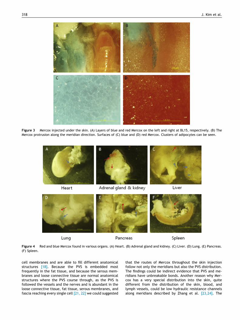
Figure 3 Mercox injected under the skin. (A) Layers of blue and red Mercox on the left and right at BL15, respectively. (B) TheMercox protrusion along the meridian direction. Surfaces of (C) blue and (D) red Mercox. Clusters of adipocytes can be seen.
Figure 4 Red and blue Mercox found in various organs. (A) Heart. (B) Adrenal gland and kidney. (C) Liver. (D) Lung. (E) Pancreas.(F) Spleen.
318 J. Kim et al.
cell membranes and are able to fill different anatomicalstructures [18]. Because the PVS is embedded mostfrequently in the fat tissue, and because the serous mem-branes and loose connective tissue are normal anatomicalstructures where the PVS course through, as the PVS isfollowed the vessels and the nerves and is abundant in theloose connective tissue, fat tissue, serous membranes, andfascia reaching every single cell [21, 22] we could suggested
that the routes of Mercox throughout the skin injectionfollow not only the meridians but also the PVS distribution.The findings could be indirect evidence that PVS and me-ridians have unbreakable bonds. Another reason why Mer-cox has a very special distribution into the skin, quitedifferent from the distribution of the skin, blood, andlymph vessels, could be low hydraulic resistance channelsalong meridians described by Zhang et al. [23,24]. The
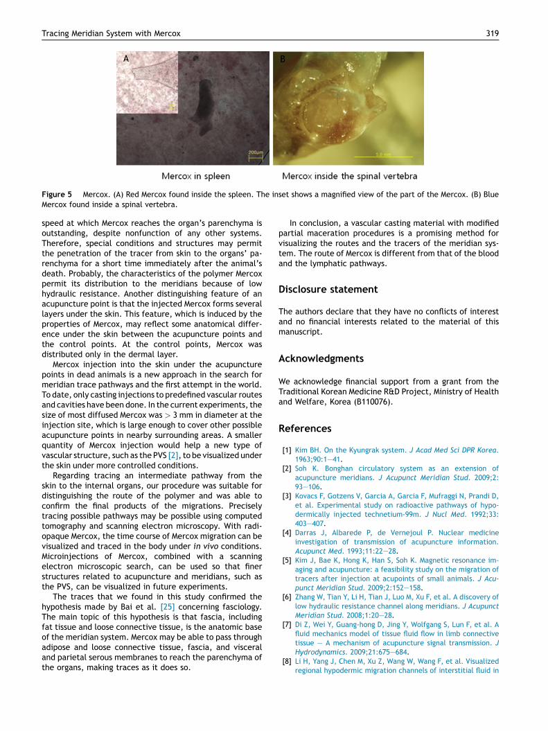
Figure 5 Mercox. (A) Red Mercox found inside the spleen. The inset shows a magnified view of the part of the Mercox. (B) BlueMercox found inside a spinal vertebra.
Tracing Meridian System with Mercox 319
speed at which Mercox reaches the organ’s parenchyma isoutstanding, despite nonfunction of any other systems.Therefore, special conditions and structures may permitthe penetration of the tracer from skin to the organs’ pa-renchyma for a short time immediately after the animal’sdeath. Probably, the characteristics of the polymer Mercoxpermit its distribution to the meridians because of lowhydraulic resistance. Another distinguishing feature of anacupuncture point is that the injected Mercox forms severallayers under the skin. This feature, which is induced by theproperties of Mercox, may reflect some anatomical differ-ence under the skin between the acupuncture points andthe control points. At the control points, Mercox wasdistributed only in the dermal layer.
Mercox injection into the skin under the acupuncturepoints in dead animals is a new approach in the search formeridian trace pathways and the first attempt in the world.To date, only casting injections to predefined vascular routesand cavities have been done. In the current experiments, thesize of most diffused Mercox was > 3 mm in diameter at theinjection site, which is large enough to cover other possibleacupuncture points in nearby surrounding areas. A smallerquantity of Mercox injection would help a new type ofvascular structure, such as the PVS [2], to be visualized underthe skin under more controlled conditions.
Regarding tracing an intermediate pathway from theskin to the internal organs, our procedure was suitable fordistinguishing the route of the polymer and was able toconfirm the final products of the migrations. Preciselytracing possible pathways may be possible using computedtomography and scanning electron microscopy. With radi-opaque Mercox, the time course of Mercox migration can bevisualized and traced in the body under in vivo conditions.Microinjections of Mercox, combined with a scanningelectron microscopic search, can be used so that finerstructures related to acupuncture and meridians, such asthe PVS, can be visualized in future experiments.
The traces that we found in this study confirmed thehypothesis made by Bai et al. [25] concerning fasciology.The main topic of this hypothesis is that fascia, includingfat tissue and loose connective tissue, is the anatomic baseof the meridian system. Mercox may be able to pass throughadipose and loose connective tissue, fascia, and visceraland parietal serous membranes to reach the parenchyma ofthe organs, making traces as it does so.
In conclusion, a vascular casting material with modifiedpartial maceration procedures is a promising method forvisualizing the routes and the tracers of the meridian sys-tem. The route of Mercox is different from that of the bloodand the lymphatic pathways.
Disclosure statement
The authors declare that they have no conflicts of interestand no financial interests related to the material of thismanuscript.
Acknowledgments
We acknowledge financial support from a grant from theTraditional Korean Medicine R&D Project, Ministry of Healthand Welfare, Korea (B110076).
References
[1] Kim BH. On the Kyungrak system. J Acad Med Sci DPR Korea.1963;90:1e41.
[2] Soh K. Bonghan circulatory system as an extension ofacupuncture meridians. J Acupunct Meridian Stud. 2009;2:93e106.
[3] Kovacs F, Gotzens V, Garcia A, Garcia F, Mufraggi N, Prandi D,et al. Experimental study on radioactive pathways of hypo-dermically injected technetium-99m. J Nucl Med. 1992;33:403e407.
[4] Darras J, Albarede P, de Vernejoul P. Nuclear medicineinvestigation of transmission of acupuncture information.Acupunct Med. 1993;11:22e28.
[5] Kim J, Bae K, Hong K, Han S, Soh K. Magnetic resonance im-aging and acupuncture: a feasibility study on the migration oftracers after injection at acupoints of small animals. J Acu-punct Meridian Stud. 2009;2:152e158.
[6] Zhang W, Tian Y, Li H, Tian J, Luo M, Xu F, et al. A discovery oflow hydraulic resistance channel along meridians. J AcupunctMeridian Stud. 2008;1:20e28.
[7] Di Z, Wei Y, Guang-hong D, Jing Y, Wolfgang S, Lun F, et al. Afluid mechanics model of tissue fluid flow in limb connectivetissue e A mechanism of acupuncture signal transmission. JHydrodynamics. 2009;21:675e684.
[8] Li H, Yang J, Chen M, Xu Z, Wang W, Wang F, et al. Visualizedregional hypodermic migration channels of interstitial fluid in

320 J. Kim et al.
human beings: are these ancient meridians? J Altern Com-plement Med. 2008;14:621e628.
[9] Dickie R, Bachoo R, Rupnick M, Dallabrida S, DeLoid G, Lai J,et al. Three-dimensional visualization of microvessel archi-tecture of whole-mount tissue by confocal microscopy.Microvasc Res. 2006;72:20e26.
[10] Duvernoy H, Delon S, Vannson J. Cortical blood vessels of thehuman brain. Brain Res Bull. 1981;7:519e579.
[11] Rautenfeld D, Lubach D, Wenzel-Hora B, Klanke J,Hunneshagen C. New techniques of demonstrating lymph vesselsin skin biopsy specimens and intact skin with the scanning elec-tron microscope. Arch Dermatol Res. 1987;279:327e334.
[12] Kondo S. Microinjection methods for visualization of thevascular architecture of the mouse embryo for light andscanning electron microscopy. J Electron Microsc (Tokyo).1998;47:101e113.
[13] Hossler F. Vascular corrosion casting can provide quantitativeas well as morphological information on the microvasculatureof organs and tissues. Microscopy Today. 1998;7:14e15.
[14] Lokmic Z, Mitchell G. Visualisation and stereological assess-ment of blood and lymphatic vessels. Histol Histopathol.2011;26:781e796.
[15] Lim S, Andrews F, Christophi C, O’Brien P. Microvascularchanges in liver after ischemia-reperfusion injury. Dig Dis Sci.1994;39:1683e1690.
[16] Less J, Skalak T, Sevick E, Jain R. Microvascular architecturein a mammary carcinoma: branching patterns and vessel di-mensions. Cancer Res. 1991;51:265e273.
[17] Krucker T, Schuler A, Meyer E, Staufenbiel M, Beckmann N.Magnetic resonance angiography and vascular corrosion cast-ing as tools in biomedical research: application to transgenic
mice modeling Alzheimer’s disease. Neurol Res. 2004;26:507e516.
[18] Stefanov M, Kim J, Nam M, Soh K. New approach of corrosioncasting using direct injection of Mercox into the parenchymaof different organs. Anat Rec (Hoboken). 2013;296:724e725.
[19] Yin C, Jeong H, Park H, Baik Y, Yoon M, Choi C, et al. A pro-posed transpositional acupoint system in a mouse and ratmodel. Res Vet Sci. 2008;84:159e165.
[20] WHO standard acupuncture point locations in the WesternPacific Region. WHO Library Cataloguing in Publication Data.Geneva: World Health Organization; 2008.
[21] Stefanov M, Kim J. Primo vascular system as a new morpho-functional integrated system. J Acupunct Meridian Stud.2012;5:193e200.
[22] Stefanov M, Potroz M, Kim J, Lim J, Cha R, Nam M. Primovascular system as new anatomical system. J Acupunct Me-ridian Stud. 2013;6:331e338.
[23] Zhang W, Tian Y, Li H, Tian JH, Luo MF, Xu FL, et al. A dis-covery of low hydraulic resistance channel along meridians.J Acupunct Meridian Stud. 2008;1:20e28.
[24] ZhangW,WangG,FuxeK.Classicandmodernmeridianstudies:Areview of low hydraulic resistance channels alongmeridians andtheir relevance for therapeutic effects in traditional Chinesemedicine. Evid Based Complement Alternat Med. 2015;2015:1e14. http://dx.doi.org/10.1155/2015/410979. Article ID:410979.
[25] Bai, Y, Yuan L, Huang Y, Wang C, Wang J, Wang J, et al. Fromthe anatomical discovery of meridians and collaterals to fas-ciology theory. In: Soh K, Kang K, Harrison D, eds. The PrimoVascular System: Its Role in Cancer and Regeneration. NewYork: Springer; 2012. pp. 283e287.