Repair Clear - Acupuncture San Diego | San Diego Acupuncture

Available online at www.sciencedirect.com
Journal of Acupuncture and Meridian Studies
journal homepage: www. jams-kpi .com
J Acupunct Meridian Stud 2017;10(2):81e89
phªC
REV IEW ART ICLE
Acupuncture Points and Their Relationshipwith Multireceptive Fields of Neurons
Salvador Quiroz-Gonzalez 1,*, Sergio Torres-Castillo 1,Rosa Estela Lopez-Gomez 1, Ismael Jimenez Estrada 2
1 Department of Acupuncture and Rehabilitation, State University of Ecatepec Valley,Ecatepec, State of Mexico, Mexico2 Department of Physiology, Biophysics and Neuroscience, Center for Research and AdvancedStudies, National Polytechnic Institute, Mexico City, Mexico
Available online 21 February 2017Received: Jun 10, 2016Revised: Jan 17, 2017Accepted: Jan 17, 2017
KEYWORDS
acupuncture points;neurons;neurophysiology;receptive field
* CEE
ISSNttp20C B
orresponding author. Departmentsq. Leona Vicario, Colonia Valle d-mail: [email protected] (S. Q
2005-2901 eISSN 2093-8152://dx.doi.org/10.1016/j.jams.20117 Medical Association of PharmY-NC-ND license (http://creativ
AbstractIn Traditional Chinese Medicine, acupuncture points (APs) have been emphasized as keyelements that generate the therapeutic effects of acupuncture. At the spinal cord or su-praspinal level, sensory neurons located in the dorsal horn receive an extensive supply ofsensory information from skin and muscle receptors through peripheral afferent nerves.The stimulated skin area that influences the activity of a spinal sensory neuron is knownas the peripheral receptive field (RF) of that neuron. By considering that a particular APlocation involves the activation of one or various RFs, it can be assumed that several sen-sory central neurons are the site of convergence of the peripheral input generated byacupuncture stimulation. However, stimulation on nonacupoint sites could also activateskin areas with RFs that have been sensitized, and they could be involved in the genera-tion of nonspecific effects of acupuncture, as seen in clinical practice. From the latter, itis suggested that effective APs, and even nonacupoints, are associated with a particulararrangement of RFs, and their study will be useful for understanding the intrinsic mech-anisms of acupuncture and for the development and identification of more efficient sitesand modes of acupuncture stimulation to evoke optimal therapeutic actions.
of Acupuncture and Rehabilitation, State University of Ecatepec Valley, Mexico Avenida Central s/n,e Anahuac, Seccion “A”, Codigo Postal 55210, Ecatepec, Estado de Mexico, Mexico.uiroz-Gonzalez).
7.01.006acopuncture Institute, Publishing services by Elsevier B.V. This is an open access article under theecommons.org/licenses/by-nc-nd/4.0/).

82 S. Quiroz-Gonzalez et al.
1. Introduction
Acupuncture is a therapeutic modality that emerged fromtraditional Chinese medicine (TCM). Although the WorldHealth Organization recommends its use for the treatmentof diseases [1], a systematic review and meta-analysis havequestioned the real efficacy of acupuncture [2]. Contra-dictory results are associated with some variables thatremain unsolved in acupuncture research, such as stan-dardization and/or individualization of points to be stimu-lated, selection of credible control procedures, effectduration, session length, and time intervals of treatments[3]. TCM recognizes approximately 361 acupuncture points(APs) and indicates that the required therapeutic effect isevoked only by the precise stimulation of specific AP sites[4]. Until now, numerous efforts have been made to iden-tify the clinical specificity of APs. Some evidence haspointed to the specificity of APs [5], whereas other lines ofevidence do not support such notion [6,7]. Some reportshave questioned the existence of APs as discrete entities[3,8], and their electrical characterization remains un-solved [9,10]. Experimental evidence has indicated thatacupuncture works through the activation of particularcentral nervous pathways [11]. Skin and muscle sensoryreceptors and their axons are responsible for generatingand transmitting the sensory signals from APs to first- andsecond-order neurons located at spinal and supraspinallevels. Dorsal horn neurons in the spinal cord receiveextensive sensory inputs from peripheral skin and musclereceptors, which can be activated by electroacupuncture(EA), by several propriospinal and descending pathways andeven visceral afferents [12e14]. The synaptic afferentinput to a particular spinal or thalamic neuron is known asthe receptive field (RF) of that neuron [14]. In this review,we analyze some neurophysiological concepts to explainthe possible relationship between APs and RFs of multi-receptive neurons as an example of the convergence ofskin, muscle, and visceral synaptic influences exerted onindividual spinal neurons [14]. We also analyzed lines ofevidence suggesting that the average size of RFs could bemodified by visceral pathologies. This study could yieldadditional information about APs and the intrinsic mecha-nisms of acupuncture in order to reveal more efficient sitesfor acupuncture stimulation to induce optimal therapeuticeffects.
2. Acupoints and meridians are located close toperipheral nerves
By means of morphological studies, it has been shown thatmost APs are located on or adjacent to peripheral nervetrunks or branches, capillary vessels, blood vessels,lymphatic vessels, nerve receptors, nerve endings, andmast cells. The meridians correspond to trajectories ofrelevant deep peripheral nerves including blood vessels[15,16]. Early studies identified that myelinated and un-myelinated fibers are stimulated during acupuncture byrecording the compound action potential [17]. Theacupuncture-signal transmission to the central nervoussystem occurred through such afferent fibers [11]. Even the
sensation that occurs during positioning of an acupunctureneedle during treatment (“De qi sensation”), it is nowgenerally accepted that it involves a multitude of fibertypes, ranging from fast-conducting myelinated Ab fibers ofhigh threshold to slow-conducting unmyelinated C fiberswith relatively low thresholds [18,19]. In addition, lesion ofnerves, block of neuronal activity, reduction of neuro-transmitter release (by pharmacologic intervention), or bylesioning the spinal cord or supraspinal regions, preventsthe action of acupuncture on the function of the followingsystems: cardiovascular function [15,20], nociceptive ornonnociceptive pathways [11,21], immune responses[22,23], digestive system [24,25], and neuroendocrineregulation [26,27]. Therefore, the nerves within thesemeridians and acupoints are probably involved in thetherapeutic effects of acupuncture (Figure 1).
3. RFs and multireceptive neurons
RF is generally defined as the area of the skin from which asensory neuron can be activated by a specific stimulus [14].Sherrington [28] was the first to use the concept of RFs todescribe the area of skin from which a scratch reflex couldbe evoked. The properties of peripheral RFs, such as theirlocalization, size, type of afferent fibers, and the strengthof excitatory drives, are usually interpreted as indicators ofthe role played by central neurons in processing sensoryinformation [29]. Sensory neurons, located in the spinalcord or in supraspinal levels, are classified according to thetype of sensory modality that activates them. Neuronsdriven by visceral nerves, excited by the cutaneous acti-vation of the corresponding dermatome, are consideredviscerosomatic neurons [30], and they are involved in thecutaneousevisceral reflex induced by acupuncture[24,26,31]. In contrast, neurons activated by afferents fromsomatic sensory receptors, but not by visceral afferents,are known as somatic neurons [30]. A subclassificationcorresponds to neurons that respond to both noxious andnonnoxious inputs and participates in the antinociceptiveeffect exerted by acupuncture on somatic pain [11].Another characteristic of a particular RF is that it shows achange in response owing to excitatory and/or inhibitoryinfluences from segmental or descending pathways [14,32].In addition, RF could be modified by experience or by injuryto sensory nerves.
There are several other groups of neurons that receivesynaptic inputs from receptors responding to different skinsense modalities (i.e., from light touch to noxious deeppressure), which are called “multireceptive neurons” [14].Visceral and skin RF inputs converge on the same spinal vis-cerosomatic neuron [14], even at the supraspinal level [33].This converging somatic and visceral influence into neuronsallows peripheral stimulation to modulate the excitabilityandfiring patterns of neural networks [11,17]. As an example,visceral or somatic nociceptive inputs arrive to multi-receptive spinal neurons,whichare inhibitedby tactile, heat,or noxious conditioning stimuli applied to the skin, viscera,and/or muscle pathways [30,34,35], or by activation ofdescending pathways involved in analgesia, such as theraphespinal, reticulospinal, or ceruleospinal tracts [17,36].

Figure 1 Relations between (A) peripheral nerves with (B) some channels of acupuncture.
Acupuncture Points and Multireceptive Fields of Neurons 83
4. Relation of acupoints with multireceptiveneurons
In TCM, acupoints are located on body surfaces where theQi of meridians and internal organs is streaming. Accordingto the TCM theory, the therapeutic effects of AP stimula-tion work through 12 principal meridians that representchannels, where Qi flows and connect with various internalorgans. The abnormal flow of Qi in a meridian is related todisease, and its natural flow can be restored by stimulationof appropriate APs [4].
The convergence of sensory inputs on multireceptivespinal neurons could explain some relationships betweeninternal organs and APs [37,38]. It has been shown thatacupuncture exerts its therapeutic effects through themodulation of neuronal activity at the level of the dorsalroot ganglion [11,30,39], spinal cord [38,40,41], trigeminalnucleus [42], thalamus [34], and brain [17], whereacupuncture might inhibit the neuronal discharges inducedby somatic and visceral pain [11]. Multireceptive neuronscould be the target of converging input from acupuncturestimulation to reduce visceral pain (Figure 2). Convergentinputs from visceral and somatic territories have been re-ported at the level of the dorsolateral medulla [41,43,44],solitary nucleus [45,46], spinal cord [46], thalamus [47],and midbrain periaqueductal gray [33]; acupuncture alsoevokes neuronal responses in those areas [11,17]. In thisway, the interrelation between body surface sensory re-ceptors, multireceptive neurons, and internal organ re-ceptors, and/or between meridians and internal organsmay have certain common neurophysiological andmorphological basis.
An important mechanism demonstrating that it is pro-duced by injury of viscera structures is the long-term
sensitization that results in the progressive amplificationof the nociceptive response (Figure 3). As an example,central sensitization is associated with an expansion of theRF of various central neurons, resulting in a larger topo-graphic distribution of pain [35,48,49]. In addition, arepeated nociceptive input from muscle or stimulation ofperipheral RF of spinal neurons results in an expansion ofthe central RF [49]. As a result of the colorectal inflam-matory reaction, the average size of the RF of multi-receptive neurons increased as the visceral inflammatoryreaction intensified. Multiple distending of the esophagusby means of an air sac results in the expansion of the re-ceptor field of dorsal horn neurons in T2eT4 [50]. In thisway, the output of multireceptive neurons should be un-derstood in ample dynamic terms because the size of theperipheral field of individual neurons may change as aresult of neuronal plasticity.
4.1. Do acupoints expand under pathologicalconditions such as dynamic RFs?
Recent lines of evidence showed that when internal organsare under pathological conditions, acupoints become moresensitive [35], and the size of acupoints varies according tovisceral alterations [48,51]. A study in dysmenorrheal pa-tients found that the most sensitive points were divergentfrom the standard location of the Diji (SP 8) acupoint,which indicates that the location of the Diji area in path-ological states is different from the standardized location[52]. Ben and collaborators [53] investigated the sensiti-zation of human skin points along meridians related tovisceral disease by using the pressure-pain threshold as anindicator. Their results showed the existence of pain-sensitive points on the abdomen and back regions of
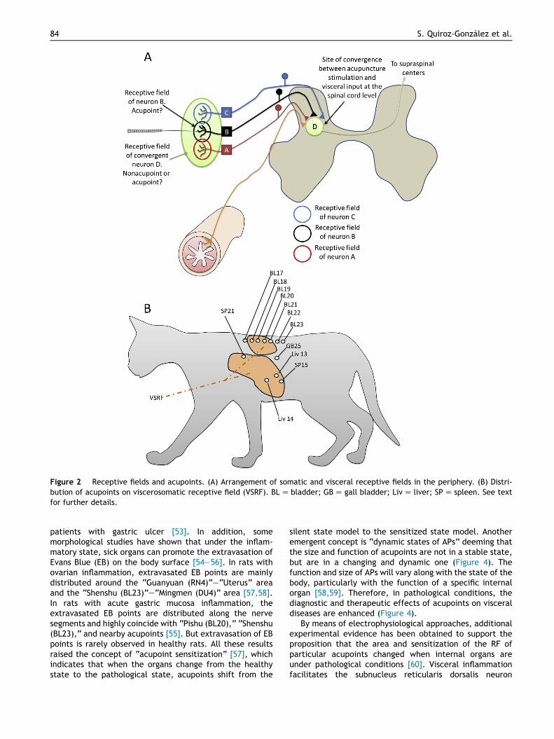
Figure 2 Receptive fields and acupoints. (A) Arrangement of somatic and visceral receptive fields in the periphery. (B) Distri-bution of acupoints on viscerosomatic receptive field (VSRF). BLZ bladder; GBZ gall bladder; LivZ liver; SPZ spleen. See textfor further details.
84 S. Quiroz-Gonzalez et al.
patients with gastric ulcer [53]. In addition, somemorphological studies have shown that under the inflam-matory state, sick organs can promote the extravasation ofEvans Blue (EB) on the body surface [54e56]. In rats withovarian inflammation, extravasated EB points are mainlydistributed around the “Guanyuan (RN4)”e“Uterus” areaand the “Shenshu (BL23)”e“Mingmen (DU4)” area [57,58].In rats with acute gastric mucosa inflammation, theextravasated EB points are distributed along the nervesegments and highly coincide with “Pishu (BL20),” “Shenshu(BL23),” and nearby acupoints [55]. But extravasation of EBpoints is rarely observed in healthy rats. All these resultsraised the concept of “acupoint sensitization” [57], whichindicates that when the organs change from the healthystate to the pathological state, acupoints shift from the
silent state model to the sensitized state model. Anotheremergent concept is “dynamic states of APs” deeming thatthe size and function of acupoints are not in a stable state,but are in a changing and dynamic one (Figure 4). Thefunction and size of APs will vary along with the state of thebody, particularly with the function of a specific internalorgan [58,59]. Therefore, in pathological conditions, thediagnostic and therapeutic effects of acupoints on visceraldiseases are enhanced (Figure 4).
By means of electrophysiological approaches, additionalexperimental evidence has been obtained to support theproposition that the area and sensitization of the RF ofparticular acupoints changed when internal organs areunder pathological conditions [60]. Visceral inflammationfacilitates the subnucleus reticularis dorsalis neuron
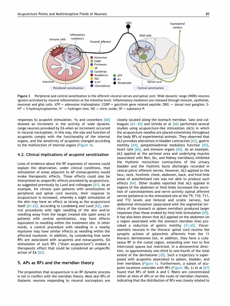
Figure 3 Peripheral and central sensitization in the afferent visceral nerves and spinal cord. Wide dynamic range (WDR) neurons(green) activated by visceral inflammation at the intestine level. Inflammatory mediators are released through immune, epithelial,neuronal and glial cells. ATPZ adenosine triphosphate; CGRPZ galcitinin gene related peptide; DRG Z dorsal root ganglion; 5-HTZ 5-hydroxytryptamine; HþZ hydrogen ions; NOZ nitric oxide; SPZ substance P.
Acupuncture Points and Multireceptive Fields of Neurons 85
responses to acupoint stimulation. Yu and coworkers [60]showed an increment in the activity of wide dynamicrange neurons provoked by EA when an increment occurredin visceral nociception. In this way, the size and function ofacupoints comply with the functionality of the internalorgans, and the sensitivity of acupoints changed accordingto the malfunction of internal organs (Figure 4).
4.2. Clinical implications of acupoint sensitization
Lines of evidence about the RF expansion of neurons couldexplain the observation, under clinical conditions, thatstimulation of areas adjacent to AP (nonacupoints) wouldevoke therapeutic effects. Those effects could also beinterpreted as unspecific effects provoked by acupuncture,as suggested previously by Lund and colleagues [61]. As anexample, for chronic pain patients with sensitization ofperipheral and spinal cord neurons, their response toacupuncture is increased, whereby a slight stimulation ofthe skin may have an effect as strong as the acupunctureitself [61,62]. According to Lundeberg and Lund [62], con-trol procedures with light needling of the skin and/orneedling away from the target treated site (pain area) inpatients with central sensitization, may have effectsequivalent to needling within the treatment site. In otherwords, a control procedure with needling in a nearbymyotome may have similar effects as needling within theaffected myotome. In addition, by considering that someRFs are associated with acupoints and nonacupoints, EAstimulation of such RFs (“sham acupuncture”) evoked atherapeutic effect that could be considered an unspecificaction of EA [62].
5. APs as RFs and the meridian theory
The proposition that acupuncture is an RF dynamic processis not in conflict with the meridian theory. Most skin RFs ofthalamic neurons responding to visceral nociceptors are
closely located along the stomach meridian. Sato and col-leagues [63e65] and Uchida et al [66] performed severalstudies using acupuncture-like stimulation (ALS) in whichthe acupuncture needles are placed extensively throughoutthe body RFs of experimental animals. They observed thatALS provokes alterations in bladder contraction [63], gastricmotility [64], sympathoadrenal medullary function [65],heart rate [66], and immune organs [65]. As an example,ALS applied at the perineal area and underlying muscles(associated with Ren, Du, and Kidney meridians) inhibitedthe rhythmic micturition contractions of the urinarybladder and the rhythmic burst discharges recorded invesical pelvic efferent nerves. However, ALS applied to theface, neck, forelimb, chest, abdomen, back, and hind limbareas of anesthetized rats was not able to produce sucheffects [64]. Other studies reported that ALS applied inregions of the abdomen or hind limbs increased the secre-tion of catecholamines and nerve activity (spinal afferentnerves ipsilateral to the stimulated site at the T9, T10, T11,and T12 levels and femoral and sciatic nerves), butabdominal stimulation (associated with the segmental ter-ritory of the stomach or spleen meridian) produced largerresponses than those evoked by hind limb stimulation [65].It has also been shown that ALS applied on the abdomen (ina region associated with the stomach meridian line) pro-voked a reduction of gastric motility [31,65]. Viscer-osomatic neurons in the thoracic spinal cord receive thesynaptic actions of splanchnic afferents from the 11thoracic dermatomes but, in addition, they have a cuta-neous RF in the costal region, extending over two to fiveintercostal spaces but restricted, in a dorsoventral direc-tion, to approximately one-third to one-fourth of the totalextent of the dermatome [30]. Such a trajectory is super-posed with acupoints associated to spleen, bladder, andliver meridians (Figure 1). Furthermore, a subset of acu-point locations coincides with RFs (Figure 1B). Li et al [67]found that RFs of both A and C fibers are concentratedeither at sites of APs or on the route of meridian channels,indicating that the distribution of RFs was closely related to
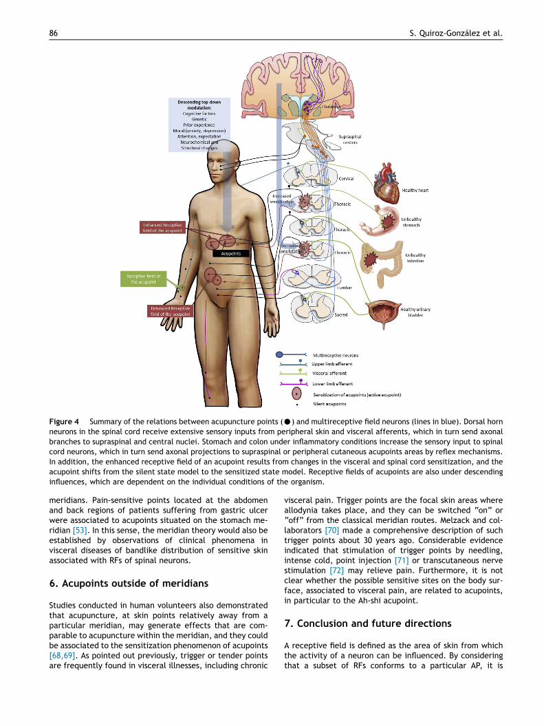
Figure 4 Summary of the relations between acupuncture points (C) and multireceptive field neurons (lines in blue). Dorsal hornneurons in the spinal cord receive extensive sensory inputs from peripheral skin and visceral afferents, which in turn send axonalbranches to supraspinal and central nuclei. Stomach and colon under inflammatory conditions increase the sensory input to spinalcord neurons, which in turn send axonal projections to supraspinal or peripheral cutaneous acupoints areas by reflex mechanisms.In addition, the enhanced receptive field of an acupoint results from changes in the visceral and spinal cord sensitization, and theacupoint shifts from the silent state model to the sensitized state model. Receptive fields of acupoints are also under descendinginfluences, which are dependent on the individual conditions of the organism.
86 S. Quiroz-Gonzalez et al.
meridians. Pain-sensitive points located at the abdomenand back regions of patients suffering from gastric ulcerwere associated to acupoints situated on the stomach me-ridian [53]. In this sense, the meridian theory would also beestablished by observations of clinical phenomena invisceral diseases of bandlike distribution of sensitive skinassociated with RFs of spinal neurons.
6. Acupoints outside of meridians
Studies conducted in human volunteers also demonstratedthat acupuncture, at skin points relatively away from aparticular meridian, may generate effects that are com-parable to acupuncture within the meridian, and they couldbe associated to the sensitization phenomenon of acupoints[68,69]. As pointed out previously, trigger or tender pointsare frequently found in visceral illnesses, including chronic
visceral pain. Trigger points are the focal skin areas whereallodynia takes place, and they can be switched “on” or“off” from the classical meridian routes. Melzack and col-laborators [70] made a comprehensive description of suchtrigger points about 30 years ago. Considerable evidenceindicated that stimulation of trigger points by needling,intense cold, point injection [71] or transcutaneous nervestimulation [72] may relieve pain. Furthermore, it is notclear whether the possible sensitive sites on the body sur-face, associated to visceral pain, are related to acupoints,in particular to the Ah-shi acupoint.
7. Conclusion and future directions
A receptive field is defined as the area of skin from whichthe activity of a neuron can be influenced. By consideringthat a subset of RFs conforms to a particular AP, it is

Acupuncture Points and Multireceptive Fields of Neurons 87
proposed that different classes of neurons are sites ofconvergence of the afferent input generated by acupunc-ture stimulation. Even the specificity of EA stimulation toprovoke spinal cord dorsum potentials is suggested to beprimarily related to peripheral nerve routes [73]. In spite ofthat, activation of other skin areas with active RFs butdifferent from those exerting a particular acupuncture ac-tion (nonacupoints) explains the occurrence of nonspecificeffects provoked by acupuncture procedures, as reportedin clinical trials. On the other side, the effects ofacupuncture need to be examined from the perspective ofRF, in which properties of the RF must be further consid-ered. For example, the segmental RF of multireceptiveneurons is generally located on the skin and is made up ofan excitatory field or an inhibitory field. In that sense,peripheral influences could be both excitatory and inhibi-tory, and pain can be relieved by reinforced segmentalinhibitory controls through electrical stimulation by EA orother empirical physical means.
Finally, insertion of an acupuncture needle into adesignated point on the body surface produces a stimulusthat evokes the activation of multiple central neuronalpathways [21,31]. An optimal response to acupuncturemight be a consequence of the efficiency and specificity ofthe stimulation site and activation of particular pathways inthe central nervous system. Precise identification of APbecame challenging because the size and location of RFs formultireceptive neurons could be increased or modified bychanges in the physiological condition of the individual(Figure 4); even the response of spinal dorsal horn neuronsto a particular RF would change as a result of excitatory andinhibitory influences exerted by segmental, propriospinal,or descending processes impinging on them at a particulartime. Consequently, in clinical practice, each patient couldhave sites of acupuncture stimulation that do not corre-spond strictly with traditional APs, probably because theyare associated with RFs varying dynamically in time andmagnitude, according to the general physiological state ofthe individual (Figure 4). Considering AP stimulation as adynamic and complex process might help us understand thedifferent degrees of acupoint specificity observed duringdiverse clinical conditions. From a neurophysiologicalperspective, APs considered as dynamic RFs could be ofconsiderable importance for the characterization ofacupuncture actions and for the development and/orimprovement of acupuncture procedures to obtain optimaltherapeutic treatments.
Disclosure statement
None.
Acknowledgments
We thank the American Journal Experts for editing (English)this manuscript. This work was partially supported by afellowship granted to I. Jimenez-Estrada from the SistemaNacional de Investigadores and to S. Quiroz-Gonzalez by agrant from PROMEP (No. 103.5-13-6729) and SNI-CONACyT.
References
[1] Barnes PM, Bloom B, Nahin RL. Complementary and alterna-tive medicine use among adults and children. Natl Health StatRep. 2008;10:1e23.
[2] Birch S, Hesselink JK, Jonkman FAM, Hekker TAM, Bos AAT.Clinical research on acupuncture: Part 1. What have reviewsof the efficacy and safety of acupuncture told us so far? JAltern Complement Med. 2004;10:468e480.
[3] Ernst E. Acupunctured critical analysis. J Intern Med. 2006;259:125e137.
[4] Zhou F, Huang D, Xia Y, Xia Y, Wu G, Cao X. AcupunctureTherapy for Neurological Diseases: A Neurobiological View.Beijing, China: Tsinghua University Press; 2010:32e80.
[5] Zhao L, Chen J, Liu CZ, Li Y, Cai DJ, Tang Y, et al. A review ofacupoint specificity research in china: status quo and pros-pects. Evidence Based Complement Alternative Med. 2012;2012:5439e5443.
[6] Assefi NP, Sherman KJ, Jacobsen C, Goldberg J, Smith WR,Buchwald D. A randomized clinical trial of acupuncturecompared with sham acupuncture in fibromyalgia. Ann IntMed. 2005;143:10e19.
[7] Harris RE, TianX,WilliamsDA,TianTX, CuppsTR, PetzkeF, et al.Treatment of fibromyalgia with formula acupuncture: investi-gation of needle placement, needle stimulation, and treatmentfrequency. J Altern Complement Med. 2005;11:663e671.
[8] Ramey DW. Acupuncture points do not exist. Sci Rev AlternMed. 2001;5:140e145.
[9] Ahn AC, Martinsen OG. Electrical characterization ofacupuncture points: technical issues and challenges. J AlternComplement Med. 2007;13:817e824.
[10] Ahn AC, Colbert AP, Anderson BJ, Martinsen OG,Hammerschlag R, Cina S, et al. Electrical properties ofacupuncture points and meridians: a systematic review. Bio-electromagnetics. 2008;29:245e256.
[11] Zhao ZQ. Neural mechanism underlying acupuncture anal-gesia. Prog Neurobiol. 2008;85:355e375.
[12] Zhang ZJ, Wang XM, McAlonan GM. Neural acupuncture unit: anew concept for interpreting effects and mechanisms ofacupuncture. Evid Based Complement Altern Med. 2012;2012:429412e429423.
[13] Manjarrez E, Jimenez I, Rudomin P. Intersegmental synchro-nization of spontaneous activity of dorsal horn neurons in thecat spinal cord. Exp Brain Res. 2003;148:401e413.
[14] Le Bars D. The whole body receptive field of dorsal hornmultireceptive neurones. Brain Res Rev. 2002;40:29e44.
[15] Li P, Longhurst JC. Neural mechanism of electroacupunctureshypotensive effects. Auton Neurosci. 2010;157:24e30.
[16] Chapple W. Proposed catalog of the neuroanatomy and thestratified anatomy for the 361 acupuncture points of 14channels. J Acupunct Meridian Stud. 2013;5:270e274.
[17] Zhang R, Lao L, Ren K, Berman BM. Mechanisms of acu-punctureeelectroacupuncture on persistent pain. Anesthesi-ology. 2014;120:482e503.
[18] Kagitani F, Sae U, Hotta H. Afferent nerve fibers andacupuncture. Auton Neurosci. 2010;157:2e8.
[19] Kim SA, Lee BH, Bae JH, Kim KJ, Steffensen SC, Ryu YH, et al.Peripheral afferent mechanisms underlying acupuncture in-hibition of cocaine behavioral effects in rats. PLoS One. 2013;8:81018.
[20] Zhou W, Fu LW, Guo Z, Longhurst JC. Role of glutamate inrostral ventrolateral medulla in acupuncture-related modu-lation of visceral reflex sympathoexcitation. Am J PhysiolHeart Circ Physiol. 2007;292:H1868eH1875.
[21] Quiroz-Gonzalez S, Segura-Alegrıa B, Guadarrama-Olmos JC,Jimenez-Estrada I. Cord dorsum potentials evoked by

88 S. Quiroz-Gonzalez et al.
electroacupuncture applied to the hind limbs of rats. J Acu-punct Meridian Stud. 2014;7:25e32.
[22] Ding SS, Hong SH, Wang C, Gou Y, Wang ZK, Xu Y. Acupuncturemodulates the neuro-endocrine-immune network. Q J Med.2014;107:341e345.
[23] Kim SK, Bae H. Acupuncture and immune modulation. AutonNeurosci. 2010;157:38e41.
[24] Takahashi T. Mechanism of acupuncture on neuromodulationin the gutda review. Neuromodulation. 2011;14:8e12.
[25] Takahashi T. Effect and mechanism of acupuncture ongastrointestinal diseases. Int Rev Neurobiol. 2013;111:273e294.
[26] Yu JS, Zeng BY, Hsieh CL. Acupuncture stimulation and neuro-endocrine regulation. Int Rev Neurobiol. 2013;111:125e140.
[27] Langevin HM, Yandow JA. Relationship of acupuncture pointsand meridians to connective tissue planes. Anat Rec. 2002;269:257e265.
[28] Sherrington CS. Observations on the scratch-reflex in thespinal dog. J Physiol. 1906;34:1e50.
[29] Weiss NS, Ohara KO, Johnson FA, Lenz. The human thalamicsomatic sensory nucleus [ventral caudal (Vc)] shows neuronalmechanoreceptor-like responses to optimal stimuli for pe-ripheral mechanoreceptors. J Neurophysiol. 2009;101:1033e1042.
[30] Cervero F. Sensory innervation of the viscera: peripheral basisof visceral pain. Physiol Rev. 1994;75:95e138.
[31] Sato A, Schmidt RF. The modulation of visceral functions bysomatic afferent activity. Jpn J Physiol. 1997;37:1e17.
[32] Contreras-Hernandez E, Chavez D, Rudomin P. Dynamic syn-chronization of ongoing neuronal activity across spinal seg-ments regulates sensory information flow. J Physiol. 2015;593:2343e2363.
[33] Keay KA, Clement CL, Owler B, Depaulis A, Bandler R.Convergence of deep somatic and visceral nociceptive infor-mation onto a discrete ventrolateral midbrain periaqueductalgray region. Neuroscience. 1994;61:727e732.
[34] Zhang JL, Zhang SP, Zhang HQ. Effect of electroacupunctureon thalamic neuronal response to visceral nociception. Eur JPain. 2009;13:366e372.
[35] Rong PJ, Li S, Ben H, Li L, Yu LL, Cui CX, et al. Peripheral andspinal mechanisms of acupoint sensitization phenomenon.Evid Based Complement Altern Med. 2013;2013:742195.
[36] Millan MJ. Descending control of pain. Prog Neurobiol. 2002;66:355e474.
[37] Wang SJ, Yang HY, Wang F, Li ST. Acupoint specificity oncolorectal hypersensitivity alleviated by acupuncture and thecorrelation with the brain-gut axis. Neurochem Res. 2015;40:1274e1282.
[38] Rong P, Zhu B, Li Y, Gao X, Ben H, Li Y, et al. Mechanism ofacupuncture regulating visceral sensation and mobility. FrontMed. 2011;5:151e156.
[39] Cameron DM, Brennan TJ, Gebhart GF. Hind paw incision inthe rat produces long-lasting colon hypersensitivity. J Pain.2008;9:246e253.
[40] Lee JH, Beitz AJ. Electroacupuncture modifies the expressionof c-fos in the spinal cord induced by noxious stimulation.Brain Res. 1992;577:80e91.
[41] Luz LL, Fernandes EC, Sivado M, Kokai E, Szucs P, Safronov BV.Monosynaptic convergence of somatic and visceral C-fiberafferents on projection and local circuit neurons in lamina I: asubstrate for referred pain. Pain. 2015;156:2042e2051.
[42] Bing Z, Villanueva L, Le Bars D. Acupuncture and diffusenoxious inhibitory controls: naloxone-reversible depression ofactivities of trigeminal convergent neurons. Neuroscience.1990;37:809e818.
[43] Blair RW, Thompson GM. Convergence of multiple sensory in-puts onto in the dorsolateral medulla in cats. Neuroscience.1995;67:721e729.
[44] Zhuo M, Gebhart GF. Facilitation and attenuation of a visceralnociceptive reflex from the rostroventral medulla in the rat.Gastroenterology. 2002;122:1007e1019.
[45] Hubscher CH. Responses of neurons in caudal solitary nucleusof female rats to stimulation of vagina, cervix, uterine hornand colon. Brain Res. 1994;664:1e8.
[46] Berkley KJ, Hubscher CH, Wall PD. Neuronal responses tostimulation of the cervix, uterus, colon, and skin in the ratspinal cord. J Neurophysiol. 1993;69:545e556.
[47] Horn AC, Vahle-Hinz C, Bruggemann J, Petersen M, Kniffki KD.Responses of neurons in the lateral thalamus of the cat tostimulation of urinary bladder, colon, esophagus, and skin.Brain Res. 1999;851:164e174.
[48] Baron R, Baron Y, Disbrow E, Roberts TP. Activation of thesomatosensory cortex during Abeta-fiber mediated hyper-algesia. A MSI study. Brain Res. 2000;871:75e82.
[49] Li YQ, Rong PJ, Xu WD, Zhu B. Conditioning stimulationinduced change in receptive field of spinal neuron. Chin JNeurosci. 1999;15:75e82.
[50] Euchner-Wamser I, Sengupta JN, Gebhart GF, Meller ST.Characterization of responses of T2eT4 spinal cord neurons toesophageal distension in the rat. J Neurophysiol. 1993;69:868e883.
[51] Cheng B, Shi H, Ji CF, Li JH, Chen SL, Jing XH. Distribution ofthe activated acupoints after acute gastric mucosal injury inthe rat. Acupunct Res. 2010;35:193e197.
[52] Chen S, Miao Y, Nan Y, Wang Y, Zhao Q, He E, et al. The studyof dynamic characteristic of acupoints based on the primarydysmenorrhea patients with the tenderness reflection on Diji(SP 8). Evid Based Complement Altern Med. 2015;2015:158012.
[53] Ben H, Li L, Rong PJ, Jin ZG, Zhang JL, Li YH, et al. Obser-vation of pain-sensitive points along the meridians in patientswith gastric ulcer or gastritis. Evid Based Complement AlternMed. 2012;2012:130802.
[54] Wang SJ, Zhu B. Study on relation of ovaryebody surfacecorrelativity with acupoints. Chin Acupunct Moxibustion.2007;27:761e765.
[55] Yu S, Yang J, Yang M, Gao Y, Chen J, Ren Y, et al. Applicationof acupoints and meridians for the treatment of primarydysmenorrhea: a data mining-based literature study. EvidBased Complement Altern Med. 2015;2015:1e8.
[56] Xiaochun Y, Bing Z, Junhong G. Scientific foundation of dy-namic process of acupoints. J Tradit Chin Med. 2007;48:971e973.
[57] Yu X, Zhu B, Gao JH. The scientific basis of the points dynamicprocess. J Tradit Chin Med. 2007;48:17e23.
[58] Baldry P. Large tender areas, not discrete points, observed inpatients with fibromyalgia. Acupunct Med. 2007;25:203.
[59] Baliki MN, Geha PY, Apkarian AV, Chialvo DR. Beyond feeling:chronic pain hurts the brain, disrupting the default-modenetwork dynamics. J Neurosci. 2008;28:1398e1403.
[60] Yu LL, Li L, Rong PJ, Zhu B, Qin QG, Ben H, et al. Changes inresponses of neurons in spinal and medullary subnucleusreticularis dorsalis to acupoint stimulation in rats with visceralhyperalgesia. Evid Based Complement Alternat Med. 2014;2014:768634.
[61] Lund I, Naslund J, Lundeberg T. Minimal acupuncture is not avalid placebo control in randomised controlled trials ofacupuncture: a physiologist’s perspective. Chin Med. 2009;30,4e1.
[62] Lundeberg T, Lund I. Are reviews based on sham acupunctureprocedures in fibromyalgia syndrome (FMS) valid? AcupunctMed. 2007;25:100e106.
[63] Sato A, Sato Y, Suzuki A. Mechanism of the reflex inhibition ofmicturition contractions of the urinary bladder elicited byacupuncture-like stimulation in anesthetized rats. NeurosciRes. 1992;15:189e198.
Acupuncture Points and Multireceptive Fields of Neurons 89
[64] Sato A, Sato Y, Suzuki A, Uchida S. Neural mechanisms of thereflex inhibition and excitation of gastric motility elicited byacupuncture-like stimulation in anesthetized rats. NeurosciRes. 1993;18:53e62.
[65] Sato A, Sato Y, Suzuki A, Uchida S. Reflex modulation ofcatecholamine secretion and adrenal sympathetic nerve ac-tivity by acupuncture-like stimulation in anesthetized rat. JpnJ Physiol. 1996;46:411e421.
[66] Uchida S, Kagitani F, Hotta H. Mechanism of the reflex inhi-bition of heart rate elicited by acupuncture-like stimulation inanesthetized rats. Auton Neurosci. 2008;143:12e19.
[67] Li AH, Zhang JM, Xie JK. Human acupuncture points mapped inrats areassociatedwith excitablemuscle/skin-nerve complexeswith enriched nerve endings. Brain Res. 2004;1012:154e159.
[68] Haake M, Muller HH, Schade-Brittinger C, Basler HD,Schafer H, Maier C, et al. German Acupuncture Trials (GERAC)for chronic low back pain: randomized, multicenter, blinded,parallel-group trial with 3 groups. Arch Intern Med. 2007;167:1892e1898.
[69] Liu YQ, Liu WX, Wang Q, Yu XX, Kang MJ. A clinical study onthe prevention of visceral traction pain during cesarean sec-tion using HANS-assisted epidural anesthesia. Chin J Pain Med.1996;2:215e219.
[70] Melzack R, Stillwell DM, Fox EJ. Trigger points and acupunc-ture points for pain: correlations and implications. Pain. 1997;3:3e23.
[71] Graboski CL, Gray DS, Burnham RS. Botulinum toxin A versusbupivacaine trigger point injections for the treatment ofmyofascial pain syndrome: a randomised double blind cross-over study. Pain. 2005;118:170e175.
[72] Vance CG, Dailey DL, Rakel BA, Sluka KA. Using TENS for paincontrol: the state of the evidence. Pain Manag. 2014;4:197e209.
[73] Quiroz-Gonzalez S, Segura-Alegrıa B, Jimenez-Estrada I.Depressing effect of electroacupuncture on the spinal non-painful sensory input of the rat. Exp Brain Res. 2014;232:2721e2729.