Topics covered Scope and applications of insilico modeling in modern biology. Comparative modeling...
30
Topics covered • Scope and applications of insilico modeling in modern biology. • Comparative modeling • Constructing an initial model • refining the model • manipulating the model • Molecular superposition and structural alignment • concept of energy minimization • Different types of interactions and formulation of force fields. • Basic MD algorithm, its limitations, treatment of long range forces. • Structure Visualization and Graphical representation of molecular structures: • Small molecules (low molecular weight – peptides, nucleotides, disaccharides, simple drugs molecules) • Macromolecules (high molecular weight molecules - proteins, DNA, RNA, membranes).
-
Upload
darlene-elisabeth-clark -
Category
Documents
-
view
213 -
download
0
Transcript of Topics covered Scope and applications of insilico modeling in modern biology. Comparative modeling...
Topics covered• Scope and applications of insilico modeling in modern biology. • Comparative modeling• Constructing an initial model• refining the model• manipulating the model• Molecular superposition and structural alignment• concept of energy minimization• Different types of interactions and formulation of force fields. • Basic MD algorithm, its limitations, treatment of long range forces. • Structure Visualization and Graphical representation of molecular
structures:• Small molecules (low molecular weight – peptides, nucleotides,
disaccharides, simple drugs molecules) • Macromolecules (high molecular weight molecules - proteins, DNA,
RNA, membranes).
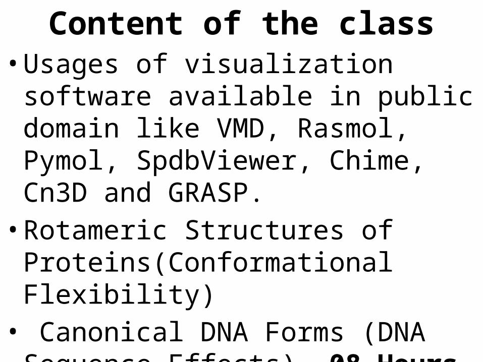
Content of the class• Usages of visualization software available
in public domain like VMD, Rasmol, Pymol, SpdbViewer, Chime, Cn3D and GRASP. • Rotameric Structures of
Proteins(Conformational Flexibility)• Canonical DNA Forms (DNA Sequence
Effects). 08 Hours
Usages of visualization software available in public domain like VMD, Rasmol, Pymol,
SpdbViewer, Chime, Cn3D and GRASP.
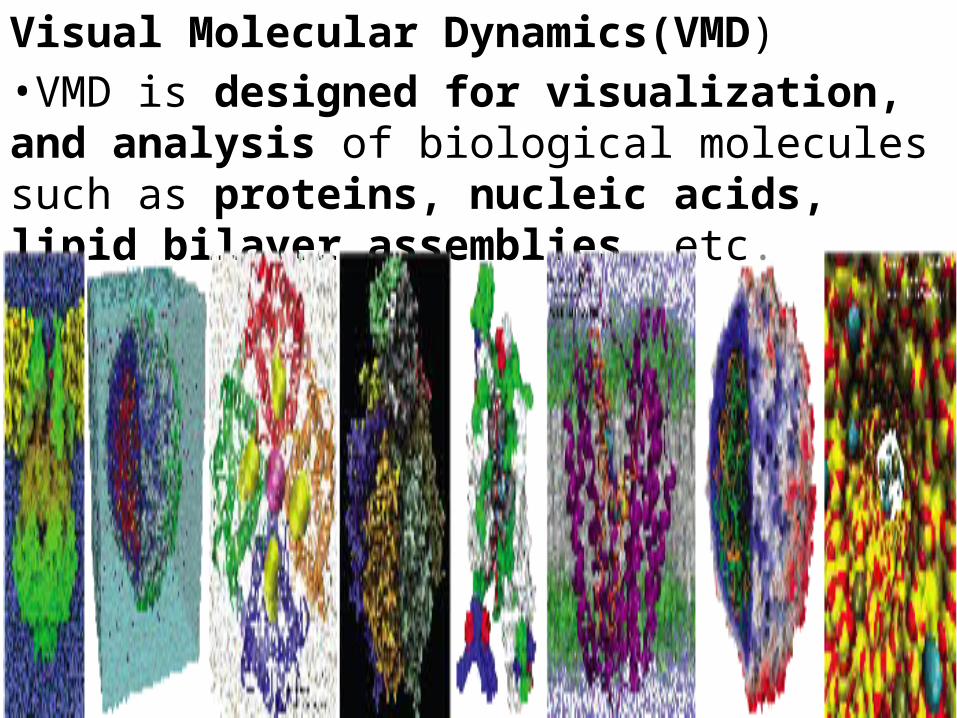
Visual Molecular Dynamics(VMD)•VMD is designed for visualization, and analysis of biological molecules such as proteins, nucleic acids, lipid bilayer assemblies, etc.
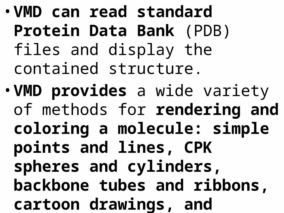
• VMD can read standard Protein Data Bank (PDB) files and display the contained structure. • VMD provides a wide variety of methods
for rendering and coloring a molecule: simple points and lines, CPK spheres and cylinders, backbone tubes and ribbons, cartoon drawings, and others.
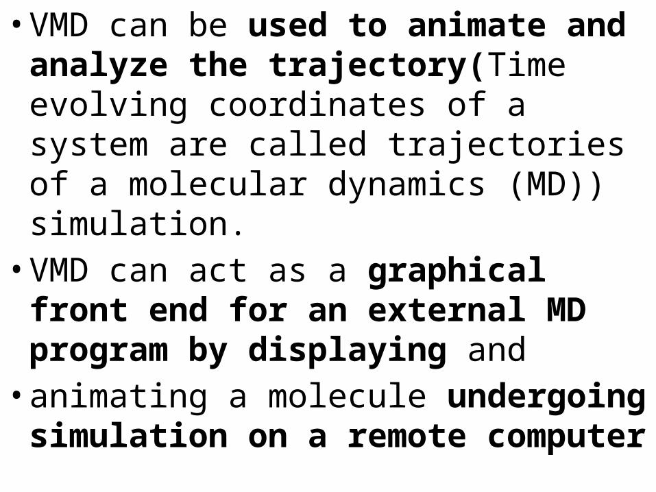
• VMD can be used to animate and analyze the trajectory(Time evolving coordinates of a system are called trajectories of a molecular dynamics (MD)) simulation. • VMD can act as a graphical front end for
an external MD program by displaying and • animating a molecule undergoing
simulation on a remote computer
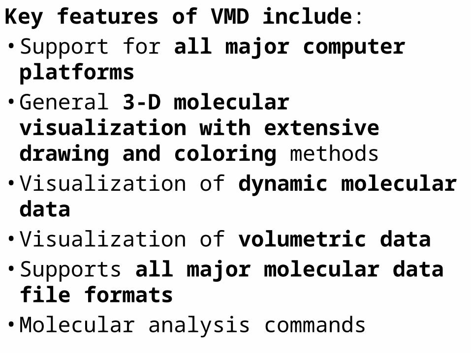
Key features of VMD include: • Support for all major computer
platforms • General 3-D molecular visualization with
extensive drawing and coloring methods • Visualization of dynamic molecular data • Visualization of volumetric data • Supports all major molecular data file
formats• Molecular analysis commands
• Rendering high-resolution, publication-quality molecule images
• Movie making capability • Building and preparing systems for molecular
dynamics simulations • Interactive molecular dynamics simulations • Extensions to the Tcl/Python scripting
languages • Extensible source code written in C and C++
Cn3D

• Cn3D is a visualization tool for biomolecular structures, sequences, and sequence alignments. • Cn3D can correlate structure and
sequence information:• for example, a scientist can quickly find
the residues in a crystal structure that correspond to known disease mutations, • or conserved active site residues from a
family of sequence homologs.

• Cn3D displays structure-structure alignments along with their structure-based sequence alignments, • to emphasize what regions of a group of
related proteins are most conserved in structure and sequence. • Also included are custom labeling
features, high-quality graphics, and
• A variety of file exports that together make Cn3D a powerful tool for literature annotation • With version 4, Cn3D is now a complete
multiple alignment editor as well, and • Includes algorithms for aligning
sequences to other sequences and to structures. • You can now create and even annotate
multiple alignments.
RasMol

• RasMol is a program that allows you to view molecular structures on the computer screen• RasMol was written by Roger Sayle, now
at Glaxo, and is available free, for Windows and Mac computers (and others). (RasMol for Windows is also known as RasWin.) • RasMol is a molecular graphics program
intended for the visualisation of proteins, nucleic acids and small molecules.
• The program reads in a molecule co-ordinate file and interactively displays the molecule on the screen in a variety of colour schemes and molecule representations.
• Currently available representations include depth-cued wireframes, 'Dreiding' sticks, spacefilling (CPK) spheres, ball and stick, solid and strand biomolecular ribbons, atom labels and dot surfaces.
• Supported input file formats include Brookhaven Protein Databank (PDB), Tripos Associates' Alchemy and Sybyl Mol2 formats, Molecular Design Limited's (MDL) Mol file format, Minnesota Supercomputer Centre's (MSC) XYZ (XMol) format and CHARMm format files
• The displayed molecule may be rotated, translated, zoomed and z-clipped (slabbed) interactively using either the mouse, the scroll bars, the command line or an attached dial box
Spdb Viewer

• Swiss-PdbViewer provides a user friendly interface allowing to analyze several proteins at the same time.
• The proteins can be superimposed in order to deduce structural alignments and compare their active sites or any other relevant parts.
• Amino acid mutations, H-bonds, angles and distances between atoms are easy to obtain
• Swiss-PdbViewer has been developed since 1994 by Nicolas Guex.
• Swiss-PdbViewer is tightly linked to SWISS-MODEL, an automated homology modeling server developed within the Swiss Institute of Bioinformatics (SIB) at the Structural Bioinformatics Group at the Biozentrum in Basel.
• Swiss-PdbViewer can also read electron density maps, and provides various tools to build into the density.
• In addition, various modeling tools are integrated and command files for popular energy minimization packages can be generated.

Pymol
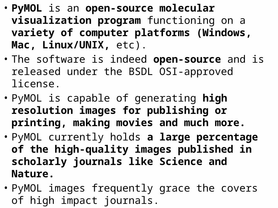
• PyMOL is an open-source molecular visualization program functioning on a variety of computer platforms (Windows, Mac, Linux/UNIX, etc).
• The software is indeed open-source and is released under the BSDL OSI-approved license.
• PyMOL is capable of generating high resolution images for publishing or printing, making movies and much more.
• PyMOL currently holds a large percentage of the high-quality images published in scholarly journals like Science and Nature.
• PyMOL images frequently grace the covers of high impact journals.
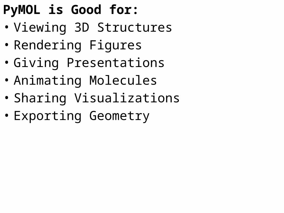
PyMOL is Good for: • Viewing 3D Structures • Rendering Figures • Giving Presentations • Animating Molecules • Sharing Visualizations • Exporting Geometry

Chime

• MDL Chime (or simply "Chime" for short) is a free program to show molecular structure in three dimensions.
• Its images look like RasMol's because Chime is derived, from RasMol.
• MDL Chime differs from RasMol in that Chime sits directly on a web page (runs inside your browser as a plug-in),
• However, some Chime websites also provide support for showing any molecule of interest, and doing self-directed exploration similar to RasMol
Rotameric structures of proteins (conformational flexibility)
•A Rotamer library describes the conformation of amino-acid side chains•The torsions are described by the Chi angles. Former Rotamer libraries where established for folding prediction•Rotamers are usually defined as low energy side-chain conformations. •The use of a build-library of rotamers allows anyone determining or modeling a structure
• To try the most likely side-chain conformations, saving time and producing a structure that is more likely to be correct.• This is, of course, only the case if the
rotamers really are the correct low energy conformations. • There is a database of Penultimate
Rotamer
• The Penultimate Rotamer Library results are presented as tables of confomers.
• Thus, one possible use of it is as a build library for model fitting.
• rotamer datasets are used in several software packages:
• KiNG & mage -- two kinemage display programs both use our rotamers in their build/mutate functions.
• rotamer -- a program in the CCP4 suite that identifies suspect rotamers.
• AFITT -- model building software from OpenEye
Canonical DNA forms (DNA sequence effects)
The “canonical” base pairsSee Figure 11.7
• The canonical A:T and G:C base pairs have nearly identical overall dimensions.
• A and T share two H bonds. • G and C share three H bonds. • G:C-rich regions of DNA are more stable. • Polar atoms in the sugar-phosphate backbone
also form H bonds.


![RESEARCHARTICLE InSilico AnalysisofUsherEncodingGenesin · including theadhesionsubunitsthat enablethebacteriatospecificallytarget acellcomponent orasurface [2].Studies onthebiochemistryandgenetics](https://static.fdocuments.in/doc/165x107/5c88c0ed09d3f23d648c06be/researcharticle-insilico-analysisofusherencodinggenesin-including-theadhesionsubunitsthat.jpg)