Tongue posture improvement and pharyngeal
11
Tongue posture improvement and pharyngeal airway enlargement as secondary effects of rapid maxillary expansion: A cone-beam computed tomography study Tomonori Iwasaki, a Issei Saitoh, b Yoshihiko Takemoto, c Emi Inada, c Eriko Kakuno, d Ryuzo Kanomi, d Haruaki Hayasaki, e and Youichi Yamasaki f Kagoshima, Niigata, and Himeji, Japan Introduction: Rapid maxillary expansion (RME) is known to improve nasal airway ventilation. Recent evidence suggests that RME is an effective treatment for obstructive sleep apnea in children with maxillary constriction. However, the effect of RME on tongue posture and pharyngeal airway volume in children with nasal airway ob- struction is not clear. In this study, we evaluated these effects using cone-beam computed tomography. Methods: Twenty-eight treatment subjects (mean age 9.96 6 1.21 years) who required RME treatment had cone-beam computed tomography images taken before and after RME. Twenty control subjects (mean age 9.68 6 1.02 years) received regular orthodontic treatment. Nasal airway ventilation was analyzed by using computational fluid dynamics, and intraoral airway (the low tongue space between tongue and palate) and pharyngeal airway volumes were measured. Results: Intraoral airway volume decreased significantly in the RME group from 1212.9 6 1370.9 mm 3 before RME to 279.7 6 472.0 mm 3 after RME. Nasal airway ventilation was significantly correlated with intraoral airway volume. The increase of pharyngeal airway volume in the con- trol group (1226.3 6 1782.5 mm 3 ) was only 41% that of the RME group (3015.4 6 1297.6 mm 3 ). Conclusions: In children with nasal obstruction, RME not only reduces nasal obstruction but also raises tongue posture and enlarges the pharyngeal airway. (Am J Orthod Dentofacial Orthop 2013;143:235-45) N asal breathing allows proper growth and devel- opment of the craniofacial complex. In contrast, nasal obstruction that leads to mouth breathing results in lower tongue posture (with greater intraoral airway volume) and a constricted and V-shaped maxil- lary dental arch. 1 Rapid maxillary expansion (RME) has been widely used by orthodontists to increase the maxillary transverse di- mensions of young patients. Recent studies have suggested that RME also increases nasal width and volume. 2-6 Therefore, RME is generally thought to diminish the resistance to nasal airflow. 6,7 Gray 8 investigated the medi- cal results of RME in 310 patients and found that over 80% of them changed their breathing pattern from mouth breathing to nasal breathing. Furthermore, the efficacy of RME to treat obstructive sleep apnea syndrome (OSAS) in children has been reported. 9-11 However, the mechanism behind the RME effect is not clear. OSAS in children has various causes. 12 Our purpose was to clarify a mechanism by which RME improves the symptoms. Upper airway obstruction has also been associated with low tongue posture; among its other effects, RME a Lecturer, Field of Developmental Medicine, Health Research Course, Graduate School of Medical and Dental Sciences, Kagoshima University, Kagoshima, Japan. b Associate professor, Division of Pediatric Dentistry, Department of Oral Health Science, Course of Oral Life Science, Graduate School of Medical and Dental Sciences, Niigata University, Niigata, Japan. c Research associate, Field of Developmental Medicine, Health Research Course, Graduate School of Medical and Dental Sciences, Kagoshima University, Kagoshima, Japan. d Private practice, Himeji, Japan. e Professor and chairman, Division of Pediatric Dentistry, Department of Oral Health Science, Course of Oral Life Science, Graduate School of Medical and Dental Sciences, Niigata University, Niigata, Japan. f Professor and chairman, Field of Developmental Medicine, Health Research Course, Graduate School of Medical and Dental Sciences, Kagoshima University, Kagoshima, Japan. The authors report no commercial, proprietary, or financial interest in the products or companies described in this article. Supported by KAKENHI from Japan Society for the Promotion of Science (22390392, 22592292, and 22792061). Reprint requests to: Tomonori Iwasaki, Graduate School of Medical and Dental Sciences, Kagoshima University, 8-35-1, Sakuragaoka Kagoshima-City, Kagoshima, 890-8544, Japan; e-mail, [email protected]. Submitted, April 2012; revised and accepted, September 2012. 0889-5406/$36.00 Copyright Ó 2013 by the American Association of Orthodontists. http://dx.doi.org/10.1016/j.ajodo.2012.09.014 235 ORIGINAL ARTICLE
-
Upload
daengarjuna -
Category
Documents
-
view
216 -
download
0
Transcript of Tongue posture improvement and pharyngeal
ORIGINAL ARTICLE
Tongue posture improvement and pharyngealairway enlargement as secondary effects of rapidmaxillary expansion: A cone-beam computedtomography study
Tomonori Iwasaki,a Issei Saitoh,b Yoshihiko Takemoto,c Emi Inada,c Eriko Kakuno,d Ryuzo Kanomi,d
Haruaki Hayasaki,e and Youichi Yamasakif
Kagoshima, Niigata, and Himeji, Japan
aLectuSchooJapanbAssoScienSciencReseGraduKagosdPrivaeProfeHealtDentafProfeCoursKagosThe aproduSuppo(2239ReprinScienKagosSubm0889-Copyrhttp:/
Introduction: Rapid maxillary expansion (RME) is known to improve nasal airway ventilation. Recent evidencesuggests that RME is an effective treatment for obstructive sleep apnea in children with maxillary constriction.However, the effect of RME on tongue posture and pharyngeal airway volume in children with nasal airway ob-struction is not clear. In this study, we evaluated these effects using cone-beam computed tomography.Methods: Twenty-eight treatment subjects (mean age 9.96 6 1.21 years) who required RME treatment hadcone-beam computed tomography images taken before and after RME. Twenty control subjects (mean age9.68 6 1.02 years) received regular orthodontic treatment. Nasal airway ventilation was analyzed by usingcomputational fluid dynamics, and intraoral airway (the low tongue space between tongue and palate) andpharyngeal airway volumes were measured. Results: Intraoral airway volume decreased significantly in theRME group from 1212.96 1370.9 mm3 before RME to 279.76 472.0 mm3 after RME. Nasal airway ventilationwas significantly correlated with intraoral airway volume. The increase of pharyngeal airway volume in the con-trol group (1226.36 1782.5 mm3) was only 41% that of the RME group (3015.46 1297.6 mm3). Conclusions:In children with nasal obstruction, RME not only reduces nasal obstruction but also raises tongue posture andenlarges the pharyngeal airway. (Am J Orthod Dentofacial Orthop 2013;143:235-45)
rer, Field of Developmental Medicine, Health Research Course, Graduatel of Medical and Dental Sciences, Kagoshima University, Kagoshima,.ciate professor, Division of Pediatric Dentistry, Department of Oral Healthce, Course of Oral Life Science, Graduate School of Medical and Dentalces, Niigata University, Niigata, Japan.arch associate, Field of Developmental Medicine, Health Research Course,ate School of Medical and Dental Sciences, Kagoshima University,hima, Japan.te practice, Himeji, Japan.ssor and chairman, Division of Pediatric Dentistry, Department of Oralh Science, Course of Oral Life Science, Graduate School of Medical andl Sciences, Niigata University, Niigata, Japan.ssor and chairman, Field of Developmental Medicine, Health Researche, Graduate School of Medical and Dental Sciences, Kagoshima University,hima, Japan.uthors report no commercial, proprietary, or financial interest in thects or companies described in this article.rted by KAKENHI from Japan Society for the Promotion of Science0392, 22592292, and 22792061).t requests to: Tomonori Iwasaki, Graduate School of Medical and Dentalces, Kagoshima University, 8-35-1, Sakuragaoka Kagoshima-City,hima, 890-8544, Japan; e-mail, [email protected], April 2012; revised and accepted, September 2012.5406/$36.00ight � 2013 by the American Association of Orthodontists./dx.doi.org/10.1016/j.ajodo.2012.09.014
Nasal breathing allows proper growth and devel-opment of the craniofacial complex. In contrast,nasal obstruction that leads to mouth breathing
results in lower tongue posture (with greater intraoralairway volume) and a constricted and V-shaped maxil-lary dental arch.1
Rapid maxillary expansion (RME) has been widely usedby orthodontists to increase the maxillary transverse di-mensions of youngpatients. Recent studies have suggestedthat RME also increases nasal width and volume.2-6
Therefore, RME is generally thought to diminish theresistance to nasal airflow.6,7 Gray8 investigated the medi-cal results of RME in 310 patients and found that over 80%of them changed their breathing pattern from mouthbreathing to nasal breathing. Furthermore, the efficacyof RME to treat obstructive sleep apnea syndrome (OSAS)in children has been reported.9-11 However, themechanism behind the RME effect is not clear. OSAS inchildren has various causes.12 Our purpose was to clarifya mechanism by which RME improves the symptoms.
Upper airway obstruction has also been associatedwith low tongue posture; among its other effects, RME
235

236 Iwasaki et al
is thought to change tongue posture.13 Previously,cephalograms were used to evaluate tongue posture,but precise measurements of tongue posture with thesemethods are difficult because tongue forms differamong patients.13,14 Ozbek et al13 reported that RMEin children with maxillary constriction, posterior cross-bite, and no signs of respiratory disturbance resulted inhigher tongue posture. This result indicates that lowtongue posture, without respiratory disturbance,changes when intermolar width is expanded.
Zhao et al15 compared absolute and percentagechanges in the retropalatal and retroglossal airways afterRME treatment and found no significant difference be-tween the treated and control groups. However, theydid not control tongue position when the cone-beamcomputed tomography (CBCT) images were taken, andthe nasal ventilation condition, which is thought to influ-ence tongue posture, was not considered. Because tongueposture is an important anatomic factor that affects theshape and size of the oropharyngeal airway volume, theabsence of control over tongue position when the CBCTimages were taken limits the conclusions from their study.
Therefore, further detailed studies are necessary todetermine how RME changes tongue posture or pharyn-geal airway volume in children with nasal airway ob-struction. Thus, we comprehensively evaluated thesecondary effects of RME by analyzing nasal airway ven-tilation, tongue posture, and pharyngeal airway volumefrom the same CBCT data. The purpose of this study wasto clarify the effect of RME on tongue posture and pha-ryngeal airway volume in children with nasal airway ob-struction.
MATERIAL AND METHODS
Records from 85 patients who visited a private ortho-dontic office in Himeji, Japan, to receive orthodontictreatment were screened for this longitudinal retrospec-tive study. Because airway volume is influenced by headposture, craniocervical inclinations of all subjects wereexamined to ensure that their inclinations were between90� and 105�.16-19 The criteria for selection included (1)Class II skeletal relationship, (2) no previous orthodontictreatment, (3) no craniofacial or growth abnormalities,and (4) no enlarged adenoids or tonsils. Forty-eight pa-tients met these selection criteria.
CBCT data were taken before and after RME treat-ment (RME group) or at corresponding times but with-out RME treatment (control group). The RME groupconsisted of serial CBCT images of 28 subjects (13boys, 15 girls) with mean ages before and after RME of9.96 6 1.21 and 11.23 6 1.12 years, respectively.They required approximately 5 mm of maxillary expan-sion as part of their orthodontic treatment. No passive
February 2013 � Vol 143 � Issue 2 American
retention appliance was used before full orthodontictreatment. The mean treatment time with the RME ap-pliance was 5.5 6 1.0 months. The control group con-sisted of serial CBCT images of 20 subjects (8 boys, 12girls) with no history of RME appliance treatment.Control CBCT images were taken at age 9.68 6 1.02years (corresponding to before RME) and at age11.13 6 1.31 years (corresponding to after RME). Thecontrol subjects were approximately matched by sex,age, and dentition with the RME subjects.
This study was reviewed and approved by the ethicscommittee of the Graduate School of Medical and DentalSciences, Kagoshima University, Kagoshima, Japan.
Each subject was seated in a chair with his or herFrankfort horizontal plane parallel to the floor. Eachsubject was asked to hold his or her breath after theend of expiration, without swallowing, because the pha-ryngeal airway caliber when awake is smallest at thistime. Breath holding at this moment provides a staticpharyngeal airway size that can be recorded consistentlyin all CBCT scans, thereby reducing variations caused bychanges in pharyngeal airway caliber during the respira-tory cycle.20 This position is stable and has high repro-ducibility for measurement. A CBCT device (CBMercuRay; Hitachi Medical, Tokyo, Japan) was set tomaximum 120 kV, maximum 15 mA, and exposuretime of 9.6 seconds. Data were sent directly to a personalcomputer and stored in digital imaging and communica-tions in medicine (DICOM) format.
We made morphologic evaluations of the airways(nasal, intraoral, and pharyngeal) (Figs 1 and 2). Volumerendering software (INTAGE Volume Editor; CYBERNET,Tokyo, Japan) was used to create the 3-dimensional (3D)volume data of the airways. Because the airway is a voidsurrounded by hard and soft tissues, inversion of the 3Drendered image is required: ie, converting a negativevalue to a positive value and vice versa. Threshold seg-mentation was used to select the computed tomographyunits in the airway. The inverted air space has a signifi-cantly greater positive computed tomography unit thando the denser surrounding soft tissues. The distincthigh-contrast border produces a clean segmentation ofthe airway. By modifying the threshold limits, an appro-priate range defined the tissues of interest in the volumeof interest for a particular scan. By using this concept,a threshold of computed tomography units was selectedto isolate all empty spaces in the airway region.21 Subse-quently, by using an appropriate smoothing algorithmwith a moving average, the 3D model was converted toa smoothed model without losing the patient-specificcharacter of the airway shape.22 The rendered volumedata was in a 512 3 512 matrix with a voxel size of0.377 mm.
Journal of Orthodontics and Dentofacial Orthopedics

Fig 1. Evaluation of nasal airway obstruction from 3D nasal airway forms in 3 subjects (top image, su-perior view; bottom image, lateral view): A, obvious complete obstruction (red arrow); B, rhinostenosis,but the presence or absence of complete obstruction cannot be determined (yellow arrow); C, no rhi-nostenosis or obstruction.6
Fig 2. Measurement of airway volumes. A, Landmarks and planes for the axial section of the airway:1, Palatal plane; 2, soft palatal plane (parallel to the palatal plane passing through the soft palatalplane); 3, epiglottis plane (parallel to the palatal plane passing through the base of the epiglottis); 4,soft palatal plane (inferior-most point on the uvula); 5, base of the epiglottis.B,Parts of the airway: nasalairway;RAv, Retropalatal airway volume, between the palatal and soft palatal planes;OAv, oropharyn-geal airway volume, between the soft palatal and epiglottis planes; IAv, intraoral airway volume, be-tween the palate and the tongue.
Iwasaki et al 237
American Journal of Orthodontics and Dentofacial Orthopedics February 2013 � Vol 143 � Issue 2

238 Iwasaki et al
The nasal airway (from the external nares to thechoanae, including the paranasal sinuses) is shown inFigure 1. When the continuity of the bilateral nasalmeatus was broken, a 3D obstruction was assumed(Fig 1, A).6
The intraoral and pharyngeal airways are shown inFigure 2. Intraoral airway volume between the tongueand palate was measured as an indication of verticaltongue position.23 Pharyngeal airway volumes werealso measured.
The cross-sectional planes (Fig 2) included (1) thepalatal plane, a plane parallel to the hard palate pass-ing through the posterior nasal spine; (2) the soft pal-atal plane, a plane parallel to the palatal plane passingthrough the inferior-most point on the uvula; and (3)the base of the epiglottis plane, a plane parallel tothe palatal plane passing through the base of the epi-glottis.
The following pharyngeal airway volumes (Fig 2)were measured: (1) total pharyngeal airway volume,the airway between the palatal plane and the epiglottisplane; (2) retropalatal airway volume, the airway be-tween the palatal plane and the soft palatal plane; and(3) oropharyngeal airway volume, the airway betweenthe soft palatal plane and the epiglottis plane.
We then evaluated nasal airway ventilation condi-tions. Computed fluid dynamics were used to determinethe presence of any functional obstruction of the nasalairway (Fig 3).6,24 This method has been shown toprovide a more accurate estimate of any obstructionthan CBCT images alone. The constructed 3D imagesfor the nasal airway were exported to fluid-dynamicsoftware (PHOENICS; CHAM-Japan, Tokyo, Japan) instereolithographic format. This software can simulateand evaluate various kinds of computed fluid dynamicsunder a set of given conditions. The simulation esti-mated airflow pressure and velocity.
In our simulation, air flowed from the choana hori-zontally, and air was exhaled through both nostrils.The flow was assumed to be a newtonian, homoge-neous, and incompressible fluid.25 Elliptic-staggeredequations and the continuity equation were used inthe study.26 The computed fluid dynamics of the nasalairway were used under the following conditions withPHOENICS: (1) the volume of airflow with a velocity of200 m per second, which is the rate of respiration ofa subject of this age at rest27; (2) the wall surface wasnonslip; and (3) the simulation was repeated 1000 timesto calculate the mean values. Convergence was judgedby monitoring the magnitude of the absolute residualsources of mass and momentum, normalized by the re-spective inlet fluxes. The iteration was continued until allresiduals fell below 0.2%.
February 2013 � Vol 143 � Issue 2 American
When the 3D CBCT reconstructions indicated nasalairway obstruction, computed fluid dynamics was notused. When computed fluid dynamics indicated a maxi-mal pressure of more than 100 Pa (with an inflow rate of200 mL/sec) and a maximum velocity of more than 10 mper second, an obstruction was assumed.24
In 1 analysis, the RME subjects were divided into 2groups by their nasal airway condition before and afterRME: (1) the obstruction group included patients inwhom a nasal obstruction was detected with the 3D im-ages or the computed fluid dynamics evaluation, and (2)the nonobstruction group included patients in whom nonasal obstruction was found with either method (Fig 4).
In a separate analysis, the RME subjects were classi-fied into 3 groups by the changing pattern of their nasalairway obstruction after RME: (1) the nonimprovementgroup, with nasal airway obstructions both before andafter RME; (2) the improvement group, with nasal airwayobstruction before RME but not after RME; and (3) theventilation group, with no nasal airway obstruction be-fore or after RME.
Statistical analysis
The significance of treatment changes (before andafter RME) of all variables (airway volume, nasal ventila-tion, pressure, and velocity) was determined with thepaired t test. When a variable had a nonnormal distribu-tion of data or differing variances, the significance of thetreatment changes was determined with the nonpara-metric Wilcoxon rank test. Comparisons between groupsat each time interval were made with the Student t test.All variables compared with this test had normal distri-butions and similar variances. When a variable hada nonnormal distribution of data or differing variances,the group comparison was made with the nonparametricMann-Whitney U test. Spearman correlation coefficientswere calculated to evaluate the relationships among na-sal airway ventilation conditions, intraoral airway vol-umes, and pharyngeal airway volumes. One-wayanalysis of variance (ANOVA) and the post-hoc Bonfer-roni test were used to compare the 3 groups (nonim-provement, improvement, and ventilation). Statisticalsignificance was set at P\0.05.
To assess the measurement error of the airway vol-ume, 10 randomly selected computed tomography im-ages from the 96 had the 3D rendering of the airwaymeasured twice with the manual method by the sameoperator (T.I.) within 1 week. The differences betweenpaired linear measurements were calculated, and Dahl-berg’s error28 (double determination method) was com-puted. The errors for airway volume were 83.72 mm3 forintraoral airway volume, 103.53 mm3 for total pharyn-geal airway volume, 75.36 mm3 for retropalatal airway
Journal of Orthodontics and Dentofacial Orthopedics

Fig 3. Steps in the evaluation of nasal airway ventilation by computed fluid dynamics: A, extraction ofthe nasal airway data; B, volume rendering and smoothing; C, construction of the stereolithographicmodel and numeric simulation; D, evaluation of the nasal airway ventilation condition.6
Fig 4. Classification of nasal airway obstruction by using 3D models and computed fluid dynamics(CFD) (3D obstruction, red arrow; CFD obstruction; yellow arrow).6
Iwasaki et al 239
volume, and 62.31 mm3 for oropharyngeal airway vol-ume. Intraclass correlations were used to calculate thereliability between the first and second measurements.The values ranged from 0.965 to 0.998 (P \0.001;df 5 8). According to all repeated analyses, the methoderrors were considered negligible.
RESULTS
After RME, the intraoral airway volume decreasedsignificantly in the RME group (Table I), whereas totalpharyngeal airway volume, retropalatal airway volume,and oropharyngeal airway volume all increased
American Journal of Orthodontics and Dentofacial Orthoped
significantly in the RME group. In the control group, to-tal pharyngeal and oropharyngeal airway volumes bothincreased significantly from before to after RME. How-ever, intraoral and retropalatal airway volumes did notchange significantly. The intraoral airway volume ofthe RME group (1212.9 mm3) was significantly greaterthan that of the control group (415.1 mm3) beforeRME. The intraoral airway volume treatment change inthe RME group (�933.3 mm3) was significantly lessthan that of the control group (155.1 mm3). As a result,there was no significant difference for intraoral airwayvolume between the 2 groups after RME.
ics February 2013 � Vol 143 � Issue 2
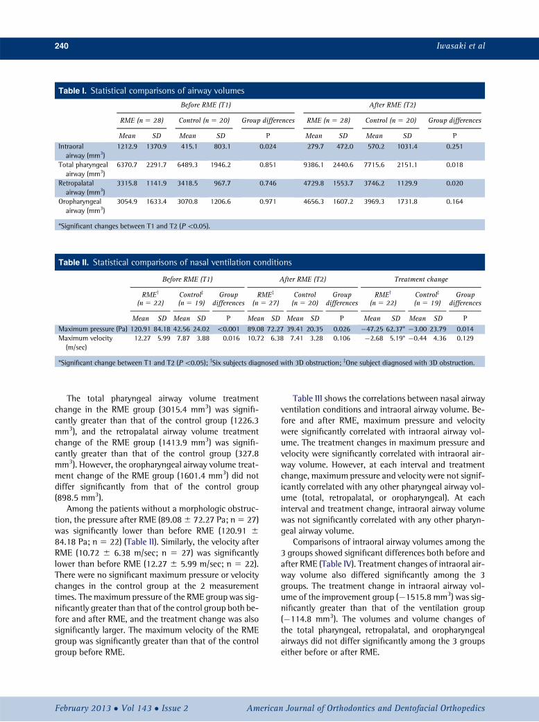
Table I. Statistical comparisons of airway volumes
Before RME (T1) After RME (T2)
RME (n 5 28) Control (n 5 20) Group differences RME (n 5 28) Control (n 5 20) Group differences
Mean SD Mean SD P Mean SD Mean SD PIntraoralairway (mm3)
1212.9 1370.9 415.1 803.1 0.024 279.7 472.0 570.2 1031.4 0.251
Total pharyngealairway (mm3)
6370.7 2291.7 6489.3 1946.2 0.851 9386.1 2440.6 7715.6 2151.1 0.018
Retropalatalairway (mm3)
3315.8 1141.9 3418.5 967.7 0.746 4729.8 1553.7 3746.2 1129.9 0.020
Oropharyngealairway (mm3)
3054.9 1633.4 3070.8 1206.6 0.971 4656.3 1607.2 3969.3 1731.8 0.164
*Significant changes between T1 and T2 (P\0.05).
Table II. Statistical comparisons of nasal ventilation conditions
Before RME (T1) After RME (T2) Treatment change
RMEy
(n 5 22)Controlz
(n 5 19)Group
differencesRMEz
(n 5 27)Control(n 5 20)
Groupdifferences
RMEy
(n 5 22)Controlz
(n 5 19)Group
differences
Mean SD Mean SD P Mean SD Mean SD P Mean SD Mean SD PMaximum pressure (Pa) 120.91 84.18 42.56 24.02 \0.001 89.08 72.27 39.41 20.35 0.026 �47.25 62.37* �3.00 23.79 0.014Maximum velocity(m/sec)
12.27 5.99 7.87 3.88 0.016 10.72 6.38 7.41 3.28 0.106 �2.68 5.19* �0.44 4.36 0.129
*Significant change between T1 and T2 (P\0.05); ySix subjects diagnosed with 3D obstruction; zOne subject diagnosed with 3D obstruction.
240 Iwasaki et al
The total pharyngeal airway volume treatmentchange in the RME group (3015.4 mm3) was signifi-cantly greater than that of the control group (1226.3mm3), and the retropalatal airway volume treatmentchange of the RME group (1413.9 mm3) was signifi-cantly greater than that of the control group (327.8mm3). However, the oropharyngeal airway volume treat-ment change of the RME group (1601.4 mm3) did notdiffer significantly from that of the control group(898.5 mm3).
Among the patients without a morphologic obstruc-tion, the pressure after RME (89.086 72.27 Pa; n5 27)was significantly lower than before RME (120.91 684.18 Pa; n 5 22) (Table II). Similarly, the velocity afterRME (10.72 6 6.38 m/sec; n 5 27) was significantlylower than before RME (12.27 6 5.99 m/sec; n 5 22).There were no significant maximum pressure or velocitychanges in the control group at the 2 measurementtimes. The maximum pressure of the RME group was sig-nificantly greater than that of the control group both be-fore and after RME, and the treatment change was alsosignificantly larger. The maximum velocity of the RMEgroup was significantly greater than that of the controlgroup before RME.
February 2013 � Vol 143 � Issue 2 American
Table III shows the correlations between nasal airwayventilation conditions and intraoral airway volume. Be-fore and after RME, maximum pressure and velocitywere significantly correlated with intraoral airway vol-ume. The treatment changes in maximum pressure andvelocity were significantly correlated with intraoral air-way volume. However, at each interval and treatmentchange, maximum pressure and velocity were not signif-icantly correlated with any other pharyngeal airway vol-ume (total, retropalatal, or oropharyngeal). At eachinterval and treatment change, intraoral airway volumewas not significantly correlated with any other pharyn-geal airway volume.
Comparisons of intraoral airway volumes among the3 groups showed significant differences both before andafter RME (Table IV). Treatment changes of intraoral air-way volume also differed significantly among the 3groups. The treatment change in intraoral airway vol-ume of the improvement group (�1515.8 mm3) was sig-nificantly greater than that of the ventilation group(�114.8 mm3). The volumes and volume changes ofthe total pharyngeal, retropalatal, and oropharyngealairways did not differ significantly among the 3 groupseither before or after RME.
Journal of Orthodontics and Dentofacial Orthopedics

Treatment change
RME (n 5 28) Control (n 5 20) Group differences
Mean SD Mean SD P�933.3 1308.8* 155.1 1096.7 0.004
3015.4 1297.6* 1226.3 1782.5* \0.001
1413.9 1172.0* 327.8 958.4 0.001
1601.4 1459.9* 898.5 1335.9* 0.095
Table I. (Continued)
Table III. Spearman rank correlation coefficients and P values (in parentheses) between intraoral airway volume andventilation condition
Before RME After RME Treatment change
Maximumpressure
Maximumvelocity
Maximumpressure
Maximumvelocity
Maximumpressure
Maximumvelocity
Intraoral airway volume before RME(n 5 22)y
0.617 (0.002)* 0.630 (0.002)* – – – –
Intraoral airway volume after RME(n 5 27)z
– – 0.473 (0.013)* 0.518 (0.006)* – –
Intraoral airway volume treatment change(n 5 22)y
– – – – 0.599 (0.003)* 0.520 (0.013)*
*Statistically significant at P\0.05; ySix subjects had 3D obstruction; zOne subject had 3D obstruction.
Iwasaki et al 241
DISCUSSION
The main purpose of this study was to clarify the ef-fect of RME on changes of tongue posture and pharyn-geal airway volume in children with nasal obstruction.Improvement of nasal airway ventilation6,29-31 andincreases in the volume of the bone and soft tissues ofthe palate have been reported as secondary effects ofRME.32 However, the effect of RME on tongue postureand pharyngeal airway volume and their associationwith improved nasal airway ventilation have not beenfirmly established. So, this study used 3D computed to-mography and computed fluid dynamics to clarify thechange of tongue posture by RME.
The purpose of our study was to clarify the relation-ship between nasal airway ventilation condition andtongue posture. Previous studies have evaluated tongueposture, hyoid posture, and tongue height by usingcephalograms33,34 and reported changes of the hyoiddistance from �0.4 to �1.9 mm after RME.13,35,36
However, in our study, the relative lingual position ofthe palate was used to evaluate tongue posture. Ozbeket al13 cephalometrically evaluated the relative tongueposture for the palate as 8 different tongue-to-
American Journal of Orthodontics and Dentofacial Orthoped
palatine bone distances. However, in subjects witha low tongue posture, variability in the shape of the dor-sum of the tongue and dehiscence of the palate make itdifficult to quantitatively evaluate tongue posture from2-dimensional cephalograms (Fig 5). Therefore, we mea-sured the intraoral airway to estimate tongue posturerelative to the palate as an indirect evaluation methodof low tongue posture. Our intraoral airway measure-ment expresses the actual volume between the palatalmucosa and the dorsum of the tongue. When the tonguecontacts the palate without a gap, this value becomeszero, indicating that tongue posture is not low. Also,this method can evaluate the degree of the low tongueposture without being affected by various forms of thepalate and the tongue. Therefore, we believe that ourmethod (intraoral airway) is better able to evaluate lowtongue posture.23
In our previous study, the intraoral airway volume ofsimilarly aged children with normal occlusion was 702.06 289.2 mm3.23 Because the intraoral airway volume ofour RME group before treatment was 1212.9 6 1370.9mm3, we considered those subjects to have low tongueposture (Table I). After RME, the intraoral airway volume
ics February 2013 � Vol 143 � Issue 2
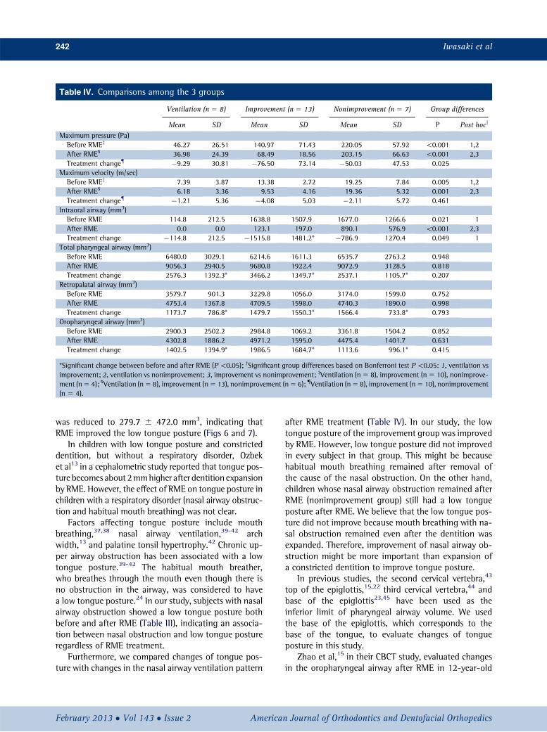
Table IV. Comparisons among the 3 groups
Ventilation (n 5 8) Improvement (n 5 13) Nonimprovement (n 5 7) Group differences
Mean SD Mean SD Mean SD P Post hocy
Maximum pressure (Pa)Before RMEz 46.27 26.51 140.97 71.43 220.05 57.92 \0.001 1,2After RME§ 36.98 24.39 68.49 18.56 203.15 66.63 \0.001 2,3Treatment change{ �9.29 30.81 �76.50 73.14 �50.03 47.53 0.025
Maximum velocity (m/sec)Before RMEz 7.39 3.87 13.38 2.72 19.25 7.84 0.005 1,2After RME§ 6.18 3.36 9.53 4.16 19.36 5.32 0.001 2,3Treatment change{ �1.21 5.36 �4.08 5.03 �2.11 5.72 0.461
Intraoral airway (mm3)Before RME 114.8 212.5 1638.8 1507.9 1677.0 1266.6 0.021 1After RME 0.0 0.0 123.1 197.0 890.1 576.9 \0.001 2,3Treatment change �114.8 212.5 �1515.8 1481.2* �786.9 1270.4 0.049 1
Total pharyngeal airway (mm3)Before RME 6480.0 3029.1 6214.6 1611.3 6535.7 2763.2 0.948After RME 9056.3 2940.5 9680.8 1922.4 9072.9 3128.5 0.818Treatment change 2576.3 1392.3* 3466.2 1349.7* 2537.1 1105.7* 0.207
Retropalatal airway (mm3)Before RME 3579.7 901.3 3229.8 1056.0 3174.0 1599.0 0.752After RME 4753.4 1367.8 4709.5 1598.0 4740.3 1890.0 0.998Treatment change 1173.7 786.8* 1479.7 1550.3* 1566.4 733.8* 0.793
Oropharyngeal airway (mm3)Before RME 2900.3 2502.2 2984.8 1069.2 3361.8 1504.2 0.852After RME 4302.8 1886.2 4971.2 1595.0 4475.4 1401.7 0.631Treatment change 1402.5 1394.9* 1986.5 1684.7* 1113.6 996.1* 0.415
*Significant change between before and after RME (P\0.05); ySignificant group differences based on Bonferroni test P\0.05: 1, ventilation vsimprovement; 2, ventilation vs nonimprovement; 3, improvement vs nonimprovement; zVentilation (n5 8), improvement (n5 10), nonimprove-ment (n5 4); §Ventilation (n5 8), improvement (n5 13), nonimprovement (n5 6); {Ventilation (n5 8), improvement (n5 10), nonimprovement(n 5 4).
242 Iwasaki et al
was reduced to 279.7 6 472.0 mm3, indicating thatRME improved the low tongue posture (Figs 6 and 7).
In children with low tongue posture and constricteddentition, but without a respiratory disorder, Ozbeket al13 in a cephalometric study reported that tongue pos-ture becomes about 2mmhigher after dentition expansionby RME. However, the effect of RME on tongue posture inchildren with a respiratory disorder (nasal airway obstruc-tion and habitual mouth breathing) was not clear.
Factors affecting tongue posture include mouthbreathing,37,38 nasal airway ventilation,39-42 archwidth,13 and palatine tonsil hypertrophy.42 Chronic up-per airway obstruction has been associated with a lowtongue posture.39-42 The habitual mouth breather,who breathes through the mouth even though there isno obstruction in the airway, was considered to havea low tongue posture.24 In our study, subjects with nasalairway obstruction showed a low tongue posture bothbefore and after RME (Table III), indicating an associa-tion between nasal obstruction and low tongue postureregardless of RME treatment.
Furthermore, we compared changes of tongue pos-ture with changes in the nasal airway ventilation pattern
February 2013 � Vol 143 � Issue 2 American
after RME treatment (Table IV). In our study, the lowtongue posture of the improvement group was improvedby RME. However, low tongue posture did not improvedin every subject in that group. This might be becausehabitual mouth breathing remained after removal ofthe cause of the nasal obstruction. On the other hand,children whose nasal airway obstruction remained afterRME (nonimprovement group) still had a low tongueposture after RME. We believe that the low tongue pos-ture did not improve because mouth breathing with na-sal obstruction remained even after the dentition wasexpanded. Therefore, improvement of nasal airway ob-struction might be more important than expansion ofa constricted dentition to improve tongue posture.
In previous studies, the second cervical vertebra,43
top of the epiglottis,15,22 third cervical vertebra,44 andbase of the epiglottis23,45 have been used as theinferior limit of pharyngeal airway volume. We usedthe base of the epiglottis, which corresponds to thebase of the tongue, to evaluate changes of tongueposture in this study.
Zhao et al,15 in their CBCT study, evaluated changesin the oropharyngeal airway after RME in 12-year-old
Journal of Orthodontics and Dentofacial Orthopedics
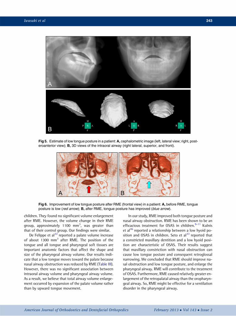
Fig 5. Estimate of low tongue posture in a patient:A, cephalometric image (left, lateral view; right, post-eroanterior view); B, 3D views of the intraoral airway (right lateral, superior, and front).
Fig 6. Improvement of low tongue posture after RME (frontal view) in a patient: A, before RME, tongueposture is low (red arrow); B, after RME, tongue posture has improved (blue arrow).
Iwasaki et al 243
children. They found no significant volume enlargementafter RME. However, the volume change in their RMEgroup, approximately 1100 mm3, was greater thanthat of their control group. Our findings were similar.
De Felippe et al32 reported a palate volume increaseof about 1300 mm3 after RME. The position of thetongue and all tongue and pharyngeal soft tissues areimportant anatomic factors that affect the shape andsize of the pharyngeal airway volume. Our results indi-cate that a low tongue moves toward the palate becausenasal airway obstruction was reduced by RME (Table III).However, there was no significant association betweenintraoral airway volume and pharyngeal airway volume.As a result, we believe that total airway volume enlarge-ment occurred by expansion of the palate volume ratherthan by upward tongue movement.
American Journal of Orthodontics and Dentofacial Orthoped
In our study, RME improved both tongue posture andnasal airway obstruction. RME has been shown to be anefficacious treatment for OSAS in children.9-11 Kulniset al46 reported a relationship between a low hyoid po-sition and OSAS in children. Seto et al33 reported thata constricted maxillary dentition and a low hyoid posi-tion are characteristic of OSAS. Their results suggestthat maxillary constriction with nasal obstruction cancause low tongue posture and consequent retroglossalnarrowing. We concluded that RME should improve na-sal obstruction and low tongue posture, and enlarge thepharyngeal airway. RME will contribute to the treatmentof OSAS. Furthermore, RME caused relatively greater en-largement of the retropalatal airway than the oropharyn-geal airway. So, RME might be effective for a ventilationdisorder in the pharyngeal airway.
ics February 2013 � Vol 143 � Issue 2

Fig 7. Improvement of low tongue posture and enlargement of the pharyngeal airway after RME in a pa-tient: A, before RME, tongue posture is low (red arrow), and the oropharyngeal airway is narrow; B, afterRME, tongueposture has improved (yellowarrow), and the pharyngeal airway hasenlarged (bluearrows).
244 Iwasaki et al
Because our study was retrospective, it was limited tochildren without adenoids or hyperplasia of the palatinetonsils.42 Adenoids and hyperplasia of the palatine ton-sils are common in pediatric OSAS. Therefore, data indi-cating airway enlargement by RME of children withthese problems are still required. Future studies shouldalso take the computed tomography data in the supineposition during sleep to match the usual clinical exami-nation. Furthermore, a study evaluating actual respira-tory status is required in the future.
CONCLUSIONS
We comprehensively examined the effect of RME onnasal airway ventilation condition, tongue posture, andpharyngeal airway volume.
Children with nasal airway obstruction have a lowtongue posture regardless of RME treatment.
Improvement of the nasal airway ventilation condi-tion by RME is associated with improved low tongueposture.
RME enlarges the pharyngeal airway both with andwithout improvement in nasal obstruction.
We thank Gaylord Throckmorton for reviewing thisarticle for English usage.
REFERENCES
1. Mattar SE, Anselmo-Lima WT, Valera FC, Matsumoto MA. Skeletaland occlusal characteristics in mouth-breathing pre-school chil-dren. J Clin Pediatr Dent 2004;28:315-8.
2. Cross DL, McDonald JP. Effect of rapid maxillary expansion onskeletal, dental, and nasal structures: a postero-anterior cephalo-metric study. Eur J Orthod 2000;22:519-28.
3. Basciftci FA, Mutlu N, Karaman AI, Malkoc S, Kucukkolbasi H.Does the timing and method of rapid maxillary expansion havean effect on the changes in nasal dimensions? Angle Orthod2002;72:118-23.
February 2013 � Vol 143 � Issue 2 American
4. Chung CH, Font B. Skeletal and dental changes in the sagittal, ver-tical, and transverse dimensions after rapid palatal expansion. AmJ Orthod Dentofacial Orthop 2004;126:569-75.
5. Haralambidis A, Ari-Demirkaya A, Acar A, Kucukkeles N, Ates M,Ozkaya S. Morphologic changes of the nasal cavity induced byrapid maxillary expansion: a study on 3-dimensional computed to-mography models. Am J Orthod Dentofacial Orthop 2009;136:815-21.
6. Iwasaki T, Saitoh I, Takemoto Y, Inada E, Kanomi R, Hayasaki H,et al. Improvement of nasal airway ventilation after rapid maxillaryexpansion evaluated with computational fluid dynamics. Am J Or-thod Dentofacial Orthop 2012;141:269-78.
7. Compadretti GC, Tasca I, Bonetti GA. Nasal airway measurementsin children treated by rapid maxillary expansion. Am J Rhinol2006;20:385-93.
8. Gray LP. Results of 310 cases of rapid maxillary expansion selectedfor medical reasons. J Laryngol Otol 1975;89:601-14.
9. Cistulli PA, Palmisano RG, Poole MD. Treatment of obstructive sleepapnea syndromeby rapidmaxillary expansion. Sleep1998;21:831-5.
10. Pirelli P, Saponara M, Guilleminault C. Rapid maxillary expansionin children with obstructive sleep apnea syndrome. Sleep 2004;27:761-6.
11. Villa MP, Malagola C, Pagani J, Montesano M, Rizzoli A,Guilleminault C, et al. Rapid maxillary expansion in children withobstructive sleep apnea syndrome: 12-month follow-up. SleepMed 2007;8:128-34.
12. Guilleminault C, Lee JH, Chan A. Pediatric obstructive sleep apneasyndrome. Arch Pediatr Adolesc Med 2005;159:775-85.
13. Ozbek MM, Memikoglu UT, Altug-Atac AT, Lowe AA. Stability ofmaxillary expansion and tongue posture. Angle Orthod 2009;79:214-20.
14. Malkoc S, Usumez S, Iseri H. Long-term effects of symphyseal dis-traction and rapid maxillary expansion on pharyngeal airway di-mensions, tongue, and hyoid position. Am J Orthod DentofacialOrthop 2007;132:769-75.
15. Zhao Y, Nguyen M, Gohl E, Mah JK, Sameshima G, Enciso R. Oro-pharyngeal airway changes after rapid palatal expansion evaluatedwith cone-beam computed tomography. Am J Orthod DentofacialOrthop 2010;137(Supp):S71-8.
16. Muto T, Takeda S, Kanazawa M, Yamazaki A, Fujiwara Y,Mizoguchi I. The effect of head posture on the pharyngeal airwayspace (PAS). Int J Oral Maxillofac Surg 2002;31:579-83.
Journal of Orthodontics and Dentofacial Orthopedics

Iwasaki et al 245
17. Lowe AA, Fleetham JA, Adachi S, Ryan CF. Cephalometricand computed tomographic predictors of obstructive sleepapnea severity. Am J Orthod Dentofacial Orthop 1995;107:589-95.
18. Solow B, Sonnesen L. Head posture and malocclusions. Eur J Or-thod 1998;20:685-93.
19. Anegawa E, Tsuyama H, Kusukawa J. Lateral cephalometric anal-ysis of the pharyngeal airway space affected by head posture. Int JOral Maxillofac Surg 2008;37:805-9.
20. Li HY, Chen NH,Wang CR, Shu YH,Wang PC. Use of 3-dimensionalcomputed tomography scan to evaluate upper airway patency forpatients undergoing sleep-disordered breathing surgery. Otolar-yngol Head Neck Surg 2003;129:336-42.
21. Tso HH, Lee JS, Huang JC, Maki K, Hatcher D, Miller AJ. Evaluationof the human airway using cone-beam computerized tomography.Oral Surg Oral Med Oral Pathol Oral Radiol Endod 2009;108:768-76.
22. Kim YJ, Hong JS, Hwang YI, Park YH. Three-dimensional analysisof pharyngeal airway in preadolescent children with different an-teroposterior skeletal patterns. Am J Orthod Dentofacial Orthop2010;137:306.e301-11; discussion 306-7.
23. Iwasaki T, Hayasaki H, Takemoto Y, Kanomi R, Yamasaki Y. Oro-pharyngeal airway in children with Class III malocclusion evaluatedby cone-beam computed tomography. Am J Orthod DentofacialOrthop 2009;136:318.e311-9; discussion 318-9.
24. Iwasaki T, Saitoh I, Takemoto Y, Inada E, Kanomi R, Hayasaki H,et al. Evaluation of upper airway obstruction in Class II childrenwith fluid-mechanical simulation. Am J Orthod Dentofacial Orthop2011;139:e135-45.
25. De Backer JW, Vanderveken OM, Vos WG, Devolder A, Verhulst SL,Verbraecken JA, et al. Functional imaging using computationalfluid dynamics to predict treatment success of mandibular ad-vancement devices in sleep-disordered breathing. J Biomech2007;40:3708-14.
26. Gami~no B, Aguill�on J. Numerical simulation of syngas combustionwith a multi-spark ignition system in a diesel engine adapted towork at the Otto cycle. Fuel 2010;89:581-91.
27. Xu C, Sin S, McDonough JM, Udupa JK, Guez A, Arens R, et al.Computational fluid dynamics modeling of the upper airway ofchildren with obstructive sleep apnea syndrome in steady flow. JBiomech 2006;39:2043-54.
28. Dahlberg G. Statistical methods for medical and biological stu-dents. New York: Interscience Publications; 1940.
29. Wertz RA. Changes in nasal airflow incident to rapid maxillary ex-pansion. Angle Orthod 1968;38:1-11.
30. Hershey HG, Stewart BL, Warren DW. Changes in nasal airway re-sistance associated with rapid maxillary expansion. Am J Orthod1976;69:274-84.
American Journal of Orthodontics and Dentofacial Orthoped
31. Hartgerink DV, Vig PS, Abbott DW. The effect of rapid maxillaryexpansion on nasal airway resistance. Am J Orthod Dentofacial Or-thop 1987;92:381-9.
32. De Felippe NL, Bhushan N, Da Silveira AC, Viana G, Smith B. Long-term effects of orthodontic therapy on the maxillary dental archand nasal cavity. Am J Orthod Dentofacial Orthop 2009;136:490.e491-8; discussion 490-1.
33. Seto BH, Gotsopoulos H, Sims MR, Cistulli PA. Maxillary morphol-ogy in obstructive sleep apnoea syndrome. Eur J Orthod 2001;23:703-14.
34. Malkoc S, Usumez S, Nur M, Donaghy CE. Reproducibility of air-way dimensions and tongue and hyoid positions on lateral cepha-lograms. Am J Orthod Dentofacial Orthop 2005;128:513-6.
35. Phoenix A, Valiathan M, Nelson S, Strohl KP, Hans M. Changes inhyoid bone position following rapid maxillary expansion in adoles-cents. Angle Orthod 2011;81:632-8.
36. Schutz TC, Dominguez GC, Hallinan MP, Cunha TC, Tufik S. ClassII correction improves nocturnal breathing in adolescents. AngleOrthod 2011;81:222-8.
37. Principato JJ. Upper airway obstruction and craniofacial morphol-ogy. Otolaryngol Head Neck Surg 1991;104:881-90.
38. Harari D, RedlichM,Miri S, Hamud T, GrossM. The effect of mouthbreathing versus nasal breathing on dentofacial and craniofacialdevelopment in orthodontic patients. Laryngoscope 2010;120:2089-93.
39. Harvold EP, Tomer BS, Vargervik K, Chierici G. Primate experi-ments on oral respiration. Am J Orthod 1981;79:359-72.
40. Vargervik K, Harvold EP. Experiments on the interaction between or-ofacial function andmorphology. Ear Nose Throat J 1987;66:201-8.
41. Behlfelt K. Enlarged tonsils and the effect of tonsillectomy. Char-acteristics of the dentition and facial skeleton. Posture of the head,hyoid bone and tongue. Mode of breathing. Swed Dent J Suppl1990;72:1-35.
42. Behlfelt K, Linder-Aronson S, Neander P. Posture of the head, thehyoid bone, and the tongue in children with and without enlargedtonsils. Eur J Orthod 1990;12:458-67.
43. El H, Palomo JM. Measuring the airway in 3 dimensions: a reliabil-ity and accuracy study. Am J Orthod Dentofacial Orthop 2010;137(Supp):S50.e51-9; discussion S50-2.
44. Grauer D, Cevidanes LS, Styner MA, Ackerman JL, Proffit WR. Pha-ryngeal airway volume and shape from cone-beam computed to-mography: relationship to facial morphology. Am J OrthodDentofacial Orthop 2009;136:805-14.
45. Abramson ZR, Susarla S, Tagoni JR, Kaban L. Three-dimensionalcomputed tomographic analysis of airway anatomy. J Oral Maxil-lofac Surg 2010;68:363-71.
46. Kulnis R, Nelson S, Strohl K, Hans M. Cephalometric assessment ofsnoring and nonsnoring children. Chest 2000;118:596-603.
ics February 2013 � Vol 143 � Issue 2