Development of the Pharyngeal Arches Part II of the Pharyngeal Arches ... lingual swelling artery...
69
Development of the Pharyngeal Arches Part II Thomas A. Marino, Ph.D. Temple University School of Medicine
Transcript of Development of the Pharyngeal Arches Part II of the Pharyngeal Arches ... lingual swelling artery...
Development of the Pharyngeal Arches
Part II
Thomas A. Marino, Ph.D.Temple University School of Medicine
Competencies: Upon completion of this section of the course, the student must be able to:
1. Recall the embryonic precursors that give rise to the adult structures of the head and neck.
2. Describe how these precursors, especially the pharyngeal arches, form the different structures in the head and neck.
3. Determine how the congenital abnormalities thyroglossal duct cysts, and cervical fistulas would occur.
4. Compare and contrast the development of the different pharyngeal pouches, clefts, arches, mesoderm, nerves, and connective tissues.
5. Use this information to figure out the cause of other congenital defects that you might see clinically.
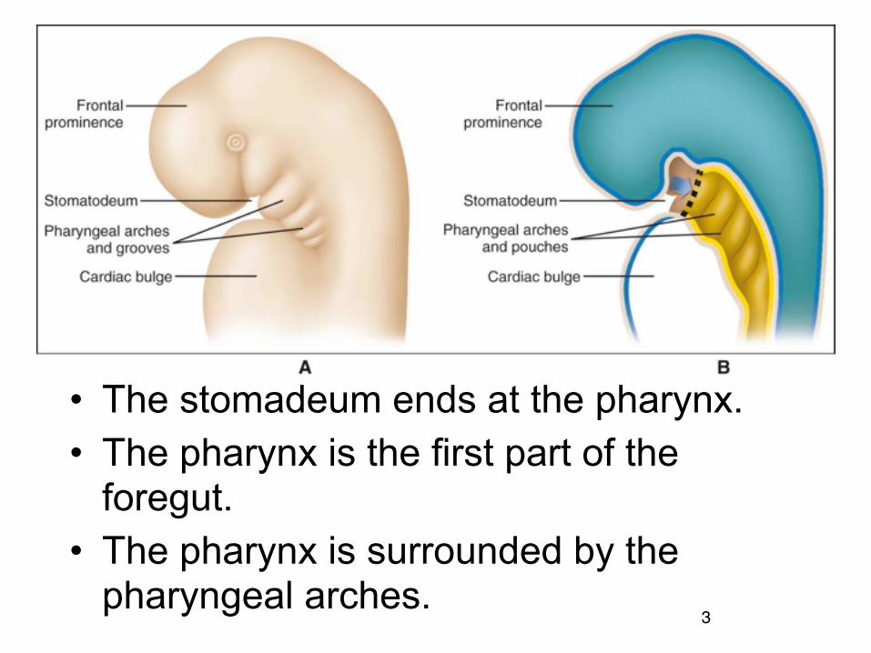
• The stomadeum ends at the pharynx. • The pharynx is the first part of the
foregut. • The pharynx is surrounded by the
pharyngeal arches.3
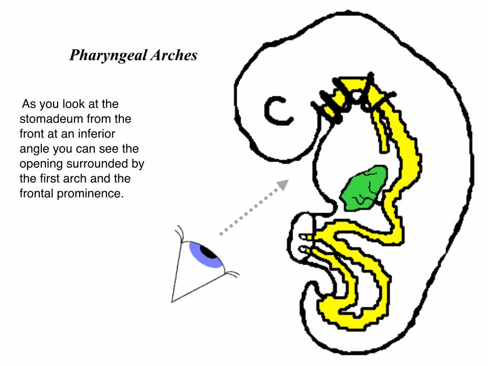
Pharyngeal Arches
As you look at the stomadeum from the front at an inferior angle you can see the opening surrounded by the first arch and the frontal prominence.
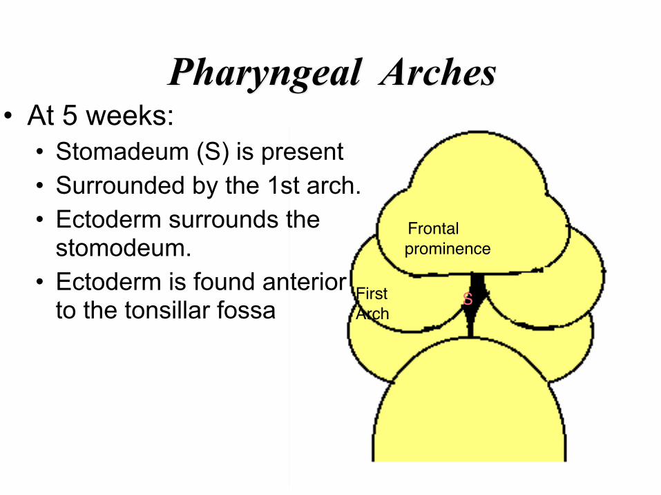
Pharyngeal Arches
Frontal prominence
• At 5 weeks: • Stomadeum (S) is present • Surrounded by the 1st arch. • Ectoderm surrounds the
stomodeum. • Ectoderm is found anterior
to the tonsillar fossa SFirst!Arch
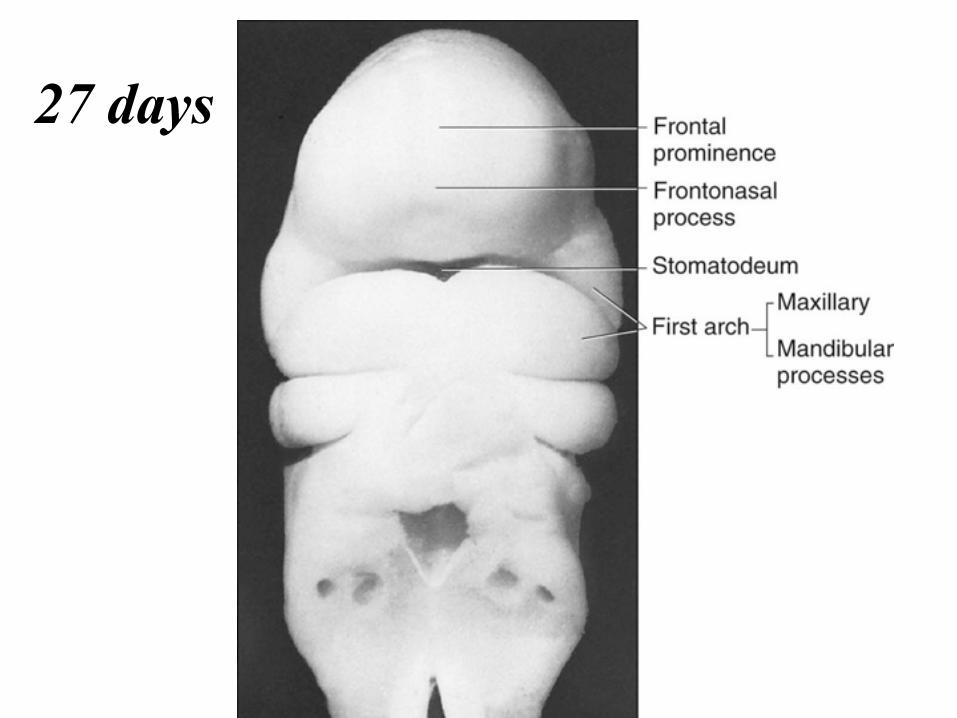
27 days
6
Neural crest cells
• migrate into: • Pharyngeal arches - from midbrain and hindbrain • Form pharyngeal arch skeletal structures
• form bones of the face and the skull • form hyoid cartilage (from 2nd and 3rd arches) • plus cartilage, bone, dentin, tendon, dermis,
meninges, sensory neurons and glandular stroma
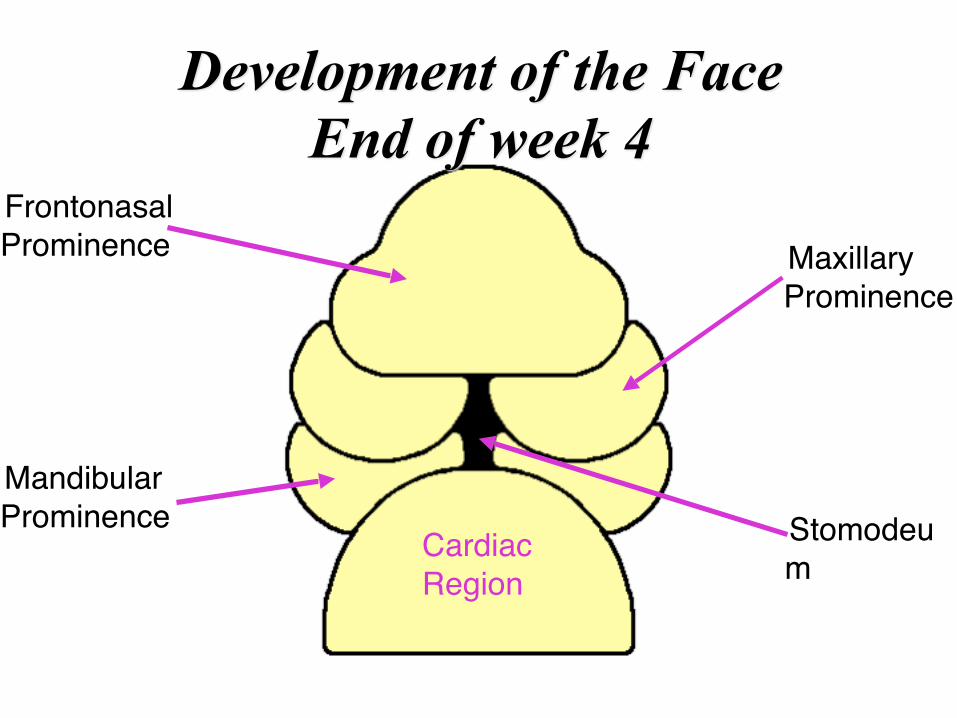
Development of the Face End of week 4
Frontonasal Prominence Maxillary
Prominence
Mandibular Prominence
Cardiac!Region
Stomodeum
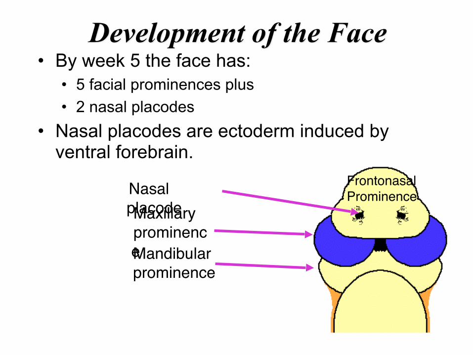
Development of the Face• By week 5 the face has:
• 5 facial prominences plus • 2 nasal placodes
• Nasal placodes are ectoderm induced by ventral forebrain.
Nasal placodeMaxillary!prominence
Frontonasal!Prominence
Mandibular!prominence
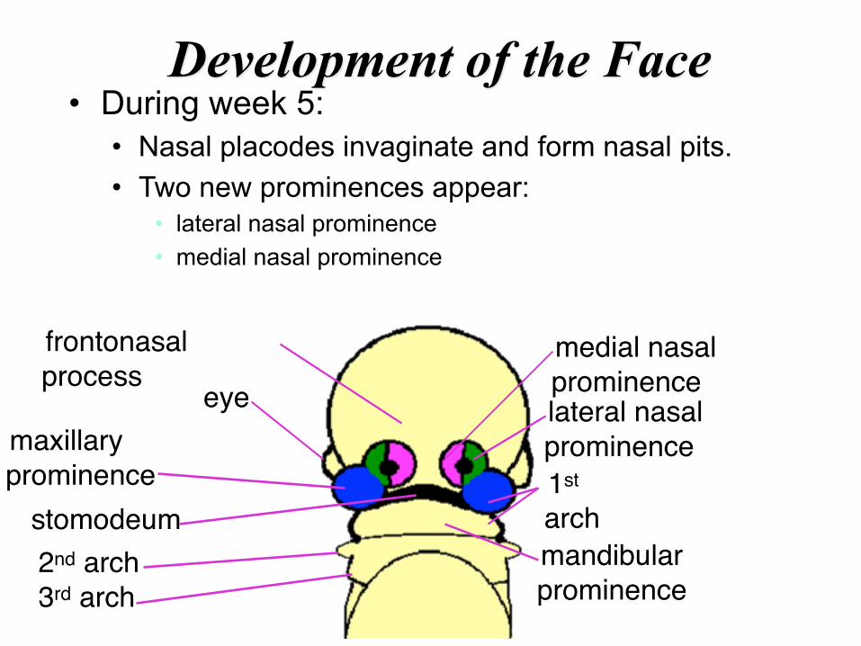
Development of the Face• During week 5:
• Nasal placodes invaginate and form nasal pits. • Two new prominences appear:
• lateral nasal prominence • medial nasal prominence
frontonasal process
eyemaxillary prominence
stomodeum2nd arch!3rd arch
medial nasal prominencelateral nasal prominence1st archmandibular prominence
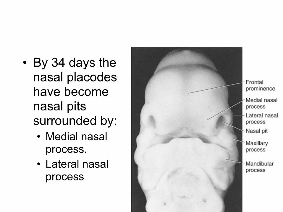
• By 34 days the nasal placodes have become nasal pits surrounded by: • Medial nasal
process. • Lateral nasal
process
11
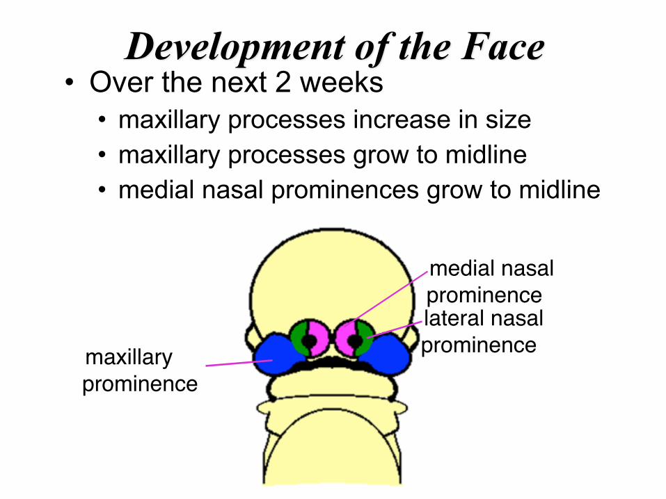
Development of the Face• Over the next 2 weeks
• maxillary processes increase in size • maxillary processes grow to midline • medial nasal prominences grow to midline
maxillary prominence
medial nasal prominencelateral nasal prominence
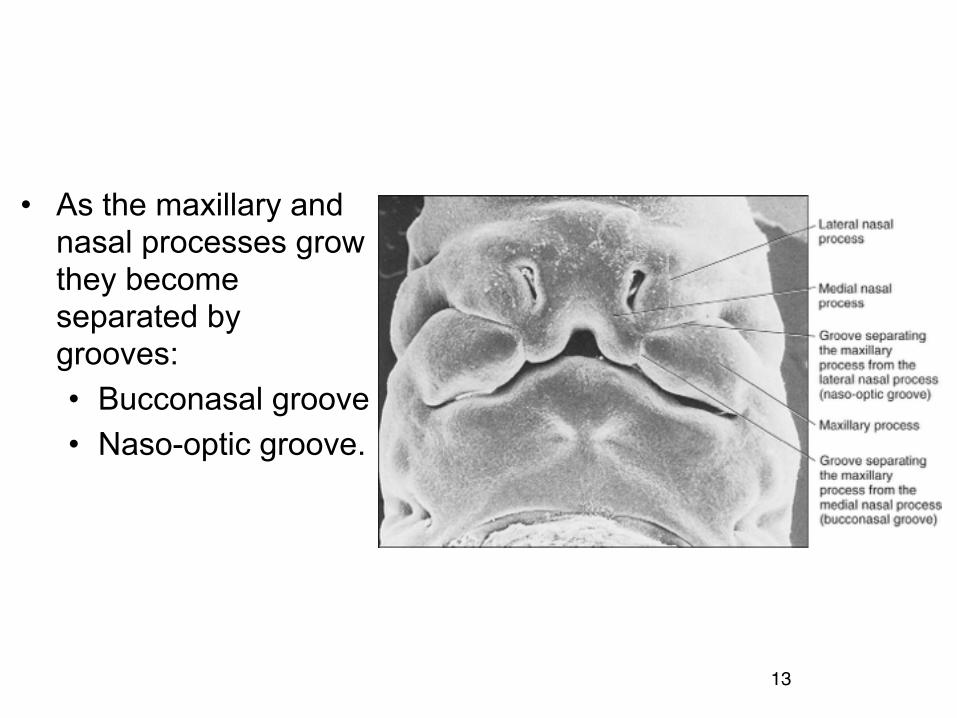
• As the maxillary and nasal processes grow they become separated by grooves: • Bucconasal groove • Naso-optic groove.
13
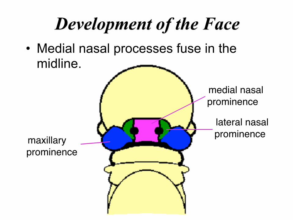
Development of the Face• Medial nasal processes fuse in the
midline.
maxillary prominence
medial nasal prominence
lateral nasal prominence
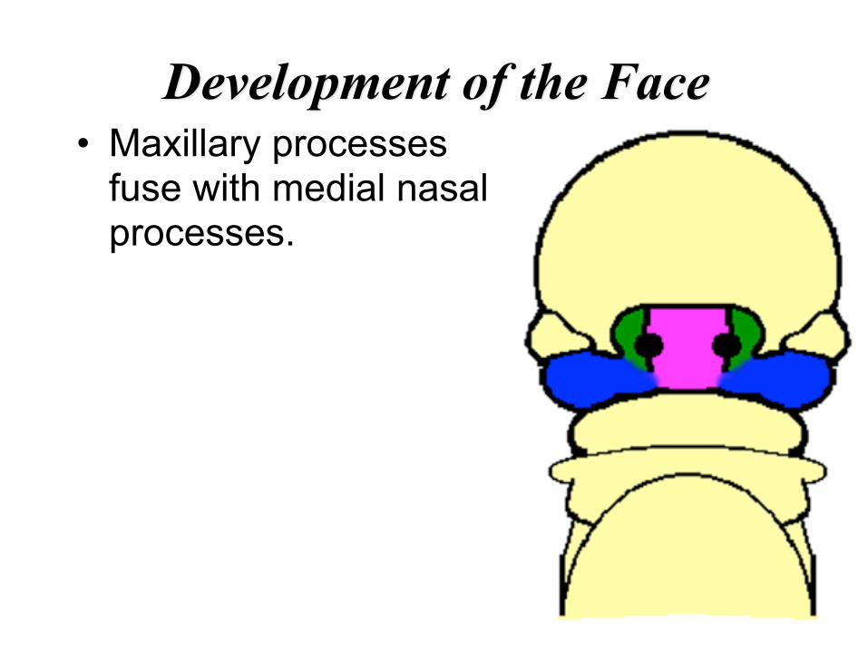
Development of the Face• Maxillary processes
fuse with medial nasal processes.
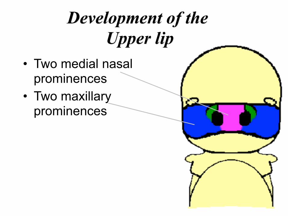
Development of the Upper lip
• Two medial nasal prominences
• Two maxillary prominences
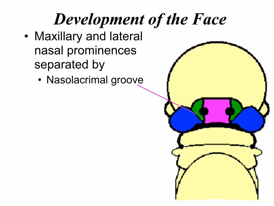
Development of the Face• Maxillary and lateral
nasal prominences separated by • Nasolacrimal groove
Development of the Face
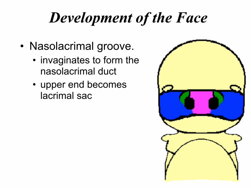
• Nasolacrimal groove. • invaginates to form the
nasolacrimal duct • upper end becomes
lacrimal sac
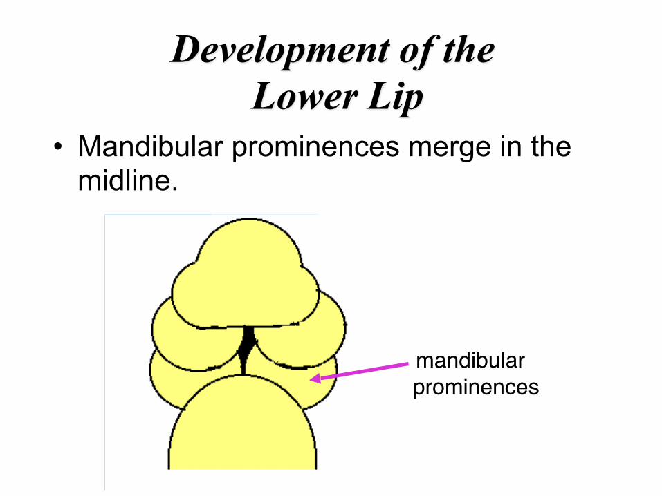
Development of the Lower Lip
• Mandibular prominences merge in the midline.
mandibular prominences
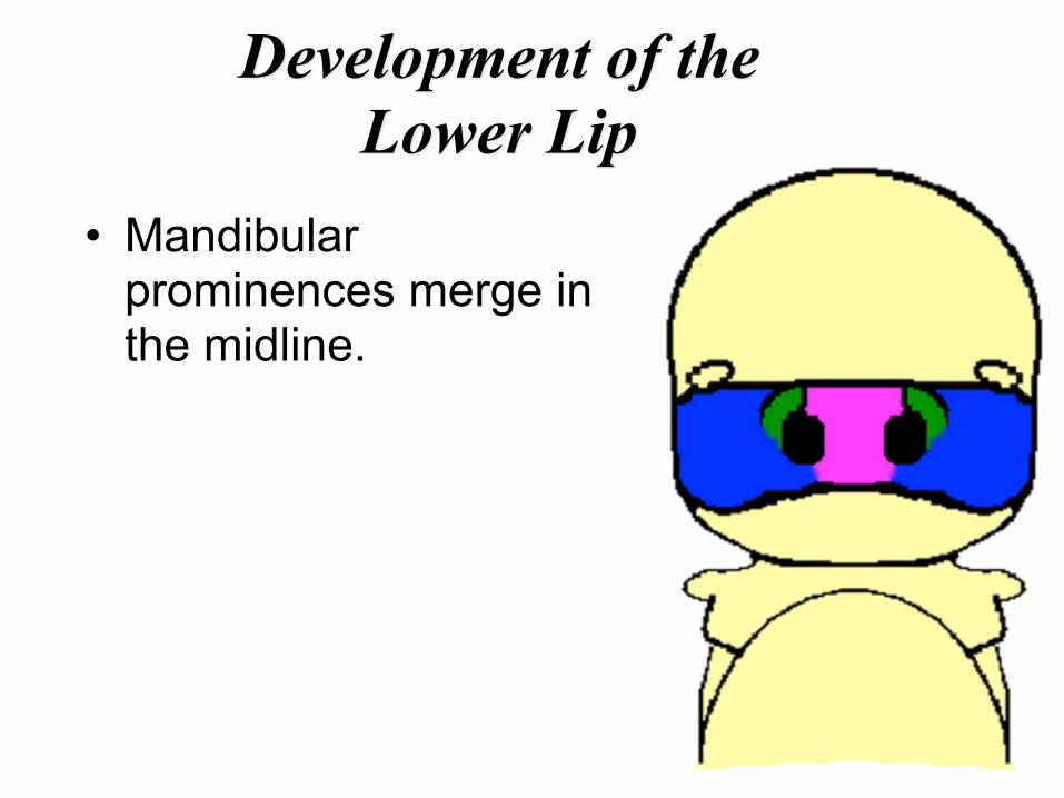
Development of the Lower Lip
• Mandibular prominences merge in the midline.
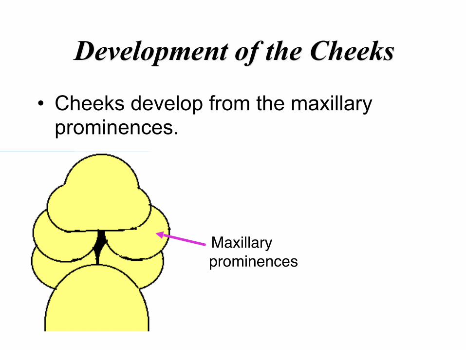
Development of the Cheeks
• Cheeks develop from the maxillary prominences.
Maxillary prominences
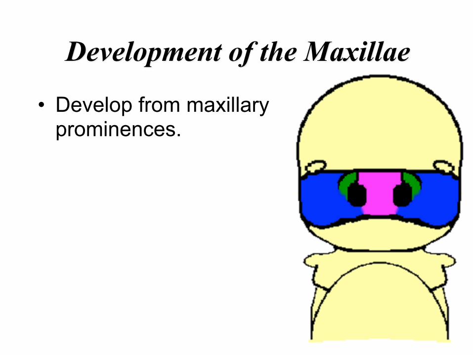
Development of the Maxillae
• Develop from maxillary prominences.
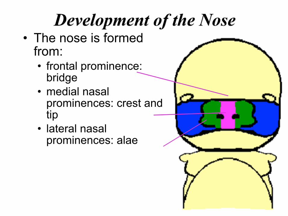
Development of the Nose• The nose is formed
from: • frontal prominence:
bridge • medial nasal
prominences: crest and tip
• lateral nasal prominences: alae
24
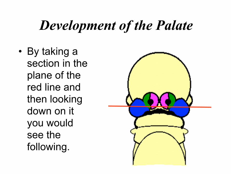
Development of the Palate
• By taking a section in the plane of the red line and then looking down on it you would see the following.
25
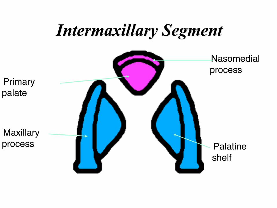
Intermaxillary SegmentNasomedial process
Primary palate
Maxillary process Palatine
shelf
Intermaxillary Segment
• 2 medial nasal prominences • labial component - philtrum of upper lip • upper jaw component - 4 incisor teeth • palatal component - primary palate • fuses with nasal septum from frontal
prominence
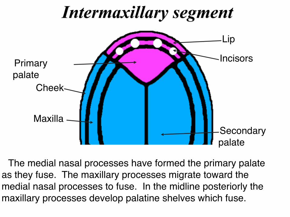
Intermaxillary segmentLip
Maxilla
Primary palate
Secondary palate
Cheek
Incisors
The medial nasal processes have formed the primary palate as they fuse. The maxillary processes migrate toward the medial nasal processes to fuse. In the midline posteriorly the maxillary processes develop palatine shelves which fuse.
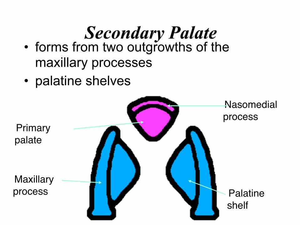
Secondary Palate• forms from two outgrowths of the
maxillary processes • palatine shelves
Nasomedial process
Primary palate
Maxillary process Palatine
shelf

30
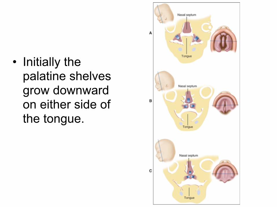
• Initially the palatine shelves grow downward on either side of the tongue.
31

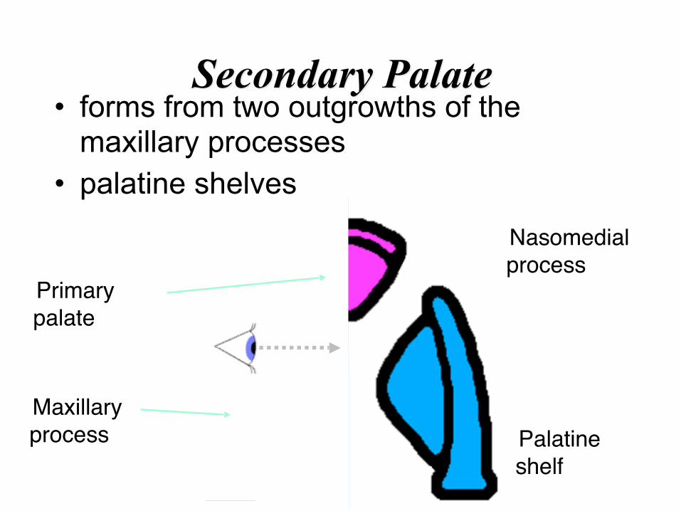
Secondary Palate• forms from two outgrowths of the
maxillary processes • palatine shelves
Nasomedial process
Primary palate
Maxillary process Palatine
shelf
Secondary Palate• forms from two outgrowths of the
maxillary processes • palatine shelves
Nasomedial process
Primary palate
Maxillary process Palatine
shelf

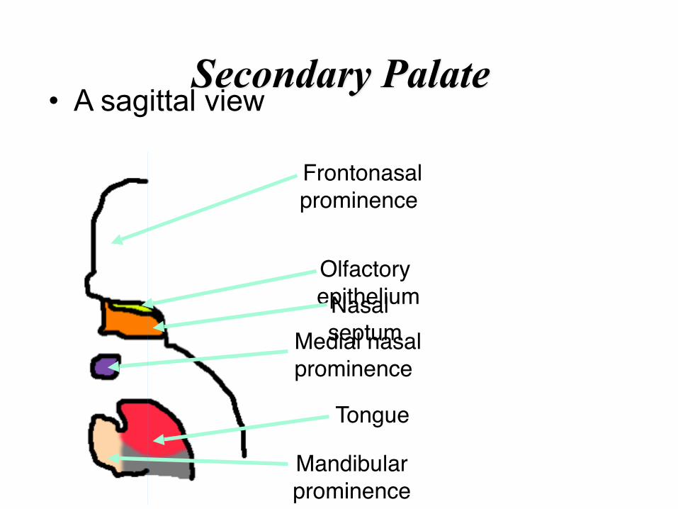
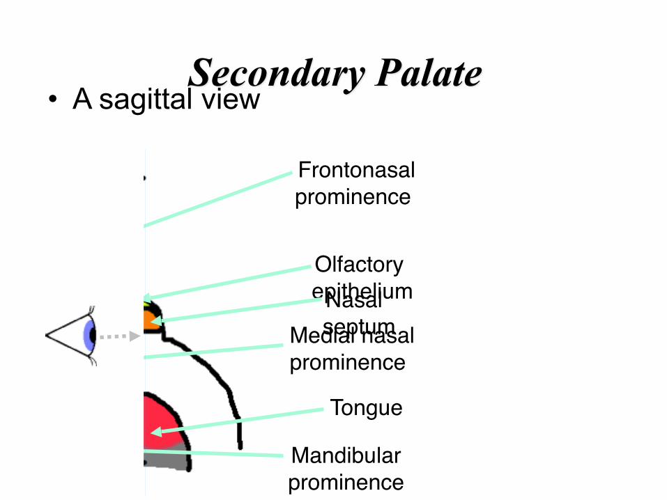
Secondary Palate• A sagittal view
Frontonasal prominence
Olfactory epitheliumNasal septumMedial nasal !
prominence
Tongue
Mandibular prominence
Secondary Palate• A sagittal view
Frontonasal prominence
Olfactory epitheliumNasal septumMedial nasal !
prominence
Tongue
Mandibular prominence
Secondary Palate• A sagittal view
Frontonasal prominence
Olfactory epitheliumNasal septumMedial nasal !
prominence
Tongue
Mandibular prominence
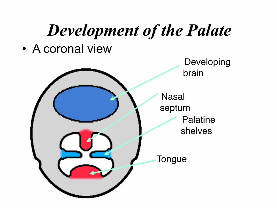
Development of the Palate• A coronal view
Developing brain
Nasal septum
Palatine shelves
Tongue
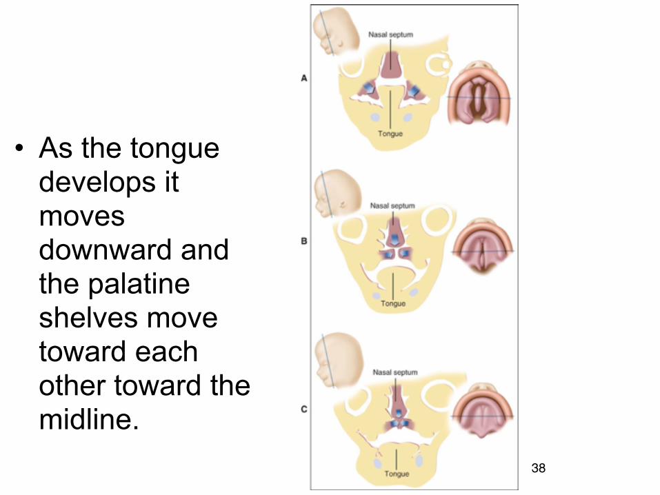
• As the tongue develops it moves downward and the palatine shelves move toward each other toward the midline.
38
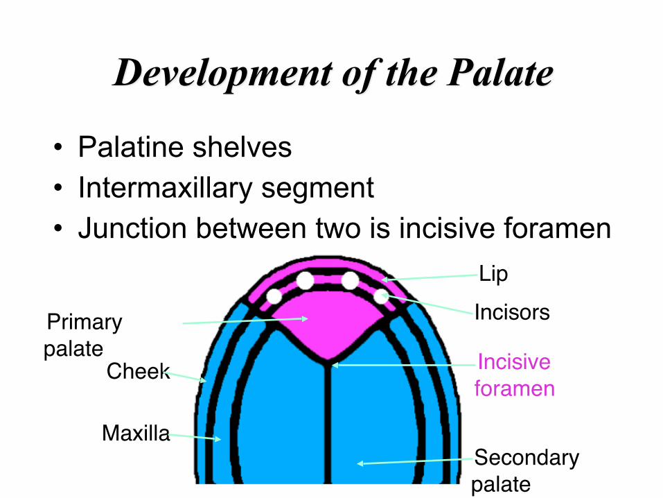
Development of the Palate
• Palatine shelves • Intermaxillary segment • Junction between two is incisive foramen
Lip
Maxilla
Primary palate
Secondary palate
Cheek
Incisors
Incisive foramen
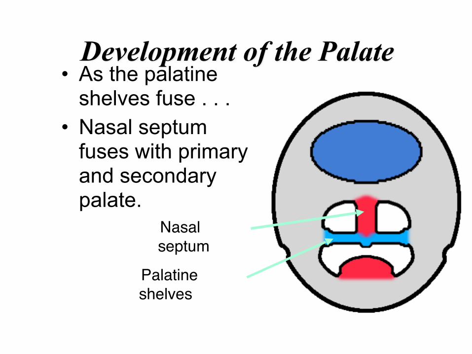
Development of the Palate• As the palatine
shelves fuse . . . • Nasal septum
fuses with primary and secondary palate.
Nasal septum
Palatine shelves
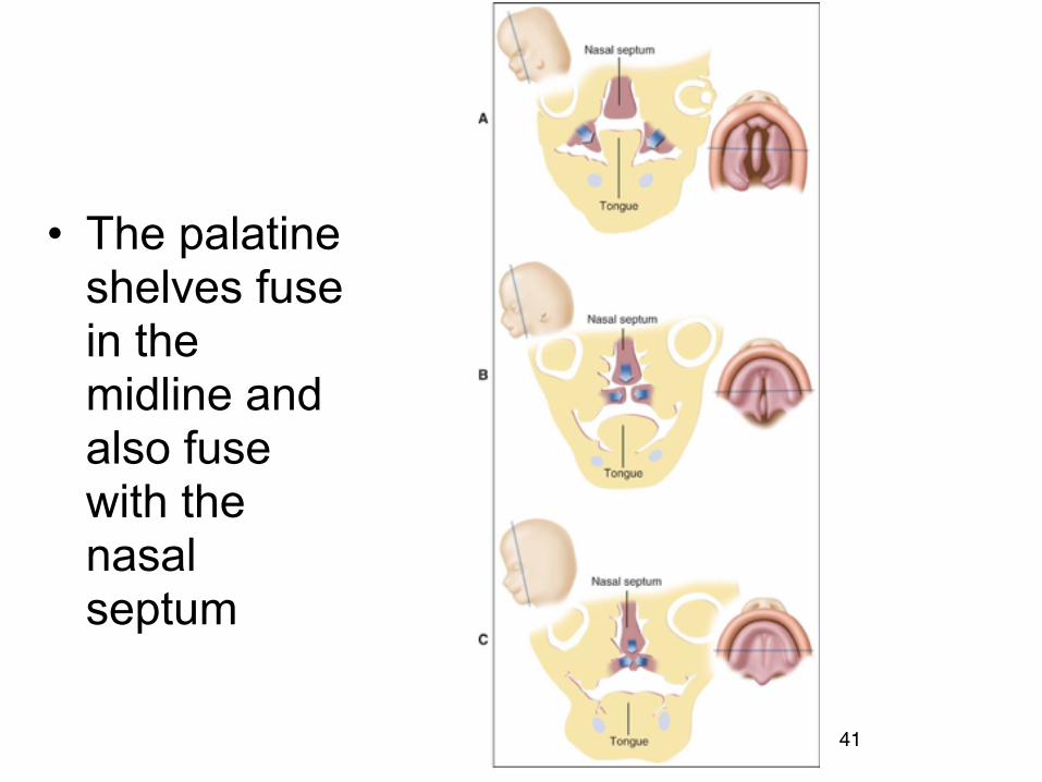
• The palatine shelves fuse in the midline and also fuse with the nasal septum
41
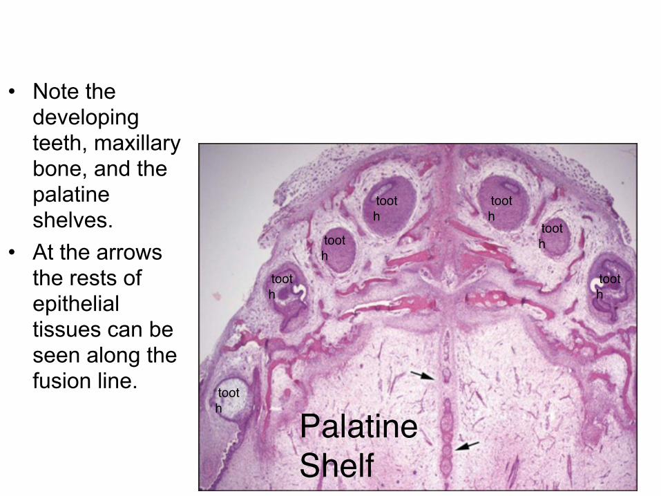
• Note the developing teeth, maxillary bone, and the palatine shelves.
• At the arrows the rests of epithelial tissues can be seen along the fusion line.
42
tooth
tooth
tooth
tooth
tooth
tooth
tooth
Palatine !Shelf
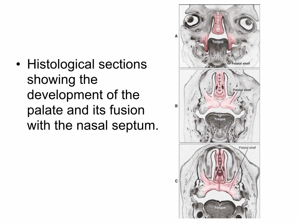
• Histological sections showing the development of the palate and its fusion with the nasal septum.
43
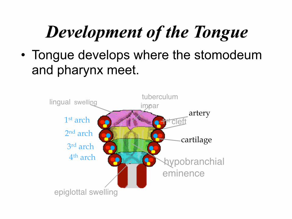
Development of the Tongue • Tongue develops where the stomodeum
and pharynx meet.
1st arch
2nd arch
3rd arch4th arch hypobranchial
eminence
epiglottal swelling
tuberculum impar
1st cleft
lingual swellingartery
cartilage
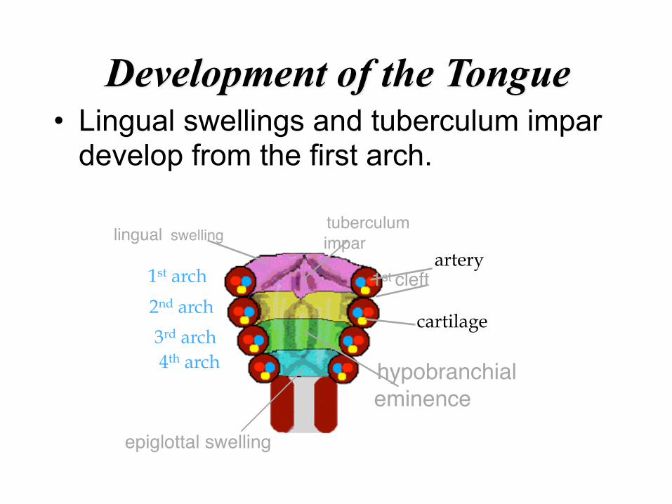
Development of the Tongue • Lingual swellings and tuberculum impar
develop from the first arch.
1st arch
2nd arch
3rd arch4th arch hypobranchial
eminence
epiglottal swelling
tuberculum impar
1st cleft
lingual swellingartery
cartilage
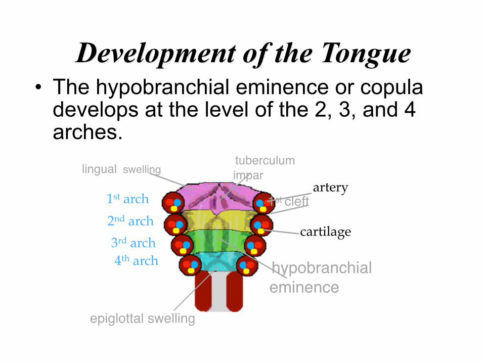
Development of the Tongue • The hypobranchial eminence or copula
develops at the level of the 2, 3, and 4 arches.
1st arch
2nd arch
3rd arch4th arch hypobranchial
eminence
epiglottal swelling
tuberculum impar
1st cleft
lingual swellingartery
cartilage
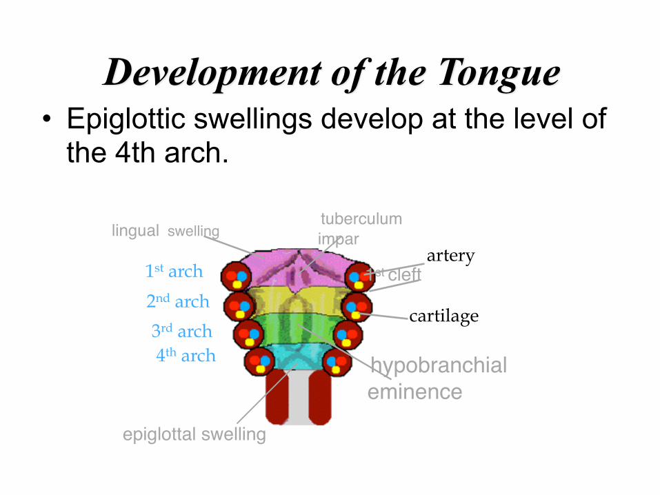
Development of the Tongue • Epiglottic swellings develop at the level of
the 4th arch.
1st arch
2nd arch
3rd arch4th arch hypobranchial
eminence
epiglottal swelling
tuberculum impar
1st cleft
lingual swellingartery
cartilage
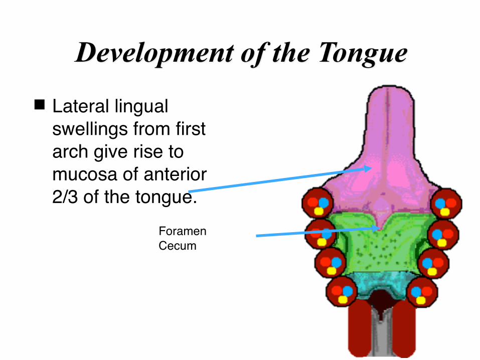
Development of the Tongue
Foramen!Cecum
■ Lateral lingual swellings from first arch give rise to mucosa of anterior 2/3 of the tongue.
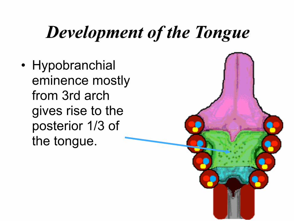
Development of the Tongue
• Hypobranchial eminence mostly from 3rd arch gives rise to the posterior 1/3 of the tongue.
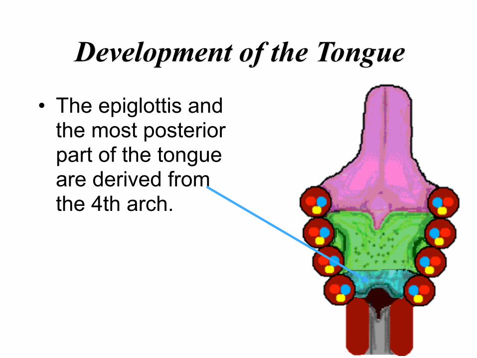
• The epiglottis and the most posterior part of the tongue are derived from the 4th arch.
Development of the Tongue
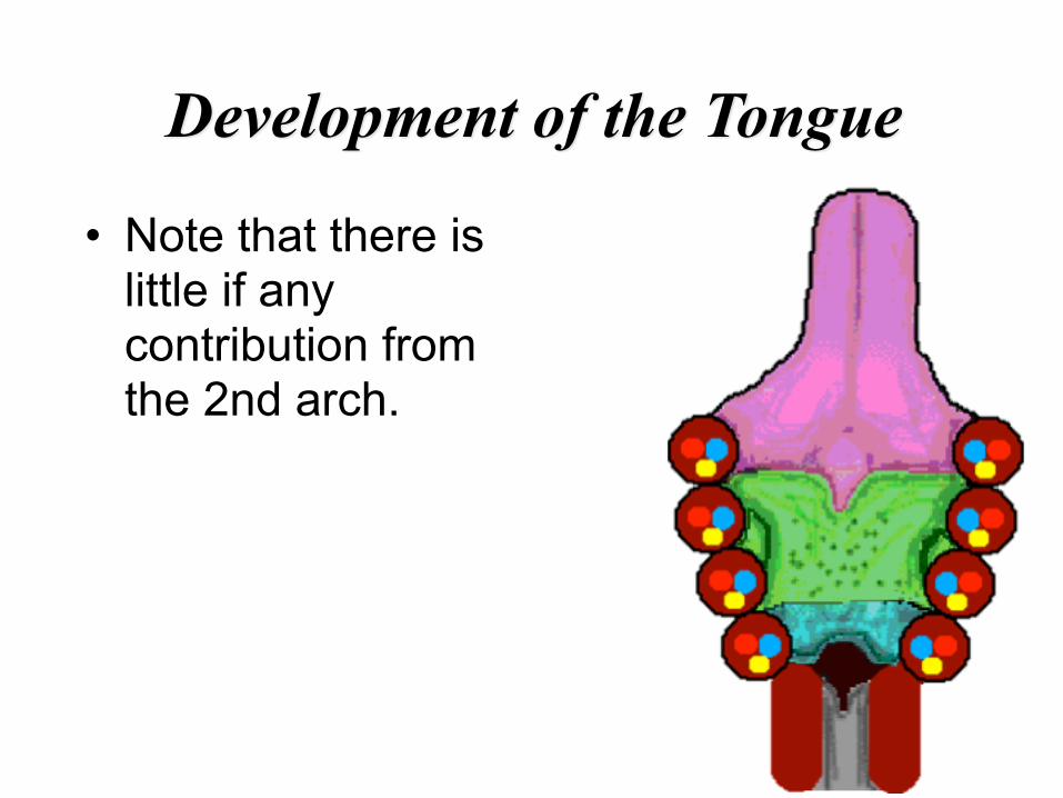
• Note that there is little if any contribution from the 2nd arch.
Development of the Tongue
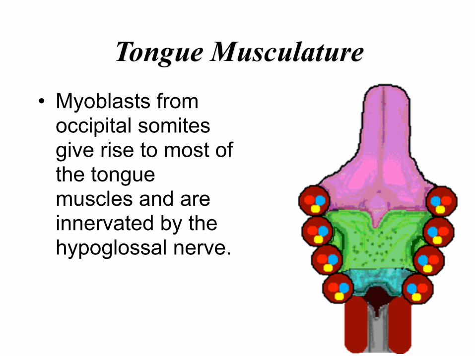
Tongue Musculature• Myoblasts from
occipital somites give rise to most of the tongue muscles and are innervated by the hypoglossal nerve.
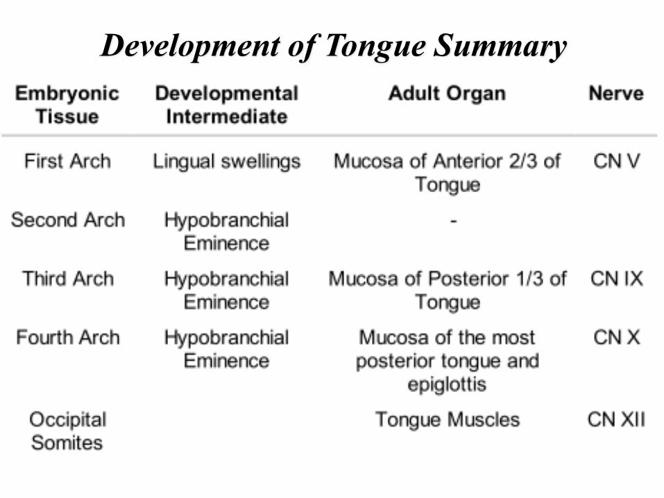
Development of Tongue Summary
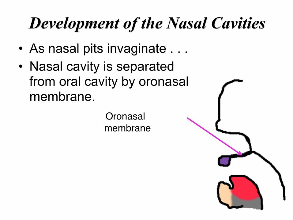
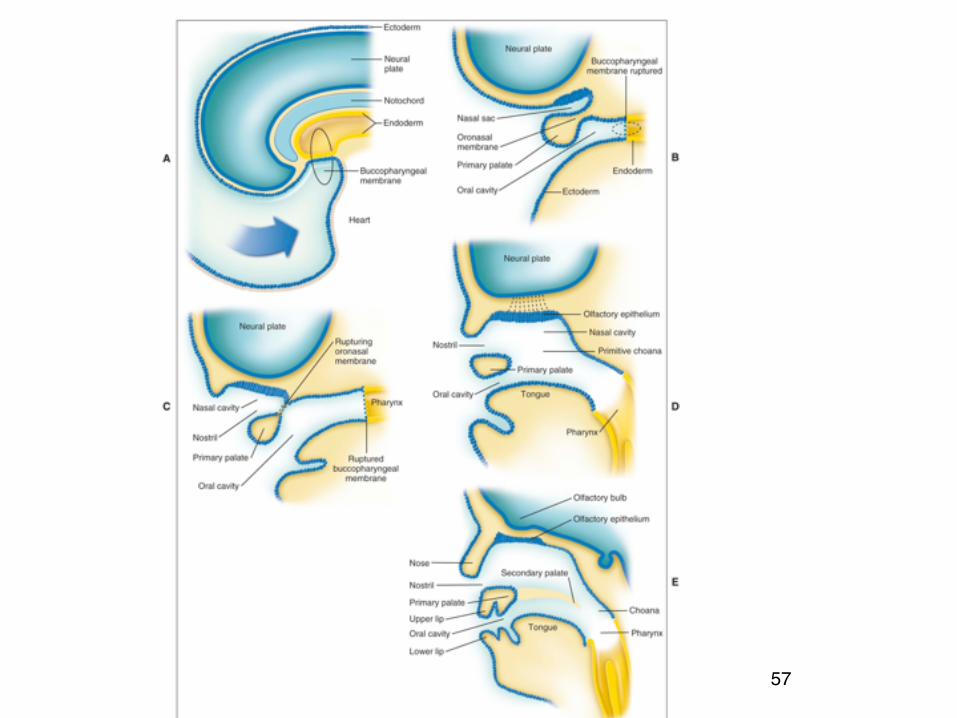
Development of the Nasal Cavities• As nasal pits invaginate . . . • Nasal cavity is separated
from oral cavity by oronasal membrane.
Oronasal membrane
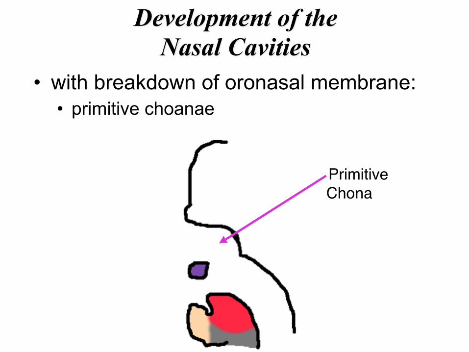
Development of the Nasal Cavities
• with breakdown of oronasal membrane: • primitive choanae
Primitive Chona
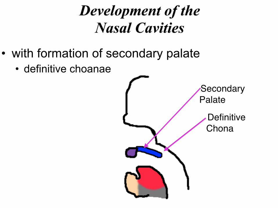
Development of the Nasal Cavities
• with formation of secondary palate • definitive choanae
Definitive Chona
Secondary Palate
57
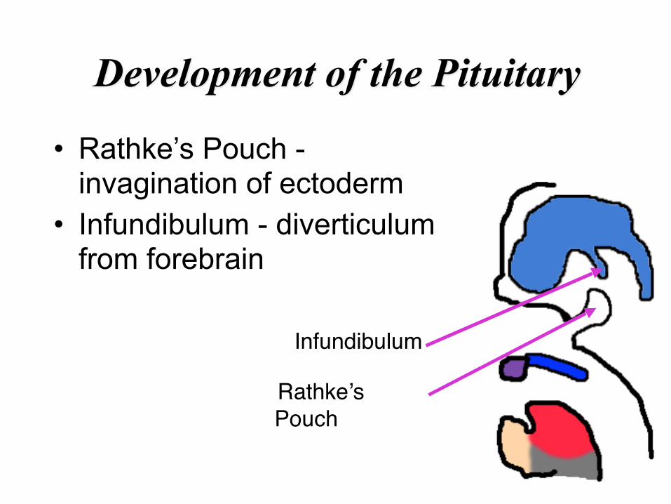
Development of the Pituitary
• Rathke’s Pouch - invagination of ectoderm
• Infundibulum - diverticulum from forebrain
Rathke’s Pouch
Infundibulum
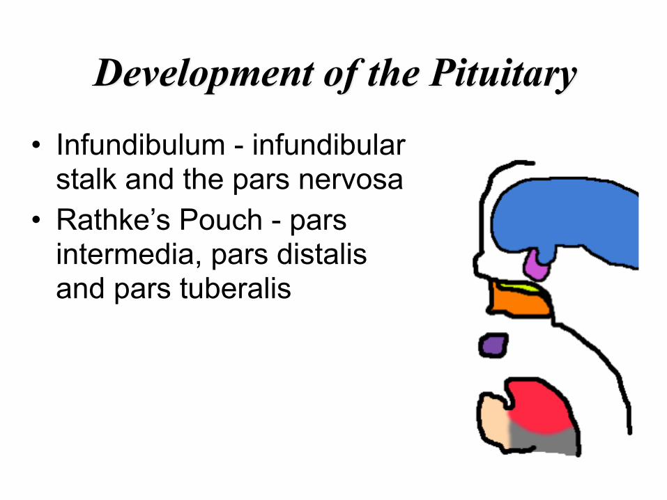
Development of the Pituitary
• Infundibulum - infundibular stalk and the pars nervosa
• Rathke’s Pouch - pars intermedia, pars distalis and pars tuberalis
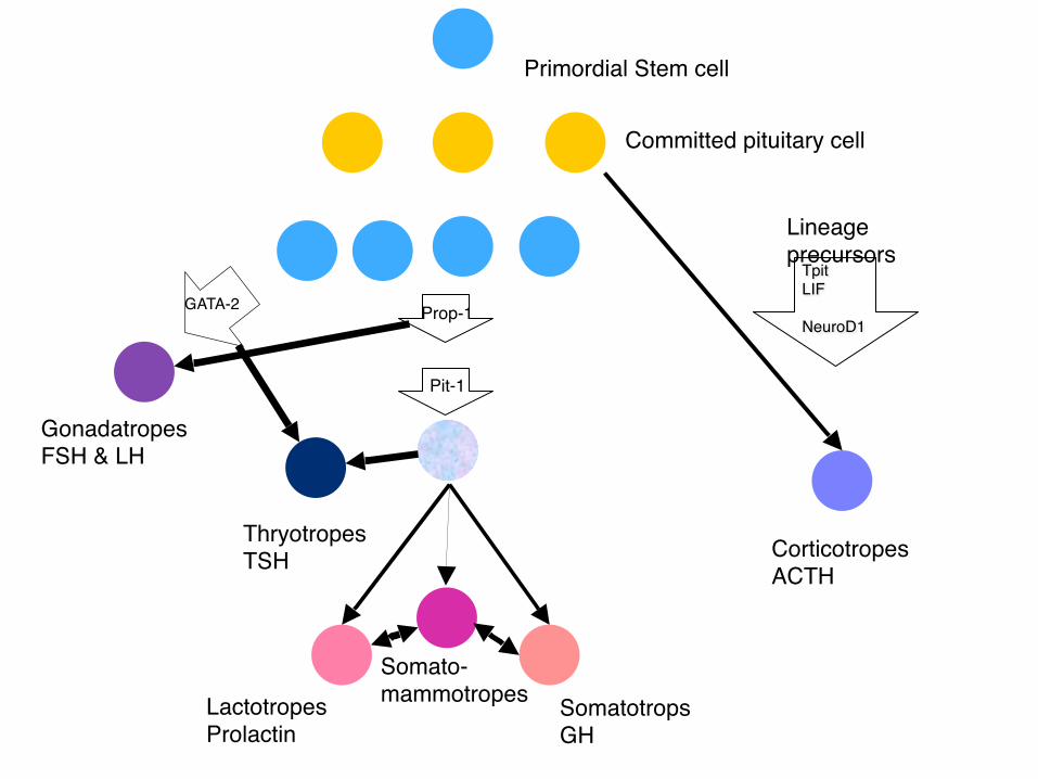
Primordial Stem cell
Committed pituitary cell
Lineage precursors
Prop-1GATA-2
Gonadatropes!FSH & LH
Pit-1
Thryotropes!TSH Corticotropes!
ACTH
Tpit!LIF! !NeuroD1
Lactotropes!Prolactin
Somato-!mammotropes Somatotrops!
GH
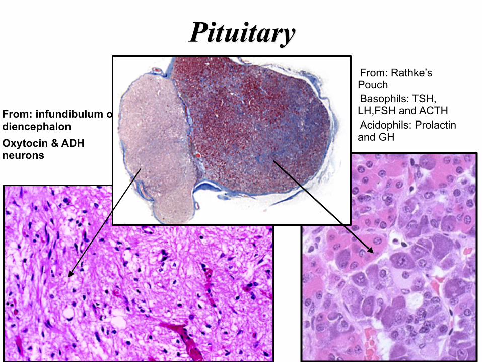
Pituitary
From: infundibulum of diencephalon Oxytocin & ADH neurons
From: Rathke’s Pouch Basophils: TSH, LH,FSH and ACTH Acidophils: Prolactin and GH
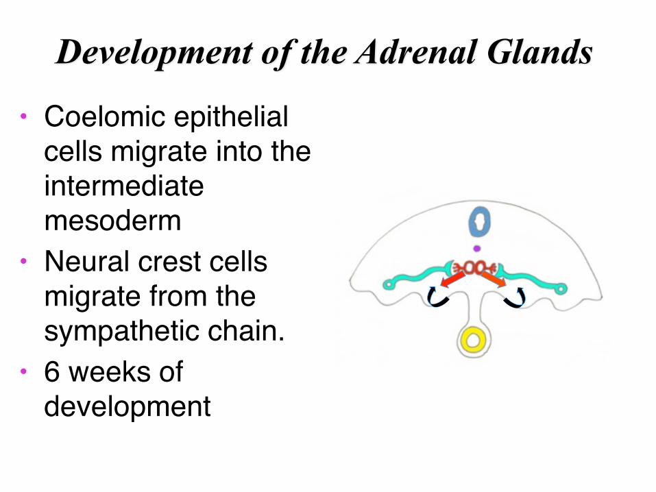
Development of the Adrenal Glands
• Coelomic epithelial cells migrate into the intermediate mesoderm!
• Neural crest cells migrate from the sympathetic chain.!
• 6 weeks of development
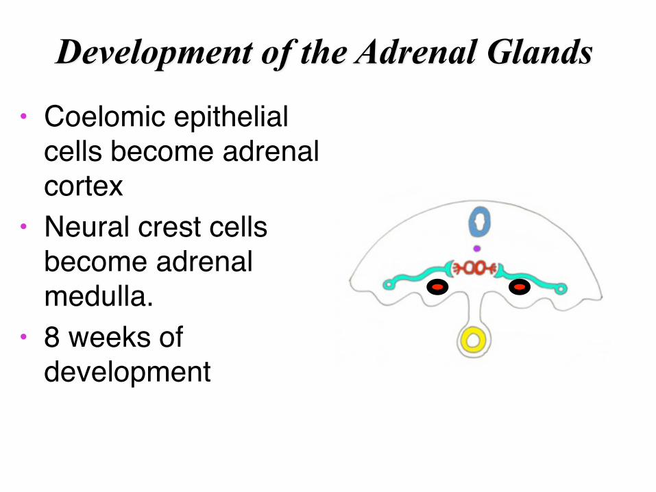
Development of the Adrenal Glands
• Coelomic epithelial cells become adrenal cortex!
• Neural crest cells become adrenal medulla.!
• 8 weeks of development
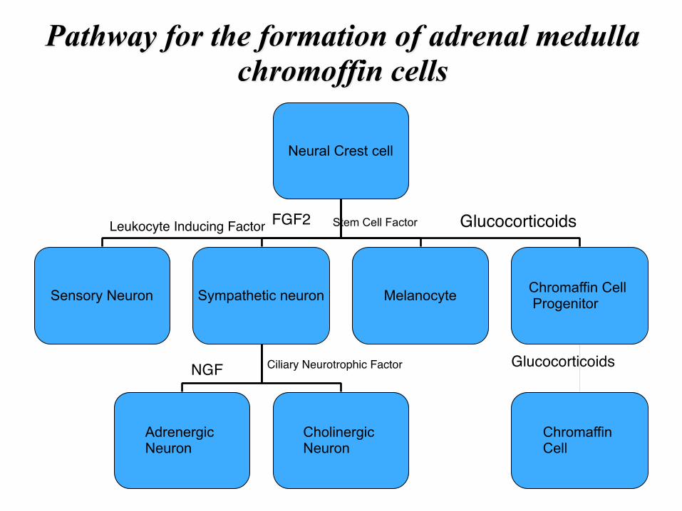
Pathway for the formation of adrenal medulla chromoffin cells
Neural Crest cell
Sensory Neuron Sympathetic neuron Melanocyte Chromaffin Cell Progenitor
Adrenergic Neuron
Cholinergic Neuron
Chromaffin Cell
Stem Cell FactorFGF2Leukocyte Inducing Factor Glucocorticoids
GlucocorticoidsCiliary Neurotrophic FactorNGF
Adrenal Gland• Fetal Adrenal
• Capsule • Definitive zone • Transition zone • Fetal zone. • Medulla.
• Functions • Maturation of: • Lungs • Liver • GI epithelium
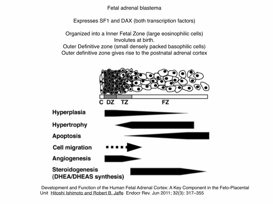
Fetal adrenal blastema!!Expresses SF1 and DAX (both transcription factors)!!
Organized into a Inner Fetal Zone (large eosinophilic cells)!Involutes at birth.!
Outer Definitive zone (small densely packed basophilic cells)!Outer definitive zone gives rise to the postnatal adrenal cortex
Development and Function of the Human Fetal Adrenal Cortex: A Key Component in the Feto-Placental Unit Hitoshi Ishimoto and Robert B. Jaffe Endocr Rev. Jun 2011; 32(3): 317–355
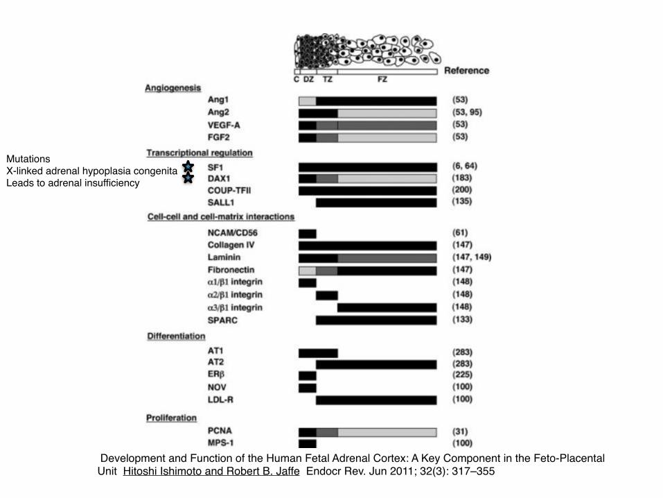
Mutations !X-linked adrenal hypoplasia congenita!Leads to adrenal insufficiency
Development and Function of the Human Fetal Adrenal Cortex: A Key Component in the Feto-Placental Unit Hitoshi Ishimoto and Robert B. Jaffe Endocr Rev. Jun 2011; 32(3): 317–355
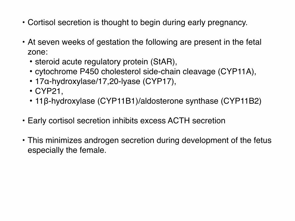
• Cortisol secretion is thought to begin during early pregnancy.!!• At seven weeks of gestation the following are present in the fetal
zone:!• steroid acute regulatory protein (StAR), !• cytochrome P450 cholesterol side-chain cleavage (CYP11A), !• 17α-hydroxylase/17,20-lyase (CYP17), !• CYP21, !• 11β-hydroxylase (CYP11B1)/aldosterone synthase (CYP11B2)! !
• Early cortisol secretion inhibits excess ACTH secretion !!• This minimizes androgen secretion during development of the fetus
especially the female.
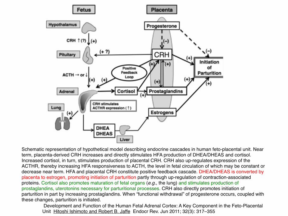
Schematic representation of hypothetical model describing endocrine cascades in human feto-placental unit. Near term, placenta-derived CRH increases and directly stimulates HFA production of DHEA/DHEAS and cortisol. Increased cortisol, in turn, stimulates production of placental CRH. CRH also up-regulates expression of the ACTHR, thereby increasing HFA responsiveness to ACTH, the level in fetal circulation of which may be constant or decrease near term. HFA and placental CRH constitute positive feedback cascade. DHEA/DHEAS is converted by placenta to estrogen, promoting initiation of parturition partly through up-regulation of contraction-associated proteins. Cortisol also promotes maturation of fetal organs (e.g., the lung) and stimulates production of prostaglandins, uterotonins necessary for parturitional processes. CRH also directly promotes initiation of parturition in part by increasing prostaglandins. When “functional withdrawal” of progesterone occurs, coupled with these changes, parturition is initiated.
Development and Function of the Human Fetal Adrenal Cortex: A Key Component in the Feto-Placental Unit Hitoshi Ishimoto and Robert B. Jaffe Endocr Rev. Jun 2011; 32(3): 317–355