TKR in the Valgus Knee - New Hampshire Musculoskeletal ... TKR in the Valgus Knee.pdf · TKR in the...
4
1/4/15 1 TKR in the Valgus Knee Peter Brooks MD, FRCS(C) Cleveland Clinic Disclosures Consultant Smith and Nephew Stryker Valgus Deformity Less common than varus Bone deformity lateral femoral hypoplasia Soft tissue problems tight lateral side stretched medial side Bone Deformity Primarily femoral unlike varus: tibial deformity distal deficiency posterior deficiency Complicates femoral component position Distal Deficiency Sloping joint line Must be corrected Final joint line parallel to floor Sloping Joint Line 5mm -> 0mm -> 5mm Distal Lateral Augment Posterior Lateral Deficiency Cannot rely on posterior referencing Results in femoral internal rotation Patellar mal-tracking
Transcript of TKR in the Valgus Knee - New Hampshire Musculoskeletal ... TKR in the Valgus Knee.pdf · TKR in the...

1/4/15
1
TKR in the Valgus Knee
Peter Brooks MD, FRCS(C) Cleveland Clinic
Disclosures
n Consultant n Smith and Nephew n Stryker
Valgus Deformity
n Less common than varus n Bone deformity
n lateral femoral hypoplasia n Soft tissue problems
n tight lateral side n stretched medial side
Bone Deformity
n Primarily femoral n unlike varus: tibial deformity n distal deficiency n posterior deficiency
n Complicates femoral component position
Distal Deficiency
n Sloping joint line n Must be corrected n Final joint line parallel to floor
Sloping Joint Line
5mm -> 0mm ->
5mm Distal Lateral Augment Posterior Lateral Deficiency
n Cannot rely on posterior referencing
n Results in femoral internal rotation
n Patellar mal-tracking

1/4/15
2
Use the Epicondylar Axis
n Avoids femoral internal rotation n Patellar maltracking
n Check with Whiteside’s Line n Helps correct soft tissue balance
in flexion
Whiteside’s Line
n Deepest part of trochlea is normally centered on axis of rotation
n A line from deepest part of trochlea to highest point of intercondylar notch is at right angles to epicondylar axis
Normal Knee
Whiteside’s Line
3°
Valgus Knee
Whiteside’s Line
10°
Incorrect Rotation
Tibia in Valgus Knee
n Not the primary deformity n May have secondary changes n Lateral plateau deficiency
Soft Tissue Balancing
n More difficult than varus knees n Sequential releases n Tight lateral, +/- stretched medial n Tight in flexion or extension, or both n Flexion contracture or hyperextension
1/4/15
3
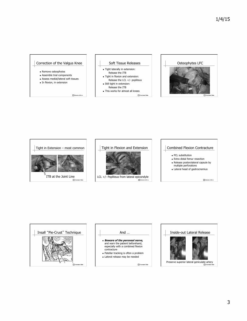
Correction of the Valgus Knee
n Remove osteophytes n Assemble trial components n Assess medial/lateral soft tissues n In flexion, in extension
Soft Tissue Releases
n Tight laterally in extension: Release the ITB
n Tight in flexion and extension: Release the LCL +/- popliteus
n Still tight in extension: Release the ITB
n This works for almost all knees
Osteophytes LFC
Tight in Extension – most common
ITB at the Joint Line
Tight in Flexion and Extension
LCL +/- Popliteus from lateral epicondyle
Combined Flexion Contracture
n PCL substitution n Extra distal femur resection n Release posterolateral capsule by
multiple perforations n Lateral head of gastrocnemius
Insall “Pie-Crust” Technique And …
n Beware of the peroneal nerve, and warn the patient beforehand, especially with a combined flexion contracture
n Patellar tracking is often a problem n Lateral release may be needed
Inside-out Lateral Release
Preserve superior lateral geniculate artery

1/4/15
4
Implant Selection
n Mild valgus n CR or PS
n Moderate valgus n PS, may need augments
n Severe valgus with medial instability n Constrained (TS or hinge) n Stems, augments
Clinical Example
n JK, 72 yr old female n OA right knee n Severe valgus deformity n Marked instability
Instability: valgus, hyperextension Implant Selection
n Severe Valgus n Medial stretching n Hyperextension
n Stabilizes medial side n Prevents over-stretching peroneal nerve
Total Stabilized TKR