


Languages
Pages
Legal


1/4/15
1
TKR in the Valgus Knee
Peter Brooks MD, FRCS(C) Cleveland Clinic
Disclosures
n Consultant n Smith and Nephew n Stryker
Valgus Deformity
n Less common than varus n Bone deformity
n lateral femoral hypoplasia n Soft tissue problems
n tight lateral side n stretched medial side
Bone Deformity
n Primarily femoral n unlike varus: tibial deformity n distal deficiency n posterior deficiency
n Complicates femoral component position
Distal Deficiency
n Sloping joint line n Must be corrected n Final joint line parallel to floor
Sloping Joint Line
5mm -> 0mm ->
5mm Distal Lateral Augment Posterior Lateral Deficiency
n Cannot rely on posterior referencing
n Results in femoral internal rotation
n Patellar mal-tracking

1/4/15
2
Use the Epicondylar Axis
n Avoids femoral internal rotation n Patellar maltracking
n Check with Whiteside’s Line n Helps correct soft tissue balance
in flexion
Whiteside’s Line
n Deepest part of trochlea is normally centered on axis of rotation
n A line from deepest part of trochlea to highest point of intercondylar notch is at right angles to epicondylar axis
Normal Knee
Whiteside’s Line
3°
Valgus Knee
Whiteside’s Line
10°
Incorrect Rotation
Tibia in Valgus Knee
n Not the primary deformity n May have secondary changes n Lateral plateau deficiency
Soft Tissue Balancing
n More difficult than varus knees n Sequential releases n Tight lateral, +/- stretched medial n Tight in flexion or extension, or both n Flexion contracture or hyperextension
1/4/15
3
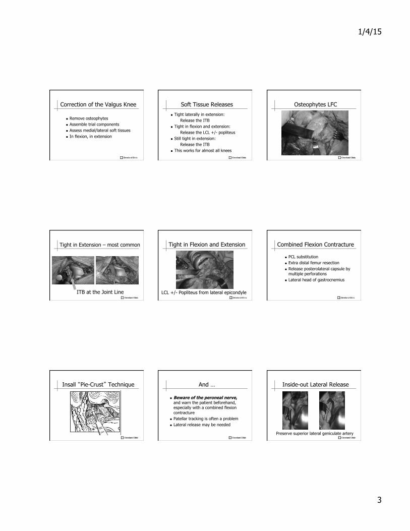
Correction of the Valgus Knee
n Remove osteophytes n Assemble trial components n Assess medial/lateral soft tissues n In flexion, in extension
Soft Tissue Releases
n Tight laterally in extension: Release the ITB
n Tight in flexion and extension: Release the LCL +/- popliteus
n Still tight in extension: Release the ITB
n This works for almost all knees
Osteophytes LFC
Tight in Extension – most common
ITB at the Joint Line
Tight in Flexion and Extension
LCL +/- Popliteus from lateral epicondyle
Combined Flexion Contracture
n PCL substitution n Extra distal femur resection n Release posterolateral capsule by
multiple perforations n Lateral head of gastrocnemius
Insall “Pie-Crust” Technique And …
n Beware of the peroneal nerve, and warn the patient beforehand, especially with a combined flexion contracture
n Patellar tracking is often a problem n Lateral release may be needed
Inside-out Lateral Release
Preserve superior lateral geniculate artery

1/4/15
4
Implant Selection
n Mild valgus n CR or PS
n Moderate valgus n PS, may need augments
n Severe valgus with medial instability n Constrained (TS or hinge) n Stems, augments
Clinical Example
n JK, 72 yr old female n OA right knee n Severe valgus deformity n Marked instability
Instability: valgus, hyperextension Implant Selection
n Severe Valgus n Medial stretching n Hyperextension
n Stabilizes medial side n Prevents over-stretching peroneal nerve
Total Stabilized TKR
Top Related