Tinea Faciei in a Mother and Daughter Caused by ... · Brief Report 242 Ann Dermatol Fig. 2. (A)...
2
Brief Report Vol. 30, No. 2, 2018 241 Received December 27, 2016, Revised March 15, 2017, Accepted for publication April 9, 2017 Corresponding author: Weon Ju Lee, Department of Dermatology, Kyungpook National University Hospital, 130 Dongdeok-ro, Jung-gu, Daegu 41944, Korea. T el: 82-53-420-5838, Fax: 82-53-426-0770, E-mail: [email protected] This is an Open Access article distributed under the terms of the Creative Commons Attribution Non-Commercial License (http://creativecommons.org/ licenses/by-nc/4.0) which permits unrestricted non-commercial use, distribution, and reproduction in any medium, provided the original work is properly cited. Copyright © The Korean Dermatological Association and The Korean Society for Investigative Dermatology Fig. 1. (A) Peripherally radiating and centrally raised, granular and downy colonies cultured from mother and (B) her daughter. https://doi.org/10.5021/ad.2018.30.2.241 Tinea Faciei in a Mother and Daughter Caused by Arthroderma benhamiae Weon Ju Lee, Dong Hyuk Eun, Yong Hyun Jang, Seok-Jong Lee, Yong Jun Bang 1 , Jae Bok Jun 1 Department of Dermatology, Kyungpook National University School of Medicine, 1 Institute of Medical Mycology, Catholic Skin Clinic, Daegu, Korea Dear Editor: Two patients presented with peripherally spreading, an- nular, inflammatory patches on the face for several months. The patients were a 46-year-old woman and her 8-year-old daughter. Both had contact with a rabbit with inflam- matory skin lesions, but they had no other specific past medical or family history. They were diagnosed with der- matophytosis caused by Arthroderma benhamiae using KOH examination, fungal culture, lactophenol cotton blue stain, reverse blot hybridization assay (REBA) and DNA gene sequencing. KOH examination results were positive in both patients. Resembling Trichophyton interdigitale, fungal culture on potato-corn meal-Tween 80 agar showed white, granular, and downy colonies with a radiating pe- riphery and raised center (Fig. 1). The long mycelium had numerous small, round microconidia and several macro- conidia or spiral hyphae on lactophenol cotton blue stain (Fig. 2). REBA and gene sequencing using gapped BLAST and position-specific iterated-BLAST programs identified A. benhamiae. The program revealed 99% or 100% homology with accession number Z98016, JX413540, JX122298, JX122297, AB458188, AB458165, AB458176, AB458143, AB458145, JN134088, KC253946, AB686489, AB686487, AB686486, AB686485, AB686484, AB686483, AB686482,
Transcript of Tinea Faciei in a Mother and Daughter Caused by ... · Brief Report 242 Ann Dermatol Fig. 2. (A)...
Brief Report
Vol. 30, No. 2, 2018 241
Received December 27, 2016, Revised March 15, 2017, Accepted for publication April 9, 2017
Corresponding author: Weon Ju Lee, Department of Dermatology, Kyungpook National University Hospital, 130 Dongdeok-ro, Jung-gu, Daegu 41944, Korea. Tel: 82-53-420-5838, Fax: 82-53-426-0770, E-mail: [email protected]
This is an Open Access article distributed under the terms of the Creative Commons Attribution Non-Commercial License (http://creativecommons.org/licenses/by-nc/4.0) which permits unrestricted non-commercial use, distribution, and reproduction in any medium, provided the original work is properly cited.
Copyright © The Korean Dermatological Association and The Korean Society for Investigative Dermatology
Fig. 1. (A) Peripherally radiating and centrally raised, granular and downy colonies cultured from mother and (B) her daughter.
https://doi.org/10.5021/ad.2018.30.2.241
Tinea Faciei in a Mother and Daughter Caused by Arthroderma benhamiae
Weon Ju Lee, Dong Hyuk Eun, Yong Hyun Jang, Seok-Jong Lee, Yong Jun Bang1, Jae Bok Jun1
Department of Dermatology, Kyungpook National University School of Medicine, 1Institute of Medical Mycology, Catholic Skin Clinic, Daegu, Korea
Dear Editor:Two patients presented with peripherally spreading, an-nular, inflammatory patches on the face for several months. The patients were a 46-year-old woman and her 8-year-old daughter. Both had contact with a rabbit with inflam-matory skin lesions, but they had no other specific past medical or family history. They were diagnosed with der-matophytosis caused by Arthroderma benhamiae using KOH examination, fungal culture, lactophenol cotton blue stain, reverse blot hybridization assay (REBA) and DNA gene sequencing. KOH examination results were positive in both patients. Resembling Trichophyton interdigitale,
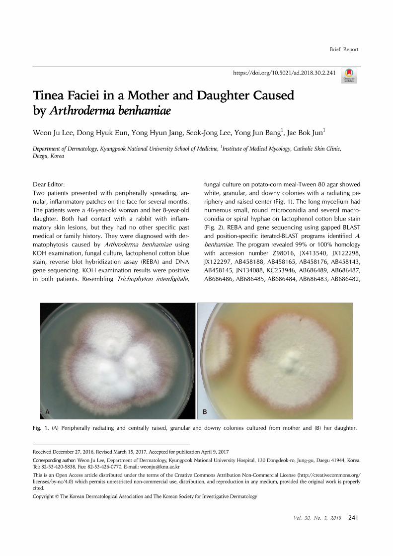
fungal culture on potato-corn meal-Tween 80 agar showed white, granular, and downy colonies with a radiating pe-riphery and raised center (Fig. 1). The long mycelium had numerous small, round microconidia and several macro-conidia or spiral hyphae on lactophenol cotton blue stain (Fig. 2). REBA and gene sequencing using gapped BLAST and position-specific iterated-BLAST programs identified A. benhamiae. The program revealed 99% or 100% homology with accession number Z98016, JX413540, JX122298, JX122297, AB458188, AB458165, AB458176, AB458143, AB458145, JN134088, KC253946, AB686489, AB686487, AB686486, AB686485, AB686484, AB686483, AB686482,
Brief Report
242 Ann Dermatol
Fig. 2. (A) Septate mycelium with a great number of small round microconidia and a few macroconidia from mother. (B) Septate mycelium with a great number of small round microconidia and spiral hyphae from her daughter (A, B: lactophenol cotton blue stain, ×200).
AB686481, and AB686475. The lesions resolved with oral antifungal medication (terbinafine: 500 mg for mother and 250 mg for daughter) for 1 month. Although molecular methods such as gene sequencing en-able precise identification, A. benhamiae resembles Micro-sporum canis and T. interdigitale. A. benhamiae is an emerging cause of inflammatory dermatophytosis, such as tinea corporis, tinea faciei, tinea capitis, and kerion celsi. A. benhamiae was isolated in Japan in 19981. A case with A. benhamiae infection was reported in Germany in 20102. Jun et al.3 published the first report of dermatophy-tosis caused by A. benhamiae in Korea, but there have been no subsequent Korean reports. A. benhamiae is usu-ally transmitted from animals to humans. Guinea pigs, hamsters, rats, and rabbits are potential carriers. Conven-tional diagnostic methods for dermatophytosis include KOH examination and culture. A. benhamiae on Sabouraud agar forms radiating colonies with beige to yellow myce-lium and a dense velvety surface. A smaller percentage of A. benhamiae cultures exhibit white granular colonies. A. benhamiae on lactophenol cotton blue stain shows septate mycelium with small round microconidia, grape-like mi-croconidia, macroconidia, and/or chlamydospores. Urea hydrolysis on Christensen’s urea agar and chromogenic agar have also been used for A. benhamiae identification4. Moreover, direct genetic detection using molecular meth-ods for pathogens in specimens is useful5. Polymerase chain reaction-enzyme linked immunosorbent assay, se-quencing of internal transcribed spacer regions of 28S
rRNA genes, and matrix-assisted laser desorption/ioniza-tion time of flight mass spectrometry were recently in-troduced for the identification of A. benhamiae. We here-in described two cases of tinea faciei caused by A. benha-miae identified with REBA and gene sequencing.
CONFLICTS OF INTEREST
The authors have nothing to disclose.
REFERENCES
1. Kano R, Nakamura Y, Yasuda K, Watari T, Watanabe S, Takahashi H, et al. The first isolation of Arthroderma
benhamiae in Japan. Microbiol Immunol 1998;42:575-578.
2. Budihardja D, Freund V, Mayser P. Widespread erosive tinea corporis by Arthroderma benhamiae in a renal
transplant recipient: case report. Mycoses 2010;53:530-532.
3. Jun JB, Sang YH, Chung SL, Choi JS, Suh SB. The mycological and molecular biological studies on Arthro-
derma benhamiae isolated for the first time in Korea. Korean
J Med Mycol 2004;9:12-27.4. Mayser P, Budihardja D. A simple and rapid method to
differentiate Arthroderma benhamiae from Microsporum
canis. J Dtsch Dermatol Ges 2013;11:322-327. 5. Nenoff P, Uhrlaß S, Krüger C, Erhard M, Hipler UC, Seyfarth
F, et al. Trichophyton species of Arthroderma benhamiae-a
new infectious agent in dermatology. J Dtsch Dermatol Ges 2014;12:571-581.