Grainy head promotes expression of septate junction proteins and ...
6
747 Short Report Introduction During Drosophila development, the Grainy head (Grh) transcription factor is expressed in the epidermis and a subset of other epithelia that form strongly adhesive layers exposed to the external environment (e.g. trachea) (Bray and Kafatos, 1991; Hemphala et al., 2003; Uv et al., 1994). In the absence of grh, these epithelial cells have altered morphology and lose expression of enzymes that cross-link the apical extracellular matrix (cuticle) (Bray and Kafatos, 1991; Hemphala et al., 2003; Mace et al., 2005; Ostrowski et al., 2002). Similarly, in mice lacking the Grh-related gene GRHL3, the outer protective layer of the skin, the stratum corneum, is defective (Ting et al., 2005; Yu et al., 2006), and in Xenopus embryos, GRHL3 and GRHL1 are expressed in the outer cells and regulate the expression of keratins (Chalmers et al., 2006; Tao et al., 2005). Thus, Grh proteins have highly conserved roles in regulating terminal differentiation of robust protective epithelia. In addition to regulating terminal differentiation per se, Grh might also have other functions in these epithelia. For example, at stages before the stratum corneum is formed, Grhl3 mutant mice have defects in re-epithelialisation following wounding (Stramer and Martin, 2005; Ting et al., 2005). They also have altered levels of many tight-junction-associated proteins, including occludins and claudins (Yu et al., 2006). Likewise, in Drosophila, grh expression commences prior to cuticle secretion and correlates with stages at which these epithelia acquire occluding junctions (septate junctions) (Tepass and Hartenstein, 1994). Nevertheless, Grh is dispensable for the establishment of basic barrier properties, because septate junctions are still present in grh-mutant tracheal cells (Hemphala et al., 2003). However, because the barrier characteristics of occluding junctions vary between epithelia (Furuse and Tsukita, 2006), the conserved expression of grh family proteins in the highly impermeable surface epithelia led us to investigate further whether Grh could directly regulate expression of epithelial junction components in Drosophila. We began our investigations by expressing Grh ectopically in the amnioserosa (AS), a single-layered epithelium that has no septate junctions (Tepass and Hartenstein, 1994; Gorfinkiel and Martinez Arias, 2007), to determine whether Grh could convert this tissue into one with barrier epithelia characteristics. The AS is normally devoid of Grh expression and plays an important role in co-ordinating the fusion between the epidermal sheets during dorsal closure (Fig. 1A,B). From these studies, we uncovered a role for Grh in regulating expression of septate junction proteins, which we have further confirmed using loss-of-function mutations and by showing that the genes contain Grh-binding sites. Thus, in addition to co-ordinating expression of matrix proteins, Grh also regulates the intrinsic barrier properties of epithelia through its effects on components of cell junctions. Results and Discussion To test the role of Grh in regulating epithelial characteristics, we specifically expressed the epidermal splice forms (N/K) in the amnioserosa, an epithelial tissue normally devoid of Grh (using c381::Gal4 and G332::Gal4; Fig. 1C,F-H). This was sufficient to block dorsal closure (Fig. 1G,H), an effect previously seen with ubiquitous Grh overexpression (Attardi et al., 1993). The effects were most penetrant with c381::Gal4 (hereafter referred to as AS c381 >grh) which resulted in 100% of embryos having dorsal holes at stage 17/hatching, when all wild-type embryos had completed dorsal closure (Fig. 1H; AS G332 >grh resulted in >50% Transcription factors of the Grainy head (Grh) family are required in epithelia to generate the impermeable apical layer that protects against the external environment. This function is conserved in vertebrates and invertebrates, despite the differing molecular composition of the protective barrier. Epithelial cells also have junctions that create a paracellular diffusion barrier (tight or septate junctions). To examine whether Grh has a role in regulating such characteristics, we used an epidermal layer in the Drosophila embryo that has no endogenous Grh and lacks septate junctions, the amnioserosa. Expression of Grh in the amnioserosa caused severe defects in dorsal closure, a process similar to wound closure, and induced robust expression of the septate junction proteins Coracle, Fasciclin 3 and Sinuous. Grh-binding sites are present within the genes encoding these proteins, consistent with them being direct targets. Removal of Grh from imaginal disc cells caused a reduction in Fasciclin 3 and Coracle levels, suggesting that Grh normally fine tunes their epithelial expression and hence contributes to barrier properties. The fact that ectopic Grh arrests dorsal closure also suggests that this dynamic process relies on epithelia having distinct adhesive properties conferred by differential deployment of Grh. Key words: Drosophila, Grainy head, Septate junctions Summary Grainy head promotes expression of septate junction proteins and influences epithelial morphogenesis Maithreyi Narasimha 1,2,3 , Anne Uv 2,4 , Alena Krejci 2 , Nicholas H. Brown 1,2 and Sarah J. Bray 2, * 1 Wellcome Trust/Cancer Research UK Gurdon Institute of Developmental Biology and Cancer, and 2 Department of Physiology Development and Neuroscience, University of Cambridge, Downing Street, Cambridge, CB2 3DY, UK 3 Department of Biological Sciences, Tata Institute for Fundamental Research, Colaba, Mumbai 400 005, India 4 Institute för Medicinsk och Fysiologisk Kemi, Medicinaregatan 9A, Göteborgs Universitet, Göteborg, Sweden *Author for correspondence (e-mail: [email protected]) Accepted 9 December 2007 Journal of Cell Science 121, 747-752 Published by The Company of Biologists 2008 doi:10.1242/jcs.019422 Journal of Cell Science
Transcript of Grainy head promotes expression of septate junction proteins and ...
747Short Report
IntroductionDuring Drosophila development, the Grainy head (Grh)transcription factor is expressed in the epidermis and a subset ofother epithelia that form strongly adhesive layers exposed to theexternal environment (e.g. trachea) (Bray and Kafatos, 1991;Hemphala et al., 2003; Uv et al., 1994). In the absence of grh, theseepithelial cells have altered morphology and lose expression ofenzymes that cross-link the apical extracellular matrix (cuticle)(Bray and Kafatos, 1991; Hemphala et al., 2003; Mace et al., 2005;Ostrowski et al., 2002). Similarly, in mice lacking the Grh-relatedgene GRHL3, the outer protective layer of the skin, the stratumcorneum, is defective (Ting et al., 2005; Yu et al., 2006), and inXenopus embryos, GRHL3 and GRHL1 are expressed in the outercells and regulate the expression of keratins (Chalmers et al., 2006;Tao et al., 2005). Thus, Grh proteins have highly conserved rolesin regulating terminal differentiation of robust protective epithelia.
In addition to regulating terminal differentiation per se, Grhmight also have other functions in these epithelia. For example, atstages before the stratum corneum is formed, Grhl3 mutant micehave defects in re-epithelialisation following wounding (Stramerand Martin, 2005; Ting et al., 2005). They also have altered levelsof many tight-junction-associated proteins, including occludins andclaudins (Yu et al., 2006). Likewise, in Drosophila, grh expressioncommences prior to cuticle secretion and correlates with stages atwhich these epithelia acquire occluding junctions (septatejunctions) (Tepass and Hartenstein, 1994). Nevertheless, Grh isdispensable for the establishment of basic barrier properties,because septate junctions are still present in grh-mutant trachealcells (Hemphala et al., 2003). However, because the barriercharacteristics of occluding junctions vary between epithelia
(Furuse and Tsukita, 2006), the conserved expression of grh familyproteins in the highly impermeable surface epithelia led us toinvestigate further whether Grh could directly regulate expressionof epithelial junction components in Drosophila.
We began our investigations by expressing Grh ectopically inthe amnioserosa (AS), a single-layered epithelium that has noseptate junctions (Tepass and Hartenstein, 1994; Gorfinkiel andMartinez Arias, 2007), to determine whether Grh could convert thistissue into one with barrier epithelia characteristics. The AS isnormally devoid of Grh expression and plays an important role inco-ordinating the fusion between the epidermal sheets duringdorsal closure (Fig. 1A,B). From these studies, we uncovered a rolefor Grh in regulating expression of septate junction proteins, whichwe have further confirmed using loss-of-function mutations and byshowing that the genes contain Grh-binding sites. Thus, in additionto co-ordinating expression of matrix proteins, Grh also regulatesthe intrinsic barrier properties of epithelia through its effects oncomponents of cell junctions.
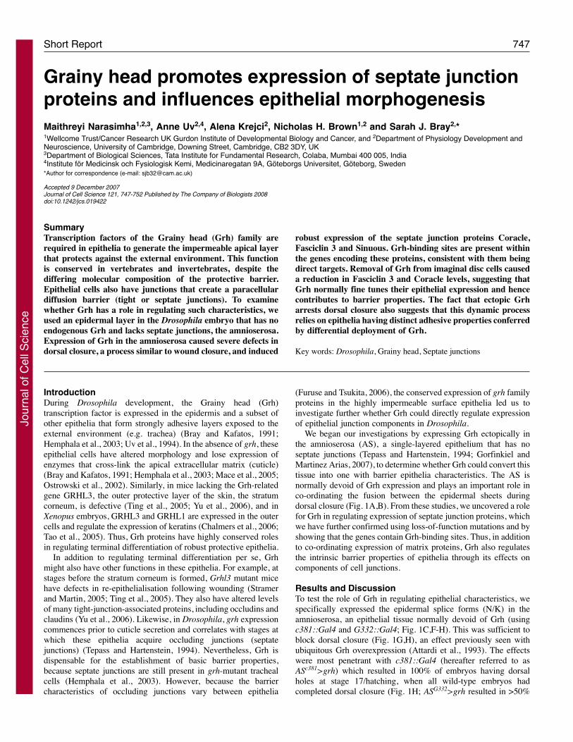
Results and DiscussionTo test the role of Grh in regulating epithelial characteristics, wespecifically expressed the epidermal splice forms (N/K) in theamnioserosa, an epithelial tissue normally devoid of Grh (usingc381::Gal4 and G332::Gal4; Fig. 1C,F-H). This was sufficient toblock dorsal closure (Fig. 1G,H), an effect previously seen withubiquitous Grh overexpression (Attardi et al., 1993). The effectswere most penetrant with c381::Gal4 (hereafter referred to asASc381>grh) which resulted in 100% of embryos having dorsalholes at stage 17/hatching, when all wild-type embryos hadcompleted dorsal closure (Fig. 1H; ASG332>grh resulted in >50%
Transcription factors of the Grainy head (Grh) family arerequired in epithelia to generate the impermeable apical layerthat protects against the external environment. This functionis conserved in vertebrates and invertebrates, despite thediffering molecular composition of the protective barrier.Epithelial cells also have junctions that create a paracellulardiffusion barrier (tight or septate junctions). To examinewhether Grh has a role in regulating such characteristics, weused an epidermal layer in the Drosophila embryo that has noendogenous Grh and lacks septate junctions, the amnioserosa.Expression of Grh in the amnioserosa caused severe defects indorsal closure, a process similar to wound closure, and induced
robust expression of the septate junction proteins Coracle,Fasciclin 3 and Sinuous. Grh-binding sites are present withinthe genes encoding these proteins, consistent with them beingdirect targets. Removal of Grh from imaginal disc cells causeda reduction in Fasciclin 3 and Coracle levels, suggesting thatGrh normally fine tunes their epithelial expression and hencecontributes to barrier properties. The fact that ectopic Grharrests dorsal closure also suggests that this dynamic processrelies on epithelia having distinct adhesive properties conferredby differential deployment of Grh.
Key words: Drosophila, Grainy head, Septate junctions
Summary
Grainy head promotes expression of septate junctionproteins and influences epithelial morphogenesisMaithreyi Narasimha1,2,3, Anne Uv2,4, Alena Krejci2, Nicholas H. Brown1,2 and Sarah J. Bray2,*1Wellcome Trust/Cancer Research UK Gurdon Institute of Developmental Biology and Cancer, and 2Department of Physiology Development andNeuroscience, University of Cambridge, Downing Street, Cambridge, CB2 3DY, UK3Department of Biological Sciences, Tata Institute for Fundamental Research, Colaba, Mumbai 400 005, India4Institute för Medicinsk och Fysiologisk Kemi, Medicinaregatan 9A, Göteborgs Universitet, Göteborg, Sweden*Author for correspondence (e-mail: [email protected])
Accepted 9 December 2007Journal of Cell Science 121, 747-752 Published by The Company of Biologists 2008doi:10.1242/jcs.019422
Jour
nal o
f Cel
l Sci
ence
748
with dorsal holes). Defects were already evident earlier (stage 14-16). In ASc381>grh embryos, the amnioserosa was less contractedthan wild type and contained cells with abnormal morphology. Inaddition, the epidermal edges failed to meet at the poles (Fig. 1G).Thus, expression of Grh disrupted the ability of amnioserosa cellsto function in dorsal closure, suggesting that it altered theirfundamental properties and/or perturbed their interactions with theepidermis.
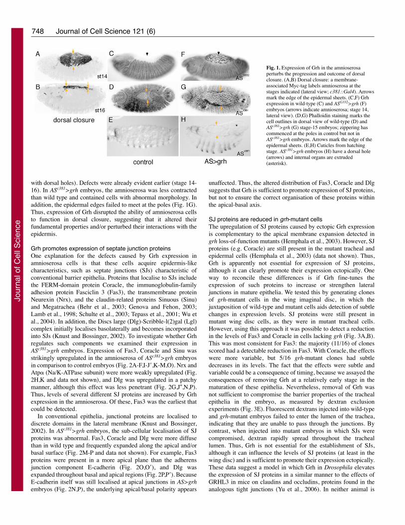
Grh promotes expression of septate junction proteinsOne explanation for the defects caused by Grh expression inamnioserosa cells is that these cells acquire epidermis-likecharacteristics, such as septate junctions (SJs) characteristic ofconventional barrier epithelia. Proteins that localise to SJs includethe FERM-domain protein Coracle, the immunoglobulin-familyadhesion protein Fasciclin 3 (Fas3), the transmembrane proteinNeurexin (Nrx), and the claudin-related proteins Sinuous (Sinu)and Megatrachea (Behr et al., 2003; Genova and Fehon, 2003;Lamb et al., 1998; Schulte et al., 2003; Tepass et al., 2001; Wu etal., 2004). In addition, the Discs large (Dlg)-Scribble-l(2)gal (Lgl)complex initially localises basolaterally and becomes incorporatedinto SJs (Knust and Bossinger, 2002). To investigate whether Grhregulates such components we examined their expression inASc381>grh embryos. Expression of Fas3, Coracle and Sinu wasstrikingly upregulated in the amnioserosa of ASc381>grh embryosin comparison to control embryos (Fig. 2A-F,I-J�,K-M,O). Nrx andAtp� (Na/K-ATPase subunit) were more weakly upregulated (Fig.2H,K and data not shown), and Dlg was upregulated in a patchymanner, although this effect was less penetrant (Fig. 2G,J�,N,P).Thus, levels of several different SJ proteins are increased by Grhexpression in the amnioserosa. Of these, Fas3 was the earliest thatcould be detected.
In conventional epithelia, junctional proteins are localised todiscrete domains in the lateral membrane (Knust and Bossinger,2002). In ASc381>grh embryos, the sub-cellular localisation of SJproteins was abnormal. Fas3, Coracle and Dlg were more diffusethan in wild type and frequently expanded along the apical and/orbasal surface (Fig. 2M-P and data not shown). For example, Fas3proteins were present in a more apical plane than the adherensjunction component E-cadherin (Fig. 2O,O�), and Dlg wasexpanded throughout basal and apical regions (Fig. 2P,P�). BecauseE-cadherin itself was still localised at apical junctions in AS>grhembryos (Fig. 2N,P), the underlying apical/basal polarity appears
unaffected. Thus, the altered distribution of Fas3, Coracle and Dlgsuggests that Grh is sufficient to promote expression of SJ proteins,but not to ensure the correct organisation of these proteins withinthe apical-basal axis.
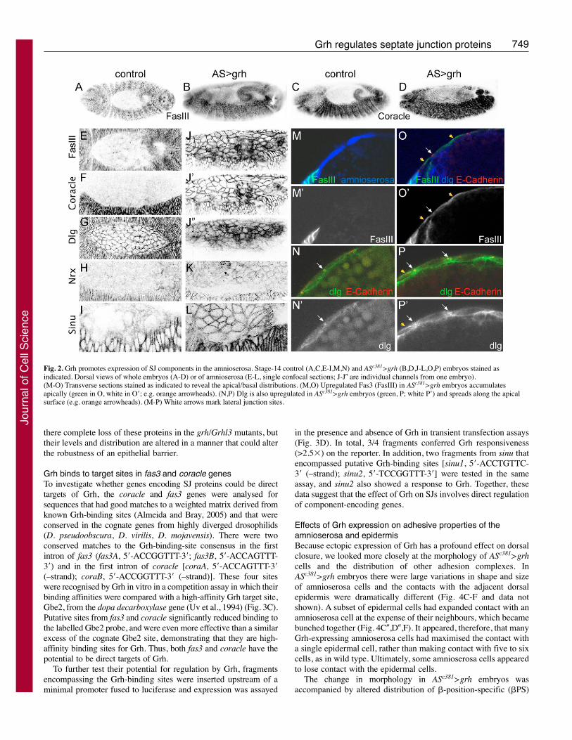
SJ proteins are reduced in grh-mutant cellsThe upregulation of SJ proteins caused by ectopic Grh expressionis complementary to the apical membrane expansion detected ingrh loss-of-function mutants (Hemphala et al., 2003). However, SJproteins (e.g. Coracle) are still present in the mutant tracheal andepidermal cells (Hemphala et al., 2003) (data not shown). Thus,Grh is apparently not essential for expression of SJ proteins,although it can clearly promote their expression ectopically. Oneway to reconcile these differences is if Grh fine-tunes theexpression of such proteins to increase or strengthen lateraljunctions in mature epithelia. We tested this by generating clonesof grh-mutant cells in the wing imaginal disc, in which thejuxtaposition of wild-type and mutant cells aids detection of subtlechanges in expression levels. SJ proteins were still present inmutant wing disc cells, as they were in mutant tracheal cells.However, using this approach it was possible to detect a reductionin the levels of Fas3 and Coracle in cells lacking grh (Fig. 3A,B).This was most consistent for Fas3: the majority (11/16) of clonesscored had a detectable reduction in Fas3. With Coracle, the effectswere more variable, but 5/16 grh-mutant clones had subtledecreases in its levels. The fact that the effects were subtle andvariable could be a consequence of timing, because we assayed theconsequences of removing Grh at a relatively early stage in thematuration of these epithelia. Nevertheless, removal of Grh wasnot sufficient to compromise the barrier properties of the trachealepithelia in the embryo, as measured by dextran exclusionexperiments (Fig. 3E). Fluorescent dextrans injected into wild-typeand grh-mutant embryos failed to enter the lumen of the trachea,indicating that they are unable to pass through the junctions. Bycontrast, when injected into mutant embryos in which SJs werecompromised, dextran rapidly spread throughout the tracheallumen. Thus, Grh is not essential for the establishment of SJs,although it can influence the levels of SJ proteins (at least in thewing disc) and is sufficient to promote their expression ectopically.These data suggest a model in which Grh in Drosophila elevatesthe expression of SJ proteins in a similar manner to the effects ofGRHL3 in mice on claudins and occludins, proteins found in theanalogous tight junctions (Yu et al., 2006). In neither animal is
Journal of Cell Science 121 (6)
Fig. 1. Expression of Grh in the amnioserosaperturbs the progression and outcome of dorsalclosure. (A,B) Dorsal closure: a membrane-associated Myc-tag labels amnioserosa at thestages indicated (lateral view; c381::Gal4). Arrowsmark the edge of the epidermal sheets. (C,F) Grhexpression in wild-type (C) and ASG332>grh (F)embryos (arrows indicate amnioserosa; stage 14,lateral view). (D,G) Phalloidin staining marks thecell outlines in dorsal view of wild-type (D) andASc381>grh (G) stage-15 embryos; zippering hascommenced at the poles in control but not inASc381>grh embryos. Arrows mark the edge of theepidermal sheets. (E,H) Cuticles from hatchingstage. ASc381>grh embryos (H) have a dorsal hole(arrows) and internal organs are extruded(asterisk).
Jour
nal o
f Cel
l Sci
ence
749Grh regulates septate junction proteins
there complete loss of these proteins in the grh/Grhl3 mutants, buttheir levels and distribution are altered in a manner that could alterthe robustness of an epithelial barrier.
Grh binds to target sites in fas3 and coracle genesTo investigate whether genes encoding SJ proteins could be directtargets of Grh, the coracle and fas3 genes were analysed forsequences that had good matches to a weighted matrix derived fromknown Grh-binding sites (Almeida and Bray, 2005) and that wereconserved in the cognate genes from highly diverged drosophilids(D. pseudoobscura, D. virilis, D. mojavensis). There were twoconserved matches to the Grh-binding-site consensus in the firstintron of fas3 (fas3A, 5�-ACCGGTTT-3�; fas3B, 5�-ACCAGTTT-3�) and in the first intron of coracle [coraA, 5�-ACCAGTTT-3�(–strand); coraB, 5�-ACCGGTTT-3� (–strand)]. These four siteswere recognised by Grh in vitro in a competition assay in which theirbinding affinities were compared with a high-affinity Grh target site,Gbe2, from the dopa decarboxylase gene (Uv et al., 1994) (Fig. 3C).Putative sites from fas3 and coracle significantly reduced binding tothe labelled Gbe2 probe, and were even more effective than a similarexcess of the cognate Gbe2 site, demonstrating that they are high-affinity binding sites for Grh. Thus, both fas3 and coracle have thepotential to be direct targets of Grh.
To further test their potential for regulation by Grh, fragmentsencompassing the Grh-binding sites were inserted upstream of aminimal promoter fused to luciferase and expression was assayed
in the presence and absence of Grh in transient transfection assays(Fig. 3D). In total, 3/4 fragments conferred Grh responsiveness(>2.5�) on the reporter. In addition, two fragments from sinu thatencompassed putative Grh-binding sites [sinu1, 5�-ACCTGTTC-3� (–strand); sinu2, 5�-TCCGGTTT-3�] were tested in the sameassay, and sinu2 also showed a response to Grh. Together, thesedata suggest that the effect of Grh on SJs involves direct regulationof component-encoding genes.
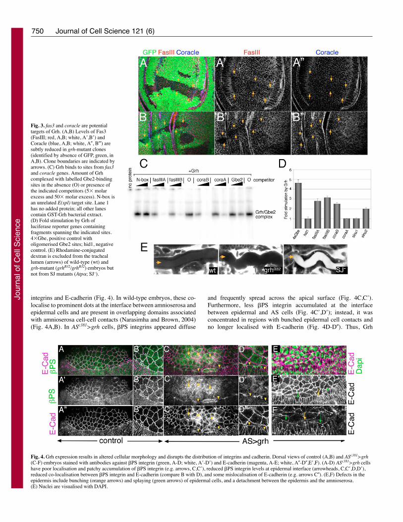
Effects of Grh expression on adhesive properties of theamnioserosa and epidermisBecause ectopic expression of Grh has a profound effect on dorsalclosure, we looked more closely at the morphology of ASc381>grhcells and the distribution of other adhesion complexes. InASc381>grh embryos there were large variations in shape and sizeof amnioserosa cells and the contacts with the adjacent dorsalepidermis were dramatically different (Fig. 4C-F and data notshown). A subset of epidermal cells had expanded contact with anamnioserosa cell at the expense of their neighbours, which becamebunched together (Fig. 4C�,D�,F). It appeared, therefore, that manyGrh-expressing amnioserosa cells had maximised the contact witha single epidermal cell, rather than making contact with five to sixcells, as in wild type. Ultimately, some amnioserosa cells appearedto lose contact with the epidermal cells.
The change in morphology in ASc381>grh embryos wasaccompanied by altered distribution of �-position-specific (�PS)
Fig. 2. Grh promotes expression of SJ components in the amnioserosa. Stage-14 control (A,C,E-I,M,N) and ASc381>grh (B,D,J-L,O,P) embryos stained asindicated. Dorsal views of whole embryos (A-D) or of amnioserosa (E-L, single confocal sections; J-J� are individual channels from one embryo).(M-O) Transverse sections stained as indicated to reveal the apical/basal distributions. (M,O) Upregulated Fas3 (FasIII) in ASc381>grh embryos accumulatesapically (green in O, white in O�; e.g. orange arrowheads). (N,P) Dlg is also upregulated in ASc381>grh embryos (green, P; white P�) and spreads along the apicalsurface (e.g. orange arrowheads). (M-P) White arrows mark lateral junction sites.
Jour
nal o
f Cel
l Sci
ence
750
integrins and E-cadherin (Fig. 4). In wild-type embryos, these co-localise to prominent dots at the interface between amnioserosa andepidermal cells and are present in overlapping domains associatedwith amnioserosa cell-cell contacts (Narasimha and Brown, 2004)(Fig. 4A,B). In ASc381>grh cells, �PS integrins appeared diffuse
and frequently spread across the apical surface (Fig. 4C,C�).Furthermore, less �PS integrin accumulated at the interfacebetween epidermal and AS cells (Fig. 4C�,D�); instead, it wasconcentrated in regions with bunched epidermal cell contacts andno longer localised with E-cadherin (Fig. 4D-D�). Thus, Grh
Journal of Cell Science 121 (6)
Fig. 3. fas3 and coracle are potentialtargets of Grh. (A,B) Levels of Fas3(FasIII; red, A,B; white, A�,B�) andCoracle (blue, A,B; white, A�, B�) aresubtly reduced in grh-mutant clones(identified by absence of GFP, green, inA,B). Clone boundaries are indicated byarrows. (C) Grh binds to sites from fas3and coracle genes. Amount of Grhcomplexed with labelled Gbe2-bindingsites in the absence (O) or presence ofthe indicated competitors (5� molarexcess and 50� molar excess). N-box isan unrelated E(spl) target site. Lane 1has no added protein; all other lanescontain GST-Grh bacterial extract.(D) Fold stimulation by Grh ofluciferase reporter genes containingfragments spanning the indicated sites.4�Gbe, positive control witholigomerised Gbe2 sites; hid1, negativecontrol. (E) Rhodamine-conjugateddextran is excluded from the tracheallumen (arrows) of wild-type (wt) andgrh-mutant (grhB32/grhB32) embryos butnot from SJ mutants (Atp�; SJ–).
Fig. 4. Grh expression results in altered cellular morphology and disrupts the distribution of integrins and cadherin. Dorsal views of control (A,B) and ASc381>grh(C-F) embryos stained with antibodies against �PS integrin (green, A-D; white, A�-D�) and E-cadherin (magenta, A-E; white, A�-D�,E�,F). (A-D) ASc381>grh cellshave poor localisation and patchy accumulation of �PS integrin (e.g. arrows, C,C�), reduced �PS integrin levels at epidermal interface (arrowheads, C,C�,D,D�),reduced co-localisation between �PS integrin and E-cadherin (compare B with D), and some mislocalisation of E-cadherin (e.g. arrows C�). (E,F) Defects in theepidermis include bunching (orange arrows) and splaying (green arrows) of epidermal cells, and a detachment between the epidermis and the amnioserosa.(E) Nuclei are visualised with DAPI.
Jour
nal o
f Cel
l Sci
ence
751Grh regulates septate junction proteins
perturbs other adhesive characteristics of amnioserosa cells. Thiscould be an indirect consequence of the increase in SJ proteins,causing altered distribution of apical and basal adhesion receptors,or Grh could additionally regulate the expression levels of cadherinand integrins (Almeida and Bray, 2005). Whichever themechanism, amnioserosa cells acquire altered adhesion propertieswith neighbouring epidermal cells, which could explain why dorsalclosure is perturbed.
Concluding commentsThe results we obtained from ectopic Grh expression have helpeduncover functions that are not easily evident from loss-of-functionexperiments and suggest that Grh is normally involved in fine-tuning the expression levels of proteins, such as Fas3, Coracle,Sinu, Nrx and Dlg, which are involved in conferring robust barrierfunction on the epidermis. Given the observation that GRHL3 alsofine-tunes the levels of junction proteins in mice (Yu et al., 2006),it appears that this represents a highly conserved aspect of Grhfunction. In addition, Grh might be intrinsic to the observed cross-talk between the extracellular matrix and junctional complexes(Tonning et al., 2005; Wang et al., 2006), because it plays a role inregulating both elements.
Grh transcription factors are also components in a conservedmechanism for wound healing, in part via their effect onextracellular matrix deposition/synthesis (Mace et al., 2005;Stramer and Martin, 2005). Our results suggest that regulation ofcell junctions might also be important for epidermal ‘sealing’. Theyfurther suggest that differences in Grh levels or activity couldregulate morphogenesis within an epithelium, as well as the abilityof epithelia to adhere to one another, by influencing the levels anddistribution of septate/tight junction proteins and other adhesionmolecules. This could also explain the role of GRHL3 duringneural tube closure in mice (Ting et al., 2003), an epithelial fusionevent that shares features with dorsal closure.
Materials and MethodsGeneticsFor ectopic expression of Grh, c381::Gal4 and G332-Gal4 drivers were combinedwith UAS::grhN/K, and embryos from 4- to 6-hour collections were aged at 25°C toenrich for stages 13-16. grh-mutant clones were induced (1 hour at 37°C) in larvaeof the genotype hsFLP/w; FRT42D grhB32/FRT42D PcEGFP.
ImmunochemistryWhole-mount staining of embryos was performed according to standard procedures.Primary antibodies were monoclonal mouse anti-�PS (CF6G11, 1:3), anti-Fas3(7G10 1:30), anti-Dlg (4F3, 1:100) and anti-Atp� (a5, 1:100), all obtained from theDevelopmental Studies Hybridoma Bank; rabbit anti-Dlg (1:500-1000), anti-Sinu(Wu et al., 2004); rat anti-E-cadherin [1:20 (Oda et al., 1994)]; guinea-pig anti-Coracle [1:1000 (Fehon et al., 1994)] and anti-Nrx (Baumgartner et al., 1996).Alexa-Fluor-488/568 (Molecular Probes) Cy2, Cy3 or Cy5 (JacksonImmunodiagnostics)-conjugated secondary antibodies were used at 1:200. Thicksections of fluorescently stained embryos were made as previously described(Narasimha and Brown, 2006).
Fluorescent images were acquired on a BioRad Radiance 2000 confocalmicroscope. Where projections are presented, sections were scanned at 0.2-0.5micron steps. Note that, in Fig. 3A,B the GFP channel from a projection issuperimposed on one optical section of the protein staining.
Target-site analysis, electrophoretic mobility-shift assays andluciferase assaysDNA sequence spanning the fas3, coracle and sinu genes was searched for a matchto a weighted matrix derived from known Grh sites (Almeida and Bray, 2005)using Target-Explorer (Sosinsky et al., 2003). The UCSC vista browserhttp://pipeline.lbl.gov/ cgi-bin/gateway2?bg=dm1 was used to determineconservation between D. melanogaster and other Drosophila species (Couronne etal., 2003). Positions of sites with respect to starting ATG in D. melanogaster are:fas3A, 31,097 bp downstream; fas3B, 34,852 bp downstream; coraA, 1169 bpupstream; coraB, 4402 bp upstream.
Electrophoretic mobility-shift assays (EMSAs) were carried out as describedpreviously (Uv et al., 1994). Reactions contained 0.5 �l of a 1:10 dilution of bacterialextract containing Gst-P/E fusion protein, 20 femtomoles of 32P-labelled gbe2double-stranded oligonucleotide (5�-CTAGCGATTGAACCGGTCCTGCGGT-3�;underlined oligonucleotides correspond to putative Grh-binding sites) and 100 fM(femtomoles) or 1 pM (picomole) of the following cold competitors where indicated:fas3A, 5�-CTAGATCGCAACCGGTTTGGGT-3�; fas3B, 5�-CTAGAGGGAAC -CAG TTTT GCCT-3�; coraA, 5�-CTAGAGCAAACTGGTTCAGCT-3�; coraB, 5�-CTAGAAAAAACCGGTTGTT-3�; and N-box, 5�-GATCAGCCACGAG CCAC -AAGGATTG-3�.
For luciferase assays, fragments encompassing the Grh-binding sites wereamplified from genomic DNA by PCR and subcloned into a pGL3-min luciferasereporter vector containing the minimal hsp70 promoter. Details available on request.Resulting plasmids were transfected into Drosophila S2 cells with a renilla controlplasmid in the presence or absence of a plasmids expressing Grh (pMT-Gal4 + UAS-GrhN). Transfection conditions and luciferase assays (Promega) were carried out asdescribed previously (Nagel et al., 2005).
We thank Greg Beitel, Manzoor Bhat, Peter Bryant, Rick Fehon andHiroki Oda for antibodies, and members of our labs for discussions.This work was supported by grants from the Medical Research Council(S.J.B.), the Wellcome Trust (N.H.B.) the Swedish Research Council(A.U.) and TIFR (M.N.).
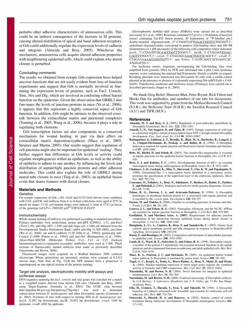
ReferencesAlmeida, M. S. and Bray, S. J. (2005). Regulation of post-embryonic neuroblasts by
Drosophila Grainyhead. Mech. Dev. 122, 1282-1293.Attardi, L. D., Von Seggern, D. and Tjian, R. (1993). Ectopic expression of wild-type
or a dominant-negative mutant of transcription factor NTF-1 disrupts normal Drosophiladevelopment. Proc. Natl. Acad. Sci. USA 90, 10563-10567.
Baumgartner, S., Littleton, J. T., Broadie, K., Bhat, M. A., Harbecke, R., Lengyel, J.A., Chiquet-Ehrismann, R., Prokop, A. and Bellen, H. J. (1996). A Drosophilaneurexin is required for septate junction and blood-nerve barrier formation and function.Cell 87, 1059-1068.
Behr, M., Riedel, D. and Schuh, R. (2003). The claudin-like megatrachea is essentialin septate junctions for the epithelial barrier function in Drosophila. Dev. Cell 5, 611-620.
Bray, S. J. and Kafatos, F. C. (1991). Developmental function of Elf-1: an essentialtranscription factor during embryogenesis in Drosophila. Genes Dev. 5, 1672-1683.
Chalmers, A. D., Lachani, K., Shin, Y., Sherwood, V., Cho, K. W. and Papalopulu, N.(2006). Grainyhead-like 3, a transcription factor identified in a microarray screen,promotes the specification of the superficial layer of the embryonic epidermis. Mech.Dev. 123, 702-718.
Couronne, O., Poliakov, A., Bray, N., Ishkhanov, T., Ryaboy, D., Rubin, E., Pachter,L. and Dubchak, I. (2003). Strategies and tools for whole-genome alignments. GenomeRes. 13, 73-80.
Fehon, R. G., Dawson, I. A. and Artavanis-Tsakonas, S. (1994). A Drosophilahomologue of membrane-skeleton protein 4.1 is associated with septate junctions andis encoded by the coracle gene. Development 120, 545-557.
Furuse, M. and Tsukita, S. (2006). Claudins in occluding junctions of humans and flies.Trends Cell Biol. 16, 181-188.
Genova, J. L. and Fehon, R. G. (2003). Neuroglian, Gliotactin, and the Na+/K+ ATPaseare essential for septate junction function in Drosophila. J. Cell Biol. 161, 979-989.
Gorfinkiel, N. and Martinez Arias, A. (2007). Requirements for adherens junctioncomponents in the interaction between epithelial tissues during dorsal closure inDrosophila. J. Cell Sci. 120, 3289-3298
Hemphala, J., Uv, A., Cantera, R., Bray, S. and Samakovlis, C. (2003). Grainy headcontrols apical membrane growth and tube elongation in response to Branchless/FGFsignalling. Development 130, 249-258.
Knust, E. and Bossinger, O. (2002). Composition and formation of intercellular junctionsin epithelial cells. Science 298, 1955-1959.
Lamb, R. S., Ward, R. E., Schweizer, L. and Fehon, R. G. (1998). Drosophila coracle,a member of the protein 4.1 superfamily, has essential structural functions in the septatejunctions and developmental functions in embryonic and adult epithelial cells. Mol. Biol.Cell 9, 3505-3519.
Mace, K. A., Pearson, J. C. and McGinnis, W. (2005). An epidermal barrier woundrepair pathway in Drosophila is mediated by grainy head. Science 308, 381-385.
Nagel, A. C., Krejci, A., Tenin, G., Bravo-Patino, A., Bray, S., Maier, D. and Preiss,A. (2005). Hairless-mediated repression of notch target genes requires the combinedactivity of Groucho and CtBP corepressors. Mol. Cell. Biol. 25, 10433-10441.
Narasimha, M. and Brown, N. H. (2004). Novel functions for integrins in epithelialmorphogenesis. Curr. Biol. 14, 381-385.
Narasimha, M. and Brown, N. H. (2006). Confocal microscopy of Drosophila embryos.In Cell Biology: A Laboratory Handbook (ed. J. E. Celis), pp. 77-86. San Diego:Academic Press.
Oda, H., Uemura, T., Harada, Y., Iwai, Y. and Takeichi, M. (1994). A Drosophilahomolog of Cadherin associated with Armadillo and essential for embryonic cell-celladhesion. Dev. Biol. 165, 716-726.
Ostrowski, S., Dierick, H. A. and Bejsovec, A. (2002). Genetic control of cuticleformation during embryonic development of Drosophila melanogaster. Genetics 161,171-182.
Jour
nal o
f Cel
l Sci
ence
752
Schulte, J., Tepass, U. and Auld, V. J. (2003). Gliotactin, a novel marker of tricellularjunctions, is necessary for septate junction development in Drosophila. J. Cell Biol. 161,991-1000.
Sosinsky, A., Bonin, C. P., Mann, R. S. and Honig, B. (2003). Target Explorer: anautomated tool for the identification of new target genes for a specified set oftranscription factors. Nucleic Acids Res. 31, 3589-3592.
Stramer, B. and Martin, P. (2005). Cell biology: master regulators of sealing and healing.Curr. Biol. 15, R425-R427.
Tao, J., Kuliyev, E., Wang, X., Li, X., Wilanowski, T., Jane, S. M., Mead, P. E. andCunningham, J. M. (2005). BMP4-dependent expression of Xenopus Grainyhead-like1 is essential for epidermal differentiation. Development 132, 1021-1034.
Tepass, U. and Hartenstein, V. (1994). The development of cellular junctions in theDrosophila embryo. Dev. Biol. 161, 563-596.
Tepass, U., Tanentzapf, G., Ward, R. and Fehon, R. (2001). Epithelial cell polarity andcell junctions in Drosophila. Annu. Rev. Genet. 35, 747-784.
Ting, S. B., Wilanowski, T., Auden, A., Hall, M., Voss, A. K., Thomas, T., Parekh,V., Cunningham, J. M. and Jane, S. M. (2003). Inositol- and folate-resistant neuraltube defects in mice lacking the epithelial-specific factor Grhl-3. Nat. Med. 9, 1513-1519.
Ting, S. B., Caddy, J., Hislop, N., Wilanowski, T., Auden, A., Zhao, L. L., Ellis, S.,Kaur, P., Uchida, Y., Holleran, W. M. et al. (2005). A homolog of Drosophila grainyhead is essential for epidermal integrity in mice. Science 308, 411-413.
Tonning, A., Hemphala, J., Tang, E., Nannmark, U., Samakovlis, C. and Uv, A. (2005).A transient luminal chitinous matrix is required to model epithelial tube diameter in theDrosophila trachea. Dev. Cell 9, 423-430.
Uv, A. E., Thompson, C. R. and Bray, S. J. (1994). The Drosophila tissue-specific factorGrainyhead contains novel DNA-binding and dimerization domains which areconserved in the human protein CP2. Mol. Cell. Biol. 14, 4020-4031.
Wang, S., Jayaram, S. A., Hemphala, J., Senti, K. A., Tsarouhas, V., Jin, H. andSamakovlis, C. (2006). Septate-junction-dependent luminal deposition of chitindeacetylases restricts tube elongation in the Drosophila trachea. Curr. Biol. 16, 180-185.
Wu, V. M., Schulte, J., Hirschi, A., Tepass, U. and Beitel, G. J. (2004). Sinuous is aDrosophila claudin required for septate junction organization and epithelial tube sizecontrol. J. Cell Biol. 164, 313-323.
Yu, Z., Lin, K. K., Bhandari, A., Spencer, J. A., Xu, X., Wang, N., Lu, Z., Gill, G. N.,Roop, D. R., Wertz, P. et al. (2006). The Grainyhead-like epithelial transactivator Get-1/Grhl3 regulates epidermal terminal differentiation and interacts functionally withLMO4. Dev. Biol. 299, 122-136.
Journal of Cell Science 121 (6)
Jour
nal o
f Cel
l Sci
ence