THIQAR- PROVINCE Alaa Abdul hassen Naif · Basrah Journal of Veterinary Research,Vol.15, No.3,2016...
12
Basrah Journal of Veterinary Research,Vol.15, No.3,2016 Proceeding of 5 th International Scientific Conference,College of Veterinary Medicine University of Basrah,Iraq 520 STUDY THE PREVALENCE AND HISTOPATHOLOGICAL CHANGES OF CUTANEOUS LEISHMANIASIS IN NASSIRRIYAH CITY \ THIQAR- PROVINCE Amal KH. Khalaf , Salih K. Majeed , Alaa Abdul hassen Naif Department of Microbiology,college of Medicine, University of Thiqar, Thiqar, Iraq College of Veterinary Medicine, University of Basrah,Basrah,Iraq Department of Medicine,College of Medicine, University of Thiqar , Thiqar ,Iraq, Key words : Cutaneous leishmaniasis , Baghdad boil , histopathology. ABSTRACT Cutaneous leishmaniasis is an endemic disease in Iraq and it is become epidemic in Nassirriyah city recently . the current study has reported high rate of infection among population at any age and gender , location or occupation . males have recorded high infection compared with female and also there is some differences among age of patients with Baghdad boil . The following histopathological study were achieved in cancer research unit for college of Medicine at University of Thiqar, targeting Nassiriyah city in south of Iraq where Baghdad boil is highly distributed during the period of during the period from August 2015 to April 2016. several of tissue (skin) biopsies were obtained by dermatologist under sterilizing condition and at Al-Hussain teaching hospital where the patients have entered for treatment . the result explain the following changing : (1) Granuloma like associated with several layers of vaculated cells filled with parasite around it (2) Dermal areas of chronic granulomatous inflammation mainly formed by vaculated macrophages filled with parasite . (3) Epidermis with keratinization and vaculated prickle cells undered it vaculated macrophages with parasite.
Transcript of THIQAR- PROVINCE Alaa Abdul hassen Naif · Basrah Journal of Veterinary Research,Vol.15, No.3,2016...

Basrah Journal of Veterinary Research,Vol.15, No.3,2016
Proceeding of 5th International Scientific Conference,College of Veterinary Medicine
University of Basrah,Iraq
520
STUDY THE PREVALENCE AND HISTOPATHOLOGICAL
CHANGES OF CUTANEOUS LEISHMANIASIS IN NASSIRRIYAH
CITY \ THIQAR- PROVINCE
Amal KH. Khalaf , Salih K. Majeed , Alaa Abdul hassen Naif
Department of Microbiology,college of Medicine, University of Thiqar, Thiqar,
Iraq
College of Veterinary Medicine, University of Basrah,Basrah,Iraq
Department of Medicine,College of Medicine, University of Thiqar , Thiqar ,Iraq,
Key words : Cutaneous leishmaniasis , Baghdad boil , histopathology.
ABSTRACT
Cutaneous leishmaniasis is an endemic disease in Iraq and it is become epidemic in
Nassirriyah city recently . the current study has reported high rate of infection among
population at any age and gender , location or occupation . males have recorded high
infection compared with female and also there is some differences among age of patients
with Baghdad boil .
The following histopathological study were achieved in cancer research unit for college
of Medicine at University of Thiqar, targeting Nassiriyah city in south of Iraq where
Baghdad boil is highly distributed during the period of during the period from August 2015
to April 2016. several of tissue (skin) biopsies were obtained by dermatologist under
sterilizing condition and at Al-Hussain teaching hospital where the patients have entered
for treatment . the result explain the following changing :
(1) Granuloma like associated with several layers of vaculated cells filled with parasite
around it
(2) Dermal areas of chronic granulomatous inflammation mainly formed by vaculated
macrophages filled with parasite .
(3) Epidermis with keratinization and vaculated prickle cells undered it vaculated
macrophages with parasite.
Basrah Journal of Veterinary Research,Vol.15, No.3,2016
Proceeding of 5th International Scientific Conference,College of Veterinary Medicine
University of Basrah,Iraq
521
(4) Small granuloma and area of granulamatous inflammatory reaction mostly formed
by vaculated macrophages with parasites , few lymphocytes present between the
macrophages.
(5) Nest like of vaculated macrophages associated with dermal infiltration of
lymphocytes .
INTRODUCTION
Leishmaniasis is a parasitic disease caused by haemoflagellate Leishmania. The disease is
widespread and may cause serious health problems in communities throughout the
Mediterranean regions and the Middle East, including Iraq [1-3]. There are an estimated 12
million cases worldwide, and there are about 1.5 million new cases of cutaneous
leishmaniasis each year, of which over 90% occur in Afghanistan, Algeria, Iran, Iraq,
Saudi Arabia, Syria, Brazil and Peru [4]. Old World disease primarily is caused by
Leishmania tropica in urban areas and Leishmania major in dry desert areas [3].
In Iraq, two species are present: L tropica, the agent of anthroponotic cutaneous
leishmaniasis (ACL), and L. major, the agent of zoonotic cutaneous leishmaniasis (ZCL).
Both ACL and ZCL were reported as causative agents of leishmaniasis in Iraq, but ACL is
found mainly in suburban areas [5]. The disease is epidemiologically unstable, with large
and unpredictable fluctuations in the number of cases. The total incidence rate of cutaneous
leishmaniasis in Iraq varies from 2.3 / 100000 to 45.5 / 100000 [5].
Cutaneous leishmaniasis is characterized by one or more cutaneous lesions on areas
where sandflies have fed. Persons who have cutaneous leishmaniasis have one or more
sores on their skin. The sores can change in size and appearance over time. They often end
up looking somewhat like a volcano, with a raised edge and central crater. A scab covers
some sores. The sores can be painless or painful. Some people have swollen glands near the
sores (for example, in the armpit if the sores are on the arm or hand).(6)
The aim of the following study was to estimate the prevalence of cutaneous leishmaniasis
and to explain the histopathological changes from it.
Basrah Journal of Veterinary Research,Vol.15, No.3,2016
Proceeding of 5th International Scientific Conference,College of Veterinary Medicine
University of Basrah,Iraq
522
MATERIALS AND METHODS
- Period of study : the current study were started from distribution of the infection in
the city at August from 2015 where collected about 3400 of cases during August from
2015 to April persisting to January and February from 2016.
- Data collection : Baghdad boil were clinically diagnosed by dermatologist in hospital
and also by making direct skin smear to examine the presence of parasite in skin. Data
were collected from all patients who entered to Al- Hussain teaching hospital in
Nassiriyah city during the period from August 2015 to April 2016 and include : age ,
gender and location of patients , type and location of ulcer , number of ulcer .
- Skin biopsy : Biopsies were taken from patients by the same dermatologist and placed
in 10% of formalin to prepare it for tissue processing (9) .
- Histology : Tissues biopsy were processed according (9), briefly the tissue from the
skin collected and placed in 10% formalin for histopathological studies and dehydrated
by several dilutions of ethanol alcohol dealcoholization with xylol, then embedded with
paraffin wax blocked (3-5) mm thickness sections were obtained by microtome. The
sections were put on glass slides deparaffinised with xylol, rehydrated by alcohol and
stained by hematoxyline and eosin.
Statistical analysis :
Results were analyze based on Chi- square test using SPSS program .
RESULTS
- prevalence of infection :
A total of 3400 patient who entered Al- Hussain teaching hospital in Nassiriyah city
suffering from Baghdad boil during the period started from August 2015 to April 2016 and
they having the following data documented in tables and explained in the following pictures,
Basrah Journal of Veterinary Research,Vol.15, No.3,2016
Proceeding of 5th International Scientific Conference,College of Veterinary Medicine
University of Basrah,Iraq
523
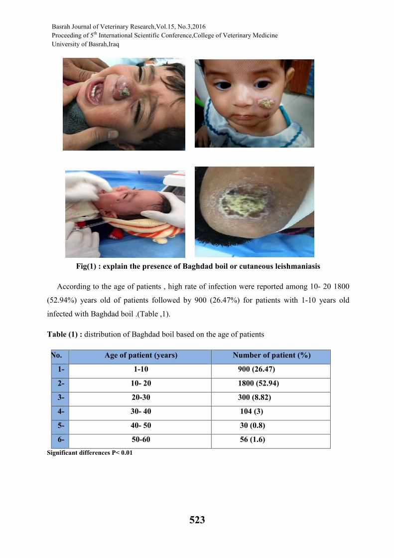
Fig(1) : explain the presence of Baghdad boil or cutaneous leishmaniasis
According to the age of patients , high rate of infection were reported among 10- 20 1800
(52.94%) years old of patients followed by 900 (26.47%) for patients with 1-10 years old
infected with Baghdad boil .(Table ,1).
Table (1) : distribution of Baghdad boil based on the age of patients
No. Age of patient (years) Number of patient (%)
1- 1-10 900 (26.47)
2- 10- 20 1800 (52.94)
3- 20-30 300 (8.82)
4- 30- 40 104 (3)
5- 40- 50 30 (0.8)
6- 50-60 56 (1.6)
Significant differences P< 0.01
Basrah Journal of Veterinary Research,Vol.15, No.3,2016
Proceeding of 5th International Scientific Conference,College of Veterinary Medicine
University of Basrah,Iraq
524
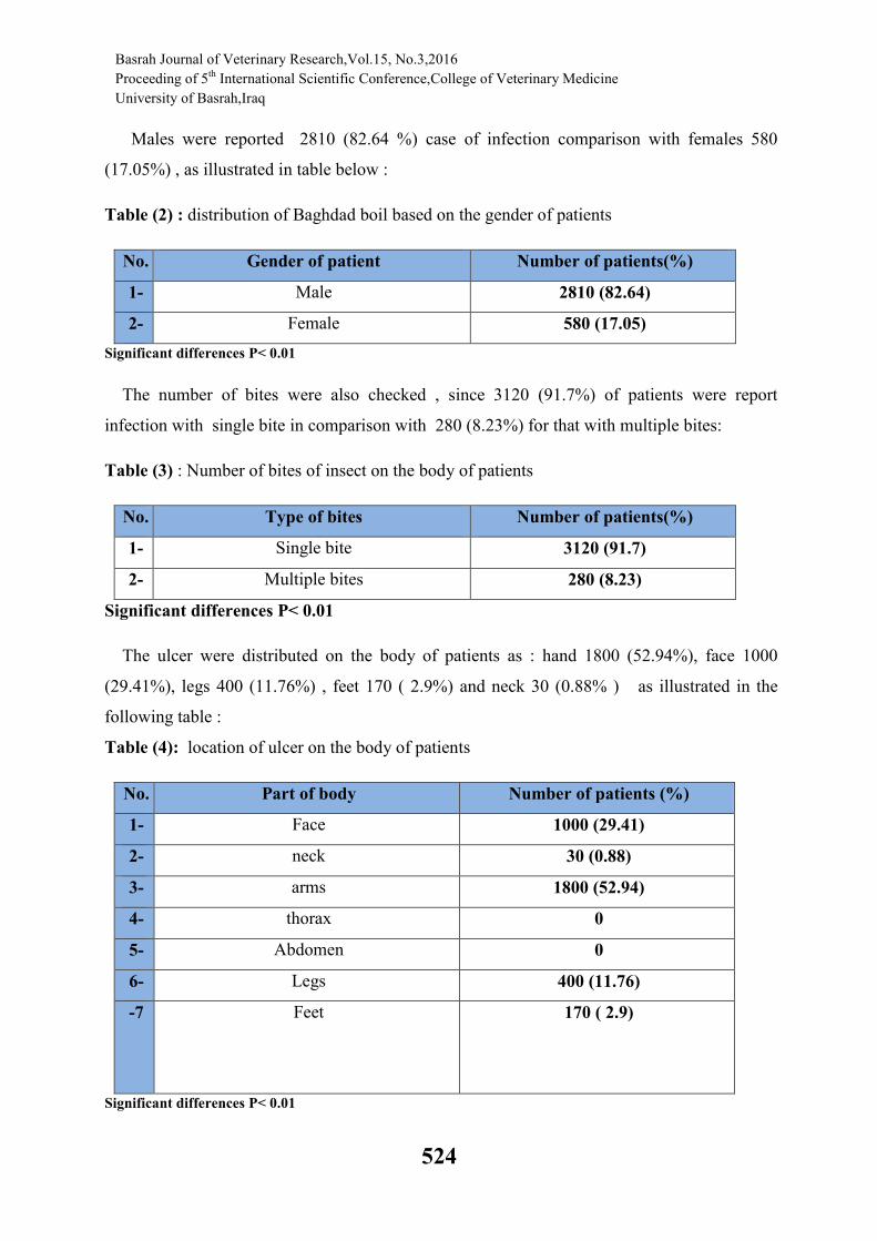
Males were reported 2810 (82.64 %) case of infection comparison with females 580
(17.05%) , as illustrated in table below :
Table (2) : distribution of Baghdad boil based on the gender of patients
No. Gender of patient Number of patients(%)
1- Male 2810 (82.64)
2- Female 580 (17.05)
Significant differences P< 0.01
The number of bites were also checked , since 3120 (91.7%) of patients were report
infection with single bite in comparison with 280 (8.23%) for that with multiple bites:
Table (3) : Number of bites of insect on the body of patients
No. Type of bites Number of patients(%)
1- Single bite 3120 (91.7)
2- Multiple bites 280 (8.23)
Significant differences P< 0.01
The ulcer were distributed on the body of patients as : hand 1800 (52.94%), face 1000
(29.41%), legs 400 (11.76%) , feet 170 ( 2.9%) and neck 30 (0.88% ) as illustrated in the
following table :
Table (4): location of ulcer on the body of patients
No. Part of body Number of patients (%)
1- Face 1000 (29.41)
2- neck 30 (0.88)
3- arms 1800 (52.94)
4- thorax 0
5- Abdomen 0
6- Legs 400 (11.76)
-7 Feet
170 ( 2.9)
Significant differences P< 0.01
Basrah Journal of Veterinary Research,Vol.15, No.3,2016
Proceeding of 5th International Scientific Conference,College of Veterinary Medicine
University of Basrah,Iraq
525
Dry ulcer is the type of ulcer that was highly distributed among patients 3400 (100%) with
Baghdad boil in comparison with wet ulcer which is consist the total infection in the city , as
tabled (5) below :
Table (5): type of ulcer on the body of patients
No. Type of ulcer Type of ulcers(%)
1- Dry ulcer 3400 (100)
2- Wet ulcer 0
Significant differences P< 0.01
- Histopathological study :
The following histopathological changing has associated with Baghdad boil , the
clinical infection with cutaneous leishmaniasis cased by the protozoan Leishmania tropica
, as illustrated in the following figures;
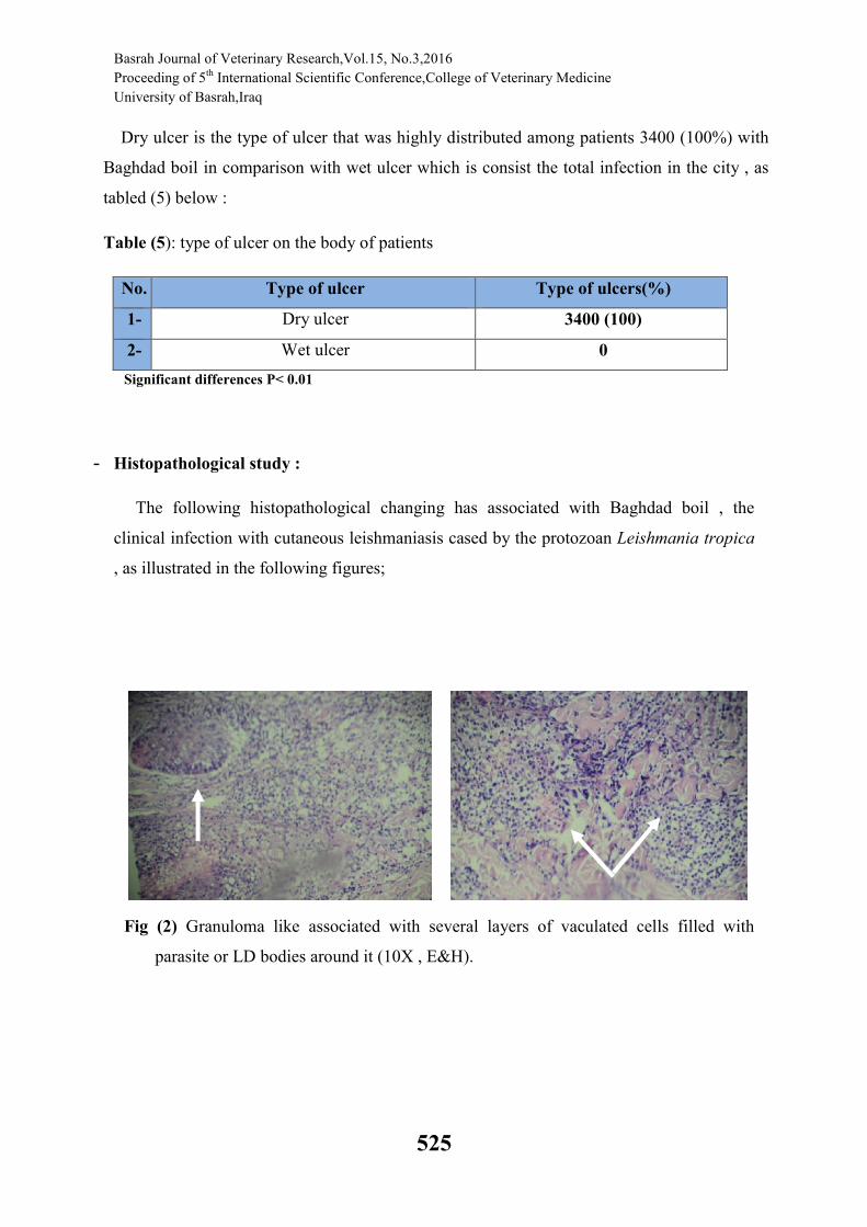
Fig (2) Granuloma like associated with several layers of vaculated cells filled with
parasite or LD bodies around it (10X , E&H).

Basrah Journal of Veterinary Research,Vol.15, No.3,2016
Proceeding of 5th International Scientific Conference,College of Veterinary Medicine
University of Basrah,Iraq
526
Fig(3) :Dermal areas of chronic granulomatous inflammation mainly formed by
vaculated macrophages filled with parasites or LD bodieis (40X, E&H)
Fig(4) Epidermis with keratinization and vaculated prickle cells under it vaculated
macrophages with parasites (40X, , E&H)
Basrah Journal of Veterinary Research,Vol.15, No.3,2016
Proceeding of 5th International Scientific Conference,College of Veterinary Medicine
University of Basrah,Iraq
527
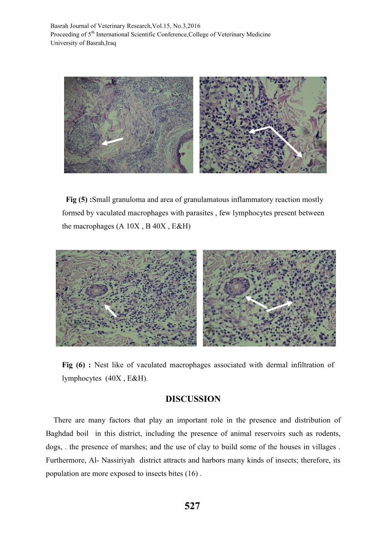
Fig (5) :Small granuloma and area of granulamatous inflammatory reaction mostly
formed by vaculated macrophages with parasites , few lymphocytes present between
the macrophages (A 10X , B 40X , E&H)
Fig (6) : Nest like of vaculated macrophages associated with dermal infiltration of
lymphocytes (40X , E&H).
DISCUSSION
There are many factors that play an important role in the presence and distribution of
Baghdad boil in this district, including the presence of animal reservoirs such as rodents,
dogs, . the presence of marshes; and the use of clay to build some of the houses in villages .
Furthermore, Al- Nassiriyah district attracts and harbors many kinds of insects; therefore, its
population are more exposed to insects bites (16) .
Basrah Journal of Veterinary Research,Vol.15, No.3,2016
Proceeding of 5th International Scientific Conference,College of Veterinary Medicine
University of Basrah,Iraq
528
In this study the infection was reported about 3400 of cases during August to April
persisting to January and February from 2016 . This rate was higher than those reported for
other geographical areas in Iraq. In two community-based studies, the incidence was 2.5 case /
10,000 for Tikrit [11] in 2000 and 15 cases / 10,000 for Kirkuk city [12] in 2000. Additionally,
in a hospital-based study performed in Samara [13], the incidence rate was 5.5 cases / 10,000
for the year 1994. However, the incidence rate was similar to that reported for Afghanistan
[14] and Eastern Venezuela [15] .
Our findings also indicate that new cases of CL tended to increase in October and reach a
maximum in January and February. The incidence rate of infection then starts to decline from
March and reaches its lowest point in April. It was observed that the majority of CL patients
attended the hospital between the months of October and March. This concurs with that
reported by (16), but not with those reported for Iran [19] or Afghanistan [14]. This variation
in seasonal peak could be due to the existence of various dominant reservoir species in each
study area as well as to the activity of the sand flies. The differences in monthly distribution of
CL patients might also be related to the development of female insects and their requirement
of blood during their life cycle for the maturation and development of eggs, especially in
spring season. The lapse of time between when the patient was bitten and the appearance of
skin lesions might be related to the long incubation period of leishmaniasis (two to four
months).
CL cases occurred more in males than in females. This result was in agreement with those
reported by [16,17], [18 [19] for Iran, and Arfan for Pakistan. In contrast, [20] in Tikrit
reported higher rates in females. These differences may be explained on the basis of variations
between studies with regard to factors such as the size of the study population, the study
design, climatic variations, and culture.
The incidence rate of CL in patients was higher in males as compared to females. This
difference in the incidence rate of infection could be due to males in this age group playing
outdoors without clothes and swimming in the rivers or lakes. This finding was in agreement
with those reported for Iraq [16,21,]. The high incidence of ZCL may be due to the presence of
reservoir animals in large numbers in this area, especially rodents and dogs. However, today,
Afghanistan, Algeria, Brazil, Iran, Iraq, Peru, Saudi Arabia and Syria account for over of the
90% of the world’s estimated 1.5 million cutaneous leishmaniasis cases [14,18]. Obviously,
dense populations of natural ZCL hosts, together with abundant vector sand flies, are the key
elements responsible for the high rate of human infection in the Nassiriyah area. ZCL in our

Basrah Journal of Veterinary Research,Vol.15, No.3,2016
Proceeding of 5th International Scientific Conference,College of Veterinary Medicine
University of Basrah,Iraq
529
country may primarily affect farmers and nomads, who are chiefly exposed to night biting
sand flies. In addition, the presence of high gerbil population densities in the area may be
blamed as reservoirs of infection that are supported by the crops for which the irrigation canals
had been constructed. Furthermore, the canal embankments serve as densely populated and
favored rodent/sand fly infestation areas.
Histopathological changing of skin during cutanous leishmaniasis explain the granulomatous
reaction in the site of infection together with presence of amastigote stage or LD bodies in the
lesion based on the current study. The essential feature of CL pathology is the colonization by
amastigotes of cells of the mononuclear phagocytic system and the resulting granulomatous
inflammatory response. A granuloma is defined as a compact collection of mature
mononuclear phagocytes, not necessarily accompanied by accessory features such as necrosis.
Conceptually granuloma evolves in three stages: 1) the development of an infiltrate of young
mononuclear phagocytes; 2) the maturation and aggregation of these cells into an unorganized
granuloma; and 3) the potential maturation of these cells into an epithelioid or organized
granuloma (7,8,9.10)
Cutaneous Leishmaniasis presents a spectrum of manifestations both clinically and
histologically. Lesions can present as nodule, plaque or ulcer mostly present on exposed sites
(8,9). Histopathological findings in acute CL include dermal infiltrate predominantly
consisting of macrophages containing large number of leishmania organism called LD
bodies.5,6 In addition plasma cells and dense mixed inflammatory cell infiltrate are also
present in dermis. When ulceration occurs secondary infiltration with neutrophils occur. (7, 8)
The morphology of LD bodies in histopathological sections were rounded with a nucleus and
kinetoplast, in some sections spindle shape form similar to smear morphology were detected.
محافظة ذي / راسة التغییرات المرضیة النسجیة وانتشار داء اللشمانیا الجلدیة في مدینة الناصریة د
قار
عالء عبد الحسن نایف،صالح مجید كاظم ، امل خضیر خلف
الخالصة
ؤخرا بشكل وباء في مدینة یعتبر مرض اللشمانیا الجلدیة من االمراض المتوطنھ في العراق والتي انتشرت م
سجلت الدراسة الحالیة . 2016لغایة شھر نیسان 2015مركز محافظة ذي قار خالل الفترة بین شھر اب / الناصریة
اذ سجلت اعلى نسبة اصابة بین الذكور ، معدل عالي لھذا المرض بین االشخاص من كافة االعمار ولكال الجنسین

Basrah Journal of Veterinary Research,Vol.15, No.3,2016
Proceeding of 5th International Scientific Conference,College of Veterinary Medicine
University of Basrah,Iraq
530
كما شملت الدراسة التعرف على التغییرات النسیجیة الحاصلھ في الجلد . ث العمر اضافة الى بعض االختالفات من حی
من خالل تحضیر وفحص ودراسة المقاطع النسیجیة في وحدة بحوث السرطان التابعة لكلیة طب ذي قار بعد متابعة
: رات التالیة وقد تم مالحظة التغیی، المرضى في مستشفى الحسین التعلیمي التابع لمركز مدینة الناصریة
.ظھور الطفیلي بشكل تجمعات ملتھمة داخل خالیا البلعم الكبیر ذات مظھر حبیبي -1
. انتشار المظھر الحبیبي للطفیلي في مناطق متعددة من طبقة االدمھ من الجلد والخاصة بوجود البثرة -2
. یلي البیضوي الشكل تصلب والتھاب مزمن في البشرة ناتج عن تجمع الخالیا البلعمیة الحاویة على الطف -3
بعض المناطق الحبیبة الحاویة على الخالیا البلعمیة الملتھمة للطفیلي شھدت تجمع للخالیا الدمویة البیضاء -4
.االمفاویة
. كما ظھرت مجامیع لھذه الخالیا مع الخالیا الدمویة البیضاء اللمفاویة في مناطق اخرى من البشرة -5
REFERENCES
1. CDC (2004) Update CL in US. Military personnel. Southwest / Central Asia, 2002-
2004. MMWR 53:264-265.
2. Hepburn NC (2003) Cutaneous leishmaniasis: an overview. JPGM 49:50-54.
3. Markle WH and Makhoul K (2004) Cutaneous leishmaniasis Recognition and
Treatment. American Family Physician. 69: 1-9.
4. Ashford RW, Desjeux P, de Raadt P (1992) Estimation of population at risk of
infection and number of cases of leishmaniasis. Parasitol Today 8:104-105.
5. WHO (2003) Communicable Disease Working Group on Emergencies, HQ Division
of Communicable Disease Control, EMRO, WHO OFFICE, Baghdad. WHO Office,
Baghdad. Communicable Disease Toolkit, IRAQ CRISIS. WHO 2003:39-44.
6. Arfan B, Rahman S (2006) Correlation of clinical, histopathological, and
microbiological finding in 60 cases of cutaneous leishmaniasis. IJDVL 72:28-32.
7. Akilov OE, Khachemoune A, Hasan T. Clinical manifestations and classification of
Old World cutaneous leishmaniasis. Int J Dermatol 2007; 46(2): 132-42.
8. Gazozai SU, Iqbal J, Bukhari I, Bashir S. Comparison of diagnostic methods in
cutaneous leishmaniasis (histopathology compared to skin smears).Pak J Pharm Sci
2010; 23(4):363-6.
9. Rahman SB, Bari A U. Histopathological patterns of Cutaneous Leishmaniasis with
clinical correlation seen in Pakistan. PakArmed Forces Med J 2003; 53(2):142-7.
Basrah Journal of Veterinary Research,Vol.15, No.3,2016
Proceeding of 5th International Scientific Conference,College of Veterinary Medicine
University of Basrah,Iraq
531
10. Mashhood AA, Khan I, Nazir S. Histopathological spectrum of cutaneous
leishmaniasis in North West Frontier Province. J Pak Assoc Derma 2004; 14(4):210-
4.
11. Alaa NH (2002) Epidemiology of skin diseases in Tikrit and vicinity: a community
based study. M Sc thesis, Tikrit University College of Medicine.
12. Murtada SJ (2001) Epidemiology of skin diseases in Kirkuk. MSc thesis, Tikrit
University College of Medicine.
13. Alsamarai AGM. Prevalence of Skin Diseases in Samara, Iraq. MEJIM. In press.
14. Faulde M, Schrader J, Heyl G, Amirih M (2008) Differences in transmission
seasons as an epidemiological tool for characterization of anthroponotic and
zoonotic cutaneous leishmaniasis in northern Afghanistan. Acta Tropica 105:131-
138.
15. Jourquera A, Ledezma E, Sousa L, et al. (1998) Epidemiologic characterization of
American cutaneous leishmaniasis in an endemic region of Eastern Veezula. Am J
Trop Med Hyg 58:589-593.
16. 16- AlSamarai and AlObaidi – Cutaneous leishmaniasis in Iraq J Infect Developing
Countries 2009; 3(2):123-129. 129 .
17. Sarhan ER. (1998) Study on Epidemiology of Cutaneous leishmaniasis in Baghdad.
MSc Thesis submitted to College of Medicine, University of Baghdad.
18. Sharifi I, Ferkeri AR and Aflatonian MR (1998) Cutaneous leishmaniasis in
primary school children in the South- eastern Iranian city of Bam, 1994-1995. Bull.
WHO 76: 289-293.
19. Talari SA, Shajari G, Talaei R (2006) Clinical finding of cutaneous leishmaniasis
as a new focus of Iran. Internet J Infec Dis 1 (2).
20. AL-Zaidawi KA (1997) New approach for treatment of cutaneous leishmaniasis by
manitol. Diploma dissertation, College of Medicine, University of Tikrit, Iraq.