Thin Layer Chromatography of KREBS Cycle Acids
5
Kraiker and Burch: Thin layer chromatography of KREBS cycle acids 393 Z. Klin. Chem. Klin. Biochem. 11. Jg. 1973, S. 393—397 Thin Layer Chromatography of KREBS Cycle Acids By H. P. KRAIKER and R. E. BuRCH 1 ) From the Medical Service, Francis Delafield Hospital and The Department of Medicine', Columbia University College of Physicians and Surgeons, New York, N. Y. 10032 (Eingegangen am 6. September 1972/3. Mai 1973) A systematic appraisal of a large number of solvent systems was carried out for the separation of lactic, pyruvic and 3-hydroxybutyric acids and all of the KREBS cycle acids, except oxalosuccinic, using silica gel G and cellulose thin layer Chromatographie plates. Four solvent systems which allowed separation of various combinations of acids are described and the limits of this method of separation are detailed. Für die Trennung organischer Säuren wurde eine Wertung einer großen Zahl von Lösungsmittelsystemen durchgeführt. Untersucht wurde die dünnschichtchromatographische Trennung von Milch-, Brenztrauben- und 3-Hydroxybuttersäure sowie aller Säuren des Citro- nensäurezyklus außer Oxalbernsteinsäure anSilicagel-G- und Celluloseplatten. Vier Lösungsmittelsysteme, die die Trennung verschiedener Kombinationen dieser Säuren gestatten, werden mitgeteilt. Die Grenzen dieser Trennungsmethode werden eingehend diskutiert. To our knowledge the only procedures which satis- factorily separate the KREBS cycle acids as well as some other acids of biological importance (lactic, pyruvic and ß-hydroxybutyric acids) are column partition and ion exchange chromatography. The first method uses the principle of an inert support material and a gradient elution system of non-polar and polar solvents (1, 2). The latter procedure applies the principle of gradient elution in connection with an ion exchange resin (3, 4, 5) (e. g. formic acid in increasing concentrations and the formate form of a Dowex-I column [3]). Column parti- tion chromatography has been the basic principle of an organic acid autoanalyzer recently developed by KESNER and MUNTWYLER (6). Gas-liquid chromatography has also been used to separate weak organic acids (7, 8) but is still in the early stages of development. Both in paper and thin-layer chromatography only a few reports have been published dealing with the se- paration of non-amino organic acids, including a few belonging to the KREBS cycle as well as lactic, pyruvic and other acids. Whereas mixtures of an homologous series, e. g. the w-monocarboxylic acids (9), can be separated quite easily, the separation of complex mix- tures of acids can hardly be expected to be complete. The difficulties of separation have been reflected in a series of papers by KAUFMANN and his co-workers (10—15) who almost always achieved incomplete sepa- ration of complex acid mixtures of biological or indus- trial importance on silica-gel and alumina columns. Mix- tures of saturated dicarboxylic acids (C 2 —C 8 ) including succinic acid have been satisfactorily separated on silica- gel G using benzene-methanol-glacial acetic acid and benzene-dioxane-glaciäl acetic acid as the solvent system by PETROWITZ and PASTUSKA (16). BRAUN and GEENEN achieved separation of the same acids on silica-gel G using a mixture of ethanol-ammonia-H 2 O (17). PREY et. al. separated formic, lactic and pyruvic acids (18); PASTUSKA and PETROWITZ (19) separated the cis-trans isomers of maleic, crotonic, //^-crotonic, tiglic, citraconic, mesaconic, itaconic, and aconitic acids. It is noteworthy that the trans forms of these acids migrated faster than the cis forms. Since thin layer chromatography is fast and inexpensive and has not been used extensively as a means of sep- arating KREBS cycle acids, we compared a wide spec- trum of thin layer systems and evaluated their efficacy in separating lactic, pyruvic, /S-hydroxybutyric and the KREBS cycle acids. As will be seen below some of these methods are sensitive enough for many investigational purposes in which only a limited number of acids might be of interest. Methods and Materials (1) Pre-coated plates, 20 X 20 cm in size, were purchased from E. Merck, Darmstadt, Germany (distributed by Brinkmann Instru- ments, Inc. in the U. S.). The types used were Silica Gel G (7731) and Silica Gel GF-254 (7730), and Cellulose and Cellulose F. (2) DL-malic, DL-Iactic, DL-/Vo-citric, succinic, pyruvic, «V-aconitic and fumaric acids were purchased from Sigma Chemical Company, St. Louis, Missouri. Citric, 2-oxoglutaric and oxaloacetic acids were obtained from Calbiochem, Los Angeles, California, and 3-hydroxybutyric acid from Mann Research Laboratory, New York. . . Standard solutions were prepared by dissolving 100 mg of each acid except fumaric in 4 ml of acetone-water (1:1). Fumaric acid was dissolved in ethanol. Spotting was done with micro- pipettes. (3) BRINKMANN tanks were lined with filter paper and equilibrated overnight, except in a few instances where they were equilibrated for at least 6 hours. The room temperature was 23i2°C. For orientation the solvent was allowed to run at least 10 cm. To check reproducibility in promising solvents the development was inter- rupted after a pre-marked 15 cm line was reached. The plates were air-dried overnight or heated for 30—45 min at 110° C. (particul- arly when the solvent contained acetic or formic acid). x ) Supported by Grants HE-00052 from the National Institutes of Health and U-1562 from The Health Research Council of the City of New York. Z. Klin. Chem. Klin. Biochem. / 11. Jahrg. 1973 / Heft 9
Transcript of Thin Layer Chromatography of KREBS Cycle Acids

Kraiker and Burch: Thin layer chromatography of KREBS cycle acids 393
Z. Klin. Chem. Klin. Biochem.11. Jg. 1973, S. 393—397
Thin Layer Chromatography of KREBS Cycle AcidsBy H. P. KRAIKER and R. E. BuRCH1)
From the Medical Service, Francis Delafield Hospital and The Department of Medicine', Columbia UniversityCollege of Physicians and Surgeons, New York, N. Y. 10032
(Eingegangen am 6. September 1972/3. Mai 1973)
A systematic appraisal of a large number of solvent systems was carried out for the separation of lactic, pyruvic and 3-hydroxybutyricacids and all of the KREBS cycle acids, except oxalosuccinic, using silica gel G and cellulose thin layer Chromatographie plates. Four solventsystems which allowed separation of various combinations of acids are described and the limits of this method of separation are detailed.
Für die Trennung organischer Säuren wurde eine Wertung einer großen Zahl von Lösungsmittelsystemen durchgeführt. Untersuchtwurde die dünnschichtchromatographische Trennung von Milch-, Brenztrauben- und 3-Hydroxybuttersäure sowie aller Säuren des Citro-nensäurezyklus außer Oxalbernsteinsäure anSilicagel-G- und Celluloseplatten. Vier Lösungsmittelsysteme, die die Trennung verschiedenerKombinationen dieser Säuren gestatten, werden mitgeteilt. Die Grenzen dieser Trennungsmethode werden eingehend diskutiert.
To our knowledge the only procedures which satis-factorily separate the KREBS cycle acids as well as someother acids of biological importance (lactic, pyruvic andß-hydroxybutyric acids) are column partition and ionexchange chromatography. The first method uses theprinciple of an inert support material and a gradientelution system of non-polar and polar solvents (1, 2).The latter procedure applies the principle of gradientelution in connection with an ion exchange resin (3, 4, 5)(e. g. formic acid in increasing concentrations and theformate form of a Dowex-I column [3]). Column parti-tion chromatography has been the basic principle of anorganic acid autoanalyzer recently developed by KESNERand MUNTWYLER (6). Gas-liquid chromatography hasalso been used to separate weak organic acids (7, 8) butis still in the early stages of development.Both in paper and thin-layer chromatography only afew reports have been published dealing with the se-paration of non-amino organic acids, including a fewbelonging to the KREBS cycle as well as lactic, pyruvicand other acids. Whereas mixtures of an homologousseries, e. g. the w-monocarboxylic acids (9), can beseparated quite easily, the separation of complex mix-tures of acids can hardly be expected to be complete.The difficulties of separation have been reflected in aseries of papers by KAUFMANN and his co-workers(10—15) who almost always achieved incomplete sepa-ration of complex acid mixtures of biological or indus-trial importance on silica-gel and alumina columns. Mix-tures of saturated dicarboxylic acids (C2—C8) includingsuccinic acid have been satisfactorily separated on silica-gel G using benzene-methanol-glacial acetic acid andbenzene-dioxane-glaciäl acetic acid as the solvent systemby PETROWITZ and PASTUSKA (16). BRAUN and GEENENachieved separation of the same acids on silica-gelG using a mixture of ethanol-ammonia-H2O (17).PREY et. al. separated formic, lactic and pyruvic acids
(18); PASTUSKA and PETROWITZ (19) separated thecis-trans isomers of maleic, crotonic, //^-crotonic, tiglic,citraconic, mesaconic, itaconic, and aconitic acids. It isnoteworthy that the trans forms of these acids migratedfaster than the cis forms.Since thin layer chromatography is fast and inexpensiveand has not been used extensively as a means of sep-arating KREBS cycle acids, we compared a wide spec-trum of thin layer systems and evaluated their efficacyin separating lactic, pyruvic, /S-hydroxybutyric and theKREBS cycle acids. As will be seen below some of thesemethods are sensitive enough for many investigationalpurposes in which only a limited number of acids mightbe of interest.
Methods and Materials(1) Pre-coated plates, 20 X 20 cm in size, were purchased fromE. Merck, Darmstadt, Germany (distributed by Brinkmann Instru-ments, Inc. in the U. S.). The types used were Silica Gel G (7731)and Silica Gel GF-254 (7730), and Cellulose and Cellulose F.(2) DL-malic, DL-Iactic, DL-/Vo-citric, succinic, pyruvic, «V-aconiticand fumaric acids were purchased from Sigma Chemical Company,St. Louis, Missouri. Citric, 2-oxoglutaric and oxaloacetic acidswere obtained from Calbiochem, Los Angeles, California, and3-hydroxybutyric acid from Mann Research Laboratory, New York.
. . Standard solutions were prepared by dissolving 100 mg ofeach acid except fumaric in 4 ml of acetone-water (1:1). Fumaricacid was dissolved in ethanol. Spotting was done with micro-pipettes.(3) BRINKMANN tanks were lined with filter paper and equilibratedovernight, except in a few instances where they were equilibratedfor at least 6 hours. The room temperature was 23i2°C. Fororientation the solvent was allowed to run at least 10 cm. To checkreproducibility in promising solvents the development was inter-rupted after a pre-marked 15 cm line was reached. The plates wereair-dried overnight or heated for 30—45 min at 110° C. (particul-arly when the solvent contained acetic or formic acid).x) Supported by Grants HE-00052 from the National Institutes ofHealth and U-1562 from The Health Research Council of the Cityof New York.
Z. Klin. Chem. Klin. Biochem. / 11. Jahrg. 1973 / Heft 9

394 Kraiker and Burch: Thin layer chromatpgraphy of KREBS cycle acids
(4) Spots were visualized by:a) spraying with bromocresolgreen (22) (0.04 gm of bromocresol-grcen in 100 ml 96% cthanol adjusted with 0.1 mol/1 NaOH untilblue coloration just appears);,b) by inserting the plate into a tank with iodine vapor; orc) with potassium permanganate spray reagent (22) (0.5 g ofKMnO4 very cautiously dissolved in 15 ml of concentrated sulfuticacid). Depending on the sorbent/solvent combination used it wasnecessary in some instances to re-heat the plates for a few minutesafter spraying with bromocresol-green. To check detection byfluorescence the indicator-containing plates were used (typesGF-254 and Cellulose F); the regular plates were sprayed, afterdevelopment with the solvent, with a rhodamine spray (2 ml of0.5% rhodamine in methanol solution + 100 ml of 2 mol/1NH4OH).
ResultsSorbents and solventsInitially, separation of all the KREBS cycle acids exceptoxalosuccinic (extremely unstable) plus lactic, pyruvicand 3-hydroxybutyric acids was tried on silica gel Gwith solvent systems reported by different authors:Methanol: 5 mol/1 Ammonia (80 ml + 20 ml) (20)96% Ethanol: H2O: 25% Ammonia (100 ml + 12 ml+ 16 ml) (17)Benzene: Methanol: Acetic acid (90 ml + 16 ml +8 ml) (16)Benzene: Dioxane: Acetic acid (90 ml + 25 ml +4ml) (16)Only succinic and citric acid could be separated (citricacid tailed in each system) and they always had highlydifferent RF values. Empirically, the combination ofethanol-ammonia was most promising. By gradually in-creasing the volume percentage of ammonia the optimalratio was found to be 65 (96% ethanol) to 35 (cone,ammonia) with no additional water added. This com-bination turned out to be even better suited for celluloselayers with regard to sensitivity and sharpness of spots.
On both silica gel G and cellulose, however, separationwas incomplete (Column I in Table 1 and 2).Because of the incomplete separation obtained with thesolvent systems above, a systematic evaluation of a largenumber of solvent systems was undertaken. Again,highly "active" silica gel G layers were used as well ascomparatively "inactive" cellulose layers. With 25 μξ ofeach acid spotted, the following cornnion pure solventswere evaluated in accordance with their dielectricconstants:;/-hexane (1.9), petroleum ether (2.0), cyclohexane (2.0),carbon tetrachloride (2.2), benzene (2.3), toluene (2.4),diethyl ether (4.3), chloroform (4.8), ethyl acetate (6.0),butanol (17.8), /j-0-propanol (18.3), «-propanoi (20.3),acetone (20.7), ethanol (24.3), methanol (32.6) andwater (78.5).In regard to dielectric constants the suitable range provedto be from butanol (17.8) to ethanol (24.3). With sol-vents more polar than ethanol the acids migrated withor very close to the solvent front. With solvents lesspolar than butanol the samples remained at or close tothe origin. The only unexplained exceptions to this
. general observation, confirmed by the circular technique(21, 22), were benzene and methylbenzene (toluene).Both led to promising separation of the acids butstreaking and tailing could not be eliminated by varyingthe pH from 2 to 10 (adjusted with acetic acid, formicacid or ammonia). With diethyl ether as the solvent theacids could not be detected, most probably because ofthe fact that all of them — though to a different degree —are soluble in ether and diffuse into the plate. Ethanol,also a good solvent for most of the acids, has a muchhigher dielectric constant and does not lead to diffusionof the samples into the layer.To suppress streaks and tails «-butanol, /jtf-prop nol,0-propanol, acetone and ethanol were subjected to
Tab. 1/?F values of KREBS cycle acids chromatographed on silica gel G thin layer plates with various solvent systems
Solvent system I. Separates acids with high Ry values (3-hydroxybutyric, lactic, fumaric and 2-oxoglutaric acids) from acids with lower /?pvalues (malic, succinic and citric plus iso-citric acids). 3-Hydroxybutyric acid can be distinguished from 2^oxoglutaric if these acids are present inamounts smaller than about 150 jug. — Solvent sys t eml l . Separates lactic and 3-hydroxybutyric acids from fumaric and citric acids. If 2^oxo-glutaric acid is present it will overlap with as-aconitic and fumaric acids. 7— Solvent system III. Separates succinic from citric, fumaric andmalic acids. If a's-aconitic acid is present it will overlap with fumaric and malic acids. — Solvent system IV. Separates lactic and 3-hydroxy-butyric acids from citric plus cis-aconitic acids. Fumaric, 2-oxoglutaric, malic and succinic acids stay close to the origin and, if present, interfere
with citric plus cis-aconitic acids
Solvent system* I II III IV
c/s-AconiticCitricDL-/so-CitricFumaricDL-3-Hydroxy^butyric2-Oxoglutaric
DL-LacticDL-MalicOxaloacetate
Pyruvic
Succinic
0.13 (yellow)0.12 (yellow)origin; hard to detect0.61 (yellow)0.70 (yellow)0.58 (yellow)
0.62 (yellow)0.32 (yellow)origin (yellow)streak (iodine vapor)origin (blue)
0.46 (yellow)
close to originorigin (yellow)not detected0.14 (yellow)0.29 (yellow)0.1 (yellow)
0.31 (yellow)close to originclose to origin
not detected
clpse to .origin
0.40 (yellow)0.14 (yellow)not detected0.42 (yellow)not detected0.36 (brown;iodine vapor)not detected0.32 (yellow)0.42 (brown;0.18 iodine vapor)
- 0.41 (brown;iodine vapor)0.58 (yellow)
close to originorigin (yellow)not detected0.14 (yellow)0.27 (yellow)**0.12 (yellow)
0.23 (yellow)** origin (blue)close to originstreak (iodine vapor)
streak (iodine vapor)0
0.10 (yellow)
* The Roman numerals correspond to the solvent systems described in the text.** Detected only with 100 μ% spotted.
2. Klin. Chem. Klin. Biochem.̂ 11. Jahrg. 1973 / Heft 9
Kraiker and Burch: Thin layer chromatography of KREBS cycle acids 395
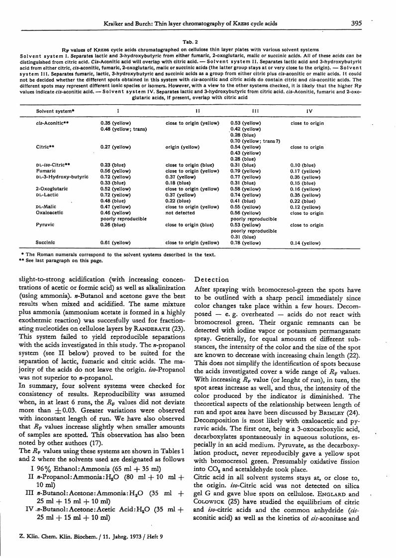
Tab. 2RF values of KREBS cycle acids chromatographed on cellulose thin layer plates with various solvent systems
Solvent system I. Separates lactic and 3-hydroxybutyric from either fumaric, 2-oxogIutaric, malic or succinic acids. All of these acids can bedistinguished from citric acid. C/s-Aconitic acid will overlap with citric acid. — Solvent system II. Separates lactic acid and 3-hydroxybutyricacid from either citric, cis-aconitic, fumaric, 2-oxogIutaric, malic or succinic acids (the latter group stays at or very close to the origin). — Solventsystem III . Separates fumaric, lactic, 3-hydroxybutyric and succinic acids as a group from either citric plus c/s-aconitic or malic acids. It couldnot be decided whether the different spots obtained in this system with c/s-aconitic and citric acids do contain citric and c/s-aconitic acids. Thedifferent spots may represent different ionic species or isomers. However, with a view to the other systems checked, it is likely that the higher Rpvalues indicate cis-aconitic acid. — Solvent system IV. Separates lactic and 3-hydroxybutyric from citric acid. c/s-Aconitic, fumaric and 2-oxo-
glutaric acids, if present, overlap with citric acid
Solvent system*
cis-Aconitic**
Citric**
DL-iso-Citric**FumaricDL-3-Hydroxy-butyric
2-OxoglutaricDL-Lactic
DL-MalicOxaloacetic
Pyruvic
Succinic
I
0.35 (yellow)0.48 (yellow; trans)
0.27 (yellow)
0.23 (blue)0.56 (yellow)0.72 (yellow)0.33 (blue)0.52 (yellow)0.72 (yellow)0.48 (blue)0.47 (yellow)0.46 (yellow)poorly reproducible0.26 (blue)
0.61 (yellow)
I I
close to origin (yellow)
origin (yellow)
close to origin (blue)close to origin (yellow)0.37 (yellow)0.18 (blue)close to origin (yellow)0.37 (yellow)0.22 (blue)close to origin (yellow)not detected
close to origin (blue)
close to origin (yellow)
I I I
0.53 (yellow)0.42 (yellow)0.28 (blue)0.70 (yellow; trans?)0.54 (yellow)0.43 (yellow)0.28 (blue)0.31 (blue)0.79 (yellow)0.77 (yellow)0.31 (blue)0.58 (yellow)0.74 (yellow)0.41 (blue)0.55 (yellow)0.56 (yellow)poorly reproducible0.53 (yellow)poorly reproducible0.31 (blue)0.78 (yellow)
IV
close to origin
close to origin
0.10 (blue)0.17 (yellow)0.35 (yellow)0.15 (blue)0.16 (yellow)0.35 (yellow)0.22 (blue)0.12 (yellow)close to origin
close to origin
0.14 (yellow)
* The Roman numerals correspond to the solvent systems described in the text.** See last paragraph on this page.
slight-to-strong acidification (with increasing concen-trations of acetic or formic acid) as well as alkalinization(using ammonia). 0-Butanol and acetone gave the bestresults when mixed and acidified. The same mixtureplus ammonia (ammonium acetate is formed in a highlyexothermic reaction) was succesfully used for fraction-ating nucleotides on cellulose layers by RANDERATH (23).This system failed to yield reproducible separationswith the acids investigated in this study. The //-propanolsystem (see Π below) proved to be suited for theseparation of lactic, fumaric and citric acids. The ma-jority of the acids do not leave the origin. /J -Propanolwas not superior to «-propanol.In summary, four solvent systems were checked forconsistency of results. Reproducibility was assumedwhen, in at least 6 runs, the RF values did not deviatemore than ±0.03. Greater variations were observedwith inconstant length of run. We have also observedthat Rp values increase slightly when smaller amountsof samples are spotted. This observation has also beennoted by other authors (17).The Rp values using these systems are shown in Tables 1and 2 where the solvents used are designated as follows
I 96% Ethanol: Ammonia (65 ml + 35 ml)II Λ-Propanol: Ammonia :H2O (80 ml + 10 ml+
10ml)III 0-Butanol:Acetone:Ammonia:H2O (35 ml -f-
25 ml + 15 ml + 10 ml)IV ,/r-Butanol: Acetone: Acetic AcidiHjO (35 ml+
25 ml + 15 ml + 10 ml)
DetectionAfter spraying with bromocresol-green the spots haveto be outlined with a sharp pencil immediately sincecolor changes take place within a few hours. Decom-posed — e. g. overheated — acids do not react withbromocresol green. Their organic remnants can bedetected with iodine vapor or potassium permanganatespray. Generally, for equal amounts of different sub-stances, the intensity of the color and the size of the spotare known to decrease with increasing chain length (22).This does not simplify the identification of spots becausethe acids investigated cover a wide range of Rp values.With increasing Rp value (of lenght of run), in turn, thespot areas increase as well, and thus, the intensity of thecolor produced by the indicator is diminished. Thetheoretical aspects of the relationship between length ofrun and spot area have been discussed by BRIMLEY (24).Decomposition is most likely with oxaloacetic and py-ruvic acids. The first one, being a 3-oxocarboxylic acid,decarboxylates spontaneously in aqueous solutions, es-pecially in an acid medium. Pyruvate, as the decarboxy-lation product, never reproducibly gave a yellow spotwith bromocresol green. Presumably oxidative fissioninto CO2 and acetaldehyde took place.Citric acid in all solvent systems stays at, or close to,the origin. w-Citric acid was not detected on silicagel G and gave blue spots on cellulose. ENGLARD andCOLOWICK (25) have studied the equilibrium of citricand /Jo-citric acids and the common anhydride (cis-aconitic acid) as well as the kinetics of Λτ-aconitase and
Z. Klin. Chcm. Klin. Biochem. / 11. Jahrg. 1973 / Heft 9
396 Kralkcr and Burch: Thin layer chromatography of KREBS cycle acids
jso-cituc dehydrogenase. Since in this equilibrium citricacid is the strongest and most stable compound it is notsurprising that it appears as a bright yellow spot. If alarge amount of «/-aconitic acid (ISO^g) was spotted oncellulose and run in solvent system III an additionalyellow spot with the RP value of citric acid was ob-served as well as a blue spot close to the origin. Thisblue spot probably represents decomposed iso-citnc acid.If «j-aconitic acid was spotted in large amounts (150 μ%)on cellulose and run in solvent system I, an additionalyellow spot with an AF higher than citric acid was ob-served. It is likely that this spot represents formation ofsome /ra/;.r-aconitic acid (the commercial acid used inthis study was trans-isomet free) since the trans formsare known to migrate faster (19), although this was notproven by elution and en2ymatic determination. Oncellulose the yellow spots of lactic and 3-hydroxybu-tyric acids had almost identical Rp values and werefollowed by blue spots which had RF values differentfrom each other.Detectability by fluorescence was not superior to bromo-cresol green in terms oi sensitivity. Further, bromo-cresol green is more specific for organic acids.
SensitivityLactic, malic, fumaric, succinic, 2-oxoglutaric, citric and«V-aconitic acids could be visualized with 6 μ§ spottedon silica gel G. When sprayed with bromocresol green,equal amounts of a given acid gave a considerablybrighter stain on cellulose than on silica gel plates.3-Hydroxybutyric acid on both types of plates couldonly be visualized in quantities of at least 25 μξ. Withpyruvic, oxaloacetic and tso-cituc acids or their decom-position products, respectively, detectability varieswidely with such factors as the age of the standard so-lutions and whether or not the plates were heated.Generally, with these unstable acids detectability waspoor.
Quantitative estimationTheoretically, as was shown by GIDDINGS and KELLER(26), the density of the chromatographed substancealong the axes χ and y of the spot follows a GAussiandistribution. FISHER et al. (27, 28) have shown that thearea of the spot increases as the logarithm of the spotcontent. This finding indicates that exact linearitybetween the spot content and the area cannot beassumed. However, planimetric measurements has beendone by several authors with considerable accuracy(28, 29). Counting the area of spots in mm2 we foundan almost linear relationship between the area of thespot and the amount of the applied acid in the rangeof 10 to 100 ̂ g.
Two-dimensional separationTwo-dimensional separation, using a combination ofsolvent systems I—IV, is unsatisfactory for several rea-sons. First, acids having a high RF value in one systembehave similarly in other systems as well (Tables 1
and 2). Second, the spot area increases with the ex-tended lenght of run in the second dimension (24) thushampering the detection limit.However, by using system III in the first, and system Iin the second dimension on cellulose a better separationof some of the acids is obtained than in a unidimensionalsystem, provided a minimal amount of acid is present fordetection. This depends on the individual acid andgenerally is about 40 to 70 μ%. With large amounts(200 μ%) succinic and fumaric, 3-hydroxybutyric andlactic as well as 2-oxoglutaric and malic acids willpartially overlap. To avoid elution and enzymatic iden-tification procedures, 11 plates were run under identicalconditions. At the origin a mixture containing 75 μξ ofeach acid was spotted on all plates. To assure an excessof one of the eleven standard acids on each plate (forpurposes of identification) an additional 75 μξ of thestandard acid which was to be added in excess wasspotted at the origin. Thus, each plate contained 75 μ§of 10 standard acids and 150 ^g of the individualstandard which was to be present in excess. Lactic(tailing), 3-hydroxybutyric, fumaric, succinic, malic andcitric acids could be located by this procedure. 2-Oxo-glutaric acid after the second-dimension could only bevisualized with iodine vapor and did not have the RFvalues expected from the unidimensional runs. Theo-retically, the spots of 2-oxoglutaric, oxaloacetic andmalic acids are overlapping. In practice, the first twodecompose and only malic acid will be visualized withbromocresol green. Citric acid theoretically interfereswith the blurred blue spot of pyruvate. The latter couldnot be visualized in the citric acid area. m-Aconitic acidwas not detected after the second-dimension run. A largeblue spot in the lower left quadrant of the plate prob-ably corresponds to a mixture of decomposition pro-ducts of /Jo-citric, 3-hydroxybutyric and pyruvic acids.
DiscussionTheoretically, heterogenous acids such as the ones stud-ied could not be expected to be easily separated bystandardized thin layer chromatography procedures.Since they do not represent a particular structural group.(e. g. 2-oxo-acids, 3-hydroxy-acids, aliphatic series etc.)separation depends on the respective molecule structureas related to given sorbent/solvent-systems. Acids form-ing a biological cycle, such as the KREBS-cycle, arehighly different with regard to their physico-chemicalproperties, particularly their oxidation state, tendencytowards isomerization, pH-dependency and heat sta-bility.Thus, this article must be understood 'as an empiricalapproach to an optimal separation with solvents ofgradually increasing dielectric constants. This is one principleout of other possible guidelines which could have beenapplied (e. g. checking a large number -of coatingmaterials). As a general rule, this article confirms thatwhen biological materials are to be studied by thin layerchromatography, the suitable separation procedure de-
Z. Klin. Chem. Klin. Biocheqif*/ 11. Jahrg. 1973 / Heft 9

Kraiker and Burch: Thin layer chromatography of KREBS cycle acids 397
pends largely on which substances can be expected, orwhich predominate from a "clinical" point of view.Thin layer chromatography will hardly be satisfactoryfor the complete analysis fo biological samples, eitherqualitatively or quantitatively, since biological materialsin general contain too many known and unknownheterogenous compounds. Pilot studies will have todecide whether relatively inexpensive thin layer chromato-graphy will sujt a given problem.The study of more recent thin layer chromatographyliterature reveals, consequently, that the thin layer chro-matography-analysis of biological material is most oftenrestricted to certain structurally related compounds,particularly those differing only in chain length. The2-oxo-carbonic acids of urine have been separated invitro by BAY&ER (30) and by LUTZ and v. REUTERN (31)with particular regard to phenylketonuria and cystinuria,as well as by STAN and SCHORMÜLLER (32). RÜDIGER,NIKISCH and GOEDDE (33) and TROP, GROSMANN andPINSKY (34) applied special procedures to distinguish2-oxo- und 3-hydroxy-acids. CHAN, CHANG andSTOFFORD (35) published a remarkable study on thinlayer chromatography separation of non-volatile bio-logical acids (out of papaya fruit material). CHURACEK
(36) has published new thin layer chromatography-reagents for the identification of acids, alcohols andamino-compounds both in vitro and in biologicalmaterial.THOMA (37) has described a method for predicting theChromatographie behaviour of a third solvent from chro-matographic data of another two. Planimetrie, photo-metric or surface significance was studied by KLAUS (38),KLOUBEK (39) and KÖHLER (40). More recently, STANand SCHORMÜLLER (32) published a method for quan-titative determination after elution, whereas RÜDIGERet al. introduced isotope techniques to separate com-pounds of close or overlapping RF-values (33).Studies such as the ones indicated confirm that thethin layer chromatography separation of biological ma-terials can be predicted only to a certain degree', since toomany parameters are usually involved. For a given pro-blem, therefore, empirical studies must be carried outsince theoretical prediction is limited.Recent papers underline the fact that thin layer chro-matography-procedures must be worked out to suit agiven problem or modified whenever a standardized pro-cedure is applied to separate biological material otherthan the one the original method was designed for.
References1. KESNER, L. & MUNTWYLER, E. (1963), J. Lab. Clin. Med. 61,604—619. — 2. SWIM, H. E. & UTTER, M. F. (1957), MethodsEnzymol. 4> 584—609. — 3. BUSCH, H., HURLBERT, R. B. & POT-TER, V. R. (1952), J. Biol. Chem. 198, 71—77. — 4. OWENS, H. S.,GOODHAN, A. E. & STARK, J. B. (1953), Anal. Chem. 25, 1507—1511. — 5. SCHENKER, H. H. & RIEMANN, W. (1953), Anal.Chem. 25, 1637—1641. — 6. KESNER, L. & MUNTWYLER, E.(1966), Anal. Chem. 38,1164—1168. — 7. ALCOCK, N. W. (1965),Anal. Biochem. //, 335—343. — 8. GEE, M. (1965), Anal. Chem.37, 926—934. — 9. HROMATKA, O. & AUE, W. A. (1963), Mh.Chemie 93, 503—512. — 10. KAUFMANN, H. P. (1939), Fette,Seifen, Anstrichmit. 46,268—274. — 11. KAUFMANN, H. P. (1940),Fette, Seifen, Anstrichmit. 47, 460—473. — 12. KAUFMANN, H. P.(1940), Angew. Chem. 53, 98—99. Int. Ed. Engl — 13. KAUF-MANN, H. P. & KIRSCH, P. (1943), Fette, Seifen, Anstrichmit. 50,519—524. —14. KAUFMANN, H. P. & SCHMIDT, O. (1940), Fette,Seifen, Anstrichmit. 47, 294—305. — 15. KAUFMANN, H. P.& WOLF, W. (1943), Fette, Seifen, Anstrichmit. 50, 519—527. —16. PETROWITZ, H. J. & PASTUSKA, G. (1962), J. Chromatogr. 7,128—130. —17. BRAUN, D. & GEENEN, B. (1962), J. Chromatogr.7, 56—59. — 18. PREY, V., BERBALK, H. & KAUSZ, M. (1962),Mikrochim. Acta, 449—461. — 19. PASTUSKA, G. & PETRO-WITZ, H. J. (1963), J. Chromatogr. 10, 517—518. — 20. WALDI, D.,unpublished, quoted after Stahl (22), 1st ed., p. 357. — 21. Iz-
MAILOW, H. & SCHRAIBER, S. (1938), Farmazia 3, 1—7. — 22.STAHL, E. (1972), Thin Layer Chromatography, 2nd ed., SpringerVerlag, Berlin-Heidelberg-New York. — 23. RANDERATH, K.(1962), Nature (London) 194, 768—769. — 24. BRIMLEY, R. C.(1949), Nature (London) 163, 215—216. — 25. ENGLARD, S.& COLOWICK, S. P. (1957), J. Biol. Chem., 226, 1047—1058. —26. GIDDINGS, J. C. & KELLER, R. A. (1959), J. Chromatogr. 2,626—633. — 27. FISHER, R. B. & HOLMES, R. (1949), Biochemistry44, . — 28. FISHER, R. B., PEARSONS, D. S. & MORRISON, G. A.(1948), Nature (London) 161, 764—765. — 29. REID, R. L.& LEDERER, M. (1951), Biochem. J., 50, 60—73. — 30. BAYZER, H.(1971), J. Chromatogr. 57, 281—286. — 31. LUTZ, P. & v. REU-TERN, G. M. (1969), this j. 7, 586—589. — 32. STAN, H. J. &SCHORMÜLLER, J. (1969), J. Chromatogr. 43, 103—109. — 33.RÜDIGER, H. W., NIKISCH, M. & GOEDDE, H. W. (1971), J.Chromatogr. 61, 373—375. — 34. TROP, M., GROSSMANN, S.& PINSKY, A. (1968), J. Chromatogr. 38, 411—413. — 35. CHAN,H. T., CHANG, T. S., STOFFORD, A. E. & BREKKE, J. E. (1971),J. Agr. Food Chem. 19, 263—265. — 36. CHURACEK, J. (1970),J. Chromatogr. 48, 241—249. — 37. THOMA, J. A. (1965), Anal.Chem. 37, 500—508. — 38. KLAUS, R. (1967), Pharm. Ztg., 2. Mit-teilung 112, 480—484. — 39. KLOUBEK, J. (1965), Chemistry 37,500—509. — 40. KÖHLER, F. (1972), J. Chromatogr. 68, 275—279.
Dr. H. P. KraikerBundesknappschaft Klinik6603 Sulzbach/Saar
Z. Klin. Chem. Klin. Biochem. /ll. Jahrg. 1973 / Heft 9 52