Therapeutics Sorafenib, a Multikinase Inhibitor, Enhances...
13
Research Article Sorafenib, a Multikinase Inhibitor, Enhances the Response of Melanoma to Regional Chemotherapy Christina K. Augustine 1,5 , Hiroaki Toshimitsu 1,5 , Sin-Ho Jung 3 , Patricia A. Zipfel 1,5 , Jin S. Yoo 1 , Yasunori Yoshimoto 6 , M. Angelica Selim 2 , James Burchette 2 , Georgia M. Beasley 1 , Nicole McMahon 1 , James Padussis 1 , Scott K. Pruitt 1,5 , Francis Ali-Osman 4 , and Douglas S. Tyler 1,5 Abstract Melanoma responds poorly to standard chemotherapy due to its intrinsic chemoresistance. Multiple genet- ic and molecular defects, including an activating mutation in the BRaf kinase gene, are associated with mel- anoma, and the resulting alterations in signal transduction pathways regulating proliferation and apoptosis are thought to contribute to its chemoresistance. Sorafenib, a multikinase inhibitor that targets BRaf kinase, is Food and Drug Administration approved for use in advanced renal cell and hepatocellular carcinomas. Al- though sorafenib has shown little promise as a single agent in melanoma patients, recent clinical trials suggest that, when combined with chemotherapy, it may have more benefit. We evaluated the ability of sorafenib to augment the cytotoxic effects of melphalan, a regional chemotherapeutic agent, and temozolomide, used in systemic and regional treatment of melanoma, on a panel of 24 human melanoma-derived cell lines and in an animal model of melanoma. Marked differences in response to 10 μmol/L sorafenib alone were observed in vitro across cell lines. Response to sorafenib significantly correlated with extracellular signal-regulated ki- nase (ERK) downregulation and loss of Mcl-1 expression (P < 0.05). Experiments with the mitogen-activated protein kinase/ERK kinase inhibitor U0126 suggest a unique role for ERK downregulation in the observed effects. Sorafenib in combination with melphalan or temozolomide led to significantly improved responses in vitro (P < 0.05). In the animal model of melanoma, sorafenib in combination with regional melphalan or regional temozolomide was more effective than either treatment alone in slowing tumor growth. These results show that sorafenib in combination with chemotherapy provides a novel approach to enhance chemothera- peutic efficacy in the regional treatment of in-transit melanoma. Mol Cancer Ther; 9(7); OF1–12. ©2010 AACR. Introduction Melanoma is a potentially life-threatening skin cancer with nearly 160,000 new cases worldwide each year (1). In addition to the dramatic increase in incidence, melanoma accounts for one of the highest rates of pro- ductive years of life lost from malignancy due to its propensity to occur in younger individuals (2). To date, the most effective single-agent chemotherapies for the management of metastatic melanoma are the alkylating agents dacarbazine and temozolomide, for systemic disease, and melphalan, for regionally advanced dis- ease of the extremity (3–5). Long-term response rates, especially to systemically administered chemotherapeu- tic agents, however, are generally poor. Recent evidence suggests that alterations in signal transduction path- ways regulating proliferation and apoptosis may con- tribute to the intrinsically chemoresistant phenotype associated with melanoma (6). One of the most common genetic alterations, ob- served in 50% to 70% of melanomas, is a mutation that results in substitution of glutamate for valine at codon 600 of the gene encoding the BRaf serine/thre- onine kinase (7). This mutation leads to constitutive activation of BRaf kinase with the resultant unchecked stimulation of the mitogen-activated protein kinase (MAPK)/extracellular signal-regulated kinase (ERK) kinase and the ERK pathways leading to increased ac- tivation of proliferative, survival/antiapoptotic, and angiogenic pathways and, ultimately, to enhanced growth and progression of melanomas (7). The high incidence of this activating BRaf mutation and the multitude of downstream effectors that enhance tumor growth and metastasis make BRaf an attractive thera- peutic target. Authors' Affiliations: Departments of 1 Surgery, 2 Pathology, 3 Biostatistics and Bioinformatics, and 4 Surgical Sciences, Duke University Medical Center; 5 Surgical Service, Durham Veteran's Affairs Medical Center, Durham, North Carolina; and 6 Department of Surgery II, Yamaguchi University School of Medicine, Yamaguchi, Japan Note: Supplementary material for this article is available at Molecular Cancer Therapeutics Online (http://mct.aacrjournals.org/). C.K. Augustine and H. Toshimitsu contributed equally to this work. Corresponding Author: Christina K. Augustine, Durham Veteran's Affairs Medical Center, 508 Fulton Street, E4001, Durham, NC 27705. Phone: 919-286-0411, ext. 5191; Fax: 919-684-6044. E-mail: Christi. [email protected] doi: 10.1158/1535-7163.MCT-10-0073 ©2010 American Association for Cancer Research. Molecular Cancer Therapeutics www.aacrjournals.org OF1 Published OnlineFirst on June 22, 2010 as 10.1158/1535-7163.MCT-10-0073 on June 13, 2018. © 2010 American Association for Cancer Research. mct.aacrjournals.org Downloaded from Published OnlineFirst June 22, 2010; DOI: 10.1158/1535-7163.MCT-10-0073
Transcript of Therapeutics Sorafenib, a Multikinase Inhibitor, Enhances...
Published OnlineFirst on June 22, 2010 as 10.1158/1535-7163.MCT-10-0073Published OnlineFirst June 22, 2010; DOI: 10.1158/1535-7163.MCT-10-0073
Research Article Molecular
CancerTherapeutics
Sorafenib, a Multikinase Inhibitor, Enhances the Responseof Melanoma to Regional Chemotherapy
Christina K. Augustine1,5, Hiroaki Toshimitsu1,5, Sin-Ho Jung3, Patricia A. Zipfel1,5, Jin S. Yoo1,Yasunori Yoshimoto6, M. Angelica Selim2, James Burchette2, Georgia M. Beasley1, Nicole McMahon1,James Padussis1, Scott K. Pruitt1,5, Francis Ali-Osman4, and Douglas S. Tyler1,5
Abstract
Authors'3BiostatisUniversityMedical CeII, Yamagu
Note: SupCancer The
C.K. Augus
CorresponAffairs MedPhone: 91augustine@
doi: 10.115
©2010 Am
www.aacr
Dow
Melanoma responds poorly to standard chemotherapy due to its intrinsic chemoresistance. Multiple genet-ic and molecular defects, including an activating mutation in the BRaf kinase gene, are associated with mel-anoma, and the resulting alterations in signal transduction pathways regulating proliferation and apoptosisare thought to contribute to its chemoresistance. Sorafenib, a multikinase inhibitor that targets BRaf kinase, isFood and Drug Administration approved for use in advanced renal cell and hepatocellular carcinomas. Al-though sorafenib has shown little promise as a single agent in melanoma patients, recent clinical trials suggestthat, when combined with chemotherapy, it may have more benefit. We evaluated the ability of sorafenib toaugment the cytotoxic effects of melphalan, a regional chemotherapeutic agent, and temozolomide, used insystemic and regional treatment of melanoma, on a panel of 24 human melanoma-derived cell lines and in ananimal model of melanoma. Marked differences in response to 10 μmol/L sorafenib alone were observedin vitro across cell lines. Response to sorafenib significantly correlated with extracellular signal-regulated ki-nase (ERK) downregulation and loss of Mcl-1 expression (P < 0.05). Experiments with the mitogen-activatedprotein kinase/ERK kinase inhibitor U0126 suggest a unique role for ERK downregulation in the observedeffects. Sorafenib in combination with melphalan or temozolomide led to significantly improved responsesin vitro (P < 0.05). In the animal model of melanoma, sorafenib in combination with regional melphalan orregional temozolomide was more effective than either treatment alone in slowing tumor growth. These resultsshow that sorafenib in combination with chemotherapy provides a novel approach to enhance chemothera-peutic efficacy in the regional treatment of in-transit melanoma. Mol Cancer Ther; 9(7); OF1–12. ©2010 AACR.
Introduction
Melanoma is a potentially life-threatening skin cancerwith nearly 160,000 new cases worldwide each year(1). In addition to the dramatic increase in incidence,melanoma accounts for one of the highest rates of pro-ductive years of life lost from malignancy due to itspropensity to occur in younger individuals (2). To date,the most effective single-agent chemotherapies for themanagement of metastatic melanoma are the alkylating
Affi l iations: Departments of 1Surgery, 2Pathology,tics and Bioinformatics, and 4Surgical Sciences, DukeMedical Center; 5Surgical Service, Durham Veteran's Affairsnter, Durham, North Carolina; and 6Department of Surgerychi University School of Medicine, Yamaguchi, Japan
plementary material for this article is available at Molecularrapeutics Online (http://mct.aacrjournals.org/).
tine and H. Toshimitsu contributed equally to this work.
ding Author: Christina K. Augustine, Durham Veteran'sical Center, 508 Fulton Street, E4001, Durham, NC 27705.
9-286-0411, ext. 5191; Fax: 919-684-6044. E-mail: Christi.duke.edu
8/1535-7163.MCT-10-0073
erican Association for Cancer Research.
journals.org
on June 13, 2018. mct.aacrjournals.org nloaded from
agents dacarbazine and temozolomide, for systemicdisease, and melphalan, for regionally advanced dis-ease of the extremity (3–5). Long-term response rates,especially to systemically administered chemotherapeu-tic agents, however, are generally poor. Recent evidencesuggests that alterations in signal transduction path-ways regulating proliferation and apoptosis may con-tribute to the intrinsically chemoresistant phenotypeassociated with melanoma (6).One of the most common genetic alterations, ob-
served in 50% to 70% of melanomas, is a mutationthat results in substitution of glutamate for valine atcodon 600 of the gene encoding the BRaf serine/thre-onine kinase (7). This mutation leads to constitutiveactivation of BRaf kinase with the resultant uncheckedstimulation of the mitogen-activated protein kinase(MAPK)/extracellular signal-regulated kinase (ERK)kinase and the ERK pathways leading to increased ac-tivation of proliferative, survival/antiapoptotic, andangiogenic pathways and, ultimately, to enhancedgrowth and progression of melanomas (7). The highincidence of this activating BRaf mutation and themultitude of downstream effectors that enhance tumorgrowth and metastasis make BRaf an attractive thera-peutic target.
OF1
© 2010 American Association for Cancer Research.
Augustine et al.
OF2
Published OnlineFirst June 22, 2010; DOI: 10.1158/1535-7163.MCT-10-0073
Sorafenib (Nexavar), an inhibitor of multiple kinasesincluding the Raf kinases, is approved by the Food andDrug Administration for the treatment of renal cell andinoperable hepatocellular carcinomas (8, 9). Sorafenibblocks cell proliferation, cell survival, and angiogenesis,all of which can be driven by the Raf-MAPK/ERKkinase-ERK pathway (7). Sorafenib may also lead toapoptosis via downregulation of the antiapoptotic pro-tein Mcl-1 and inhibition of the nuclear translocation ofapoptosis-inducing factor (10–12). In addition, sorafenibeffectively inhibits the activity of vascular endothelialgrowth factor receptors, platelet-derived growth factorreceptor β, FLT-3, and c-Kit (9). Several of these receptorshave been implicated in melanoma biology (13, 14), mak-ing sorafenib an attractive agent for melanoma therapy.Whereas sorafenib treatment of melanoma cell linesand tumor xenografts results in cell death and tumorgrowth delay (15, 16), its use as a single agent in the treat-ment of patients with metastatic melanoma has yieldeddisappointing clinical results (17).Despite its relative ineffectiveness as a single agent in
melanoma, the ability of sorafenib to alter cell prolifera-tion and/or survival suggests that it may be useful incombination with other cytotoxic therapies. Indeed, sor-afenib can enhance the effects of radiation, rapamycin,and the small-molecule inhibitor ABT-737, which inhibitsthe Bcl-2 protein (18–20). In phase I and II studies, mela-noma patients treated with sorafenib combined with car-boplatin and paclitaxel, temozolomide, dacarbazine, orIFNα-2a showed a greater response compared with his-torical control and, in one study of dacarbazine alone,with little enhancement of toxicity (14, 21–23). To date,however, no studies have examined whether sorafenibcan augment the response to the alkylating agentsmelphalan and temozolomide in the unique setting ofregional therapy of advanced in-transit melanomausing either isolated limb infusion (ILI) or isolated limbperfusion.This study was designed to address this important ques-
tion. We characterized a panel of human melanoma-derived cell lines for the presence of the V600E BRafmutation and examined their response to either sorafenibalone or sorafenib in combination with melphalan ortemozolomide chemotherapy, both in vitro and in vivo, ina rat model of regional isolated-limb infusion chemother-apy. The results presented also provide insight into someof the mechanisms by which sorafenib enhances the cyto-toxicity of chemotherapy and the molecular changes thatoccur in melanoma cells in response to treatment.
Materials and Methods
Cell cultureMelanoma cell lines were maintained at 37°C, 5% CO2
in Iscove's modified Dulbecco's medium supplementedwith 10% fetal bovine serum, 2 mmol/L L-glutamine,and 1% penicillin/streptomycin. Duke melanoma (DM)cell lines, a generous gift from Dr. Hilliard Seigler (Duke
Mol Cancer Ther; 9(7) July 2010
on June 13, 2018. mct.aacrjournals.org Downloaded from
University Medical Center, Durham, NC), were derivedfrom human melanoma patient samples under an institu-tional review board–approved protocol. A2058 andSkMel28 were purchased from the American TypeCulture Collection (2006). All cell lines were confirmedMycoplasma-free.
ReagentsMelphalan and U0126 were purchased from Sigma.
Sorafenib was provided by Bayer Pharmaceuticals.Temozolomide was provided by Schering-Plough. Anti-bodies against β-actin, ERK, phosphorylated T202/Y204-ERK (E10), and Bim were purchased from CellSignaling Technology. Antibodies against Mcl-1 and BRafwere obtained from Santa Cruz Biotechnology. For RNAinterference studies, lipid transfection reagents wereobtained from Dharmacon (Dharmafect 2) or Invitrogen(Lipofectamine 2000). Control (AllStars Negative ControlsiRNA) and Mcl-1–specific siRNA (Hs_Mcl1_6) wereobtained from Qiagen.
BRaf and NRas mutation assayTo analyze the BRaf V600E and the NRas (codon 61)
mutations, DNA was isolated from melanoma cells linesusing the DNeasy kit (Qiagen). PCR amplification wasdone using HotStart Taq DNA polymerase (Qiagen) ina 50-μL reaction volume. See Supplementary Table S1for primer sequences and Supplementary Methods forthermocycle settings. The PCR products were purified(Qiaquick PCR Purification Kit, Qiagen) and sequencedby the Duke University DNA Analysis Facility usingthe Applied Biosystems Dye Terminator Cycle Sequenc-ing system with AmpliTaq DNA Polymerase and ABI377 PRISM DNA sequencing instruments and analysissoftware.
Cell survival assayThe sensitivity of each cell line to drug treatment
was measured with a colorimetric assay using WST-1cell proliferation reagent (Roche). Temozolomide cyto-toxicity was assessed using a 12-day assay, which, aspreviously described (24), yields in vitro responses totemozolomide that more closely mimic in vivo re-sponse. Briefly, 1 × 10⁵ cells/mL treated with temozo-lomide for 2.5 hours were plated in 96-well plates at 50to 500 cells/100 μL culture medium per well. The cul-ture medium was changed after 6 days, and cell sur-vival measured after 12 days with the WST-1 assayas described above. For testing sorafenib in combina-tion with temozolomide, cells were treated with 10μmol/L sorafenib (or 0.1% DMSO control) for 2 hours,followed by temozolomide (d0) for 2.5 hours, and plat-ed as described above. After 6 days (d6), the culturemedium was replaced with fresh medium containing0.1% DMSO or 10 μmol/L sorafenib. For melphalansensitivity, cells were plated at 3,000 to 5,000 cells/100 μL per well of a 96-well plate and incubated over-night; DMSO (vehicle control) or drugs (melphalan
Molecular Cancer Therapeutics
© 2010 American Association for Cancer Research.
Sorafenib Enhances Regional Chemotherapy in Melanoma
Published OnlineFirst June 22, 2010; DOI: 10.1158/1535-7163.MCT-10-0073
and/or sorafenib and/or U0126 in DMSO) were addedat the required concentrations. In the combinationtreatment, sorafenib was added 2 hours before melpha-lan. The final DMSO concentration in each well was≤1%. Cell line sensitivity to melphalan treatment wasmeasured after 48 hours of incubation, as describedabove. Cell survival, defined as the absorbance of thetreatment group divided by the absorbance of the con-trol group, was plotted as a function of temozolomideor melphalan concentration (see SupplementaryFig. S1), and the area under the dose-response curve(AUCdrug) was computed using GraphPad Prism v4.0sof tware over a concentrat ion range of 0 to0.5 mmol/L for temozolomide and 0 to 125 μmol/Lfor melphalan. The level of resistance to chemotherapywas defined as Rdrug (fraction resistant to drug), where
Rdrug ¼ AUCdrug=AUCmax
AUCmax represents no loss of cell survival at the drugdoses tested (AUCmax = 0.5 for temozolomide assayand 125 for melphalan assay).
Western blot analysisSubconfluent cultures of melanoma cell lines were trea-
ted with DMSO or sorafenib (10 μmol/L in DMSO) ingrowth medium for 5 or 24 hours. Cells were washedwith ice-cold PBS and lysed in a modified radioimmuno-precipitation assay buffer (see Supplementary Methods).For tumor lysis, pieces of excised tumor were homoge-nized on ice with a hand-held homogenizer in radioim-munoprecipitation assay buffer. Clarified cell and tumorlysates (10–25 μg protein) were analyzed by SDS-PAGE,followed by Western blotting and detection using theVisualizer Western Blot Detection Kit (Upstate Cell Sig-naling Solutions). Blots were visualized and quantifiedusing a Bio-Rad Versa Doc 4000 and Quantity One Imagesoftware.
siRNA transfectionA suspension of cells (41 × 104/mL) prepared in Opti-
MEM (Invitrogen) was incubated with 100 nmol/LsiRNA and 4 μg/mL lipid transfection reagent. An equalvolume of full medium was added after 5 hours and cellswere cultured overnight. Cells were plated 24 hours aftertransfection into 96-well plates at a density of 2,000 cells/well and incubated overnight. Cells were treated withdrugs 48 hours after transfection and cell survival wasmeasured after 48 hours (4 days after transfection) as de-scribed above.
Measurement of apoptosisSubconfluent cultures of cells were treated with DMSO
or sorafenib (10 μmol/L) for 2 hours before the additionof melphalan or DMSO (vehicle control). After 24 to48 hours, cells were harvested by trypsinization and thenwashed, and the level of apoptosis was measured usingthe APO-BrdU TUNEL Assay Kit (Invitrogen), as direc-
www.aacrjournals.org
on June 13, 2018. mct.aacrjournals.org Downloaded from
ted by the manufacturer with some modifications (seeSupplementary Methods). Samples were analyzed byfluorescence-activated cell sorting using a FACScan flowcytometer (BD Biosciences) and results were analyzed us-ing FloJo software (Tree Star, Inc.).
Isolated limb infusionTumor initiation was done essentially as we described
previously (25) by a s.c. injection of DM443 cells (5 × 10⁶
in 2:1 PBS/Matrigel; BD Biosciences) into the right hindlimb (see Supplementary Methods). Xenografts weremeasured with Vernier calipers and tumor volume wascalculated as (length × width2)/2. Once the tumor vol-ume had reached approximately 1 cm3, daily treatmentwith sorafenib or vehicle (60 mg/kg by oral gavage) for10 days was initiated. See Supplementary Methods fordetails of drug preparations. On day 8, ILI with saline,melphalan, or temozolomide was done as previously de-scribed (see also Supplementary Methods; ref. 25). Stocksolutions of melphalan and temozolomide were preparedfresh each day. Tumor volume was followed every otherday, as described above, for 30 additional days. All ani-mal protocols were approved by the Duke UniversityMedical Center and the Durham Veterans Affairs Medi-cal Center Institutional Animal Care and Use Commit-tees. For each treatment arm, group size was five to sixanimals. Tumor growth data are calculated as foldchange in tumor volume from that measured at the timeof ILI (d0) and plotted as a function of time following in-fusion. Quintupling time is calculated as the number ofdays for tumor volume to increase 5-fold over untreatedvolume (d0).
Immunohistochemical analysis of tumorsImmunohistochemistry was done as we previously
described (26, 27). The MP5/73 antibody to detect mel-phalan adducts was kindly provided by Dr. M.J. Tilby(Northern Institute for Cancer Research, NewcastleUniversity, Newcastle upon Tyne, United Kingdom;ref. 28). Tumor specimens were also stained for micro-vascular density using the anti-CD31 mouse monoclo-nal antibody (Dako) as well as for apoptosis withKlenow Frag-EL (EMD) according to the manufacturer'sspecifications.
StatisticsStatistical analysis of data from cell lines was done us-
ing GraphPad Prism 4 software. Differences in tumorgrowth rate were compared by taking the log transfor-mation of the fold change in tumor volume and calculat-ing the slopes for each treatment group. The logtransformation was chosen to improve the linearity ofthe time trajectory in the fold change in tumor volume.To account for possible dependency among repeated tu-mor volume measurements, two-sided P values were cal-culated using the generalized estimating equationmethod (29, 30) implemented by Proc Genmod of SASwith the working independent correlation structure.
Mol Cancer Ther; 9(7) July 2010 OF3
© 2010 American Association for Cancer Research.
Augustine et al.
OF4
Published OnlineFirst June 22, 2010; DOI: 10.1158/1535-7163.MCT-10-0073
P values ≤0.05 were considered significant. Because ofthe sample size, no multiple testing adjustments weredone when comparing the slopes between different treat-ment groups.
Results
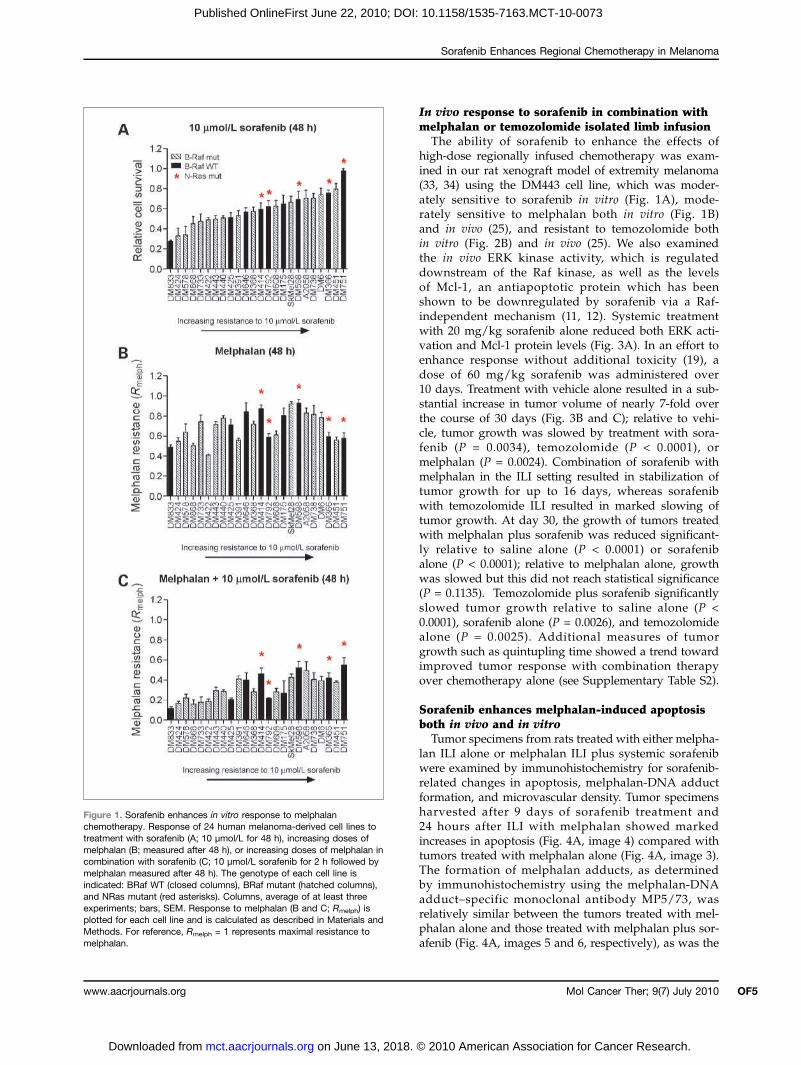
In vitro response to sorafenib, melphalan, andcombination therapyOf the 24 human melanoma-derived cell lines evalu-
ated, 15 were shown to harbor the V600E BRaf muta-tion. Of the 9 BRaf wild-type (WT) cell lines, 5 had anactivating mutation at codon 61 of the NRas gene (seeTable 1). The frequency of mutations in this panel ofhuman melanoma-derived cell lines (62.5% BRaf muta-tion and 20.8% NRas mutation) was consistent withpreviously reported rates of mutation for both genesin melanoma (7).The 24 cell lines were evaluated for baseline sensitivity
to sorafenib. The results showed modest sensitivity to5 μmol/L sorafenib across all cell lines (SupplementaryFig. S1A) at 48 hours; however, at 10 μmol/L sorafenib(Fig. 1A), there was a broad spectrum of responseranging from very sensitive (DM833; cell survival, 27%of control) to resistant (DM751; cell survival, 98% of
Mol Cancer Ther; 9(7) July 2010
on June 13, 2018. mct.aacrjournals.org Downloaded from
control). No correlation was observed between sensitivityto sorafenib alone measured at 48 hours and BRaf muta-tion status (P > 0.50), consistent with previous reports(19, 31, 32).The results of the analysis of responsiveness to mel-
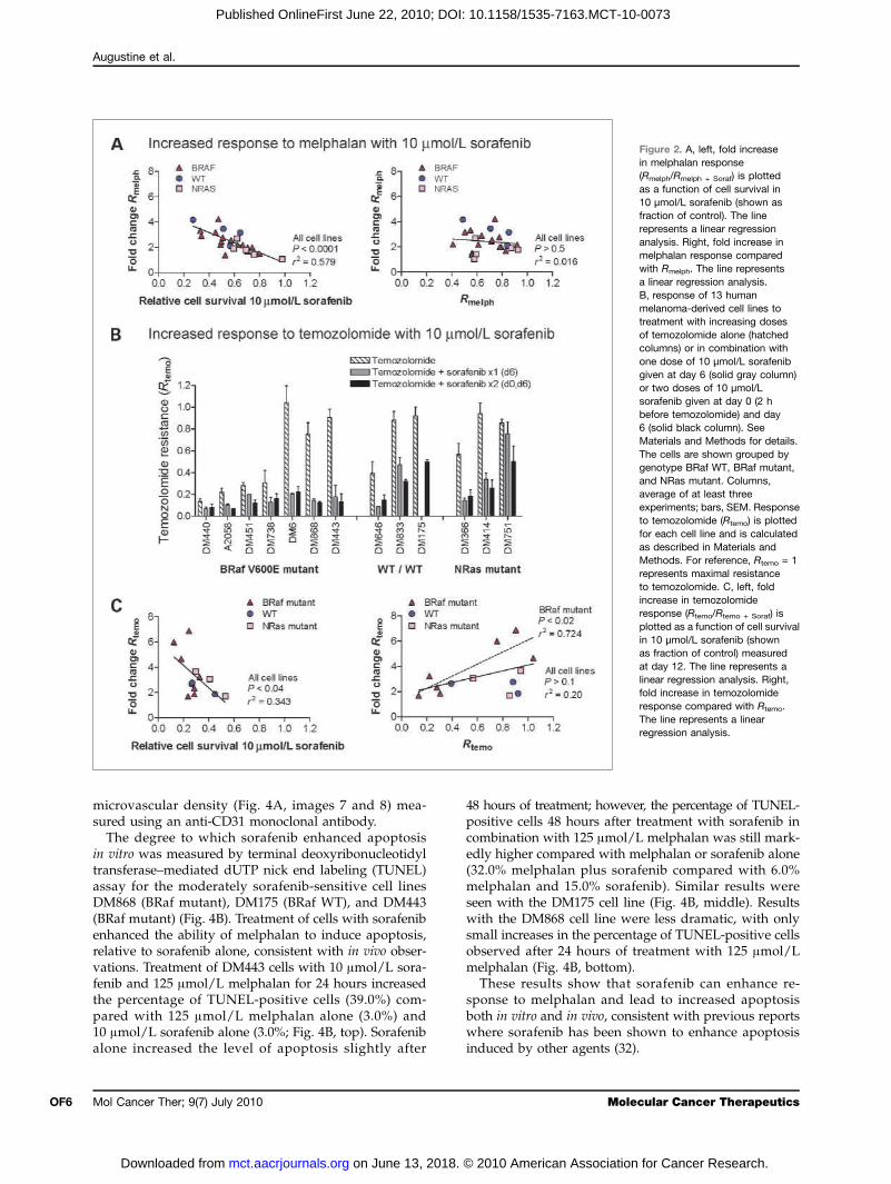
phalan chemotherapy in melanoma examined at 48 hoursfollowing treatment with melphalan alone and in combi-nation with 5 or 10 μmol/L sorafenib are summarized inFig. 1B and C. Response to melphalan alone ranged frommoderately sensitive (DM422; Rmelph = 0.41) to resistant(DM598; Rmelph = 0.93) and showed no relationship toeither BRaf mutation status (P > 0.40) or sensitivity tosorafenib (P > 0.10; Fig. 1B; Supplementary Fig. S1B).Following a 2-hour exposure to 10 μmol/L sorafenib,sensitivity to melphalan was significantly increased overcontrol (two-tailed unpaired t test, P < 0.05) in all celllines with the exception of DM751, which was the mostresistant to sorafenib (Fig. 1C; Supplementary Fig. S1C).The increase in sensitivity to melphalan with 10 μmol/Lsorafenib correlated significantly (P < 0.0001) with re-sponse to sorafenib alone (Fig. 2A, left) but not with BRafmutation status (P > 0.90) or baseline melphalan sensiti-vity (P > 0.50; Fig. 2A, right).
Effects of sorafenib on in vitro response totemozolomideResponse to temozolomide alone and in combination
with 10 μmol/L sorafenib was evaluated using the 12-day assay. As shown in Fig. 2B, sensitivity to sorafenibmeasured with the 12-day assay was significantly (P <0.04) more pronounced than that observed at 48 hoursacross 10 of the 13 cell lines tested (see also Supple-mentary Fig. S2). Response to sorafenib treatment mea-sured at day 12 showed a slight but significantdifference between cell lines with the BRaf mutationand those with the NRas mutation (P < 0.02; see Sup-plementary Fig. S2B).In all cell lines, 10 μmol/L sorafenib enhanced the
response to temozolomide chemotherapy at day 12(Fig. 2B). DM751, the most sorafenib-resistant cell line,when measured at 48 hours (see Fig. 1A), showed a1.7-fold increase in sensitivity (among the lowest) to te-mozolomide with 10 μmol/L sorafenib (P = 0.038).DM440, the most temozolomide-sensitive cell line,showed little increase in temozolomide sensitivity withsorafenib (fold increase, 1.68). Across all cell lines, therewas a significant correlation (P < 0.04) between base-line sensitivity to sorafenib, measured at day 12, andthe fold increase in temozolomide sensitivity with10 μmol/L sorafenib (Fig. 2C, left). Among the sevencell lines with the BRaf mutation, there was a signifi-cant correlation (P < 0.02) between the fold increase intemozolomide sensitivity with 10 μmol/L sorafeniband baseline sensitivity to temozolomide such that te-mozolomide-sensitive cells showed less enhancementwith sorafenib compared with temozolomide-resistantcells (Fig. 2C, right). This pattern was not observedin the six BRaf WT cell lines.
Table 1. Mutation status across 24 humanmelanoma-derived cell lines
Cell line
BRAF (bp 1799) NRAS (bp 1336)DM175
WT WT DM366 WT CAA>CGA (Q>R) DM368 GTG>GAG (V>E) WT DM391 GTG>GAG (V>E) WT DM414 WT CAA>CGA (Q>R) DM422 GTG>GAG (V>E) not determined DM424 GTG>GAG (V>E) WT DM425 WT WT DM440 GTG>GAG (V>E) WT DM443 GTG>GAG (V>E) WT DM451 GTG>GAG (V>E) WT DM578 GTG>GAG (V>E) not determined DM598 WT CAA>CGA (Q>R) DM6 GTG>GAG (V>E) WT DM608 GTG>GAG (V>E) WT DM646 WT WT DM733 GTG>GAG (V>E) WT DM738 GTG>GAG (V>E) WT DM751 WT CAA>CTA (Q>L) DM792 WT CAA>CTA (Q>L) DM833 WT WT DM868 GTG>GAG (V>E) WT A2058 GTG>GAG (V>E) WT SkMel28 GTG>GAG (V>E) WTMolecular Cancer Therapeutics
© 2010 American Association for Cancer Research.
Sorafenib Enhances Regional Chemotherapy in Melanoma
www.aacrjournals.org
on June 13, 2018. mct.aacrjournals.org Downloaded from
Published OnlineFirst June 22, 2010; DOI: 10.1158/1535-7163.MCT-10-0073
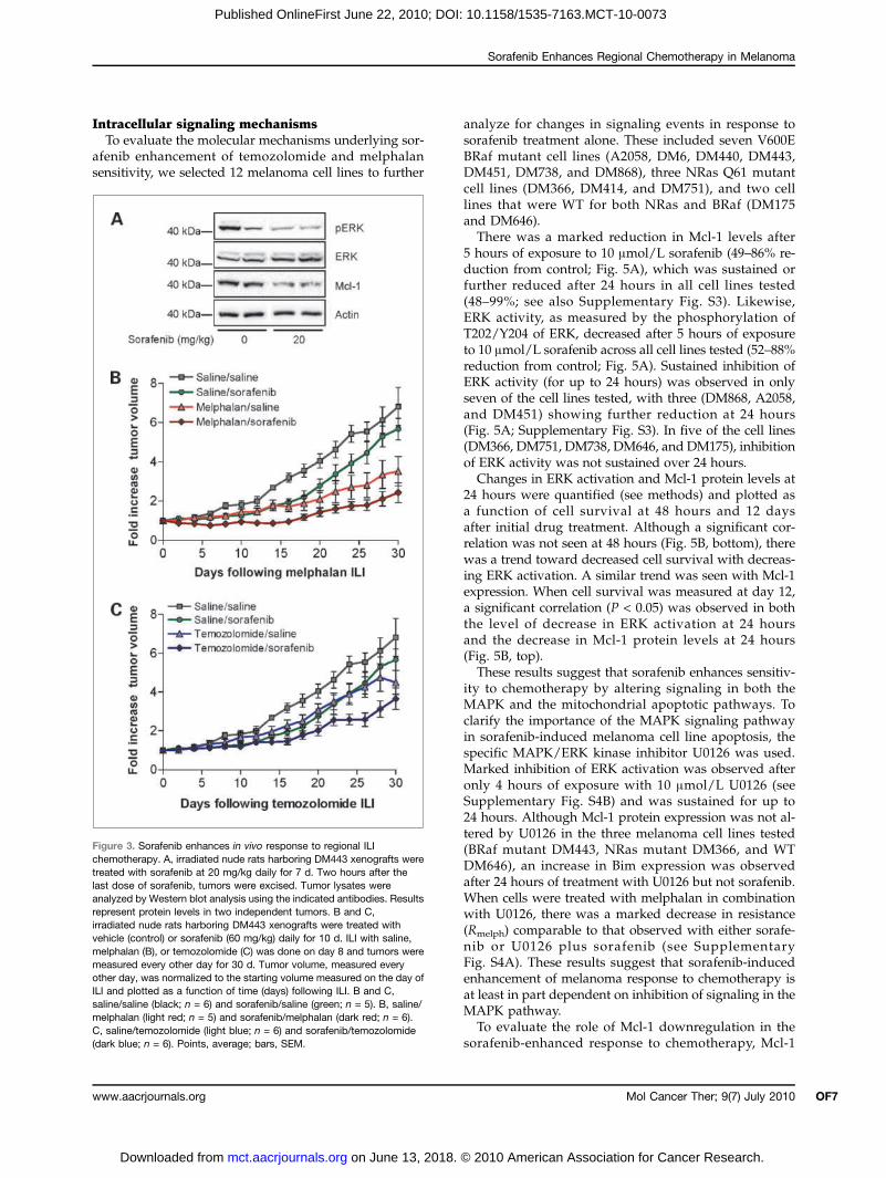
In vivo response to sorafenib in combination withmelphalan or temozolomide isolated limb infusionThe ability of sorafenib to enhance the effects of
high-dose regionally infused chemotherapy was exam-ined in our rat xenograft model of extremity melanoma(33, 34) using the DM443 cell line, which was moder-ately sensitive to sorafenib in vitro (Fig. 1A), mode-rately sensitive to melphalan both in vitro (Fig. 1B)and in vivo (25), and resistant to temozolomide bothin vitro (Fig. 2B) and in vivo (25). We also examinedthe in vivo ERK kinase activity, which is regulateddownstream of the Raf kinase, as well as the levelsof Mcl-1, an antiapoptotic protein which has beenshown to be downregulated by sorafenib via a Raf-independent mechanism (11, 12). Systemic treatmentwith 20 mg/kg sorafenib alone reduced both ERK acti-vation and Mcl-1 protein levels (Fig. 3A). In an effort toenhance response without additional toxicity (19), adose of 60 mg/kg sorafenib was administered over10 days. Treatment with vehicle alone resulted in a sub-stantial increase in tumor volume of nearly 7-fold overthe course of 30 days (Fig. 3B and C); relative to vehi-cle, tumor growth was slowed by treatment with sora-fenib (P = 0.0034), temozolomide (P < 0.0001), ormelphalan (P = 0.0024). Combination of sorafenib withmelphalan in the ILI setting resulted in stabilization oftumor growth for up to 16 days, whereas sorafenibwith temozolomide ILI resulted in marked slowing oftumor growth. At day 30, the growth of tumors treatedwith melphalan plus sorafenib was reduced significant-ly relative to saline alone (P < 0.0001) or sorafenibalone (P < 0.0001); relative to melphalan alone, growthwas slowed but this did not reach statistical significance(P = 0.1135). Temozolomide plus sorafenib significantlyslowed tumor growth relative to saline alone (P <0.0001), sorafenib alone (P = 0.0026), and temozolomidealone (P = 0.0025). Additional measures of tumorgrowth such as quintupling time showed a trend towardimproved tumor response with combination therapyover chemotherapy alone (see Supplementary Table S2).
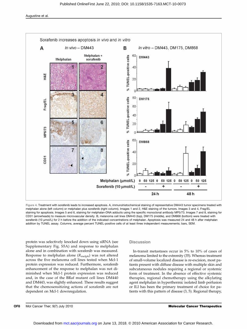
Sorafenib enhances melphalan-induced apoptosisboth in vivo and in vitroTumor specimens from rats treated with either melpha-
lan ILI alone or melphalan ILI plus systemic sorafenibwere examined by immunohistochemistry for sorafenib-related changes in apoptosis, melphalan-DNA adductformation, and microvascular density. Tumor specimensharvested after 9 days of sorafenib treatment and24 hours after ILI with melphalan showed markedincreases in apoptosis (Fig. 4A, image 4) compared withtumors treated with melphalan alone (Fig. 4A, image 3).The formation of melphalan adducts, as determinedby immunohistochemistry using the melphalan-DNAadduct–specific monoclonal antibody MP5/73, wasrelatively similar between the tumors treated with mel-phalan alone and those treated with melphalan plus sor-afenib (Fig. 4A, images 5 and 6, respectively), as was the
Figure 1. Sorafenib enhances in vitro response to melphalanchemotherapy. Response of 24 human melanoma-derived cell lines totreatment with sorafenib (A; 10 μmol/L for 48 h), increasing doses ofmelphalan (B; measured after 48 h), or increasing doses of melphalan incombination with sorafenib (C; 10 μmol/L sorafenib for 2 h followed bymelphalan measured after 48 h). The genotype of each cell line isindicated: BRaf WT (closed columns), BRaf mutant (hatched columns),and NRas mutant (red asterisks). Columns, average of at least threeexperiments; bars, SEM. Response to melphalan (B and C; Rmelph) isplotted for each cell line and is calculated as described in Materials andMethods. For reference, Rmelph = 1 represents maximal resistance tomelphalan.
Mol Cancer Ther; 9(7) July 2010 OF5
© 2010 American Association for Cancer Research.
Augustine et al.
OF6
Published OnlineFirst June 22, 2010; DOI: 10.1158/1535-7163.MCT-10-0073
microvascular density (Fig. 4A, images 7 and 8) mea-sured using an anti-CD31 monoclonal antibody.The degree to which sorafenib enhanced apoptosis
in vitro was measured by terminal deoxyribonucleotidyltransferase–mediated dUTP nick end labeling (TUNEL)assay for the moderately sorafenib-sensitive cell linesDM868 (BRaf mutant), DM175 (BRaf WT), and DM443(BRaf mutant) (Fig. 4B). Treatment of cells with sorafenibenhanced the ability of melphalan to induce apoptosis,relative to sorafenib alone, consistent with in vivo obser-vations. Treatment of DM443 cells with 10 μmol/L sora-fenib and 125 μmol/L melphalan for 24 hours increasedthe percentage of TUNEL-positive cells (39.0%) com-pared with 125 μmol/L melphalan alone (3.0%) and10 μmol/L sorafenib alone (3.0%; Fig. 4B, top). Sorafenibalone increased the level of apoptosis slightly after
Mol Cancer Ther; 9(7) July 2010
on June 13, 2018. mct.aacrjournals.org Downloaded from
48 hours of treatment; however, the percentage of TUNEL-positive cells 48 hours after treatment with sorafenib incombination with 125 μmol/L melphalan was still mark-edly higher compared with melphalan or sorafenib alone(32.0% melphalan plus sorafenib compared with 6.0%melphalan and 15.0% sorafenib). Similar results wereseen with the DM175 cell line (Fig. 4B, middle). Resultswith the DM868 cell line were less dramatic, with onlysmall increases in the percentage of TUNEL-positive cellsobserved after 24 hours of treatment with 125 μmol/Lmelphalan (Fig. 4B, bottom).These results show that sorafenib can enhance re-
sponse to melphalan and lead to increased apoptosisboth in vitro and in vivo, consistent with previous reportswhere sorafenib has been shown to enhance apoptosisinduced by other agents (32).
Mole
© 2010 American Association
Figure 2. A, left, fold increasein melphalan response(Rmelph/Rmelph + Soraf) is plottedas a function of cell survival in10 μmol/L sorafenib (shown asfraction of control). The linerepresents a linear regressionanalysis. Right, fold increase inmelphalan response comparedwith Rmelph. The line representsa linear regression analysis.B, response of 13 humanmelanoma-derived cell lines totreatment with increasing dosesof temozolomide alone (hatchedcolumns) or in combination withone dose of 10 μmol/L sorafenibgiven at day 6 (solid gray column)or two doses of 10 μmol/Lsorafenib given at day 0 (2 hbefore temozolomide) and day6 (solid black column). SeeMaterials and Methods for details.The cells are shown grouped bygenotype BRaf WT, BRaf mutant,and NRas mutant. Columns,average of at least threeexperiments; bars, SEM. Responseto temozolomide (Rtemo) is plottedfor each cell line and is calculatedas described in Materials andMethods. For reference, Rtemo = 1represents maximal resistanceto temozolomide. C, left, foldincrease in temozolomideresponse (Rtemo/Rtemo + Soraf) isplotted as a function of cell survivalin 10 μmol/L sorafenib (shownas fraction of control) measuredat day 12. The line represents alinear regression analysis. Right,fold increase in temozolomideresponse compared with Rtemo.The line represents a linearregression analysis.
cular Cancer Therapeutics
for Cancer Research.
Sorafenib Enhances Regional Chemotherapy in Melanoma
Published OnlineFirst June 22, 2010; DOI: 10.1158/1535-7163.MCT-10-0073
Intracellular signaling mechanismsTo evaluate the molecular mechanisms underlying sor-
afenib enhancement of temozolomide and melphalansensitivity, we selected 12 melanoma cell lines to further
www.aacrjournals.org
on June 13, 2018. mct.aacrjournals.org Downloaded from
analyze for changes in signaling events in response tosorafenib treatment alone. These included seven V600EBRaf mutant cell lines (A2058, DM6, DM440, DM443,DM451, DM738, and DM868), three NRas Q61 mutantcell lines (DM366, DM414, and DM751), and two celllines that were WT for both NRas and BRaf (DM175and DM646).There was a marked reduction in Mcl-1 levels after
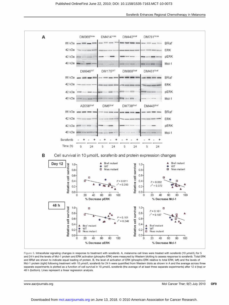
5 hours of exposure to 10 μmol/L sorafenib (49–86% re-duction from control; Fig. 5A), which was sustained orfurther reduced after 24 hours in all cell lines tested(48–99%; see also Supplementary Fig. S3). Likewise,ERK activity, as measured by the phosphorylation ofT202/Y204 of ERK, decreased after 5 hours of exposureto 10 μmol/L sorafenib across all cell lines tested (52–88%reduction from control; Fig. 5A). Sustained inhibition ofERK activity (for up to 24 hours) was observed in onlyseven of the cell lines tested, with three (DM868, A2058,and DM451) showing further reduction at 24 hours(Fig. 5A; Supplementary Fig. S3). In five of the cell lines(DM366, DM751, DM738, DM646, and DM175), inhibitionof ERK activity was not sustained over 24 hours.Changes in ERK activation and Mcl-1 protein levels at
24 hours were quantified (see methods) and plotted asa function of cell survival at 48 hours and 12 daysafter initial drug treatment. Although a significant cor-relation was not seen at 48 hours (Fig. 5B, bottom), therewas a trend toward decreased cell survival with decreas-ing ERK activation. A similar trend was seen with Mcl-1expression. When cell survival was measured at day 12,a significant correlation (P < 0.05) was observed in boththe level of decrease in ERK activation at 24 hoursand the decrease in Mcl-1 protein levels at 24 hours(Fig. 5B, top).These results suggest that sorafenib enhances sensitiv-
ity to chemotherapy by altering signaling in both theMAPK and the mitochondrial apoptotic pathways. Toclarify the importance of the MAPK signaling pathwayin sorafenib-induced melanoma cell line apoptosis, thespecific MAPK/ERK kinase inhibitor U0126 was used.Marked inhibition of ERK activation was observed afteronly 4 hours of exposure with 10 μmol/L U0126 (seeSupplementary Fig. S4B) and was sustained for up to24 hours. Although Mcl-1 protein expression was not al-tered by U0126 in the three melanoma cell lines tested(BRaf mutant DM443, NRas mutant DM366, and WTDM646), an increase in Bim expression was observedafter 24 hours of treatment with U0126 but not sorafenib.When cells were treated with melphalan in combinationwith U0126, there was a marked decrease in resistance(Rmelph) comparable to that observed with either sorafe-nib or U0126 plus sorafenib (see SupplementaryFig. S4A). These results suggest that sorafenib-inducedenhancement of melanoma response to chemotherapy isat least in part dependent on inhibition of signaling in theMAPK pathway.To evaluate the role of Mcl-1 downregulation in the
sorafenib-enhanced response to chemotherapy, Mcl-1
Figure 3. Sorafenib enhances in vivo response to regional ILIchemotherapy. A, irradiated nude rats harboring DM443 xenografts weretreated with sorafenib at 20 mg/kg daily for 7 d. Two hours after thelast dose of sorafenib, tumors were excised. Tumor lysates wereanalyzed by Western blot analysis using the indicated antibodies. Resultsrepresent protein levels in two independent tumors. B and C,irradiated nude rats harboring DM443 xenografts were treated withvehicle (control) or sorafenib (60 mg/kg) daily for 10 d. ILI with saline,melphalan (B), or temozolomide (C) was done on day 8 and tumors weremeasured every other day for 30 d. Tumor volume, measured everyother day, was normalized to the starting volume measured on the day ofILI and plotted as a function of time (days) following ILI. B and C,saline/saline (black; n = 6) and sorafenib/saline (green; n = 5). B, saline/melphalan (light red; n = 5) and sorafenib/melphalan (dark red; n = 6).C, saline/temozolomide (light blue; n = 6) and sorafenib/temozolomide(dark blue; n = 6). Points, average; bars, SEM.
Mol Cancer Ther; 9(7) July 2010 OF7
© 2010 American Association for Cancer Research.
Augustine et al.
OF8
Published OnlineFirst June 22, 2010; DOI: 10.1158/1535-7163.MCT-10-0073
protein was selectively knocked down using siRNA (seeSupplementary Fig. S5A) and response to melphalanalone and in combination with sorafenib was measured.Response to melphalan alone (Rmelph) was not alteredacross the five melanoma cell lines tested when Mcl-1protein expression was reduced. Furthermore, sorafenibenhancement of the response to melphalan was not di-minished when Mcl-1 protein expression was reducedand, in the case of the BRaf mutant cell lines DM440and DM443, was slightly enhanced. These results suggestthat the chemosensitizing actions of sorafenib are notdependent on Mcl-1 downregulation.
Mol Cancer Ther; 9(7) July 2010
on June 13, 2018. mct.aacrjournals.org Downloaded from
Discussion
In-transit metastases occur in 5% to 10% of cases ofmelanoma limited to the extremity (35). Whereas treatmentof small-volume localized disease is re-excision, most pa-tients present with diffuse disease with multiple skin andsubcutaneous nodules requiring a regional or systemicform of treatment. In the absence of effective systemictherapies, regional chemotherapy using the alkylatingagent melphalan in hyperthermic isolated limb perfusionor ILI has been the primary treatment of choice for pa-tients with this pattern of disease (3, 5). Regional therapy,
Figure 4. Treatment with sorafenib leads to increased apoptosis. A, immunohistochemical staining of representative DM443 tumor specimens treated withmelphalan alone (left column) or melphalan plus sorafenib (right column). Images 1 and 2, H&E staining of the tumors. Images 3 and 4, Frag/ELstaining for apoptosis. Images 5 and 6, staining for melphalan-DNA adducts using the specific monoclonal antibody MP5/73. Images 7 and 8, staining forCD31 (arrowheads) to measure microvascular density. B, melanoma cell lines DM443 (top), DM175 (middle), and DM868 (bottom) were treated withsorafenib (10 μmol/L) for 2 h before the addition of the indicated concentrations of melphalan. Apoptosis was measured 24 and 48 h after melphalanaddition by TUNEL assay. Columns, average percent TUNEL-positive cells of at least three independent measurements; bars, SEM.
Molecular Cancer Therapeutics
© 2010 American Association for Cancer Research.
Sorafenib Enhances Regional Chemotherapy in Melanoma
Published OnlineFirst June 22, 2010; DOI: 10.1158/1535-7163.MCT-10-0073
Figure 5. Intracellular signaling changes in response to treatment with sorafenib. A, melanoma cell lines were treated with sorafenib (10 μmol/L) for 5and 24 h and the levels of Mcl-1 protein and ERK activation (phospho-ERK) were measured by Western blotting to assess response to sorafenib. Total ERKand BRaf are shown to indicate equal loading of protein. B, the level of activation of ERK (phospho-ERK relative to total ERK; left) and the levels ofMcl-1 protein (right) following treatment with 10 μmol/L sorafenib for 24 h were quantified from Western blots as shown in A and the average across threeseparate experiments is plotted as a function of cell survival in 10 μmol/L sorafenib (the average of at least three separate experiments) after 12 d (top) or48 h (bottom). Lines represent a linear regression analysis.
Mol Cancer Ther; 9(7) July 2010www.aacrjournals.org OF9
on June 13, 2018. © 2010 American Association for Cancer Research. mct.aacrjournals.org Downloaded from
Augustine et al.
OF10
Published OnlineFirst June 22, 2010; DOI: 10.1158/1535-7163.MCT-10-0073
which, because the drug is limited to the affected extrem-ity, allows for the use of much higher levels of chemo-therapy than achievable with standard systemictreatment, has led to response rates of 70% to 90% withabout one half of these responses being complete (36, 37).Temozolomide is a second-generation alkylating agentwith a mechanism of action similar to that of dacarbazine(DTIC), currently the drug of choice for the treatment ofsystemic melanoma and the only widely used drug thatis Food and Drug Administration approved for melano-ma. Recent clinical trials have shown that for systemictreatment of metastatic melanoma, temozolomide is aseffective as DTIC (38). Furthermore, in an animal modelof extremity melanoma, ILI with temozolomide wasshown to be as effective or more effective than melphalanin four of five melanoma xenografts studied (25).Earlier attempts to improve the response rate of region-
ally administered chemotherapy in melanoma focusedon adding tumor necrosis factor (TNF) to the regional cir-cuit. A completed randomized prospective phase III trialcomparing melphalan alone to melphalan plus TNF wasstopped at its interim analysis because no response ad-vantage to TNF was identified and there were three timesthe number of grade 4 adverse events in the TNF arm(39). More recent approaches to combination therapyhave been designed so that the targeted agent or modu-lator of drug resistance is administered systemicallyaround a window of time during which high-dose re-gional chemotherapy is given. One such combinationtherapy approach has been to target cell signaling path-ways involved in tumor growth and survival. In the pres-ent study, we show that the multikinase inhibitorsorafenib can increase the cytotoxic response of melano-ma cells to the alkylating chemotherapeutics melphalanand temozolomide. In vitro, across a panel of 24 cell lines,the sorafenib-mediated augmentation of chemotherapy-induced cytotoxicity was independent of BRaf or NRasmutational status (see Supplementary Fig. S2C). In vivo,in the context of a high-dose regional chemotherapy ratmodel, sorafenib enhanced the cytotoxic response of ahuman melanoma xenograft to both melphalan and te-mozolomide. These results provide compelling evidencethat combination therapy of systemic sorafenib withregional chemotherapy infusion could provide a novelregimen for augmenting the treatment of melanomapatients with localized in-transit extremity disease.Sorafenib can induce cell cycle arrest and both caspase-
dependent and caspase-independent apoptosis via a va-riety of mechanisms (10–12, 19, 20, 40). In our cohort ofcell lines, treatment with sorafenib was associated withdecreased levels of ERK activation and Mcl-1 protein le-vels. Furthermore, the level of response of cell lines tosorafenib measured at day 12 was significantly correlatedwith the magnitude to which both ERK activation andMcl-1 protein levels were decreased. Although previous-ly published reports using both tumor cell lines and xe-nografts in mice suggest a lack of correlation betweenresponse and alterations in the Raf/ERK signaling path-
Mol Cancer Ther; 9(7) July 2010
on June 13, 2018. mct.aacrjournals.org Downloaded from
way (10, 32, 40), our results are in agreement with thosereported for liver cancer cells lines, in which the antitu-mor activity of sorafenib corresponded with downregula-tion of ERK activity (40), and suggest that in melanomathe ability of sorafenib to downregulate ERK activity is acritical factor in the chemosensitizing actions of the drug.With respect to Mcl-1, previous studies have implicatedthe negative regulation of Mcl-1 as one of the main me-chanisms of action of sorafenib alone and in combinationwith other agents (11, 12, 41). However, more recent datasuggest that downregulation of Mcl-1 alone is not suffi-cient to induce cell death or enhance the sensitivity ofcells to chemotherapy or radiation (19, 42), consistentwith our observations.Systemic treatment of rats with a small peptide inhib-
itor of N-cadherin has recently been shown to increasethe levels of melphalan adducts in tumor cells in ourxenograft melphalan ILI animal model (43). Likewise,alterations in drug delivery have been reported for othervascular targeting agents, including Gleevec (imatinib)and Avastin (bevacizumab; refs. 44–47). These observa-tions led us to hypothesize that sorafenib-mediatedinhibition of the vascular endothelial growth factor re-ceptor tyrosine kinase, which can lead to reduced micro-vessel density (16, 32), may similarly increase melphalandelivery to tumors. Although our results failed to simi-larly show alterations in microvascular density, it is pos-sible that 9 days of treatment with sorafenib was notsufficient to elicit significant changes in microvasculardensity similar to that observed after 15 days of treat-ment in a mouse model (16). Our results suggest that,for at least some melanoma cells and tumor xenografts,sorafenib actions may be able to reduce the cellularthreshold for induction of apoptosis, thereby makingcells more susceptible to other apoptotic-inducing ther-apies such as melphalan (Fig. 4). Others have shownsimilar enhancements in cell death when sorafenibwas used in combination with radiation, the platinum-based chemotherapy agents oxaliplatin and cisplatin, ra-pamycin, the proteasome inhibitor MG-132, the Bcl-2family inhibitor ABT-737, and tumor necrosis factor-related apoptosis-inducing ligand (12, 18–20, 41, 42, 48).It is noteworthy that responses to sorafenib are likely to
be dependent on the context of the cells treated, as sug-gested by our observation that DM868 cells did not res-pond to the combination of sorafenib and melphalancompared with the level of enhancement of apoptosis thatwe observed with the DM175 and DM443 cell lines(Fig. 4B). Similarly, others have observed that liver cancercell lines responded differentially to sorafenib in terms ofcaspase-independent versus caspase-dependent apoptosis(40) and that the effects of sorafenib on the cell cycle variedremarkably between cell lines (19). These observations sug-gest that although a broad range of cells and tumors maybe responsive to sorafenib alone or in combination, themechanism by which the response occurs may dependon the molecular background and biology of the indivi-dual cell line or tumor.
Molecular Cancer Therapeutics
© 2010 American Association for Cancer Research.
Sorafenib Enhances Regional Chemotherapy in Melanoma
Published OnlineFirst June 22, 2010; DOI: 10.1158/1535-7163.MCT-10-0073
The promising results we describe here on the efficacyof combinations of sorafenib and chemotherapy providea strong rationale for the development of other systemi-cally administered targeted agents to be used in conjunc-tion with regionally administered chemotherapeutics fortreatment of regionally advanced in-transit melanoma.Our results show that this approach may be effectivenot only with regionally administered chemotherapeuticagents like temozolomide (currently being evaluated in aphase I dose escalation ILI trial) but also with other sys-temically administered targeted agents or modulators. Inaddition to sorafenib, as described here, preclinical stud-ies using systemically administered agents that target theglutathione detoxification system, such as the reducedglutathione–depleting agent buthionine sulfoximine,and cell adhesion, such as the small peptide inhibitor ofN-cadherin ADH-1 (43, 49), have proved very effective inimproving tumor responses. Phase I/II clinical trials ex-amining the efficacy of these agents, including sorafenib,to enhance the response of melanoma to ILI with melpha-lan are currently in progress or under development. Inthe case of ADH-1, marked improvements in completeresponse rates from 30% to 50% have already been ob-served in phase I clinical trials (50). Correlative science
www.aacrjournals.org
on June 13, 2018. mct.aacrjournals.org Downloaded from
components of these trials will aim to understand themechanism by which these targeted therapeutics affectresponse to chemotherapy. Through a better understand-ing of the molecular mechanism underlying the ability ofsorafenib to mediate chemosensitization in regional che-motherapy, we hope to provide the basis for developingstrategies to use sorafenib more effectively in patientssuch that, ultimately, it can be optimized for use in sys-temic chemotherapy strategies for metastatic melanoma.
Disclosure of Potential Conflicts of Interest
No potential conflicts of interest were disclosed.
Grant Support
Duke Melanoma Research Fund (D.S. Tyler), VA Merit Review Grant(D.S. Tyler), and a grant from the Institute for Genomic Sciences and Pol-icy, Duke University (D.S. Tyler and F. Ali-Osman).
The costs of publication of this article were defrayed in part by thepayment of page charges. This article must therefore be hereby markedadvertisement in accordance with 18 U.S.C. Section 1734 solely to indicatethis fact.
Received 01/21/2010; revised 04/23/2010; accepted 05/05/2010;published OnlineFirst 06/22/2010.
References
1. Gray-Schopfer VC, da Rocha Dias S, Marais R. The role of B-RAF inmelanoma. Cancer Metastasis Rev 2005;24:165–83.2. Jemal A, Siegel R, Ward E, Hao Y, Xu J, Thun MJ. Cancer statistics,
2009. CA Cancer J Clin 2009;59:225–49.3. Brady MS, Brown K, Patel A, Fisher C, Marx W. A phase II trial of
isolated limb infusion with melphalan and dactinomycin for regionalmelanoma and soft tissue sarcoma of the extremity. Ann Surg Oncol2006;13:1123–9.
4. Eggermont AM, Kirkwood JM. Re-evaluating the role of dacarbazinein metastatic melanoma: what have we learned in 30 years? Eur JCancer 2004;40:1825–36.
5. Linder P, Doubrovsky A, Kam PC, Thompson JF. Prognostic factorsafter isolated limb infusion with cytotoxic agents for melanoma. AnnSurg Oncol 2002;9:127–36.
6. Smalley KS, Herlyn M. Targeting intracellular signaling pathways as anovel strategy in melanoma therapeutics. Ann N Y Acad Sci 2005;1059:16–25.
7. Gray-Schopfer V, Wellbrock C, Marais R. Melanoma biology and newtargeted therapy. Nature 2007;445:851–7.
8. Lang L. FDA approves sorafenib for patients with inoperable livercancer. Gastroenterology 2008;134:379.
9. Stein MN, Flaherty KT. CCR drug updates: sorafenib and sunitinib inrenal cell carcinoma. Clin Cancer Res 2007;13:3765–70.
10. Panka DJ, Wang W, Atkins MB, Mier JW. The Raf inhibitor BAY 43-9006 (sorafenib) induces caspase-independent apoptosis in melano-ma cells. Cancer Res 2006;66:1611–9.
11. Rahmani M, Davis EM, Bauer C, Dent P, Grant S. Apoptosis inducedby the kinase inhibitor BAY 43-9006 in human leukemia cells involvesdown-regulation of Mcl-1 through inhibition of translation. J BiolChem 2005;280:35217–27.
12. Yu C, Bruzek LM, Meng XW, et al. The role of Mcl-1 downregulationin the proapoptotic activity of the multikinase inhibitor BAY 43-9006.Oncogene 2005;24:6861–9.
13. Lazar-Molnar E, Hegyesi H, Toth S, Falus A. Autocrine and paracrineregulation by cytokines and growth factors in melanoma. Cytokine2000;12:547–54.
14. Bharti AC, Donato N, Singh S, Aggarwal BB. Curcumin (diferuloyl-methane) down-regulates the constitutive activation of nuclear fac-tor-κB and IκBα kinase in human multiple myeloma cells, leading tosuppression of proliferation and induction of apoptosis. Blood 2003;101:1053–62.
15. Gray-Schopfer VC, Karasarides M, Hayward R, Marais R. Tumor ne-crosis factor-α blocks apoptosis in melanoma cells when BRAF sig-naling is inhibited. Cancer Res 2007;67:122–9.
16. Sharma A, Trivedi NR, Zimmerman MA, Tuveson DA, Smith CD, Ro-bertson GP. Mutant V599EB-Raf regulates growth and vascular de-velopment of malignant melanoma tumors. Cancer Res 2005;65:2412–21.
17. Eisen T, Ahmad T, Flaherty KT, et al. Sorafenib in advanced melano-ma: a phase II randomised discontinuation trial analysis. Br J Cancer2006;95:581–6.
18. Molhoek K, Brautigan D, Slingluff C. Synergistic inhibition of humanmelanoma proliferation by combination treatment with B-Raf inhibi-tor BAY43-9006 and mTOR inhibitor rapamycin. J Transl Med 2005;3:39–49.
19. Plastaras JP, Kim S-H, Liu YY, et al. Cell cycle dependent andschedule-dependent antitumor effects of sorafenib combined withradiation. Cancer Res 2007;67:9443–54.
20. Zhang W, Konopleva M, Ruvolo VR, et al. Sorafenib induces apopto-sis of AML cells via Bim-mediated activation of the intrinsic apoptoticpathway. Leukemia 2008;22:808–18.
21. Escudier B, Lassau N, Angevin E, et al. Phase I trial of sorafenib incombination with IFNα-2a in patients with unresectable and/or meta-static renal cell carcinoma or malignant melanoma. Clin Cancer Res2007;13:1801–9.
22. McDermott DF, Sosman JA, Gonzalez R, et al. Double-blind random-ized phase II study of the combination of sorafenib and dacarbazinein patients with advanced melanoma: a report from the 11715 StudyGroup. J Clin Oncol 2008;26:2178–85.
23. Amaravadi RK, Schuchter LM, McDermott DF, et al. Phase II trial oftemozolomide and sorafenib in advanced melanoma patients with orwithout brain metastases. Clin Cancer Res 2009;15:7711–8.
Mol Cancer Ther; 9(7) July 2010 OF11
© 2010 American Association for Cancer Research.
Augustine et al.
OF12
Published OnlineFirst June 22, 2010; DOI: 10.1158/1535-7163.MCT-10-0073
24. Augustine CK, Yoo JS, Potti A, et al. Genomic and molecular profil-ing predicts response to temozolomide in melanoma. Clin CancerRes 2009;15:502–10.
25. Yoshimoto Y, Augustine CK, Yoo JS, et al. Defining regional infusiontreatment strategies for extremity melanoma: comparative analysisof melphalan and temozolomide as regional chemotherapeuticagents. Mol Cancer Ther 2007;6:1492–500.
26. Attis MG, Burchette JL, Selim MA, Pham T, Soler AP. Differential ex-pression of N-cadherin distinguishes a subset of metastasizing des-moplastic melanomas. Hum Pathol 2006;37:899–905.
27. Pham TTN, Selim MA, Burchette JL, Madden J, Turner J, Herman C.CD10 expression in trichoepithelioma and basal cell carcinoma.J Cutan Pathol 2006;33:123–8.
28. Tilby MJ, Styles JM, Dean CJ. Immunological detection of DNA dam-age caused by melphalan using monoclonal antibodies. Cancer Res1987;47:1542–6.
29. Liang KY, Zeger SL. Longitudinal data analysis using generalized lin-ear models. Biometrika 1986;73:13–22.
30. Jung SH, Ahn C. Sample size estimation for GEE method for com-paring slopes in repeated measurements data. Stat Med 2003;22:1305–15.
31. Kim S, Yazici YD, Calzada G, et al. Sorafenib inhibits the angiogen-esis and growth of orthotopic anaplastic thyroid carcinoma xeno-grafts in nude mice. Mol Cancer Ther 2007;6:1785–92.
32. Wilhelm SM, Carter C, Tang L, et al. BAY 43-9006 exhibits broadspectrum oral antitumor activity and targets the RAF/MEK/ERK path-way and receptor tyrosine kinases involved in tumor progression andangiogenesis. Cancer Res 2004;64:7099–109.
33. Ko SH, Ueno T, Yoshimoto Y, et al. Optimizing a novel regional che-motherapeutic agent against melanoma: hyperthermia-induced en-hancement of temozolomide cytotoxicity. Clin Cancer Res 2006;12:289–97.
34. Ueno T, Ko SH, Grubbs E, et al. Modulation of chemotherapy resis-tance in regional therapy: a novel therapeutic approach to advancedextremity melanoma using intra-arterial temozolomide in combina-tion with systemic O6-benzylguanine. Mol Cancer Ther 2006;5:732–8.
35. Pawlik TM, Ross MI, Johnson MM, et al. Predictors and natural his-tory of in-transit melanoma after sentinel lymphadenectomy. AnnSurg Oncol 2005;12:587–96.
36. Gander M, Leyvraz S, Decosterd L, et al. Sequential administration oftemozolomide and fotemustine: depletion of O6-alkyl guanine-DNAtransferase in blood lymphocytes and in tumours. Ann Oncol 1999;10:831–8.
37. Thompson JF, Hunt JA, Shannon KF, Kam PC. Frequency and du-ration of remission after isolated limb perfusion for melanoma. ArchSurg 1997;132:903–7.
Mol Cancer Ther; 9(7) July 2010
on June 13, 2018. mct.aacrjournals.org Downloaded from
38. Middleton MR, Grob JJ, Aaronson N, et al. Randomized phase IIIstudy of temozolomide versus dacarbazine in the treatment of pa-tients with advanced metastatic malignant melanoma. J Clin Oncol2000;18:158–66.
39. Cornett WR, McCall LM, Petersen RP, et al. Randomized multicentertrial of hyperthermic isolated limb perfusion with melphalan alonecompared with melphalan plus tumor necrosis factor: American Col-lege of Surgeons Oncology Group Trial Z0020. J Clin Oncol 2006;24:4196–201.
40. Liu L, Cao Y, Chen C, et al. Sorafenib blocks the RAF/MEK/ERKpathway, inhibits tumor angiogenesis, and induces tumor cell apo-ptosis in hepatocellular carcinoma model PLC/PRF/5. Cancer Res2006;66:11851–8.
41. Ricci MS, Kim S-H, Ogi K, et al. Reduction of TRAIL-induced Mcl-1and cIAP2 by c-Myc or sorafenib sensitizes resistant human cancercells to TRAIL-induced death. Cancer Cell 2007;12:66–80.
42. Rosato RR, Almenara JA, Coe S, Grant S. The multikinase inhibitorsorafenib potentiates TRAIL lethality in human leukemia cells in as-sociation with Mcl-1 and cFLIPL down-regulation. Cancer Res 2007;67:9490–500.
43. Augustine CK, Yoshimoto Y, Gupta M, et al. Targeting N-cadherinenhances antitumor activity of cytotoxic therapies in melanomatreatment. Cancer Res 2008;68:3777–84.
44. Bertino P, Piccardi F, Porta C, et al. Imatinib mesylate enhances ther-apeutic effects of gemcitabine in human malignant mesothelioma xe-nografts. Clin Cancer Res 2008;14:541–8.
45. Ogawa Y, Kawamura T, Furuhashi M, Tsukamoto K, Shimada S. Im-proving chemotherapeutic drug penetration in melanoma by imatinibmesylate. J Dermatol Sci 2008;51:190–9.
46. Pietras K, Stumm M, Hubert M, et al. STI571 enhances the therapeu-tic index of epothilone B by a tumor-selective increase of drug up-take. Clin Cancer Res 2003;9:3779–87.
47. Dickson PV, Hamner JB, Sims TL, et al. Bevacizumab-induced tran-sient remodeling of the vasculature in neuroblastoma xenografts re-sults in improved delivery and efficacy of systemically administeredchemotherapy. Clin Cancer Res 2007;13:3942–50.
48. Heim M, Scharifi M, Zisowsky J, et al. The Raf kinase inhibitor BAY43-9006 reduces cellular uptake of platinum compounds and cyto-toxicity in human colorectal carcinoma cell lines. Anticancer Drugs2005;16:129–36.
49. Grubbs EG, Ueno T, Abdel-Wahab O, et al. Modulation of resistanceto regional chemotherapy in the extremity melanoma model. Surgery2004;136:210–8.
50. Beasley G, McMahon N, Sanders G, et al. A phase I/II study of sys-temic ADH-1 in combination with isolated limb infusion with melpha-lan (ILI-M) in patients (pts) with locally advanced in-transit melanoma.J Clin Oncol 2008;26:abstr 9013.
Molecular Cancer Therapeutics
© 2010 American Association for Cancer Research.
Published OnlineFirst June 22, 2010.Mol Cancer Ther Christina K. Augustine, Hiroaki Toshimitsu, Sin-Ho Jung, et al. Melanoma to Regional ChemotherapySorafenib, a Multikinase Inhibitor, Enhances the Response of
Updated version
10.1158/1535-7163.MCT-10-0073doi:
Access the most recent version of this article at:
Material
Supplementary
http://mct.aacrjournals.org/content/suppl/2010/06/22/1535-7163.MCT-10-0073.DC1
Access the most recent supplemental material at:
E-mail alerts related to this article or journal.Sign up to receive free email-alerts
Subscriptions
Reprints and
To order reprints of this article or to subscribe to the journal, contact the AACR Publications
Permissions
Rightslink site. Click on "Request Permissions" which will take you to the Copyright Clearance Center's (CCC)
.http://mct.aacrjournals.org/content/early/2010/06/18/1535-7163.MCT-10-0073To request permission to re-use all or part of this article, use this link
on June 13, 2018. © 2010 American Association for Cancer Research. mct.aacrjournals.org Downloaded from
Published OnlineFirst June 22, 2010; DOI: 10.1158/1535-7163.MCT-10-0073