Resistance to multikinase inhibitor actions mediated by ...
10
RESEARCH Open Access Resistance to multikinase inhibitor actions mediated by insulin like growth factor-1 Catia Lippolis 1 , Maria Grazia Refolo 1 , Rosalba D’Alessandro 1 , Nicola Carella 1 , Caterina Messa 1 , Aldo Cavallini 1 and Brian Irving Carr 2* Abstract Background: Blood platelet numbers are correlated with growth and aggressiveness of several tumor types, including hepatocellular carcinoma (HCC). We previously found that platelet lysates (hPLs) both stimulated HCC cell growth and migration, and antagonized the growth-inhibitory and apoptotic effects of Regorafenib, multikinase growth inhibitor, on HCC cell lines. We evaluated the effects of human insulin-like growth factor-1 (IGF1), a mitogen contained in platelets, on the Regorafenib-mediated growth inhibition. Methods: An Elisa kit was used to evaluate hPL IGF1 concentrations. The effects of IGF1 on cell proliferation were assessed with MTT assay and analysis of cell cycle progression. Apoptosis assays, scratch assay and Transwell assay were performed to measure apoptosis, cell migration and invasion respectively. Western blots were performed by standard protocols. Results: IGF1 antagonized growth inhibition exerted by Regorafenib on HCC cell lines. Moreover the mitogen blocked Regorafenib-induced apoptosis and decreased the rate of cell migration and invasion. The IGF1 effects were in turn antagonized by actions of a potent IGF1 receptor inhibitor, GSK1838705A, showing that the IGF1 receptor was involved in the mechanisms of IGF1-mediated blocking of Regorafenib action. GSK1838705A also partially blocked the effects of hPLs in antagonizing Regorafenib-mediated growth inhibition, showing that IGF1 was an important component of hPL actions. Conclusions: These results show that IGF1 antagonized Regorafenib-mediated growth, migration and invasion inhibition, as well as the drug-mediated induction of apoptosis in HCC cells and reinforce the idea that microenvironmental factors can influence cancer drug actions. Keywords: Platelets, Insulin growth factor, Regorafenib, HCC cells Introduction Hepatocellular carcinoma (HCC) occurs most frequently in livers that have been chronically damaged by hepatitis B or C, chronic alcohol ingestion, or a wide range of chronic metabolic disturbances. Most cases develop in association with liver fibrosis or cirrhosis, especially in the presence of hepatitis C, although chronic infection with hepatitis B can lead to HCC without cirrhosis and HCC [1, 2]. A consequence of the liver fibrosis is portal hypertension, with associated splenomegaly, that can cause thrombocytopenia. The latter has been considered to be a warning sign of impending HCC development in patients with chronic virus hepatitis or cirrhosis [3–5]. Thrombocytopenia-associated HCC has recently been shown to be associated with smaller-size tumors [6, 7]. In contrast, several reports have shown that large size HCCs often have normal or elevated (thrombocytosis) platelet counts [8–13], likely due to less portal hyperten- sion. There are multiple reports of thrombosis and thrombocytosis in various cancer types [14–20]. Platelets have been shown to be involved in tumor me- tastasis, as well as in HCC growth, in addition to their well-recognized role in blood coagulation [21, 22]. Hu- man platelet lysates (hPL) are a source of many growth factors and have been recently introduced into clinical practice as an adjunct to wound healing [23–27]. We * Correspondence: [email protected] 2 Izmir Biomedicine and Genome Center, Dokuz Eylul University, Izmir, Turkey Full list of author information is available at the end of the article © 2015 Lippolis et al. Open Access This article is distributed under the terms of the Creative Commons Attribution 4.0 International License (http://creativecommons.org/licenses/by/4.0/), which permits unrestricted use, distribution, and reproduction in any medium, provided you give appropriate credit to the original author(s) and the source, provide a link to the Creative Commons license, and indicate if changes were made. The Creative Commons Public Domain Dedication waiver (http://creativecommons.org/publicdomain/zero/1.0/) applies to the data made available in this article, unless otherwise stated. Lippolis et al. Journal of Experimental & Clinical Cancer Research (2015) 34:90 DOI 10.1186/s13046-015-0210-1
Transcript of Resistance to multikinase inhibitor actions mediated by ...
RESEARCH Open Access
Resistance to multikinase inhibitor actionsmediated by insulin like growth factor-1Catia Lippolis1, Maria Grazia Refolo1, Rosalba D’Alessandro1, Nicola Carella1, Caterina Messa1, Aldo Cavallini1
and Brian Irving Carr2*
Abstract
Background: Blood platelet numbers are correlated with growth and aggressiveness of several tumor types,including hepatocellular carcinoma (HCC). We previously found that platelet lysates (hPLs) both stimulated HCC cellgrowth and migration, and antagonized the growth-inhibitory and apoptotic effects of Regorafenib, multikinasegrowth inhibitor, on HCC cell lines. We evaluated the effects of human insulin-like growth factor-1 (IGF1), a mitogencontained in platelets, on the Regorafenib-mediated growth inhibition.
Methods: An Elisa kit was used to evaluate hPL IGF1 concentrations. The effects of IGF1 on cell proliferation wereassessed with MTT assay and analysis of cell cycle progression. Apoptosis assays, scratch assay and Transwell assaywere performed to measure apoptosis, cell migration and invasion respectively. Western blots were performed bystandard protocols.
Results: IGF1 antagonized growth inhibition exerted by Regorafenib on HCC cell lines. Moreover the mitogenblocked Regorafenib-induced apoptosis and decreased the rate of cell migration and invasion. The IGF1 effectswere in turn antagonized by actions of a potent IGF1 receptor inhibitor, GSK1838705A, showing that the IGF1receptor was involved in the mechanisms of IGF1-mediated blocking of Regorafenib action. GSK1838705A alsopartially blocked the effects of hPLs in antagonizing Regorafenib-mediated growth inhibition, showing that IGF1was an important component of hPL actions.
Conclusions: These results show that IGF1 antagonized Regorafenib-mediated growth, migration and invasioninhibition, as well as the drug-mediated induction of apoptosis in HCC cells and reinforce the idea thatmicroenvironmental factors can influence cancer drug actions.
Keywords: Platelets, Insulin growth factor, Regorafenib, HCC cells
IntroductionHepatocellular carcinoma (HCC) occurs most frequentlyin livers that have been chronically damaged by hepatitisB or C, chronic alcohol ingestion, or a wide range ofchronic metabolic disturbances. Most cases develop inassociation with liver fibrosis or cirrhosis, especially inthe presence of hepatitis C, although chronic infectionwith hepatitis B can lead to HCC without cirrhosis andHCC [1, 2]. A consequence of the liver fibrosis is portalhypertension, with associated splenomegaly, that cancause thrombocytopenia. The latter has been considered
to be a warning sign of impending HCC development inpatients with chronic virus hepatitis or cirrhosis [3–5].Thrombocytopenia-associated HCC has recently beenshown to be associated with smaller-size tumors [6, 7].In contrast, several reports have shown that large sizeHCCs often have normal or elevated (thrombocytosis)platelet counts [8–13], likely due to less portal hyperten-sion. There are multiple reports of thrombosis andthrombocytosis in various cancer types [14–20].Platelets have been shown to be involved in tumor me-
tastasis, as well as in HCC growth, in addition to theirwell-recognized role in blood coagulation [21, 22]. Hu-man platelet lysates (hPL) are a source of many growthfactors and have been recently introduced into clinicalpractice as an adjunct to wound healing [23–27]. We
* Correspondence: [email protected] Biomedicine and Genome Center, Dokuz Eylul University, Izmir, TurkeyFull list of author information is available at the end of the article
© 2015 Lippolis et al. Open Access This article is distributed under the terms of the Creative Commons Attribution 4.0International License (http://creativecommons.org/licenses/by/4.0/), which permits unrestricted use, distribution, andreproduction in any medium, provided you give appropriate credit to the original author(s) and the source, provide a link tothe Creative Commons license, and indicate if changes were made. The Creative Commons Public Domain Dedication waiver(http://creativecommons.org/publicdomain/zero/1.0/) applies to the data made available in this article, unless otherwise stated.
Lippolis et al. Journal of Experimental & Clinical Cancer Research (2015) 34:90 DOI 10.1186/s13046-015-0210-1
previously found that hPL have anti-apoptotic effects inHCC cells and can antagonize the apoptotic and cellgrowth inhibitory actions of the multikinase inhibitors,Sorafenib and Regorafenib [28, 29]. The current workextends previous findings, by showing that growth in-hibitory actions mediated by Regorafenib [30] in HCCcells in vitro can be antagonized by insulin like growthfactor 1 (IGF1), one of the well-described mitogens con-tained in platelets [25–27]. Furthermore, an IGF1 recep-tor inhibitor can partially block the drug resistanceactions of hPL, supporting the idea that platelet-associated IGF1 may modulate HCC resistance to multi-kinase inhibitor effects.
Materials and methodsCells and drugsRegorafenib was gifts from the Bayer Corp (West Haven,CT, USA), recombinant human IGF1 was purchasedfrom Pepro-Tech (Rocky Hill, NJ, USA), GSK1838705Awas purchased from Selleckchem (Houston, TX, USA).Hep3B, HepG2 and PLC/PRF/5 human HCC cells were
purchased from the American Type Culture Collection(ATCC, Rockville, MD, USA). The culture medium wasDulbecco’s Modified Eagle’s Medium (DMEM). All cellculture components were purchased from Sigma- Aldrich(Milan, Italy).
Cell cultureCells were cultured in DMEM in monolayer culture, andsupplemented with 10 % fetal bovine serum (FBS), 100U/ml penicillin, 100 μg/ml streptomycin, and incubatedat 37 °C in a humidified atmosphere containing 5 % CO2
in air.
Platelet lysatesThe hPL were blood bank time-expired bags, fromhealthy volunteers. The study protocol was approved bythe institutional review boards of the University of Bariand “Saverio de Bellis” Institute of Castellana G. (BA),Italy. Additionally, written informed consent was ob-tained from participants for the use of their blood in thisstudy. The platelet-rich plasma was obtained using anautomated hemapheresis procedure in a local bloodtransfusion centre. The platelets obtained from differentvolunteers were pooled and then divided into aliquots.Each aliquot was subjected to three freeze-thaw cyclesto disrupt their membranes and release the growth fac-tors stored in the granules, producing hPLs.
IGF1 concentrations in platelet lysatesThe Human IGF1 ELISA kit (Wuhan Boster BiologicalTechnology LTD, Wuhan, China) was used for the invitro quantitative determination of human IGF1 in FBS
(control) and serial dilution of hPL, according to theuser’s guide.
Growth assayThe cells were cultured in 1 % FBS medium containingIGF1 40 ng/ml, the concentration was derived from theIGF1 ELISA dosage in hPL, or hPL corresponding to3.75 × 107 platelets/ml or equivalent percentage of FBS inpresence of 1 μM (HepG2 cells) or 5 μM (Hep3B andPLC/RFP/5) of Regorafenib. In the same growth condi-tion, HCC cells were cultured in absence or presence ofIGFR inhibitor, GSK1838705A 1 μM. After defined incu-bation times, the proliferative response was estimated bycolorimetric 3-(4,5 di-methylthiazol-2-yl)-2,5-diphenyltet-razolium bromide (MTT) test. The trypan blue exclusionassay was used to evaluate cell viability. Each experimentwas performed in triplicate and repeated three times.
Cell cycle analysisPLC/PRF/5 were synchronized by using thymidine0.2 M added to the medium. After 18 h of incubation,the medium containing thymidine was replaced withfresh medium for 9 h, and then cells were treated withthymidine for an additional 17 h. Cells were separatedinto two groups: one group was collected for cell cycleanalysis and the other one continued culturing; Regoraf-enib 5 μM, IGF1 40 ng/ml and GSK1838705A 1 μMwere added, and after 6 h of treatment cells were col-lected to be processed, according to the user’s guide,with the Muse Cell Cycle Kit (Millipore, Darmstadt,Germany) which determines the percentage of cells inthe G0/G1, S and G2/M phases of cell cycle with theMuse Cell Analyzer.
Migration assayA scratch assay was performed as previously described[31, 32]. Briefly, a wound was generated with a pipettetip, after rinsing, medium containing IGF1 40 ng/ml or1 % FBS (control) alone or in combination with Regoraf-enib 1 μM and/or GSK1838705A 1 μM. Photographswere taken of each well immediately (T0) and after 24 h(T1), 48 h (T2) and 72 h (T3). The values were ex-pressed as percentage of migration, with 100 % beingwhen the wound was completely closed. The resultswere representative of three independent experiments.
Invasion assayCell invasion assays were performed using Matrigel (BDTransduction, San Jose, CA, USA)-coated Transwells(8 μm pore PET membrane, Millipore, Billerica, MA,USA) as previously described [31]. Briefly, Regorafenib5 μM and/or GSK1838705A 1 μM treated cells were sus-pended in low serum medium. Medium containing IGF40 ng/ml or FBS was added to the bottom wells. After
Lippolis et al. Journal of Experimental & Clinical Cancer Research (2015) 34:90 Page 2 of 10
incubation of 24 h, the invading cells were fixed andstained. The images were acquired and analyzed count-ing the cells with Image J Software (National Institute ofHealth, USA). Values obtained were expressed as per-centage of invading cells, setting the cell counts of con-trol cells as 100 %. Results were representative of threeindependent experiments.
Apoptosis assays - Annexin VThe Muse Annexin V/Dead Cell Assay Kit (Millipore,Darmstadt, Germany) for quantitative analysis of live,early/ late apoptotic and dead cells was used with aMuse Cell Analyzer (Millipore). Briefly, the assay utilizesAnnexin V to detect PS on the external membrane ofapoptotic cells. A dead cell marker (7-AAD) is also used.PLC/PRF/5 cell line, including positive and negativecontrols, were cultured in 1 % FBS medium in presenceof Regorafenib 5 μM alone (control cells) or in combin-ation with IGF1 40 ng/ml or IGF1 40 ng/ml andGSK1838705A 1 μM for 48 h. The cells were then proc-essed as described in the user’s guide.
Western blotsMAPK signaling and anti-apoptosis markers in cellstreated with Regorafenib 5 μM and IGF1 40 ng/mL wereanalyzed by Western blot, as previously described [31].Briefly, cells were washed twice with cold PBS and thenlysed in RIPA buffer (Sigma-Aldrich, Milan; Italy). Afterquantization of protein concentration, equal amount ofprotein (50 μg) were resolved on SDS–PAGE and trans-ferred to polyvinyldifluoride (PVDF) filters. The blotswere blocked with 5 % (w/v) nonfat dry milk for 2 h atroom temperature and then probed with primary anti-body overnight at 4 °C.The primary antibodies were directed against the follow-
ing proteins: IGFR-1 and phospho-IGFR-1 (p-IGFR-1),ERK and phospho-ERK (p-ERK), BRAF and phospho-BRAF (p-BRAF), c-Myc and phospho-c-Myc (p-c-Myc),JNK and phospho-JNK (p-JNK), STAT3 and phospho-STAT3 (Tyr705, Ser727) (p-STAT3), p38 and phospho-p38 (p-p38), Bim, Bid, Bad, phospho-survivin (p-survivin),survivin, Bcl-xL, Bcl-2 and β-actin (Cell Signaling, Beverly,MA, USA). After three washes, incubation was followedby the reaction with horseradish peroxidase-conjugatedsecondary antibody for 1 h at room temperature. The im-munoreactive bands were visualized and analyzed usingenhanced chemiluminescence detection reagents, accord-ing to the manufacturer’s instructions, and chemilumines-cence detection system (ChemiDoc XRS apparatus andsoftware, Bio-Rad, Milan, Italy).
Statistical analysisGraphPad Prism 5.0 software (La Jolla, CA, USA) was usedfor all statistical analysis. Mann–Whitney nonparametric
test was employed to assess the statistical significance ofdifferences between two groups. For multiple comparisonswas used one-way Anova test followed by appropriatepost-test. p < 0.05 was considered statistically significant.All experiments were done in triplicate and data are pre-sented as mean ± standard deviation (SD).
ResultsAntagonism by IGF1 of Regorafenib-mediated inhibitionof HCC cell growthhPL were previously examined for the ability to antago-nize Regorafenib-mediated inhibition of human HCC cellline growth [29]. To further investigate the role of IGF1,one of the known platelet growth factors, in antagonizingRegorafenib actions on HCC cells, we initially measuredIGF1 levels in hPL as described in Materials and Methods.The results showed concentrations of 40 ng/ml of IGF1were present in hPL, corresponding to 3.75 × 10⁷ plate-lets/ml. This IGF1 concentration range was used in thesubsequent experiments.Hep3B, PLC/PRF/5 and HepG2 human HCC cell lines
were treated in log phase growth in culture dishes withRegorafenib 1–5 μM or IGF1 40 ng/ml alone or in com-bination with appropriate controls and proliferation wasevaluated by MTT assay.We found that IGF1 significantly antagonized the
growth inhibitory actions of Regorafenib. We found thatin all HCC cell lines that were examined, IGF1, added incombination with Regorafenib, significantly increasedthe proliferation rate of about 60 %, compared to thesame cells treated only with Regorafenib. This effect wasonly partial because the proliferation rate of IGF1/Rtreated cells was still lower than both IGF1 (42 %) andcontrol (15 %) cells and was blocked by GSK1838705A,a potent inhibitor of IGF1 receptor, used at a non- toxicconcentration (1 μM) that did not by itself affect prolif-eration (Fig. 1a).We next investigated if the timing of the IGF1 addition
might affect Regorafenib-mediated growth inhibition. Forthis purpose, two different culture conditions were used.In the first condition, cells that had been previously cul-tured for 48 h with IGF1 40 ng/ml or equivalent percent-age of FBS (control) were then treated with Regorafenib5 μM for the next 24 h. In the second condition, cells thathad been pre-treated for 24 h with Regorafenib were sub-sequently cultured for the next 48 h in the presence ofIGF1 or FBS. In Regorafenib-treated cells, IGF1 pre-treatment led to an increase in the proliferation rate(40 %) compared to the proliferation rate observed incells treated only with drug (Fig. 1b). Moreover, IGF1partially reversed (28 %) the Regorafenib-mediatedgrowth inhibition if added after drug administration.These results confirmed our previous findings in whichthe reversible effect of Regorafenib was shown [33].
Lippolis et al. Journal of Experimental & Clinical Cancer Research (2015) 34:90 Page 3 of 10
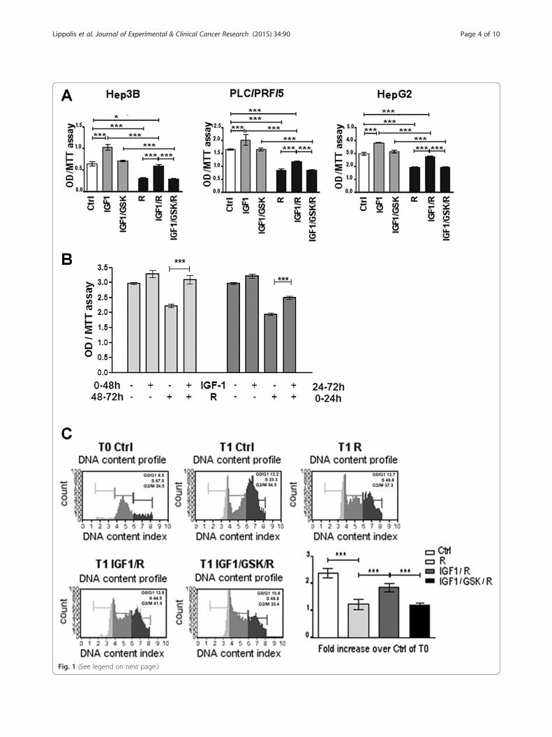
Fig. 1 (See legend on next page.)
Lippolis et al. Journal of Experimental & Clinical Cancer Research (2015) 34:90 Page 4 of 10
The antagonism exerted by IGF1 on Regorafenib-mediated growth inhibitory actions was also observedon cell cycle progression. Regorafenib caused an inhibi-tion in the progression from S phase of the cell cycle toG2/M phase. After 6 h (T1) from block release (T0),Regorafenib treated cells in G2/M phase were only 0.2times more than the control cells at T0, while thenumber of control cells in G2/M phase at T1 were 1.5times more with respect the number of control cells atT0 (Fig. 1c). IGF1 counteracted the Regorafenib-mediated block in cell cycle progression (0.85 timesmore than the control cells at T0), and the IGFRantagonist, GSK1838705A, abrogated this effect.
Antagonism by IGF1 of Regorafenib-mediated inductionof apoptosisThe effects of IGF1 on Regorafenib–mediated apoptosis, amajor aspect of its growth-inhibitory effects, were thenexamined. Regorafenib induced an increase in cellularAnnexin V. When IGF1 was also added to the cellmedium together with Regorafenib, a pronounced and sig-nificant antagonism of apoptosis induction was found andthis antagonism was abrogated by the IGFR inhibitorGSK1838705A 1 μM (Fig. 2a). Major apoptosis markers incells treated with Regorafenib alone or in combinationwith IGF1 were then examined. We found that pro-apoptotic marker (Bim, tBid and Bad) levels increasedin the presence of Regorafenib alone and anti-apoptoticmarkers (p-survivin, Bcl-xL and Bcl-2) decreased underthe same conditions. However, in cells treated withRegorafenib in combination with IGF1 (Fig. 2b), wefound that IGF1 antagonized these Regorafenib effectson induction of apoptosis.
Antagonism by IGF-1 of Regorafenib-mediated inhibitionof cell migration and invasionRegorafenib inhibits both HCC cell migration, as well ascell invasion through Matrigel membranes [31]. IGF140 ng/ml was then added to cells in the presence ofRegorafenib 5 μM, a concentration that can inhibit bothmigration and invasion in HCC cells. We found that IGF1antagonized the inhibition by Regorafenib of both migra-tion and invasion and these effects were also abrogated byGSK1838705A 1 μM (Fig. 3a/b).
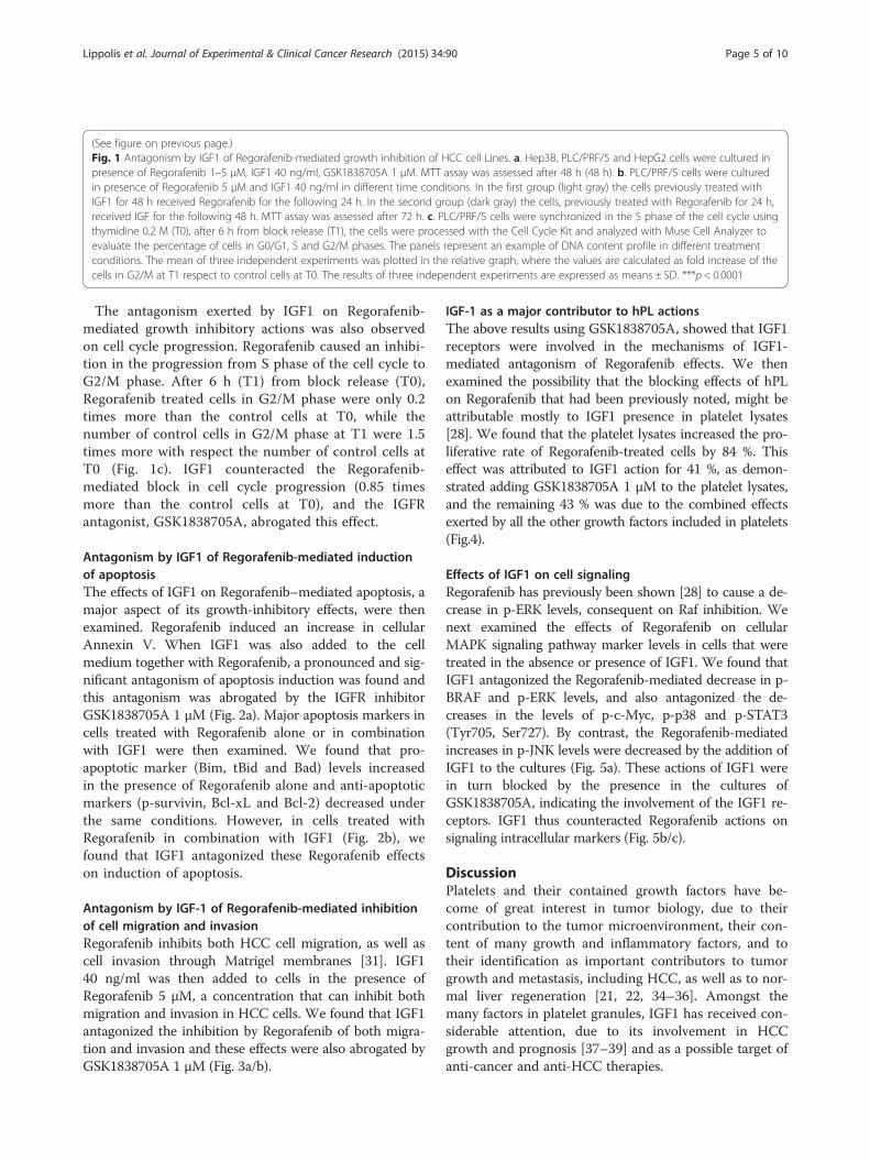
IGF-1 as a major contributor to hPL actionsThe above results using GSK1838705A, showed that IGF1receptors were involved in the mechanisms of IGF1-mediated antagonism of Regorafenib effects. We thenexamined the possibility that the blocking effects of hPLon Regorafenib that had been previously noted, might beattributable mostly to IGF1 presence in platelet lysates[28]. We found that the platelet lysates increased the pro-liferative rate of Regorafenib-treated cells by 84 %. Thiseffect was attributed to IGF1 action for 41 %, as demon-strated adding GSK1838705A 1 μM to the platelet lysates,and the remaining 43 % was due to the combined effectsexerted by all the other growth factors included in platelets(Fig.4).
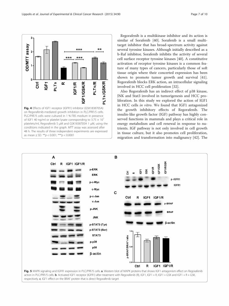
Effects of IGF1 on cell signalingRegorafenib has previously been shown [28] to cause a de-crease in p-ERK levels, consequent on Raf inhibition. Wenext examined the effects of Regorafenib on cellularMAPK signaling pathway marker levels in cells that weretreated in the absence or presence of IGF1. We found thatIGF1 antagonized the Regorafenib-mediated decrease in p-BRAF and p-ERK levels, and also antagonized the de-creases in the levels of p-c-Myc, p-p38 and p-STAT3(Tyr705, Ser727). By contrast, the Regorafenib-mediatedincreases in p-JNK levels were decreased by the addition ofIGF1 to the cultures (Fig. 5a). These actions of IGF1 werein turn blocked by the presence in the cultures ofGSK1838705A, indicating the involvement of the IGF1 re-ceptors. IGF1 thus counteracted Regorafenib actions onsignaling intracellular markers (Fig. 5b/c).
DiscussionPlatelets and their contained growth factors have be-come of great interest in tumor biology, due to theircontribution to the tumor microenvironment, their con-tent of many growth and inflammatory factors, and totheir identification as important contributors to tumorgrowth and metastasis, including HCC, as well as to nor-mal liver regeneration [21, 22, 34–36]. Amongst themany factors in platelet granules, IGF1 has received con-siderable attention, due to its involvement in HCCgrowth and prognosis [37–39] and as a possible target ofanti-cancer and anti-HCC therapies.
(See figure on previous page.)Fig. 1 Antagonism by IGF1 of Regorafenib-mediated growth inhibition of HCC cell Lines. a. Hep3B, PLC/PRF/5 and HepG2 cells were cultured inpresence of Regorafenib 1–5 μM, IGF1 40 ng/ml, GSK1838705A 1 μM. MTT assay was assessed after 48 h (48 h). b. PLC/PRF/5 cells were culturedin presence of Regorafenib 5 μM and IGF1 40 ng/ml in different time conditions. In the first group (light gray) the cells previously treated withIGF1 for 48 h received Regorafenib for the following 24 h. In the second group (dark gray) the cells, previously treated with Regorafenib for 24 h,received IGF for the following 48 h. MTT assay was assessed after 72 h. c. PLC/PRF/5 cells were synchronized in the S phase of the cell cycle usingthymidine 0.2 M (T0), after 6 h from block release (T1), the cells were processed with the Cell Cycle Kit and analyzed with Muse Cell Analyzer toevaluate the percentage of cells in G0/G1, S and G2/M phases. The panels represent an example of DNA content profile in different treatmentconditions. The mean of three independent experiments was plotted in the relative graph, where the values are calculated as fold increase of thecells in G2/M at T1 respect to control cells at T0. The results of three independent experiments are expressed as means ± SD. ***p < 0.0001
Lippolis et al. Journal of Experimental & Clinical Cancer Research (2015) 34:90 Page 5 of 10

Fig. 2 Antagonism by IGF1 of Regorafenib-mediated induction of apoptosis. a. PLC/PRF/5 cell line cultured in 1 % FBS medium was treated with IGF140 ng/ml in combination with Regorafenib 5 μM and GSK 1 μM. The Muse Annexin V kit was used to evaluate the percentage of apoptotic cells. Themeans ± SD of three independent experiments is plotted in the relative graph. ***p < 0.0001. b. Representative Western blot that shows anti-apoptotic(phospho-survivin, Bcl-xL and Bcl-2) and pro-apoptotic (Bim, truncated-Bid and Bad) proteins in PLC/PRF/5 cells treated as described above
Fig. 3 Antagonism by IGF1 of regorafenib-mediated inhibition of migration and invasion. PLC/PRF/5 cell line cultured in 1 % FBS medium wastreated with IGF1 40 ng/ml alone or in combination with Regorafenib 5 μM and GSK 1 μM. a. Migration assay was performed as described andthe microscopic analysis was assessed at the time of the scratch (T0) and after 48 h (T2). The values were expressed as percentage of migration,where 100 % represents the scratch completely closed. b. The percentage of invasion was calculated comparing the invading drug-treated cellsto drug-untreated control cells (100 %). The results of three independent experiments are expressed as means ± SD. ***p < 0.0001
Lippolis et al. Journal of Experimental & Clinical Cancer Research (2015) 34:90 Page 6 of 10
Regorafenib is a multikinase inhibitor and its action issimilar of Sorafenib [40]. Sorafenib is a small multi-target inhibitor that has broad-spectrum activity againstseveral tyrosine kinases. Although initially described as ab-Raf inhibitor, Sorafenib inhibits the activity of severalcell surface receptor tyrosine kinases [40]. A constitutiveactivation of receptor tyrosine kinases is a common fea-ture of many types of cancers, particularly those of softtissue origin where their concerted expression has beenshown to promote tumor growth and survival [41].Regorafenib blocks ERK action, an intracellular signalinginvolved in HCC cell proliferation [32].Also Regorafenib has an indirect effect of p38 kinase,
JNK and Stat3 involved in tumorigenesis and HCC pro-liferation. In this study we explored the action of IGF1in HCC cells in vitro. We found that IGF1 antagonizedthe growth inhibitory effects of Regorafenib. Theinsulin-like growth factor (IGF) pathway has highly con-served functions in mammals and plays a critical role inenergy metabolism and cell renewal in response to nu-trients. IGF pathway is not only involved in cell growthin tissue culture, but it also promotes cell proliferation,migration and transformation into malignancy [42]. The
Fig. 4 Effects of IGF1 receptor (IGFR1) inhibitor (GSK1838705A)on Regorafenib-mediated growth inhibition in PLC/PRF/5 cells.PLC/PRF/5 cells were cultured in 1 % FBS medium in presenceof IGF1 40 ng/ml or platelet lysate corresponding to 3.75 × 10⁷
platelets/ml, Regorafenib 5 μM and GSK1838705A 1 μM, using theconditions indicated in the graph. MTT assay was assessed after48 h. The results of three independent experiments are expressedas mean ± SD. **p < 0.001; ***p < 0.0001
Fig. 5 MAPK signaling and IGFR1 expression in PLC/PRF/5 cells. a. Western blot of MAPK proteins that shows IGF1 antagonism effect on Regorafenibaction in PLC/PRF/5 cells. b. Activated IGF1 receptor (IGFR1) after treatment with Regorafenib (R), IGF1, IGF1 + R, IGF1 + GSK and IGF1 + R + GSK,respectively. c. IGF1 effect on the BRAF protein that is direct Regorafenib target
Lippolis et al. Journal of Experimental & Clinical Cancer Research (2015) 34:90 Page 7 of 10

anti-apoptotic property of IGF-1R was shown in its re-sponse to p53, the tumor suppressor gene that promotesapoptosis [42]. Wild type p53 expression inhibited thegene expression of IGF-1R, while mutant p53 increasedthe gene expression of IGF-1R. Oncogenes such as Srckinase and Akt kinase both stimulated the gene expres-sion of IGF-1R, providing more evidence that IGF-1R isvital in carcinogenesis [43]. In addition, IGF-1R alsostimulates cell mobility, as demonstrated by its activityin melanoma cell lines. Another important role of IGF-1R in carcinogenesis is its ability to transform and main-tain the transformed phenotype [42]. IGF1 and its bind-ing to its receptor induce activations of two majorintracellular cascades, the phosphatidyl inositol 3-kinase(PI3K) and the mitogen-activated protein kinase (MAPK),both of which result in cell differentiation, proliferationand anti-apoptosis [43]. Particularly in HCC, IGF-1R isoverexpressed and can induce carcinogenesis. In a studywhere 10 HCC cell lines (including PLC/PRF/5 cell line)were tested, all of them showed elevated IGF-1R mRNA.Furthermore, the addition of IGF-1 to the PLC/PRF/5 cellline induced increased cell proliferation in a dosedependent manner, showing that the major tumor pro-moting effects of IGF ligands on HCC are exerted throughIGF-1R [42–44].The IGF1 signaling pathway provides an important
regulatory mechanism for tumorigenesis and drug resist-ance in HCC [41].We found that IGF1, used in the same concentration
as was measured in hPL, significantly antagonized thegrowth inhibitory actions of Regorafenib. This effectwas blocked by GSK1838705A, a potent inhibitor ofIGF1 receptors, used at non-toxic concentrations thatdo not affect proliferation. We next found that IGFpre-treatment protected the cells from subsequentaddition of Regorafenib to the cultures and antagonizedRegorafenib-mediated growth inhibition, which was re-duced by 40 % when the cells were pre-treated withIGF1. This suggested that IGF1 signalling is implicatedin the observed Regorafenib resistance or that a majormechanism of Regorafenib-mediated growth control isexerted through interference in the IGFR pathway,which is then counteracted by addition of IGF1 [42].Moreover, the Regorafenib pre-treatment modified thestimulatory action ensuing to the addition of the IGF1.Regorafenib-mediated inhibition of cell growth wasonly partially rescued by subsequent IGF1 treatment(28 %), showing that Regorafenib treatment, even if re-versible, modified the cells [43].A corollary is that IGFR inhibitors might enhance the
growth inhibitory actions of Regorafenib, since IGF1 canblock the drug effects on cell growth. Our results on cellcycle progression support the idea of antagonism exertedby IGF on Regorafenib-mediated growth inhibition.
An important mechanism of resistance to IGFR inhibi-tors is the compensatory activation of related signallingpathways [44]. We observed that IGF1 changed intracel-lular signalling in HCC cells. We found that levels of theproliferation markers p-BRAF, p-ERK, p-p38 and p-Stat3(Tyr705, Ser727) were decreased by Regorafenib action.By contrast, IGF1 addition to the cultures antagonizedRegorafenib action and levels of the proliferationmarkers increased. We, also, found that levels of apop-tosis markers were influenced by IGF1 actions, sincepro-apoptotic markers (p-JNK, p-c-Jun, Bim, Bad andBid) decreased with IGF1, while anti-apoptosis markers(survivin, BCL-xL and Bcl-2) increased. Survivin is anapoptosis-inhibitory protein that is over-expressed inmultiple cancer types, including HCC, plays critical rolesin regulating apoptosis, cell proliferation and survivaland has been shown to be a direct downstream target ofIGF1 pathway [44].Regorafenib decreased p-survivin levels and this was
also antagonized by addition of IGF1. GSK1838705Areversed the IGF1 actions, showing that the antagonisteffects of IGF are exerted through its receptor. The an-tagonism exerted by IGF on Regorafenib effects wasalso shown for drug mediated apoptosis, as a decreasein Annexin V levels and in the Caspase 3/7 activation.Pre-clinical studies have shown that the efficacy in anti-cancer therapy for HCC can be improved by inhibitingthe IGF signalling pathway in HCC cells [45].Several new inhibitors of IGF1 or its receptor are in cur-
rent clinical trials for HCC, including Octreotide (Novartis),a Somatostatin analog and MEDI 573 (Astrazeneca), tar-geting either IGF1 and or IGFII, as well as Linsitinib (OSIPharmaceuticals), Cixutumumab (ImClone), AVE1642(Sanofi-Aventis), which are IGF receptor antagonists.However, the relationship of IGF1 levels in serum andtumor tissue and of its receptor levels with HCC growthand prognosis are very complex [37]. It has recently beenshown that one of the many mechanisms of Sorafenib(Regorafenib is Fluoro-Sorafenib) action on HCC cellgrowth is via an inhibition of IGF1 [39]. Furthermore,IGF1 has been shown to be involved in resistance tocytotoxic chemotherapy by several drugs and on a varietyof tumor types, mainly through an anti-apoptotic effect[33, 46–51], as we have found in the current work.
ConclusionThese experiments highlight the importance of themicroenvironment, including IGF1, in modulating thegrowth inhibitory effects of anti-HCC therapeutic drugs.By contrast, anti-IGF or anti-IGF receptor agents mightbe predicted to be therapeutically useful in enhancingthe activity of anti-HCC therapies. Finally, we re-enforcethe concept of drug resistance mediated by factors in thetumor microenvironment, and also summarize potential
Lippolis et al. Journal of Experimental & Clinical Cancer Research (2015) 34:90 Page 8 of 10

drug targets based on the current knowledge of thetumor microenvironment [52].
AbbreviationsHCC: Hepatocellular carcinoma; hPL: Human platelet lysates; ERK: Extracellularsignal-regulated kinase; JNK: c-Jun NH2-terminal kinase; STAT: Signaltransducer and activator of transcription-3; WB: Western blot; MTT: 3-(4,5-Dimethylthiazol-2-yl)-2,5-diphenyltetrazolium bromide; BrdU 5-bromo-2’-de-oxy-uridine; IGF1: Insulin like growth factor 1.
Competing interestThe authors declare that they have no competing interests.
Authors’ contributionBIC and CL are fully responsible for study designing, drafting and finalizingthe manuscript; MGR and RD’A executed in vitro experiments; NC and CLcarried out protein measurement by Western blot; AC and CM conductedstatistical analysis and participated in coordination manuscript providingimportant suggestions. All authors read and approved the final manuscript.
AcknowledgmentsThis research was supported by Italian Ministry of Public Health.
Author details1Department Clinical Pathology, Laboratory of Cellular and Molecular Biology,National Institute for Digestive Diseases, IRCCS “Saverio de Bellis”, Via Turi 27,70013 Castellana Grotte, BA, Italy. 2Izmir Biomedicine and Genome Center,Dokuz Eylul University, Izmir, Turkey.
Received: 12 May 2015 Accepted: 20 August 2015
References1. Nordenstedt H, White DL, El-Serag HB. The changing pattern of
epidemiology in hepatocellular carcinoma. Dig Liver Dis. 2010;42:S206–14.2. Carr BI. Understanding Liver Cancer: a tale of two diseases. New York:
Springer Press; 2014. ISBN-13: 978–1910315019.3. Lu SN, Wang JH, Liu SL, Hung CH, Chen CH, Tung HD, et al.
Thrombocytopenia as a surrogate for cirrhosis and a marker for theidentification of patients at high-risk for hepatocellular carcinoma. Cancer.2006;107:2212–22.
4. Kumada T, Toyoda H, Kiriyama S, Sone Y, Tanikawa M, Hisanaga Y, et al.Incidence of hepatocellular carcinoma in patients with chronic hepatitis Bvirus infection who have normal alanine aminotransferase values. J MedVirol. 2010;82:539–45.
5. Lok AS, Seeff LB, Morgan TR, di Bisceglie AM, Sterling RK, Curto TM, et al.HALT-C Trial Group. Incidence of hepatocellular carcinoma and associatedrisk factors in hepatitis C-related advanced liver disease. Gastroenterology.2009;136:138–48.
6. Carr BI, Guerra V, Pancoska P. Thrombocytopenia in relation to tumor size inpatients with hepatocellular carcinoma. Oncology. 2012;83:339–45.
7. Carr BI, Guerra V, De Giorgio M, Fagiuoli S, Pancoska P. Small hepatocellularcarcinomas and thrombocytopenia. Oncology. 2012;83:331–8.
8. Trevisani F, D’Intino PE, Caraceni P, Pizzo M, Stefanini GF, Mazziotti A, et al.Etiologic factors and clinical presentation of hepatocellular carcinoma.Differences between cirrhotic and noncirrhotic Italian patients. Cancer.1995;75:2220–32.
9. Truant S, Boleslawsky E, Duhamel A, Bouras AF, Louvet A, Febvay C, et al.Tumor size of hepatocellular carcinoma in noncirrhotic liver: A controversialpredictive factor for outcome after resection. Eur J Surg Oncol.2012;38:1189–96.
10. Carr BI, Guerra V, Giannini EG, Farinati F, Ciccarese F, Rapaccini GL, et al.Significance of platelet and AFP levels and liver function parameters forHCC size and survival. Int J Biol Markers. 2014;29:e215–23.
11. Carr BI, Guerra V. Features of massive hepatocellular carcinomas. Eur JGastroenterol Hepatol. 2014;26:101–8.
12. Carr BI, Guerra V. Thrombocytosis and hepatocellular carcinoma. Dig DisSci. 2013;58:1790–6.
13. Hwang SJ, Luo JC, Li CP, Chu CW, Wu JC, Lai CR, et al. Thrombocytosis: aparaneoplastic syndrome in patients with hepatocellular carcinoma. World JGastroenterol. 2004;10:2472–7.
14. Levin J, Conley CL. Thrombocytosis associated with malignant disease. ArchInt Med. 1964;114:497–500.
15. Stone RL, Nick AM, McNeish IA, Balkwill F, Han HD, Bottsford-Miller J, et al.Paraneoplastic thrombocytosis in ovarian cancer. N Engl J Med. 2012;366:610–8.
16. Voutsadakis IA. Thrombocytosis as a prognostic marker in gastrointestinalcancers. World J Gastrointest Oncol. 2014;6:34–40.
17. Li FX, Wei LJ, Zhang H, Li SX, Liu JT. Significance of thrombocytosis inclinicopathologic characteristics and prognosis of gastric cancer. Asian Pac JCancer Prev. 2014;15:6511–7.
18. Kim M, Chang H, Yang HC, Kim YJ, Lee CT, Lee JH, et al. Preoperativethrombocytosis is a significant unfavorable prognostic factor for patients withresectable non-small cell lung cancer. World J Surg Oncol. 2014;12:37.
19. Njolstad TS, Engerud H, Werner HM, Salvesen HB, Trovik J. Preoperativeanemia, leukocytosis and thrombocytosis identify aggressive endometrialcarcinomas. Gynecol Oncol. 2013;131:410–5.
20. Goubran HA, Stakiw J, Radosevic M, Burnouf T. Platelets effects on tumorgrowth. Semin Oncol. 2014;41:359–69.
21. Labelle M, Begum S, Hynes RO. Direct signaling between platelets andcancer cells induces an epithelial-mesenchymal-like transition and promotesmetastasis. Cancer Cell. 2011;20:576–90.
22. Everts PA, Brown Mahoney C, Hoffmann JJ, Schönberger JP, Box HA, vanZundert A, et al. Platelet-rich plasma preparation using three devices:implications for platelet activation and platelet growth factor release.Growth Factors. 2006;24:165–71.
23. Christgau M, Moder D, Hiller KA, Dada A, Schmitz G, Schmalz G. Growthfactors and cytokines in autologous platelet concentrate and theircorrelation to periodontal regeneration outcomes. J Clin Periodontol.2006;33:837–45.
24. Eppley BL, Woodell JE, Higgins J. Platelet quantification and growth factoranalysis from platelet-rich plasma: implications for wound healing. PlastReconstr Surg. 2004;114:1502–8.
25. Weibric G, Buch RS, Kleis WK, Hafner G, Hitzler WE, Wagner W.Quantification of thrombocyte growth factors in platelet concentratesproduced by discontinuous cell separation. Growth Factors.2002;20:93–7.
26. Karey KP, Marquardt H, Sirbasku DA. Human platelet-derived mitogens.Identification of insulin like growth factors I and II by purification and Nalpha amino acid sequence analysis. Blood. 1989;74:1084–92.
27. Carr BI, Cavallini A, D'Alessandro R, Refolo MG, Lippolis C, Mazzocca A, et al.Platelet extracts induce growth, migration and invasion in humanhepatocellular carcinoma in vitro. BMC Cancer. 2014;14:43.
28. D'Alessandro R, Refolo MG, Lippolis C, Giannuzzi G, Carella N, Messa C, et al.Antagonism of Sorafenib and Regorafenib actions by platelet factors inhepatocellular carcinoma cell lines. BMC Cancer. 2014;14:351.
29. Ettrich TJ1, Seufferlein T. Regorafenib. Recent Results. Cancer Res.2014;201:185–96.
30. Sartore-Bianchi A1, Zeppellini A, Amatu A, Ricotta R, Bencardino K, Siena S.Regorafenib in metastatic colorectal cancer. Expert Rev Anticancer Ther.2014;14:255–65.
31. Carr BI, D’Alessandro R, Refolo MG, Iacovazzi PA, Lippolis C, Messa C, etal. Effects of low concentrations of Regorafenib and Sorafenib on humanHCC cell AFP, migration, invasion, and growth In Vitro. J Cell Physiol.2013;228:1344–50.
32. Carr BI, Cavallini A, Lippolis C, D'Alessandro R, Messa C, Refolo MG, et al.Fluoro Sorafenib (Regorafenib) effects on hepatoma cells: growth inhibition,quiescence, and recovery. J Cell Physiol. 2013;228:292–7.
33. D'Alessandro R, Refolo MG, Lippolis C, Messa C, Cavallini A, Rossi R, et al.Reversibility of Regorafenib effects in hepatocellular carcinoma cells. CancerChemother Pharmacol. 2013;72:869–77.
34. Morimoto Y, Nouso K, Wada N, Takeuchi Y, Kinugasa H, Miyahara K, et al.Involvement of platelets in extrahepatic metastasis of hepatocellularcarcinoma. Hepatol Res. 2014;44:e353–9.
35. Hoshi R, Murata S, Matsuo R, Myronovych A, Hashimoto I, Ikeda H, et al.Freeze-dried platelets promote hepatocyte proliferation in mice.Cryobiology. 2007;55:255–60.
36. Enguita-Germán M, Fortes P. Targeting the insulin-like growth factorpathway in hepatocellular carcinoma. World J Hepatol. 2014;6:716–37.
37. Kaseb AO, Morris JS, Hassan MM, Siddiqui AM, Lin E, Xiao L, et al. Clinicaland prognostic implications of plasma insulin-like growth factor-1 andvascular endothelial growth factor in patients with hepatocellularcarcinoma. J Clin Oncol. 2011;29:3892–9.
Lippolis et al. Journal of Experimental & Clinical Cancer Research (2015) 34:90 Page 9 of 10

38. Shao YY, Huang CC, Lin SD, Hsu CH, Cheng AL. Serum insulin- like growthfactor-1 levels predict outcomes of patients with advanced hepatocellularcarcinoma receiving antiangiogenic therapy. Clin Cancer Res.2012;18:3992–7.
39. Sprinzl MF, Puschnik A, Schlitter AM, Schad A, Ackermann K, Esposito I, et al.Sorafenib inhibits macrophage-induced growth of HCC cells by interferencewith insulin-like growth factor-1 secretion. J Hepatol. 2014;62:863–70.
40. Maruwge W, D’Arcy P, Folin A, Brnjic S, Wejde J, Davis A, et al. Sorafenibinhibits tumor growth and vascularization of rhabdomyosarcoma cells byblocking IGF-1R-mediated signaling. Onco Targets Ther. 2008;1:67–78.
41. Caro JF, Poulos J, Ittoop O, Pories WJ, Flickinger EG, Sinha MK. Insulin-likegrowth factor I binding in hepatocytes from human liver, humanhepatoma, and normal, regenerating, and fetal rat liver. J Clin Invest.1988;81:976–81.
42. Tovar V, Alsinet C, Villanueva A, Hoshida Y, Chiang DY, Sole M, et al. IGFactivation in a molecular subclass of hepatocellular carcinoma and pre-clinical efficacy of IGF-1R blockage. J Hepatol. 2010;52:550–9.
43. Ou DL, Lee BS, Lin LI, Liou JY, Liao SC, Hsu C, et al. Vertical blockade of theIGFR- PI3K/Akt/mTOR pathway for the treatment of hepatocellularcarcinoma: the role of survivin. Mol Cancer. 2014;13:2.
44. Gao J, Chang YS, Jallal B, Viner J. Targeting the insulin-like growth factor axisfor the development of novel therapeutics in oncology. Cancer Res.2012;72:3–12.
45. Scharf JG, Schmidt-Sandte W, Pahernik SA, Ramadori G, Braulke T, HartmannH. Characterization of the insulin-like growth factor axis in a humanhepatoma cell line (PLC). Carcinogenesis. 1998;19:2121–8.
46. Wu J, Zhu AX. Targeting insulin-like growth factor axis in hepatocellularcarcinoma. J Hematol Oncol. 2011;4:30.
47. Pal S, Shankar BS, Sainis KB. Cytokines from the tumor microenvironmentmodulate sirtinol cytotoxicity in A549 lung carcinoma cells. Cytokine.2013;64:196–207.
48. Sun Y, Zheng S, Torossian A, Speirs CK, Schleicher S, Giacalone NJ, et al. Roleof insulin-like growth factor-1 signaling pathway in cisplatin-resistant lungcancer cells. Int J Radiat Oncol Biol Phys. 2012;82:e563–72.
49. Juan HC, Tsai HT, Chang PH, Huang CY, Hu CP, Wong FH. Insulin-likegrowth factor 1 mediates 5-fluorouracil chemoresistance in esophagealcarcinoma cells through increasing survivin stability. Apoptosis.2011;16:174–83.
50. Zhang YW, Yan DL, Wang W, Zhao HW, Lu X, Wu JZ, et al. Knockdown ofinsulin-like growth factor I receptor inhibits the growth and enhanceschemo-sensitivity of liver cancer cells. Curr Cancer Drug Targets.2012;12:74–84.
51. Eckstein N, Servan K, Hildebrandt B, Pölitz A, von Jonquières G,Wolf-Kümmeth S, et al. Hyperactivation of the insulin-like growth factorreceptor I signaling pathway is an essential event for cisplatin resistanceof ovarian cancer cells. Cancer Res. 2009;69:2996–3003.
52. Gao F, Liang B, Reddy ST, Farias-Eisner R, Su X. Role of inflammation-associated microenvironment in tumorigenesis and metastasis. CurrCancer Drug Targets. 2014;14:30–45.
Submit your next manuscript to BioMed Centraland take full advantage of:
• Convenient online submission
• Thorough peer review
• No space constraints or color figure charges
• Immediate publication on acceptance
• Inclusion in PubMed, CAS, Scopus and Google Scholar
• Research which is freely available for redistribution
Submit your manuscript at www.biomedcentral.com/submit
Lippolis et al. Journal of Experimental & Clinical Cancer Research (2015) 34:90 Page 10 of 10


![An Auxin Transport Inhibitor Targets Villin-Mediated · An Auxin Transport Inhibitor Targets Villin-Mediated Actin Dynamics to Regulate Polar Auxin Transport1[OPEN] Minxia Zou,a Haiyun](https://static.fdocuments.in/doc/165x107/5f495bd623de363ead44b1aa/an-auxin-transport-inhibitor-targets-villin-an-auxin-transport-inhibitor-targets.jpg)


![Therapeutics A Novel Small-Molecule Inhibitor of Protein ...transferase–mediated dUTP nick end labeling (TUNEL)– positive cells], and abrogated the expression of several NF-κB–dependent](https://static.fdocuments.in/doc/165x107/6118340d0013286be8293532/therapeutics-a-novel-small-molecule-inhibitor-of-protein-transferaseamediated.jpg)


