Differential Effects of Sorafenib on Liver Versus Tumor ...
20
Differential Effects of Sorafenib on Liver Versus Tumor Fibrosis Mediated by Stromal-Derived Factor 1 alpha/ C-X-C Receptor Type 4 Axis and Myeloid Differentiation Antigen- Positive Myeloid Cell Infiltration in Mice The Harvard community has made this article openly available. Please share how this access benefits you. Your story matters Citation Chen, Yunching, Yuhui Huang, Thomas Reiberger, Annique M. Duyverman, Peigen Huang, Rekha Samuel, Lotte Hiddingh, and et al. 2014. Differential effects of sorafenib on liver versus tumor fibrosis mediated by stromal-derived factor 1 alpha/C-X-C receptor type 4 axis and myeloid differentiation antigen-positive myeloid cell infiltration in mice. Hepatology 59(4): 1435–1447. Published Version doi:10.1002/hep.26790 Citable link http://nrs.harvard.edu/urn-3:HUL.InstRepos:32518371 Terms of Use This article was downloaded from Harvard University’s DASH repository, and is made available under the terms and conditions applicable to Other Posted Material, as set forth at http:// nrs.harvard.edu/urn-3:HUL.InstRepos:dash.current.terms-of- use#LAA
Transcript of Differential Effects of Sorafenib on Liver Versus Tumor ...

Differential Effects of Sorafenib onLiver Versus Tumor Fibrosis Mediatedby Stromal-Derived Factor 1 alpha/
C-X-C Receptor Type 4 Axis andMyeloid Differentiation Antigen-
Positive Myeloid Cell Infiltration in MiceThe Harvard community has made this
article openly available. Please share howthis access benefits you. Your story matters
Citation Chen, Yunching, Yuhui Huang, Thomas Reiberger, Annique M.Duyverman, Peigen Huang, Rekha Samuel, Lotte Hiddingh, andet al. 2014. Differential effects of sorafenib on liver versus tumorfibrosis mediated by stromal-derived factor 1 alpha/C-X-C receptortype 4 axis and myeloid differentiation antigen-positive myeloid cellinfiltration in mice. Hepatology 59(4): 1435–1447.
Published Version doi:10.1002/hep.26790
Citable link http://nrs.harvard.edu/urn-3:HUL.InstRepos:32518371
Terms of Use This article was downloaded from Harvard University’s DASHrepository, and is made available under the terms and conditionsapplicable to Other Posted Material, as set forth at http://nrs.harvard.edu/urn-3:HUL.InstRepos:dash.current.terms-of-use#LAA

Differential effects of sorafenib on liver versus tumor fibrosismediated by SDF1α/CXCR4 axis and Gr-1+ myeloid cellinfiltration in mice
Yunching Chen1,2, Yuhui Huang1, Thomas Reiberger1, Annique M. Duyverman1,3, PeigenHuang1, Rekha Samuel1,4, Lotte Hiddingh1,5, Sylvie Roberge1, Christina Koppel1, GregoryY. Lauwers6, Andrew X. Zhu7, Rakesh K. Jain1, and Dan G. Duda1,*
Yunching Chen: [email protected]; Yuhui Huang: [email protected]; Thomas Reiberger:[email protected]; Annique M. Duyverman: [email protected]; Peigen Huang:[email protected]; Rekha Samuel: [email protected]; Lotte Hiddingh: [email protected];Sylvie Roberge: [email protected]; Christina Koppel: [email protected]; Gregory Y.Lauwers: [email protected]; Andrew X. Zhu: [email protected]; Rakesh K. Jain: [email protected];Dan G. Duda: [email protected] Laboratory for Tumor Biology, Department of Radiation Oncology, MassachusettsGeneral Hospital, Harvard Medical School, Boston, MA 021146Department of Pathology, Massachusetts General Hospital, Harvard Medical School, Boston,MA 021147Department of Medicine, Massachusetts General Hospital, Harvard Medical School, Boston, MA02114
AbstractSorafenib—a broad kinase inhibitor—is a standard therapy for advanced hepatocellular carcinoma(HCC), and has been shown to exert anti-fibrotic effects in liver cirrhosis, a precursor of HCC.However, the effects of sorafenib on tumor desmoplasia—and its consequences on treatmentresistance — remain unknown. We demonstrate that sorafenib has differential effects on tumorfibrosis versus liver fibrosis in orthotopic models of HCC in mice. Sorafenib intensifies tumorhypoxia, which increases stromal-derived factor 1α (SDF1α) expression in cancer and stromalcells, and subsequently Gr-1+ myeloid cell infiltration. The SDF1α/CXCR4 pathway directlypromotes hepatic stellate cell (HSC) differentiation and activation via MAP kinase pathway. Thisis consistent with the association between SDF1α expression with fibrotic septa in cirrhotic livertissues as well as with desmoplastic regions of human HCC samples. We demonstrate that aftertreatment with sorafenib, SDF1α increased the survival of HSCs and their α-SMA and Collagen Iexpression, thus increasing tumor fibrosis. Finally, we show that Gr-1+ myeloid cells mediateHSC differentiation/activation in a paracrine manner. CXCR4 inhibition using AMD3100 incombination with sorafenib treatment prevents the increase in tumor fibrosis—despite persistentlyelevated hypoxia—in part by reducing Gr-1+ myeloid cell infiltration, and inhibits HCC growth.Similarly, antibody blockade of Gr-1 reduces tumor fibrosis and inhibited HCC growth whencombined with sorafenib treatment.
Conclusion—Blocking SDF1α/CXCR4 or Gr-1+ myeloid cell infiltration may reduce hypoxia-mediated HCC desmoplasia and increase the efficacy of sorafenib treatment.
*Corresponding author: Dan G. Duda, DMD, PhD, Steele Laboratory for Tumor Biology, Massachusetts General Hospital, Cox-734,100 Blossom Street, Boston, MA 02114; phone: (617) 726-4648; fax: (617) 726-1962; [email protected] of Biomedical Engineering, National Tsing Hua University, Taiwan3Universty of Utrecht, The Netherlands4Christian Medical College, Vellore, India5VU Medical Center Amsterdam, The Netherlands.
NIH Public AccessAuthor ManuscriptHepatology. Author manuscript; available in PMC 2015 April 01.
Published in final edited form as:Hepatology. 2014 April ; 59(4): 1435–1447. doi:10.1002/hep.26790.
NIH
-PA Author Manuscript
NIH
-PA Author Manuscript
NIH
-PA Author Manuscript

Keywordshepatocellular carcinoma; hypoxia; collagen I; hepatic stellate cell; α-smooth muscle actin
INTRODUCTORY STATEMENTHepatocellular carcinoma (HCC) almost exclusively arises in cirrhotic livers, and thepreexisting chronic inflammation and fibrosis fuel hepatocarcinogenesis and HCC growth(1–3). Fibrosis is the consequence of hepatic stellate cell (HSC) activation and proliferation,and myofibroblast differentiation leading to increased collagen deposition (4). This dualpathology of the liver contributes to an aggressive and systemic treatment-refractorycharacteristic in HCCs (1). Recently, the tyrosine kinase inhibitor (TKI) sorafenib hasemerged as the first systemic therapy for HCC. Sorafenib is an antiangiogenic drug that hasa broad tyrosine kinase inhibition spectrum (5). However, despite this progress, the mortalityrate from HCC remains high, making this disease the third leading cause of cancer-relateddeath worldwide (6–10).
Sorafenib is widely considered as an anti-angiogenic/anti-vascular drug through inhibitionof VEGF receptors (VEGFRs) and platelet-derived growth factor receptors (PDGFRs).However, more potent and selective anti-VEGF agents or more broad antiangiogenic agents(e.g., VEGFR/FGFR and anti-VEGFR/PDGFR inhibitors) have failed so far to match theefficacy of sorafenib in phase III trials in HCC (10–13). Moreover, anti-angiogenic therapyhas not led to tumor regression in patients or in experimental models in mice: The benefitseen with sorafenib in HCC patients is likely due to a transient delay in HCC growth, afterwhich most tumors resume their growth (10). Whereas the mechanisms of acquiredresistance to sorafenib and other anti-VEGF inhibitors in HCC remain unknown, it is likelythat tumor stroma-mediated survival pathways might play a key role (1, 10). Of these,increased hypoxia has been proposed as a mechanism of resistance to multitargeted TKItherapy (14–17). The challenge is to identify the key molecular pathways regulating stroma-mediated resistance to sorafenib treatment in HCC.
Hypoxia and other cellular stresses can promote the expression of the chemokine stromal-derived factor 1 alpha (SDF1α or CXCL12) and of its receptor CXCR4 (18–22). In clinicalstudies, we showed that SDF1α level increased in plasma circulation in HCC patients aftertreatment with sunitinib or cediranib (both anti-VEGFR and anti-PDGFR TKIs) (23, 24).Moreover, we showed that elevated circulating levels of SDF1α correlated with poortreatment outcome in HCC patients after sunitinib treatment (23). Systemic activation ofSDF1α/CXCR4 axis is known to mediate intra-tumoral infiltration of inflammatory cells,including Gr-1+ myeloid (CD11b+) cells (25–28). Gr-1+ myeloid cells can drive tumorrecurrence after anti-VEGF therapy in various tumor models (29). Finally, clinicalcorrelative data also strongly suggest that the effects on multi-targeted TKI treatment ontumor vasculature and on myeloid cells may mediate the response and resistance therapy inHCC patients (23, 30). However, a causal role of Gr-1+ myeloid cells in HCC resistance toanti-angiogenic treatment has not been characterized. Furthermore, a mechanisticunderstanding of the interplay between treatment-induced hypoxia, SDF1α/CXCR4 pathwayactivation, and Gr-1+ myeloid cell infiltration and tumor fibrosis in HCC is currentlylacking. Here, we examined in orthotopic HCC models whether the SDF1α/CXCR4pathway is activated and causally related to Gr-1+ myeloid cell infiltration and tumor-associated fibrosis and, ultimately, to sorafenib resistance.
Chen et al. Page 2
Hepatology. Author manuscript; available in PMC 2015 April 01.
NIH
-PA Author Manuscript
NIH
-PA Author Manuscript
NIH
-PA Author Manuscript

EXPERIMENTAL PROCEDURESCells and Materials
We used the C3H mouse-derived HCC cell line HCA-1 (31). Human hepatic stellate cells(HSCs; Catalog #5300) were purchased from ScienCell Research Laboratories (San Diego,CA). These primary HSCs have been previously characterized (32). We purchasedAMD3100 and FR180204 from Sigma (St. Louis, MO), AZD6244 and sorafenib fromSelleck Chemicals (Houston, TX), recombinant PDGF-B and SDF1α from R&D system(Minneapolis, MN), and anti-Gr-1 antibody (LeafTH purified anti-mouse Gr-1 antibody)from Biolegend (San Diego, CA). For co-culture experiments, we isolated Gr-1+ myeloidcells from enzymatically digested HCA-1 tumors.
AnimalsTo induce liver fibrosis, we treated 5-week-old male C3H or Mst1−/−Mst2F/− mice withcarbon tetrachloride (CCl4, 16 %v/v in olive oil, 100μL gavage, 3 times per week) prior totumor implantation/induction. HCA-1 cells were orthotopically implanted in mice 2 weeksafter the last CCl4 treatment, as previously described (33). Mst1−/−Mst2F/− mice developspontaneous HCCs after i.v. injection of Ad-Cre (34), and were a kind gift from Dr. NabeelBardeesy (MGH). All animals received humane care according to the criteria outlined in the“Guide for the Care and Use of Laboratory Animals” prepared by the National Academy ofSciences and published by the National Institutes of Health (NIH publication 86–23 revised1985).
Treatment studiesMice were treated daily by gavage with sorafenib (40mg/kg) or vehicle (PBS) alone.AMD3100 (10mg/kg/day) was delivered continuously using Alzet micro-osmotic pumps(DURECT Corporation, Cupertino, CA) over 2 weeks.
Cell viability assaysWe assessed cell viability using 3-[4,5-dimethylthiazol-2-yl]-2,5-diphenyl tetrazoliumbromide (MTT) assay (Sigma).
Western blot analysisWestern blotting was performed using antibodies against collagen I and α-SMA (Abcam,Cambridge, MA), and phosphorylated (p)-ERK, ERK, p-AKT and AKT (Cell Signaling,Danvers, MA).
siRNA knockdownON-TARGETplus CXCR4 siRNA and non-targeting control siRNA were purchased fromDharmacon (Lafayette, CO) and used for transfection of HCA-1 cells.
ImmunohistochemistryFor murine samples, we used antibodies against α-SMA, CAIX (Abcam), collagen I (LF-67,kindly provided by Dr. L. Fisher, National Institute of Dental Research), and SDF1α(BioVision). For apoptosis detection, frozen tumor sections were stained using TACS™ TdTKit (R&D Systems, Minneapolis, MN). To assess expression level, we measured thefluorescently stained area for each marker and normalized it to DAPI area (used as measureof cellularity in viable tumor regions), as previously described (28). In addition, weperformed immunostaining for SDF1α in tumor samples from patients who underwent HCCresection. Hepatic fibrosis in the non-HCC liver area was evaluated according to the
Chen et al. Page 3
Hepatology. Author manuscript; available in PMC 2015 April 01.
NIH
-PA Author Manuscript
NIH
-PA Author Manuscript
NIH
-PA Author Manuscript

Laennec system as previously described (35). Co-localization between SDF1α expressionand desmoplasia was assessed in 10 randomly selected high-power fields at 4xmagnification.
Flow cytometryWe used fluorescently labeled rat monoclonal antibodies anti-mouse CD45-PE-Cy7, Gr-1-APC and CD11b-APC-Cy7 (BD Biosciences) to perform flow cytometric analysis indigested tumor tissue, as previously described (36).
Quantitative RT-PCRWe determined the relative gene expression of SDF1α, CXCR4, MCP-1, 5-LO, TGF-α,TGF-β, PDGF-α, PDGF-β, MMP9, MMP13, and β-actin in tumor-infiltrating Gr-1+ cellsusing specific primers (Table S1), Real-Time SYBR Green PCR master mix (AppliedBiosystems, Branchburg, NJ) and the Stratagene Mx3000P QPCR System, as previouslydescribed (36).
Statistical analysisComparisons between treatment groups were performed using the Mann-Whitney U-test. Ap-value of less than 0.05 was considered to denote statistical significance. (See more detailsin Supplemental Experimental Methods.)
RESULTSSorafenib treatment increases hypoxia and SDF1α expression in orthotopic HCC models
To model the clinical features of HCC, we first induced liver fibrosis in mice by CCl4treatment for 11 weeks (Fig. S1). Then, we generated orthotopic tumors by intrahepaticHCA-1 cell implantation in C3H mice or by inducing spontaneous HCC using Cre-adenovirus (Ad-Cre) i.v. injection in Mst1−/− Mst2F/− mice (Fig. S2). When tumors becameestablished, we treated the mice with sorafenib for 14 days and then measured the changesin tissue oxygenation. Sorafenib treatment significantly increased the hypoxic tissue fraction—measured by carbonic anhydrase IX (CAIX) immunostaining—when compared tocontrol-treated tumors (Fig. 1A, B). In contrast, hypoxic tissue surface area did not increasein the surrounding fibrotic liver tissue after treatment (Fig. 1E).
To examine the consequence of treatment-induced increase in tumor hypoxia, we nextevaluated whether the increase in SDF1α expression—suggested by studies in HCC patients(23, 24)—is recapitulated in these HCC models. Indeed, we found a 2-fold elevation inSDF1α expression after sorafenib treatment in HCA-1 tumor grafts and in spontaneousHCCs in Mst1−/− Mst2F/− mice (Fig. 1A–D, Fig. S3). In contrast, SDF1α expression did notsignificantly increase in the fibrotic liver after sorafenib treatment (Fig. 1F).
To confirm that hypoxia is the driver of SDF1α expression in cancer and stromal cells, wecultured HCA-1 cells as well as hepatic stellate cells (HSCs) for 48hr in hypoxic (1% O2) ornormoxic (21% O2) conditions and measured SDF1α expression by qPCR. Exposure tohypoxic conditions increased SDF1α expression in both HCC cells and HSCs (Fig. S4).
To evaluate the effect of SDF1α/CXCR4 pathway inhibition on tumor tissue oxygenationand SDF1α expression after treatment, we combined sorafenib treatment with the CXCR4inhibitor AMD3100 (Sigma, 10mg/kg/day). Interestingly, the combination therapy furtherincreased hypoxic tissue fraction and SDF1α expression in orthotopic HCA-1 tumor graftsand spontaneous HCCs in Mst1−/− Mst2F/− mice when compared to sorafenib treatmentalone (Fig. 1, Fig. S3).
Chen et al. Page 4
Hepatology. Author manuscript; available in PMC 2015 April 01.
NIH
-PA Author Manuscript
NIH
-PA Author Manuscript
NIH
-PA Author Manuscript

Inhibition of CXCR4 prevents the increase in HCC fibrosis after sorafenib in vivoTo establish if fibrosis is associated with elevated SDF1α expression in HCC patients, weexamined surgical specimens for HCC patients with cirrhosis. We observed that SDF1αexpression co-localized with extracellular matrix components/fibrotic septa in both HCCand cirrhotic liver tissues (Fig. 2). Thus, we next evaluated the effect of sorafenib treatmentwith or without the inhibition of SDF1α/CXCR4 pathway on fibrosis, a hallmark of HCCprogression (4), in the mouse models. To this end, we measured the expression of collagen Ias well as the number of α SMA+ myofibroblasts after sorafenib treatment both in the tumorregion and in the surrounding liver tissue. Of note, the α SMA+ myofibroblasts were alsopositive for GFAP (Fig. S5). IHC analyses revealed that the HCCs growing in the face ofsorafenib treatment showed significantly increased tumor desmoplasia. Treatment resultedin increased intra- and peri-tumoral collagen I and α-SMA+ myofibroblasts infiltration inorthotopic HCA-1 HCCs in mice with CCl4-induced liver fibrosis and in spontaneous HCCsin Mst1−/− Mst2F/− mice (Fig. 3A–E). Of note, sorafenib treatment reduced collagen Iexpression levels in the surrounding liver tissue in mice with CCl4-induced liver fibrosis(Fig. 3F).
To assess the effect of SDF1α/CXCR4 pathway inhibition on tumor-associated fibrosis aftersorafenib treatment, we tested the combined administration of sorafenib with the CXCR4inhibitor AMD3100. This combination therapy prevented the increase in collagen Iexpression and α SMA+ myofibroblasts after sorafenib treatment in spontaneous andimplanted HCCs, without significantly changing collagen I expression in the fibrotic liver inCCl4-treated mice (Fig. 3). Of note, this effect was seen despite a persistent increase inhypoxia and SDF1α expression in the HCCs treated with sorafenib and AMD3100 (Fig.1A–D). Thus, CXCR4 inhibition can prevent the pro-fibrotic effects of sorafenib treatmentin HCC in vivo, despite persistent tumor hypoxia.
SDF-1α/CXCR4 axis directly mediates hepatic stellate cell (HSC) differentiation tomyofibroblasts in HCC despite PDGFR blockade by sorafenib
We next examined the role of SDF1α/CXCR4 axis in selective promotion of tumor fibrosisafter sorafenib treatment in vitro. To determine if increased SDF1α expression in the tumorcould mediate the increase in tumor fibrosis after sorafenib treatment, we first exposedprimary HSCs to recombinant (r)SDF1α in the presence or absence of sorafenib or rPDGF-B. Consistent with previous reports (37), we found that rPDGF-B stimulated proliferationand α-SMA expression in HSCs (Fig. 4A, B). Sorafenib treatment prevented the effects ofrPDGF-B and led to significant decrease in HSC viability (as evidenced by an increase incleaved caspase 3 expression and in the number of apoptotic HSCs) and inhibited α-SMAand collagen I expression (Fig. 4A–C). These results indicate that sorafenib treatment maydirectly reduce liver fibrosis by blocking PDGFR pathway in HSCs. Exposure to rSDF1αinduced HSC differentiation into myofibroblast, as evidenced by dose-dependent increasesin α-SMA and collagen I expression as well as in ERK and Akt activation (Fig. 4D). Wethen evaluated whether SDF1α can drive HSC differentiation and activation of HSCs in theface of PDGFR blockade by sorafenib treatment. We found that SDF1α increased cellproliferation and viability, and promoted the differentiation of HSCs despite sorafenibtreatment (Fig. 4A–C).
Finally, to determine if SDF1α/CXCR4 axis mediates these effects in HSCs, we usedpharmacologic and genetic approaches to inhibit CXCR4. To this end, we cultured HSCs inthe presence of rSDF1α with or without the CXCR4 inhibitor AMD3100 or with or withoutCXCR4 expression knockdown using siRNA. In both settings, inhibition of CXCR4prevented the effects of SDF1α on HSC differentiation and activation (i.e., prevented theincrease in α-SMA and collagen I expression induced by SDF1α in HSCs) (Fig. 4D, E, Fig.
Chen et al. Page 5
Hepatology. Author manuscript; available in PMC 2015 April 01.
NIH
-PA Author Manuscript
NIH
-PA Author Manuscript
NIH
-PA Author Manuscript

S6). The inhibitory effects were mediated in part by preventing the activation of the MAPKpathway activation by SDF1α, as shown by ERK inhibition with FR180204 (2μM) or MEKinhibition with AZD6244 in HSCs treated with rSDF1α (Fig. 4D–F, Fig. S7).
Gr1+ myeloid cell infiltration increases in HCC after sorafenib in an SDF1α/CXCR4dependent manner
We next examined the effects of treatment with sorafenib—with or without inhibition ofCXCR4—on inflammatory cell infiltration in HCC. To this end, we evaluated enzymaticallydigested HCC tissue by flow cytometric analysis. We found that the number of Gr-1+
myeloid cells increased by over 2-fold in HCA-1 transplanted HCC models and inspontaneous HCCs in Mst1−/− Mst2F/− mice after sorafenib treatment (Fig. 5A, B). Incontrast, we found that sorafenib treatment reduced the accumulation of Gr-1+ myeloid cellsin the surrounding fibrotic liver tissue (Fig. 5C). Finally, inhibition of CXCR4 usingAMD3100 in combination with sorafenib decreased the number of tumor-infiltrating Gr-1+myeloid cells to levels comparable to control-treated spontaneous and transplanted HCCs(Fig. 5A–B).
Gr1+ myeloid cell infiltration increases fibrosis in HCC after sorafenib in a SDF1α/CXCR4dependent manner
We next tested whether direct Gr-1+ myeloid cell blockade could prevent the increase infibrosis after sorafenib treatment in HCC. To achieve this, we used an anti-Gr-1-blockingantibody with or without sorafenib treatment in mice with CCl4-induced liver fibrosis withorthotopically implanted HCA-1 tumors. Gr-1 blockade prevented the increase in Gr-1+myeloid cell infiltration in HCC after sorafenib treatment to levels comparable to tumorsfrom control-treated mice (Fig. 6A). Moreover, inhibition of Gr-1+ myeloid cell infiltrationalso prevented the increase in HCC-associated fibrosis seen after sorafenib treatment (i.e.,decreased the levels of collagen I and α–SMA expression) (Fig. 6B, C). To examine themechanisms by which Gr-1+ myeloid cells promote fibrosis, we isolated HCC-infiltratingGr-1+ (Ly-6G/Ly-6C) myeloid cells from HCA-1 digested tumor tissue by way of magneticseparation, and co-cultured them with HSCs – either with or without direct contact. In bothco-culture systems, Gr-1+ cells stimulated collagen I and α–SMA expression (measured inHSCs) in a dose-dependent manner (Fig. 6D, Fig. S8A). This indicates that soluble factorsreleased by Gr-1+ myeloid cells promote fibrosis. To determine whether the effects of Gr-1+myeloid cells on HSCs can overcome the PDGFR blockade by sorafenib, we repeated theco-culture experiments in the presence or absence of sorafenib and rPDGF-B. We found thatGr-1+ myeloid cells can drive, in a dose-dependent manner, HSC differentiation andactivation despite effective blockade of PDGFR by sorafenib treatment (Fig. 6E, Fig. S8B).To examine the pro-fibrotic factors involved in this paracrine interaction, we sorted Gr1+myeloid cells from digested HCC tissue from mice treated with sorafenib or vehicle controland then extracted mRNA. We next performed real-time PCR analysis to measure theexpression of pro-fibrotic cytokines (4). Gr-1+ myeloid cells from sorafenib-treated tumorsshowed higher expression of several pro-fibrotic factors (5-lipoxygenase (5-LO), PDGF-Band MCP-1 as well as of SDF1α and CXCR4) compared to Gr-1+ myeloid cells fromcontrol-treated tumors (Table S2). To evaluate if SDF1α/CXCR4 axis mediates not onlyGr-1+ myeloid cell infiltration in HCC but also their paracrine interaction with HSCsleading to fibrosis, we inhibited CXCR4 with AMD3100 in the co-culture systems describedabove. AMD3100 treatment prevented the upregulation of collagen I and α-SMA expressionin HSCs co-cultured with tumor-derived Gr-1+ myeloid cells, in a dose-dependent manner(Fig. 6F). Taken together, these data show that SDF1α/CXCR4 pathway mediates both theincreased infiltration of Gr-1+ myeloid cells in HCC after by sorafenib treatment as well astheir pro-fibrotic effects on HSCs.
Chen et al. Page 6
Hepatology. Author manuscript; available in PMC 2015 April 01.
NIH
-PA Author Manuscript
NIH
-PA Author Manuscript
NIH
-PA Author Manuscript

Inhibition in CXCR4 or Gr-1 in combination with sorafenib inhibits HCC growth comparedto sorafenib alone
Finally, given the SDF1α mediation of pro-fibrotic and pro-inflammatory effects in HCCafter sorafenib treatment, we next evaluated the specific impact of fibrosis on HCC growthafter treatment with sorafenib or the CXCR4 inhibitor AMD3100. First, we found that thegrowth of spontaneous and grafted HCC was accelerated in mice with fibrotic livercompared to mice with normal liver (Fig. 7A, Fig. S2). AMD3100 treatment alone showedno significant inhibition of HCC growth. However, when combined with sorafenib,AMD3100 induced a significant additional tumor growth inhibition of orthotopic HCA-1tumors in immunocompetent C3H mice with underlying liver fibrosis (Fig. 7A). Moreover,inhibition of CXCR4 with AMD3100 in combination of sorafenib induced a significantincrease in cell apoptosis compared to sorafenib or AMD3100 alone in both grafted andspontaneous orthotopic HCC models (up to 20% in HCA-1 growing in fibrotic liver) (Fig.7B–E). Of note, the liver function parameters (ALT, AST and ALP) remained unchangedafter combination treatments (not shown). Finally, combination of anti-Gr-1 antibody withsorafenib also induced a significant delay in HCC growth compared to sorafenib alone (Fig.7F).
DISCUSSIONDespite its inhibitory effect on hepatic fibrogenesis, the efficacy of sorafenib treatment inHCC may be thwarted ensuing increase in hypoxia that leads to increased tumordesmoplasia and inflammation. We show that although sorafenib can reduce chemicallyinduced liver fibrosis in mice, its anti-vascular effects in tumors lead to increased hypoxia,inflammation and fibrosis in the tumor tissue. Our results confirm the anti-fibrotic effectsseen in cirrhotic livers with sorafenib and other TKIs (vatalanib) (38, 39), but also revealthat tumor-associated fibrosis/desmoplasia is increased after sorafenib treatment. Thisindicates a potential role of hypoxia-induced tumor fibrosis during development ofresistance to sorafenib treatment in HCC.
PDGF-B is a pro-fibrotic growth factor whose signaling is blocked by sorafenib (5, 40, 41).However, the conversion of HSCs to myofibroblasts during hepatic fibrogenesis or theactivation of fibroblasts during development of desmoplasia in malignant tumors (such asbreast cancer) may be directly mediated by other pro-fibrotic and pro-inflammatory factorssuch as SDF1α (42–47). We demonstrate here in mouse models of HCC that increasedtumor-associated fibrosis is due to increased myofibroblast infiltration and differentiationmediated by the SDF1α CXCR4 pathway (Fig. 8). Our data show that SDF1α can directlyinduce HSC differentiation and proliferation via MAPK activation after sorafenib treatment.We also show that SDF1α can counteract the anti-fibrotic effects of sorafenib via PDGFR-inhibition – thus leading to increased tumor-associated fibrosis. This led us to test whetherCXCR4 blockade could prevent the increase in fibrosis in HCC after sorafenib treatment.Indeed, addition of CXCR4 to sorafenib treatment prevented the increase in tumor-associated fibrosis in the face of persistent hypoxia. Moreover, this combination therapysignificantly inhibited HCC growth compared to sorafenib alone.
Increased SDF1α expression can also lead to accumulation of tumor-promoting (pro-angiogenic and immune-suppressive) inflammatory cells (18, 25, 48, 49). We previouslydemonstrated that CXCR4 is critical for myeloid cell infiltration in tumors and cancompensate for VEGFR1 inhibition in bone marrow-derived cells by inhibiting CXCR4 withpharmacologic agents and in genetic models (28). Indeed, we detected an increasedinfiltration in Gr-1+ myeloid cells in HCC after sorafenib treatment. Paracrine interactionsbetween stellate cells and inflammatory cells leading to liver fibrosis are also critical in viralhepatitis and in pancreatic malignancies (43–45, 50). Here, we demonstrate that Gr-1+
Chen et al. Page 7
Hepatology. Author manuscript; available in PMC 2015 April 01.
NIH
-PA Author Manuscript
NIH
-PA Author Manuscript
NIH
-PA Author Manuscript

myeloid cells from sorafenib-treated tumors showed higher expression of multiple pro-fibrotic factors, including SDF1α and CXCR4, compared to Gr-1+ myeloid cells fromcontrol-treated tumors. Furthermore, we show Gr-1+ cells directly stimulated thedifferentiation of HSCs and the CXCR4 blockade prevented the upregulation of collagen Iand α-SMA expression in HSCs co-cultured with tumor-derived Gr-1+ myeloid cells. Itindicates that SDF1α/CXCR4 axis plays an important role mediating not only Gr-1+myeloid cell infiltration in HCC but also their paracrine interaction with HSCs leading tofibrosis. Finally, antibody blockade of Gr-1 reduced Gr-1+ myeloid cell infiltration, tumordesmoplasia, and HCC growth.
In conclusion, targeting the SDF1α/CXCR4 pathway or Gr-1+ myeloid cell infiltration maybe an effective approach to block hypoxia-induced HCC desmoplasia and overcomeresistance to sorafenib therapy in HCC.
Supplementary MaterialRefer to Web version on PubMed Central for supplementary material.
AcknowledgmentsFinancial Support: This study was supported by NIH grant P01-CA080124. DGD’s work has been supportedthrough NIH grants R01-CA159258, R21-CA139168 and Proton Beam/Federal Share Program, and the AmericanCancer Society grant 120733-RSG-11-073-01-TBG. RKJ’s work has been supported through NIH grants R01-CA126642 and Proton Beam/Federal Share Program, and the Department of Defense Breast Cancer InnovatorAward W81XWH-10-1-0016. TR’s work is supported by a Max Kade Fellowship.
We thank D Nguyen and C Smith for outstanding technical support, Drs. JA Engelman and Dr. CH Benes (MGH)for useful discussions and Dr. Bardeesy for providing the Mst1−/− Mst2F/− transgenic mice.
List of Abbreviations
HCC hepatocellular carcinoma
SDF1α stromal-derived factor 1 alpha
CXCR4 C-X-C receptor type 4
HSC hepatic stellate cell
MAPK mitogen-activated protein kinase
Gr-1 myeloid differentiation antigen
TKI tyrosine kinase inhibitor
VEGFR vascular endothelial growth factor receptor
PDGFR platelet-derived growth factor receptor
FGFR basic fibroblast growth factor receptor
CXCL12 C-X-C ligand 12
PBS phosphate buffered solution
Ad-Cre Cre-expressing adenovirus
CCl4 carbon tetrachloride
α–SMA alpha smooth muscle actin
MCP-1 monocyte chemoattractant protein-1
Chen et al. Page 8
Hepatology. Author manuscript; available in PMC 2015 April 01.
NIH
-PA Author Manuscript
NIH
-PA Author Manuscript
NIH
-PA Author Manuscript

5-LO 5-lipoxygenase
TGF transforming growth factor
MMP matrix metalloproteinase
CAIX carbonic anhydrase 9
References1. Hernandez-Gea V, Toffanin S, Friedman SL, Llovet JM. Role of the microenvironment in the
pathogenesis and treatment of hepatocellular carcinoma. Gastroenterology. 2013; 144:512–527.[PubMed: 23313965]
2. Hui CK, Leung N, Shek TW, Yao H, Lee WK, Lai JY, Lai ST, et al. Sustained disease remissionafter spontaneous HBeAg seroconversion is associated with reduction in fibrosis progression inchronic hepatitis B Chinese patients. Hepatology. 2007; 46:690–698. [PubMed: 17680649]
3. Luedde T, Schwabe RF. NF-kappaB in the liver--linking injury, fibrosis and hepatocellularcarcinoma. Nat Rev Gastroenterol Hepatol. 2011; 8:108–118. [PubMed: 21293511]
4. Friedman SL. Evolving challenges in hepatic fibrosis. Nat Rev Gastroenterol Hepatol. 2010; 7:425–436. [PubMed: 20585339]
5. Wilhelm SM, Carter C, Tang L, Wilkie D, McNabola A, Rong H, Chen C, et al. BAY 43-9006exhibits broad spectrum oral antitumor activity and targets the RAF/MEK/ERK pathway andreceptor tyrosine kinases involved in tumor progression and angiogenesis. Cancer Res. 2004;64:7099–7109. [PubMed: 15466206]
6. Almhanna K, Philip PA. Safety and efficacy of sorafenib in the treatment of hepatocellularcarcinoma. Onco Targets Ther. 2009; 2:261–267. [PubMed: 20616913]
7. Bruix J, Boix L, Sala M, Llovet JM. Focus on hepatocellular carcinoma. Cancer Cell. 2004; 5:215–219. [PubMed: 15050913]
8. Cheng AL, Kang YK, Chen Z, Tsao CJ, Qin S, Kim JS, Luo R, et al. Efficacy and safety ofsorafenib in patients in the Asia-Pacific region with advanced hepatocellular carcinoma: a phase IIIrandomised, double-blind, placebo-controlled trial. Lancet Oncol. 2009; 10:25–34. [PubMed:19095497]
9. Llovet JM, Ricci S, Mazzaferro V, Hilgard P, Gane E, Blanc JF, de Oliveira AC, et al. Sorafenib inadvanced hepatocellular carcinoma. N Engl J Med. 2008; 359:378–390. [PubMed: 18650514]
10. Zhu AX, Duda DG, Sahani DV, Jain RK. HCC and angiogenesis: possible targets and futuredirections. Nat Rev Clin Oncol. 2011; 8:292–301. [PubMed: 21386818]
11. Cheng A, Kang Y, Lin D, Park J, Kudo M, Qin S, Omata S, et al. Phase III trial of sunitinib versussorafenib in advanced hepatocellular carcinoma. J Clin Oncol. 2011; (suppl):abstr 4000.
12. Eckel F, von Delius S, Mayr M, Dobritz M, Fend F, Hosius C, Schleyer E, et al. Pharmacokineticand clinical phase II trial of imatinib in patients with impaired liver function and advancedhepatocellular carcinoma. Oncology. 2005; 69:363–371. [PubMed: 16319507]
13. Kaseb AO, Hanbali A, Cotant M, Hassan MM, Wollner I, Philip PA. Vascular endothelial growthfactor in the management of hepatocellular carcinoma: a review of literature. Cancer. 2009;115:4895–4906. [PubMed: 19637355]
14. Loges S, Mazzone M, Hohensinner P, Carmeliet P. Silencing or fueling metastasis with VEGFinhibitors: antiangiogenesis revisited. Cancer Cell. 2009; 15:167–170. [PubMed: 19249675]
15. Sennino B, McDonald DM. Controlling escape from angiogenesis inhibitors. Nat Rev Cancer.2012; 12:699–709. [PubMed: 23001349]
16. Carmeliet P, Jain RK. Molecular mechanisms and clinical applications of angiogenesis. Nature.2011; 473:298–307. [PubMed: 21593862]
17. Jain RK. Normalizing tumor microenvironment to treat cancer: bench to bedside to biomarkers. JClin Oncol. 2013; 31:2205–2218. [PubMed: 23669226]
Chen et al. Page 9
Hepatology. Author manuscript; available in PMC 2015 April 01.
NIH
-PA Author Manuscript
NIH
-PA Author Manuscript
NIH
-PA Author Manuscript

18. Duda DG, Kozin SV, Kirkpatrick ND, Xu L, Fukumura D, Jain RK. CXCL12 (SDF1alpha)-CXCR4/CXCR7 pathway inhibition: an emerging sensitizer for anticancer therapies? Clin CancerRes. 2011; 17:2074–2080. [PubMed: 21349998]
19. Farazi PA, DePinho RA. Hepatocellular carcinoma pathogenesis: from genes to environment. NatRev Cancer. 2006; 6:674–687. [PubMed: 16929323]
20. Friand V, Haddad O, Papy-Garcia D, Hlawaty H, Vassy R, Hamma-Kourbali Y, Perret GY, et al.Glycosaminoglycan mimetics inhibit SDF-1/CXCL12-mediated migration and invasion of humanhepatoma cells. Glycobiology. 2009; 19:1511–1524. [PubMed: 19717493]
21. Schimanski CC, Bahre R, Gockel I, Muller A, Frerichs K, Horner V, Teufel A, et al. Disseminationof hepatocellular carcinoma is mediated via chemokine receptor CXCR4. Br J Cancer. 2006;95:210–217. [PubMed: 16819541]
22. Xiang ZL, Zeng ZC, Tang ZY, Fan J, Zhuang PY, Liang Y, Tan YS, et al. Chemokine receptorCXCR4 expression in hepatocellular carcinoma patients increases the risk of bone metastases andpoor survival. BMC Cancer. 2009; 9:176. [PubMed: 19508713]
23. Zhu AX, Sahani DV, Duda DG, di Tomaso E, Ancukiewicz M, Catalano OA, Sindhwani V, et al.Efficacy, safety, and potential biomarkers of sunitinib monotherapy in advanced hepatocellularcarcinoma: a phase II study. J Clin Oncol. 2009; 27:3027–3035. [PubMed: 19470923]
24. Zhu AX, Ancukiewicz M, Supko JG, Sahani DV, Blaszkowsky LS, Meyerhardt JA, Abrams TA, etal. Efficacy, Safety, Pharmacokinetics, and Biomarkers of Cediranib Monotherapy in AdvancedHepatocellular Carcinoma: A Phase II Study. Clin Cancer Res. 2013; 19:1557–1566. [PubMed:23362324]
25. Du R, Lu KV, Petritsch C, Liu P, Ganss R, Passegue E, Song H, et al. HIF1alpha induces therecruitment of bone marrow-derived vascular modulatory cells to regulate tumor angiogenesis andinvasion. Cancer Cell. 2008; 13:206–220. [PubMed: 18328425]
26. Littlepage LE, Egeblad M, Werb Z. Coevolution of cancer and stromal cellular responses. CancerCell. 2005; 7:499–500. [PubMed: 15950897]
27. Sutton A, Friand V, Brule-Donneger S, Chaigneau T, Ziol M, Sainte-Catherine O, Poire A, et al.Stromal cell-derived factor-1/chemokine (C-X-C motif) ligand 12 stimulates human hepatoma cellgrowth, migration, and invasion. Mol Cancer Res. 2007; 5:21–33. [PubMed: 17259344]
28. Hiratsuka S, Duda DG, Huang Y, Goel S, Sugiyama T, Nagasawa T, Fukumura D, et al. C-X-Creceptor type 4 promotes metastasis by activating p38 mitogen-activated protein kinase in myeloiddifferentiation antigen (Gr-1)-positive cells. Proc Natl Acad Sci U S A. 2011; 108:302–307.[PubMed: 21173223]
29. Shojaei F, Wu X, Malik AK, Zhong C, Baldwin ME, Schanz S, Fuh G, et al. Tumor refractorinessto anti-VEGF treatment is mediated by CD11b+Gr1+ myeloid cells. Nat Biotechnol. 2007;25:911–920. [PubMed: 17664940]
30. Zhu AX, Duda DG, Ancukiewicz M, di Tomaso E, Clark JW, Miksad R, Fuchs CS, et al.Exploratory analysis of early toxicity of sunitinib in advanced hepatocellular carcinoma patients:kinetics and potential biomarker value. Clin Cancer Res. 2011; 17:918–927. [PubMed: 20843836]
31. Tofilon PJ, Basic I, Milas L. Prediction of in vivo tumor response to chemotherapeutic agents bythe in vitro sister chromatid exchange assay. Cancer Res. 1985; 45:2025–2030. [PubMed:4039220]
32. Das A, Shergill U, Thakur L, Sinha S, Urrutia R, Mukhopadhyay D, Shah VH. Ephrin B2/EphB4pathway in hepatic stellate cells stimulates Erk-dependent VEGF production and sinusoidalendothelial cell recruitment. Am J Physiol Gastrointest Liver Physiol. 2010; 298:G908–915.[PubMed: 20338920]
33. Kim W, Seong J, Oh HJ, Koom WS, Choi KJ, Yun CO. A novel combination treatment of armedoncolytic adenovirus expressing IL-12 and GM-CSF with radiotherapy in murinehepatocarcinoma. J Radiat Res. 2011; 52:646–654. [PubMed: 21952320]
34. Zhou D, Conrad C, Xia F, Park JS, Payer B, Yin Y, Lauwers GY, et al. Mst1 and Mst2 maintainhepatocyte quiescence and suppress hepatocellular carcinoma development through inactivation ofthe Yap1 oncogene. Cancer Cell. 2009; 16:425–438. [PubMed: 19878874]
Chen et al. Page 10
Hepatology. Author manuscript; available in PMC 2015 April 01.
NIH
-PA Author Manuscript
NIH
-PA Author Manuscript
NIH
-PA Author Manuscript

35. Kim SU, Oh HJ, Wanless IR, Lee S, Han KH, Park YN. The Laennec staging system forhistological sub-classification of cirrhosis is useful for stratification of prognosis in patients withliver cirrhosis. J Hepatol. 2012; 57:556–563. [PubMed: 22617153]
36. Huang Y, Yuan J, Righi E, Kamoun WS, Ancukiewicz M, Nezivar J, Santosuosso M, et al.Vascular normalizing doses of antiangiogenic treatment reprogram the immunosuppressive tumormicroenvironment and enhance immunotherapy. Proc Natl Acad Sci U S A. 2012; 109:17561–17566. [PubMed: 23045683]
37. Bai Q, An J, Wu X, You H, Ma H, Liu T, Gao N, et al. HBV promotes the proliferation of hepaticstellate cells via the PDGF-B/PDGFR-beta signaling pathway in vitro. Int J Mol Med. 2012;30:1443–1450. [PubMed: 23042547]
38. Liu Y, Lui EL, Friedman SL, Li L, Ye T, Chen Y, Poon RT, et al. PTK787/ZK22258 attenuatesstellate cell activation and hepatic fibrosis in vivo by inhibiting VEGF signaling. Lab Invest. 2009;89:209–221. [PubMed: 19114984]
39. Wang Y, Gao J, Zhang D, Zhang J, Ma J, Jiang H. New insights into the antifibrotic effects ofsorafenib on hepatic stellate cells and liver fibrosis. J Hepatol. 2010; 53:132–144. [PubMed:20447716]
40. Pinzani M, Milani S, Herbst H, DeFranco R, Grappone C, Gentilini A, Caligiuri A, et al.Expression of platelet-derived growth factor and its receptors in normal human liver and duringactive hepatic fibrogenesis. Am J Pathol. 1996; 148:785–800. [PubMed: 8774134]
41. Lederle W, Stark HJ, Skobe M, Fusenig NE, Mueller MM. Platelet-derived growth factor-BBcontrols epithelial tumor phenotype by differential growth factor regulation in stromal cells. Am JPathol. 2006; 169:1767–1783. [PubMed: 17071599]
42. Kojima Y, Acar A, Eaton EN, Mellody KT, Scheel C, Ben-Porath I, Onder TT, et al. AutocrineTGF-beta and stromal cell-derived factor-1 (SDF-1) signaling drives the evolution of tumor-promoting mammary stromal myofibroblasts. Proc Natl Acad Sci U S A. 2010; 107:20009–20014.[PubMed: 21041659]
43. Mitra P, Shibuta K, Mathai J, Shimoda K, Banner BF, Mori M, Barnard GF. CXCR4 mRNAexpression in colon, esophageal and gastric cancers and hepatitis C infected liver. Int J Oncol.1999; 14:917–925. [PubMed: 10200342]
44. Terada R, Yamamoto K, Hakoda T, Shimada N, Okano N, Baba N, Ninomiya Y, et al. Stromalcell-derived factor-1 from biliary epithelial cells recruits CXCR4-positive cells: implications forinflammatory liver diseases. Lab Invest. 2003; 83:665–672. [PubMed: 12746476]
45. Wald O, Pappo O, Safadi R, Dagan-Berger M, Beider K, Wald H, Franitza S, et al. Involvement ofthe CXCL12/CXCR4 pathway in the advanced liver disease that is associated with hepatitis Cvirus or hepatitis B virus. Eur J Immunol. 2004; 34:1164–1174. [PubMed: 15048728]
46. Hong F, Tuyama A, Lee TF, Loke J, Agarwal R, Cheng X, Garg A, et al. Hepatic stellate cellsexpress functional CXCR4: role in stromal cell-derived factor-1alpha-mediated stellate cellactivation. Hepatology. 2009; 49:2055–2067. [PubMed: 19434726]
47. Sawitza I, Kordes C, Reister S, Haussinger D. The niche of stellate cells within rat liver.Hepatology. 2009; 50:1617–1624. [PubMed: 19725107]
48. Facciabene A, Peng X, Hagemann IS, Balint K, Barchetti A, Wang LP, Gimotty PA, et al. Tumourhypoxia promotes tolerance and angiogenesis via CCL28 and T(reg) cells. Nature. 2011; 475:226–230. [PubMed: 21753853]
49. Hanahan D, Coussens LM. Accessories to the crime: functions of cells recruited to the tumormicroenvironment. Cancer Cell. 2012; 21:309–322. [PubMed: 22439926]
50. Beatty GL, Chiorean EG, Fishman MP, Saboury B, Teitelbaum UR, Sun W, Huhn RD, et al. CD40agonists alter tumor stroma and show efficacy against pancreatic carcinoma in mice and humans.Science. 2011; 331:1612–1616. [PubMed: 21436454]
Chen et al. Page 11
Hepatology. Author manuscript; available in PMC 2015 April 01.
NIH
-PA Author Manuscript
NIH
-PA Author Manuscript
NIH
-PA Author Manuscript

Figure 1. Tumor hypoxia and SDF1α expression are increased after sorafenib treatment inorthotopic HCCA, Representative immunofluorescence staining of CAIX and SDF1α in HCA-1 tumorimplanted in mice with liver fibrosis. Images are 636μm across. B,C, The hypoxic tumortissue fraction (B) and SDF1α expression (C) increased significantly after sorafenibtreatment in orthotopic HCA-1 tumors in mice (n=5–9). D, Immunofluorescence imaginganalysis showed that SDF1α expression is increased after 14 days of sorafenib treatment inspontaneous HCCs in Mst1−/− Mst2F/− transgenic mice. The increased hypoxia and SDF1αexpression persisted when sorafenib treatment was combined with AMD3100 (n=5–9). E,F,Expression of CAIX (E) and SDF1α (F) in the liver was not significantly changed aftersorafenib, AMD3100 or combination treatment (n=4–7). The number of random regions ofinterest used for quantification is shown in parentheses. Data are presented as mean ± SEM;*P<0.05, **P<0.01.
Chen et al. Page 12
Hepatology. Author manuscript; available in PMC 2015 April 01.
NIH
-PA Author Manuscript
NIH
-PA Author Manuscript
NIH
-PA Author Manuscript

Figure 2. Immunostaining for SDF1α in liver tissue samples from HCC patientsParaffin embedded tissue sections were stained for SDF1α by immunohistochemistry. A,BSDF1α expression in non-malignant liver tissue (A) was consistently lower than inmalignant HCC tumor area (B). Both in cirrhotic liver tissue (C) and in HCC nodules (D),SDF1α expression was colocalized with ECM components/fibrous septa supporting thepotential pro-fibrotic role of SDF1α in human hepatic fibrogenesis and HCC desmoplasia.E, Specimens of tumors that re-occurred after transcatheter arterial chemoembolization andradiofrequency ablation – both of which are known to cause severe hypoxia – consistentlyshowed high expression of SDF1α, which was also associated with more pronounceddesmoplasia in HCC area. Error bars represent score ± SEM (n=10 regions of interest persample).
Chen et al. Page 13
Hepatology. Author manuscript; available in PMC 2015 April 01.
NIH
-PA Author Manuscript
NIH
-PA Author Manuscript
NIH
-PA Author Manuscript
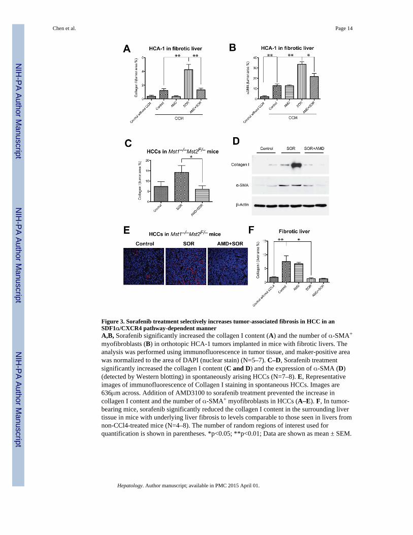
Figure 3. Sorafenib treatment selectively increases tumor-associated fibrosis in HCC in anSDF1α/CXCR4 pathway-dependent mannerA,B, Sorafenib significantly increased the collagen I content (A) and the number of α-SMA+
myofibroblasts (B) in orthotopic HCA-1 tumors implanted in mice with fibrotic livers. Theanalysis was performed using immunofluorescence in tumor tissue, and maker-positive areawas normalized to the area of DAPI (nuclear stain) (N=5–7). C–D, Sorafenib treatmentsignificantly increased the collagen I content (C and D) and the expression of α-SMA (D)(detected by Western blotting) in spontaneously arising HCCs (N=7–8). E, Representativeimages of immunofluorescence of Collagen I staining in spontaneous HCCs. Images are636μm across. Addition of AMD3100 to sorafenib treatment prevented the increase incollagen I content and the number of α-SMA+ myofibroblasts in HCCs (A–E). F, In tumor-bearing mice, sorafenib significantly reduced the collagen I content in the surrounding livertissue in mice with underlying liver fibrosis to levels comparable to those seen in livers fromnon-CCl4-treated mice (N=4–8). The number of random regions of interest used forquantification is shown in parentheses. *p<0.05; **p<0.01; Data are shown as mean ± SEM.
Chen et al. Page 14
Hepatology. Author manuscript; available in PMC 2015 April 01.
NIH
-PA Author Manuscript
NIH
-PA Author Manuscript
NIH
-PA Author Manuscript

Figure 4. SDF1α/CXCR4 axis promotes HSC to myofibroblast differentiation in the face ofPDGFR blockade by sorafenibA, Exposure to recombinant PDGF-B increased HSC proliferation, while treatment withsorafenib reduced the viability of HSCs. Exposure to recombinant SDF1α increased theviability of HSCs despite PDGFR inhibition using sorafenib treatment, in a dose-dependentmanner. HSC viability was measured by MTT assay (N=6 experimental repeats). B,Exposure to recombinant SDF1α increased the expression of α-SMA and collagen I andreduced cleaved caspase-3 expression (evaluated by Western blotting) despite sorafenibtreatment, in a dose-dependent manner. C, Exposure to recombinant SDF1α increasedviability of HSCs despite sorafenib treatment (N=3–5 experimental repeats). D, Exposure torecombinant SDF1α upregulated collagen I and α-SMA expression levels as well as ERKand AKT activation in HSCs, consistent with their myofibroblast differentiation. Inhibitionof CXCR4 with AMD3100 (D) or using siRNA (E) prevented the effects of SDF1α. F, ERKinhibition with FR-180204 (2μM) decreased α-SMA expression in HSCs treated withrecombinant SDF1α. Data are presented as mean ± SEM.
Chen et al. Page 15
Hepatology. Author manuscript; available in PMC 2015 April 01.
NIH
-PA Author Manuscript
NIH
-PA Author Manuscript
NIH
-PA Author Manuscript

Figure 5. Intratumoral infiltration of Gr1+ myeloid cells is increased after sorafenib (SOR)treatment, and is prevented by CXCR4 inhibition in HCCA–B, After SOR treatment, the number of 7AAD–CD11b+Gr1+ monocytes (measured byflow cytometry) significantly increased in HCA-1 tumors growing C3H mice with liverfibrosis (A) of as well as in spontaneous HCCs in Mst1−/− Mst2F/− transgenic mice (B).Treatment with the CXCR4 inhibitor AMD3100 prevented this effect (A–B). C, The numberof 7AAD–CD11b+Gr1+ myeloid cells was significantly reduced in the fibrotic liver tissuesfrom sorafenib-treated mice. Data are shown as percentages of the total number of cellsevaluated in enzymatically digested tissue. Data are presented as mean ± SEM (N=5–14mice per group). **P<0.01.
Chen et al. Page 16
Hepatology. Author manuscript; available in PMC 2015 April 01.
NIH
-PA Author Manuscript
NIH
-PA Author Manuscript
NIH
-PA Author Manuscript
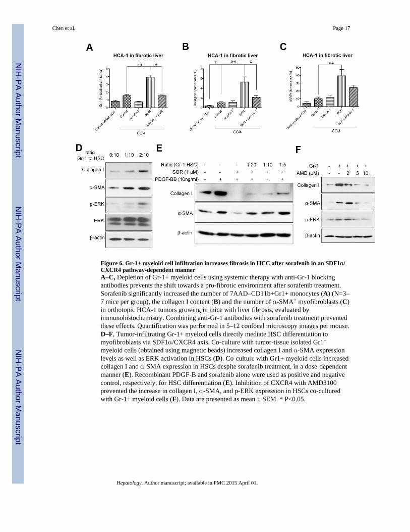
Figure 6. Gr-1+ myeloid cell infiltration increases fibrosis in HCC after sorafenib in an SDF1α/CXCR4 pathway-dependent mannerA–C, Depletion of Gr-1+ myeloid cells using systemic therapy with anti-Gr-1 blockingantibodies prevents the shift towards a pro-fibrotic environment after sorafenib treatment.Sorafenib significantly increased the number of 7AAD–CD11b+Gr1+ monocytes (A) (N=3–7 mice per group), the collagen I content (B) and the number of α-SMA+ myofibroblasts (C)in orthotopic HCA-1 tumors growing in mice with liver fibrosis, evaluated byimmunohistochemistry. Combining anti-Gr-1 antibodies with sorafenib treatment preventedthese effects. Quantification was performed in 5–12 confocal microscopy images per mouse.D–F, Tumor-infiltrating Gr-1+ myeloid cells directly mediate HSC differentiation tomyofibroblasts via SDF1α/CXCR4 axis. Co-culture with tumor-tissue isolated Gr1+
myeloid cells (obtained using magnetic beads) increased collagen I and α-SMA expressionlevels as well as ERK activation in HSCs (D). Co-culture with Gr1+ myeloid cells increasedcollagen I and α-SMA expression in HSCs despite sorafenib treatment, in a dose-dependentmanner (E). Recombinant PDGF-B and sorafenib alone were used as positive and negativecontrol, respectively, for HSC differentiation (E). Inhibition of CXCR4 with AMD3100prevented the increase in collagen I, α-SMA, and p-ERK expression in HSCs co-culturedwith Gr-1+ myeloid cells (F). Data are presented as mean ± SEM. * P<0.05.
Chen et al. Page 17
Hepatology. Author manuscript; available in PMC 2015 April 01.
NIH
-PA Author Manuscript
NIH
-PA Author Manuscript
NIH
-PA Author Manuscript
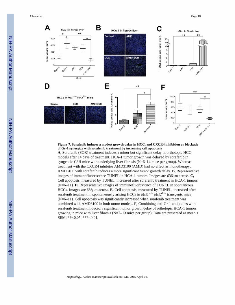
Figure 7. Sorafenib induces a modest growth delay in HCC, and CXCR4 inhibition or blockadeof Gr-1 synergize with sorafenib treatment by increasing cell apoptosisA, Sorafenib (SOR) treatment induces a minor but significant delay in orthotopic HCCmodels after 14 days of treatment. HCA-1 tumor growth was delayed by sorafenib insyngeneic C3H mice with underlying liver fibrosis (N=6–14 mice per group). Whereastreatment with the CXCR4 inhibitor AMD3100 (AMD) had no effect as monotherapy,AMD3100 with sorafenib induces a more significant tumor growth delay. B, Representativeimages of immunofluorescence TUNEL in HCA-1 tumors. Images are 636μm across. C,Cell apoptosis, measured by TUNEL, increased after sorafenib treatment in HCA-1 tumors(N=6–11). D, Representative images of immunofluorescence of TUNEL in spontaneousHCCs. Images are 636μm across. E, Cell apoptosis, measured by TUNEL, increased aftersorafenib treatment in spontaneously arising HCCs in Mst1−/− Mst2F/− transgenic mice(N=6–11). Cell apoptosis was significantly increased when sorafenib treatment wascombined with AMD3100 in both tumor models. F, Combining anti-Gr-1 antibodies withsorafenib treatment induced a significant tumor growth delay of orthotopic HCA-1 tumorsgrowing in mice with liver fibrosis (N=7–13 mice per group). Data are presented as mean ±SEM; *P<0.05, **P<0.01.
Chen et al. Page 18
Hepatology. Author manuscript; available in PMC 2015 April 01.
NIH
-PA Author Manuscript
NIH
-PA Author Manuscript
NIH
-PA Author Manuscript

Figure 8. Differential impact of sorafenib on liver versus tumor-associated fibrosis mediated bySDF1α/CXCR4 axis and Gr-1+ cells in HCCThe differential effects of sorafenib are the result of increased intratumoral hypoxia, leadingto elevated SDF1α expression and Gr-1+ myeloid cell infiltration. Blocking CXCR4prevents Gr-1+ myeloid cell infiltration and hepatic stellate cells differentiation andactivation, and synergizes with the anti-tumor effects of sorafenib.
Chen et al. Page 19
Hepatology. Author manuscript; available in PMC 2015 April 01.
NIH
-PA Author Manuscript
NIH
-PA Author Manuscript
NIH
-PA Author Manuscript