The Proteasome and Its Network: Engineering for Adaptability
27
The Proteasome and Its Network: Engineering for Adaptability Daniel Finley and Miguel A. Prado Department of Cell Biology, Harvard Medical School, Boston, Massachusetts 02115 Correspondence: [email protected] The proteasome, the most complex protease known, degrades proteins that have been con- jugated to ubiquitin. It faces the unique challenge of acting enzymatically on hundreds and perhaps thousands of structurally diverse substrates, mechanically unfolding them from their native state and translocating them vectorially from one specialized compartment of the enzyme to another. Moreover, substrates are modified by ubiquitin in myriad configurations of chains. The many unusual design features of the proteasome may have evolved in part to endow this enzyme with a robust ability to process substrates regardless of their identity. The proteasome plays a major role in preserving protein homeostasis in the cell, which requires adaptation to a wide variety of stress conditions. Modulation of proteasome function is achieved through a large network of proteins that interact with it dynamically, modify it enzymatically, or fine-tune its levels. The resulting adaptability of the proteasome, which is unique among proteases, enables cells to control the output of the ubiquitin–proteasome pathway on a global scale. Q uality control (QC) of proteins and organ- elles in eukaryotic cells is mediated by a vast and incompletely charted set of activities. QC pathways can target proteins that are misfolded, aggregated, mutated, chemically modified, mis- localized, mistranslated, or that have failed to assemble into a multisubunit complex. The sig- nificance of QC to human disease as well as aging is well recognized and owes to the marked toxicity of many misfolded proteins. Molecular chaperones, autophagy, and the ubiquitin–pro- teasome system (UPS) are all key players in QC pathways (see review by Hegde and Zavodszky 2019). While molecular chaperones work in part to prevent and reverse misfolding events, they cannot correct all QC problems by any means, and therefore the activity of molecular chaper- ones is complemented by autophagy and the UPS, which safeguard proteostasis by destroying misfolded and toxic species. At a mechanistic level, molecular chaperones, autophagy, and the UPS often work hand-in-hand. For example, molecular chaperones frequently assist in target- ing proteins to the UPS, and selective autophagy is often driven by ubiquitination of autophagic cargo. Here we focus on the UPS, and in particular on the proteasome holoenzyme, also known as the 26S proteasome, a 2.5–3 MDa protease that degrades proteins that have been conjugated to ubiquitin (Hough et al. 1987; Waxman et al. 1987). The proteasome is of interest as the Editors: Richard I. Morimoto, F. Ulrich Hartl, and Jeffery W. Kelly Additional Perspectives on Protein Homeostasis available at www.cshperspectives.org Copyright © 2019 Cold Spring Harbor Laboratory Press; all rights reserved Advanced Online Article. Cite this article as Cold Spring Harb Perspect Biol doi: 10.1101/cshperspect.a033985 1 on December 4, 2021 - Published by Cold Spring Harbor Laboratory Press http://cshperspectives.cshlp.org/ Downloaded from
Transcript of The Proteasome and Its Network: Engineering for Adaptability

The Proteasome and Its Network: Engineeringfor Adaptability
Daniel Finley and Miguel A. Prado
Department of Cell Biology, Harvard Medical School, Boston, Massachusetts 02115
Correspondence: [email protected]
The proteasome, the most complex protease known, degrades proteins that have been con-jugated to ubiquitin. It faces the unique challenge of acting enzymatically on hundreds andperhaps thousands of structurally diverse substrates, mechanically unfolding them from theirnative state and translocating them vectorially from one specialized compartment of theenzyme to another. Moreover, substrates are modified by ubiquitin in myriad configurationsof chains. The many unusual design features of the proteasome may have evolved in part toendow this enzymewith a robust ability to process substrates regardless of their identity. Theproteasome plays a major role in preserving protein homeostasis in the cell, which requiresadaptation to a wide variety of stress conditions. Modulation of proteasome function isachieved through a large network of proteins that interact with it dynamically, modify itenzymatically, or fine-tune its levels. The resulting adaptability of the proteasome, which isunique among proteases, enables cells to control the output of the ubiquitin–proteasomepathway on a global scale.
Quality control (QC) of proteins and organ-elles in eukaryotic cells ismediated byavast
and incompletely charted set of activities. QCpathways can target proteins that are misfolded,aggregated, mutated, chemically modified, mis-localized, mistranslated, or that have failed toassemble into a multisubunit complex. The sig-nificance ofQC tohumandisease aswell as agingis well recognized and owes to the markedtoxicity of many misfolded proteins. Molecularchaperones, autophagy, and the ubiquitin–pro-teasome system (UPS) are all key players in QCpathways (see review by Hegde and Zavodszky2019).Whilemolecular chaperones work in partto prevent and reverse misfolding events, theycannot correct all QC problems by any means,
and therefore the activity of molecular chaper-ones is complemented by autophagy and theUPS, which safeguard proteostasis by destroyingmisfolded and toxic species. At a mechanisticlevel, molecular chaperones, autophagy, andtheUPS often work hand-in-hand. For example,molecular chaperones frequently assist in target-ing proteins to the UPS, and selective autophagyis often driven by ubiquitination of autophagiccargo.
Here we focus on the UPS, and in particularon the proteasome holoenzyme, also known asthe 26S proteasome, a 2.5–3 MDa protease thatdegrades proteins that have been conjugated toubiquitin (Hough et al. 1987; Waxman et al.1987). The proteasome is of interest as the
Editors: Richard I. Morimoto, F. Ulrich Hartl, and Jeffery W. KellyAdditional Perspectives on Protein Homeostasis available at www.cshperspectives.org
Copyright © 2019 Cold Spring Harbor Laboratory Press; all rights reservedAdvanced Online Article. Cite this article as Cold Spring Harb Perspect Biol doi: 10.1101/cshperspect.a033985
1
on December 4, 2021 - Published by Cold Spring Harbor Laboratory Press http://cshperspectives.cshlp.org/Downloaded from
enzyme at which all substrates converge in theUPS, as one of the most complex enzymes innature, as a regulatory hub of the UPS, and asa major therapeutic target. Excellent recent re-views have covered proteasome structure andfunction (Collins and Goldberg 2017; Bard etal. 2018), ubiquitin recognition by the protea-some (Saeki 2017), substrate processing by theproteasome (Yu andMatouschek 2017), protea-somal deubiquitinating enzymes (de Poot et al.2017), and proteasome assembly (Budenholzeret al. 2017; Rousseau and Bertolotti 2018).
ASSEMBLY OF THE PROTEASOME FROMTHE REGULATORY AND CORE PARTICLES
All cells carry out selective protein degradationprimarily through ATP-dependent proteaseswhose proteolytic sites are sequestered fromthe cytoplasmic space to minimize nonspecificproteolytic events. The proteasome is on thesame evolutionary lineage as the archaeal prote-ase PAN, although the latter is formed fromthree distinct gene products and the proteasome33 gene products. The PAN protease has a pro-teolytic core particle ([CP], also known as the20S complex) composed of α-type and β-typesubunits arranged in rings that are stacked intoa barrel-like α7β7β7α7 assembly (Lowe et al.1995). Thus, the heptameric α rings occupythe ends of the barrel, whereas the inner ringsare formed by β subunits, which are proteolyti-cally active. The CP of the eukaryotic protea-some differs mainly in that the α and β ringsare heteromeric rather than homomeric (Grollet al. 1997).
Sequestration of the CP’s proteolytic sites,which face the interior of the barrel, restrictstheir enzymatic activity when the CP is in anisolated state. However, a variety of activatingcomplexes can derepress the CP by opening agate in the center of the α ring through whichsubstrates will pass (Groll et al. 2000; Whitbyet al. 2000; Stadtmueller and Hill 2011). Thisgate provides tightly regulated access into theproteolytic chamber of the CP. In the case ofPAN, a homohexameric ATPase ring mediatesactivation. The carboxyl termini of the ATPasesinsert into intersubunit pockets within the α
ring, which controls the opening of the gate(Smith et al. 2007; Majumder et al. 2019).
Gate opening is required but not sufficientfor rapid and specific protein degradation. Fold-ed domains within proteins prevent their pas-sage through the narrow channel into the CP;thus, domains must be unfolded prior to degra-dation. The ATPase ring uses the energy of ATPhydrolysis to actively unfold the substrate as it isvectorially translocated into CP (Fig. 1). Unfold-ing is driven mechanically by ATP-dependenttranslocation, as the substrate is forced to nego-tiate the narrow channel (Prakash et al. 2004;Yu and Matouschek 2017). The substrate trans-location channel of the ATPase ring is linedwith substrate-contacting “pore loops” on eachATPase, whose ATP-directed motion drivessubstrates into the CP (de la Peña et al. 2018;Dong et al. 2019). Favorable proteasome sub-strates must contain an element that can makeinitial contact with the proteasome, the target ofwhich is thought to be the pore loops, so as toinitiate degradation. This step is known as en-gagement and requires a flexible segment of atleast 20–30 amino acids in the initiator element,though sequence requirements are minimal (Yuet al. 2016; Yu and Matouschek 2017).
Gate opening and substrate unfolding arestill not sufficient for protein degradation bythe proteasome, as ubiquitin modification is re-quired for most substrates. Ubiquitin docks asubstrate on the proteasome, forming an en-zyme–substrate complex that is sufficiently sta-ble that the substrate can be productively en-gaged by the pore loops, and translocation caninitiate.With the innovation of ubiquitination inthe eukaryotic lineage, the PAN-like ancestorprotease was greatly elaborated to form the pro-teasome. As we have seen for the CP, the homo-meric ATPase ring evolved into a heteromericring (subunits Rpt1-Rpt6), but more important-lymany new components were added (Rpn-typesubunits) to generate a 19-subunit assembly re-ferred to as the proteasome regulatory particle([RP], also known as the 19S complex). Manyof the new subunits and associated factors adaptthe proteasome to recognize and process ubiq-uitin conjugates, functioning either as ubiquitinreceptors or deubiquitinating enzymes (Fig. 2).
D. Finley and M.A. Prado
2 Advanced Online Article. Cite this article as Cold Spring Harb Perspect Biol doi: 10.1101/cshperspect.a033985
on December 4, 2021 - Published by Cold Spring Harbor Laboratory Press http://cshperspectives.cshlp.org/Downloaded from

Other CP activators, such as PA28 and PA200,seem to lack both ATPase and ubiquitin-recog-nizing activities. Theywill not be consideredherebut the reader is referred to other reviews (Stadt-mueller and Hill 2011; Tanaka et al. 2012). Fi-nally, we note that numerous tissue-specific iso-forms of the proteasome have been described,most notably the thymoproteasome and immu-noproteasome, which are critical for the ontog-enyand selectivityof adaptive immunity, respec-tively (Qian et al. 2013; Huang et al. 2016a;Murata et al. 2018).
UBIQUITIN RECOGNITION ANDPROCESSING
The most critical ubiquitin receptor of the pro-teasome is Rpn10. This subunit recognizes ubiq-uitin through an amphipathic α-helix known asthe ubiquitin-interacting motif (UIM). In mice,the UIM appears to be essential for embryonicdevelopment, and liver-specific conditional mu-
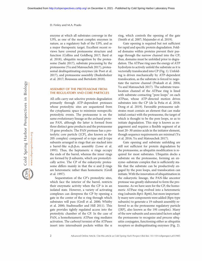
tants lacking the UIM exhibit substantial defectsin the breakdown of ubiquitin-protein conju-gates in this tissue (Hamazaki et al. 2007). De-spite the importance of the UIM, inactivatingmutants do not have a striking growth pheno-type inyeast (Fu et al. 1998), indicating thatotherreceptors also have significant roles in ubiquitinrecognition.ThePrudomainofRpn13 (Husnjaket al. 2008; Schreiner et al. 2008) and the T1element of Rpn1 (Chen et al. 2016b; Shi et al.2016; Dong et al. 2019) have also been shownto function in ubiquitin recognition (Fig. 2).However, subunits Rpn13, Rpn1, and Rpn10do not function only as ubiquitin receptors—they can also recognize ubiquitin indirectly byreversibly binding extrinsic ubiquitin receptors,which themselves carry substrate. The canonicalreceptors of this class are the ubiquitin-like–ubiq-uitin-activating (UBL–UBA) proteins (Fig. 3),which bind ubiquitin via their UBA domainsand the proteasome via their UBL domains. Anelegant proteomic study concluded that the
BA
State EBDeubiquitination
State EC1Translocation initiation
RPN1
RPN10
C
State ED2Translocation
RPN11
Ub
Figure 1. Conformational states of substrate-bound human proteasomes. Cryo-electron microscopy (cryo-EM)density maps of substrate (orange)-bound human proteasomes in three different stages of substrate processing arerepresented. (A) A conformation that is compatible with the deubiquitylation of the bound substrate. (B,C) Twoconsecutive conformations of substrate translocation (EC1 and ED2) into the core particle (CP). Color coding for theproteasome lid, the RPT/ATPase ring, and CP is given in the legend to Fig. 2. RPT1 and RPT5 density maps wereremoved from theATPase rings to expose the substrate translocation channel of the regulatory particle (RP). In ED2,subunitα1 (PSMA1)was also removed to show the substrate translocation channel of theCP. EB (ProteinDataBank[PDB]:6MSE),EC1(PDB:6MSG),andED2(PDB:6MSK)densitymapswereobtainedfromdata inDongetal. (2018).
The Proteasome and Its Network
Advanced Online Article. Cite this article as Cold Spring Harb Perspect Biol doi: 10.1101/cshperspect.a033985 3
on December 4, 2021 - Published by Cold Spring Harbor Laboratory Press http://cshperspectives.cshlp.org/Downloaded from

UBL–UBA pathway is the major route by whichproteins are targeted to the proteasome in yeast(Tsuchiya et al. 2017), though this study trackedbiochemical interactions with the proteasomerather than protein degradation per se. The threeUBL–UBA proteins in yeast, Rad23, Dsk2, andDdi1, have each been amplified into a small genefamily in mammals (see Fig. 3; Saeki 2017),and mutations in a Dsk2 homolog, Ubiquilin2,underlie a familial form of amyotrophic lateralsclerosis (fALS) (Deng et al. 2011).
Yeast strains in which the RAD23, DSK2,and DDI1 genes have all been deleted, and theubiquitin-binding elements of Rpn10, Rpn13,and Rpn1 mutationally inactivated, are sensitiveto stress but nonetheless viable, suggesting thatadditional ubiquitin receptors for the protea-some remain to be identified (Shi et al. 2016).Why are there so many proteasomal ubiquitinreceptors? One reason may be to promotesubstrate association with the proteasome inmultiple alternative orientations—“orientationsampling” (Shi et al. 2016). According to thismodel, some substrates docked at the protea-some may initiate degradation inefficientlybecause their initiation elements are not posi-tioned close enough to the substrate entry port.However, if this were the casewhen the ubiquitinchain is bound to one receptor, it might not bewhen bound to another. Thus, positioning ubiq-uitin receptors at different locations in the RPcould allow the proteasome to function morerobustly as a protease.
A second, not mutually exclusive model, isthat having multiple receptors is advantageousin allowing avid or multipoint recognition ofubiquitin conjugates (Meyer and Rape 2014; Luet al. 2015; Shi et al. 2016). Multipoint recogni-tionwould enhance the abilityof the proteasometo recognize substrates modified with branchedubiquitin chains and with chains attached atmultiple lysineswithin the substrate.Aswithori-entation sampling, themost general effect wouldbe amore robust capacity to degrade diverse cel-lular proteins.
Deubiquitinating enzymes on the protea-some prevent ubiquitin from being degradedtogether with the substrate to which it is at-tached (de Poot et al. 2017). Release of ubiquitincan promote substrate degradation, becauseubiquitin has an unusually stable folded struc-ture, and thus it can impede translocation kinet-ically (Verma et al. 2002; Yao and Cohen 2002;Worden et al. 2017). Deubiquitinating enzymeRpn11 is positioned directly above the substrateentry port of the actively translocating protea-some (Figs. 1 and 2). Due in part to this posi-tioning, it seems to remove ubiquitin primarilyfrom substrates that have already been commit-ted to degradation by virtue of engagement with
UCH37
RPN10RPN11
USP14
RPN1
RPN13
Entry port
RPT ring
α ring
β ring
CP
RP
Figure 2. Overview of deubiquitinating enzymes(DUBs) and Ub/Ubl receptors of the human protea-some. A cryo-electron microscopy (cryo-EM) densitymapof thehumanproteasome is representedwithall ofits known ubiquitin receptors (RPN1, RPN10, andRPN13 in purple tones) and DUBs (RPN11, USP14,and UCH37 in red tones). The lid subassembly of theRP is in light gray. The cryo-EM structure of the pro-teasome bound to USP14-ubiquitin-aldehyde was ob-tained from data presented in Huang et al. (2016b)(Protein Data Bank [PDB]: 5GJQ). The location ofUCH37 and RPN13 were modeled as shown in dePoot et al. (2017) using data in densitymaps from Sah-toe et al. (2015) and Vander Linden et al. (2015) (PDB:4UEL and 4WLQ). All six subunits of the RPT ring areshown in light blue and ubiquitinmonomers bound tothe DUBs are shown in yellow ribbon representations.
D. Finley and M.A. Prado
4 Advanced Online Article. Cite this article as Cold Spring Harb Perspect Biol doi: 10.1101/cshperspect.a033985
on December 4, 2021 - Published by Cold Spring Harbor Laboratory Press http://cshperspectives.cshlp.org/Downloaded from

the pore loops of the ATPase ring. The ATPdependence of Rpn11 activity is a signature ofthis coupling to substrate engagement, giventhat Rpn11 is not itself an ATPase. Proteasomaldeubiquitinating enzymes that function inde-pendently of ATP, such as USP14/Ubp6 andUCH37/UCHL5 (Fig. 2), can remove ubiquitinprior to substrate engagement and therefore canantagonize degradation by promoting substratedissociation from the proteasome prior to the“engagement” step (Yao and Cohen 2002; Leeet al. 2010, 2016). The catalytic activities ofUCH37/UCHL5, USP14/Ubp6, and Rpn11/PSMD14 are all activated by the proteasomeand subject to other types of regulation as re-viewed below and elsewhere (de Poot et al. 2017;Bard et al. 2018).
CONFORMATIONAL STATES OF THEPROTEASOME
Cryo-EM has recently been applied to the pro-teasome with great success, and many distinctconformational states have been resolved (Bardet al. 2018). Although the total number of dis-tinct populated states is high and remains to beestablished, studies of the yeast proteasome haveto date resolved six conformational states, s1–s6(Eisele et al. 2018), even in the absence of a sub-strate. The s1 state is the principal species inidling proteasomes, that is, in the presence of
ATP and the absence of a substrate. (Substrate-free proteasomes continually hydrolyze ATP.)Key features of the proteasome are misalignedin s1; the substrate translocation channel of theATPase ring is not flush with the CP gate, andimportantly the active site of Rpn11 is 25 Å awayfrom the substrate entry port of the ATPase ring.Although the CP gatewas formany years viewedas being opened as a direct consequence of CP-RP association, this is clearly not the case as thegate is closed in the s1, s2, and s3 states (Chen etal. 2016c;Wehmer et al. 2017; Eisele et al. 2018).The increased access to the CP of small-mole-cule probes such as LLVY-AMC upon holoen-zyme assembly in the absence of a substrate mayreflect a mixture of holoenzyme states in thesample, although the open-gate states s4, s5,and s6 are not well populated. In any case, theclosed CP gate seen in s1 state proteasomes isan additional hallmark of their latency.
The proteasome can be converted into a deg-radation-competent state even in the absence ofa substrate, for example by replacing ATP withthe slowly hydrolyzed nucleotide ATPγS. Underthis condition, themajor form is s3,which showsproductive axial alignment of Rpn11, the RPsubstrate translocation channel, and the CPgate (Matyskiela et al. 2013; Śledź et al. 2013).But the gate remains closed, being opened onlyin the s4 state (Wehmer et al. 2017; Eisele et al.2018). In general, gate opening has been regard-
scRad23
hHR23A
hHR23B
Rad
23 fa
mily
hUBQLN2
hUBQLN3
hUBQLN4
hUBQLN1
scDsk2D
sk2
fam
ily
Ddi
1 fa
mily scDdi1
hDDI2
hDDI1
Zfa
nd fa
mily
hZFAND1
hZFAND5
hZFAND2A(AIRAP)
hZFAND2B(AIRAPL)
scTmc1
PXXUBL UBA RVP ZnF AN1 ZnF A20 UIMSTI
scCuz1
Figure 3. Extrinsic ubiquitin receptors of the proteasome. Shown is a schematic representation of four proteinfamilies of Saccharomyces cerevisiae andHomo sapiens, which include proteins thought to deliver ubiquitinatedtargets to the proteasome (Rad23, Ddi1, Dsk2, and Zfand). Some of these proteins do not appear to be functionalubiquitin receptors (see text for details). The simple modular architecture research tool (SMART) (Letunic andBork 2018) was used to identify SMARTand Pfam protein domains. All proteins and domains are drawn to scale.
The Proteasome and Its Network
Advanced Online Article. Cite this article as Cold Spring Harb Perspect Biol doi: 10.1101/cshperspect.a033985 5
on December 4, 2021 - Published by Cold Spring Harbor Laboratory Press http://cshperspectives.cshlp.org/Downloaded from

ed, both from biochemical and initial cryo-EMstudies, as a consequence of the insertion of“HbYX”-type ATPases into their cognate αpockets. The HbYX motif, whose signature is apenultimate tyrosine, was identified in the car-boxyl terminus of PAN, and found to be presentas well in three of the six proteasomal ATPases(Rpt2, Rpt3, andRpt5) (Smith et al. 2007). How-ever, a key role was subsequently found for αpocket contacts to the carboxyl terminus ofRpt6 (Park et al. 2013; Sokolova et al. 2015; Eiseleet al. 2018), which does not conform to theHbYX consensus. Optimal gate opening is ap-parently only observedwhen the carboxyl termi-ni of both Rpt6 and Rpt1 are engaged with the αring (Eisele et al. 2018), whichwill depend on thenucleotide state of the proteasome’s ATPasering, or “Rpt ring,” as discussed below.
Recent work has visualized numerous sub-strate-engaged states by substrate trapping, usingapproaches such as stripping Zn++ ion fromRpn11 or adding ATPγS to proteasomes thatare in the act of substrate degradation (Maty-skiela et al. 2013; de la Peña et al. 2018; Donget al. 2019). These studies present a detailedmodel of the proteasome’s ATP hydrolytic cycleand coupling to substrate translocation. The hy-drolytic cycle of the engaged proteasome isthought to involve a “rotary” mechanism inwhich an ATP hydrolytic event in one Rpt pro-tein is followed by one in the adjacent subunit,proceeding continuously around the ring (de laPeña et al. 2018; Dong et al. 2019; Majumderet al. 2019).
Substrate-engaged pore loops travel “down-ward,” or toward the CP, in conveyor-belt fash-ion, under the direction of the sequential ATPhydrolytic events. When a pore loop reaches theCP-proximal position, it disengages from thesubstrate, then returns to a CP-distal positionwhere it will subsequently engage another seg-ment of the translocating substrate. Disengagedsubunits are displaced outward from the ring sothat their pore loops lose contact with the sub-strate (Fig. 4). The coupling of disengagement todistal movement of the pore loop underlies thevectoriality of substrate movement, in that onlyengaged pore loops move toward the CP. Con-temporaneously with these motions, the four
subunits that are engaged with substrate movein concert, with one step of the process translo-cating the substrate toward the CP by a distancecorresponding to approximately two aminoacids in the substrate. The dynamics of the Rptring and the central role of the pore loops indriving substrate translocation are similar tothose generally observed in AAA ATPase com-plexes (Puchades et al. 2017), which includeother proteases, helicases, and additional bio-chemical activities.
TOMOGRAPHIC RESOLUTION OFPROTEASOME STATES WITHIN CELLS
Remarkable advances in cryo-electron tomogra-phy have recently enabled the visualization ofproteasomes under essentially native conditionswithin cells (Asano et al. 2015; Guo et al. 2018).The level of resolution is sufficient to assign con-formational states in reference to cryo-EM dataon purified proteasomes. The tomographic ap-proach has been particularly revealing in thestudy of familial ALS (fALS). The C9orf72gene is often expanded in G4C2 repeats infALS, and these are aberrantly translated into avariety of toxic dipeptide repeat containing pro-teins. One of these, poly-Gly-Ala (poly-GA),forms densely packed cytoplasmic fibrils thatspecifically recruit proteasomes, with poly-GAaggregates concentrating proteasomes ∼30-foldover neighboring sites within the cell (Guo et al.2018). Unlike the general pool of proteasomes,sequestered proteasomes are frequently found inthe s4 state and thus are likely to be engagedwithsubstrate. Yet poly-GA proteins are not favor-able proteasome substrates. These findings sug-gest that the sequestered proteasomes may beunproductively engaged with the substrate (i.e.,stalled). The sequestration and inhibition of aproteasome by poly-GA aggregates could poten-tially play a significant role in their neurodegen-erative effect.
SUBSTRATE RECOGNITION AND THEUBIQUITIN CODE
Unlike many protein modifications, ubiquitina-tion exhibitsmarked structural variation, arising
D. Finley and M.A. Prado
6 Advanced Online Article. Cite this article as Cold Spring Harb Perspect Biol doi: 10.1101/cshperspect.a033985
on December 4, 2021 - Published by Cold Spring Harbor Laboratory Press http://cshperspectives.cshlp.org/Downloaded from

principally from the formation of ubiquitin–ubiquitin adducts. The seven lysine residues ofubiquitin as well as its amino terminal group areall subject to modification (Chau et al. 1989;Spence et al. 1995; Kirisako et al. 2006; Xu etal. 2009; Kwon and Ciechanover 2017). Ligases
and ubiquitin-conjugating enzymes with highspecificity in the formation of ubiquitin–ubiq-uitin linkages generate homogeneous chains(Chau et al. 1989; VanDemark et al. 2001), whileother ligases generate mixed linkages (Kirkpat-rick et al. 2006), or in some cases no chains
State 4D - top viewState 5D - top view
Rpt4
Rpt5
ATP
ADP
Rpt4
Rpt5
State 5D - side view State 4D - side view
90° 90°
x axis x axis
BA
Figure 4. Representation of the ATP hydrolytic cycle and substrate translocation. (A,B) Two different cryo-electron microscopy (cryo-EM) structures of the RPT ring at different states of substrate (orange mesh) trans-location in the yeast proteasome holoenzyme are shown (states 5D and 4D, respectively). Images were createdfrom data in de la Pena et al. (2018). Ribbon representations of the top view of the RPT rings are shown in thebottom panels, where substrate disengagement of Rpt5 (green) and Rpt4 (blue) can be observed (A and B,respectively). A 90° rotation about the x axis was applied to show a lateral view of the RPT ring (top panels).While the substrate polypeptide is surrounded by a spiral-staircase of pore 1 loop tyrosines (highlighted in pink)projecting from substrate-engaged RPT subunits, the Tyr of the RPT subunits that are disengaged (Rpt5 inA andRpt4 in B) are displaced from the substrate. In the bottom panels, spherical representations of ATP (yellow) andADP (blue) are also included. For a better view of the spiral-staircase model in the top panels, two RPT subunits(Rpt4 and Rpt3) and three amino acids (Arg261, Glu293, and Gly294 of RPT4) were omitted from the densitymaps. States 5D and 4D were created from data in de la Pena et al. (2018) (Protein Data Bank [PDB]: 6EF1 and6EF3, respectively).
The Proteasome and Its Network
Advanced Online Article. Cite this article as Cold Spring Harb Perspect Biol doi: 10.1101/cshperspect.a033985 7
on December 4, 2021 - Published by Cold Spring Harbor Laboratory Press http://cshperspectives.cshlp.org/Downloaded from

whatsoever (Braten et al. 2016; Nguyen et al.2017; Yanagitani et al. 2017). Some chains havebranched topologies (Meyer and Rape 2014; Yauet al. 2017; Samant et al. 2018), where an acceptorubiquitin ismodified bymore than one ubiquitindonor. Phosphorylation and acetylation of ubiq-uitinprovide further important variation (Harperet al. 2018), and another protein modifier, theubiquitin-like protein SUMO, can form mixedchains with ubiquitin, which also target proteinsto the proteasome (Liebelt and Vertegaal 2016).Chains vary in length (Tsuchiya et al. 2018), andwith approximately ∼100 distinct deubiquitinat-ing enzymes (DUBs) encoded in themammaliangenome (Leznicki and Kulathu 2017), it is notsurprising that chains are in addition highly dy-namic. Chain-editing processes may fundamen-tally alter chain structures over time (Wertz andDixit 2014; Chen et al. 2017).
The complexity of chain architectures re-flects that ubiquitin serves numerous signalingroles apart from targeting to the proteasome.Ubiquitination of cell-surface proteins targetsthem to the endosome and ultimately the lyso-some for degradation (Piper et al. 2014). Inaddition, selective autophagy is frequently de-pendent on ubiquitination of cargo proteins(Grumati andDikic 2018).Aside from thesedeg-radative pathways, many ubiquitination eventsdonot target substrate degradation at all, as com-monly seen in the signaling pathways of innateimmunity and in targets ofmodification in chro-matin, such as histones andPCNA. It is clear thatchain linkage specificity underlies much of theselectivity of these targeting events, with chainslinked through Lys63 and Met1 being instru-mental in nonproteolytic signaling. Yet whythe proteasome acts so poorly, if at all, on sub-strates modified by such chains is still unre-solved. Purified proteasomes discriminate onlyweakly against chains, such as Lys63 chains,which appear to exhibit negligible targeting ac-tivity in vivo (Hofmann and Pickart 2001). Oneexplanation may be that the in vivo activity ofproteasomes on such targets is limited by kineticcompetition with other (Lys63-specific) ubiqui-tin receptors (Nathan et al. 2013). In other cases,the Lys63 chain selectivity effect may bemiscon-strued in that it does not owe to the chain itself
but instead to the substrate components of theseconjugates being inherently poor proteasomesubstrates, for example, by being resistant to un-folding by the proteasome.
A key problem in substrate recognition bythe proteasome is how UBL–UBA proteins pro-mote the process. Because the proteasome-bind-ing UBL domain and the ubiquitin-bindingUBA domain are joined through long, flexiblelinkers, a substrate docked by these bridging fac-tors would have greater freedom of orientationwith respect to the proteasome than one dockeddirectly onto Rpn1 or Rpn13, which could resultin faster andmore efficient orientation sampling(Shi et al. 2016). The UBA domains withinUBL–UBA proteins suppress deubiquitinationof bound conjugates, which would also promotedegradation (Hartmann-Petersen et al. 2003).Binding of the proteasome by the UBL domainsof these proteins may also play a regulatory role,as purified UBL domains from mammalian ho-mologs of Rad23 and Dsk2 (hHR23B and Ubiq-uilin1, respectively; Fig. 3) stimulate peptide hy-drolysis by the proteasome, albeit partially (KimandGoldberg 2018). Asmentioned above, only asubset of the conformational states of the assem-bled proteasome holoenzyme have an open CPchannel. Therefore, such a stimulation of pep-tide hydrolysis could suggest that UBL bindingsomehow redistributes the ensemble of confor-mational states that are present at the steady statein the proteasome samples tested.
Additional, more specific functions of UBL–UBA proteins have also been reported. For ex-ample, ubiquilins (mammalian homologs ofDsk2) specifically promote the proteasomal deg-radation of mislocalized mitochondrial proteinsthrough recognizing exposed transmembrane(TM) domains (Itakura et al. 2016; Suzuki andKawahara 2016;Whiteley et al. 2017). This ubiq-uilin function is particularly evident in activatedB cells, which have elevated levels of mitochon-drial protein mislocalization (Whiteley et al.2017). The domain implicated in TM interac-tion is neither the UBL nor UBA, but onewithinthe flexible linker between them, the STI do-main, also known as the M domain (Fig. 3),which has so far been subject to little study (Ita-kura et al. 2016). As ubiquilins are hypothesized
D. Finley and M.A. Prado
8 Advanced Online Article. Cite this article as Cold Spring Harb Perspect Biol doi: 10.1101/cshperspect.a033985
on December 4, 2021 - Published by Cold Spring Harbor Laboratory Press http://cshperspectives.cshlp.org/Downloaded from
to recruit an E3 to ubiquitinate such proteins,this model holds that, at least for these sub-strates, ubiquilin binding actually precedes andhelps to direct ubiquitination events.
Studies in yeast point to a critical role forUBL–UBA proteins (Rad23 and Dsk2) in thetransfer of ubiquitin conjugates from Cdc48 tothe proteasome. Cdc48, known in mammals asp97, is an interestingATPase-ring complex fromthe same (AAA+) protein family as the Rpt pro-teins of the proteasome (van den Boom andMeyer 2018). It appears to unfold proteins, pri-marily ubiquitinated proteins, by a mechanisminvolving threading of the substrate through anarrow axial channel, as described for the pro-teasome above (Blythe et al. 2017; Bodnar andRapoport 2017). Such substrates are in many(but not all) cases destined to the proteasomefor degradation, in that the Cdc48 complex itselfhas no proteolytic activity. One possible modelof Cdc48-proteasome coupling is that theATPase ring of Cdc48 replaces the RP complexat the cylinder end of the CP, injecting the sub-strate directly into the CP (Barthelme and Sauer2012; Esaki et al. 2017). A model with moredirect support to date is that UBL–UBAproteinsferry the substrate from Cdc48 to a canonicalRP-CP proteasome (Kim et al. 2004; Richlyet al. 2005; Tsuchiya et al. 2017).
A cofactor of Cdc48 known as Ufd2, an E4ubiquitin ligase (i.e., it extends preexisting ubiq-uitin chains), directly interacts with the UBLdomains of Rad23 and Dsk2 (Hänzelmann etal. 2010). In fact, Ufd2 has a considerably higheraffinity for these UBL domains than does theproteasome. As chains are extended by aUfd2–Rad23 complex on Cdc48, the UBA do-main in Rad23 can bind the growing chain as itreaches approximately six ubiquitins in length,blocking further elongation. The Rad23–sub-strate complex can then dissociate from Ufd2,freeing its UBL domain to bind proteasomal re-ceptors such as Rpn1 and Rpn13.
Recent studies have reported a crucial role ofUbiquilin2 (Hjerpe et al. 2016) and its yeast ho-molog Dsk2 (Samant et al. 2018) in the turnoverof misfolded or aggregated proteins in the nu-cleus, including misfolded proteins generatedby heat shock. Aggregates of polyQ-expanded
Huntingtin also appear to be cleared throughUbiquilin2 (Hjerpe et al. 2016). In these studies,the UBL–UBA protein is proposed to cooperatewith Hsp70 chaperones rather than Cdc48/p97.This pathway to the proteasome may be of spe-cial importance in the nucleus because of theabsence of autophagy in this compartment.
Several ubiquitin–receptor proteins that arenot in the UBL–UBA family also bind protea-somes and appear to target proteins to the pro-teasome (for review, see Saeki 2017). TheZFAND family of zinc-finger proteins is of spe-cial interest (Fig. 3). Initial work defined AIRAP(also known as ZFAND2A) as a protein thatis induced by the proteotoxic agent arsenic,binds proteasomes, and enhances their activitythrough an unknownmechanism (Stanhill et al.2006). A constitutive homolog located in the en-doplasmic reticulum (ER),AIRAPL (also knownas ZFAND2B) was later identified, and Caeno-rhabditis elegans lacking this activity were foundto have a decreased life span and to be hypersen-sitive to the expression of misfolded proteins(Yun et al. 2008). Although the expression ofAIRAPL is not induced by arsenic, the proteinis recruited to the proteasome under theseconditions.
ZFAND1 (also known as Cuz1) was origi-nally characterized in S. cerevisiae, where itbinds both proteasomes and Cdc48 and confersresistance to arsenic (Sá-Moura et al. 2013;Hanna et al. 2014). It was recently found tobind arsenite-induced cytoplasmic stress gran-ules (SGs) in human (HEK293) cells and inyeast, and to promote binding of both protea-somes and p97 to these structures (Turakhiyaet al. 2018). ZFAND1 also mediates the elimi-nation of SGs in a proteasome- and p97-depen-dent manner. SGs form by phase separationwhen translation is suppressed, and contain ri-bonucleoprotein complexes stalled at a preini-tiation step. In general, SGs can be induced bydiverse stresses, and curiously only those in-duced by arsenite stress are ZFAND1-respon-sive (Turakhiya et al. 2018).
ZFAND5 is unique among these ZFANDfamily members in having no obvious respon-siveness to arsenic. It is instead strongly inducedin atrophic muscle after treatments such as fast-
The Proteasome and Its Network
Advanced Online Article. Cite this article as Cold Spring Harb Perspect Biol doi: 10.1101/cshperspect.a033985 9
on December 4, 2021 - Published by Cold Spring Harbor Laboratory Press http://cshperspectives.cshlp.org/Downloaded from

ing (Hishiya et al. 2006), and Zfand5-null mu-tant mice are substantially deficient in muscleatrophy. These mutants also exhibit elevatedubiquitin conjugate levels under atrophy-inducing conditions, consistent with defectiveconjugate degradation. ZFAND5 has complexstimulatory effects on the proteasome and nota-bly binds ubiquitin via its A20 domain, which isnecessary for stimulation of ubiquitin conjugatedegradation (Hishiya et al. 2006; Lee et al.2018a). The ubiquitin-binding capacity ofZFAND5 is shared with ZFAND2B/AIRAPL,although ZFAND2B binds ubiquitin via tandemUIM elements rather than an A20 domain (Ra-highi et al. 2016). In summary, two members ofthe ZFAND family are ubiquitin-binding pro-teins and are assumed to deliver conjugates tothe proteasome in analogy to UBL–UBA pro-teins. Others, such as AIRAP, also stimulateproteasome activity, ostensibly by coupling theproteasome to p97, but perhaps also throughother still unidentified mechanisms.
REGULATION OF PROTEASOME LEVELSBY Rpn4 AND NRF1
Early in the development of the ubiquitin field, itwas assumed that proteasomes were present atlevels adequate to handle the incoming flux ofubiquitin conjugates, and that therefore the ex-act level and activity of this enzyme were not ofmajor biological significance. Today control ofthe level of the proteasome is understood to bemediated by an elaborate network that contrib-utes to global regulation of protein degradation.Proteasome levels are especially significantwhen cells are under proteotoxic stress or ex-posed to proteasome inhibitors in anticancertreatments.
In elegant studies, proteasome levels wereshown in yeast to be under negative feedbackcontrol by the transcriptional factor Rpn4 (Xieand Varshavsky 2001). All genes encoding pro-teasome subunits have proteasome-associatedcontrol (PACE) elements in their promoters,which confer Rpn4 control (Fig. 5). Feedbackis mediated by proteasomal degradation ofRpn4, whose basal half-life of ∼2 min makes itone of the most rapidly turned over proteins in
yeast. Rpn4 is ubiquitinated by the ligase Ubr2but is also degraded through a ubiquitin-inde-pendent pathway (Wang et al. 2004; Ha et al.2012). The latter pathway is very unusual for ahigh-turnover protein, and ensures that Rpn4 issensitive principally to fluctuations in protea-some activity rather than ubiquitin levels, whichare separately regulated (Hanna et al. 2007) (seealso below). In the absence of proteostatic stress,proteasome levels are reduced in rpn4 null mu-tants (Xie and Varshavsky 2001), indicating thatRpn4 mediates tonic control over proteasomelevels under favorable conditions. However,multiple transcriptional sensors of proteostaticstress bind to and regulate the promoter of theRPN4 gene (Wang et al. 2008), including thosesensing heat stress (Hsf1) and oxidative stress(Yap1). Mediators of the pleiotropic drug re-sponse (Pdr1 and Pdr3) can also induce RPN4transcription. These findings indicate that theRpn4-proteasome negative feedback loop is in-tegrated into a broader stress-responsive regula-tory network. Indeed, Rpn4 itself hasmany tran-scriptional targets other than proteasomesubunit genes (Jelinsky et al. 2000).
Although proteasome levels inmammals arealso controlled by a homeostatic negative feed-back loop, the regulators are not homologous toRpn4. The design of the mammalian circuit isextremely interesting, and is conserved toC. elegans (Lehrbach and Ruvkun 2016). Tran-scription factorNRF1 is, like Rpn4, constitutive-ly degraded (with a basal half-life of ∼30 min),and like Rpn4 it controls the genes for all pro-teasome subunits in response to proteasome in-hibitors (Radhakrishnan et al. 2010; Steffen et al.2010). NRF1 recognizes well-characterized an-tioxidant response elements (AREs) in targetgenes, which encode a wide variety of activities(Fig. 5).
NRF1 differs from Rpn4 in being localizedto the ER, and its constitutive degradation pro-ceeds in part through the ERAD pathway, inwhich proteins from the ER are retrotranslo-cated into the cytoplasm (see reviews by Kara-göz et al. 2019; Needham et al. 2019). Retro-translocation is coupled to ubiquitination, inthis case by the HRD1 ligase, which resides inthe ER membrane and also appears to be the
D. Finley and M.A. Prado
10 Advanced Online Article. Cite this article as Cold Spring Harb Perspect Biol doi: 10.1101/cshperspect.a033985
on December 4, 2021 - Published by Cold Spring Harbor Laboratory Press http://cshperspectives.cshlp.org/Downloaded from

transmembrane channel for retrotranslocation(Schoebel et al. 2017). The ubiquitinated sub-strate is extracted from the membrane by p97and subsequently degraded by the proteasome.When retrotranslocated but not immediatelydegraded, NRF1 gains access to the nucleus,where it is active, though still rapidly degraded
through the action of the β-TRCP and FBXW7ligases, and thus significantly accumulating onlywhen its degradation through the proteasome isinhibited. However, translocation into the nu-cleus, and thus activation ofNRF1 in the cytosol,additionally requires an unusual endoproteo-lytic cleavage event that separates the transcrip-
A
B
Rpn4Ub
UbUb
Ubr2
Ub
Proteasome
genes
Nucleus
PACE
Rpn4
Rpn4
Proteasomeinhibition
Rpn4
transcription
Nucleus
Hsf1 Yap1Pdr1Pdr3
YREHSE PDRE
ERNRF1
Retrotranslocation
p97DDI2
HRD1NRF1
Proteasome
genes
Nucleus
ARE
NRF1NRF1
Ub
Ub NRF1
Figure 5. Feedback control of proteasome levels in yeast and mammals. (A) In S. cerevisiae, transcription factorRpn4 controls the level of expression of proteasome subunits. Rpn4 is stabilized under specific proteotoxicstresses, inducing the transcription of proteasomal subunit genes regulated by proteasome-associated control(PACE) elements. (B) In mammals, NRF1 is the key transcriptional regulator of proteasome subunit genes. Aresident of the endoplasmic reticulum (ER), NRF1 is ubiquitinated byHRD1 and retrotranslocated by p97. NRF1protein can then be degraded via the proteasome as an ERAD substrate or cleaved endoproteolytically by theprotease DDI2, resulting in its liberation from the ERmembrane. The cleaved version of NRF1 is translocated tothe nucleus where it binds ARE elements and induces the transcription of genes for proteasome subunits. Thenuclear form of NRF1 is also rapidly degraded by the proteasome (see main text).
The Proteasome and Its Network
Advanced Online Article. Cite this article as Cold Spring Harb Perspect Biol doi: 10.1101/cshperspect.a033985 11
on December 4, 2021 - Published by Cold Spring Harbor Laboratory Press http://cshperspectives.cshlp.org/Downloaded from

tional activation (TAD) and DNA-binding do-mains of NRF1 from its transmembrane do-main. Unexpectedly, the pathway does notsimply consist of the release of a cytoplasmicTAD from a membrane anchor by endoproteo-lytic cleavage, because the TAD domains ofNRF1 are situated inside the ER lumen priorto retrotranslocation (Fig. 5).
The source of endoproteolytic activity is re-markably enough a UBL–UBA domain protein,DDI2. It has been known for some time thatDdi1, the yeast homolog of mammalian DDI2,contains a functional aspartyl protease domainbetween its UBL and UBA domains (Krylov andKoonin 2001; Sirkis et al. 2006), but no substratehad been identified prior to NRF1. PossiblyDDI2 uses its ubiquitin-binding capacity to se-lectively cleave ubiquitinated NRF1, workinglike the proteasome as a ubiquitin-dependentprotease and indeed in competition with theproteasome for retrotranslocated NRF1. Such acompetition would help to explain why tran-scription by NRF1 mediates feedback controlof proteasome levels in response to proteasomeinhibition, although cleaved and nuclearly local-ized NRF1 is also stabilized by proteasome in-hibitors.
Although interest in NRF1 has focusedmainly on its role in mediating adaptation toproteasome inhibitors during chemotherapy,there are other interesting aspects to the biologyof this circuit. When mammals are exposed toambient temperatures that are below body tem-perature, they induce thermogenic pathways tosustain their temperature. Enhanced metabolicactivity in brown adipose tissue, or BAT, plays amajor role in cold adaptation. Metabolic adap-tation is accompanied by transcriptional induc-tion of the Nrf1 gene (Nfe2l1), giving rise tosignificant elevation of proteasome levels (Bar-telt et al. 2018). Under thermogenic conditions,but not at thermoneutrality, proteasome inhib-itors delivered systemically compromise theability ofmice to sustain their body temperature.In BAT-specific Nrf1 mutants, numerous BATproteins were found by global proteomics to behyperubiquitinated under thermogenic condi-tions, an accumulation that points to overloadof the proteasome in the absence of induction.
Interestingly, ∼1/3 of the hyperubiquitinatedproteins were from mitochondria, which corre-lates with impaired mitochondrial function inNrf1 mutants. This is a striking example ofhow proteasome levels are finely adjusted underphysiologically relevant stress conditions.
SUBUNIT Rpn6 PROMOTES PROTEASOMEASSEMBLY AND ORGANISMAL LONGEVITY
Whereas Rpn4 and NRF1 control proteasomelevels through coordinate regulation of all genesencoding proteasome subunits, other regulatorypathways are far more targeted and yet surpris-ingly achieve a similar effect. For example, tran-scriptional induction of the gene encodingRpn6/PSMD11, a subunit of the RP, increasesproteasome activity with dramatic physiologicalconsequences, as described below (Vilchez et al.2012a,b, 2014). Interesting studies of C. elegansrevealed the role of Rpn6 in proteasome regula-tion and set this in a critical physiological con-text. It has long been recognized that C. elegansmutants that lack a germline have an extendedlife span, which is dependent on the forkhead-class transcription factor DAF-16. The rpn-6.1gene has an apparent DAF-16-binding site in itspromoter and is induced by DAF-16. Overex-pression of rpn-6.1 conferred longevity onworms as well as a variety of proteostasis-relatedphenotypes such as resistance to oxidative andheat stress. The Rpn6 effect is conserved be-tween C. elegans and mammals, and the mech-anism underlying this effect has been examinedinmammalian cells (Vilchez et al. 2012a). High-er levels of Rpn6 were found to promote moreefficient formation of holoenzyme from RP andCP (Vilchez et al. 2012a). Elevated Rpn6 expres-sion was found in human embryonic stem cells,apparently accounting for their relatively highproteasome activities.
Although Rpn6 is to our knowledge the onlyproteasome subunit used physiologically to fine-tune proteasome holoenzyme levels, several oth-er subunits have been found to drive proteasomeassemblywhen artificially overexpressed. For ex-ample, overexpression of Rpn11 in otherwisewild-type Drosophia melanogaster resulted in asubstantial increase in mean life span from ∼27
D. Finley and M.A. Prado
12 Advanced Online Article. Cite this article as Cold Spring Harb Perspect Biol doi: 10.1101/cshperspect.a033985
on December 4, 2021 - Published by Cold Spring Harbor Laboratory Press http://cshperspectives.cshlp.org/Downloaded from

to ∼37 days (Tonoki et al. 2009). Rpn11 over-expression increased proteasome levels and sup-pressed the accumulation of ubiquitin–proteinconjugates that is observed in wild-type flies asthey age. Similar effects on longevity have beenreported for overexpression of CP subunit β5 inC. elegans as well (Chondrogianni et al. 2015).
POSTTRANSLATIONAL REGULATIONOF PROTEASOME LEVELS
It has longbeen recognized that cells subjected tonutritional limitation induce autophagy, whichserves to ensure stable amino acid supply fromendogenous resources. More recently, notablestudies have shown that a comparable responseis exhibited by in the UPS and can be elicitedby inhibition of the growth-regulatory kinasemTOR(Zhang et al. 2014;Zhao et al. 2015;Rous-seau and Bertolotti 2016), which may likewiseserve in stabilizing amino acid pools (Suraweeraet al. 2012). These studies, however, reach dif-ferent conclusions on the nature of mTOR reg-ulation of the UPS, which can perhaps be tracedback to the differing experimental systems used.Another study suggested that mTOR inhibitionsuppresses rather than activates the UPS (Zhanget al. 2014). The molecular mechanisms under-lying these effects, including an acute elevationof proteasome activity upon mTOR inhibition,have been debated, and the reader is referred torecent reviews that discuss this in detail (Zhaoand Goldberg 2016; Rousseau and Bertolotti2018).
The response of the UPS is surprisingly sen-sitive to the nature and duration of the nutri-tional deprivation. In particular, longer depriva-tion results in down-regulation of proteasomeactivity (in contrast to that noted above), as cellsprogressively adopt a quiescent state. Proteinsynthesis rates fall markedly, and, as proteinsynthesis and degradation must in the longrun be held in balance, down-regulation of theproteasome should be adaptive. Genetic studiessupport this view (Marshall and Vierstra 2018).Multiple mechanisms have been shown to un-derlie this down-regulation. It was first notedthat as cells proceed into quiescent phase theirproteasomes are disassembled into free RP and
CP (Bajorek et al. 2003). Upon refeeding of thesecultures, RP and CP rapidly reassemble, andthus a level of proteasome activity characteristicof growing culture is quickly reestablished (Ba-jorek et al. 2003; Laporte et al. 2008). Open-channel mutants in CP α subunits lose viabilityafter prolonged stationary phase incubation, in-dicating that these cells are indeed vulnerable tohyperactive protein degradation (Bajorek et al.2003). The CP gate may thus play a key role insuppressing protein degradation by free CP, es-pecially in stationary phase cells.
Interestingly, the free RP and CP in quies-cent cells collect into structures known as pro-teasome storage granules, or PSGs (Laporte et al.2008). PSGs contain both RPandCP but they donot assemble into a holoenzyme within thegranule. PSG formation also involves transloca-tion of RP and CP from the nucleus to the cyto-plasm (Laporte et al. 2008). Over 40 genes havebeen found to be required for PSG formation(Gu et al. 2017). Although a general understand-ing of the pathway has not yet been achieved, it isclear that Blm10/PA200 plays a key role in tar-geting CP into PSGs, while Spg5 does so for RP(Marshall and Vierstra 2018). Blm10 contains afunctional HbYXmotif at its carboxyl terminus,which inserts into the α5/α6 pocket of the CP.Through this and other interactions, Blm10 oc-cupies the cylinder end of the complex (Sadre-Bazzaz et al. 2010), explaining, at least in part,the failure of CP–RP interaction in the PSG.
PSGs are formed under carbon starvationthough surprisingly not under nitrogen starva-tion. Under the latter condition, with amino ac-ids limiting, proteasomes seem to be sacrificedfor their amino acids, and eliminated throughselective autophagy, with the CP and RP beingindependently targeted (Waite et al. 2016). Thisprocess, known as proteaphagy, is directed byCue5 (Marshall et al. 2016), the only autophagyreceptor in yeast that is known to recognize ubiq-uitinated cargo (Lu et al. 2014). The involvementof Cue5 suggests that ubiquitin modification ofthe proteasome may be required for proteaph-agy, and indeed various proteasome compo-nents are subject to ubiquitination, particularlyupon proteasome inhibition (Crosas et al. 2006;Besche et al. 2014; Marshall et al. 2015).
The Proteasome and Its Network
Advanced Online Article. Cite this article as Cold Spring Harb Perspect Biol doi: 10.1101/cshperspect.a033985 13
on December 4, 2021 - Published by Cold Spring Harbor Laboratory Press http://cshperspectives.cshlp.org/Downloaded from

What is the function of PSGs? They are notformed simply as an alternative to proteaphagy,but rather PSGs function to protect proteasomecomponents from a default pathway of autoph-agy under carbon starvation. Thus, when PSGformation is prevented in carbon-starved cells,their proteasomes are degraded via autophagy,essentially reverting to the program of nitrogenstarvation. This has been elegantly shown usingnull mutants in the above-mentioned PSG re-cruitment factors Blm10 and Spg5 (Marshalland Vierstra 2018).
REGULATION OF PROTEASOME ACTIVITYBY USP14
As discussed above, during substrate degrada-tion, attached ubiquitin groups constitute a ki-netic impediment to translocation, a problemsolved by the incorporation of deubiquitinatingenzymes into the proteasome. In yeast, deletionof theUBP6 gene results in accelerated ubiquitindegradation via the proteasome (Hanna et al.2003). Therefore, Rpn11 cannot be fully compe-tent to rescue substrate-attached ubiquitin fromdegradation. Perhaps there is a distinct class ofubiquitin conjugates that are weak Rpn11 sub-strates and preferredUbp6 substrates but there isno indication of what features may underlie this.The specificity of Ubp6 and its mammalian or-tholog USP14 is highly unusual in that these en-zymes rapidly disassemble ubiquitin–proteinconjugates that carry more than one ubiquitinchain (Lee et al. 2016). Conjugates of this typecan be deubiquitinated by proteasome-associ-ated, or “activated”Ubp6 on a time scale of mil-liseconds to seconds, whereas the deubiquitina-tion of single-chain conjugates and disassemblyof free chains byactivatedUbp6 is so slowas tobechallenging to measure. If a conjugate carryingmultiple chains docks at the proteasome, Ubp6removes every chain but one and then stops (Leeet al. 2016). The preservation of a single chainensures that the substrate will still have a chanceof being degraded, depending on the length ofthe remaining chain, which governs the dissoci-ation rate of the substrate from the proteasome.
Ubp6 and Usp14 have long been viewed assuppressive of substrate degradation by the pro-
teasome (Hanna et al. 2006, 2007; Lee et al. 2010,2016; Bashore et al. 2015; Kim and Goldberg2017). This suppression has two distinct com-ponents, one catalytic and one noncatalytic. Forbrevity, the latter will not be discussed here, as itwas recently reviewed (de Poot et al. 2017). Themultichain specificity of deubiquitination byUSP14 is in agreement with the model thatUSP14 suppresses substrate degradation, as-suming that more than one chain on a substratecan contribute to the strength of binding to theproteasome. Recent work supports this assump-tion (Lu et al. 2015), and the concept of multi-valent recognition of ubiquitin conjugates is inagreement with the multiplicity of ubiquitin re-ceptors on the proteasome (see above).
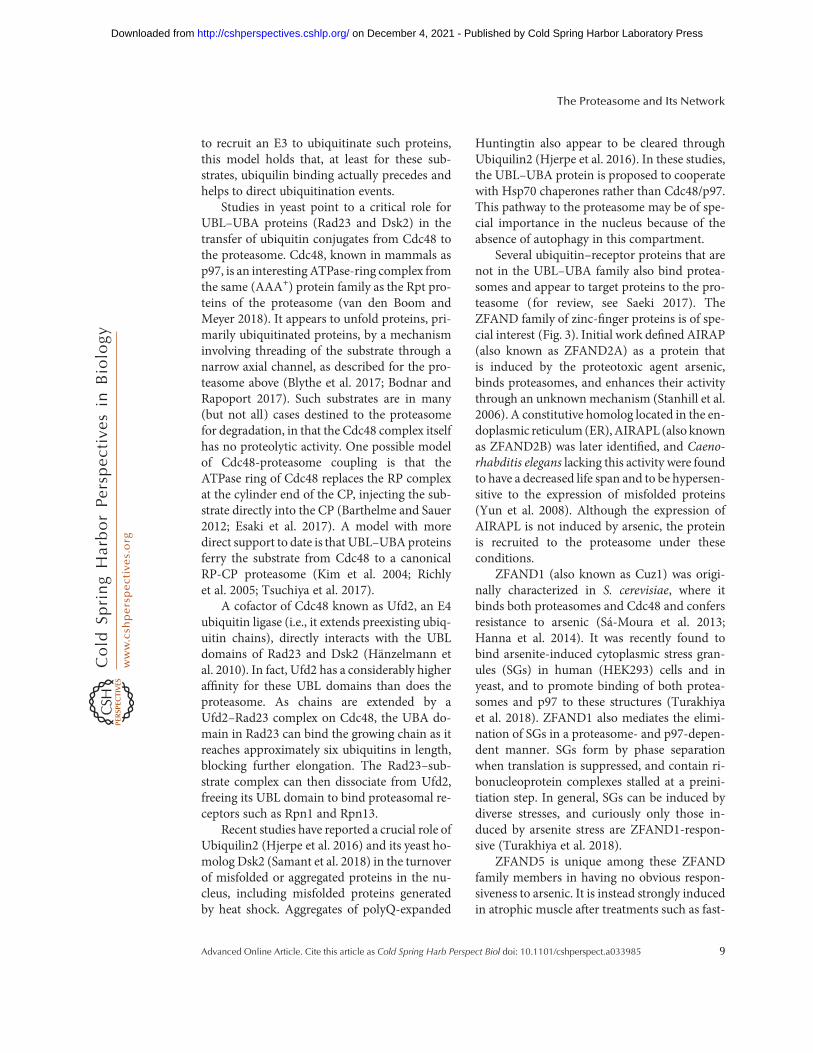
Themodel that deubiquitination byUbp6 orUSP14 can suppress degradation requires thatdeubiquitination is very fast, so as to outracesubstrate degradation. This has been validatedby quench-flow and single-molecule analyses(Lee et al. 2016). The multichain model hasnot been validated with endogenous substrates,but many groups have identified endogenousclients of USP14 that are more rapidly degradedwhen this activity is inhibited, or when USP14expression is knocked down (Lee et al. 2010;Homma et al. 2015; Chen et al. 2016a; McKin-non et al. 2016; Nakashima et al. 2016; Sareen-Khanna et al. 2016; Xu et al. 2016; Boselli et al.2017; Liao et al. 2017, 2018; Min et al. 2017b;Song et al. 2017; Wei et al. 2017; Zhu et al. 2017;Liu et al. 2018; Massa et al. 2018; Li et al. 2019).These experiments have relied mainly on thespecific small-molecule inhibitors of USP14,IU1, and IU1-47 (Lee et al. 2010; Boselli et al.2017). IU1 has recently been cocrystallized withUSP14 and shown to inhibit enzymatic activityby blocking access of the carboxyl terminus ofubiquitin to the active site of USP14, as shown inFigure 6 (Wang et al. 2018).
In addition to suppressing the degradationof numerous proteins, USP14 has remarkablybeen found to also suppress the degradation ofan organelle (Chakraborty et al. 2018). In bothmammalian cells and D. melanogaster, USP14controls the rate of basal mitophagy. AlthoughspecificUSP14 substrates thatmediate this effecthave not been identified, it may reflect that basal
D. Finley and M.A. Prado
14 Advanced Online Article. Cite this article as Cold Spring Harb Perspect Biol doi: 10.1101/cshperspect.a033985
on December 4, 2021 - Published by Cold Spring Harbor Laboratory Press http://cshperspectives.cshlp.org/Downloaded from

mitophagy can be signaled by ubiquitination ofproteins of the mitochondrial membrane (Villaet al. 2018), as described for the depolarization-induced mitophagy pathway that is mediated bythe ubiquitin ligase Parkin (Harper et al. 2018).In D. melanogaster mutants that lack Parkin-dependent mitophagy, reduction of USP14activity confers substantial life span extensionand enhances motor function. These effects ap-parently result from improved mitochondrialfunction, as increased basal mitophagy providesfor more efficient clearance of dysfunctional mi-tochondria that would otherwise be cleared bythe Parkin pathway. Suppression of Parkinmu-tant phenotypes was evident both with Usp14knockdown mutations and with IU1 providedin the flies’ food. Phenotypic suppression by lossof USP14 activity was also seen with mutants inPink1, the protein kinase that activates Parkin(Chakraborty et al. 2018).
Ubp6/USP14 is activated 300- to 800-fold byproteasome association when assayed with a
model substrate having a small leaving group,such as ubiquitin-AMC (Leggett et al. 2002;Lee et al. 2010). Repression of the free form ofUbp6/USP14 is thought to be mediated, at leastin part, by two loops, BL1 and BL2 (Fig. 6),which block access of the carboxyl terminus ofubiquitin to the active site of the enzyme (Huet al. 2005; Huang et al. 2016b). Free Ubp6/USP14 is often assayes using diubiquitin, butinterestingly no activation is evident with thissubstrate (Lee et al. 2016). A likely resolutionto this conundrum is that complex formationwith the proteasome, as seen by cryo-EM (Auf-derheide et al. 2015; Huang et al. 2016b), bringsthe active site of Ubp6/USP14 so close to theproteasome that the folded structure of the prox-imal ubiquitin is occluded (Lee et al. 2016). Ac-cordingly, Ubp6/USP14 releases chains en blocrather than whittling them down from their tips.Interestingly, because proximal ubiquitin in adiubiquitin adduct occupies the position of sub-strate in a ubiquitin–substrate adduct, the idea
BL2
CBA
BL1
Ub
Figure 6. Activation and inhibition of USP14. (A) Structure of the substrate-engaged USP14 (red) bound toubiquitin aldehyde (Ub-Al, in yellow), a model of the enzyme’s activated state. The principal difference from theubiquitin-free (inactive) state of USP14 is in the positioning of the two blocking (BL) loops (Hu et al. 2005). TheBL loops of USP14-Ub-Al are in green, those of the free USP14 in blue. To the right of these loops, the active-siteCys is represented as a light green patch. In the free form of USP14, the BL loops block the access of the carboxylterminus of ubiquitin to the active site. This occlusion is relieved in the substrate-engaged form. Ser432 (gray),located within the BL2 loop, is subject to phosphorylation by AKT, providing partial activation (Xu et al. 2015).(B) Structure of USP14 bound to IU1-47 (white) shows that this inhibitor occludes access of the carboxylterminus of ubiquitin to the catalytic site (Wang et al. 2018). To better visualize the IU1-47-binding pocket,both BL loopswere removed from the densitymaps. (C) Structure of free USP14with IU1-47 superimposed. Thismodel shows how the BL2 loop (blue) occludes access of IU1-47, consistent with previous evidence that IU1series compounds do not inhibit the free form of USP14 (Lee et al. 2016). Density maps of the USP14 catalyticdomain in its free form (Protein Data Bank [PDB]: 2AYN), USP14 bound to Ub-Al (PDB: 2AYO), and theUSP14-IU1-47 complex (PDB: 6IIL) were created from data in Hu et al. (2005) andWang et al. (2018). To bettervisualize the catalytic site of USP14, Gln197 was removed from all USP14 structures in this figure.
The Proteasome and Its Network
Advanced Online Article. Cite this article as Cold Spring Harb Perspect Biol doi: 10.1101/cshperspect.a033985 15
on December 4, 2021 - Published by Cold Spring Harbor Laboratory Press http://cshperspectives.cshlp.org/Downloaded from
that the body of the proteasome blocks access ofbulky leaving groups to Ubp6/USP14 also car-ries the prediction that Ubp6/USP14 discrimi-nates against folded domains in the substrateprotein itself. Ubiquitin groups or chains at-tached to domains that are folded when the sub-strate docks at the proteasome may therefore bereserved for Rpn11, because substrate transloca-tion draws these domains toward Rpn11, where-as their accessibility to Rpn11’s active site is en-sured by ATP-dependent unfolding.
As discussed above, Ubp6 is needed in yeastto prevent excessive ubiquitin degradation. Inyeast engineered to express low levels of ubi-quitin, Ubp6 is dramatically induced by a ho-meostatic feedback circuit (Hanna et al. 2007).USP14 activity is subject to multiple controlsthough the mechanisms differ from that ofUbp6 in yeast. Perhaps surprisingly, four distinctmicroRNAs control the expression of the pro-tein, exerting a variety of physiological effects(Wu et al. 2014; Sun et al. 2016; Zhu et al.2017; Lee et al. 2018b). At the posttranslationallevel, USP14 is phosphorylated on Ser432 by thegrowth-regulatory kinase AKT, which resultsin partial activation of its deubiquitinatingactivity (Xu et al. 2015). As Ser432 lies withinthe BL2 loop (Fig. 6), its modification presum-ably antagonizes the above-described autoinhib-itory mechanism. In summary, growth signalssuch as insulin activate AKT, which among itsmany activities will potentiate USP14, likelyleading to reduced protein breakdown by theproteasome.
The most dramatic regulation of USP14 de-scribed to date is posttranslational and involvesthe protein TRIM11, which binds the amino-terminal ubiquitin-like domain of USP14 incompetition with the proteasome (Chen et al.2018). Thus, TRIM11 functions as an endo-genous USP14 inhibitor. Heat shock inducesTRIM11, promoting cell survival apparently byreleasing the proteasome from the inhibitory ef-fect of USP14 (Chen et al. 2018). The intricateregulation of USP14 on multiple levels in mam-mals seems to imply that USP14 is used notsimply to promote ubiquitin recovery from pro-teasome substrates but also as a vehicle for finetuning of proteasome activity.
PROTEASOME-ASSOCIATED UBIQUITIN-CONJUGATING ACTIVITY
The proteasome is associated with not only de-ubiquitinating activity but also ubiquitin ligaseactivity. A number of ligases have been shown tointeract with the proteasome, if in some casesweakly (Verma et al. 2000; Xie and Varshavsky2000, 2002; Leggett et al. 2002; Crosas et al.2006; Besche et al. 2009; Martinez-Noel et al.2012; Kühnle et al. 2018). One view of this phe-nomenon is that the potency of a ligase will beenhanced if it ubiquitinates its substrates at theproteasome, because the temporal lag betweenubiquitination and proteasome docking, duringwhich the conjugate is vulnerable to deubiquiti-nation, is reduced (Xie and Varshavsky 2000).Another view, proposed for the yeast Hul5 ligase(Crosas et al. 2006), is that conjugating activitycould be directed preferentially toward protea-some-bound proteins—typically proteins thatare already ubiquitinated by another ligase—and function as a general regulator of the pro-teasome. In vitro,Hul5 efficiently adds ubiquitinto model target proteins such as tetraubiquitinand ubiquitinated cyclin B, although not takingunmodified cyclin B as a substrate (Crosas et al.2006). These reactions are dependent on thepresence of the proteasome. Because Hul5mod-ified cyclin B only if it had already been ubiqui-tinated, Hul5 appeared to act in this case as an“E4” or (essentially) a ubiquitin chain-extend-ing enzyme (Crosas et al. 2006).
Hul5 and its mammalian homolog UBE3Chave repeatedly been identified as proteasomeprocessivity factors in studies carried out withartificial fusion proteins that are ubiquitinatedby an E3 other than Hul5 and degraded by theproteasome (Kohlmann et al. 2008; Aviram andKornitzer 2010; Martinez-Noel et al. 2012; Chuet al. 2013). In hul5 and Ube3C loss of functionmutants, the proteasome fails to fully degradethese proteins, releasing a truncated product.The activity seems to be unique to Hul5/UBE3C. These observations are consistent witha generalized E4 activity inHul5, which serves topromote the degradation of stalled substratesthat may otherwise fail to complete transloca-tion from the RP to the CP. Seemingly in agree-
D. Finley and M.A. Prado
16 Advanced Online Article. Cite this article as Cold Spring Harb Perspect Biol doi: 10.1101/cshperspect.a033985
on December 4, 2021 - Published by Cold Spring Harbor Laboratory Press http://cshperspectives.cshlp.org/Downloaded from

ment with these findings, Hul5 and UBE3C arerapidly recruited to proteasomes whose activityhas been compromised by mutation, inhibitorsof the proteasome, or inhibitors of USP14 (Parket al. 2011; Kuo and Goldberg 2017). This re-cruitment effect is seen with USP14 as well (Bo-rodovsky et al. 2001; Kuo and Goldberg 2017),and the presence of USP14 enhances UBE3Crecruitment just as Ubp6 enhancesHul5 recruit-ment (Crosas et al. 2006; Kuo and Goldberg2017). Such inducible recruitment of USP14 tothe proteasome may underlie the cytoprotectiveeffects ofUSP14 inhibitors observed under somestress conditions (Sareen-Khanna et al. 2016;Min et al. 2017a; Chen et al. 2018; VerPlanket al. 2018). Defective proteasomes also recruitEcm29, a large, HEAT-repeat-containing pro-tein (Park et al. 2011; De La Mota-Peynadoet al. 2013). LikeUSP14, Ecm29 exerts a negativeinfluence on the proteasome. Binding in prox-imity to Rpt5, it suppresses ATPase activity, gateopening, and the degradation of ubiquitin–pro-tein conjugates (De La Mota-Peynado et al.2013).
An interesting inducer of Hul5-dependentubiquitination is heat stress, pointing to therole of Hul5 in QC degradation (Fang et al.2011). hul5Δ mutants show a strong delay inthe resumption of cell division after a heat shock.Moreover, as much as 50% of the ubiquitinationthat is acutely induced by heat shock is Hul5-dependent, with assays of the degradation ofnewly synthesized proteins giving comparableresults. Most Hul5 substrates identified in Fanget al. (2011) were still monoubiquitinated in theabsence of Hul5, in agreement with the modelthat Hul5 functions as an E4 in heat-shockedcells.
The conjugating activity of Hul5 andUBE3C is not strictly limited to proteasomesubstrates; they can also modify proteasomesubunits as well, such as ubiquitin receptorsRpn10 and Rpn13 (Crosas et al. 2006; Bescheet al. 2014). Ubiquitin modification of ubiquitinreceptors typically results in their functional in-activation (Hoeller and Dikic 2010). Therefore,ubiquitination of Rpn10 and Rpn13 could me-diate negative regulation of the proteasome un-der certain, perhaps extreme, stress conditions,
in contrast to the positive regulation seen inmany studies as described above.
CONTROLOF PROTEASOME ACTIVITYBY PHOSPHORYLATION
Protein modification by phosphorylation iswidely used to integrate signaling, metabolic,and gene expression networks.Hundreds of siteson the proteasome are subject to phosphoryla-tion but to date only a few have been character-ized (reviewed by Guo et al. 2017; VerPlank andGoldberg 2018). Modification of RPT3 at Thr25by the DYRK2 kinase places proteasome activi-ty under cell-cycle control, with a Thr25Alaknockin mutant being defective in total proteindegradation to a remarkable 66% (Guo et al.2016). Among other things this suggests mini-mal substrate specificity in the effect of phosphor-ylation. The mutation impairs cellular prolifera-tion aswell as the in vivo growth of triple-negativebreast cancer cells, perhaps to be expected giventhe extent towhich these proteasomes are inhib-ited. Why the knockin proteasomes function sopoorly remains mysterious. The mutation doesnot affect proteasome levels or ostensibly theirassembly (Guo et al. 2016). It has not been pos-sible to visualize RPT3-Thr25 in the proteasometo date but it would seem to be located far fromthe core elements of the RP, such as the translo-cation channel and RPN11.
Another critical phosphorylation site isSer14 of RPN6, a target of the cAMP-dependentprotein kinase A (PKA) (Lokireddy et al. 2015).Overexpression of a nonphosphorylatableSer14Ala mutant of RPN6 is suppressive ofprotein degradation, whereas that of the phos-phomimetic Ser14Aspmutant stimulates degra-dation. As cAMP is used for signaling in diversephysiological and cellular contexts, includingfasting and exercise, this pathway may be widelyutilized. cAMP levels can be elevated experi-mentally using rolipram, a specific inhibitor ofphosphodiesterase-4, which acts on cAMP. Ro-lipram increases the levels of assembled holoen-zyme in cells, particularly the double-cappedRP2CP form of holoenzyme, possibly account-ing for the stimulation of protein degradation intreated cells (Lokireddy et al. 2015).
The Proteasome and Its Network
Advanced Online Article. Cite this article as Cold Spring Harb Perspect Biol doi: 10.1101/cshperspect.a033985 17
on December 4, 2021 - Published by Cold Spring Harbor Laboratory Press http://cshperspectives.cshlp.org/Downloaded from
Rolipram accentuates proteasome activity inthe mouse brain, presumably by inducing thephosphorylation of RPN6-Ser14 (Myeku et al.2016). In mice expressing a pathogenic form ofthe tau protein (tau-Pro301Leu) that inducesneurodegeneration, rolipram significantly re-duced the levels of insoluble tau species. Thesefindings suggest that the stimulation of protea-some phosphorylation and activity in the brainattenuates tauopathies and may have implica-tions for other neurodegenerative disorders.
Another phosphorylation event observedwith brain proteasomes is the modification ofRPT6-Ser120 by the calcium/calmodulin-de-pendent kinase CaMKIIα, a critical and veryabundant activity in the brain. Phosphorylationat Ser120 stimulates proteasome activity and in-terestingly it is induced in particular by cocaine(Gonzales et al. 2018). For reasons that areunclear, phosphomimetic RPT6-Ser120Aspmouse mutants fail to develop cocaine sensiti-zation, an effect closely linked to addiction.
CONCLUDING REMARKS
Although derived from a common type of ATP-dependent protease expressed in archaea, theproteasome has evolved within the eukaryoticlineage into a unique molecular machine. Pre-sumably the initial driving force behind the evo-lution of the complexity of the proteasome wasthe inherent complications involved in degrad-ing ubiquitin–protein conjugates, as reflected inthe unanticipated multiplicity of ubiquitin re-ceptors and deubiquitinating enzymes associat-ed with the proteasome. The complexity of theproteasome does not stop at its traditional bor-ders; numerous cofactors, regulators, and post-translational modifications of the proteasomeare integral to its function, and this broader net-work provides for striking adaptability in re-sponse to growth stimuli, nutritional limitation,and proteostatic stress. Thus, contrary to long-standing perceptions of the proteasome, exten-sive variation of the magnitude and nature of itsactivity are tolerated, and indeed these varia-tions are promoted through conserved regula-tory mechanisms. Because of the importance ofthe proteasome in proteostasis, these insights
may have significant implications for our under-standing of disease and aging. Future studiesshould define more clearly how proteasome reg-ulators impact the increasingly well-definedcore mechanisms of the enzyme and its ensem-ble of conformational states. In addition, prote-omic studies will play a central role in defininghow specific modes of proteasome regulationperturb its output on a global scale.
ACKNOWLEDGMENTS
We gratefully acknowledge members of theFinley laboratory for helpful comments on themanuscript. Patents 8933087 and 9201073 areheld on IU1, IU1-47, and USP14 inhibition,filed by Harvard University on behalf of D.F.and others. These patents have been licensedto Proteostasis Therapeutics.
REFERENCES�Reference is also in this collection.
Asano S, Fukuda Y, Beck F, Aufderheide A, Forster F, DanevR, BaumeisterW. 2015. Proteasomes. Amolecular censusof 26S proteasomes in intact neurons. Science 347: 439–442. doi:10.1126/science.1261197
Aufderheide A, Beck F, Stengel F, Hartwig M, Schweitzer A,Pfeifer G, Goldberg AL, Sakata E, Baumeister W, FörsterF. 2015. Structural characterization of the interaction ofUbp6 with the 26S proteasome. Proc Natl Acad Sci 112:8626–8631. doi:10.1073/pnas.1510449112
Aviram S, Kornitzer D. 2010. The ubiquitin ligase Hul5 pro-motes proteasomal processivity. Mol Cell Biol 30: 985–994. doi:10.1128/MCB.00909-09
Bajorek M, Finley D, Glickman MH. 2003. Proteasome dis-assembly and downregulation is correlated with viabilityduring stationary phase. Curr Biol 13: 1140–1144. doi:10.1016/S0960-9822(03)00417-2
Bard JAM, Goodall EA, Greene ER, Jonsson E, Dong KC,Martin A. 2018. Structure and function of the 26S pro-teasome. Annu Rev Biochem 87: 697–724. doi:10.1146/annurev-biochem-062917-011931
Bartelt A, Widenmaier SB, Schlein C, Johann K, GoncalvesRLS, Eguchi K, Fischer AW, Parlakgul G, SnyderNA, Nguyen TB, et al. 2018. Brown adipose tissue ther-mogenic adaptation requires Nrf1-mediated proteasomalactivity. Nat Med 24: 292–303. doi:10.1038/nm.4481
Barthelme D, Sauer RT. 2012. Identification of the Cdc48·20Sproteasome as an ancient AAA+ proteolytic machine. Sci-ence 337: 843–846. doi:10.1126/science.1224352
Bashore C, Dambacher CM, Goodall EA, Matyskiela ME,Lander GC, Martin A. 2015. Ubp6 deubiquitinase con-trols conformational dynamics and substrate degradation
D. Finley and M.A. Prado
18 Advanced Online Article. Cite this article as Cold Spring Harb Perspect Biol doi: 10.1101/cshperspect.a033985
on December 4, 2021 - Published by Cold Spring Harbor Laboratory Press http://cshperspectives.cshlp.org/Downloaded from

of the 26S proteasome. Nat Struct Mol Biol 22: 712–719.doi:10.1038/nsmb.3075
Besche HC, Haas W, Gygi SP, Goldberg AL. 2009. Isolationofmammalian 26S proteasomes and p97/VCP complexesusing the ubiquitin-like domain from HHR23B revealsnovel proteasome-associated proteins. Biochemistry 48:2538–2549. doi:10.1021/bi802198q
Besche HC, Sha Z, Kukushkin NV, Peth A, Hock EM, KimW, Gygi S, Gutierrez JA, Liao H, Dick L, et al. 2014.Autoubiquitination of the 26S proteasome on Rpn13 reg-ulates breakdown of ubiquitin conjugates. EMBO J 33:1159–1176. doi:10.1002/embj.201386906
Blythe EE, Olson KC, Chau V, Deshaies RJ. 2017. Ubiqui-tin- and ATP-dependent unfoldase activity of P97/VCP·NPLOC4·UFD1L is enhanced by a mutation thatcauses multisystem proteinopathy. Proc Natl Acad Sci114: E4380–E4388. doi:10.1073/pnas.1706205114
Bodnar NO, Rapoport TA. 2017. Molecular mechanism ofsubstrate processing by the Cdc48 ATPase complex. Cell169: 722–735.e9. doi:10.1016/j.cell.2017.04.020
Borodovsky A, Kessler BM, Casagrande R, Overkleeft HS,Wilkinson KD, Ploegh HL. 2001. A novel active site-di-rected probe specific for deubiquitylating enzymes revealsproteasome association of USP14. EMBO J 20: 5187–5196.
Boselli M, Lee BH, Robert J, Prado MA, Min SW, Cheng C,Silva MC, Seong C, Elsasser S, Hatle KM, et al. 2017. Aninhibitor of the proteasomal deubiquitinating enzymeUSP14 induces tau elimination in cultured neurons. JBiol Chem 292: 19209–19225. doi:10.1074/jbc.M117.815126
Braten O, Livneh I, Ziv T, Admon A, Kehat I, Caspi LH,Gonen H, Bercovich B, Godzik A, Jahandideh S, et al.2016. Numerous proteins with unique characteristicsare degraded by the 26S proteasome following monoubi-quitination. Proc Natl Acad Sci 113: E4639–E4647. doi:10.1073/pnas.1608644113
Budenholzer L, Cheng CL, Li Y, Hochstrasser M. 2017. Pro-teasome structure and assembly. J Mol Biol 429: 3500–3524. doi:10.1016/j.jmb.2017.05.027
Chakraborty J, von Stockum S, Marchesan E, Caicci F,Ferrari V, Rakovic A, Klein C, Antonini A, Bubacco L,Ziviani E. 2018. USP14 inhibition corrects an in vivomodel of impaired mitophagy. EMBO Mol Med 10:e9014. doi:10.15252/emmm.201809014
Chau V, Tobias JW, Bachmair A, Marriott D, Ecker DJ,Gonda DK, Varshavsky A. 1989. A multiubiquitin chainis confined to specific lysine in a targeted short-livedprotein. Science 243: 1576–1583. doi:10.1126/science.2538923
ChenM,MengQ, Qin Y, Liang P, Tan P, He L, ZhouY, ChenY, Huang J,Wang RF, et al. 2016a. TRIM14 Inhibits cGASdegradation mediated by selective autophagy receptorp62 to promote innate immune responses. Mol Cell 64:105–119. doi:10.1016/j.molcel.2016.08.025
Chen X, Randles L, Shi K, Tarasov SG, Aihara H,Walters KJ.2016b. Structures of Rpn1T1:Rad23 and hRpn13:hPLIC2reveal distinct binding mechanisms between substrate re-ceptors and shuttle factors of the proteasome. Structure24: 1257–1270. doi:10.1016/j.str.2016.05.018
Chen S,Wu J, Lu Y, Ma YB, Lee BH, Yu Z, Ouyang Q, FinleyDJ, Kirschner MW, Mao Y. 2016c. Structural basis for
dynamic regulation of the human 26S proteasome. ProcNatl Acad Sci 113: 12991–12996. doi:10.1073/pnas.1614614113
Chen L, Paquette N, Mamoor S, Rus F, Nandy A, Leszyk J,Shaffer SA, Silverman N. 2017. Innate immune signalinginDrosophila is regulated by transforming growth factor β(TGFβ)-activated kinase (Tak1)-triggered ubiquitin edit-ing. J Biol Chem 292: 8738–8749. doi:10.1074/jbc.M117.788158
Chen L, Zhu G, Johns EM, Yang X. 2018. TRIM11 activatesthe proteasome and promotes overall protein degradationby regulating USP14. Nat Commun 9: 1223. doi:10.1038/s41467-018-03499-z
Chondrogianni N, Georgila K, Kourtis N, Tavernarakis N,Gonos ES. 2015. 20S proteasome activation promotes lifespan extension and resistance to proteotoxicity in Caeno-rhabditis elegans. FASEB J 29: 611–622. doi:10.1096/fj.14-252189
Chu BW, Kovary KM, Guillaume J, Chen LC, Teruel MN,Wandless TJ. 2013. The E3 ubiquitin ligase UBE3C en-hances proteasome processivity by ubiquitinating partial-ly proteolyzed substrates. J Biol Chem 288: 34575–34587.doi:10.1074/jbc.M113.499350
Collins GA, Goldberg AL. 2017. The logic of the 26S protea-some. Cell 169: 792–806. doi:10.1016/j.cell.2017.04.023
Crosas B, Hanna J, Kirkpatrick DS, Zhang DP, Tone Y,Hathaway NA, Buecker C, Leggett DS, Schmidt M, KingRW, et al. 2006. Ubiquitin chains are remodeled at theproteasome by opposing ubiquitin ligase and deubiquiti-nating activities. Cell 127: 1401–1413. doi:10.1016/j.cell.2006.09.051
De La Mota-Peynado A, Lee SY, Pierce BM, Wani P, SinghCR, Roelofs J. 2013. The proteasome-associated proteinEcm29 inhibits proteasomal ATPase activity and in vivoprotein degradation by the proteasome. J Biol Chem 288:29467–29481. doi:10.1074/jbc.M113.491662
de la Peña AH,Goodall EA, Gates SN, Lander GC,Martin A.2018. Substrate-engaged 26S proteasome structures revealmechanisms for ATP-hydrolysis-driven translocation.Science 362: eaav0725. doi:10.1126/science.aav0725
Deng HX, Chen W, Hong ST, Boycott KM, Gorrie GH,Siddique N, Yang Y, Fecto F, Shi Y, Zhai H, et al. 2011.Mutations in UBQLN2 cause dominant X-linked juvenileand adult-onset ALS and ALS/dementia. Nature 477:211–215. doi:10.1038/nature10353
de Poot SAH, Tian G, Finley D. 2017. Meddling with fate:The proteasomal deubiquitinating enzymes. J Mol Biol429: 3525–3545. doi:10.1016/j.jmb.2017.09.015
Dong Y, Zhang S, Wu Z, Li X, Wang WL, Zhu Y, Stoilova-McPhie S, Lu Y, Finley D, Mao Y. 2019. Cryo-EM struc-tures and dynamics of substrate-engaged human 26S pro-teasome. Nature 565: 49–55. doi:10.1038/s41586-018-0736-4
EiseleMR, Reed RG, Rudack T, Schweitzer A, Beck F, Nagy I,Pfeifer G, Plitzko JM, Baumeister W, Tomko RJ, et al.2018. Expanded coverage of the 26S proteasome confor-mational landscape reveals mechanisms of peptidase gat-ing. Cell Rep 24: 1301–1315.e5. doi:10.1016/j.celrep.2018.07.004
Esaki M, IslamMT, Tani N, Ogura T. 2017. Deviation of thetypical AAA substrate-threading pore prevents fatal pro-
The Proteasome and Its Network
Advanced Online Article. Cite this article as Cold Spring Harb Perspect Biol doi: 10.1101/cshperspect.a033985 19
on December 4, 2021 - Published by Cold Spring Harbor Laboratory Press http://cshperspectives.cshlp.org/Downloaded from

tein degradation in yeast Cdc48. Sci Rep 7: 5475. doi:10.1038/s41598-017-05806-y
Fang NN, Ng AH, Measday V, Mayor T. 2011. Hul5 HECTubiquitin ligase plays a major role in the ubiquitylationand turnover of cytosolicmisfolded proteins.Nat Cell Biol13: 1344–1352. doi:10.1038/ncb2343
FuH, Sadis S, Rubin DM, GlickmanM, van Nocker S, FinleyD, Vierstra RD. 1998. Multiubiquitin chain binding andprotein degradation are mediated by distinct domainswithin the 26 S proteasome subunit Mcb1. J Biol Chem273: 1970–1981. doi:10.1074/jbc.273.4.1970
Gonzales FR, Howell KK, Dozier LE, Anagnostaras SG, Pat-rick GN. 2018. Proteasome phosphorylation regulates co-caine-induced sensitization.Mol Cell Neurosci 88: 62–69.doi:10.1016/j.mcn.2017.12.001
GrollM,Ditzel L, Löwe J, StockD, BochtlerM, BartunikHD,Huber R. 1997. Structure of 20S proteasome from yeastat 2.4 Å resolution. Nature 386: 463–471. doi:10.1038/386463a0
Groll M, Bajorek M, Köhler A, Moroder L, Rubin DM, Hu-ber R, Glickman MH, Finley D. 2000. A gated channelinto the proteasome core particle.Nat Struct Biol 7: 1062–1067. doi:10.1038/80992
Grumati P,Dikic I. 2018.Ubiquitin signaling and autophagy.J Biol Chem 293: 5404–5413. doi:10.1074/jbc.TM117.000117
GuZC,WuE, Sailer C, Jando J, Styles E, Eisenkolb I, KuschelM, Bitschar K, Wang X, Huang L, et al. 2017. Ubiquitinorchestrates proteasome dynamics between proliferationand quiescence in yeast. Mol Biol Cell 28: 2479–2491.doi:10.1091/mbc.e17-03-0162
Guo X, Wang X, Wang Z, Banerjee S, Yang J, Huang L,Dixon JE. 2016. Site-specific proteasome phosphoryla-tion controls cell proliferation and tumorigenesis. NatCell Biol 18: 202–212. doi:10.1038/ncb3289
Guo X, Huang X, Chen MJ. 2017. Reversible phosphoryla-tion of the 26S proteasome. Protein Cell 8: 255–272.doi:10.1007/s13238-017-0382-x
Guo Q, Lehmer C, Martínez-Sánchez A, Rudack T, Beck F,Hartmann H, Pérez-Berlanga M, Frottin F, Hipp MS,Hartl FU, et al. 2018. In situ structure of neuronalC9orf72 Poly-GA Aggregates reveals proteasome recruit-ment. Cell 172: 696–705.e12. doi:10.1016/j.cell.2017.12.030
Ha SW, Ju D, Xie Y. 2012. The N-terminal domain of Rpn4serves as a portable ubiquitin-independent degron and isrecognized by specific 19S RP subunits. Biochem BiophysRes Commun 419: 226–231. doi:10.1016/j.bbrc.2012.01.152
Hamazaki J, Sasaki K, Kawahara H, Hisanaga S, Tanaka K,Murata S. 2007. Rpn10-mediated degradation of ubiqui-tinated proteins is essential for mouse development.MolCell Biol 27: 6629–6638. doi:10.1128/MCB.00509-07
Hanna J, LeggettDS, FinleyD. 2003. Ubiquitin depletion as akey mediator of toxicity by translational inhibitors. MolCell Biol 23: 9251–9261. doi:10.1128/MCB.23.24.9251-9261.2003
Hanna J, Hathaway NA, Tone Y, Crosas B, Elsasser S, Kirk-patrickDS, Leggett DS, Gygi SP, King RW, Finley D. 2006.Deubiquitinating enzyme Ubp6 functions noncatalyti-cally to delay proteasomal degradation. Cell 127: 99–111. doi:10.1016/j.cell.2006.07.038
Hanna J, Meides A, Zhang DP, Finley D. 2007. A ubiquitinstress response induces altered proteasome composition.Cell 129: 747–759. doi:10.1016/j.cell.2007.03.042
Hanna J, Waterman D, Isasa M, Elsasser S, Shi Y, Gygi S,Finley D. 2014. Cuz1/Ynl155w, a zinc-dependent ubiqui-tin-binding protein, protects cells from metalloid-in-duced proteotoxicity. J Biol Chem 289: 1876–1885.doi:10.1074/jbc.M113.534032
Hänzelmann P, Stingele J, Hofmann K, Schindelin H, RaasiS. 2010. The yeast E4 ubiquitin ligase Ufd2 interacts withthe ubiquitin-like domains of Rad23 and Dsk2 via a noveland distinct ubiquitin-like binding domain. J Biol Chem285: 20390–20398. doi:10.1074/jbc.M110.112532
Harper JW, Ordureau A, Heo JM. 2018. Building and de-coding ubiquitin chains for mitophagy. Nat Rev Mol CellBiol 19: 93–108. doi:10.1038/nrm.2017.129
Hartmann-Petersen R, Hendil KB, Gordon C. 2003. Ubiq-uitin binding proteins protect ubiquitin conjugates fromdisassembly. FEBS Lett 535: 77–81. doi:10.1016/S0014-5793(02)03874-7
� Hegde RS, Zavodszky E. 2019. Recognition and degradationofmislocalized proteins in health and disease.Cold SpringHarb Perspect Biol doi:10.1101/cshperspect.033902
Hishiya A, Iemura S, Natsume T, Takayama S, Ikeda K,Watanabe K. 2006. A novel ubiquitin-binding proteinZNF216 functioning in muscle atrophy. EMBO J 25:554–564. doi:10.1038/sj.emboj.7600945
Hjerpe R, Bett JS, Keuss MJ, Solovyova A, McWilliams TG,Johnson C, Sahu I, Varghese J, Wood N,WightmanM, etal. 2016. UBQLN2mediates autophagy-independent pro-tein aggregate clearance by the proteasome.Cell 166: 935–949. doi:10.1016/j.cell.2016.07.001
HoellerD,Dikic I. 2010. Regulation of ubiquitin receptors bycoupled monoubiquitination. Subcell Biochem 54: 31–40.doi:10.1007/978-1-4419-6676-6_3
Hofmann RM, Pickart CM. 2001. In vitro assembly andrecognition of Lys-63 polyubiquitin chains. J Biol Chem276: 27936–27943. doi:10.1074/jbc.M103378200
HommaT, Ishibashi D, Nakagaki T, Fuse T,Mori T, SatohK,Atarashi R, Nishida N. 2015. Ubiquitin-specific protease14 modulates degradation of cellular prion protein. SciRep 5: 11028. doi:10.1038/srep11028
Hough R, Pratt G, Rechsteiner M. 1987. Purification of twohigh molecular weight proteases from rabbit reticulocytelysate. J Biol Chem 262: 8303–8313.
HuM, Li P, Song L, Jeffrey PD, Chenova TA,Wilkinson KD,Cohen RE, Shi Y. 2005. Structure and mechanisms of theproteasome-associated deubiquitinating enzyme USP14.EMBO J 24: 3747–3756. doi:10.1038/sj.emboj.7600832
Huang L, Haratake K, Miyahara H, Chiba T. 2016a. Protea-some activators, PA28γ and PA200, play indispensableroles in male fertility. Sci Rep 6: 23171. doi:10.1038/srep23171
Huang X, Luan B,Wu J, Shi Y. 2016b. An atomic structure ofthe human 26S proteasome. Nat Struct Mol Biol 23: 778–785. doi:10.1038/nsmb.3273
Husnjak K, Elsasser S, Zhang N, Chen X, Randles L, Shi Y,Hofmann K, Walters KJ, Finley D, Dikic I. 2008. Protea-some subunit Rpn13 is a novel ubiquitin receptor.Nature453: 481–488. doi:10.1038/nature06926
D. Finley and M.A. Prado
20 Advanced Online Article. Cite this article as Cold Spring Harb Perspect Biol doi: 10.1101/cshperspect.a033985
on December 4, 2021 - Published by Cold Spring Harbor Laboratory Press http://cshperspectives.cshlp.org/Downloaded from

Itakura E, Zavodszky E, Shao S, Wohlever ML, Keenan RJ,Hegde RS. 2016. Ubiquilins chaperone and triage mito-chondrial membrane proteins for degradation. Mol Cell63: 21–33. doi:10.1016/j.molcel.2016.05.020
Jelinsky SA, Estep P, Church GM, Samson LD. 2000. Regu-latory networks revealed by transcriptional profiling ofdamaged Saccharomyces cerevisiae cells: Rpn4 links baseexcision repair with proteasomes.Mol Cell Biol 20: 8157–8167. doi:10.1128/MCB.20.21.8157-8167.2000
� Karagöz GE, Acosta-Alvear D,Walter P. 2019. The unfoldedprotein response: Detecting and responding to fluctua-tions in the protein folding capacity of the endoplasmicreticulum. Cold Spring Harb Perspect Biol doi:10.1101/cshperspect.033886
Kim HT, Goldberg AL. 2017. The deubiquitinating enzymeUsp14 allosterically inhibits multiple proteasomal activi-ties and ubiquitin-independent proteolysis. J Biol Chem292: 9830–9839. doi:10.1074/jbc.M116.763128
Kim HT, Goldberg AL. 2018. UBL domain of Usp14 andother proteins stimulates proteasome activities and pro-tein degradation in cells. Proc Natl Acad Sci 115: E11642–E11650. doi:10.1073/pnas.1808731115
Kim I, Mi K, Rao H. 2004. Multiple interactions of rad23suggest a mechanism for ubiquitylated substrate deliveryimportant in proteolysis. Mol Biol Cell 15: 3357–3365.doi:10.1091/mbc.e03-11-0835
Kirisako T, Kamei K, Murata S, KatoM, FukumotoH, KanieM, Sano S, Tokunaga F, Tanaka K, Iwai K. 2006. A ubiq-uitin ligase complex assembles linear polyubiquitinchains. EMBO J 25: 4877–4887. doi:10.1038/sj.emboj.7601360
Kirkpatrick DS, Hathaway NA, Hanna J, Elsasser S, Rush J,Finley D, King RW, Gygi SP. 2006. Quantitative analysisof in vitro ubiquitinated cyclin B1 reveals complex chaintopology. Nat Cell Biol 8: 700–710. doi:10.1038/ncb1436
Kohlmann S, Schäfer A, Wolf DH. 2008. Ubiquitin ligaseHul5 is required for fragment-specific substrate degrada-tion in endoplasmic reticulum-associated degradation.J Biol Chem 283: 16374–16383. doi:10.1074/jbc.M801702200
Krylov DM, Koonin EV. 2001. A novel family of predictedretroviral-like aspartyl proteases with a possible key rolein eukaryotic cell cycle control. Curr Biol 11: R584–R587.doi:10.1016/S0960-9822(01)00357-8
Kühnle S,Martinez-Noël G, Leclere F, Hayes SD,Harper JW,Howley PM. 2018. Angelman syndrome-associated pointmutations in the Zn2+-binding N-terminal (AZUL) do-main of UBE3A ubiquitin ligase inhibit binding to theproteasome. J Biol Chem 293: 18387–18399. doi:10.1074/jbc.RA118.004653
Kuo CL, Goldberg AL. 2017. Ubiquitinated proteins pro-mote the association of proteasomes with the deubiquiti-nating enzyme Usp14 and the ubiquitin ligase Ube3c.Proc Natl Acad Sci 114: E3404–E3413. doi:10.1073/pnas.1701734114
Kwon YT, Ciechanover A. 2017. The ubiquitin code in theubiquitin-proteasome system and autophagy. Trends Bio-chem Sci 42: 873–886. doi:10.1016/j.tibs.2017.09.002
Laporte D, Salin B, Daignan-Fornier B, Sagot I. 2008. Re-versible cytoplasmic localization of the proteasome inquiescent yeast cells. J Cell Biol 181: 737–745. doi:10.1083/jcb.200711154
Lee BH, LeeMJ, Park S,OhDC, Elsasser S, ChenPC,GartnerC, DimovaN, Hanna J, Gygi SP, et al. 2010. Enhancementof proteasome activity by a small-molecule inhibitor ofUSP14. Nature 467: 179–184. doi:10.1038/nature09299
Lee BH, Lu Y, Prado MA, Shi Y, Tian G, Sun S, Elsasser S,Gygi SP, King RW, FinleyD. 2016.USP14 deubiquitinatesproteasome-bound substrates that are ubiquitinatedat multiple sites. Nature 532: 398–401. doi:10.1038/nature17433
Lee D, Takayama S, Goldberg AL. 2018a. ZFAND5/ZNF216is an activator of the 26S proteasome that stimulates over-all protein degradation. Proc Natl Acad Sci 115: E9550–E9559. doi:10.1073/pnas.1809934115
Lee SW, Oh YM, Lu YL, Kim WK, Yoo AS. 2018b. Micro-RNAs overcome cell fate barrier by reducing EZH2-con-trolled REST stability during neuronal conversion of hu-man adult fibroblasts.DevCell 46: 73–84.e7. doi:10.1016/j.devcel.2018.06.007
Leggett DS, Hanna J, Borodovsky A, Crosas B, Schmidt M,Baker RT, Walz T, Ploegh H, Finley D. 2002. Multipleassociated proteins regulate proteasome structure andfunction. Mol Cell 10: 495–507. doi:10.1016/S1097-2765(02)00638-X
LehrbachNJ, RuvkunG. 2016. Proteasome dysfunction trig-gers activation of SKN-1A/Nrf1 by the aspartic proteaseDDI-1. eLife 5: e17721. doi:10.7554/eLife.17721
Letunic I, Bork P. 2018. 20 years of the SMART proteindomain annotation resource. Nucleic Acids Res 46:D493–D496. doi:10.1093/nar/gkx922
Leznicki P, Kulathu Y. 2017. Mechanisms of regulation anddiversification of deubiquitylating enzyme function. J CellSci 130: 1997–2006. doi:10.1242/jcs.201855
Li H, Zhao Z, Ling J, Pan L, Zhao X, ZhuH, Yu J, Xie B, ShenJ, ChenW. 2019. USP14 promotes K63-linked RIG-I deu-biquitination and suppresses antiviral immune responses.Eur J Immunol 49: 42–53. doi:10.1002/eji.201847603
Liao Y, Liu N, Hua X, Cai J, Xia X, Wang X, Huang H, Liu J.2017. Proteasome-associated deubiquitinase ubiquitin-specific protease 14 regulates prostate cancer proliferationby deubiquitinating and stabilizing androgen receptor.Cell Death Dis 8: e2585. doi:10.1038/cddis.2016.477
Liao Y, Xia X, Liu N, Cai J, Guo Z, Li Y, Jiang L, Dou QP,Tang D, Huang H, et al. 2018. Growth arrest and apopto-sis induction in androgen receptor-positive human breastcancer cells by inhibition of USP14-mediated androgenreceptor deubiquitination. Oncogene 37: 1896–1910.doi:10.1038/s41388-017-0069-z
Liebelt F, Vertegaal AC. 2016. Ubiquitin-dependent and in-dependent roles of SUMO in proteostasis. Am J PhysiolCell Physiol 311: C284–C296. doi:10.1152/ajpcell.00091.2016
Liu B, Jiang S, Li M, Xiong X, Zhu M, Li D, Zhao L, Qian L,Zhai L, Li J, et al. 2018. Proteome-wide analysis of USP14substrates revealed its role in hepatosteatosis via stabiliza-tion of FASN.Nat Commun 9: 4770. doi:10.1038/s41467-018-07185-y
Lokireddy S, Kukushkin NV, Goldberg AL. 2015. cAMP-induced phosphorylation of 26S proteasomes on Rpn6/PSMD11 enhances their activity and the degradation ofmisfolded proteins. Proc Natl Acad Sci 112: E7176–E7185. doi:10.1073/pnas.1522332112
The Proteasome and Its Network
Advanced Online Article. Cite this article as Cold Spring Harb Perspect Biol doi: 10.1101/cshperspect.a033985 21
on December 4, 2021 - Published by Cold Spring Harbor Laboratory Press http://cshperspectives.cshlp.org/Downloaded from

Lowe J, Stock D, Jap B, Zwickl P, Baumeister W, Huber R.1995. Crystal structure of the 20S proteasome from thearchaeon T. acidophilum at 3.4 Å resolution. Science 268:533–539. doi:10.1126/science.7725097
Lu K, Psakhye I, Jentsch S. 2014. Autophagic clearance ofpolyQ proteins mediated by ubiquitin-Atg8 adaptors ofthe conserved CUET protein family. Cell 158: 549–563.doi:10.1016/j.cell.2014.05.048
Lu Y, Lee BH, King RW, Finley D, Kirschner MW. 2015.Substrate degradation by the proteasome: A single-mole-cule kinetic analysis. Science 348: 1250834. doi:10.1126/science.1250834
Majumder P, Rudack T, Beck F, Danev R, Pfeifer G, Nagy I,Baumeister W. 2019. Cryo-EM structures of the archaealPAN-proteasome reveal an around-the-ring ATPase cy-cle. Proc Natl Acad Sci 116: 534–539. doi:10.1073/pnas.1817752116
Marshall RS, Vierstra RD. 2018. Proteasome storage gran-ules protect proteasomes from autophagic degradationupon carbon starvation. eLife 7: e34532. doi:10.7554/eLife.34532
Marshall RS, Li F, Gemperline DC, Book AJ, Vierstra RD.2015. Autophagic degradation of the 26S proteasome ismediated by the dual ATG8/ubiquitin receptor RPN10 inArabidopsis. Mol Cell 58: 1053–1066. doi:10.1016/j.molcel.2015.04.023
Marshall RS, McLoughlin F, Vierstra RD. 2016. Autophagicturnover of inactive 26S proteasomes in yeast is directedby the ubiquitin receptor Cue5 and the Hsp42 chaperone.Cell Rep 16: 1717–1732. doi:10.1016/j.celrep.2016.07.015
Martinez-Noel G, Galligan JT, Sowa ME, Arndt V, OvertonTM, Harper JW, Howley PM. 2012. Identification andproteomic analysis of distinct UBE3A/E6AP proteincomplexes. Mol Cell Biol 32: 3095–3106. doi:10.1128/MCB.00201-12
Massa F, Tammaro R, Prado MA, Cesana M, Lee BH, FinleyD, Franco B, Morleo M. 2018. The deubiquitinating en-zyme USP14 controls ciliogenesis and hedgehog signal-ling. Hum Mol Genet doi:10.1093/hmg/ddy380
Matyskiela ME, Lander GC, Martin A. 2013. Conformation-al switching of the 26S proteasome enables substrate deg-radation. Nat Struct Mol Biol 20: 781–788. doi:10.1038/nsmb.2616
McKinnon C, Goold R, Andre R, Devoy A, Ortega Z,Moonga J, Linehan JM, Brandner S, Lucas JJ, Collinge J,et al. 2016. Prion-mediated neurodegeneration is associ-ated with early impairment of the ubiquitin-proteasomesystem. Acta Neuropathol 131: 411–425. doi:10.1007/s00401-015-1508-y
Meyer HJ, Rape M. 2014. Enhanced protein degradation bybranched ubiquitin chains. Cell 157: 910–921. doi:10.1016/j.cell.2014.03.037
Min JW, Lü L, Freeling JL, Martin DS, Wang H. 2017a.USP14 inhibitor attenuates cerebral ischemia/reperfu-sion-induced neuronal injury in mice. J Neurochem140: 826–833. doi:10.1111/jnc.13941
Min Y, Lee S, Kim MJ, Chun E, Lee KY. 2017b. Ubiqu-specific protease 14 negatively regulates Toll-like receptor4-mediated signaling and autophagy induction by inhib-iting ubiquitination of TAK1-binding protein 2 and Bec-lin 1. Front Immunol 8: 1827. doi:10.3389/fimmu.2017.01827
Murata S, Takahama Y, Kasahara M, Tanaka K. 2018. Theimmunoproteasome and thymoproteasome: Functions,evolution and human disease. Nat Immunol 19: 923–931. doi:10.1038/s41590-018-0186-z
Myeku N, Clelland CL, Emrani S, Kukushkin NV, Yu WH,Goldberg AL, Duff KE. 2016. Tau-driven 26S proteasomeimpairment and cognitive dysfunction can be preventedearly in disease by activating cAMP-PKA signaling. NatMed 22: 46–53. doi:10.1038/nm.4011
Nakashima A, Ohnuma S, Kodani Y, Kaneko YS, NagasakiH, Nagatsu T, Ota A. 2016. Inhibition of deubiquitinatingactivity of USP14 decreases tyrosine hydroxylase phos-phorylated at Ser19 in PC12D cells. Biochem BiophysRes Commun 472: 598–602. doi:10.1016/j.bbrc.2016.03.022
Nathan JA, Kim HT, Ting L, Gygi SP, Goldberg AL. 2013.Why do cellular proteins linked to K63-polyubiquitinchains not associate with proteasomes? EMBO J 32:552–565. doi:10.1038/emboj.2012.354
� Needham PG, Guerriero CJ, Brodsky JL. 2019. Chaperoningendoplasmic reticulum-associated degradation (ERAD)and protein conformational diseases. Cold Spring HarbPerspect Biol doi:10.1101/cshperspect.033928
Nguyen AT, Prado MA, Schmidt PJ, Sendamarai AK, Wil-son-Grady JT, Min M, Campagna DR, Tian G, Shi Y,Dederer V, et al. 2017. UBE2O remodels the proteomeduring terminal erythroid differentiation. Science 357:eaan0218. doi:10.1126/science.aan0218
Park S, Kim W, Tian G, Gygi SP, Finley D. 2011. Structuraldefects in the regulatory particle-core particle interface ofthe proteasome induce a novel proteasome stress re-sponse. J Biol Chem 286: 36652–36666. doi:10.1074/jbc.M111.285924
Park S, Li X, KimHM, Singh CR, Tian G, HoytMA, Lovell S,Battaile KP, Zolkiewski M, Coffino P, et al. 2013. Recon-figuration of the proteasome during chaperone-mediatedassembly.Nature 497: 512–516. doi:10.1038/nature12123
Piper RC, Dikic I, Lukacs GL. 2014. Ubiquitin-dependentsorting in endocytosis. Cold Spring Harb Perspect Biol 6:a016808. 10.1101/cshperspect.a016808
Prakash S, Tian L, Ratliff KS, Lehotzky RE, Matouschek A.2004. An unstructured initiation site is required for effi-cient proteasome-mediated degradation. Nat Struct MolBiol 11: 830–837. doi:10.1038/nsmb814
Puchades C, Rampello AJ, Shin M, Giuliano CJ, WisemanRL, Glynn SE, Lander GC. 2017. Structure of the mito-chondrial inner membrane AAA+ protease YME1 givesinsight into substrate processing. Science 358: eaao0464.doi:10.1126/science.aao0464
Qian MX, Pang Y, Liu CH, Haratake K, Du BY, Ji DY, WangGF, Zhu QQ, Song W, Yu Y, et al. 2013. Acetylation-mediated proteasomal degradation of core histones dur-ing DNA repair and spermatogenesis. Cell 153: 1012–1024. doi:10.1016/j.cell.2013.04.032
Radhakrishnan SK, Lee CS, Young P, Beskow A, Chan JY,Deshaies RJ. 2010. Transcription factor Nrf1mediates theproteasome recovery pathway after proteasome inhibitionin mammalian cells. Mol Cell 38: 17–28. doi:10.1016/j.molcel.2010.02.029
Rahighi S, Braunstein I, Ternette N, Kessler B, Kawasaki M,Kato R, Matsui T, Weiss TM, Stanhill A, Wakatsuki S.2016. Selective binding of AIRAPL tandem UIMs to
D. Finley and M.A. Prado
22 Advanced Online Article. Cite this article as Cold Spring Harb Perspect Biol doi: 10.1101/cshperspect.a033985
on December 4, 2021 - Published by Cold Spring Harbor Laboratory Press http://cshperspectives.cshlp.org/Downloaded from

Lys48-linked tri-ubiquitin chains. Structure 24: 412–422.doi:10.1016/j.str.2015.12.017
Richly H, Rape M, Braun S, Rumpf S, Hoege C, Jentsch S.2005. A series of ubiquitin binding factors connectsCDC48/p97 to substrate multiubiquitylation and protea-somal targeting. Cell 120: 73–84. doi:10.1016/j.cell.2004.11.013
Rousseau A, Bertolotti A. 2016. An evolutionarily conservedpathway controls proteasome homeostasis. Nature 536:184–189. doi:10.1038/nature18943
Rousseau A, Bertolotti A. 2018. Regulation of proteasomeassembly and activity in health and disease. Nat Rev MolCell Biol 19: 697–712. doi:10.1038/s41580-018-0040-z
Sadre-Bazzaz K, Whitby FG, Robinson H, Formosa T, HillCP. 2010. Structure of a Blm10 complex reveals commonmechanisms for proteasome binding and gate opening.Mol Cell 37: 728–735. doi:10.1016/j.molcel.2010.02.002
Saeki Y. 2017. Ubiquitin recognition by the proteasome. JBiochem 161: 113–124.
Sahtoe DD, van Dijk WJ, El Oualid F, Ekkebus R, Ovaa H,Sixma TK. 2015. Mechanism of UCH-L5 activation andinhibition byDEUBADdomains in RPN13 and INO80G.Mol Cell 57: 887–900. doi:10.1016/j.molcel.2014.12.039
Samant RS, Livingston CM, Sontag EM, Frydman J. 2018.Distinct proteostasis circuits cooperate in nuclear andcytoplasmic protein quality control. Nature 563: 407–411. doi:10.1038/s41586-018-0678-x
Sá-Moura B, FunakoshiM, Tomko RJ Jr, Dohmen RJ,Wu Z,Peng J, Hochstrasser M. 2013. A conserved protein withAN1 zinc finger and ubiquitin-like domains modulatesCdc48 (p97) function in the ubiquitin-proteasome path-way. J Biol Chem 288: 33682–33696. doi:10.1074/jbc.M113.521088
Sareen-Khanna K, Papillon J, Wing SS, Cybulsky AV. 2016.Role of the deubiquitinating enzyme ubiquitin-specificprotease-14 in proteostasis in renal cells. Am J PhysiolRenal Physiol 311: F1035–F1046. doi:10.1152/ajprenal.00252.2016
Schoebel S, Mi W, Stein A, Ovchinnikov S, Pavlovicz R,DiMaio F, Baker D, Chambers MG, Su H, Li D, et al.2017. Cryo-EM structure of the protein-conductingERAD channel Hrd1 in complex with Hrd3. Nature548: 352–355. doi:10.1038/nature23314
Schreiner P, Chen X, Husnjak K, Randles L, Zhang N, El-sasser S, Finley D, Dikic I, Walters KJ, Groll M. 2008.Ubiquitin docking at the proteasome through a novelpleckstrin-homology domain interaction. Nature 453:548–552. doi:10.1038/nature06924
Shi Y, Chen X, Elsasser S, Stocks BB, Tian G, Lee BH, Shi Y,Zhang N, de Poot SA, Tuebing F, et al. 2016. Rpn1 pro-vides adjacent receptor sites for substrate binding anddeubiquitination by the proteasome. Science 351:aad9421. doi:10.1126/science.aad9421
Sirkis R, Gerst JE, Fass D. 2006. Ddi1, a eukaryotic proteinwith the retroviral protease fold. J Mol Biol 364: 376–387.doi:10.1016/j.jmb.2006.08.086
Śledź P, Unverdorben P, Beck F, Pfeifer G, Schweitzer A,Förster F, Baumeister W. 2013. Structure of the 26S pro-teasome with ATP-γS bound provides insights into themechanism of nucleotide-dependent substrate transloca-tion. Proc Natl Acad Sci 110: 7264–7269. doi:10.1073/pnas.1305782110
Smith DM, Chang SC, Park S, Finley D, Cheng Y, GoldbergAL. 2007. Docking of the proteasomal ATPases’ carboxyltermini in the 20S proteasome’s α ring opens the gate forsubstrate entry. Mol Cell 27: 731–744. doi:10.1016/j.molcel.2007.06.033
Sokolova V, Li F, Polovin G, Park S. 2015. Proteasome acti-vation is mediated via a functional switch of the Rpt6 C-terminal tail following chaperone-dependent assembly.Sci Rep 5: 14909. doi:10.1038/srep14909
Song C, Ma R, Yang X, Pang S. 2017. The deubiquitinatingenzyme USP14 regulates leukemic chemotherapy drugs-induced cell apoptosis by suppressing ubiquitination ofAurora Kinase B. Cell Physiol Biochem 42: 965–973.doi:10.1159/000478679
Spence J, Sadis S, Haas AL, Finley D. 1995. A ubiquitinmutant with specific defects in DNA repair and multi-ubiquitination.Mol Cell Biol 15: 1265–1273. doi:10.1128/MCB.15.3.1265
Stadtmueller BM, Hill CP. 2011. Proteasome activators.MolCell 41: 8–19. doi:10.1016/j.molcel.2010.12.020
Stanhill A, Haynes CM, Zhang Y, Min G, Steele MC, Kali-nina J, Martinez E, Pickart CM, Kong XP, Ron D. 2006.An arsenite-inducible 19S regulatory particle-associatedprotein adapts proteasomes to proteotoxicity.MolCell 23:875–885. doi:10.1016/j.molcel.2006.07.023
Steffen J, Seeger M, Koch A, Krüger E. 2010. Proteasomaldegradation is transcriptionally controlled by TCF11 viaan ERAD-dependent feedback loop. Mol Cell 40: 147–158. doi:10.1016/j.molcel.2010.09.012
Sun Y, Qin Z, Li Q,Wan JJ, ChengMH,Wang PY, Su DF, YuJG, Liu X. 2016.MicroRNA-124 negatively regulates LPS-induced TNF-α production in mouse macrophages bydecreasing protein stability. Acta Pharmacol Sin 37:889–897. doi:10.1038/aps.2016.16
Suraweera A, Münch C, Hanssum A, Bertolotti A. 2012.Failure of amino acid homeostasis causes cell death fol-lowing proteasome inhibition. Mol Cell 48: 242–253.doi:10.1016/j.molcel.2012.08.003
Suzuki R, Kawahara H. 2016. UBQLN4 recognizes mislocal-ized transmembrane domain proteins and targets theseto proteasomal degradation. EMBO Rep 17: 842–857.doi:10.15252/embr.201541402
Tanaka K, Mizushima T, Saeki Y. 2012. The proteasome:Molecular machinery and pathophysiological roles. BiolChem 393: 217–234. doi:10.1515/hsz-2011-0285
Tonoki A, Kuranaga E, Tomioka T, Hamazaki J, Murata S,Tanaka K, Miura M. 2009. Genetic evidence linking age-dependent attenuation of the 26S proteasome with theaging process. Mol Cell Biol 29: 1095–1106. doi:10.1128/MCB.01227-08
Tsuchiya H, Ohtake F, Arai N, Kaiho A, Yasuda S, Tanaka K,Saeki Y. 2017. In vivo ubiquitin linkage-type analysis re-veals that theCdc48-Rad23/Dsk2 axis contributes toK48-linked chain specificity of the proteasome. Mol Cell 66:488–502.e7. doi:10.1016/j.molcel.2017.04.024
Tsuchiya H, Burana D, Ohtake F, Arai N, Kaiho A, KomadaM, Tanaka K, Saeki Y. 2018. Ub-ProT reveals globallength and composition of protein ubiquitylation in cells.Nat Commun 9: 524. doi:10.1038/s41467-018-02869-x
Turakhiya A,Meyer SR,Marincola G, Böhm S, Vanselow JT,Schlosser A, Hofmann K, Buchberger A. 2018. ZFAND1recruits p97 and the 26S proteasome to promote the clear-
The Proteasome and Its Network
Advanced Online Article. Cite this article as Cold Spring Harb Perspect Biol doi: 10.1101/cshperspect.a033985 23
on December 4, 2021 - Published by Cold Spring Harbor Laboratory Press http://cshperspectives.cshlp.org/Downloaded from

ance of arsenite-induced stress granules. Mol Cell 70:906–919.e7. doi:10.1016/j.molcel.2018.04.021
VanDemark AP, Hofmann RM, Tsui C, Pickart CM, Wol-berger C. 2001. Molecular insights into polyubiquitinchain assembly: Crystal structure of the Mms2/Ubc13heterodimer. Cell 105: 711–720. doi:10.1016/S0092-8674(01)00387-7
van den Boom J,MeyerH. 2018. VCP/p97-mediated unfold-ing as a principle in protein homeostasis and signaling.Mol Cell 69: 182–194. doi:10.1016/j.molcel.2017.10.028
Vander LindenRT,HemmisCW, Schmitt B, NdojaA,Whit-by FG, Robinson H, Cohen RE, Yao T, Hill CP. 2015.Structural basis for the activation and inhibition of theUCH37 deubiquitylase. Mol Cell 57: 901–911. doi:10.1016/j.molcel.2015.01.016
Verma R, Chen S, Feldman R, Schieltz D, Yates J, Dohmen J,Deshaies RJ. 2000. Proteasomal proteomics: Identifica-tion of nucleotide-sensitive proteasome-interacting pro-teins by mass spectrometric analysis of affinity-purifiedproteasomes. Mol Biol Cell 11: 3425–3439. doi:10.1091/mbc.11.10.3425
Verma R, Aravind L, Oania R, McDonald WH, Yates JR III,Koonin EV, Deshaies RJ. 2002. Role of Rpn11 metallo-protease in deubiquitination and degradation by the 26Sproteasome. Science 298: 611–615. doi:10.1126/science.1075898
VerPlank JJS, Goldberg AL. 2018. Exploring the regulationof proteasome function by subunit phosphorylation.Methods Mol Biol 1844: 309–319. doi:10.1007/978-1-4939-8706-1_20
VerPlank JJS, Lokireddy S, Feltri ML, Goldberg AL,WrabetzL. 2018. Impairment of protein degradation and protea-some function in hereditary neuropathies. Glia 66: 379–395. doi:10.1002/glia.23251
Vilchez D, Boyer L, Morantte I, Lutz M, Merkwirth C, JoyceD, Spencer B, Page L, Masliah E, Berggren WT, et al.2012a. Increased proteasome activity in human embry-onic stem cells is regulated by PSMD11.Nature 489: 304–308. doi:10.1038/nature11468
Vilchez D, Morantte I, Liu Z, Douglas PM, MerkwirthC, Rodrigues AP, Manning G, Dillin A. 2012b. RPN-6determines C. elegans longevity under proteotoxicstress conditions. Nature 489: 263–268. doi:10.1038/nature11315
Vilchez D, Saez I, Dillin A. 2014. The role of proteinclearance mechanisms in organismal ageing and age-related diseases. Nat Commun 5: 5659. doi:10.1038/ncomms6659
Villa E,Marchetti S, Ricci JE. 2018. No Parkin zone:Mitoph-agy without Parkin. Trends Cell Biol 28: 882–895. doi:10.1016/j.tcb.2018.07.004
Waite KA, De LaMota-Peynado A, Vontz G, Roelofs J. 2016.Starvation induces proteasome autophagy with differentpathways for core and regulatory particles. J Biol Chem291: 3239–3253. doi:10.1074/jbc.M115.699124
Wang L, Mao X, Ju D, Xie Y. 2004. Rpn4 is a physiologicalsubstrate of the Ubr2 ubiquitin ligase. J Biol Chem 279:55218–55223. doi:10.1074/jbc.M410085200
Wang X, Xu H, Ju D, Xie Y. 2008. Disruption of Rpn4-induced proteasome expression in Saccharomyces cerevi-siae reduces cell viability under stressed conditions. Ge-netics 180: 1945–1953. doi:10.1534/genetics.108.094524
Wang Y, Jiang Y, Ding S, Li J, SongN, Ren Y, HongD,WuC,Li B, Wang F, et al. 2018. Small molecule inhibitors revealallosteric regulation of USP14 via steric blockade.Cell Res28: 1186–1194. doi:10.1038/s41422-018-0091-x
WaxmanL, Fagan JM,GoldbergAL. 1987.Demonstration oftwo distinct high molecular weight proteases in rabbitreticulocytes, one of which degrades ubiquitin conjugates.J Biol Chem 262: 2451–2457.
Wehmer M, Rudack T, Beck F, Aufderheide A, Pfeifer G,Plitzko JM, Förster F, Schulten K, Baumeister W, SakataE. 2017. Structural insights into the functional cycle of theATPasemodule of the 26S proteasome. Proc Natl Acad Sci114: 1305–1310. doi:10.1073/pnas.1621129114
Wei J, Dong S, Bowser RK, Khoo A, Zhang L, Jacko AM,ZhaoY, Zhao J. 2017. Regulation of the ubiquitylation anddeubiquitylation of CREB-binding protein modulateshistone acetylation and lung inflammation. Sci Signal10: eaak9660. doi:10.1126/scisignal.aak9660
Wertz I, Dixit V. 2014. A20—A bipartite ubiquitin editingenzyme with immunoregulatory potential. Adv Exp MedBiol 809: 1–12.
Whitby FG, Masters EI, Kramer L, Knowlton JR, Yao Y,Wang CC, Hill CP. 2000. Structural basis for the activa-tion of 20S proteasomes by 11S regulators. Nature 408:115–120. doi:10.1038/35040607
Whiteley AM, Prado MA, Peng I, Abbas AR, Haley B,Paulo JA, Reichelt M, Katakam A, Sagolla M, ModrusanZ, et al. 2017. Ubiquilin1 promotes antigen-receptor me-diated proliferation by eliminating mislocalized mito-chondrial proteins. eLife 6: e26435. doi:10.7554/eLife.26435
Worden EJ, Dong KC, Martin A. 2017. An AAA motor-driven mechanical switch in Rpn11 controls deubiquiti-nation at the 26S proteasome. Mol Cell 67: 799–811.e8.doi:10.1016/j.molcel.2017.07.023
Wu N, Zhang C, Bai C, Han YP, Li Q. 2014. MiR-4782-3p inhibited non-small cell lung cancer growth viaUSP14. Cell Physiol Biochem 33: 457–467. doi:10.1159/000358626
Xie Y, Varshavsky A. 2000. Physical association of ubiquitinligases and the 26S proteasome. Proc Natl Acad Sci 97:2497–2502. doi:10.1073/pnas.060025497
Xie Y, Varshavsky A. 2001. RPN4 is a ligand, substrate, andtranscriptional regulator of the 26S proteasome: A nega-tive feedback circuit. Proc Natl Acad Sci 98: 3056–3061.doi:10.1073/pnas.071022298
Xie Y, Varshavsky A. 2002. UFD4 lacking the proteasome-binding region catalyses ubiquitination but is impairedin proteolysis. Nat Cell Biol 4: 1003–1007. doi:10.1038/ncb889
Xu P, Duong DM, Seyfried NT, Cheng D, Xie Y, Robert J,Rush J, Hochstrasser M, Finley D, Peng J. 2009. Quanti-tative proteomics reveals the function of unconventionalubiquitin chains in proteasomal degradation. Cell 137:133–145. doi:10.1016/j.cell.2009.01.041
Xu D, Shan B, Lee BH, Zhu K, Zhang T, Sun H, LiuM, Shi L,Liang W, Qian L, et al. 2015. Phosphorylation and acti-vation of ubiquitin-specific protease-14 by Akt regulatesthe ubiquitin-proteasome system. eLife 4: e10510. doi:10.7554/eLife.10510
XuD, Shan B, SunH, Xiao J, Zhu K, Xie X, Li X, LiangW, LuX, Qian L, et al. 2016. USP14 regulates autophagy by
D. Finley and M.A. Prado
24 Advanced Online Article. Cite this article as Cold Spring Harb Perspect Biol doi: 10.1101/cshperspect.a033985
on December 4, 2021 - Published by Cold Spring Harbor Laboratory Press http://cshperspectives.cshlp.org/Downloaded from

suppressing K63 ubiquitination of Beclin 1.Genes Dev 30:1718–1730. doi:10.1101/gad.285122.116
Yanagitani K, Juszkiewicz S, Hegde RS. 2017. UBE2O is aquality control factor for orphans of multiprotein com-plexes. Science 357: 472–475. doi:10.1126/science.aan0178
Yao T, Cohen RE. 2002. A cryptic protease couples deubi-quitination and degradation by the proteasome. Nature419: 403–407. doi:10.1038/nature01071
Yau RG,Doerner K, Castellanos ER, HaakonsenDL,WernerA, Wang N, Yang XW, Martinez-Martin N, MatsumotoML, Dixit VM, et al. 2017. Assembly and function ofheterotypic ubiquitin chains in cell-cycle and proteinquality control. Cell 171: 918–933.e20. doi:10.1016/j.cell.2017.09.040
YuH,MatouschekA. 2017. Recognition of client proteins bythe proteasome. Annu Rev Biophys 46: 149–173. doi:10.1146/annurev-biophys-070816-033719
Yu H, Singh Gautam AK, Wilmington SR, Wylie D, Marti-nez-Fonts K, Kago G, Warburton M, Chavali S, Inobe T,Finkelstein IJ, et al. 2016. Conserved sequence preferencescontribute to substrate recognition by the proteasome.J Biol Chem 291: 14526–14539. doi:10.1074/jbc.M116.727578
Yun C, Stanhill A, Yang Y, Zhang Y, Haynes CM, Xu CF,Neubert TA, Mor A, Philips MR, Ron D. 2008. Proteaso-mal adaptation to environmental stress links resistance toproteotoxicity with longevity in Caenorhabditis elegans.Proc Natl Acad Sci 105: 7094–7099. doi:10.1073/pnas.0707025105
Zhang Y, Nicholatos J, Dreier JR, Ricoult SJ,Widenmaier SB,Hotamisligil GS, Kwiatkowski DJ, Manning BD. 2014.Coordinated regulation of protein synthesis and degrada-tion by mTORC1. Nature 513: 440–443. doi:10.1038/nature13492
Zhao J, GoldbergAL. 2016. Coordinate regulation of autoph-agy and the ubiquitin proteasome system by MTOR.Autophagy 12: 1967–1970. doi:10.1080/15548627.2016.1205770
Zhao J, Zhai B, Gygi SP, Goldberg AL. 2015. mTOR inhibi-tion activates overall protein degradation by the ubiquitinproteasomesystemaswell as byautophagy.ProcNatlAcadSci 112: 15790–15797. doi:10.1073/pnas.1521919112
Zhu Y, Zhang Y, Sui Z, Zhang Y, Liu M, Tang H. 2017.USP14 de-ubiquitinates vimentin and miR-320a modu-lates USP14 and vimentin to contribute to malignancy ingastric cancer cells. Oncotarget 8: 48725–48736.
The Proteasome and Its Network
Advanced Online Article. Cite this article as Cold Spring Harb Perspect Biol doi: 10.1101/cshperspect.a033985 25
on December 4, 2021 - Published by Cold Spring Harbor Laboratory Press http://cshperspectives.cshlp.org/Downloaded from

published online March 4, 2019Cold Spring Harb Perspect Biol Daniel Finley and Miguel A. Prado The Proteasome and Its Network: Engineering for Adaptability
Subject Collection Protein Homeostasis
Proteostasis in Living CellsProteome-Scale Mapping of Perturbed
Isabel Lam, Erinc Hallacli and Vikram KhuranaBiology and MedicineThe Amyloid Phenomenon and Its Significance in
Michele VendruscoloChristopher M. Dobson, Tuomas P.J. Knowles and
Ameliorate Protein Conformational DiseasesProteostasis in the Secretory Pathway to Pharmacologic Approaches for Adapting
Jeffery W. Kelly
HSP90 and Beyond−−in Cancer A Chemical Biology Approach to the Chaperome
Tony Taldone, Tai Wang, Anna Rodina, et al.
in Aging and DiseaseCell-Nonautonomous Regulation of Proteostasis
Richard I. MorimotoChaperone Interplay−Complex Virus
Proteostasis in Viral Infection: Unfolding the
Ranen Aviner and Judith Frydman
NeurodegenerationThe Autophagy Lysosomal Pathway and
Steven FinkbeinerAdaptabilityThe Proteasome and Its Network: Engineering for
Daniel Finley and Miguel A. PradoFunctional Modules of the Proteostasis Network
Gopal G. Jayaraj, Mark S. Hipp and F. Ulrich HartlFunctional Amyloids
Daniel Otzen and Roland Riek
Method in the Study of Protein HomeostasisProtein Solubility Predictions Using the CamSol
Pietro Sormanni and Michele Vendruscolo
Chaperone Interactions at the Ribosome
KreftElke Deuerling, Martin Gamerdinger and Stefan G.
Proteins in Health and DiseaseRecognition and Degradation of Mislocalized
Ramanujan S. Hegde and Eszter Zavodszky
Mechanisms of Small Heat Shock Proteins
Christopher N. Woods, et al.Maria K. Janowska, Hannah E.R. Baughman,
Chaperone NetworkThe Nuclear and DNA-Associated Molecular
FreemanZlata Gvozdenov, Janhavi Kolhe and Brian C.
MachineryStructure, Function, and Regulation of the Hsp90
Maximilian M. Biebl and Johannes Buchner
http://cshperspectives.cshlp.org/cgi/collection/ For additional articles in this collection, see
Copyright © 2019 Cold Spring Harbor Laboratory Press; all rights reserved
on December 4, 2021 - Published by Cold Spring Harbor Laboratory Press http://cshperspectives.cshlp.org/Downloaded from

http://cshperspectives.cshlp.org/cgi/collection/ For additional articles in this collection, see
Copyright © 2019 Cold Spring Harbor Laboratory Press; all rights reserved
on December 4, 2021 - Published by Cold Spring Harbor Laboratory Press http://cshperspectives.cshlp.org/Downloaded from