Ligand-switchable Substrates for a Ubiquitin-Proteasome System* S

YEASTBOOK
CELL STRUCTURE & TRAFFICKING
The Ubiquitin–Proteasome System ofSaccharomyces cerevisiaeDaniel Finley,* Helle D. Ulrich,†,1 Thomas Sommer,‡ and Peter Kaiser§
*Department of Cell Biology, Harvard Medical School, Boston, Massachusetts 02115, yCancer Research UK London Research Institute, Clare HallLaboratories, South Mimms, EN6 3LD, United Kingdom, ‡Max-Delbrück Center for Molecular Medicine, 13125 Berlin, Germany, and xDepartment ofBiological Chemistry, University of California, Irvine, California 92697
ABSTRACT Protein modifications provide cells with exquisite temporal and spatial control of protein function. Ubiquitin is among themost important modifiers, serving both to target hundreds of proteins for rapid degradation by the proteasome, and as a dynamicsignaling agent that regulates the function of covalently bound proteins. The diverse effects of ubiquitylation reflect the assembly ofstructurally distinct ubiquitin chains on target proteins. The resulting ubiquitin code is interpreted by an extensive family of ubiquitinreceptors. Here we review the components of this regulatory network and its effects throughout the cell.
TABLE OF CONTENTS
Abstract 319
Introduction 320
Ubiquitin–Protein Conjugation 321Ubiquitylation reaction 321
Topology of ubiquitin conjugates 321
Ubiquitin-activating enzyme 325
Ubiquitin-conjugating enzymes 325
Ubiquitin ligases 326HECT ubiquitin ligases: 327RING domain ubiquitin ligases: 327APC/C: 327Cullin-RING ligases: 328
Deubiquitylation 329
Proteasome 330Core particle 330
Regulatory Particle 331Subunit organization of the regulatory particle: 331Substrate recognition: 332Deubiquitylation at the proteasome: 333
Continued
Copyright © 2012 by the Genetics Society of Americadoi: 10.1534/genetics.112.140467Manuscript received March 13, 2012; accepted for publication July 28, 20121Corresponding author: Cancer Research UK London Research Institute, Clare Hall Laboratories, Blanche Lane, South Mimms, EN6 3LD, United Kingdom. E-mail: [email protected]
Genetics, Vol. 192, 319–360 October 2012 319
CONTENTS, continued
Initiation sites: 334Rpt ring: 334Interface between the RP and CP: 334
Blm10 and ubiquitin-independent protein degradation by the proteasome 334
Regulation of proteasome activity 335
Proteasome Assembly 335CP assembly 335
RP assembly 335
Cdc48 ATPase 336
Substrate Recognition in the Ubiquitin Pathway 337Quality-control protein degradation 337
Protein quality control in the endoplasmic reticulum 338Ubiquitin ligase Doa10: 338HRD ubiquitin ligase: 339
Degradation signals 340
Ubiquitylation of Membrane Proteins 341Ubiquitin function in endocytosis 341
Function of ubiquitin in the MVB pathway 342
Ubiquitylation and protein import into peroxisomes 342
Nuclear Functions of the Ubiquitin System 342Coupling cell cycle progression to DNA replication and chromosome segregation 343
Replication initiation: 343Origin licensing: 343Chromosome segregation: 343
Responses to replication stress 343Mechanisms of replication fork protection: 343Control of DNA damage bypass: 344
DNA repair 345Global genome repair: 345Transcription-coupled repair: 346
Regulation of gene expression and chromatin structure 346Modulation of the transcription machinery: 346Regulation of chromatin structure: 347Processing of mRNAs: 347Ccr4–Not complex: 348
Perspectives 348
THE modification of proteins by the covalent attachmentof ubiquitin is a regulatory process whose influence is felt
throughout the cell in all eukaryotes. Ubiquitylation targetsproteins to the proteasome to be degraded, a process thatdynamically sculpts the proteome, with hundreds of yeastproteins being rapidly and selectively degraded (Belle et al.2006). However, many ubiquitin modifications act throughnonproteolytic mechanisms, such as in DNA repair, chroma-tin dynamics, mRNA export, the extraction of proteins frommultisubunit complexes, and the trafficking of membraneproteins. These differing fates of ubiquitylated proteins arecontrolled by the nature of the ubiquitin modification; a singleubiquitin is often insufficient to target the substrate to the
proteasome, whereas substrates modified by a polyubiquitinchain can be preferentially targeted to the proteasome.Thus, the degree of processivity of a ubiquitin ligase is cru-cial in determining the consequences of the modification.
Ubiquitin is usually attached to protein lysine residues.Ubiquitin itself has seven lysines, all of which can beconjugated to a second ubiquitin molecule (Peng et al.2003). This allows for the construction of topologically dis-tinct polyubiquitin chains and a diversity of signaling modesbeyond those associated with chain length. For example,Lys48-linked chains are critical for protein degradation,whereas Lys63-linked chains are used in DNA repair andthe trafficking of membrane proteins. This flexibility in
320 D. Finley et al.

signaling is fundamental to the ubiquitin system and mayaccount for the pervasive influence of ubiquitin in cellularregulatory pathways. Ubiquitin receptors, many of which dis-play specificity or preference for ubiquitin chain linkage typeor length, play a key role in decoding the signals embedded inthe structure of ubiquitin chains (Dikic et al. 2009).
The ubiquitin–proteasome and autophagy systems rep-resent the principal modes of breakdown of intracellularproteins in eukaryotes. Autophagy, the hydrolysis of intra-cellular proteins within the vacuole (Nakatogawa et al.2009), will be described in another article from this series.Autophagy is responsible for the selective breakdown ofwhole organelles, such as mitochondria and peroxisomes,as well as at least one large protein complex, the ribosome,but in Saccharomyces cerevisiae, autophagy is otherwisethought to be nonselective as compared to proteasomal deg-radation. Rapid protein breakdown within the cytoplasmand nuclei of eukaryotic cells, as exemplified by substratessuch as cyclins, is generally mediated by the proteasome.
The paradigm of ubiquitylation, in which a small proteinwith a b-grasp fold covalently modifies other molecules viaits C-terminal glycine, extends to several ubiquitin-like pro-teins (UBLs), each with a dedicated conjugation machinery:Smt3 (this modification is known as SUMOylation), Rub1(NEDDylation), Urm1 (urmylation), and the autophagy fac-tors Atg8 and Atg12 (Hochstrasser 2009; Inoue and Klionsky2010). The target of conjugation is not always a protein; Atg8is conjugated to phosphatidylethanolamine, thus convertingit from a soluble to a membrane-bound protein, and Urm1acts both as a protein modifier and as a sulfur carrier to sup-port thiolation of tRNAs. Because of space limitations we willdiscuss these modification pathways below only as they relateto ubiquitylation itself. Space restrictions also prevent us fromgiving a complete description of the ubiquitin–proteasomesystem in yeast, and we apologize for any gaps in coverage.
We begin this review by describing the various compo-nents of the ubiquitin–proteasome system, including theconjugation cascade, the deubiquitylating enzymes, the pro-teasome, and the ubiquitin-selective chaperone Cdc48.The nature of substrate recognition in this pathway is thendiscussed, with special emphasis on the selective modifi-cation and degradation of defective proteins. Finally, weconsider the many specialized functions of ubiquitylationin the nucleus, endomembrane system, and other subcel-lular sites.
Ubiquitin–Protein ConjugationUbiquitylation reaction
Ubiquitin is typically linked to substrates through an isopep-tide bond between the e-amino group of a substrate lysineresidue and the carboxyl terminus of ubiquitin. Ubiquitinconjugation involves the E1–E2–E3 cascade of enzymes (Fig-ure 1A). The reaction is initiated by the ubiquitin-activatingenzyme E1 (Uba1), which forms a high-energy thioesterbond with the main-chain carboxyl group of the terminal
glycine residue of ubiquitin. This step consumes ATP in form-ing a ubiquitin–adenylate intermediate with subsequentrelease of AMP and pyrophosphate. Activated ubiquitin istransferred to one of the ubiquitin-conjugating enzymes(E2 or Ubc enzymes) by transesterification. Finally, E3enzymes (ubiquitin ligases) catalyze the formation of isopep-tide bonds between e-amino groups of lysine residues in sub-strate proteins and the activated carboxyl group of ubiquitin(Deshaies and Joazeiro 2009; Varshavsky 2012). Throughsuccessive rounds of conjugation, polyubiquitin chains aresynthesized. Lysines are by far the most common acceptorsites but ubiquitin ligation to the N-terminal amino group inhigher eukaryotes (Bloom et al. 2003; Ben-Saadon et al.2004; Kirisako et al. 2006; Rahighi et al. 2009; Tokunagaet al. 2009), as well as to serine, threonine, or cysteine inboth yeast and mammals, has been observed (Cadwell andCoscoy 2005; Ravid and Hochstrasser 2007; Wang et al.2007; Shimizu et al. 2010). The conjugation machineryshows hierarchical organization with one or two E1s (onein yeast), multiple E2s (11 in yeast) (Table 1), and a largefamily of E3s (60–100 in yeast) (Table 2). E3s mediate theexquisite selectivity of ubiquitylation by direct interactionwith substrates.
Topology of ubiquitin conjugates
Monoubiquitylation describes the attachment of a singleubiquitin molecule to a substrate protein, whereas the at-tachment of more than one ubiquitin is referred to aspolyubiquitylation or multiubiquitylation (Figure 1A). Poly-ubiquitylation represents the characteristic degradation signal,synthesized through isopeptide bond formation betweenlysine residues on substrate-anchored ubiquitin moleculesand activated free-ubiquitin moieties. In contrast, multiu-biquitylation is the attachment of multiple single ubiquitinmolecules to several acceptor lysine residues in one pro-tein. Marking proteins for degradation by the proteasomeis the primary function of most polyubiquitin chains. Incontrast, multi- or monoubiquitylation often, but not always(Dimova et al. 2012), mediates proteasome-independentfunctions such as protein binding, subcellular localization,intracellular trafficking, and modulation of activity (Hicke2001; Kravtsova-Ivantsiv et al. 2009; Ziv et al. 2011).
Polyubiquitin chain assembly involves the formation ofubiquitin–ubiquitin conjugates, and any of the seven lysinesof ubiquitin (K6, K11, K27, K29, K33, K48, and K63) canserve as an isopeptide bond acceptor in yeast and mammals(Figure 1B; Peng et al. 2003; Tagwerker et al. 2006;Meierhofer et al. 2008; Xu et al. 2009; Komander and Rape2012). The resulting chains may define distinct signals,though all of them except K63-linked chains appear to markproteins for degradation by the proteasome (Meierhoferet al. 2008; Xu et al. 2009; Kim et al. 2011). Dependingon the substrate, some K63 chains might do so as well (Saekiet al. 2009b). Given that K48 and K63 chains have long beenconsidered as canonical chain topologies, quantitation of thedifferent polyubiquitin chains in yeast revealed a surprisingly
Ubiquitin–Proteasome System of S. cerevisiae 321

high abundance of unconventional linkages. In unperturbedcells, �29% of all ubiquitin–ubiquitin linkages are throughK48, 16% through K63, and 28% through K11. The remain-ing topologies are less abundant, with K6 at 11%, K27 at9%, and �3% each for K29 and K33 (Xu et al. 2009).
The concept of specific signaling functions mediatedthrough different ubiquitin chain topologies emerged fromthe engineering of yeast cells expressing K48R or K63R
ubiquitin mutants to prevent formation of K48- or hypo-thetical K63-linked ubiquitin chains, respectively (Finleyet al. 1994; Spence et al. 1995). K48-linked chains werefound to target proteins for degradation and to be essentialfor viability, whereas K63-chains were dispensable duringunstressed growth and did not affect degradation of pro-teasome substrates, but were required for the DNA damageresponse. The specific functions of other chain topologies
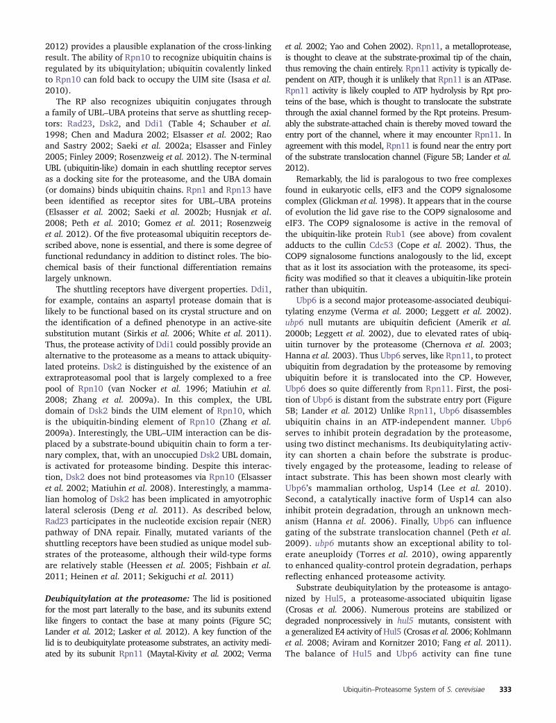
Figure 1 Protein ubiquitylation. (A)Ubiquitin is activated by E1 in an ATP-dependent step, transferred to the activesite cysteine in an ubiquitin-conjugatingenzyme (E2), and covalently attachedto substrate proteins. Substrate selectiondepends on ubiquitin ligases (E3). Conju-gation of a single ubiquitin moleculegenerates monoubiquitylated proteins.Repeated rounds of ubiquitin activationand conjugation lead to multi- or poly-ubiquitylated proteins. (B) Differentpolyubiquitin chain topologies can besynthesized depending on the specificlysine residue in ubiquitin used for chainformation. Three of the eight possibleunbranched chain topologies (K6, K11,K27, K29, K33, K48, K63, and linearchains), and only one type of the possi-ble forked polyubiquitin chains areshown. (C) Structural model for synthe-sis of K63-linked polyubiquitin chains byUbc13/Mms2. Mms2 positions the ac-ceptor ubiquitin with K63 in proximityto the active site cysteine of Ubc13.Figure adapted with permission fromMacmillan Publishers Ltd: Chan, N. L.,and C. P. Hill, 2001 Nat. Struct. Biol. 8:650–652.
Table 1 Ubiquitin conjugating enzymes of Saccharomyces cerevisiae
UBC Viablea Biological processes and/or unique features
Ubc1 + Vesicle biogenesis, ERAD, nuclear protein quality control, E2 for APCUbc2/Rad6 + DNA repair, N-end rule, H2B monoubiquitylationUbc3/Cdc34 2 Cell cycle, E2 for SCF ligasesUbc4 + Protein quality control outside the nucleus, E2 for APCUbc5 + Comparable to Ubc4 but expression is elevated in stationary phaseUbc6 + ERAD, has transmembrane region, can synthesize K11-chains in vivoUbc7 + ERADUbc8 + Regulation of gluconeogenesisUbc9b 2 E2 for Smt3 (SUMO) conjugationUbc10/Pex4 + Peroxisomal E2 important for peroxisome biogenesisUbc11 + Cytoplasmic localizationUbc12b + E2 for Rub1 (Nedd8) conjugationUbc13 + DNA repair, dimerizes with Mms2 for synthesis of K63 chainsa In rich medium at 30�.b E2s for conjugation of ubiquitin-like proteins.
322 D. Finley et al.

Table 2 Ubiquitin ligases and components of Saccharomyces cerevisiae
E3 Viable Biological processes and/or unique featuresa
HECT E3sHul4 + UnknownHul5 + Cytoplasmic PQCb, proteasome-associated protein, potential E4Rsp5 2 Nedd4 family ligase, multiple functions: MVB sorting, endocytosis, transcription,Tom1 + mRNA export, degradation of excess histonesUfd4 + Ubiquitin fusion degradation pathway, N-end rule
Rsp5 adaptorsArt1/Ldb19 + Regulation of endocytosis, localized at plasma membraneArt2/Ecm21 + Regulation of endocytosis, localized at plasma membraneArt3/Aly2 + Control of nutrient-mediated intracellular sorting of GAP1Art4/Rod1 + Regulation of endocytosis, localized at plasma membraneArt5 + Regulation of endocytosis, localized at plasma membraneArt6/Aly1 + Regulation of endocytosisArt7/Rog3 + Regulation of endocytosisArt8/Csr2 + Regulation of endocytosis, regulates use of nonfermentable carbon sourcesArt9/Rim8 + Essential for anaerobic growth, PH responseArt10 + Unknown function, cytoplasmicBsd2 + Facilitates trafficking of metal transporters, localized at Golgi/endosomeBul1 + Post-Golgi endosomal sorting, temperature sensitive, functional homolog of Bul2Bul2 + Post-Golgi endosomal sorting, functional homolog of Bul1Ear1 + Cargo sorting at multivesicular bodies, localized at Golgi/endosomeSsh4 + Cargo sorting at multivesicular bodies, localized at Golgi/endosomeTre1 + Degradation of metal stransporter smf1, function is redundant with that of Tre2Tre2 + Degradation of metal stransporter smf1, function is redundant with that of Tre1
RING E3sa
Asi1 + SPS sensor signaling of amino acids, homologous to Asi3, transmembrane proteinAsi3 + SPS sensor signaling of amino acids, homologous to Asi1, transmembrane proteinAsr1 + RNA Pol II modification, alcohol stress responseBre1 + Histone H2B monoubiquitylation on K123Cwc24 2 Pre-mRNA and snoRNA splicingDma1 + Spindle positioning, orthologs of human Rnf8, redundant with Dma2Dma2 + Spindle positioning, orthologs of human Rnf8, redundant with Dma1Doa10 2 ERAD-C, N-end rule ubiquitylation of acetylated proteinsEtp1 + Required for growth in ethanolFap1 + Response to rapamycinFar1 + G1 cyclin dependent kinase inhibitor, pheromone response, putative E3Hel2 + Degradation of excess histoneHrd1 + ERAD-M, ERAD-LGid9/Fyv10 + Degenerate ring domain, cooperates with RMD5 in ubiquitin ligation (see below)Irc20 + Unknown, localized to nucleus and mitochondria, has helicase domainMag2 + Unknown function, cytoplasmic, homologous to human Rnf10Nam7 + Nonsense mediated mrna degradation, telomere maintenanceNot4 + Subunit of Ccr4–Not complex, ubiquitylates NAC and histone demethylase Jhd2pPep3 + Vacuolar protein sortingPep5 + Vacuolar protein sortingPex2 + Peroxisomal membrane E3, peroxisomal matrix protein importPex10 + Peroxisomal membrane E3Pex12 + Peroxisomal membrane E3, required for peroxisome biogenesisPib1 + Localized in endosomal and vacuolar membranesPsh1 + Cse4 ubiquitylationRad5 + PCNA polyubiquitylation, postreplication repairRad16 + Nucleotide excision repairRad18 + PCNA-K164 monoubiquitylation, postreplication repairRkr1/Lnt1 + Ubiquitylation of proteins translated from nonstop mRNAsRmd5 + Gluconeogenesis, degradation of fructose-1,6-bisphosphataseRtc1 + Unknown functionSan1 + Nuclear PQCSlx5 + SUMO-directed ligase, genotoxic stress response, forms STUbL together with Slx8Slx8 + SUMO-directed ligase, genotoxic stress response, forms STUbL together with Slx5Snt2 + Degradation of excess histoneSsm4 + mRNA stability, localized to ER/nuclear membraneSte5 + Scaffold protein for MAPK cascade proteins
(continued)
Ubiquitin–Proteasome System of S. cerevisiae 323

Table 2, continued
E3 Viable Biological processes and/or unique featuresa
Tfb3 2 Cul3 and Rtt101 neddylation, nucleotide excision repairTul1 + Membrane protein sorting, localized to GolgiUbr1 + N-recognin (N-end rule pathway), PQCUbr2 + Rpn4 ubiquitylation, cytoplasmic PQC; Mub1 assists in recognition of Rpn4Uls1 + Degradation of SUMOylated proteinsUpf1 + RING-related, nonsense-mediated decay of mRNAYBR062C + Unknown function
U-box proteinsPrp19 2 Splicing, U-box proteinUfd2 + Ubiquitin fusion degradation pathway, U-box protein, E4 activity, Cdc48 partner
RBR E3sHel1 + Degradation of excess histone, putative RING-in-between-RING ligaseItt1 + Putative RING-in-between-RING ligase
CRL core componentsCdc53 2 Cullin 1, many functions including cell cycleCul3 + Cullin 3, RNA Pol II ubiquitylationRtt101 + Functional homolog of human cullin 4, DNA repair, rRNA decaySkp1 2 SCF ligase component, many functions including cell cycleElc1 + Elongin C, binds Cul3, RNA Pol II ubiquitylationMms1 + Adaptor for Rtt101Hrt1 2 Rbx1/Roc1, RING component of CRL ligases, many functions including cell cycle
F-box proteinsAmn1 + Mitotic exit networkCdc4 2 Cell cycle, many other functionsCos111 + Unknown function, localizes to mitochondriaCtf13 2 Subunit of centromere binding factor 3Das1 + Similarity to YDR131C, 6-azauracil sensitiveDia2 + Protection from DNA damage and replication stress, part of the RPCEla1 + Elongin A, component of CRL3 ligase, RNA Pol II degradationGrr1 + G1 cyclin degradation, regulates glucose repressionHrt3 + Unknown functionMdm30 + Mitochondrial fusionMet30 2 Cell cycle, heavy metal stress response, sulfur compound homeostasisMfb1 + Mitochondria morphology, mitochondria associatedRav1 + Component of RAVE complex, important for V-ATPase assemblyRcy1 + Recycling of internalized plasma membrane proteinsRoy1 + Intracellular trafficking, inhibits Ypt52 GTPase activitySaf1 + Entry into quiescent phaseSkp2 + Unknown function, homology to human Skp2Ufo1 + HO endonuclease degradationYDR131C + Similarity to Das1YLR224W + Unknown functionYDR306C + Unknown functionYLR352W + Unknown function
Substrate receptors of Cul3 and Rtt101 ligasesCrt10 + Substrate receptor for Rtt101 E3, ribonucleotide reductase gene expressionElc1 + Elongin C, component of CRL3 ligase, RNA Pol II degradationMms22 + Substrate receptor for Rtt101 E3, DNA damage responseRad7 + Nucleotide excision repair, putative substrate receptor of CRL3YDR132C + Unknown function, putative BTB domain proteinYIL001W + Unknown function, putative BTB domain proteinYLR108C + Unknown function, putative BTB domain protein
APC cyclosome core componentsApc1 2 Cell cycle, largest APC/C subunitApc2 2 Cell cycle, cullin homologyCdc27 2 Cell cycleApc4 2 Cell cycleApc5 2 Cell cycleCdc16 2 Cell cycleCdc23 2 Cell cycleApc9 + Cell cycleDoc1/Apc10 + Cell cycle, coreceptor for D-box recognition
(continued)
324 D. Finley et al.

are less clear. Interestingly, preventing K11 chain formationin yeast by K11R-ubiquitin replacement results in hyper-sensitivity to the ER-stress inducers DTT and tunicamycin,indicating that K11 chains are important for the endoplasmic-reticulum–associated degradation (ERAD) pathway (Xu et al.2009).
Methods for the detection and quantification of ubiquitinconjugates have recently been reviewed (Kim et al. 2011;Laney and Hochstrasser 2011).
Ubiquitin-activating enzyme
In yeast, a single E1 enzyme is responsible for activationof ubiquitin. E1 is encoded by the essential UBA1 gene(McGrath et al. 1991). Several temperature-sensitive uba1alleles exist; uba1-206 is a tight mutant, and shows rapiddepletion of ubiquitin conjugates at nonpermissive temper-ature as well as other phenotypes expected from a generalblock of ubiquitylation (Ghaboosi and Deshaies 2007).
Ubiquitin-conjugating enzymes
The first yeast E2 enzymes identified were Rad6/Ubc2(Jentsch et al. 1987) and Cdc34/Ubc3 (Goebl et al.1988). A total of 13 yeast UBC genes have been designated(Table 1), though further biochemical analyses revealedthat Ubc9 and Ubc12 do not conjugate ubiquitin, but ratherthe ubiquitin-like proteins Smt3 (mammalian SUMO) andRub1 (mammalian Nedd8), respectively (Johnson andBlobel 1997; Liakopoulos et al. 1998). Among the 11 gen-uine ubiquitin-conjugating enzymes only Cdc34/Ubc3 isessential for viability (Goebl et al. 1988). Temperature-sensitive cdc34 mutants arrest at the G1-to S-phase tran-sition of the cell cycle due to a defect in degradation of thecyclin-dependent kinase inhibitor Sic1 (Schwob et al.1994). Cdc34 has many other substrates and most areselected by the Skp1–Cdc53–F-box (SCF) ubiquitin ligasefamily for which Cdc34 serves as the main, if not only, E2enzyme (Petroski and Deshaies 2005). In addition, Cdc34together with the ubiquitin ligase San1 functions in thenuclear protein quality control pathway (Gardner et al. 2005a;see below). Ubc1 is an alternative ubiquitin-conjugating factorfor San1 (Gardner et al. 2005a).
Several other E2 enzymes are important for proteinquality control pathways outside the nucleus. Ubc4 andUbc5 are highly similar and function redundantly in conju-gation of ubiquitin to abnormal proteins in the cytosol toinduce their degradation by the proteasome (Seufert andJentsch 1990). The double mutant is inviable in somegenetic backgrounds (Panasenko et al. 2009; Stoll et al.2011); in others it shows severe growth defects (Seufertand Jentsch 1990; Chen et al. 1993). Three E2 enzymesare involved in degradation of misfolded proteins from theendoplasmatic reticulum (ERAD pathway; see below):Ubc1, Ubc6, and Ubc7. Among these, only Ubc6 is directlyanchored to the ER membrane by a C-terminal transmem-brane region (Sommer and Jentsch 1993), whereas Ubc7 isrecruited to the ER membrane and activated by ER-boundCue1 (Biederer et al. 1997; Bazirgan and Hampton 2008).Ubc6 but not Ubc7 contributes significantly to total cellularprotein modification with K11-linked polyubiquitin chains(Xu et al. 2009).
Multiple E2 enzymes can be involved in degradation ofa single substrate, the MATa2 transcriptional regulatorbeing a complex case in which four different UBCs havebeen implicated (Ubc4, Ubc5, Ubc6, and Ubc7) (Chenet al. 1993). However, many substrates may rely on a singleE2. Ubiquitin-conjugating enzymes can also operate sequen-tially for efficient substrate polyubiquitylation, as demon-strated for Ubc1 and Ubc4 in polyubiquitylation of cellcycle regulators targeted by a ubiquitin ligase known asthe anaphase promoting complex, or cyclosome (APC/C;Rodrigo-Brenni and Morgan 2007). Polyubiquitylation re-quires two distinct types of conjugation events: Attachmentof the first ubiquitin to the substrate protein in an initialmonoubiquitylation step, followed by cycles of ubiquitinchain elongation. In yeast, the rate-limiting monoubiquity-lation step for APC/C substrates is catalyzed by Ubc4,whereas efficient ubiquitin chain synthesis requires Ubc1(Rodrigo-Brenni and Morgan 2007). A C-terminal ubiquitinassociated (UBA) domain that binds ubiquitin—a feature ofUbc1 not shared with any other yeast E2 (Merkley and Shaw2004)—is required for optimal processivity of this reaction(Rodrigo-Brenni and Morgan 2007).
Table 2, continued
E3 Viable Biological processes and/or unique featuresa
Apc11 2 Cell cycle, RING-finger subunit of APC/CCdc26 + Cell cycleSwm1 + Cell cycleMnd2 + Meiosis
APC cyclosome substrate receptorsAma1 + APC/C activator for meiosisCdc20 2 APC/C activator, degradation of Pds1 and other mitotic regulatorsCdh1 + APC/C activator, degradation of mitotic cyclins
a Note that biochemical evidence for ubiquitin ligase activity has so far not been reported for many of these proteins. They are listed here because they contain RING (like)motifs, homology to F-box motifs, or other sequence features frequently associated with ubiquitin ligases. Several proteins that are possible E3s have been excluded fromthis list: Air1, Air2, Nse1, and Yvh1.
b Protein quality control.
Ubiquitin–Proteasome System of S. cerevisiae 325

Some E2s are poised for synthesis of polyubiquitin chains.For example, heterodimeric E2s such as the yeast Ubc13/Mms2 complex synthesize polyubiquitin chains by transfer-ring the thioester-bound donor ubiquitin from the catalyti-cally active subunit (Ubc13) onto an acceptor ubiquitin thatis noncovalently bound to a catalytically inactive UEV (ubiq-uitin E2 variant) binding partner (Mms2) (Hofmann andPickart 1999; Eddins et al. 2006). Other E2s may transferpreassembled polyubiquitin chains onto substrates, as de-scribed for the mammalian Ube2g2 enzyme and its yeastortholog Ubc7 (Li et al. 2007b; Ravid and Hochstrasser2007). However, the same E2 can often catalyze both mono-and polyubiquitylation. For example, Rad6/Ubc2 catalyzesmonoubiquitylation of the proliferating cell nuclear antigenPCNA (Hoege et al. 2002) and histone H2B (Robzyk et al.2000), but forms polyubiquitin chains in the context of theN-end rule (Dohmen et al. 1991), a conserved pathway thatrelates protein stability to the identity of the amino terminalresidue (Varshavsky 1992; Varshavsky 2011; Tasaki et al.2012). Rad6/Ubc2 functions with different ubiquitin ligasesin these pathways, and it appears that ligases and E2enzymes, as well as the substrates themselves, can be deter-minants deciding between mono- or polyubiquitylation.
Ubiquitin-conjugating enzymes also help to define thelinkage type during polyubiquitin chain synthesis. In particular,E2s dictate chain architecture when paired with really inter-esting new gene (RING) domain ubiquitin ligases, whereasHECT (homologous to E6-AP carboxy terminus) domain E3soverride any intrinsic chain topology preference of E2s. Syn-thesis of polyubiquitin chains with specific architectures byRING–E3/E2 pairs requires positioning of the E2 such thatthe linkage-defining lysine residue in the acceptor ubiquitinis proximal to the charged active site cysteine of the E2. Thebest-studied example in yeast is Ubc13, which synthesizes K63-linked chains (Figure 1C). In the Ubc13/Mms2 heterodimer,Mms2 positions the acceptor ubiquitin so that only K63 isallowed to approach the active site cysteine of Ubc13 (Eddinset al. 2006). A related mechanism was demonstrated for themammalian ubiquitin-conjugating enzyme Ube2S, wherea ubiquitin-binding region in the E2 orients the acceptor ubiq-uitin for K11-selective chain synthesis (Wickliffe et al. 2011).
The ubiquitin-conjugating enzymes are at the center of theE1–E2–E3 cascade. They interact with E1 and E3 but alsoensure unidirectional handoff of ubiquitin from E1 to the sub-strate. E1 and E3 use a shared binding site on E2s, preventingrecharging of E2s while bound to E3s and forcing their dis-sociation before the next round of conjugation (Eletr et al.2005). Directionality of ubiquitin transfer is ensured by E1-dependent ATP hydrolysis as well as the different affinities ofcharged and uncharged E2s for E1 and E3. The ubiquitin-activating enzyme E1 binds uncharged E2s with higher affin-ity than the E2�Ub leading to release of the loaded E2�Ub(Hershko et al. 1983; Pickart and Rose 1985). Similarly, E3shave somewhat higher affinity for E2�Ub than for the un-charged E2, facilitating processive ubiquitin chain synthesis(Siepmann et al. 2003; Saha and Deshaies 2008).
Ubiquitin ligases
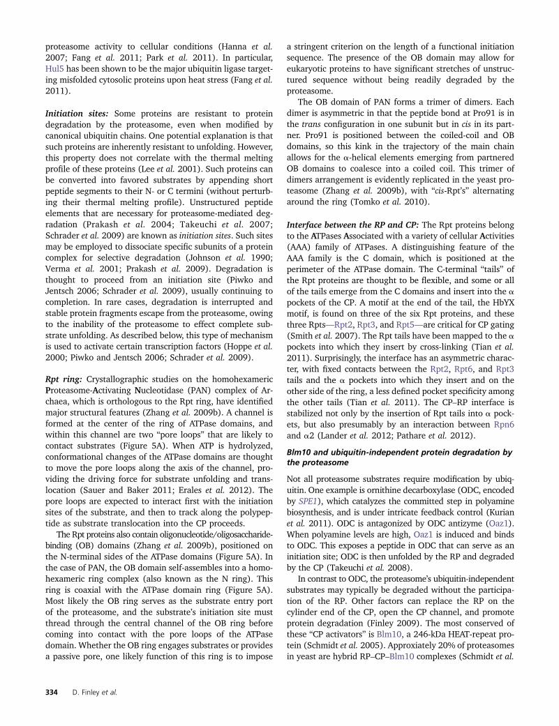
Ubiquitin ligases (E3s) form the largest group of proteinsinvolved in ubiquitylation and they confer selectivity to theprocess. They bind E2s and substrate proteins to facilitatesubstrate-specific ubiquitylation. The first E3 identified wasUbr1, a mediator of the N-end rule pathway. Ubr1 bindsprotein substrates with different affinities based on theirN-terminal amino acids (Bartel et al. 1990; Varshavsky1992). Many other E3 enzymes were subsequently identi-fied, all falling into two major classes: RING domain E3s(including the structurally related U-box domain E3s) andHECT domain E3s. Considering sequence features fre-quently associated with ubiquitin ligases, such as RING(like), F box, or HECT motifs, there are 60–100 putativeE3s in yeast (Table 2). Most belong to the class of RINGdomain E3s, and only five HECT domain E3s are encodedin the yeast genome (Table 2). RING and HECT domain E3sfollow distinct mechanisms to catalyze ubiquitylation (Fig-ure 2). HECT domain E3s contain an active site cysteinewithin the HECT domain, which forms a thioester with ubiq-uitin received from an E2 prior to its transfer to the substrate(Scheffner et al. 1995). RING E3s do not form thioesterintermediates; they instead facilitate ubiquitin transfer bypositioning the charged E2�Ub in proximity to the acceptorlysine in the substrate. In addition, RING domain ligasesseem to activate E2s to facilitate ubiquitylation (Deshaiesand Joazeiro 2009).
A subclass of RING domain E3s, the RING-in-between-RING (RBR) proteins, appear to function as RING/HECT
Figure 2 HECT and RING E3 ubiquitin ligases. Substrate ubiquitylationwith HECT E3s involves an E3�Ub thioester intermediate. Ubiquitin istransferred from the HECT E3 to the substrate. RING E3s typically donot form thioester intermediates but promote ubiquitin conjugation bybridging the interaction between E2 and substrate proteins. RING E3s alsostimulate E2 activity. A subclass of RING-based ligases, the RING-in-between-RING (RBR) proteins, function like RING/HECT hybrids and form thioesterintermediates. This mechanism remains to be confirmed for putative yeastRBR ligases.
326 D. Finley et al.

hybrids (Wenzel and Klevit 2012). They bind E2s with oneRING domain and stimulate the transfer of ubiquitin ontoa conserved cysteine residue in the other RING domain, form-ing an E3�Ub thioester before conjugation to the substrate(Wenzel et al. 2011). Homology searches revealed two puta-tive RBR ligases in yeast (Hel1 and Itt1) (Eisenhaber et al.2007). Whether they indeed form E3�Ub intermediates isunknown.
Functional interaction between RING and HECT domainE3s has been demonstrated for the N-end rule pathway(Hwang et al. 2010). The RING-type Ubr1 and HECT-typeUfd4 ligases form a complex to enhance processivity of sub-strate ubiquitylation. A similar role for the Ubr1/Ufd4 com-plex in the ubiquitin-fusion degradation pathway has alsobeen suggested (Hwang et al. 2010). Interestingly, theUbr1/Ufd4 complex may function as an E3/E4 pair. E4enzymes—a small subgroup of ubiquitin ligases—select sub-strate proteins based on their having been previously ubiq-uitylated, and E4s function to extend these ubiquitin chains(Koegl et al. 1999).
HECT ubiquitin ligases: HECT domain E3s are named aftertheir founding member E6AP, which ubiquitylates mamma-lian p53 in cells expressing the human papilloma virusprotein E6. Yeast has five HECT E3s: Rsp5, Ufd4, Hul4,Hul5, and Tom1. The HECT domain is an �350-residueregion consisting of the N-terminal lobe, which binds anE2, and the C-terminal lobe containing the active site cyste-ine, which forms a thioester intermediate with ubiquitin. Nand C lobes are connected by a flexible hinge region (Huanget al. 1999). The five yeast HECT domain ubiquitin ligasesfunction in diverse processes ranging from multivesicularbody (MVB) sorting, endocytosis, histone degradation, andprocessing of ubiquitylated proteins (Hoppe et al. 2000;Shcherbik et al. 2003; Rape and Jentsch 2004; Crosaset al. 2006; Rotin and Kumar 2009; Singh et al. 2009).
The E3�Ub thioester intermediate mediates E3-instructedubiquitin chain assembly as demonstrated for Rsp5, whichhas been shown to dictate synthesis of K63-linked chains in-dependently of the E2 enzymes used (Kim and Huibregtse2009). Although the molecular mechanism is not known indetail, mutational studies suggest that the carboxy-terminalregion of Rsp5 is involved in acceptor ubiquitin orientationto favor nucleophilic attack from lysine-63 in ubiquitin.
Rsp5 is the only yeast HECT E3 essential for viability inrich medium. Rsp5 is a particularly active E3 that mediatesubiquitylation of a large number of substrates and contrib-utes to regulation of diverse biological pathways (Guptaet al. 2007; Rotin and Kumar 2009). Rsp5 is required forupregulation of expression of the fatty acid desaturase OLE1by the homologous transcription factors Spt23 and Mga2,and accordingly the lethality of rsp5 mutants can be rescuedby addition of oleic acid to the growth medium (Hoppe et al.2000). While Spt23 and Mga2 are normally anchored inthe ER membrane, Rsp5-mediated ubiquitylation inducesproteasomal processing and release of transcriptional acti-
vation domains from these proteins (Hoppe et al. 2000;Shcherbik et al. 2003). Hul5, another HECT domain protein,is discussed below in the Proteasome section.
RING domain ubiquitin ligases: There are 44 yeast proteinscontaining RING domains and two proteins of the U-boxfamily, which are structurally related to RING E3s but do notbind zinc (Ufd2 and Prp19). Although conclusive biochem-ical evidence for ubiquitin ligase activity is not availablefor all RING domain proteins, most of them probably havethis activity. The globular RING domains bind E2 enzymes(Zheng et al. 2000) and appear to stimulate ubiquitin trans-fer by induction of subtle structural changes (Ozkan et al.2005). Substrate recruitment, the central function of ubiq-uitin ligases, is achieved either by substrate binding domainswithin the same polypeptide chain as the RING domain (sin-gle subunit RING E3s) or by engaging specialized substratereceptors to form multisubunit RING E3s (Deshaies andJoazeiro 2009). Examples of the former are the N-recogninUbr1 (Bartel et al. 1990); the ubiquitin ligase Bre1 thattogether with Rad6/Ubc2 catalyzes histone H2B ubiquityla-tion (Wood et al. 2003); the regulator of nuclear proteinquality control San1 (Gardner et al. 2005a); Rkr1/Ltn1,which ensures degradation of potentially cytotoxic transla-tion products produced from mRNAs that lack stop codons(Bengtson and Joazeiro 2010); and the two RING E3s,Rad18 and Rad5, which catalyze mono- and polyubiquityla-tion of PCNA, respectively (Hoege et al. 2002; see below).Prominent members of the multisubunit RING E3s are theAPC/C (Pesin and Orr-Weaver 2008) and the largest groupof ligases, the modular cullin-RING ligases (CRLs) (Petroskiand Deshaies 2005; Zimmerman et al. 2010; Duda et al.2011). Although one subunit (Apc2) of APC/C containsa cullin-like domain, the overall ligase architecture is verydifferent from that of true CRLs. Detailed studies of otherRING domain proteins (Table 2) may identify additionalmultisubunit E3s as has been shown for the seven-subunitGid (glucose-induced degradation-deficient) E3, which con-trols the metabolic switch between glycolysis and gluconeo-genesis (Santt et al. 2008; Menssen et al. 2012).
APC/C: APC/C is perhaps the most complex ubiquitin ligase.Its core is composed of 13 subunits (Apc1, Apc2, Cdc27,Apc4, Apc5, Cdc16, Cdc23, Apc9, Doc1, Apc11, Cdc26,Swm1, and Mnd2), with Apc11 being the RING domaincomponent that binds Ubc1 and Ubc4, the two primaryE2s functioning with yeast APC/C (McLean et al. 2011).The core APC/C associates with one of three activators thatbind substrates and are crucial targets for APC/C regulation.Cdh1 and Cdc20 are activators controlling mitotic cell cycleprogression and Ama1 recruits meiotic targets to APC/C(Visintin et al. 1997; Cooper et al. 2000).
Degradation of several important APC/C substratesensures ordered progression through the steps of chromo-some segregation. A cascade of mitotic events is unleashedby APC/C-mediated degradation of Pds1/securin to initiate
Ubiquitin–Proteasome System of S. cerevisiae 327
the metaphase-to-anaphase transition (Cohen-Fix et al.1996; Yamamoto et al. 1996). Pds1 is an inhibitor ofEsp1/separase, a protease that cleaves the cohesin Scc1 toallow sister chromatid separation (Ciosk et al. 1998;Uhlmann et al. 1999). Clb2 and other B-type cyclins aredegraded by APC/C from anaphase until the end of the sub-sequent G1 phase, which ensures a period of low cyclin-dependent kinase activity that is important for cytokinesisand the assembly of prereplication complexes (Irniger et al.1995). Many other mitotic and meiotic regulators are APC/Csubstrates, and their degradation controls both normal mi-totic processes and cell cycle checkpoint pathways (Pesinand Orr-Weaver 2008; McLean et al. 2011).
Tight regulation of Cdh1 and Cdc20 restricts APC/Cactivity to M phase and G1 of the mitotic cell cycle (Pesinand Orr-Weaver 2008). G2/M-phase–induced CDC20 ex-pression, APC/C phosphorylation-dependent binding ofCdc20 (Rudner and Murray 2000; Rudner et al. 2000),combined with active Cdc20 degradation during G1 byAPC/CCdh1, limit Cdc20 association with APC/C to M phase(Prinz et al. 1998; Foe et al. 2011). In contrast, Cdh1 levelsare largely constant throughout the cell cycle, but bindingto APC/C is prevented by Cdh1 phosphorylation during mostof the cell cycle, except late M phase and G1 (Zachariae et al.1998).
APC/C substrates share distinct degradation motifs, themost common being the classic destruction box (D box) andthe KEN box (Glotzer et al. 1991; Pfleger and Kirschner2000). Although APC/C activators play a crucial role inD-box and KEN-box recognition, the core subunit Apc10/Doc1 serves as a coreceptor in D-box recognition (Carrollet al. 2005; Da Fonseca et al. 2011). Regulation occurs atthe level of activator abundance, phosphorylation of activa-tors and core components, as well as binding of the APC/CCdc20 inhibitors Mad2 and Mad3 (McLean et al. 2011).
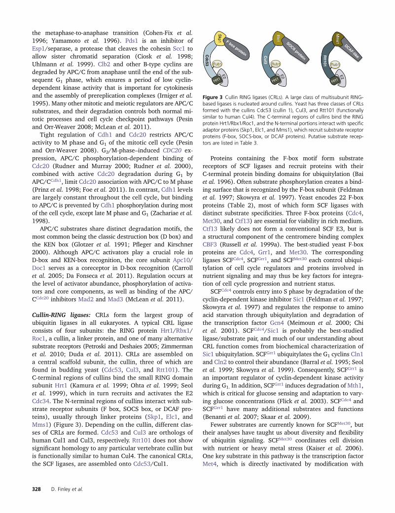
Cullin-RING ligases: CRLs form the largest group ofubiquitin ligases in all eukaryotes. A typical CRL ligaseconsists of four subunits: the RING protein Hrt1/Rbx1/Roc1, a cullin, a linker protein, and one of many alternativesubstrate receptors (Petroski and Deshaies 2005; Zimmermanet al. 2010; Duda et al. 2011). CRLs are assembled ona central scaffold subunit, the cullin, three of which arefound in budding yeast (Cdc53, Cul3, and Rtt101). TheC-terminal regions of cullins bind the small RING domainsubunit Hrt1 (Kamura et al. 1999; Ohta et al. 1999; Seolet al. 1999), which in turn recruits and activates the E2Cdc34. The N-terminal regions of cullins interact with sub-strate receptor subunits (F box, SOCS box, or DCAF pro-teins), usually through linker proteins (Skp1, Elc1, andMms1) (Figure 3). Depending on the cullin, different clas-ses of CRLs are formed. Cdc53 and Cul3 are orthologs ofhuman Cul1 and Cul3, respectively. Rtt101 does not showsignificant homology to any particular vertebrate cullin butis functionally similar to human Cul4. The canonical CRLs,the SCF ligases, are assembled onto Cdc53/Cul1.
Proteins containing the F-box motif form substratereceptors of SCF ligases and recruit proteins with theirC-terminal protein binding domains for ubiquitylation (Baiet al. 1996). Often substrate phosphorylation creates a bind-ing surface that is recognized by the F-box subunit (Feldmanet al. 1997; Skowyra et al. 1997). Yeast encodes 22 F-boxproteins (Table 2), most of which form SCF ligases withdistinct substrate specificities. Three F-box proteins (Cdc4,Met30, and Ctf13) are essential for viability in rich medium.Ctf13 likely does not form a conventional SCF E3, but isa structural component of the centromere binding complexCBF3 (Russell et al. 1999a). The best-studied yeast F-boxproteins are Cdc4, Grr1, and Met30. The correspondingligases SCFCdc4, SCFGrr1, and SCFMet30 each control ubiqui-tylation of cell cycle regulators and proteins involved innutrient signaling and may thus be key factors for integra-tion of cell cycle progression and nutrient status.
SCFCdc4 controls entry into S phase by degradation of thecyclin-dependent kinase inhibitor Sic1 (Feldman et al. 1997;Skowyra et al. 1997) and regulates the response to aminoacid starvation through ubiquitylation and degradation ofthe transcription factor Gcn4 (Meimoun et al. 2000; Chiet al. 2001). SCFCdc4/Sic1 is probably the best-studiedligase/substrate pair, and much of our understanding aboutCRL function comes from biochemical characterization ofSic1 ubiquitylation. SCFGrr1 ubiquitylates the G1 cyclins Cln1and Cln2 to control their abundance (Barral et al. 1995; Seolet al. 1999; Skowyra et al. 1999). Consequently, SCFGrr1 isan important regulator of cyclin-dependent kinase activityduring G1. In addition, SCFGrr1 induces degradation of Mth1,which is critical for glucose sensing and adaptation to vary-ing glucose concentrations (Flick et al. 2003). SCFCdc4 andSCFGrr1 have many additional substrates and functions(Benanti et al. 2007; Skaar et al. 2009).
Fewer substrates are currently known for SCFMet30, buttheir analyses have taught us about diversity and flexibilityof ubiquitin signaling. SCFMet30 coordinates cell divisionwith nutrient or heavy metal stress (Kaiser et al. 2006).One key substrate in this pathway is the transcription factorMet4, which is directly inactivated by modification with
Figure 3 Cullin RING ligases (CRLs). A large class of multisubunit RING-based ligases is nucleated around cullins. Yeast has three classes of CRLsformed with the cullins Cdc53 (cullin 1), Cul3, and Rtt101 (functionallysimilar to human Cul4). The C-terminal regions of cullins bind the RINGprotein Hrt1/Rbx1/Roc1, and the N-terminal portions interact with specificadaptor proteins (Skp1, Elc1, and Mms1), which recruit substrate receptorproteins (F-box, SOCS-box, or DCAF proteins). Putative substrate recep-tors are listed in Table 3.
328 D. Finley et al.

a K48-linked ubiquitin chain, but degradation is preventedbecause two ubiquitin binding motifs in Met4 shield thepolyubiquitin chain from signaling degradation (Flick et al.2006; Tyrrell et al. 2010). Although ubiquitylated Met4 isinactive as a transcription factor, it functions as a substratereceptor in the context of the extended SCFMet30/Met4 ubiq-uitin ligase to trigger ubiquitylation and degradation of sev-eral Met4 binding factors, including Met32, which inducescell cycle arrest when stabilized (Ouni et al. 2010). The dualfunction of Met4 as transcription factor and ubiquitin ligasecomponent allows it to coordinate cell cycle progressionwith response to nutrient or heavy metal stress.
An interesting aspect of CRL regulation is a ubiquitin-likemodification found on cullins. Cullins are covalently mod-ified on a conserved lysine residue in the C-terminal regionby the ubiquitin-like protein Rub1, the yeast ortholog ofmetazoan Nedd8 (Lammer et al. 1998; Liakopoulos et al.1998). Cullin modification with Nedd8 induces a major con-formational change such that the E2-binding interface of theRING component Hrt1 extends out from the cullin surface,remaining tethered only by a flexible linker region. This notonly allows the E2 to closely approach the substrate, but alsoprovides the flexibility to adopt different conformations nec-essary for polyubiquitin chain synthesis (Duda et al. 2008).Rub1 modification is not essential for viability of buddingyeast, but it is required for robust CRL activity and is essen-tial in other organisms (Willems et al. 2004).
Deubiquitylation
Deubiquitylating enzymes catalyze the hydrolysis of theisopeptide bonds that link ubiquitin to its targets (Reyes-Turcu et al. 2009). Twenty deubiquitylating enzymes(DUBs) are found in yeast (Table 3), falling into four fam-
ilies: the Usp family, including 16 members; the Otu family,with two members; and the JAMM and Uch families, withone member each. Additional paralogs exist, with specificityfor ubiquitin-like proteins such as Smt3 and Rub1. Yuh1, thelone Uch-type DUB in yeast, may serve primarily in theremoval of Rub1 from target proteins, although capable ofdeubiquitylation as well (Linghu et al. 2002). Most DUBs arethiol proteases, the only exception being Rpn11, a zinc met-alloprotease (Verma et al. 2002; Yao and Cohen 2002). Thethree-dimensional structures of several DUBs from yeast andother organisms are available (Johnston et al. 1999; Huet al. 2005; Li et al. 2007a; Sato et al. 2008; Reyes-Turcuet al. 2009; Köhler et al. 2010).
The DUBs are highly diverse functionally, reflecting boththeir subcellular localization and their inherent substratespecificities. For example, Ubp8 is a component of the SAGAcomplex, a nuclear particle involved in chromatin remodeling(Henry et al. 2003). Ubp10 is also a specific regulator of nu-clear processes such as the silencing of gene expression (seebelow). Other DUBs seem to function specifically on endo-somes and multivesicular bodies, such as Doa4/Ubp4 (Luhtalaand Odorizzi 2004; Amerik et al. 2006). One DUB, Ubp16, isthought to be an integral membrane protein and fractionateswith mitochondria (Kinner and Kölling 2003). The enzymaticspecificity of DUBs from yeast is only partially characterized(Amerik et al. 2000b; Schaefer and Morgan 2011). DUBs arepresented with potential substrates that must number in thehundreds and possibly thousands, given the breadth of theubiquitin pathway (Kim et al. 2011). Systematic identificationof DUB substrates in yeast has not been attempted, and it iseven unclear in general how rapidly ubiquitin modifications ofprotein substrates are reversed within cells.
DUB activity is required not only for the disassemblyof ubiquitin–protein conjugates but also for biosynthetic
Table 3 Deubiquitylating enzymes of Saccharomyces cerevisiae
DUB Type Localization/complex Phenotypea
Ubp1 USP Cytoplasmic, ER MildUbp2 USP Ubp2/Rsp5/Rup1 PleiotropicUbp3 USP Ubp3/Bre5 PleiotropicDoa4/Ubp4 USP Endosomal, Doa4/Bro1 Ub deficient, partial ts, cans
Ubp5 USP Bud neck Assorted mild phenotpyesUbp6 USP Proteasomal Ub deficient; enhanced proteolysis, cans
Ubp7 USP Cytoplasmic Increased prion formationUbp8 USP Nuclear; SAGA Sensitive to heat and g-rays; partial tsUbp9 USP Cytoplasmic MildUbp10 USP Nuclear Decreased silencing, partial cs, cans
Ubp11 USP Pleiotropically stress sensitive, cans
Ubp12 USP cans
Ubp13 USP Pleiotropically stress sensitiveUbp14 USP Elevated free ubiquitin chains, cans
Ubp15 USP Stress sensitive, partial ts, strong cs, cans
Ubp16 USP Mitochondrial Cans, slow growth on nonfermentable carbonRpn11 JAMM Proteasomal Essential (DUB activity not essential)Otu1 OTU Cdc48 Pleiotropically stress sensitiveOtu2 OTU Ribosome associated (?) Pleiotropically stress sensitiveYuh1 UCH Cytoplasmic Acts preferentially on Rub1 (vs. ubiquitin)a cans, sensitive to amino acid analog canavanine; cs, cold-sensitive; ts, temperature-sensitive.
Ubiquitin–Proteasome System of S. cerevisiae 329

processing of the Ubi1–Ubi4 gene products, ubiquitin fusionproteins that are the sole source of ubiquitin in the cell.UBI1–UBI3, which supply most of the ubiquitin in growing,unstressed cultures, encode ubiquitin as N-terminal fusionsto ribosomal proteins L40 and S31 (Finley et al. 1989).UBI4, the stress-responsive ubiquitin gene (Finley et al.1987), has a series of six tandem repeats of the ubiquitincoding sequence (Ozkaynak et al. 1984). DUB activity isessential to release ubiquitin from these precursor forms,as their C termini are blocked. It is not known which DUBsare responsible for these cleavage events, but they exhibitfast reaction kinetics, as observed for artificial ubiquitin–b-galactosidase fusion proteins (Bachmair et al. 1986).
An important function of the DUBs is to recycle ubiquitinby recovering it from ubiquitin–protein conjugates beforethe target protein is degraded. Defects in this process giverise to reduced ubiquitin levels and pleiotropic stress sensi-tivities. The main DUBs responsible for recovering ubiquitinfrom conjugates that are en route to being degraded areUbp6, Rpn11, and Doa4 (Swaminathan et al. 1999; Ameriket al. 2000b; Leggett et al. 2002; Hanna et al. 2003, 2007;Chernova et al. 2003; Kimura et al. 2009). Ubp6 and Rpn11rescue ubiquitin from degradation by the proteasome, andDoa4 releases ubiquitin from membrane proteins that areabout to be internalized within multivesicular bodies enroute to the lysosome. Both Ubp6 and Rpn11 can releaseubiquitin from proteasome substrates in the form of unan-chored chains (Verma et al. 2002; Yao and Cohen 2002;Hanna et al. 2006). If not promptly disassembled, such chainscan inhibit the proteasome by competing with ubiquitin–protein conjugates for access to proteasomal ubiquitinreceptors. Ubp14 is dedicated to breaking down such un-anchored chains (Amerik et al. 1997). Its specificity isachieved by recognition of the free C terminus of the prox-imal ubiquitin of the chain, leading to allosteric activationand cleavage of the isopeptide bond joining the proximalubiquitin to the penultimate member of the chain (Reyes-Turcu et al. 2009). Doa4 can also disassemble free chainsand plays a major role in this process upon heat shock(Kimura et al. 2009).
DUBs often function within protein complexes, and insuch cases are typically activated by incorporation into thecomplex. For example, Ubp6 and Rpn11 are thought to beactive only when associated with the proteasome (Leggettet al. 2002; Verma et al. 2002), Ubp3 is activated by Bre5(Cohen et al. 2003), and Otu1 functions in association withCdc48 (Rumpf and Jentsch 2006). A particularly elegantexample is the activation of Ubp8 as a DUB when it isincorporated into the SAGA complex (Köhler et al. 2010).Additional modes of DUB regulation are exemplified by thetranscriptional induction of the UBP6 gene in response toreduced ubiquitin levels (Hanna et al. 2007); the inhibitionof Doa4 by Rfu1, which is relieved upon heat shock (Kimuraet al. 2009); and stimulation of Ubp3 activity by Hog1 kinase-dependent phosphorylation upon osmotic stress (Solé et al.2011).
Some DUBs antagonize specific ubiquitin ligases. Ubp2forms a complex with the ligase Rsp5, and deubiquitylatesthose proteins that Rsp5 modifies (Kee et al. 2005, 2006;Harreman et al. 2009). Other cases of DUB–ligase antago-nism involve E4 enzymes. Thus, the E4 Ufd2 is antagonizedby Otu1, with both residing on Cdc48 (Rumpf and Jentsch2006), and the E4 Hul5 is antagonized by Ubp6, with bothresiding on the proteasome (Crosas et al. 2006). It would beinteresting to understand why DUB–ligase pairs have evolvedin these cases, since most ligases do not seem to be pittedagainst a specific DUB in this way.
Because of the abundance of DUBs in yeast, it isnecessary to take precautions against postlysis deubiquity-lation when assessing the role of ubiquitylation in anysetting. DUBs that are thiol proteases are inactivated by thealkylating agent N-ethylmaleimide, but a zinc chelatingagent such as o-phenanthroline is recommended in addi-tion to neutralize the metalloprotease Rpn11 (Verma et al.2002).
Proteasome
The proteasome has 33 distinct subunits (Table 4) and is themost complex protease known (Finley 2009). Its principalfunction is to degrade ubiquitin–protein conjugates. Theproteasome is found in all eukaryotes and is highly con-served in evolution. Proteasomes are organized into twosubassemblies, the 19S regulatory particle (RP) and the20S core particle (CP). The RP recognizes substrates to bedegraded, while the CP contains the proteolytic active sites.The proteolytic sites are sequestered within an interior spaceof the CP, ensuring that access to these sites is under strictcontrol and nonspecific proteolysis is minimized (Figure 4).Substrates are routed from the RP to the CP through a nar-row substrate translocation channel, which can exist in openand closed states (Figure 4). Globular proteins must beunfolded to traverse this channel. Unfolding is an activeprocess mediated by the six distinct ATPases of the RP,Rpt1–Rpt6, which form a heteromeric ring complex (Figure5A). Simple methods are available for testing whether anunstable protein is degraded in a proteasome-dependentmanner (Fleming et al. 2002; Liu et al. 2007).
Core particle
The CP is a barrel-like structure composed of four stackedrings of subunits (Groll et al. 1997). The two outer rings areknown as a rings, the two inner rings as b rings (Figure 4).CP components are generally referred to as a1–a7 and b1–b7 (Table 4). The proteolytic activity of the proteasomeresides in the b ring; subunits b1, b2, and b5 are proteolyt-ically active and are founding members of the threonineclass of proteases. In each case, the active site nucleophileis the N-terminal a-amino group of the main chain. b1, b2,and b5 are synthesized as proenzymes and cleaved upon CPassembly to reveal a threonine residue at the new N termi-nus (Chen and Hochstrasser 1996; Arendt and Hochstrasser1997; Groll et al. 1997). The specificities of the b1, b2, and
330 D. Finley et al.

b5 active sites are trypsin-like, caspase-like, and chymotrypsin-like, in that they prefer basic, acidic, or hydrophobic residues,respectively, N-terminal to the scissile bond (Groll et al. 2005).
The a rings regulate substrate access into the inner cham-ber of the CP (Groll et al. 2000; Whitby et al. 2000; Bajoreket al. 2003). In the free form of the CP, the center of the a
ring is occupied by N termini from all seven subunits, whichconverge into a defined but irregular structure that blockssubstrate access to the chamber. Another important functionof the a ring is to serve as a docking site for the RP and otherregulators of the CP, such as Blm10. Both the RP and Blm10activate the peptidase of the CP by shifting the a N termini
away from the center of the a ring, and thus creating anopening for the passage of substrate (Finley 2009; Sadre-Bazzaz et al. 2010). The interfaces of the a subunits formseven pockets, which provide docking sites for the RP andBlm10 (Sadre-Bazzaz et al. 2010; Tian et al. 2011). The Ctermini of the Rpt proteins project into these pockets tostabilize the association between the RP and CP and driveopening of the CP channel (see below).
Regulatory Particle
Subunit organization of the regulatory particle: Thespatial organization of the RP has been resolved in recent
Table 4 Proteasome components and cofactors
Subcomplex or gene Alias Domains Notes
CPScl1 a1Pre8 a2Pre9 a3 NonessentialPre6 a4Pup2 a5Pre5 a6Pre10 a7Pre3 b1 Propeptide Proteolytically activePup1 b2 Propeptide Proteolytically activePup3 b3Pre1 b4Pre2 b5 Propeptide Proteolytically activePrs3 b6 PropeptidePre4 b7 Propeptide
RP baseRpt1 AAA, OB, CC ATPaseRpt2 AAA, OB, CC, HbYX ATPaseRpt3 AAA, OB, CC, HbYX ATPaseRpt4 AAA, OB, CC ATPaseRpt5 AAA, OB, CC, HbYX ATPaseRpt6 AAA, OB, CC ATPaseRpn1 TPR-like repeats Apparent scaffoldRpn2 TPR-like repeats Apparent scaffoldRpn13 PRU domain Ub receptor, nonessentialRpn10 VWA, UIM Ub receptor, nonessential
RP lidRpn3 PCIRpn5 PCIRpn6 PCIRpn7 PCIRpn8 MPNRpn9 PCI NonessentialRpn11 MPN DUB activityRpn12 PCISem1 Nonessential
Associated proteinsUbp6 UBL and USP DUB activityHul5 HECT Ub ligase activityUfd4 HECT Ub ligase activityUbc4 E2 E2 enzymeEcm29 HEAT Possible chaperoneBlm10 HEAT Opens CP gateRad23 UBL and UBA Ub receptorDsk2 UBL and UBA Ub receptorDdi1 UBL and UBA Ub receptor
Ubiquitin–Proteasome System of S. cerevisiae 331

electron microscopy studies (Lander et al. 2012; Lasker et al.2012; Pathare et al. 2012; Sakata et al. 2012), as summa-rized in Figure 5. The RP is composed of the 10-subunit baseand nine-subunit lid subassemblies (Table 4; Glickman et al.1998; Finley 2009). The RP is anchored to the CP principallythrough the base, but the lid subunit Rpn6 also contactsthe CP (Lander et al. 2012; Pathare et al. 2012). Dissoci-ation of the RP into base and lid is observed upon purifi-cation of proteasomes from rpn10D mutants, or uponpurification of wild-type proteasomes in the presence ofhigh salt (Glickman et al. 1998; Saeki et al. 2000). More-over, the base and lid are intermediates in RP assembly(see below). Thus, the base–lid dichotomy reflects the fun-damental organization of the RP.
Unfolding of the protein substrate and its translocationinto the CP are driven by ATP hydrolysis (Schrader et al.2009; Sauer and Baker 2011; Smith et al. 2011a). The het-erohexameric Rpt ring of the base represents the ancientcore of the machinery that defines ATP-dependent proteasesin all kingdoms of life (Figure 5A). The 13 additional com-ponents of the RP are peculiar to eukaryotes and seemdesigned in large part to recognize or process the ubiquitincomponent of the ubiquitin–protein conjugate, as discussedbelow. For example, two components of the base are ubiq-
uitin receptors, and other components of the base, Rpn1 andRpn2, are large subunits that serve as scaffolds (Figure 5C),allowing for the recruitment of a variety of factors, such asshuttling receptors (see below) with their cargo of ubiquitin–protein conjugates.
Substrate recognition: Two subunits of the RP, Rpn10, andRpn13, bind ubiquitin chains. Rpn10 binds via its a-helicalUbiquitin-Interacting Motif (UIM) element (Elsasser et al.2004; Verma et al. 2004; Mayor et al. 2007), and Rpn13 viaa pleckstrin homology (PH) domain known as the Pleckstrin-like Receptor for Ubiquitin (PRU) domain (Husnjak et al.2008). Rpn10 and Rpn13 are situated on opposite sides ofthe substrate entry port, with Rpn13 more distant from theport due its apical position (Figure 5C; Lander et al. 2012;Sakata et al. 2012). Although not proximal to one another(Figure 5B), Rpn10 and Rpn13 might simultaneously en-gage the same ubiquitin chain, given adequate chain length.The UIM element of Rpn10 appears to contact the coiled-coildomain shared by Rpt4 and Rpt5 (Figure 5B). Rpt5 hasbeen hypothesized to be a ubiquitin receptor based oncross-linking studies (Lam et al. 2002), though nevershown to bind ubiquitin directly; and the proximity of itscoiled-coil element to the UIM of Rpn10 (Lander et al.
Figure 4 Proteasome core particle. (A) Space-fillingexterior view of the CP, with subunits differentiated bycolor. Note the a7b7b7a7 organization. (B) Medial cut-away view of the CP, showing the interior cavity and activesites (red) sequestered within it. The substrate transloctionchannel is fully closed in the crystal structure of the freeCP, but brackets indicate the approximate position of thechannel in its open state. (C) Detail of the CP gate. TheN-terminal tails of the a subunits, particularly a2, a3, anda4, as shown, block substrate access. The bodies of thea subunits are rendered in gray. Arrow indicates the move-ment of the tails that constitutes gate opening, a likelyupward and outward migration (Förster et al. 2003).Images modified from Groll et al. 1997 and Tian et al.2011, with permission.
Figure 5 The proteasome holoenzyme. (A) Model of theRpt ring of the proteasome in association with the yeastCP. Medial cut-away view, with the Rpt ring modeled fromobservations of the PAN ATPase from Archaea (adaptedfrom Zhang et al. 2009b, with permission). The ATPasedomain of the Rpt ring and the smaller OB domain aboveit both in blue. Coiled-coil elements (turquoise) emergedistally from the OB domain with their trajectory influ-enced by Pro91 (pink). The CP is in green, with proteolyticsites in red. Slice surfaces of the CP and Rpt ring are inblack. The presumptive substrate translocation channel isdemarcated with yellow lines: The entry port of the trans-location channel is thought to be the OB ring, and sub-strates must migrate to the proteolytic active sites (red) to
be hydrolyzed. The driving force for translocation is thought to be axial motions of the pore loops from the ATPase domain that line the translocationchannel (gold rectangles). (B) Tilted view of the RP based on EM studies (Lander et al. 2012). The Rpt ring and CP are colored as in A. The DUB Rpn11 isin turquoise, with the presumptive substrate entry port directly beneath it (red-orange). The ubiquitin receptor Rpn13 is in orange. To its left is Ubp6(approximate position), contacting Rpn1. To the right is Rpn10, with its Von Willebrand A (VWA) domain in yellow and its ubiquitin-binding UIMdomain in red. All other RP subunits are in gray. Shown for comparison at upper right is free ubiquitin (pink). (C) Lateral view of the RP (derived fromLander et al. 2012). Highlighted are Rpn1 (red-orange), Rpn2 (pink), Rpn13 (orange), and Rpn10 (yellow). Lid subunits are in gray. B and C are from Tianet al. (2012), with permission.
332 D. Finley et al.
2012) provides a plausible explanation of the cross-linkingresult. The ability of Rpn10 to recognize ubiquitin chains isregulated by its ubiquitylation; ubiquitin covalently linkedto Rpn10 can fold back to occupy the UIM site (Isasa et al.2010).
The RP also recognizes ubiquitin conjugates througha family of UBL–UBA proteins that serve as shuttling recep-tors: Rad23, Dsk2, and Ddi1 (Table 4; Schauber et al.1998; Chen and Madura 2002; Elsasser et al. 2002; Raoand Sastry 2002; Saeki et al. 2002a; Elsasser and Finley2005; Finley 2009; Rosenzweig et al. 2012). The N-terminalUBL (ubiquitin-like) domain in each shuttling receptor servesas a docking site for the proteasome, and the UBA domain(or domains) binds ubiquitin chains. Rpn1 and Rpn13 havebeen identified as receptor sites for UBL–UBA proteins(Elsasser et al. 2002; Saeki et al. 2002b; Husnjak et al.2008; Peth et al. 2010; Gomez et al. 2011; Rosenzweiget al. 2012). Of the five proteasomal ubiquitin receptors de-scribed above, none is essential, and there is some degree offunctional redundancy in addition to distinct roles. The bio-chemical basis of their functional differentiation remainslargely unknown.
The shuttling receptors have divergent properties. Ddi1,for example, contains an aspartyl protease domain that islikely to be functional based on its crystal structure and onthe identification of a defined phenotype in an active-sitesubstitution mutant (Sirkis et al. 2006; White et al. 2011).Thus, the protease activity of Ddi1 could possibly provide analternative to the proteasome as a means to attack ubiquity-lated proteins. Dsk2 is distinguished by the existence of anextraproteasomal pool that is largely complexed to a freepool of Rpn10 (van Nocker et al. 1996; Matiuhin et al.2008; Zhang et al. 2009a). In this complex, the UBLdomain of Dsk2 binds the UIM element of Rpn10, whichis the ubiquitin-binding element of Rpn10 (Zhang et al.2009a). Interestingly, the UBL–UIM interaction can be dis-placed by a substrate-bound ubiquitin chain to form a ter-nary complex, that, with an unoccupied Dsk2 UBL domain,is activated for proteasome binding. Despite this interac-tion, Dsk2 does not bind proteasomes via Rpn10 (Elsasseret al. 2002; Matiuhin et al. 2008). Interestingly, a mamma-lian homolog of Dsk2 has been implicated in amyotrophiclateral sclerosis (Deng et al. 2011). As described below,Rad23 participates in the nucleotide excision repair (NER)pathway of DNA repair. Finally, mutated variants of theshuttling receptors have been studied as unique model sub-strates of the proteasome, although their wild-type formsare relatively stable (Heessen et al. 2005; Fishbain et al.2011; Heinen et al. 2011; Sekiguchi et al. 2011)
Deubiquitylation at the proteasome: The lid is positionedfor the most part laterally to the base, and its subunits extendlike fingers to contact the base at many points (Figure 5C;Lander et al. 2012; Lasker et al. 2012). A key function of thelid is to deubiquitylate proteasome substrates, an activity medi-ated by its subunit Rpn11 (Maytal-Kivity et al. 2002; Verma
et al. 2002; Yao and Cohen 2002). Rpn11, a metalloprotease,is thought to cleave at the substrate-proximal tip of the chain,thus removing the chain entirely. Rpn11 activity is typically de-pendent on ATP, though it is unlikely that Rpn11 is an ATPase.Rpn11 activity is likely coupled to ATP hydrolysis by Rpt pro-teins of the base, which is thought to translocate the substratethrough the axial channel formed by the Rpt proteins. Presum-ably the substrate-attached chain is thereby moved toward theentry port of the channel, where it may encounter Rpn11. Inagreement with this model, Rpn11 is found near the entry portof the substrate translocation channel (Figure 5B; Lander et al.2012).
Remarkably, the lid is paralogous to two free complexesfound in eukaryotic cells, eIF3 and the COP9 signalosomecomplex (Glickman et al. 1998). It appears that in the courseof evolution the lid gave rise to the COP9 signalosome andeIF3. The COP9 signalosome is active in the removal ofthe ubiquitin-like protein Rub1 (see above) from covalentadducts to the cullin Cdc53 (Cope et al. 2002). Thus, theCOP9 signalosome functions analogously to the lid, exceptthat as it lost its association with the proteasome, its speci-ficity was modified so that it cleaves a ubiquitin-like proteinrather than ubiquitin.
Ubp6 is a second major proteasome-associated deubiqui-tylating enzyme (Verma et al. 2000; Leggett et al. 2002).ubp6 null mutants are ubiquitin deficient (Amerik et al.2000b; Leggett et al. 2002), due to elevated rates of ubiq-uitin turnover by the proteasome (Chernova et al. 2003;Hanna et al. 2003). Thus Ubp6 serves, like Rpn11, to protectubiquitin from degradation by the proteasome by removingubiquitin before it is translocated into the CP. However,Ubp6 does so quite differently from Rpn11. First, the posi-tion of Ubp6 is distant from the substrate entry port (Figure5B; Lander et al. 2012) Unlike Rpn11, Ubp6 disassemblesubiquitin chains in an ATP-independent manner. Ubp6serves to inhibit protein degradation by the proteasome,using two distinct mechanisms. Its deubiquitylating activ-ity can shorten a chain before the substrate is produc-tively engaged by the proteasome, leading to release ofintact substrate. This has been shown most clearly withUbp6’s mammalian ortholog, Usp14 (Lee et al. 2010).Second, a catalytically inactive form of Usp14 can alsoinhibit protein degradation, through an unknown mech-anism (Hanna et al. 2006). Finally, Ubp6 can influencegating of the substrate translocation channel (Peth et al.2009). ubp6 mutants show an exceptional ability to tol-erate aneuploidy (Torres et al. 2010), owing apparentlyto enhanced quality-control protein degradation, perhapsreflecting enhanced proteasome activity.
Substrate deubiquitylation by the proteasome is antago-nized by Hul5, a proteasome-associated ubiquitin ligase(Crosas et al. 2006). Numerous proteins are stabilized ordegraded nonprocessively in hul5 mutants, consistent witha generalized E4 activity of Hul5 (Crosas et al. 2006; Kohlmannet al. 2008; Aviram and Kornitzer 2010; Fang et al. 2011).The balance of Hul5 and Ubp6 activity can fine tune
Ubiquitin–Proteasome System of S. cerevisiae 333
proteasome activity to cellular conditions (Hanna et al.2007; Fang et al. 2011; Park et al. 2011). In particular,Hul5 has been shown to be the major ubiquitin ligase target-ing misfolded cytosolic proteins upon heat stress (Fang et al.2011).
Initiation sites: Some proteins are resistant to proteindegradation by the proteasome, even when modified bycanonical ubiquitin chains. One potential explanation is thatsuch proteins are inherently resistant to unfolding. However,this property does not correlate with the thermal meltingprofile of these proteins (Lee et al. 2001). Such proteins canbe converted into favored substrates by appending shortpeptide segments to their N- or C termini (without perturb-ing their thermal melting profile). Unstructured peptideelements that are necessary for proteasome-mediated deg-radation (Prakash et al. 2004; Takeuchi et al. 2007;Schrader et al. 2009) are known as initiation sites. Such sitesmay be employed to dissociate specific subunits of a proteincomplex for selective degradation (Johnson et al. 1990;Verma et al. 2001; Prakash et al. 2009). Degradation isthought to proceed from an initiation site (Piwko andJentsch 2006; Schrader et al. 2009), usually continuing tocompletion. In rare cases, degradation is interrupted andstable protein fragments escape from the proteasome, owingto the inability of the proteasome to effect complete sub-strate unfolding. As described below, this type of mechanismis used to activate certain transcription factors (Hoppe et al.2000; Piwko and Jentsch 2006; Schrader et al. 2009).
Rpt ring: Crystallographic studies on the homohexamericProteasome-Activating Nucleotidase (PAN) complex of Ar-chaea, which is orthologous to the Rpt ring, have identifiedmajor structural features (Zhang et al. 2009b). A channel isformed at the center of the ring of ATPase domains, andwithin this channel are two “pore loops” that are likely tocontact substrates (Figure 5A). When ATP is hydrolyzed,conformational changes of the ATPase domains are thoughtto move the pore loops along the axis of the channel, pro-viding the driving force for substrate unfolding and trans-location (Sauer and Baker 2011; Erales et al. 2012). Thepore loops are expected to interact first with the initiationsites of the substrate, and then to track along the polypep-tide as substrate translocation into the CP proceeds.
The Rpt proteins also contain oligonucleotide/oligosaccharide-binding (OB) domains (Zhang et al. 2009b), positioned onthe N-terminal sides of the ATPase domains (Figure 5A). Inthe case of PAN, the OB domain self-assembles into a homo-hexameric ring complex (also known as the N ring). Thisring is coaxial with the ATPase domain ring (Figure 5A).Most likely the OB ring serves as the substrate entry portof the proteasome, and the substrate’s initiation site mustthread through the central channel of the OB ring beforecoming into contact with the pore loops of the ATPasedomain. Whether the OB ring engages substrates or providesa passive pore, one likely function of this ring is to impose
a stringent criterion on the length of a functional initiationsequence. The presence of the OB domain may allow foreukaryotic proteins to have significant stretches of unstruc-tured sequence without being readily degraded by theproteasome.
The OB domain of PAN forms a trimer of dimers. Eachdimer is asymmetric in that the peptide bond at Pro91 is inthe trans configuration in one subunit but in cis in its part-ner. Pro91 is positioned between the coiled-coil and OBdomains, so this kink in the trajectory of the main chainallows for the a-helical elements emerging from partneredOB domains to coalesce into a coiled coil. This trimer ofdimers arrangement is evidently replicated in the yeast pro-teasome (Zhang et al. 2009b), with “cis-Rpt’s” alternatingaround the ring (Tomko et al. 2010).
Interface between the RP and CP: The Rpt proteins belongto the ATPases Associated with a variety of cellular Activities(AAA) family of ATPases. A distinguishing feature of theAAA family is the C domain, which is positioned at theperimeter of the ATPase domain. The C-terminal “tails” ofthe Rpt proteins are thought to be flexible, and some or allof the tails emerge from the C domains and insert into the a
pockets of the CP. A motif at the end of the tail, the HbYXmotif, is found on three of the six Rpt proteins, and thesethree Rpts—Rpt2, Rpt3, and Rpt5—are critical for CP gating(Smith et al. 2007). The Rpt tails have been mapped to the apockets into which they insert by cross-linking (Tian et al.2011). Surprisingly, the interface has an asymmetric charac-ter, with fixed contacts between the Rpt2, Rpt6, and Rpt3tails and the a pockets into which they insert and on theother side of the ring, a less defined pocket specificity amongthe other tails (Tian et al. 2011). The CP–RP interface isstabilized not only by the insertion of Rpt tails into a pock-ets, but also presumably by an interaction between Rpn6and a2 (Lander et al. 2012; Pathare et al. 2012).
Blm10 and ubiquitin-independent protein degradation bythe proteasome
Not all proteasome substrates require modification by ubiq-uitin. One example is ornithine decarboxylase (ODC, encodedby SPE1), which catalyzes the committed step in polyaminebiosynthesis, and is under intricate feedback control (Kurianet al. 2011). ODC is antagonized by ODC antizyme (Oaz1).When polyamine levels are high, Oaz1 is induced and bindsto ODC. This exposes a peptide in ODC that can serve as aninitiation site; ODC is then unfolded by the RP and degradedby the CP (Takeuchi et al. 2008).
In contrast to ODC, the proteasome’s ubiquitin-independentsubstrates may typically be degraded without the participa-tion of the RP. Other factors can replace the RP on thecylinder end of the CP, open the CP channel, and promoteprotein degradation (Finley 2009). The most conserved ofthese “CP activators” is Blm10, a 246-kDa HEAT-repeat pro-tein (Schmidt et al. 2005). Approxiately 20% of proteasomesin yeast are hybrid RP–CP–Blm10 complexes (Schmidt et al.
334 D. Finley et al.

2005). Blm10 binds to the cylinder end of the CP in the formof a turban and inserts its C-terminal HbYX element into thea5/a6 pocket to open the CP gate (Sadre-Bazzaz et al.2010). An aperture in Blm10, though small, could provideaccess to the CP channel for an unfolded protein. Perhaps inthis way, Blm10 promotes degradation of Sfp1, a transcrip-tional activator of ribosomal protein genes (Dange et al.2011; Lopez et al. 2011). Blm10 also participates in assem-bly of the CP (Fehlker et al. 2003; Marques et al. 2007).
Proteasome activators such as Blm10 seem to lack boththe capacity to recognize ubiquitin and to hydrolyze ATP.Their ability to promote protein degradation relies on open-ing of the CP channel, to provide access to substrate. Theymay preferentially catalyze the degradation of proteins thatcan bypass an ATP-dependent unfolding step, either becausethe substrate spontaneously unfolds at a high frequency or isconstitutively unfolded (Dange et al. 2011).
Regulation of proteasome activity
The transcription factor Rpn4 recognizes consensus bindingelements upstream of all genes encoding major proteasomecomponents (Mannhaupt et al. 1999; Leggett et al. 2002).The protein is extremely unstable, being a substrate for theUbr2 ligase (Wang et al. 2004; Ju et al. 2008), and Rpn4 isalso degraded by the proteasome in a ubiquitin-independentpathway (Ju and Xie 2006; Ha et al. 2012). Consequently,when proteasome function is compromised, Rpn4 levels rise,leading to homeostatic restoration of proteasome activity(Xie and Varshavsky 2001; Metzger and Michaelis 2009;Wang et al. 2010). Under conditions of “proteasome stress,”proteasomes also exhibit altered composition (Park et al.2011). Chronic upregulation of proteasome activity by over-expression of Rpn4 leads to extended replicative lifespan inyeast (Kruegel et al. 2011; see also Chen et al. 2006).
Proteasome Assembly
CP assembly
An early step in CP assembly is formation of the seven-membered a ring. This ring is then used as a template forassembly of the b ring. The resulting structures, or “half-mers,” are subsequently joined through b ring–b ring inter-actions to form the mature a7b7b7a7 CP. The proteolyticsites of the CP are held in an inactive state until thea7b7b7a7 complex is fully assembled, so that the proteolyticsites are never active unless sequestered from the cytoplasm.This pathway is ordered through the action of five dedicatedassembly chaperones (Table 5) (Ramos et al. 1998; Le Tallecet al. 2007; Li et al. 2007c; reviewed by Kusmierczyk andHochstrasser 2008).
The Pba1–Pba2 heterodimer binds the outer, RP-bindingsurface of the a ring, and the Pba3–Pba4 heterodimer theinner surface, which abuts the b ring in the mature particle.Interestingly, Pba1 and Pba2 contain HbYX motifs, suggest-ing that they may suppress premature Rpt tail insertioninto nascent CP species (Kusmierczyk et al. 2011). The
crystal structure of a Pba3–Pba4–a5 ternary complex indi-cates that these chaperones occlude interaction surfacesbetween the a and b rings (Yashiroda et al. 2008). How-ever, the Pba3–Pba4 heterodimer has more complex effectson assembly, since its absence results in the substitution ofa4 for a3 in a subset of proteasomes (Kusmierczyk et al.2008).
The three catalytically active b subunits, as well as two ofthe catalytically inactive subunits, are synthesized withN-terminal propeptides. Propeptide removal follows uponthe joining of two half-mers, reflecting that formation ofthe interface between b rings is required for the proteolyticsites to acquire catalytic activity (Arendt and Hochstrasser1997). Interestingly, the propeptide of b5 is essential for thissubunit’s incorporation into the CP (Chen and Hochstrasser1996). The b5 propeptide also interacts physically withUmp1 (Heink et al. 2005), a chaperone that suppresses half-mer dimerization until the b ring is complete (Li et al. 2007c).The b ring is completed with the addition of the b7 subunit(Marques et al. 2007). This subunit has a C-terminal tail thatreaches to the neighboring b ring and inserts into the interfacebetween b1 and b2. As half-mers are joined, Ump1 is en-capsulated in the nascent CP and degraded (Ramos et al.1998).
RP assembly
The base and lid appear to have independent assemblypathways, and are joined to form the RP late in the pathway.Base assembly involves four dedicated and evolutionarilyconserved chaperones, which are not found in matureproteasomes (Table 5) (Funakoshi et al. 2009; Kanekoet al. 2009; Le Tallec et al. 2009; Park et al. 2009; Roelofset al. 2009; Saeki et al. 2009a). Each of these “RP chaper-ones” binds to the C domain of an Rpt protein, which con-stitutes a notable example of convergent evolution, becausethey have no sequence or structural homology.
The base is assembled from three precursor complexes ormodules. Each module is defined by a pair of Rpt proteins,containing one cis and one trans subunit with respect toPro91 (Rpt1–Rpt2, Rpt3–Rpt6, and Rpt4–Rpt5) (Funakoshiet al. 2009; Kaneko et al. 2009; Saeki et al. 2009a; Tomkoet al. 2010). Thus, the slow steps in Rpt ring assembly arethose involving the presumably weak interdimeric interfaces.
Table 5 Assembly chaperones for the proteasome
CP chaperones Domains/motifs Ligands
Pba1 HbYX An a pocket?Pba2/Add66 HbYX An a pocket?Pba3/Irc25 a5Pba4/Poc4 a5Ump1 b5 propeptide
RP chaperonesNas2 PDZ Rpt5 C domainNas6 Ankyrin repeats Rpt3 C domainRpn14 WD40 repeats Rpt6 C domainHsm3 Arm-like repeats Rpt1 C domain
Ubiquitin–Proteasome System of S. cerevisiae 335

Each module contains at least one RP chaperone as well: theRpt1–Rpt2 module is found with Hsm3 bound to Rpt1, whereasthe Rpt4–Rpt5 module has Nas2 on Rpt5. The final module hasboth Nas6 and Rpn14 bound to Rpt3 and Rpt6, respectively.
The Rpt C-terminal tails appear to be critical for assembly;deletion of a single amino acid from the C terminus of eitherRpt6 or Rpt4 leads to a dramatic defect in RP formation (Parket al. 2009). The Rpt proteins that have strong effects on gat-ing—Rpt2, Rpt3, and Rpt5—have, by contrast, little effect onproteasome assembly. The assembly phenotypes of the rpt6and rpt4 mutants suggest a role of the CP in RP assembly,and indeed, RP assembly is defective in several mutants whoseprimary defect is in CP assembly (Kusmierczyk et al. 2008;Park et al. 2011). Such CP mutants do not interfere nonspecif-ically with RP assembly but rather block an early step of Rptring formation, consistent with a templating model (Park et al.2011)
How the chaperones promote proper base assemblyremains to be solved. Interestingly, when bound to anRpt C domain, some or all of the RP chaperones mayproject partly in the direction of the CP (Roelofs et al. 2009;Barrault et al. 2012). Chaperone binding is thus hypothe-sized to occlude contacts between the Rpt tail and its cog-nate a pocket, thus minimizing the formation of prematureor incorrect RP–CP contacts (Roelofs et al. 2009). The RPchaperones Hsm3, Nas6, and Rpn14 do not bind the Rpttail, only the proximal C domain (Roelofs et al. 2009;Takagi et al. 2012). Thus, they may occlude the tail byvirtue of the proximity of the C domain to the tail. Consis-tent with this idea, Rpn14 remains stably associated withthe proteasome when the C-terminal amino acid of Rpt6 isdeleted (presumably leading to poor engagement of thistail), and Nas6 remains associated with proteasomes inthe C-terminal mutant of Rpt3 (Park et al. 2009). Thus,negative regulation of the insertion of Rpt C termini intothe a pockets of the CP may be a key mechanism of chap-
erone action. Nas2 appears to bind both the C domain andthe tail itself, so it may well conform to the model (Leeet al. 2011). Some chaperones may positively regulate lat-eral interactions between ATPases as well, as indicated byrecent work on Hsm3 (Barrault et al. 2012).
Whereas base assembly is guided by multipe chaperones,no chaperones have been identified for lid assembly. Alandmark in lid assembly is the incorporation of Rpn12, thelast subunit to join the complex (Fukunaga et al. 2010;Tomko and Hockstrasser 2011). The arrival of Rpn12 con-verts the nascent lid into a state competent to join with thebase to form the RP. This property of Rpn12 is likelyaccounted for by direct contacts between this subunit andthe base (Tomko and Hockstrasser 2011).
Cdc48 ATPase
An essential factor involved widely in ubiquitin-dependentprocesses is the chaperone Cdc48 (Meyer et al. 2012). Thisenzyme’s ortholog in mammalian cells is p97 or valosin con-taining protein (VCP). Cdc48 belongs to the AAA family ofATPases (Halawani and Latterich 2006; Ye 2006; Jentschand Rumpf 2007). It comprises two AAA ATPase domains,D1 and D2, and a terminal N domain (Figure 6). The chaper-one assembles into cylindrical homohexamers that undergonucleotide-dependent conformational changes, predominantlybetween the N and D1 domains (Pye et al. 2006). Geneticdefects in the chaperone give rise to VCP disease (Ju andWeihl 2010), a progressive autosomal disorder associatedwith inclusion body myopathy, Paget disease of the bone,and frontotemporal dementia, accompanied by a marked ac-cumulation of polyubiquitylated proteins.
The first link of Cdc48 to ubiquitin was found in a screenfor stabilizing mutants in the N-end rule pathway (Ghislainet al. 1996). However, Cdc48 participates in diverse cellu-lar processes such as cell cycle progression, homotypic
Figure 6 Structure of p97/Cdc48. Left: Ribbon represen-tations of full-length p97. Top and side views are shown.The N, D1, and D2 domains are indicated in differentcolors. Right: Ribbon representations of p97 N and D1domains interacting with p47. Top and side views, as atleft. These images were reproduced with permission fromDreveny et al. (2004).
336 D. Finley et al.

membrane fusion, DNA repair, and transcription factor pro-cessing. To support this wide range of functions, ancillaryproteins regulate Cdc48 activity toward individual sub-strates in a spatially and temporally controlled manner(Schuberth and Buchberger 2008). The majority of ancillaryfactors are ubiquitin receptors that deliver ubiquitylatedproteins to Cdc48, suggesting that it acts downstream ofubiquitylation and upstream of the proteasome. At leastsome Cdc48-associated proteins bind mutually exclusivelyto Cdc48 and thus define functionally distinct subcomplexes(Schuberth and Buchberger 2008). Interestingly, Cdc48 ac-tion may also lead to proteolysis in the vacuole. Cdc48 actson substrates modified with the ubiquitin-like moleculeAtg8 in the course of macroautophagy (Krick et al. 2010)and it plays a role in ribophagy under starvation conditions(Ossareh-Nazari et al. 2010).
Proteins that associate with Cdc48 are classified accord-ing to their Cdc48-binding domains. For example, regula-tory cofactors containing ubiquitin regulatory X (UBX)domains or suppressor of high-copy PP1 (SHP) boxes bindto distinct regions within the N domain (Schuberth andBuchberger 2008). In contrast, several proteins harboringa peptide:N-glycanase/UBA or UBX-containing (PUB) orPLAP, Ufd3, and Lub1 (PUL) domain bind near the C ter-minus of Cdc48 (Madsen et al. 2009).
Cdc48’s mechanism of action is not fully understood.Initial insight derived from analysis of the oleic acid(OLE) pathway, which controls the synthesis of unsatu-rated fatty acids in yeast, from the ERAD pathway, andfrom analysis of membrane fusion events. Transcriptionof OLE1 is driven by Spt23 and its homolog Mga2. Spt23is synthesized as an inactive precursor (p120), which isanchored in the membrane of the ER. In the absence ofunsaturated fatty acids, p120 is ubiquitylated by the ubiq-uitin ligase Rsp5 and cleaved by the proteasome. Theresulting p90 fragment lacks a transmembrane anchorand can thus drive transcription of OLE1 (Hoppe et al.2000). A complex of Cdc48 and the cofactors Ufd1 andNpl4 is involved in Spt23 activation. After activation,Spt23 exists in the ER membrane as a homodimer, onlyone subunit of which is processed. Cdc48–Ufd1–Npl4 bindsand mobilizes the ubiquitylated and processed p90, sepa-rating it from the unprocessed Spt23. These data suggestedthat Cdc48 acts as a segregase to disassemble protein com-plexes (Rape et al. 2001; Jentsch and Rumpf 2007; Shcherbikand Haines 2007).
In support of a segregase function, Cdc48 is required toremove ubiquitylated Rpb1, the largest subunit of RNA PolII, from chromatin. Rbp1 turnover is induced by UV (seebelow) and dependent on Cdc48, Ufd1, Npl4, Ubx4, andUbx5 (Verma et al. 2011). In the absence of functionalCdc48, with Rpb1 degradation inhibited, ubiquitylatedforms of Rbp1 are still delivered to the proteasome (Vermaet al. 2011). These findings suggest that Cdc48 need notfunction strictly upstream of the proteasome but might acton proteasome-bound ubiquitin–protein conjugates. In some
cases, however, the ATPases of the proteasome seem to besufficient to extract a proteolytic target from a protein com-plex (see above). The mechanism whereby Cdc48 separatessubunits of a protein complex from one another remains tobe understood at the biochemical level. In particular, it isunresolved whether this activity involves threading of sub-strates through a central channel in Cdc48, in analogy to themechanism of the proteasome.
In the turnover of ERAD substrates, Cdc48 acts subse-quently to ubiquitylation but prior to the proteasome. Thecofactors Ufd1 and Npl4 contain ubiquitin bindingdomains and participate in the export of polyubiquitylatedproteins from the lumen and membrane of the ER (Bayset al. 2001b; Ye et al. 2001; Braun et al. 2002; Jaroschet al. 2002; Rabinovich et al. 2002). The integral mem-brane protein Ubx2 recruits Cdc48–Ufd1–Npl4 to the ERmembrane and establishes its interaction with ubiquitinligase complexes involved in ERAD (Neuber et al. 2005;Schuberth and Buchberger 2005). In membrane fusionprocesses, Cdc48 associates with the cofactor Shp1/Ubx1(or, in mammals, with the homologous protein p47), topromote the homotypic fusion of membranes derived fromthe nucleus, the ER, and the Golgi apparatus (Hetzer et al.2001).
Notably, some Cdc48 partner proteins modify the ubiqui-tin chains of bound substrates, thereby regulating the fate ofthese substrates. Thus, the antagonistic actions of the E4enzyme Ufd2 and the DUB Otu1 determine the length ofpolyubiquitin chains on certain Cdc48 substrates, such asSpt23. According to a current model, Cdc48 accepts oligo-ubiquitylated substrates from ligases and then adjusts thelength of a polyubiquitin chain prior to the substrate’s dis-sociation from the Cdc48–Ufd1–Npl4 complex. The handoffof ubiquitylated proteins from Cdc48 to the proteasome isfacilitated by the interaction of Ufd2 with the UBL domainsof Rad23 and Dsk2. Once dissociated from Ufd2, these UBLdomains are free to deliver substrate to the proteasome(Richly et al. 2005; Rumpf and Jentsch 2006; Hänzelmannet al. 2010; see also Kim et al. 2004).
Substrate Recognition in the Ubiquitin Pathway
Quality-control protein degradation
One of the major functions of the ubiquitin–proteasome sys-tem is the disposal of misfolded and damaged proteins. Cellsare highly sensitive to such proteins (Geiler-Samerotte et al.2011) and possess several mechanisms, in addition to theubiquitin–proteasome system, to neutralize them (Liu et al.2011). Misfolded proteins are often localized in subcellularcompartments that may either reduce the toxicity of theseproteins or promote efficient quality-control protein turn-over (Kaganovich et al. 2008). Several E3s involved in pro-tein quality control have been discovered. They include thenuclear quality control ligase San1 (Gardner et al. 2005a)and the endoplasmic-reticulum–associated degradationE3s Hrd1 and Doa10 (see below). The principal quality
Ubiquitin–Proteasome System of S. cerevisiae 337
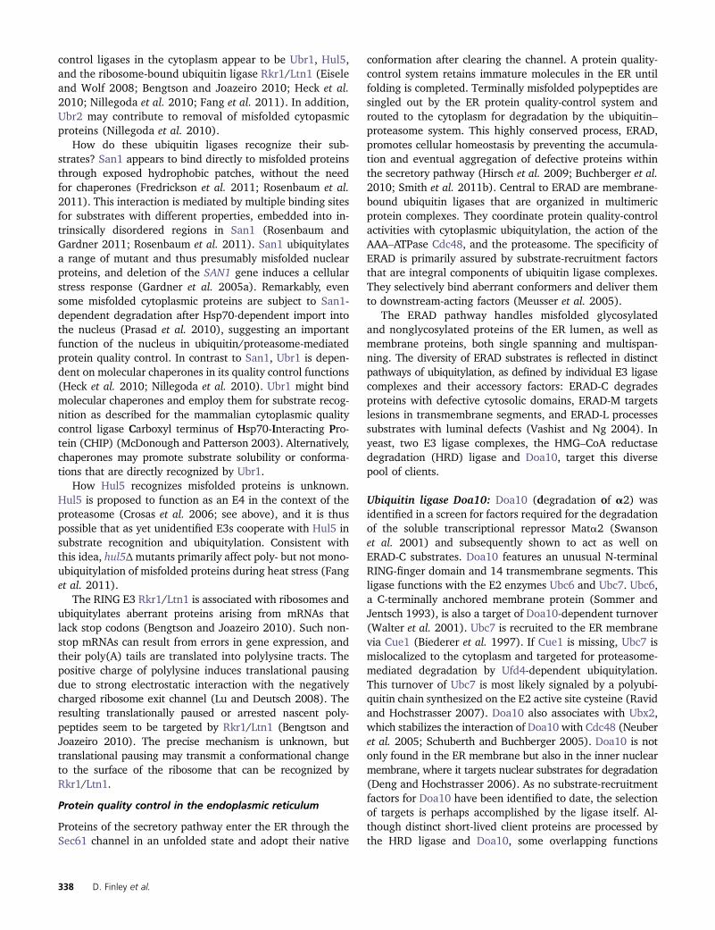
control ligases in the cytoplasm appear to be Ubr1, Hul5,and the ribosome-bound ubiquitin ligase Rkr1/Ltn1 (Eiseleand Wolf 2008; Bengtson and Joazeiro 2010; Heck et al.2010; Nillegoda et al. 2010; Fang et al. 2011). In addition,Ubr2 may contribute to removal of misfolded cytopasmicproteins (Nillegoda et al. 2010).
How do these ubiquitin ligases recognize their sub-strates? San1 appears to bind directly to misfolded proteinsthrough exposed hydrophobic patches, without the needfor chaperones (Fredrickson et al. 2011; Rosenbaum et al.2011). This interaction is mediated by multiple binding sitesfor substrates with different properties, embedded into in-trinsically disordered regions in San1 (Rosenbaum andGardner 2011; Rosenbaum et al. 2011). San1 ubiquitylatesa range of mutant and thus presumably misfolded nuclearproteins, and deletion of the SAN1 gene induces a cellularstress response (Gardner et al. 2005a). Remarkably, evensome misfolded cytoplasmic proteins are subject to San1-dependent degradation after Hsp70-dependent import intothe nucleus (Prasad et al. 2010), suggesting an importantfunction of the nucleus in ubiquitin/proteasome-mediatedprotein quality control. In contrast to San1, Ubr1 is depen-dent on molecular chaperones in its quality control functions(Heck et al. 2010; Nillegoda et al. 2010). Ubr1 might bindmolecular chaperones and employ them for substrate recog-nition as described for the mammalian cytoplasmic qualitycontrol ligase Carboxyl terminus of Hsp70-Interacting Pro-tein (CHIP) (McDonough and Patterson 2003). Alternatively,chaperones may promote substrate solubility or conforma-tions that are directly recognized by Ubr1.
How Hul5 recognizes misfolded proteins is unknown.Hul5 is proposed to function as an E4 in the context of theproteasome (Crosas et al. 2006; see above), and it is thuspossible that as yet unidentified E3s cooperate with Hul5 insubstrate recognition and ubiquitylation. Consistent withthis idea, hul5Δmutants primarily affect poly- but not mono-ubiquitylation of misfolded proteins during heat stress (Fanget al. 2011).
The RING E3 Rkr1/Ltn1 is associated with ribosomes andubiquitylates aberrant proteins arising from mRNAs thatlack stop codons (Bengtson and Joazeiro 2010). Such non-stop mRNAs can result from errors in gene expression, andtheir poly(A) tails are translated into polylysine tracts. Thepositive charge of polylysine induces translational pausingdue to strong electrostatic interaction with the negativelycharged ribosome exit channel (Lu and Deutsch 2008). Theresulting translationally paused or arrested nascent poly-peptides seem to be targeted by Rkr1/Ltn1 (Bengtson andJoazeiro 2010). The precise mechanism is unknown, buttranslational pausing may transmit a conformational changeto the surface of the ribosome that can be recognized byRkr1/Ltn1.
Protein quality control in the endoplasmic reticulum
Proteins of the secretory pathway enter the ER through theSec61 channel in an unfolded state and adopt their native
conformation after clearing the channel. A protein quality-control system retains immature molecules in the ER untilfolding is completed. Terminally misfolded polypeptides aresingled out by the ER protein quality-control system androuted to the cytoplasm for degradation by the ubiquitin–proteasome system. This highly conserved process, ERAD,promotes cellular homeostasis by preventing the accumula-tion and eventual aggregation of defective proteins withinthe secretory pathway (Hirsch et al. 2009; Buchberger et al.2010; Smith et al. 2011b). Central to ERAD are membrane-bound ubiquitin ligases that are organized in multimericprotein complexes. They coordinate protein quality-controlactivities with cytoplasmic ubiquitylation, the action of theAAA–ATPase Cdc48, and the proteasome. The specificity ofERAD is primarily assured by substrate-recruitment factorsthat are integral components of ubiquitin ligase complexes.They selectively bind aberrant conformers and deliver themto downstream-acting factors (Meusser et al. 2005).
The ERAD pathway handles misfolded glycosylatedand nonglycosylated proteins of the ER lumen, as well asmembrane proteins, both single spanning and multispan-ning. The diversity of ERAD substrates is reflected in distinctpathways of ubiquitylation, as defined by individual E3 ligasecomplexes and their accessory factors: ERAD-C degradesproteins with defective cytosolic domains, ERAD-M targetslesions in transmembrane segments, and ERAD-L processessubstrates with luminal defects (Vashist and Ng 2004). Inyeast, two E3 ligase complexes, the HMG–CoA reductasedegradation (HRD) ligase and Doa10, target this diversepool of clients.
Ubiquitin ligase Doa10: Doa10 (degradation of a2) wasidentified in a screen for factors required for the degradationof the soluble transcriptional repressor Mata2 (Swansonet al. 2001) and subsequently shown to act as well onERAD-C substrates. Doa10 features an unusual N-terminalRING-finger domain and 14 transmembrane segments. Thisligase functions with the E2 enzymes Ubc6 and Ubc7. Ubc6,a C-terminally anchored membrane protein (Sommer andJentsch 1993), is also a target of Doa10-dependent turnover(Walter et al. 2001). Ubc7 is recruited to the ER membranevia Cue1 (Biederer et al. 1997). If Cue1 is missing, Ubc7 ismislocalized to the cytoplasm and targeted for proteasome-mediated degradation by Ufd4-dependent ubiquitylation.This turnover of Ubc7 is most likely signaled by a polyubi-quitin chain synthesized on the E2 active site cysteine (Ravidand Hochstrasser 2007). Doa10 also associates with Ubx2,which stabilizes the interaction of Doa10 with Cdc48 (Neuberet al. 2005; Schuberth and Buchberger 2005). Doa10 is notonly found in the ER membrane but also in the inner nuclearmembrane, where it targets nuclear substrates for degradation(Deng and Hochstrasser 2006). As no substrate-recruitmentfactors for Doa10 have been identified to date, the selectionof targets is perhaps accomplished by the ligase itself. Al-though distinct short-lived client proteins are processed bythe HRD ligase and Doa10, some overlapping functions
338 D. Finley et al.

seem to exist: Double mutants of doa10 and hrd1 displayenhanced cadmium sensitivity and show an activated un-folded protein response (Swanson et al. 2001; for furtherreading on the unfolded protein response (UPR), see Walterand Ron 2011).
HRD ubiquitin ligase: ERAD-L and ERAD-M substrates aretargeted by the HRD ligase. Key elements of this ligasecomplex have been identified in two genetic screens. In oneof the screens, an ERAD-L substrate was used (Knop et al.1996)—a mutant version of the vacuolar enzyme carboxy-peptidase Y, CPY* (Finger et al. 1993). A mutation thatstabilized CPY* was found in UBC7, which was the first in-dication that misfolded proteins of the ER lumen are de-graded by cytoplasmic pathways (Hiller et al. 1996). SinceUBC7 was also required for turnover of a mutant form of thetranslocation component Sec61 (sec61-2; Biederer et al.1996) it became evident that ERAD-L and ERAD-M sub-strates can be degraded by the same cytoplasmic pathway.The other genetic screen was performed using an 3-hydroxy–3-methylglutaryl–CoA reductase (HMG-R) isozyme, Hmg2(Hampton et al. 1996), which is not a bona fide misfoldedprotein. Instead, Hmg2 turnover is regulated through feed-back control involving the mevalonate pathway. The twoscreens revealed overlapping genes (the “HRD” and the“DER” genes), indicating that Hmg2 is channeled into anERAD pathway that also acts on misfolded proteins. Indeed,farnesol opens the conformation of Hmg2, which could makeit accessible to the HRD ligase. Interestingly, the effect offarnesol requires an intact Hmg2 sterol-sensing domain(Shearer and Hampton 2005).
The HRD ligase complex is composed of at least sixsubunits and the requisite E2 enzymes (Figure 7A). The
central component of this complex, Hrd1/Der3, comprises sixtransmembrane segments and ubiquitylates substrates at thecytoplasmic surface of the ER through its C-terminal RING-finger domain (Bordallo et al. 1998; Bays et al. 2001a). Simi-larly to Doa10, Hrd1 functions with Cue1-tethered Ubc7. Cue1not only localizes with Ubc7 but also stimulates its enzy-matic activity (Bazirgan and Hampton 2008). Ubx2, anadditional component shared by Hrd1 and Doa10, connectsCdc48 to the HRD ligase pathway (Neuber et al. 2005;Schuberth and Buchberger 2005).
Another key element of the HRD ligase is Usa1, compris-ing two transmembrane helices and a UBA domain. Usa1mediates the interaction of Hrd1 with the small membraneprotein Der1, which spans the membrane four times and isselectively required for the breakdown of ERAD-L substrates(Knop et al. 1996; Carvalho et al. 2006). Additionally, Usa1acts as a scaffold that binds Hrd1 and promotes its dimer-ization. This function of Usa1 is generally required for pro-teolysis of ERAD-L and ERAD-M substrates (Horn et al.2009; Carvalho et al. 2010).
In vivo cross-linking studies have suggested that Hrd1may bind ERAD-L substrates (Carvalho et al. 2010). How-ever, Hrd1 carries only small loops facing the lumen of theER. Therefore, it is likely that the luminal substrate-bindingmodule of the HRD ligase is formed primarily by Hrd3 andYos9 (Denic et al. 2006; Gauss et al. 2006a,b). Hrd3 is a typeI transmembrane protein that exposes an �80-kDa domaininto the ER lumen. Yos9 interacts with the HRD ligase viaHrd3 and contains a mannose-6 phosphate receptor homol-ogy domain (MRH). The luminal Hrd3/Yos9 module linksthe ligase to the chaperone system of the ER by recruitingthe Hsp70-type chaperone Kar2 to the E3 (Denic et al.2006). Hrd3 binds misfolded CPY* irrespective of its glycan
Figure 7 HRD ubiquitin ligase. (A) HRD ubiquitin ligaseconsists of six core subunits: Hrd1 exposes a RING-fingerdomain on the cytoplasmic surface of the ER membraneand acts together with the E2 enzymes Ubc7/Cue1 andUbc1 (both not depicted). Hrd3 together with Yos9 formsthe ER luminal domain of the ligase complex. Usa1 bridgesHrd1 with Der1. Ubx2 binds Hrd1 and also, via a UBXdomain, Cdc48. The transmembrane organization of theligase complex suggests that it connects ER-luminal qual-ity-control functions, dislocation, ubiquitylation, and thegeneration of pulling forces with proteolysis by the pro-teasome. (B) Hypothetical model of how the ER-luminaldomain of the HRD ligase selects ERAD substrates. Theglycans of misfolded proteins are processed by Htm1 togenerate the glycan signal Man7GlcNAc2. Hrd3 first bindsthe misfolded protein in a “recruitment step” (left). ThenYos9 controls the identity of the glycan signal in a “com-mitment step” (center). Only when both interactions areproductive is the client protein dislocated into the cyto-plasm for proteasomal digestion.
Ubiquitin–Proteasome System of S. cerevisiae 339

modifications and also in absence of Yos9. Therefore, Hrd3was proposed to be the primary receptor for misfolded pro-teins at the ligase complex (Gauss et al. 2006b). Yos9 spe-cifically binds terminal a1,6-bonded mannose moieties onmisfolded glycoproteins (Quan et al. 2008). These are gen-erated by Mns1 and Htm1, which convert Man9GlcNAc2 intoMan7GlcNAc2 (Clerc et al. 2009; Gauss et al. 2011). Thebinding characteristics of Hrd3 and Yos9 reflect the key fea-tures of degradation signals in ERAD substrates, one beingmisfolding of the client, probably recognized by hydropho-bic interactions, and the other, a specific glycan signal,a Man7GlcNAc2 modification (Figure 7B). Since glycopro-teins that are not processed by Mns1 and Htm1 are pro-tected from degradation, these two mannosidases act asa timer that allows newly synthesized proteins to be distin-guished from those that have failed to fold correctly (Jakobet al. 1998). While these data apply to glycan-modifiedERAD-L model substrates, targeting of ERAD-M client pro-teins may differ. A mutational analysis of the Hrd1p mem-brane anchors indicated that the transmembrane segmentsmay play a crucial role in detecting misfolding of ERAD-Msubstrates (Sato et al. 2009).
Although the Hrd1 and Doa10 ligases exhibit similar ac-tivities in ERAD, their topological organization is different.While it is likely that the domains of Doa10 involved insubstrate selection and ubiquitin conjugation both residein the cytoplasm, these domains are separated by the ERmembrane in the case of the HRD ligase. Thus, at leastERAD-L clients have to be exported from the ER prior toubiquitylation. This process, termed dislocation or retro-translocation, most likely involves a proteinaceous channelin the ER membrane. It has been speculated that sucha channel may be formed by the components of the HRDligase itself (Hampton et al. 1996; Swanson et al. 2001;Horn et al. 2009). However, a function of the translocon indislocation has also been proposed, based on a physical inter-action between Hrd3 and Sec61 (Schafer and Wolf 2009).Moreover, an apparent interaction of CPY* with Sec61 is main-tained until the misfolded protein is ubiquitylated on the cyto-plasmic surface (Schafer and Wolf 2009). These findingssupport previous genetic data pointing to a function of Sec61in ERAD (Plemper et al. 1997).
Degradation signals
Mechanisms of substrate selection by ubiquitin ligasesare diverse and rely on a variety of degradation signals(degrons) (reviewed in Ravid and Hochstrasser 2008). Gen-erally we can distinguish between signal-specific degrons onregulatory proteins and degrons controlled by protein fold-ing and assembly. We briefly discussed the latter in the pre-vious section, as they are key to protein quality-controlpathways.
The first systematically studied and perhaps most sur-prising degrons are determined by the N-terminal aminoacid residue of the substrate protein (Bachmair et al. 1986;Varshavsky 2011). The N-end rule ubiquitin ligase Ubr1
(Bartel et al. 1990) binds proteins with different affinitiesdepending on the side chain of the first amino acid andthereby relates the protein’s N terminus to protein stability(Table 6) (Choi et al. 2010). The specificity of Ubr1 is es-sentially complementary to that of methionine aminopepti-dases, so that newly synthesized proteins will rarely presentdestabilizing residues; if the penultimate residue is destabi-lizing, methionine aminopeptidase will not remove the ini-tiator methionine. Rather, destabilizing N-terminal residuesare formed as a result of endoproteolytic cleavage by proteasessuch as separase (see below), or other post-translational events(Varshavsky 2011). For example, acidic N-terminal residuesgenerated by endoproteases are not recognized by Ubr1, butare substrates for Ate1, an enzyme that ligates arginine tothe substrate’s N terminus. This allows for subsequent Ubr1-mediated recognition, ubiquitylation, and degradation. N-terminal glutamine and asparagine residues are funneledinto the N-end rule pathway by the action of Nta1, anN-terminal amidase, whose reaction products are in turnsubstrates for Ate1 (Baker and Varshavsky 1995). Thesepathways mediate major regulatory events in many eukar-yotes, such as the sensing of oxygen and nitric oxide levels(Licausi et al. 2011; Varshavsky 2011).
Acetylated N termini, which are found in most proteins,are not recognized by Ubr1. Instead, they present separatedegrons recognized by the Doa10 ligase (Hwang et al.2010). Many proteins are metabolically stable despite thepresence of these targeting elements, presumably due topoor exposure of their N termini, suggesting a potential in-volvement of this pathway in recognition of misfolded pro-teins and quality control degradation (Hwang et al. 2010).
Many other regulated degradation pathways also usepost-translational modifications to activate degrons. For
Table 6 N-end rule in Saccharomyces cerevisiae
Residue atN terminus
Half-life ofX-ßgal
Arg 2 minLys 3 minPhe 3 minLeu 3 minTrp 3 minHis 3 minAsp 3 minAsn 3 minTyr 10 minGln 10 minIle 30 minGlu 30 minCys .20 hrAla .20 hrSer .20 hrThr .20 hrGly .20 hrVal .20 hrPro NDMet .20 hr
Adapted from Bachmair et al. (1986), with permission. ND, not done.
340 D. Finley et al.
example, phosphorylation often generates a high-affinityinteraction site for E3 recruitment. Such phosphodegronsare widely used by SCF ligases (Petroski and Deshaies 2005;Zimmerman et al. 2010; Duda et al. 2011). The cell cycleinhibitor Sic1 contains an array of phosphodegrons withrelatively low affinities for SCFCdc4. This arrangementrequires processive multiphosphorylation by the G1 andS-phase kinases Cln2/Cdc28 and Clb5/Cdc28 for efficientSic1 degradation and transforms the graded kinase activityinto a switch-like cell-cycle transition (Nash et al. 2001;Petroski and Deshaies 2003; Koivomagi et al. 2011).
Another interesting variety of degron formed by post-translational modification is recognized by the small ubiquitin-related modifier (SUMO)-targeted ubiquitin ligases (STUbLs)(Perry et al. 2008). These E3s contain SUMO interactingmotifs that mediate binding to SUMOylated substrate pro-teins for ubiquitylation (see below).
Other degrons are less well defined and include shortsurface-exposed hydrophobic stretches such as the N-terminaldegron in Mata2, which can be masked by heterodimeriza-tion with Mata1 (Johnson et al. 1998).
Sites in target proteins that recruit E3s and allowubiquitin conjugation constitute the canonical form of adegradation signal in the ubiquitin–proteasome system.These sites are remarkably varied, consistent with the mul-tiplicity of ubiquitin ligases and their diverse substrate rec-ognition modes. However, additional features to supportproteasome-mediated degradation of the substrate are alsocritical, particularly an unfolded segment to serve as an inita-tion site for the proteasome, as discussed above (Prakashet al. 2004).
Ubiquitylation of Membrane Proteins
As discussed earlier, membrane-associated ubiquitin ligasesplay key roles in the protein quality-control pathway of theER. However, other membrane systems of the cell are alsosites of abundant ubiquitylation, where it acts to directprotein sorting. On the one hand, ubiquitylation drivestransport from the trans-Golgi and the plasma membrane.On the other, it helps to concentrate proteins in the MVBcompartment. The MVB sorting step leads ultimately to pro-teolysis of the cargo—not in the proteasome, however, butin the vacuole (Lauwers et al. 2010).
Ubiquitin function in endocytosis
The abundance of receptors and transporters at the plasmamembrane is regulated by endocytosis, often in a signal-dependent manner. Internalized proteins are transported tothe endocytic compartment. From there, they are eitherrecycled to the plasma membrane or packaged into multi-vesicular bodies for delivery to lysosomes. Ubiquitin serves asan important internalization signal for endocytosis. In somecases, ubiquitin seems to act redundantly with other signals.
A function of ubiquitin in protein sorting at the plasmamembrane was suggested by the observation that ubiquity-
lated Ste6, the yeast pheromone transporter, accumulated atthe plasma membrane when endocytosis is blocked (Köllingand Hollenberg 1994). Moreover, ubiquitylation was foundto be necessary and sufficient for ligand-induced endocytosisof the pheromone receptor Ste2 (Hicke and Riezman 1996).Accordingly, the nitrogen permease inactivator Npi1 wasidentified as the HECT domain ligase Rsp5 (Hein et al.1995; Huibregtse et al. 1995). A number of other plasmamembrane proteins were subsequently shown to undergoubiquitylation, including the permeases Fur4 and Gap1.
After Rsp5-dependent selection and modification of thecargo, it is most likely recognized by endocytic adaptors (Shihet al. 2002). Yeast endocytic adaptors Ent1 and Ent2 bindubiquitylated cargo via UIM domains and localize to the plasmamembrane by interacting with phosphatidylinositol-(4,5)-bisphosphate and clathrin. An additional endocytic scaffoldprotein, Ede1, an EH domain protein, contains a UBA domainand it may also contribute to cargo interaction and concentra-tion (Dores et al. 2010).
Several nutrient permeases are ubiquitylated on multiplelysines by short K63 ubiquitin chains, consistent with thelinkage specificity of Rsp5 (Kim and Huibregtse 2009).Though a single ubiquitin molecule is sufficient to promoteendocytosis, multiple monoubiquitylation and short K63chains accelerate the rate of endocytosis, to an extent thatmay depend on the substrate (Galan and Haguenauer-Tsapis1997; Springael et al. 1999). Notably, the Jen1 transportershows a strict requirement for K63 chains (Paiva et al.2009).
Rsp5 contains WW domains, named for the presence oftwo highly conserved tryptophan residues, which directlyinteract with PPx(Y/F) motifs in substrate proteins. How-ever, many cargo molecules, including several permeases,do not carry such a motif. In these cases, interaction of thecargo with Rsp5 is mediated by a family of adaptor proteins,such as the arrestin-related trafficking adaptors (ARTs).Unlike mammals, yeast does not have canonical arrestins,in that the yeast ART proteins lack adaptin and clathrin-bind-ing sequences. Instead they interact with Rsp5 through a PxYsequence (Lin et al. 2008; Leon and Haguenauer-Tsapis 2009;Nikko and Pelham 2009). ART proteins (Art1–Art10) regulatethe ubiquitylation of specific cargos at the plasma membrane inresponse to specific stimuli and may be part of a quality-control system that targets damaged and misfolded mem-brane proteins for degradation in the vacuole. This variety ofadaptors explains how a single ubiquitin ligase can regulateendocytosis of many different proteins. Another level of reg-ulation is provided by differential localization of the Rsp5adaptors. For example, the adaptors Bul1 and Bul2 workboth at the plasma membrane and the trans-Golgi (Nikkoand Pelham 2009). The Art1–Art10 proteins (Lin et al. 2008;Nikko and Pelham 2009; MacGurn et al. 2011) functionmainly at the plasma membrane, while the Rsp5 adaptorproteins Ear1, Ssh4, Bsd2, Tre1, and Tre2 are located mainlyat endosomes (Liu et al. 1997; Stimpson et al. 2006; Leonet al. 2008).
Ubiquitin–Proteasome System of S. cerevisiae 341
Nutrient uptake is controlled by a regulatory loop thatadjusts the level of amino acid transporters at the plasmamembrane through Art1. Npr1 (nitrogen permease reactiva-tor 1 kinase) phosphorylates residues near the N terminus ofArt1 to inhibit its transport to the plasma membrane. Endo-cytosis of amino acid transporters is thereby suppressed.Npr1 itself is negatively regulated by the TORC1 kinase,which thus acts on ubiquitin-dependent endocytosis to finetune the activity and composition of proteins of the plasmamembrane (MacGurn et al. 2011).
Function of ubiquitin in the MVB pathway
Proteins of the late endosome destined for proteolysis aresorted into multivesicular bodies, which deliver cargo to theyeast vacuole for degradation (Katzmann et al. 2001; Henneet al. 2011). Formation of these vesicles involves invaginationof membranes into the endosomal compartment. MatureMVBs subsequently fuse with the lysosome and release theircontents. Crucial players in the MVB pathway are specificmultisubunit endosomal sorting complexes required fortransport (ESCRT): ESCRT-0, ESCRT-I, ESCRT-II, ESCRT-III, and the AAA ATPase Vps4 (part of the fifth ESCRT com-plex). These act sequentially (Katzmann et al. 2001; Babstet al. 2002a,b) in early cargo recruitment and concentration(ESCRT-0, -I, and -II) and later in cargo deubiquitylationand membrane sculpting (ESCRT-III and Vps4).
A prerequisite for selective sorting and concentration ofmembrane proteins into endosomal microdomains and, even-tually MVBs, is the ubiquitylation of cargo proteins (Katzmannet al. 2001; Lauwers et al. 2010; Henne et al. 2011). ESCRTcomplexes have several distinct ubiquitin-binding motifs forcargo recognition. Subunits of the ESCRT-0 complex bindubiquitin in several ways: Vps27 contains a VHS (Vps27 HrsSTAM) and two UIM domains in tandem, while the Hse1subunit contains both a UIM and a VHS domain (Bilodeauet al. 2002). Thus, ESCRT-0 contains five ubiquitin-bindingdomains. However, it remains unclear whether this allowsbinding of several cargoes simultaneously or binding with highaffinity to poly- or multiubiquitylated cargo (Ren and Hurley2010). In addition to its ubiquitin-binding activity, ESCRT-0may bind cargo through interactions with the clathrin vesi-cle machinery, suggesting microdomains in which clathrinlattices, ESCRT-0, and ubiquitylated cargo meet. ESCRT-Ialso contains ubiquitin-binding domains: the subunit Vps23contains a UEV domain (Pornillos et al. 2002), and Mvb12harbors a novel ubiquitin-binding domain (Shields et al.2009). So far only one ubiquitin-binding domain has beenidentified in ESCRT-II, an Npl4 Zinc Finger (NZF) motifin Vps36. Also participating in ubiquitin-dependent sortingare the ubiquitin-binding adaptor proteins Gga1 and Gga2,although their exact roles in the pathway are not under-stood (Lauwers et al. 2009, 2010).
Though it was initially assumed that monoubiquitylationis sufficient to direct targets into the MVB pathway, it is nowaccepted that K63 ubiquitin chains are needed. Whetherthese ubiquitin moieties are added at the endosome or
persist from endocytosis-associated ubiquitylation events atthe plasma membrane is unresolved. In any case, thismodification must be removed prior to packaging of cargointo vesicles to avoid depletion of ubiqutin by its uptake intothe vacuole. In yeast, this step involves the DUB Doa4(Amerik et al. 2000a). Doa4 is recruited into the ESCRT-IIIcomplex by the adaptor protein Bro1, which also stimulatesthe deubiquitylating activity of Doa4 (Luhtala and Odorizzi2004; Richter et al. 2007).
In addition to ubiquitin binding, ESCRT components alsoserve as targets for ubiquitylation. For instance, Vps27 canbe monoubiquitylated (Polo et al. 2002; Stringer and Piper2011). Although the physiological function of this modifica-tion remains unknown, Hrs, the human homolog of Vps27,is inhibited by monoubiquitylation because an intramolecu-lar interaction between the UIM and the ubiquitin modifica-tion prevents the binding of ubiquitylated cargo. Similarobservations have been made for the endocytic adaptorEps15, raising the possibility of a general role for monoubi-quitylation in downregulation of these pathways (Hoelleret al. 2006).
Ubiquitylation and protein import into peroxisomes
Peroxisomes are small cytoplasmic vesicles housing �50enzymes that mediate b oxidation of fatty acids and othermetabolic processes. Luminal proteins of the peroxisomeare imported post-translationally by a complex, receptor-mediated process (Girzalsky et al. 2010). The import appa-ratus includes three RING ligases (Pex2, Pex10, andPex12) that reside in the peroxisomal membrane, as wellas as a cognate E2 (Ubc10/Pex4) (Williams et al. 2008;Platta et al. 2009). The principal role of ubiquitin in per-oxisomes is apparently the monoubiquitylation of Pex5,a receptor for protein import into peroxisomes that deliverscargo to the peroxisome by cycling between the cytoplasmand the peroxisomal membrane (Carvalho et al. 2007;Platta et al. 2007; Grou et al. 2008). After cargo delivery,Pex5 must return to the cytoplasm for another round ofimport. This recycling step is dependent on Pex5 ubiqui-tylation. Other peroxisomal import receptors such asPex18 and Pex20 are ubiquitylated and potentially followa similar cycle. A heteromeric protein complex of the AAAfamily, composed of Pex1 and Pex6 monomers, mediatesATP-dependent extraction of Pex5 into the cytosol by anunknown mechanism (Platta et al. 2005). A hypotheticalmodel accounting for these data are that the Pex1/Pex6complex recognizes Pex5 via its ubiquitin modification,and functions analogously to Cdc48 in its extraction ofmembrane proteins from the ER in the ERAD-M pathway,as described above.
Nuclear Functions of the Ubiquitin System
Nuclear functions of ubiquitin fall into two general catego-ries: on one hand, there is a nuclear form of the above-described protein quality-control pathways. On the other
342 D. Finley et al.

hand, as described below, ubiquitylation contributes,both in its proteolytic and its noncanonical mode, tovirtually all aspects of DNA metabolism (Ulrich 2002),such as DNA replication and repair (Bergink and Jentsch2009; Ulrich and Walden 2010), gene expression andchromatin structure (Muratani and Tansey 2003; Shilati-fard 2006), and chromosome dynamics and segregation(Pines 2006).
Coupling cell cycle progression to DNA replication andchromosome segregation
DNA replication and chromosome segregation are intimatelycoupled to cell cycle progression and hence subject toregulation by the ubiquitin system, mostly by means ofproteolytic destruction of important regulators. These pro-cesses have been discussed in detail in excellent reviews(Nasmyth 1996; Pines 2006; Diffley 2010).
Replication initiation: At the G1-to-S transition, Sic1 deg-radation, initiated by SCFCdc4, allows the activation of thecyclin-dependent kinase (CDK) Cdc28 in complex with theS-phase–specific, B-type cyclins, Clb5 or Clb6, leading tophosphorylation and activation of the replication initiationfactors Sld2 and Sld3 (Tanaka et al. 2007; Zegerman andDiffley 2007). In parallel, SIC1 transcription is terminated byubiquitylation and destruction of a transcriptional activator,Swi5, also mediated by SCFCdc4 (Kishi et al. 2008).
There is also evidence for nondegradative contributionsof ubiquitin to replication initiation. The cullin Rtt101 asso-ciates with early replication origins and ubiquitylates Spc16,a subunit of the FAcilitator of Chromatin Transactions (FACT)complex, predominantly via K63-linked chains (Han et al.2010). FACT is a histone chaperone with functions in tran-scription, DNA replication, and repair (Winkler and Luger2011). Deletion of RTT101 results in a weakening of the inter-actions between FACT and the replicative helicase (the Mcm2-7 hexamer), and a partial loss of both complexes from a subsetof replication origins (Han et al. 2010). Whether or not ubiq-uitylation of Spc16 is responsible for this phenomenon has notbeen determined, but the activity of Rtt101 appears to im-pinge specifically on the replication-related functions of FACT.
Origin licensing: The mechanism that limits replication toa single round per cell cycle is called origin licensing. Thisrestricts the activation of replication origins to S phase andprevents renewed firing until the next cell cycle. All or-ganisms use multiple strategies to achieve this goal, in-cluding ubiquitin-mediated proteolysis of key regulatoryfactors (Diffley 2010). In budding yeast, the primary targetis Cdc6, a component of the prereplicative complex (pre-RC). Cdc6 is phosphorylated in late G1 and S phase, whichcauses ubiquitylation by SCFCdc4 and subsequent degradation(Drury et al. 1997). While in mammalian cells the pre-RCcomponent Cdt1 is subject to proteolysis, its yeast homologTah11 is instead inactivated by export from the nucleus(Diffley 2010).
Chromosome segregation: One of the most importantfeatures of cell division is the even distribution of replicatedchromosomes to the daughter cells, a process controlled byubiquitin-dependent proteolysis (Pines 2006). During thiscell cycle stage, the dominant E3 is the APC/C, which cou-ples mitosis to cytokinesis and ensures correct chromosomesegregation (Harper et al. 2002). Important substratesinclude cyclins; components of the spindle checkpoint thatmonitor the correct assembly of the mitotic spindle; and thesecurin protein, Pds1, an inhibitor of separase (Esp1) thatinitiates anaphase by cleaving the cohesin subunit Scc1, thusallowing sister chromatid separation (Nasmyth et al. 2000).The C-terminal proteolytic fragment of Scc1 is subject toproteasomal degradation by the N-end rule pathway, initi-ated by ubiquitylation via the RING-finger E3 Ubr1 with theE2 Rad6, and interference with this process causes chromo-some loss (Rao et al. 2001; see also Buonomo et al. 2000).
Responses to replication stress
Mechanisms of replication fork protection: Whereas thecoupling of replication initiation and origin licensing to the cellcycle mostly involves proteolytic functions of the ubiquitinsystem, the role of ubiquitylation in the course of replicationappears more diverse. A number of ubiquitin ligases have beenfound to contribute to genome stability by protecting replica-tion forks from stress, but their mechanisms of action arepoorly understood.
The F-box protein Dia2 is a constitutive component ofthe replisome progression complex (RPC), tethered to theRPC components Mrc1 and Ctf4 by means of its N-terminaldomain (Morohashi et al. 2009). Dia2 acts as a substrateadaptor for SCFDia2. Its association with replication forksappears to facilitate the replication of difficult templatesand protects cells from DNA damage and replication stress(Mimura et al. 2009; Morohashi et al. 2009). Mrc1 andCtf4 are ubiquitylated by SCFDia2 and seem to be degraded,but it is unclear whether these substrates are functionallycritical (Mimura et al. 2009). Dia2 itself is an unstable pro-tein, and it is stabilized by replication stress (Kile andKoepp 2010).
The cullin Rtt101, in addition to its role in replicationinitiation, also contributes to protecting replication forks fromcollapse when they encounter DNA lesions (Luke et al. 2006).Thus, rtt101D mutants are sensitive to DNA-damaging agentsand unable to recover from damage-induced fork stalling. Inresponse to DNA damage, Rtt101 forms a ubiquitin ligasewith the RING finger protein Hrt1, the linker protein Mms1,and the putative substrate adaptor Mms22 (Zaidi et al.2008). Interestingly, Rtt101’s function in replication forkprotection appears unrelated to its action on the FACT com-plex, as the latter does not require the presence of Mms1 orMms22 (Han et al. 2010). A second substrate adaptor,Crt10, is recruited to the Rtt101 cullin complex via Mms1in a damage-independent manner and has been suggestedto affect replication by regulating nucleotide levels (Fu andXiao 2006; Zaidi et al. 2008).
Ubiquitin–Proteasome System of S. cerevisiae 343

An interesting example of cross-talk between ubiquitin andthe small ubiquitin-related modifier SUMO has emerged fromthe identification of a class of E3s, called STUbLs, whichrecognize SUMO-modified proteins as targets for ubiquity-lation (Perry et al. 2008). In budding yeast, the RING-fingerproteins Hex3/Slx5 and Slx8 have been implicated in pre-venting the accumulation of DNA damage during replication(Zhang et al. 2006). They form a heterodimer that promotesthe ubiquitylation and degradation of highly sumoylatedcellular proteins (Uzunova et al. 2007; Xie et al. 2007).The SUMO moieties are recognized by SUMO-interactingmotifs within Hex3. It remains to be resolved whether thecontribution of SUMO-dependent ubiquitylation to rep-lication fork protection is attributable to the removal ofspecific sumoylated proteins for regulatory purposes or to pre-venting bulk accumulation of potentially toxic poly-SUMOconjugates.
Control of DNA damage bypass: An independent pathwayfor lesion processing during replication is called postreplica-tion repair, DNA damage bypass, or DNA damage tolerance(Lawrence 1994). The process provides resistance to DNA-damaging agents, but is capable of generating genomic in-stability through damage-induced mutagenesis. It is initiatedby ubiquitylation of the proliferating cell nuclear antigen(PCNA) Pol30 (Hoege et al. 2002), a homotrimeric slidingclamp that ensures processivity of the replicative polymerasesand also acts as an interaction platform for a multitude of pro-teins involved in various aspects of DNA metabolism (Moldovanet al. 2007). The PCNA modification system (Figure 8) providesan example where mono- and polyubiquitylation at a single siteelicit distinct cellular responses (Ulrich 2009), mediated bya range of ubiquitin receptors (Table 7).
Monoubiquitylation at a single conserved lysine, K164,mediated by the E2–E3 complex Rad6–Rad18 (Hoege et al.2002), is a prerequisite for a process called translesion syn-thesis (Stelter and Ulrich 2003). This reaction involves a se-ries of specialized DNA polymerases capable of usingdamaged DNA as a template for DNA synthesis (Waters et al.2009). Although there is evidence for ubiquitin-independenttranslesion synthesis in vertebrates, the principle by whichubiquitylated PCNA activates damage-tolerant polymerasesappears to be conserved: a series of ubiquitin-bindingdomains of the Ubiquitin-Binding Zinc Finger (UBZ) orUbiquitin-Binding Motif (UBM) type, present in a subset ofthe polymerases (Table 7), affords enhanced affinity for themonoubiquitylated form of PCNA and thereby allows theirrecruitment and activation in response to DNA damage(Bienko et al. 2005). In budding yeast, this applies to poly-merase h (encoded by RAD30), which mediates error-freetranslesion synthesis over UV-induced lesions, and Rev1,which in cooperation with polymerase z (encoded by REV3and REV7) is responsible for a large part of damage-inducedmutagenesis (Garg and Burgers 2005; Guo et al. 2006;Parker et al. 2007).
The consequences of PCNA polyubiquitylation are lesswell defined. The modification is a prerequisite for anerror-free pathway of template switching (Hoege et al.2002), which mediates damage bypass by avoiding theuse of damaged DNA as a replication template. PCNA poly-ubiquitylation involves the synthesis of a K63-linked chainby the E3 Rad5 in cooperation with the heterodimeric E2Ubc13–Mms2 (Hoege et al. 2002; Parker and Ulrich 2009).The modification is likely to serve a nondegradative func-tion (Zhao and Ulrich 2010) and enhances the affinity of anATPase, Mgs1, for PCNA (Hishida et al. 2006; Saugar et al.
Figure 8 Modifications of the replica-tion factor PCNA. During undisturbedreplication, PCNA (blue ring shape) pro-motes processive DNA synthesis by rep-licative polymerases d and e (Pol d/e),and is modified by SUMO (red lollipopshape). The modification prevents bind-ing of Eco1, but causes the recruitmentof Elg1 and Srs2. Srs2 prevents the for-mation of the recombinogenic Rad51filament (51), inhibiting unscheduled re-combination at replication forks. Upondamage-induced replication fork stall-ing, PCNA is modified by mono- andpolyubiquitin (black lollipop shapes)at postreplicative daughter-strand gaps.Monoubiquitylation recruits damage-tolerant DNA polymerases (TLS) fortranslesion synthesis, while K63 polyubi-quitylation causes recruitment of Mgs1and initiates damage bypass by templateswitching in an unknown manner. Con-jugating enzymes, ligases, and DUBs arehighlighted in shades of purple, green,and pink, respectively.
344 D. Finley et al.
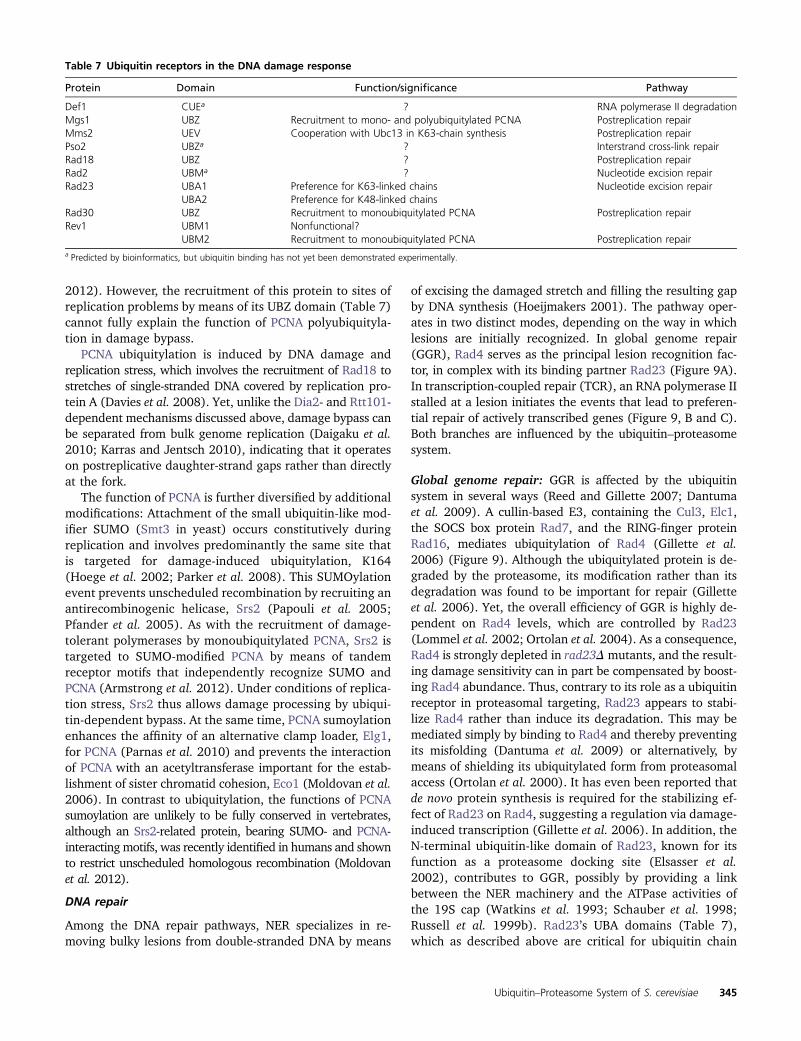
2012). However, the recruitment of this protein to sites ofreplication problems by means of its UBZ domain (Table 7)cannot fully explain the function of PCNA polyubiquityla-tion in damage bypass.
PCNA ubiquitylation is induced by DNA damage andreplication stress, which involves the recruitment of Rad18 tostretches of single-stranded DNA covered by replication pro-tein A (Davies et al. 2008). Yet, unlike the Dia2- and Rtt101-dependent mechanisms discussed above, damage bypass canbe separated from bulk genome replication (Daigaku et al.2010; Karras and Jentsch 2010), indicating that it operateson postreplicative daughter-strand gaps rather than directlyat the fork.
The function of PCNA is further diversified by additionalmodifications: Attachment of the small ubiquitin-like mod-ifier SUMO (Smt3 in yeast) occurs constitutively duringreplication and involves predominantly the same site thatis targeted for damage-induced ubiquitylation, K164(Hoege et al. 2002; Parker et al. 2008). This SUMOylationevent prevents unscheduled recombination by recruiting anantirecombinogenic helicase, Srs2 (Papouli et al. 2005;Pfander et al. 2005). As with the recruitment of damage-tolerant polymerases by monoubiquitylated PCNA, Srs2 istargeted to SUMO-modified PCNA by means of tandemreceptor motifs that independently recognize SUMO andPCNA (Armstrong et al. 2012). Under conditions of replica-tion stress, Srs2 thus allows damage processing by ubiqui-tin-dependent bypass. At the same time, PCNA sumoylationenhances the affinity of an alternative clamp loader, Elg1,for PCNA (Parnas et al. 2010) and prevents the interactionof PCNA with an acetyltransferase important for the estab-lishment of sister chromatid cohesion, Eco1 (Moldovan et al.2006). In contrast to ubiquitylation, the functions of PCNAsumoylation are unlikely to be fully conserved in vertebrates,although an Srs2-related protein, bearing SUMO- and PCNA-interacting motifs, was recently identified in humans and shownto restrict unscheduled homologous recombination (Moldovanet al. 2012).
DNA repair
Among the DNA repair pathways, NER specializes in re-moving bulky lesions from double-stranded DNA by means
of excising the damaged stretch and filling the resulting gapby DNA synthesis (Hoeijmakers 2001). The pathway oper-ates in two distinct modes, depending on the way in whichlesions are initially recognized. In global genome repair(GGR), Rad4 serves as the principal lesion recognition fac-tor, in complex with its binding partner Rad23 (Figure 9A).In transcription-coupled repair (TCR), an RNA polymerase IIstalled at a lesion initiates the events that lead to preferen-tial repair of actively transcribed genes (Figure 9, B and C).Both branches are influenced by the ubiquitin–proteasomesystem.
Global genome repair: GGR is affected by the ubiquitinsystem in several ways (Reed and Gillette 2007; Dantumaet al. 2009). A cullin-based E3, containing the Cul3, Elc1,the SOCS box protein Rad7, and the RING-finger proteinRad16, mediates ubiquitylation of Rad4 (Gillette et al.2006) (Figure 9). Although the ubiquitylated protein is de-graded by the proteasome, its modification rather than itsdegradation was found to be important for repair (Gilletteet al. 2006). Yet, the overall efficiency of GGR is highly de-pendent on Rad4 levels, which are controlled by Rad23(Lommel et al. 2002; Ortolan et al. 2004). As a consequence,Rad4 is strongly depleted in rad23Dmutants, and the result-ing damage sensitivity can in part be compensated by boost-ing Rad4 abundance. Thus, contrary to its role as a ubiquitinreceptor in proteasomal targeting, Rad23 appears to stabi-lize Rad4 rather than induce its degradation. This may bemediated simply by binding to Rad4 and thereby preventingits misfolding (Dantuma et al. 2009) or alternatively, bymeans of shielding its ubiquitylated form from proteasomalaccess (Ortolan et al. 2000). It has even been reported thatde novo protein synthesis is required for the stabilizing ef-fect of Rad23 on Rad4, suggesting a regulation via damage-induced transcription (Gillette et al. 2006). In addition, theN-terminal ubiquitin-like domain of Rad23, known for itsfunction as a proteasome docking site (Elsasser et al.2002), contributes to GGR, possibly by providing a linkbetween the NER machinery and the ATPase activities ofthe 19S cap (Watkins et al. 1993; Schauber et al. 1998;Russell et al. 1999b). Rad23’s UBA domains (Table 7),which as described above are critical for ubiquitin chain
Table 7 Ubiquitin receptors in the DNA damage response
Protein Domain Function/significance Pathway
Def1 CUEa ? RNA polymerase II degradationMgs1 UBZ Recruitment to mono- and polyubiquitylated PCNA Postreplication repairMms2 UEV Cooperation with Ubc13 in K63-chain synthesis Postreplication repairPso2 UBZa ? Interstrand cross-link repairRad18 UBZ ? Postreplication repairRad2 UBMa ? Nucleotide excision repairRad23 UBA1 Preference for K63-linked chains Nucleotide excision repair
UBA2 Preference for K48-linked chainsRad30 UBZ Recruitment to monoubiquitylated PCNA Postreplication repairRev1 UBM1 Nonfunctional?
UBM2 Recruitment to monoubiquitylated PCNA Postreplication repaira Predicted by bioinformatics, but ubiquitin binding has not yet been demonstrated experimentally.
Ubiquitin–Proteasome System of S. cerevisiae 345

recognition by the proteasome, are not specifically requiredfor NER (Bertolaet et al. 2001).
Transcription-coupled repair: The influence of ubiquityla-tion on TCR may be viewed as a solution to the problem ofan irreversibly stalled RNA polymerase II (Svejstrup 2010)(Figure 9C). In this situation, the enzyme’s large subunit,Rpb1, is ubiquitylated and degraded by the proteasome,which clears the transcription machinery from the site ofdamage and allows subsequent repair via GGR (Beaudenonet al. 1999; Woudstra et al. 2002). Degradation is de-pendent on the Coupling of Ubiquitin conjugation to ERdegradation (CUE)-domain protein Def1 (Table 7), butwhether this domain actually binds ubiquitin has not beendetermined. The E3 Rsp5 attaches either a single ubiquitinor a short K63-linked polyubiquitin chain to Rpb1, the lat-ter of which may be trimmed by the DUB enzyme Ubp2(Beaudenon et al. 1999; Harreman et al. 2009). A secondE3, containing Cul3 in complex with Elc1, Ela1, and Hrt1,but not Rad7 and Rad16, has been implicated in Rpb1modification as well (Ribar et al. 2006, 2007). This com-plex uses monoubiquitylated Rpb1 as a substrate for poly-ubiquitylation with a K48-linked chain (Harreman et al.2009). Hence, Rpb1 polyubiquitylation, like PCNA modifi-cation, is characterized by the successive action of two E3s.As discussed above, degradation of polyubiquitylated Rpb1
requires the Cdc48–Ufd1–Npl4 complex (Verma et al. 2011),and the process might be balanced by Ubp3-mediated decon-jugation (Kvint et al. 2008).
Regulation of gene expression and chromatin structure
Modulation of the transcription machinery: Tight controlover gene expression is essential for adaptation to changes ina cell’s environment. The ubiquitin–proteasome system con-tributes to this activity in many aspects (Muratani and Tansey2003; Shilatifard 2006; Ouni et al. 2011; Geng et al. 2012).Among the most direct modes of influence is control over thelevels of transcriptional regulators. Hence, many transcriptionfactors are short-lived proteins, such as Gcn4, a target ofSCFCdc4 (Meimoun et al. 2000), and Gal4, whose abundanceis limited by SCFGrr1 (Muratani et al. 2005). In many cases,including Gcn4, the transcriptional activation domain wasfound to overlap with the degradation signal (Salghettiet al. 2000). Ubiquitin-dependent proteolysis was thus foundto be intimately coupled to the activity of natural and engi-neered transcription factors (Salghetti et al. 2001; Lipfordet al. 2005; Wang et al. 2010). These observations led tothe hypothesis that periodic promoter clearance is importantfor maximal activity. In the case of Gal4, two parallel degra-dation pathways have been described: the SCFGrr1-dependentmode, which is independent of Gal4 activity and downregu-lates the protein in the absence of galactose, and a pathway
Figure 9 Ubiquitylation during nucleo-tide excision repair. (A) For global ge-nome repair, lesions are recognized byRad4 in complex with Rad23. Ubiquity-lation of Rad4 is important for sub-sequent steps of repair. UbiquitylatedRad4 is degraded by the proteasome.(B) Lesions on the transcribed strand ofactively expressed genes are repaired bytranscription-coupled repair, where RNApolymerase II (RNA Pol II) contributes tolesion recognition. Following removal ofthe enzyme by the action of Rad26,strand unwinding, excision of the lesionand resynthesis proceed as in global ge-nome repair. (C) An irreversibly stalledRNA polymerase II is targeted for ubiq-uitylation and proteasomal degradationin a Def1-dependent manner. This freesthe lesion and allows global genome re-pair. Conjugating enzymes, ligases, andDUBs are highlighted in shades of purple,green, and pink, respectively. Distinct poly-ubiquitin chain linkages are indicated asK48 or K63.
346 D. Finley et al.
mediated by the F-box protein Dsg1, which applies to acti-vated Gal4 (Muratani et al. 2005).
Ubiquitin-dependent activation of transcription factorsdoes not need to involve complete degradation, but can alsoproceed by proteolytic processing. This was observed for thetranscriptional activators Spt23 and Mga2, whose mem-brane-bound precursors are ubiquitylated by Rsp5, leadingto their processing and relocalization into the nucleus(Hoppe et al. 2000; see above).
Finally, as discussed above, RNA polymerase II itself issubject to ubiquitylation and degradation upon transcriptionstalling. In addition, ubiquitylation of Rpb1 and Rpb2 bythe RING-finger E3 Asr1 apparently causes an eviction ofthe polymerase subunits Rpb4 and Rpb7, leading to theenzyme’s inactivation (Daulny et al. 2008). It remains tobe seen, however, to what extent this strategy is used asa regulatory measure.
Several observations suggest possible nonproteolytic con-tributions of the proteasome to transcription. The RP, but notthe CP, has been implicated in transcription elongation(Ferdous et al. 2001), and its ATPase subunits were foundin association with active promoters (Gonzalez et al. 2002;Sulahian et al. 2006). Also, chromatin immunoprecipitationanalyses have suggested limited overlap between RP and CPcomponents on chromatin and have localized proteasomesubunits to internal and 39 regions of transcribed genes aswell (Auld et al. 2006; Sikder et al. 2006). These data haveresulted in a model postulating a nonproteolytic and chaper-one-like activity of the proteasomal ATPases in transcription.In the context of transcription initiation, this has been linkedto the recruitment and stimulation of the Spt-Ada-Gcn5 Ace-tyltransferase (SAGA) complex, a histone acetyltransferase(Lee et al. 2005). However, the physiological relevance ofthese findings is still debated (see for example Collins et al.2009), and the mechanism by which the proteasome mightact here remains to be elucidated.
Regulation of chromatin structure: The higher organiza-tion of genes into chromatin and their accessibility by thetranscription machinery are crucial determinants of geneexpression (Osley 2006; Shilatifard 2006). The first evi-dence for a contribution of the ubiquitin system to chromatinstructure came from the identification of monoubiquitylatedhistone H2B (Robzyk et al. 2000). The modification isattached to K123 within the C-terminal tail of H2B byRad6 in complex with the RING–E3 Bre1 (Wood et al.2003), and is a prerequisite for the subsequent di- and tri-methylation of histone H3 on K4 and K79 by the methyl-transferases Set1 and Dot1, respectively (Dover et al. 2002;Ng et al. 2002; Sun and Allis 2002). The relationshipsbetween the levels of ubiquitylated H2B and methylatedH3 are complex, and the mechanism by which one modifi-cation induces the other has not been fully explained; butthe reaction appears to occur cotranscriptionally and isimportant for telomeric gene silencing. H2B ubiquitylationis also observed on the body of transcribed genes and has
been associated with transcriptional initiation and elonga-tion, but also repression (Henry et al. 2003; Kao et al. 2004;Xiao et al. 2005; Osley 2006). In a reconstituted systemderived from mammalian cells, the effect of H2B monoubi-quitylation on elongation by RNA polymerase II is due toa stimulation of the FACT complex, and a similar situationmay apply in yeast (Pavri et al. 2006).
Interestingly, optimal transcription in vivo requires bothubiquitylation and subsequent deubiquitylation of H2B.Deconjugation is mediated by the two DUBs, Ubp8 andUbp10 (Henry et al. 2003; Emre et al. 2005; Gardner et al.2005b). Ubp8 acts as an integral component of the SAGAcomplex, and during the early steps of transcription. In con-trast, Ubp10 works independently and influences mainlytelomeric silencing, indicating nonredundant roles (Emreet al. 2005; Gardner et al. 2005b). A recent genome-wideanalysis of ubiquitin and methylation marks on H2B revealedthat the two DUBs affect different pools of cellular H2B(Schulze et al. 2011).
The cross-talk of histone ubiquitylation and methylationaffects not only gene expression, but also genome mainte-nance, via an influence of H2B monoubiquitylation andsubsequent H3 K79 methylation on the DNA damagecheckpoint (Game and Chernikova 2009). In this context,the checkpoint mediator protein Rad9 recognizes K79-dimethylated H3 via Rad9’s tudor domain and facilitatesDNA repair by homologous recombination. As a conse-quence, bre1 and dot1 mutants are equally sensitive toagents that cause double-strand breaks (Game et al. 2006).
Processing of mRNAs: Gene expression can be regulatedpost-transcriptionally by modulating the maturation, export,or stability of mRNA, all of which are affected by theubiquitin system. Maturation of pre-mRNAs is controlled bythe splicing factor Prp19, an E3 of the U-box type, whoseactivity is essential for spliceosome function (Ohi et al.2003). A possible substrate of Prp19 is the splicing factorPrp8, which is ubiquitylated in vivo (Bellare et al. 2008), butalso contains a ubiquitin-binding domain of the Jab1/Mpr1,Pad1, N-terminal (MPN) class that is essential for splicing(Bellare et al. 2006). Following splicing, mRNA is exportedfrom the nucleus, and this process is guided by two E3s ofthe HECT family, Rsp5 and Tom1 (Duncan et al. 2000;Rodriguez et al. 2003). Rsp5 together with Ubc4 ubiquity-lates the mRNA export factor Hpr1 in a transcription-dependentmanner (Gwizdek et al. 2005). Ubiquitylated Hpr1 is tar-geted to the proteasome, but at the same time, the modifi-cation enhances interaction with the mRNA export receptorMex67. This interaction is in part mediated by a UBA do-main within Mex67, which was demonstrated to bind topolyubiquitin chains but also to interact with Hpr1 directly(Gwizdek et al. 2006). As a consequence, Mex67 stabilizesubiquitylated Hpr1 by protecting it from proteasomal deg-radation. At the same time, it contributes to the recruitmentof Hpr1 to actively transcribed genes, thus coordinatingmRNA export with transcription (Gwizdek et al. 2006).
Ubiquitin–Proteasome System of S. cerevisiae 347

A relevant target of Tom1 appears to be Yra1, an adaptorprotein linking mRNA to Mex67 (Iglesias et al. 2010). Mono-ubiquitylation and K48-diubiquitylation of Yra1 do not induceproteolysis, but promote dissociation from the Mex67–mRNPcomplex, which facilitates mRNA export. Finally, nonsense-mediated decay of mRNAs containing premature terminationcodons requires an RNA-dependent ATPase, Upf1, which alsoharbors a RING-related domain and displays E3 activity thatis necessary for its function (Takahashi et al. 2008). However,the substrates and mechanism of this cytoplasmic pathwayhave not been elucidated.
Ccr4–Not complex: The multisubunit Ccr4–Not compleximpinges on chromatin modification and transcription elon-gation, but also on RNA processing, export, translation, andstability (Collart and Panasenko 2011). Its Not4 subunit,a RING-finger protein, displays E3 activity (Albert et al.2002), but it is unclear how many of the functions ascribedto the Ccr4–Not complex actually require this activity. Twotargets have been identified: Jhd2, a histone H3K4 demethy-lase involved in regulating gene expression (Mersman et al.2009), and the so-called “nascent associated polypeptidecomplex,” a chaperone for nascent peptides at the ribosomethat may be involved in protein quality control (Panasenkoet al. 2006). Although Not4 cooperates with Ubc4 and/orUbc5 in both cases, Jhd2 is polyubiquitylated and degraded,while the latter substrate undergoes monoubiquitylation. Inaddition, Not4 directly interacts with the proteasome andappears to contribute to its structural integrity (Panasenkoand Collart 2011). It has been suggested that the complexacts as a general chaperone platform by means of associatingwith multiple interaction partners, but further research isclearly needed to uncover the basis of its multifunctionality.
Perspectives
For more than 25 years, the study of ubiquitylation in yeasthas been a major driving force in the ubiquitin field, withcountless original insights that have proven to be generalacross eukaryotes. Discoveries in this area have also fertil-ized many other aspects of cell biology, such as DNA repairand protein trafficking. Our understanding of ubiquitylationin yeast is more advanced than in other species but nonethelessfar from mature.
In the coming years, this vast system will no doubt be chartedmore effectively through large-scale, mass-spectrometry–basedproteomics efforts. The major goals of such studies will beto identify the set of all yeast proteins that undergo ubiqui-tylation; to identify the sites of ubiquitylation and the topol-ogies of the ubiquitin chains at these sites, if any; todetermine the set of yeast proteins that are substrates forthe proteasome; and to match all substrates of the pathwayto ubiquitin ligases, DUBs, and ubiquitin receptors that acton them. Such work should provide many fresh insights intothe basic biology of yeast.
However, the type of pathway map that may emerge fromstudies of this kind will be limited. We will additionally need
a better understanding of how the signaling informationcaptured in the topology of ubiquitin chains is interpretedby ubiquitin receptors, and more generally deeper inroadsmust be made into the specificity, mechanisms, regulation,dynamics, and cell biology of the pathway. Vast networks ofubiquitin receptors, such as in the MVB and proteasomepathways, need to be deciphered. Ubiquitin chains are nowstudied as static entities, but they are likely to be very dynamic.It will be important, although challenging, to follow suchkey dynamics in the cell without perturbing pathwayfunction.
Ackowledgments
We thank Geng Tian, Suzanne Elsasser, Soyeon Park, AnTyrrell, Karin Flick, Ingfei Chen, and especially MarionSchmidt for critical input and assistance with the figures.Work in our laboratories is supported by National Institutesof Health grants GM43601 and GM095526 (to D.F.),GM66164 and CA112560 (to P.K.); Deutsche Forschungs-gemeinschaft grants SFB740, DIP, SPP1365 (to T.S.); andCancer Research UK (H.D.U.)
Literature Cited
Albert, T. K., H. Hanzawa, Y. I. Legtenberg, M. J. De Ruwe, F. A.Van Den Heuvel et al., 2002 Identification of a ubiquitin-protein ligase subunit within the CCR4-NOT transcriptionrepressor complex. EMBO J. 21: 355–364.
Amerik, A. Y., S. Swaminathan, B. A. Krantz, K. D. Wilkinson, andM. Hochstrasser, 1997 In vivo disassembly of free polyubiqui-tin chain by yeast Ubp14 modulates rates of protein degradationby the proteasome. EMBO J. 16: 4826–4838.
Amerik, A. Y., J. Nowak, S. Swaminathan, and M. Hochstrasser,2000a The Doa4 deubiquitinating enzyme is functionallylinked to the vacuolar protein-sorting and endocytic pathways.Mol. Biol. Cell 11: 3365–3380.
Amerik, A. Y., S. J. Li, and M. Hochstrasser, 2000b Analysis of thedeubiquitinating enzymes of the yeast Saccharomyces cerevisiae.Biol. Chem. 381: 981–992.
Amerik, A., N. Sindhi, and M. Hochstrasser, 2006 A conserved lateendosome-targeting signal required for Doa4 deubiquitylatingenzyme function. J. Cell Biol. 175: 825–835.
Arendt, C. S., and M. Hochstrasser, 1997 Identification of theyeast 20S proteasome catalytic centers and subunit interactionsrequired for active-site formation. Proc. Natl. Acad. Sci. USA 94:7156–7161.
Armstrong, A. A., F. Mohideen, and C. D. Lima, 2012 Recognitionof SUMO-modified PCNA requires tandem receptor motifs inSrs2. Nature 483: 59–63.
Auld, K. L., C. R. Brown, J. M. Casolari, S. Komili, and P. A. Silver,2006 Genomic association of the proteasome demonstratesoverlapping gene regulatory activity with transcription factorsubstrates. Mol. Cell 21: 861–871.
Aviram, S., and D. Kornitzer, 2010 The ubiquitin ligase Hul5 pro-motes proteasomal processivity. Mol. Cell. Biol. 30: 985–994.
Babst, M., D. J. Katzmann, E. J. Estepa-Sabal, T. Meerloo, and S. D.Emr, 2002a ESCRT-III: an endosome-associated heterooligo-meric protein complex required for MVB sorting. Dev. Cell 3:271–282.
Babst, M., D. J. Katzmann, W. B. Snyder, B. Wendland, and S. D.Emr, 2002b Endosome-associated complex, ESCRT-II, recruits
348 D. Finley et al.

transport machinery for protein sorting at the multivesicularbody. Dev. Cell 3: 283–289.
Bachmair, A., D. Finley, and A. Varshavsky, 1986 The in vivo half-life of a protein is a function of its aminoterminal residue. Sci-ence 234: 179–186.
Bai, C., P. Sen, K. Hofman, L. Ma, M. Goebl et al., 1996 Skp1connects cell cycle regulators to the ubiquitin proteolysis ma-chinery through a novel motif, the F-box. Cell 86: 263–274.
Bajorek, M., D. Finley, and M. H. Glickman, 2003 Proteasomedisassembly and downregulation is correlated with viability dur-ing stationary phase. Curr. Biol. 13: 1140–1144.
Baker, R. T., and A. Varshavsky, 1995 Yeast N-terminal amidase. Anew enzyme and component of the N-end rule pathway. J. Biol.Chem. 270: 12065–12074.
Barral, Y., S. Jentsch, and C. Mann, 1995 G1 cyclin turnover andnutrient uptake are controlled by a common pathway in yeast.Genes Dev. 9: 399–409.
Barrault, M.-B., N. Richet, C. Godard, B. Murciano, B. Le Tallecet al., 2012 Dual functions of the Hsm3 protein in chaperoningand scaffolding regulatory particle subunits during the protea-some assembly. Proc. Natl. Acad. Sci. USA 109: E1001–E1010.
Bartel, B., I. Wunning, and A. Varshavsky, 1990 The recognitioncomponent of the N-end rule pathway. EMBO J. 9: 3179–3189.
Bays, N. W., R. G. Gardner, L. P. Seelig, C. A. Joazeiro, and R. Y.Hampton, 2001a Hrd1p/Der3p is a membrane-anchored ubiq-uitin ligase required for ER-associated degradation. Nat. CellBiol. 3: 24–29.
Bays, N. W., S. K. Wilhovsky, A. Goradia, K. Hodgkiss-Harlow, andR. Y. Hampton, 2001b HRD4/NPL4 is required for the protea-somal processing of ubiquitinated ER proteins. Mol. Biol. Cell12: 4114–4128.
Bazirgan, O. A., and R. Y. Hampton, 2008 Cue1p is an activator ofUbc7p E2 activity in vitro and in vivo. J. Biol. Chem. 283:12797–12810.
Beaudenon, S. L., M. R. Huacani, G. Wang, D. P. Mcdonnell, and J.M. Huibregtse, 1999 Rsp5 ubiquitin-protein ligase mediatesDNA damage-induced degradation of the large subunit of RNApolymerase II in Saccharomyces cerevisiae. Mol. Cell. Biol. 19:6972–6979.
Bellare, P., A. K. Kutach, A. K. Rines, C. Guthrie, and E. J.Sontheimer, 2006 Ubiquitin binding by a variant Jab1/MPN domain in the essential pre-mRNA splicing factor Prp8p.RNA 12: 292–302.
Bellare, P., E. C. Small, X. Huang, J. A. Wohlschlegel, J. P. Staleyet al., 2008 A role for ubiquitin in the spliceosome assemblypathway. Nat. Struct. Mol. Biol. 15: 444–451.
Belle, A., A. Tanay, L. Bitincka, R. Shamir, and E. K. O’Shea,2006 Quantification of protein half-lives in the budding yeastproteome. Proc. Natl. Acad. Sci. USA 103: 13004–13009.
Ben-Saadon, R., I. Fajerman, T. Ziv, U. Hellman, A. L. Schwartzet al., 2004 The tumor suppressor protein p16(INK4a) andthe human papillomavirus oncoprotein-58 E7 are naturally oc-curring lysine-less proteins that are degraded by the ubiquitinsystem. Direct evidence for ubiquitination at the N-terminal res-idue. J. Biol. Chem. 279: 41414–41421.
Benanti, J. A., S. K. Cheung, M. C. Brady, and D. P. Toczyski,2007 A proteomic screen reveals SCFGrr1 targets that regulatethe glycolytic-gluconeogenic switch. Nat. Cell Biol. 9: 1184–1191.
Bengtson, M. H., and C. A. Joazeiro, 2010 Role of a ribosome-associated E3 ubiquitin ligase in protein quality control. Nature467: 470–473.
Bergink, S., and S. Jentsch, 2009 Principles of ubiquitin andSUMO modifications in DNA repair. Nature 458: 461–467.
Bertolaet, B. L., D. J. Clarke, M. Wolff, M. H. Watson, M. Henzeet al., 2001 UBA domains of DNA damage-inducible proteinsinteract with ubiquitin. Nat. Struct. Biol. 8: 417–422.
Biederer, T., C. Volkwein, and T. Sommer, 1996 Degradation ofsubunits of the Sec61p complex, an integral component of theER membrane, by the ubiquitin-proteasome pathway. EMBO J.15: 2069–2076.
Biederer, T., C. Volkwein, and T. Sommer, 1997 Role of Cue1p inubiquitination and degradation at the ER surface. Science 278:1806–1809.
Bienko, M., C. M. Green, N. Crosetto, F. Rudolf, G. Zapart et al.,2005 Ubiquitin-binding domains in Y-family polymerases reg-ulate translesion synthesis. Science 310: 1821–1824.
Bilodeau, P. S., J. L. Urbanowski, S. C. Winistorfer, and R. C. Piper,2002 The Vps27p-Hse1p complex binds ubiquitin and medi-ates endosomal protein sorting. Nat. Cell Biol. 4: 534–539.
Bloom, J., V. Amador, F. Bartolini, G. Demartino, and M. Pagano,2003 Proteasome-mediated degradation of p21 via N-terminalubiquitinylation. Cell 115: 71–82.
Bordallo, J., R. K. Plemper, A. Finger, and D. H. Wolf,1998 Der3p/Hrd1p is required for endoplasmic reticulum-associated degradation of misfolded lumenal and integral mem-brane proteins. Mol. Biol. Cell 9: 209–222.
Braun, S., K. Matuschewski, M. Rape, S. Thoms, and S. Jentsch,2002 Role of the ubiquitin-selective CDC48(UFD1/NPL4)chaperone (segregase) in ERAD of OLE1 and other substrates.EMBO J. 21: 615–621.
Buchberger, A., B. Bukau, and T. Sommer, 2010 Protein qualitycontrol in the cytosol and the endoplasmic reticulum: brothersin arms. Mol. Cell 40: 238–252.
Buonomo, S. B., R. K. Clyne, J. Fuchs, J. Loidl, F. Uhlmann et al.,2000 Disjunction of homologous chromosomes in meiosis Idepends on proteolysis of the meiotic cohesin Rec8 by separin.Cell 103: 387–398.
Cadwell, K., and L. Coscoy, 2005 Ubiquitination on nonlysine res-idues by a viral E3 ubiquitin ligase. Science 309: 127–130.
Carroll, C. W., M. Enquist-Newman, and D. O. Morgan, 2005 TheAPC subunit Doc1 promotes recognition of the substrate de-struction box. Curr. Biol. 15: 11–18.
Carvalho, P., V. Goder, and T. A. Rapoport, 2006 Distinct ubiquitin-ligase complexes define convergent pathways for the degradationof ER proteins. Cell 126: 361–373.
Carvalho, A. F., M. P. Pinto, C. P. Grou, I. S. Alencastre, M. Fransenet al., 2007 Ubiquitination of mammalian Pex5p, the peroxi-somal import receptor. J. Biol. Chem. 282: 31267–31272.
Carvalho, P., A. M. Stanley, and T. A. Rapoport, 2010 Retrotranslocationof a misfolded luminal ER protein by the ubiquitin-ligase Hrd1p.Cell 143: 579–591.
Chen, L., and K. Madura, 2002 Rad23 promotes the targeting ofproteolytic substrates to the proteasome. Mol. Cell. Biol. 22:4902–4913.
Chen, P., and M. Hochstrasser, 1996 Autocatalytic subunit pro-cessing couples active site formation in the 20S proteasome tocompletion of assembly. Cell 86: 961–972.
Chen, P., P. Johnson, T. Sommer, S. Jentsch, and M. Hochstrasser,1993 Multiple ubiquitin-conjugating enzymes participate inthe in vivo degradation of the yeast MAT alpha 2 repressor. Cell74: 357–369.
Chen, Q., J. Thorpe, J. R. Dohmen, F. Li, and J. N. Keller,2006 Ump1 extends yeast lifespan and enhances viability dur-ing oxidative stress: Central role for the proteasome? FreeRadic. Biol. Med. 40: 120–126.
Chernova, T. A., K. D. Allen, L. M. Wesoloski, J. R. Shanks, Y. O.Chernoff et al., 2003 Pleiotropic effects of Ubp6 loss on drugsensitivities and yeast prion are due to depletion of the freeubiquitin pool. J. Biol. Chem. 278: 52102–52115.
Chi, Y., M. J. Huddleston, X. Zhang, R. A. Young, R. S. Annan et al.,2001 Negative regulation of Gcn4 and Msn2 transcriptionfactors by Srb10 cyclin-dependent kinase. Genes Dev. 15:1078–1092.
Ubiquitin–Proteasome System of S. cerevisiae 349

Choi, W. S., B. C. Jeong, Y. J. Joo, M. R. Lee, J. Kim et al.,2010 Structural basis for the recognition of N-end rule sub-strates by the UBR box of ubiquitin ligases. Nat. Struct. Mol.Biol. 17: 1175–1181.
Ciosk, R., W. Zachariae, C. Michaelis, A. Shevchenko, M. Mannet al., 1998 An ESP1/PDS1 complex regulates loss of sisterchromatid cohesion at the metaphase to anaphase transitionin yeast. Cell 93: 1067–1076.
Clerc, A., C. Hirsch, D. M. Oggier, P. Deprez, C. Jakob et al.,2009 Htm1 protein generates the N-glycan signal for glycopro-tein degradation in the endoplasmic reticulum. J. Cell Biol. 184:159–172.
Cohen, M., F. Stutz, N. Belgareh, R. Haguenauer-Tsapis, andC. Dargemont, 2003 Ubp3 requires a cofactor, Bre5, to spe-cifically de-ubiquitinate the COPII protein, Sec23. Nat. Cell Biol.5: 661–667.
Cohen-Fix, O., J. M. Peters, M. W. Kirschner, and D. Koshland,1996 Anaphase initiation in Saccharomyces cerevisiae is con-trolled by the APC-dependent degradation of the anaphase in-hibitor Pds1p. Genes Dev. 10: 3081–3093.
Collart, M. A., and O. O. Panasenko, 2011 The Ccr4-Not complex.Gene 492: 42–53.
Collins, G. A., J. R. Lipford, R. J. Deshaies, and W. P. Tansey,2009 Gal4 turnover and transcription activation. Nature 461:E7–E8.
Cooper, K. F., M. J. Mallory, D. B. Egeland, M. Jarnik, and R. Strich,2000 Ama1p is a meiosis-specific regulator of the anaphasepromoting complex/cyclosome in yeast. Proc. Natl. Acad. Sci.USA 97: 14548–14553.
Cope, G. A., G. S. Suh, L. Aravind, S. E. Schwarz, S. L. Zipurskyet al., 2002 Role of predicted metalloprotease motif of Jab1/Csn5 in cleavage of Nedd8 from Cul1. Science 298: 608–611.
Crosas, B., J. Hanna, D. S. Kirkpatrick, D. P. Zhang, Y. Tone et al.,2006 Ubiquitin chain remodeling at the proteasome regulatesprotein degradation. Cell 127: 1401–1413.
Da Fonseca, P. C., E. H. Kong, Z. Zhang, A. Schreiber, M. A.Williams et al., 2011 Structures of APC/C(Cdh1) with sub-strates identify Cdh1 and Apc10 as the D-box co-receptor.Nature 470: 274–278.
Daigaku, Y., A. A. Davies, and H. D. Ulrich, 2010 Ubiquitin-dependent DNA damage bypass is separable from genomereplication. Nature 465: 951–955.
Dange, T., D. Smith, T. Noy, P. C. Rommel, L. Jurzitza et al.,2011 Blm10 promotes proteasomal substrate turnover by anactive gating mechanism. J. Biol. Chem. 286: 42830–42839.
Dantuma, N. P., C. Heinen, and D. Hoogstraten, 2009 The ubiq-uitin receptor Rad23: at the crossroads of nucleotide excisionrepair and proteasomal degradation. DNA Repair (Amst.) 8:449–460.
Daulny, A., F. Geng, M. Muratani, J. M. Geisinger, S. E. Salghettiet al., 2008 Modulation of RNA polymerase II subunit compo-sition by ubiquitylation. Proc. Natl. Acad. Sci. USA 105: 19649–19654.
Davies, A. A., D. Huttner, Y. Daigaku, S. Chen, and H. D. Ulrich,2008 Activation of ubiquitin-dependent DNA damage bypassis mediated by replication protein A. Mol. Cell 29: 625–636.
Deng, H.-X., W. Chen, S.-T. Hong, K. M. Boycott, G. H. Gorrie et al.,2011 Mutations in UBQLN2 cause dominant X-linked juvenileand adult-onset ALS and ALS/dementia. Nature 477: 211–215.
Deng, M., and M. Hochstrasser, 2006 Spatially regulated ubiqui-tin ligation by an ER/nuclear membrane ligase. Nature 443:827–831.
Denic, V., E. M. Quan, and J. S. Weissman, 2006 A luminal sur-veillance complex that selects misfolded glycoproteins for ER-associated degradation. Cell 126: 349–359.
Deshaies, R. J., and C. A. Joazeiro, 2009 RING domain E3 ubiq-uitin ligases. Annu. Rev. Biochem. 78: 399–434.
Diffley, J. F., 2010 The many faces of redundancy in DNA repli-cation control. Cold Spring Harb. Symp. Quant. Biol. 75: 135–142.
Dikic, I., S. Wakatsuki, and K. J. Walters, 2009 Ubiquitin-bindingdomains: from structures to functions. Nat. Rev. Mol. Cell Biol.10: 659–671.
Dimova, N. V., N. A. Hathaway, B. H. Lee, D. S. Kirkpatrick, M. L.Berkowitz et al., 2012 APC/C-mediated multiple monoubiqui-tylation provides an alternative degradation signal for cyclin B1.Nat. Cell Biol. 14: 168–176.
Dohmen, R. J., K. Madura, B. Bartel, and A. Varshavsky, 1991 TheN-end rule is mediated by the UBC2(RAD6) ubiquitin-conjugatingenzyme. Proc. Natl. Acad. Sci. USA 88: 7351–7355.
Dores, M. R., J. D. Schnell, L. Maldonado-Baez, B. Wendland, andL. Hicke, 2010 The function of yeast Epsin and Ede1 ubiquitin-binding domains during receptor internalization. Traffic 11:151–160.
Dover, J., J. Schneider, M. A. Tawiah-Boateng, A. Wood, K. Deanet al., 2002 Methylation of histone H3 by COMPASS requiresubiquitination of histone H2B by Rad6. J. Biol. Chem. 277:28368–28371.
Dreveny, I., V. E. Pye, F. Beuron, L. C. Briggs, R. L. Isaacson et al.,2004 p97 and close encounters of every kind: a brief review.Biochem. Soc. Trans. 32: 715–720.
Drury, L. S., G. Perkins, and J. F. Diffley, 1997 The Cdc4/34/53pathway targets Cdc6p for proteolysis in budding yeast. EMBOJ. 16: 5966–5976.
Duda, D. M., L. A. Borg, D. C. Scott, H. W. Hunt, M. Hammel et al.,2008 Structural insights into NEDD8 activation of cullin-RINGligases: conformational control of conjugation. Cell 134: 995–1006.
Duda, D. M., D. C. Scott, M. F. Calabrese, E. S. Zimmerman, N. Zhenget al., 2011 Structural regulation of cullin-RING ubiquitin ligasecomplexes. Curr. Opin. Struct. Biol. 21: 257–264.
Duncan, K., J. G. Umen, and C. Guthrie, 2000 A putative ubiquitinligase required for efficient mRNA export differentially affectshnRNP transport. Curr. Biol. 10: 687–696.
Eddins, M. J., C. M. Carlile, K. M. Gomez, C.M. Pickart, and C.Wolberger,2006 Mms2-Ubc13 covalently bound to ubiquitin reveals thestructural basis of linkage-specific polyubiquitin chain forma-tion. Nat. Struct. Mol. Biol. 13: 915–920.
Eisele, F., and D. H. Wolf, 2008 Degradation of misfolded proteinin the cytoplasm is mediated by the ubiquitin ligase Ubr1. FEBSLett. 582: 4143–4146.
Eisenhaber, B., N. Chumak, F. Eisenhaber, and M. T. Hauser,2007 The ring between ring fingers (RBR) protein family. Ge-nome Biol. 8: 209.
Eletr, Z. M., D. T. Huang, D. M. Duda, B. A. Schulman, and B.Kuhlman, 2005 E2 conjugating enzymes must disengage fromtheir E1 enzymes before E3-dependent ubiquitin and ubiquitin-like transfer. Nat. Struct. Mol. Biol. 12: 933–934.
Elsasser, S., and D. Finley, 2005 Delivery of ubiquitinated sub-strates to protein-unfolding machines. Nat. Cell Biol. 7: 742–749.
Elsasser, S., R. R. Gali, M. Schwickart, C. N. Larsen, D. S. Leggettet al., 2002 Proteasome subunit Rpn1 binds ubiquitin-like pro-tein domains. Nat. Cell Biol. 4: 725–730.
Elsasser, S., D. Chandler-Militello, B. Mueller, and D. Finley,2004 Rad23 and Rpn10 serve as alternative ubiquitin recep-tors for the proteasome. J. Biol. Chem. 279: 26817–26822.
Emre, N. C., K. Ingvarsdottir, A. Wyce, A. Wood, N. J. Krogan et al.,2005 Maintenance of low histone ubiquitylation by Ubp10correlates with telomere-proximal Sir2 association and gene si-lencing. Mol. Cell 17: 585–594.
Erales, J., M. A. Hoyt, F. Troll, and P. Coffino, 2012 Functionalasymmetries of proteasome translocase pore. J. Biol. Chem.287: 18535–18543.
350 D. Finley et al.

Fang, N., A. Ng, V. Measday, and T. Mayer, 2011 Hul5 HECTubiquitin ligase plays a major role in the ubiquitylation andturnover of cytosolic misfolded proteins. Nat. Cell Biol. 13:1344–1352.
Fehlker, M., P. Wendler, A. Lehmann, and C. Enenkel, 2003 Blm3is part of nascent proteasomes and is involved in a late stage ofnuclear proteasome assembly. EMBO Rep. 4: 959–963.
Feldman, R. M., C. C. Correll, K. B. Kaplan, and R. J. Deshaies,1997 A complex of Cdc4p, Skp1p, and Cdc53p/cullin cata-lyzes ubiquitination of the phosphorylated CDK inhibitor Sic1p.Cell 91: 221–230.
Ferdous, A., F. Gonzalez, L. Sun, T. Kodadek, and S. A. Johnston,2001 The 19S regulatory particle of the proteasome is re-quired for efficient transcription elongation by RNA polymeraseII. Mol. Cell 7: 981–991.
Finger, A., M. Knop, and D. H. Wolf, 1993 Analysis of two mu-tated vacuolar proteins reveals a degradation pathway in theendoplasmic reticulum or a compartment of yeast. Eur. J. Bio-chem. 218: 565–574.
Finley, D., 2009 Recognition and processing of ubiquitin-proteinconjugates by the proteasome. Annu. Rev. Biochem. 78: 477–513.
Finley, D., E. Ozkaynak, and A. Varshavsky, 1987 The yeast poly-ubiquitin gene is essential for resistance to high temperatures,starvation and other stresses. Cell 48: 1035–1046.
Finley, D., B. Bartel, and A. Varshavsky, 1989 The tails of ubiquitinprecursors are ribosomal proteins whose fusion to ubiquitin fa-cilitates ribosome biogenesis. Nature 338: 394–401.
Finley, D., S. Sadis, B. P. Monia, P. Boucher, D. J. Ecker et al.,1994 Inhibition of proteolysis and cell cycle progression ina multiubiquitination-deficient yeast mutant. Mol. Cell. Biol.14: 5501–5509.
Fishbain, S., S. Prakash, A. Herrig, S. Elsasser, and A. Matouschek,2011 Rad23 escapes degradation because it lacks a proteasomeinitiation region. Nat Commun. 2: 192.
Fleming, J. A., E. S. Lightcap, S. Sadis, V. Thoroddsen, C. E. Bulawaet al., 2002 Complementary whole-genome technologies re-veal the cellular response to proteasome inhibition by PS-341.Proc. Natl. Acad. Sci. USA 99: 1461–1466.
Flick, K., S. Raasi, H. Zhang, J. L. Yen, and P. Kaiser, 2006 Aubiquitin-interacting motif protects polyubiquitinated Met4from degradation by the 26S proteasome. Nat. Cell Biol. 8:509–515.
Flick, K. M., N. Spielewoy, T. I. Kalashnikova, M. Guaderrama, Q. Zhuet al., 2003 Grr1-dependent inactivation of Mth1 mediates glu-cose-induced dissociation of Rgt1 from HXT gene promoters. Mol.Biol. Cell 14: 3230–3241.
Foe, I. T., S. A. Foster, S. K. Cheung, S. Z. Deluca, D. O. Morganet al., 2011 Ubiquitination of Cdc20 by the APC occursthrough an intramolecular mechanism. Curr. Biol. 21: 1870–1877.
Förster, A., F. G. Whitby, and C. P. Hill, 2003 The pore of activated20S proteasomes has an ordered 7-fold symmetric conforma-tion. EMBO J. 22: 4356–4364.
Fredrickson, E. K., J. C. Rosenbaum, M. N. Locke, T. I. Milac, and R.G. Gardner, 2011 Exposed hydrophobicity is a key determi-nant of nuclear quality control degradation. Mol. Biol. Cell 22:2384–2395.
Fu, Y., and W. Xiao, 2006 Identification and characterization ofCRT10 as a novel regulator of Saccharomyces cerevisiae ribonu-cleotide reductase genes. Nucleic Acids Res. 34: 1876–1883.
Fukunaga, K., T. Kudo, A. Toh-e, K. Tanaka, and Y. Saeki,2010 Dissection of the assembly pathway of the proteasomelid in Saccharomyces cerevisiae. Biochem. Biophys. Res. Com-mun. 396: 1048–1053.
Funakoshi, M., R. J. Tomko Jr., H. Kobayashi, and M. Hochstrasser,2009 Multiple assembly chaperones govern biogenesis of theproteasome regulatory particle base. Cell 137: 887–899.
Galan, J. M., and R. Haguenauer-Tsapis, 1997 Ubiquitin Lys63 isinvolved in ubiquitination of a yeast plasma membrane protein.EMBO J. 16: 5847–5854.
Game, J. C., and S. B. Chernikova, 2009 The role of RAD6 inrecombinational repair, checkpoints and meiosis via histonemodification. DNA Repair (Amst.) 8: 470–482.
Game, J. C., M. S. Williamson, T. Spicakova, and J. M. Brown,2006 The RAD6/BRE1 histone modification pathway inSaccharomyces confers radiation resistance through aRAD51-dependent process that is independent of RAD18.Genetics 173: 1951–1968.
Gardner, R. G., Z. W. Nelson, and D. E. Gottschling,2005a Degradation-mediated protein quality control in thenucleus. Cell 120: 803–815.
Gardner, R. G., Z. W. Nelson, and D. E. Gottschling,2005b Ubp10/Dot4p regulates the persistence of ubiquiti-nated histone H2B: distinct roles in telomeric silencing and gen-eral chromatin. Mol. Cell. Biol. 25: 6123–6139.
Garg, P., and P. M. Burgers, 2005 Ubiquitinated proliferating cellnuclear antigen activates translesion DNA polymerases eta andREV1. Proc. Natl. Acad. Sci. USA 102: 18361–18366.
Gauss, R., T. Sommer, and E. Jarosch, 2006a The Hrd1p ligasecomplex forms a linchpin between ER-lumenal substrate selec-tion and Cdc48p recruitment. EMBO J. 25: 1827–1835.
Gauss, R., E. Jarosch, T. Sommer, and C. Hirsch, 2006b A complexof Yos9p and the HRD ligase integrates endoplasmic reticulumquality control into the degradation machinery. Nat. Cell Biol. 8:849–854.
Gauss, R., K. Kanehara, P. Carvalho, D. T. W. Ng, and M. Aebi,2011 A complex of Pdi1p and the mannosidase Htm1p ini-tiates clearance of unfolded glycoproteins from the endoplasmicreticulum. Mol. Cell 42: 782–793.
Geiler-Samerotte, K. A., M. F. Dion, B. A. Budnik, S. M. Wang,D. L. Hartl et al., 2011 Misfolded proteins impose a dosage-dependent fitness cost and trigger a cytosolic unfolded proteinresponse in yeast. Proc. Natl. Acad. Sci. USA 108: 680–685.
Geng, F., S. Wenzel, and W. P. Tansey, 2012 Ubiquitin and pro-teasomes in transcription. Annu. Rev. Biochem. 81: 177–201.
Ghaboosi, N., and R. J. Deshaies, 2007 A conditional yeast E1mutant blocks the ubiquitin-proteasome pathway and revealsa role for ubiquitin conjugates in targeting Rad23 to the protea-some. Mol. Biol. Cell 18: 1953–1963.
Ghislain, M., R. J. Dohmen, F. Levy, and A. Varshavsky,1996 Cdc48p interacts with Ufd3p, a WD repeat protein re-quired for ubiquitin-mediated proteolysis in Saccharomyces cer-evisiae. EMBO J. 15: 4884–4899.
Gillette, T. G., S. Yu, Z. Zhou, R. Waters, S. A. Johnston et al.,2006 Distinct functions of the ubiquitin-proteasome pathwayinfluence nucleotide excision repair. EMBO J. 25: 2529–2538.
Girzalsky, W., D. Saffian, and R. Erdmann, 2010 Peroxisomal pro-tein translocation. Biochim. Biophys. Acta 1803: 724–731.
Glickman, M. H., D. M. Rubin, O. Coux, I. Wefes, G. Pfeifer et al.,1998 A subcomplex of the proteasome regulatory particle re-quired for ubiquitin-conjugate degradation and related to theCOP9-signalosome and eIF3. Cell 94: 615–623.
Glotzer, M., A. W. Murray, and M. W. Kirschner, 1991 Cyclin isdegraded by the ubiquitin pathway. Nature 349: 132–138.
Goebl, M. G., J. Yochem, S. Jentsch, J. P. McGrath, A. Varshavskyet al., 1988 The yeast cell cycle gene CDC34 encodes a ubiquitin-conjugating enzyme. Science 241: 1331–1335.
Gomez, T. A., N. Kolawa, M. Gee, M. J. Sweredoski, and R. J.Deshaies, 2011 Identification of a functional docking site inthe Rpn1 LRR domain for the UBA-UBL domain protein Ddi1.BMC Biol. 9: 33.
Gonzalez, F., A. Delahodde, T. Kodadek, and S. A. Johnston,2002 Recruitment of a 19S proteasome subcomplex to an ac-tivated promoter. Science 296: 548–550.
Ubiquitin–Proteasome System of S. cerevisiae 351

Groll, M., L. Ditzel, J. Löwe, D. Stock, M. Bochtler et al.,1997 Structure of 20S proteasome from yeast at 2.4 Å resolu-tion. Nature 386: 463–471.
Groll, M., M. Bajorek, A. Kohler, L. Moroder, D. M. Rubin et al.,2000 A gated channel into the proteasome core particle. Nat.Struct. Biol. 7: 1062–1067.
Groll, M., M. Bochtler, H. Brandstetter, T. Clausen, and R. Huber,2005 Molecular machines for protein degradation. ChemBio-Chem 6: 222–256.
Grou, C. P., A. F. Carvalho, M. P. Pinto, S. Wiese, H. Piechura et al.,2008 Members of the E2D (UbcH5) family mediate the ubiq-uitination of the conserved cysteine of Pex5p, the peroxisomalimport receptor. J. Biol. Chem. 283: 14190–14197.
Guo, C., T. S. Tang, M. Bienko, J. L. Parker, A. B. Bielen et al.,2006 Ubiquitin-binding motifs in REV1 protein are requiredfor its role in the tolerance of DNA damage. Mol. Cell. Biol.26: 8892–8900.
Gupta, R., B. Kus, C. Fladd, J. Wasmuth, R. Tonikian et al.,2007 Ubiquitination screen using protein microarrays for com-prehensive identification of Rsp5 substrates in yeast. Mol. Syst.Biol. 3: 116.
Gwizdek, C., M. Hobeika, B. Kus, B. Ossareh-Nazari, C. Dargemontet al., 2005 The mRNA nuclear export factor Hpr1 is regulatedby Rsp5-mediated ubiquitylation. J. Biol. Chem. 280: 13401–13405.
Gwizdek, C., N. Iglesias, M. S. Rodriguez, B. Ossareh-Nazari, M.Hobeika et al., 2006 Ubiquitin-associated domain of Mex67synchronizes recruitment of the mRNA export machinery withtranscription. Proc. Natl. Acad. Sci. USA 103: 16376–16381.
Ha, S. W., D. Ju, and Y. Xie, 2012 The N-terminal domain of Rpn4serves as a portable ubiquitin-independent degron and is recog-nized by specific 19S RP subunits. Biochem. Biophys. Res. Com-mun. 419: 226–231.
Halawani, D., and M. Latterich, 2006 p97: The cell’s molecularpurgatory? Mol. Cell 22: 713–717.
Hampton, R. Y., R. G. Gardner, and J. Rine, 1996 Role of 26Sproteasome and HRD genes in the degradation of 3-hydroxy-3-methylglutaryl-CoA reductase, an integral endoplasmic retic-ulum membrane protein. Mol. Biol. Cell 7: 2029–2044.
Han, J., Q. Li, L. Mccullough, C. Kettelkamp, T. Formosa et al.,2010 Ubiquitylation of FACT by the cullin-E3 ligase Rtt101connects FACT to DNA replication. Genes Dev. 24: 1485–1490.
Hanna, J., D. L. Leggett, and D. Finley, 2003 Ubiquitin depletionas a key mediator of toxicity by translational inhibitors. Mol.Cell. Biol. 23: 9251–9261.
Hanna, J., N. A. Hathaway, Y. Tone, S. Elsasser, D. S. Kirkpatricket al., 2006 Deubiquitinating enzyme Ubp6 functions noncata-lytically to delay proteasomal degradation. Cell 127: 99–111.
Hanna, J., A. Meides, D. P. Zhang, and D. Finley, 2007 A ubiquitinstress response induces altered proteasome composition. Cell129: 747–760.
Hänzelmann, P., J. Stingele, K. Hofmann, H. Schindelin, and S.Raasi, 2010 The yeast E4 ubiquitin ligase Ufd2 interacts withthe ubiquitin-like domains of Rad23 and Dsk2 via a novel anddistinct ubiquitin-like binding domain. J. Biol. Chem. 285:20390–20398.
Harper, J. W., J. L. Burton, and M. J. Solomon, 2002 The ana-phase-promoting complex: it’s not just for mitosis any more.Genes Dev. 16: 2179–2206.
Harreman, M., M. Taschner, S. Sigurdsson, R. Anindya, J. Reidet al., 2009 Distinct ubiquitin ligases act sequentially forRNA polymerase II polyubiquitylation. Proc. Natl. Acad. Sci.USA 106: 20705–20710.
Heck, J. W., S. K. Cheung, and R. Y. Hampton, 2010 Cytoplasmicprotein quality control degradation mediated by parallel actionsof the E3 ubiquitin ligases Ubr1 and San1. Proc. Natl. Acad. Sci.USA 107: 1106–1111.
Heessen, S., M. G. Masucci, and N. P. Dantuma, 2005 The UBA2domain functions as an intrinsic stabilization signal that protectsRad23 from proteasomal degradation. Mol. Cell 18: 225–235.
Hein, C., J. Y. Springael, C. Volland, R. Haguenauer-Tsapis, and B.André, 1995 Npl1, an essential yeast gene involved in induceddegradation of Gap1 and Fur4 permeases, encodes the Rsp5ubiquitin-protein ligase. Mol. Microbiol. 18: 77–87.
Heinen, C., K. Acs, D. Hoogstraten, and N. P. Dantuma,2011 C-terminal UBA domains protect ubiquitin receptors bypreventing initiation of protein degradation. Nat Commun. 2:191.
Heink, S., D. Ludwig, P. M. Kloetzel, and E. Krüger, 2005 IFN-gamma-induced immune adaptation of the proteasome systemis an accelerated and transient response. Proc. Natl. Acad. Sci.USA 102: 9241–9246.
Henne, W. M., N. J. Buchkovich, and S. D. Emr, 2011 The ESCRTpathway. Dev. Cell 21: 77–91.
Henry, K. W., A. Wyce, W. S. Lo, L. J. Duggan, N. C. Emre et al.,2003 Transcriptional activation via sequential histone H2B ubiq-uitylation and deubiquitylation, mediated by SAGA-associatedUbp8. Genes Dev. 17: 2648–2663.
Hershko, A., H. Heller, S. Elias, and A. Ciechanover, 1983 Componentsof ubiquitin-protein ligase system. Resolution, affinity purifica-tion, and role in protein breakdown. J. Biol. Chem. 258: 8206–8214.
Hetzer, M., H. H. Meyer, T. C. Walther, D. Bilbao-Cortes, G. Warrenet al., 2001 Distinct AAA-ATPase p97 complexes function indiscrete steps of nuclear assembly. Nat. Cell Biol. 3: 1086–1091.
Hicke, L., 2001 Protein regulation by monoubiquitin. Nat. Rev.Mol. Cell Biol. 2: 195–201.
Hicke, L., and H. Riezman, 1996 Ubiquitination of a yeast plasmamembrane receptor signals its ligand-stimulated endocytosis.Cell 84: 277–287.
Hiller, M. M., A. Finger, M. Schweiger, and D. H. Wolf, 1996 ERdegradation of a misfolded luminal protein by the cytosolicubiquitin-proteasome pathway. Science 273: 1725–1728.
Hirsch, C., R. Gauss, S. C. Horn, O. Neuber, and T. Sommer,2009 The ubiquitylation machinery of the endoplasmic retic-ulum. Nature 458: 453–460.
Hishida, T., T. Ohya, Y. Kubota, Y. Kamada, and H. Shinagawa,2006 Functional and physical interaction of yeast Mgs1 withPCNA: impact on RAD6-dependent DNA damage tolerance.Mol. Cell. Biol. 26: 5509–5517.
Hochstrasser, M., 2009 Origin and function of ubiquitin-like pro-teins. Nature 458: 422–429.
Hoege, C., B. Pfander, G. L. Moldovan, G. Pyrowolakis, and S.Jentsch, 2002 RAD6-dependent DNA repair is linked to mod-ification of PCNA by ubiquitin and SUMO. Nature 419: 135–141.
Hoeijmakers, J. H., 2001 Genome maintenance mechanisms forpreventing cancer. Nature 411: 366–374.
Hoeller, D., N. Crosetto, B. Blagoev, C. Raiborg, R. Tikkanen et al.,2006 Regulation of ubiquitin-binding proteins by monoubiqui-tination. Nat. Cell Biol. 8: 163–169.
Hofmann, R. M., and C. M. Pickart, 1999 NoncanonicalMMS2-encoded ubiquitin-conjugating enzyme functions in as-sembly of novel polyubiquitin chains for DNA repair. Cell 96:645–653.
Hoppe, T., K. Matuschewski, M. Rape, S. Schlenker, H. D. Ulrichet al., 2000 Activation of a membrane-bound transcription fac-tor by regulated ubiquitin/proteasome-dependent processing.Cell 102: 577–586.
Horn, S. C., J. Hanna, C. Hirsch, C. Volkwein, A. Schütz et al.,2009 Usa1 functions as a scaffold of the HRD-ubiquitin ligase.Mol. Cell 36: 782–793.
352 D. Finley et al.

Hu, M., P. Li, L. Song, P. D. Jeffrey, T. A. Chenova et al.,2005 Structure and mechanisms of the proteasome-associateddeubiquitinating enzyme USP14. EMBO J. 24: 3747–3756.
Huang, L., E. Kinnucan, G. Wang, S. Beaudenon, P. M. Howleyet al., 1999 Structure of an E6AP-UbcH7 complex: insightsinto ubiquitination by the E2–E3 enzyme cascade. Science286: 1321–1326.
Huibregtse, J. M., M. Scheffner, S. Beaudenon, and P. M. Howley,1995 A family of proteins structurally and functionally relatedto the E6-AP ubiquitin-protein ligase. Proc. Natl. Acad. Sci. USA92: 2563–2567.
Husnjak, K., and S. Elsasser, N. Zhang, X. Chen, L. Randles et al.,2008 Proteasome subunit Rpn13 is a novel ubiquitin receptor.Nature 453: 481–488.
Hwang, C.-S., A. Shemorry, D. Auerbach, and A. Varshavsky,2010 The N-end rule pathway is mediated by a complex ofthe RING-type Ubr1 and HECT-type Ufd4 ubiquitin ligases.Nat. Cell Biol. 12: 1177–1185.
Iglesias, N., E. Tutucci, C. Gwizdek, P. Vinciguerra, E. Von Dachet al., 2010 Ubiquitin-mediated mRNP dynamics and surveil-lance prior to budding yeast mRNA export. Genes Dev. 24:1927–1938.
Inoue, Y., and D. J. Klionsky, 2010 Regulation of macroautophagyin Saccharomyces cerevisiae. Semin. Cell Dev. Biol. 21: 664–670.
Irniger, S., S. Piatti, C. Michaelis, and K. Nasmyth, 1995 Genesinvolved in sister chromatid separation are needed for B-typecyclin proteolysis in budding yeast. Cell 81: 269–277.
Isasa, M., E. J. Katz, W. Kim, V. Yugo, S. González et al.,2010 Monoubiquitination of Rpn10 regulates substraterecruitment to the proteasome. Mol. Cell 38: 733–745.
Jakob, C. A., P. Burda, J. Roth, and M. Aebi, 1998 Degradation ofmisfolded endoplasmic reticulum glycoproteins in Saccharomyescerevisae is determined by a specific oligosaccharide structure. J.Cell Biol. 142: 1223–1233.
Jarosch, E., C. Taxis, C. Volkwein, J. Bordallo, D. Finley et al.,2002 Protein dislocation from the ER requires polyubiquitina-tion and the AAA-ATPase Cdc48. Nat. Cell Biol. 4: 134–139.
Jentsch, S., and S. Rumpf, 2007 Cdc48 (p97): a ‘molecular gear-box’ in the ubiquitin pathway? Trends Biochem. Sci. 32: 6–11.
Jentsch, S., J. P. McGrath, and A. Varshavsky, 1987 The yeastDNA repair gene RAD6 encodes a ubiquitin-conjugatingenzyme. Nature 329: 131–134.
Johnson, E. S., D. K. Gonda, and A. Varshavsky, 1990 Cis-transrecognition and subunit-specific degradation of short-lived pro-teins. Nature 346: 287–291.
Johnson, E. S., and G. Blobel, 1997 Ubc9p is the conjugatingenzyme for the ubiquitin-like protein Smt3p. J. Biol. Chem.272: 26799–26802.
Johnson, P. R., R. Swanson, L. Rakhilina, and M. Hochstrasser,1998 Degradation signal masking by heterodimerization ofMATalpha2 and MATa1 blocks their mutual destruction by theubiquitin-proteasome pathway. Cell 94: 217–227.
Johnston, S. C., S. M. Riddle, R. E. Cohen, and C. P. Hill,1999 Structural basis for the specificity of ubiquitin C-terminalhydrolases. EMBO J. 18: 3877–3887.
Ju, D., and Y. Xie, 2006 Identification of the preferential ubiquiti-nation site and ubiquitin-dependent degradation signal of Rpn4.J. Biol. Chem. 281: 10657–10662.
Ju, J. S., and C. C. Weihl, 2010 Inclusion body myopathy, Paget’sdisease of the bone and fronto-temporal dementia: a disorder ofautophagy. Hum. Mol. Genet. 19: 38–45.
Ju, D., X. Wang, H. Xu, and Y. Xie, 2008 Genome-wide analysisidentifies MYND-domain protein Mub1 as an essential factor forRpn4 ubiquitylation. Mol. Cell. Biol. 28: 1404–1412.
Kaganovich, D., R. Kopito, and J. Frydman, 2008 Misfolded pro-teins partition between two distinct quality control compart-ments. Nature 454: 1088–1095.
Kaiser, P., N. Y. Su, J. L. Yen, I. Ouni, and K. Flick, 2006 The yeastubiquitin ligase SCF-Met30: connecting environmental andintracellular conditions to cell division. Cell Div. 1: 16.
Kamura, T., D. M. Koepp, M. N. Conrad, D. Skowyra, R. J. Morelandet al., 1999 Rbx1, a component of the VHL tumor suppressorcomplex and SCF ubiquitin ligase. Science 284: 657–661.
Kaneko, T., J. Hamazaki, S. Iemura, K. Sasaki, K. Furuyama et al.,2009 Assembly pathway of the mammalian proteasome basesubcomplex is mediated by multiple specific chaperones. Cell137: 914–925.
Kao, C. F., C. Hillyer, T. Tsukuda, K. Henry, S. Berger et al.,2004 Rad6 plays a role in transcriptional activation throughubiquitylation of histone H2B. Genes Dev. 18: 184–195.
Karras, G. I., and S. Jentsch, 2010 The RAD6 DNA damage toler-ance pathway operates uncoupled from the replication fork andis functional beyond S phase. Cell 141: 255–267.
Katzmann, D. J., M. Babst, and S. D. Emr, 2001 Ubiquitin-dependentsorting into the multivesicular body pathway requires the functionof a conserved endosomal protein sorting complex, ESCRT-I. Cell106: 145–155.
Kee, Y., N. Lyon, and J. M. Huibregtse, 2005 The Rsp5 ubiquitinligase is coupled to and antagonized by the Ubp2 deubiquitinat-ing enzyme. EMBO J. 24: 2414–2424.
Kee, Y., W. Muñoz, N. Lyon, and J. M. Huibregtse, 2006 Thedeubiquitinating enzyme Ubp2 modulates Rsp5-dependentLys63-linked polyubiquitin conjugates in Saccharomyces cerevi-siae. J. Biol. Chem. 281: 36724–36731.
Kile, A. C., and D. M. Koepp, 2010 Activation of the S-phasecheckpoint inhibits degradation of the F-box protein Dia2.Mol. Cell. Biol. 30: 160–171.
Kim, H. C., and J. M. Huibregtse, 2009 Polyubiquitination byHECT E3s and the determinants of chain type specificity. Mol.Cell. Biol. 29: 3307–3318.
Kim, I., K. Mi, and H. Rao, 2004 Multiple interactions of Rad23suggest a mechanism for ubiquitylated substrate delivery impor-tant in proteolysis. Mol. Biol. Cell 15: 3357–3365.
Kim, W., E. J. Bennett, E. L. Huttlin, A. Guo, J. Li et al.,2011 Systematic and quantitative assessment of the ubiquitin-modified proteome. Mol. Cell 44: 325–340.
Kimura, Y., and H. Yashiroda, T. Kudo, S. Koitabashi, and S. Murata,2009 An inhibitor of a deubiquitinating enzyme regulates ubiq-uitin homeostasis. Cell 137: 549–559.
Kinner, A., and R. Kölling, 2003 The yeast deubiquitinatingenzyme Ubp16 is anchored to the outer mitochondrial mem-brane. FEBS Lett. 549: 135–140.
Kirisako, T., K. Kamei, S. Murata, M. Kato, H. Fukumoto et al.,2006 A ubiquitin ligase complex assembles linear polyubiqui-tin chains. EMBO J. 25: 4877–4887.
Kishi, T., A. Ikeda, N. Koyama, J. Fukada, and R. Nagao, 2008 Arefined two-hybrid system reveals that SCF(Cdc4)-dependentdegradation of Swi5 contributes to the regulatory mechanismof S-phase entry. Proc. Natl. Acad. Sci. USA 105: 14497–14502.
Knop, M., A. Finger, T. Braun, K. Hellmuth, and D. H. Wolf,1996 Der1, a novel protein specifically required for endoplas-mic reticulum degradation in yeast. EMBO J. 15: 753–763.
Koegl, M., T. Hoppe, S. Schlenker, H. D. Ulrich, T. U. Mayer et al.,1999 A novel ubiquitination factor, E4, is involved in multi-ubiquitin chain assembly. Cell 96: 635–644.
Köhler, A., E. Zimmerman, M. Schneider, E. Hurt, and N. Zheng,2010 Structural basis for assembly and activation of the het-erotetrameric SAGA histone H2B deubiquitinase module. Cell141: 606–617.
Kohlmann, S., A. Schäfer, and D. H. Wolf, 2008 Ubiquitin ligaseHul5 is required for fragment-specific substrate degradation inendoplasmic reticulum-associated degradation. J. Biol. Chem.283: 16374–16383.
Ubiquitin–Proteasome System of S. cerevisiae 353
Kölling, R., and C. P. Hollenberg, 1994 The ABC-transporter Ste6accumulates in the plasma membrane in a ubiquitinated form inendocytosis mutants. EMBO J. 13: 3261–3271.
Koivomagi, M., E. Valk, R. Venta, A. Iofik, M. Lepiku et al.,2011 Cascades of multisite phosphorylation control Sic1 de-struction at the onset of S phase. Nature 480: 128–131.
Komander, D., and M. Rape, 2012 The ubiquitin code. Annu. Rev.Biochem. 81: 203–229.
Kravtsova-Ivantsiv, Y., S. Cohen, and A. Ciechanover,2009 Modification by single ubiquitin moieties rather thanpolyubiquitination is sufficient for proteasomal processing ofthe p105 NF-kB precursor. Mol. Cell 33: 496–504.
Krick, R., S. Bremer, E. Welter, P. Schlotterhose, Y. Muehe et al.,2010 Cdc48/p97 and Shp1/p47 regulate autophagosome bio-genesis in concert with ubiquitin-like Atg8. J. Cell Biol. 190:965–973.
Kruegel, U., B. Robison, T. Dange, G. Kahlert, J. R. Delaney et al.,2011 Elevated proteasome capacity extends replicative life-span in Saccharomyces cerevisiae. PLoS Genet. 7: e1002253.
Kurian, L., R. Palanimurugan, D. Godderz, and R. J. Dohmen,2011 Polyamine sensing by nascent ornithine decarboxylaseantizyme stimulates decoding of its mRNA. Nature 477: 490–494.
Kusmierczyk, A. R., and M. Hochstrasser, 2008 Some assemblyrequired: dedicated chaperones in eukaryotic proteasome bio-genesis. Biol. Chem. 389: 1143–1151.
Kusmierczyk, A. R., M. J. Kunjappu, M. Funakoshi, and M.Hochstrasser, 2008 A multimeric assembly factor controlsthe formation of alternative 20S proteasomes. Nat. Struct.Mol. Biol. 15: 237–244.
Kusmierczyk, A. R., M. J. Kunjappu, R. Y. Kim, and M. Hochstrasser,2011 A conserved 20S proteasome assembly factor requiresa C-terminal HbYX motif for proteasomal precursor binding.Nat. Struct. Mol. Biol. 18: 622–629.
Kvint, K., J. P. Uhler, M. J. Taschner, S. Sigurdsson, H. Erdjument-Bromage et al., 2008 Reversal of RNA polymerase II ubiquity-lation by the ubiquitin protease Ubp3. Mol. Cell 30: 498–506.
Lam, Y. A., T. G. Lawson, M. Velayutham, J. L. Zweier, and C. M.Pickart, 2002 A proteasomal ATPase subunit recognizes thepolyubiquitin degradation signal. Nature 416: 763–767.
Lammer, D., N. Mathias, J. M. Laplaza, W. Jiang, Y. Liu et al.,1998 Modification of yeast Cdc53p by the ubiquitin-relatedprotein Rub1p affects function of the SCFCdc4 complex. GenesDev. 12: 914–926.
Lander, G. C., E. Estrin, M. E. Matyskiela, C. Bashore, E. Nogaleset al., 2012 Complete subunit architecture of the proteasomeregulatory particle. Nature 482: 186–191.
Laney, J. D., and M. Hochstrasser, 2011 Analysis of protein ubiq-uitination. Curr. Protoc. Protein. Sci. Chap 14; Unit 14.5.
Lasker, K., F. Förster, S. Bohn, T. Walzthoeni, E. Villa et al.,2012 Molecular architecture of the 26S proteasome holocom-plex determined by an integrative approach. Proc. Natl. Acad.Sci. USA 109: 1380–1387.
Lauwers, E., C. Jacob, and B. André, 2009 K63-linked ubiquitinchains as a specific signal for protein sorting into the multive-sicular body pathway. J. Cell Biol. 185: 493–502.
Lauwers, E., Z. Erpapazoglou, R. Haguenauer-Tsapis, and B. Andre,2010 The ubiquitin code of yeast permease trafficking. TrendsCell Biol. 20: 196–204.
Lawrence, C., 1994 The RAD6 DNA repair pathway in Saccharo-myces cerevisiae: What does it do, and how does it do it? Bio-essays 16: 253–258.
Lee, B. H., M. J. Lee, S. Park, D. C. Oh, S. Elsasser et al.,2010 Enhancement of proteasome activity by a small-moleculeinhibitor of USP14. Nature 467: 179–184.
Lee, C., M. P. Schwartz, S. Prakash, M. Iwakura, and A. Matou-schek, 2001 ATP-dependent proteases degrade their substrates
by processively unraveling them from the degradation signal.Mol. Cell 7: 627–637.
Lee, D., E. Ezhkova, B. Li, S. G. Pattenden, W. P. Tansey et al.,2005 The proteasome regulatory particle alters the SAGAcoactivator to enhance its interactions with transcriptional acti-vators. Cell 123: 423–436.
Lee, S. Y.-C., A. De La Mota-Peynado, and J. Roelofs, 2011 Loss ofRpt5 interactions with the core particle and Nas2 causes theformation of faulty proteasomes that are inhibited by Ecm29.J. Biol. Chem. 286: 36641–36651.
Leggett, D. S., J. Hanna, A. Borodovsky, B. Crosas, M. Schmidt et al.,2002 Multiple associated proteins regulate proteasome struc-ture and function. Mol. Cell 10: 495–507.
Leon, S., and R. Haguenauer-Tsapis, 2009 Ubiquitin ligase adap-tors: regulators of ubiquitylation and endocytosis of plasmamembrane proteins. Exp. Cell Res. 315: 1574–1583.
Leon, S., Z. Erpapazoglou, and R. Haguenauer-Tsapis, 2008 Ear1pand Ssh4p are new adaptors of the ubiquitin ligase Rsp5p forcargo ubiquitylation and sorting at multivesicular bodies. Mol.Biol. Cell 19: 2379–2388.
Le Tallec, B., M. B. Barrault, R. Courbeyrette, R. Guérois, M. C.Marsolier-Kergoat et al., 2007 20S proteasome assembly is or-chestrated by two distinct pairs of chaperones in yeast and inmammals. Mol. Cell 27: 660–674.
Le Tallec, B., M. B. Barrault, R. Guérois, T. Carré, and A. Peyroche,2009 Hsm3/S5b participates in the assembly pathway of the19S regulatory particle of the proteasome. Mol. Cell 33: 389–399.
Li, K., B. Ossareh-Nazari, X. Liu, C. Dargemont, and R. Marmor-stein, 2007a Molecular basis for Bre5 cofactor recognition bythe Ubp3 deubiquitylating enzyme. J. Mol. Biol. 372: 194–204.
Li, W., D. Tu, A. T. Brunger, and Y. Ye, 2007b A ubiquitin ligasetransfers preformed polyubiquitin chains from a conjugating en-zyme to a substrate. Nature 446: 333–337.
Li, X., A. R. Kusmierczyk, P. Wong, A. Emili, and M. Hochstrasser,2007c b-Subunit appendages promote 20S proteasome assem-bly by overcoming an Ump1-dependent checkpoint. EMBO J.26: 2339–2349.
Liakopoulos, D., G. Doenges, K. Matuschewski, and S. Jentsch,1998 A novel protein modification pathway related to theubiquitin system. EMBO J. 17: 2208–2214.
Licausi, F., M. Kosmacz, D. A. Weits, B. Giuntoli, F. M. Giorgi et al.,2011 Oxygen sensing in plants is mediated by an N-end rulepathway for protein destabilization. Nature 479: 419–422.
Lin, C. H., J. A. MacGurn, T. Chu, C. J. Stefan, and S. D. Emr,2008 Arrestin-related ubiquitin-ligase adaptors regulate endo-cytosis and protein turnover at the cell surface. Cell 135: 714–725.
Linghu, B., J. Callis, and M. G. Goebl, 2002 Rub1p processing byYuh1p is required for wild-type levels of Rub1p conjugation toCdc53p. Eukaryot. Cell 1: 491–494.
Lipford, J. R., G. T. Smith, Y. Chi, and R. J. Deshaies, 2005 Aputative stimulatory role for activator turnover in gene expres-sion. Nature 438: 113–116.
Liu, B., L. Larsson, V. Franssens, X. Hao, S. M. Hill et al.,2011 Segregation of protein aggregates involves actin andthe polarity machinery. Cell 147: 959–961.
Liu, C., J. Apodaca, L. E. Davis, and H. Rao, 2007 Proteasomeinhibition in wild-type yeast Saccharomyces cerevisiae cells. Bio-techniques 42: 158–162.
Liu, X. F., F. Supek, N. Nelson, and V. C. Culotta, 1997 Negativecontrol of heavy metal uptake by the Saccharomyces cerevisiaeBSD2 gene. J. Biol. Chem. 272: 11763–11769.
Lommel, L., T. Ortolan, L. Chen, K. Madura, and K. S. Sweder,2002 Proteolysis of a nucleotide excision repair protein bythe 26 S proteasome. Curr. Genet. 42: 9–20.
354 D. Finley et al.

Lopez, A. D., K. Tar, U. Krügel, T. Dange, I. G. Ros et al.,2011 Proteasomal degradation of Spf1 contributes to the re-pression of ribosome biogenesis during starvation and is medi-ated by the proteasome activator Blm10. Mol. Biol. Cell 22:528–540.
Lu, J., and C. Deutsch, 2008 Electrostatics in the ribosomal tunnelmodulate chain elongation rates. J. Mol. Biol. 384: 73–86.
Luhtala, N., and G. Odorizzi, 2004 Bro1 coordinates deubiquiti-nation in the multivesicular body pathway by recruiting Doa4 toendosomes. J. Cell Biol. 166: 717–729.
Luke, B., G. Versini, M. Jaquenoud, I. W. Zaidi, T. Kurz et al.,2006 The cullin Rtt101p promotes replication fork progressionthrough damaged DNA and natural pause sites. Curr. Biol. 16:786–792.
MacGurn, J. A., P. C. Hsu, M. B. Smolka, and S. D. Emr,2011 TORC1 regulates endocytosis via Npr1-mediated phos-phoinhibition of a ubiquitin ligase adaptor. Cell 147: 1104–1117.
Madsen, L., M. Seeger, C. A. Semple, and R. Hartmann-Petersen,2009 New ATPase regulators – p97 goes to the PUB. Int. J.Biochem. Cell Biol. 41: 2380–2388.
Mannhaupt, G., R. Schnall, V. Karpov, I. Vetter, and H. Feldmann,1999 Rpn4p acts as a transcription factor by binding to PACE,a nonamer box found upstream of 26S proteasomal and othergenes in yeast. FEBS Lett. 450: 27–34.
Matiuhin, Y., D. S. Kirkpatrick, I. Ziv, W. Kim, A. Dakshinamurthyet al., 2008 Extraproteasomal Rpn10 restricts access of thepolyubiquitin-binding protein Dsk2 to proteasome. Mol. Cell32: 415–425.
Marques, A. J., C. Glanemann, P. C. Ramos, and R. J. Dohmen,2007 The C-terminal extension of the b7 subunit and activatorcomplexes stabilize nascent 20 S proteasomes and promote theirmaturation. J. Biol. Chem. 282: 34869–34876.
Mayor, T., J. Graumann, J. Bryan, M. J. MacCoss, and R. J. De-shaies, 2007 Quantitative profiling of ubiquitylated proteinsreveals proteasome substrates and the substrate repertoire influ-enced by the Rpn10 receptor pathway. Mol. Cell. Proteomics 6:1885–1895.
Maytal-Kivity, V., N. Reis, K. Hofmann, and M. H. Glickman,2002 MPN+, a putative catalytic motif found in a subset ofMPN domain proteins from eukaryotes and prokaryotes, is crit-ical for Rpn11 function. BMC Biochem. 3: 28–39.
McDonough, H., and C. Patterson, 2003 CHIP: a link between thechaperone and proteasome systems. Cell Stress Chaperones 8:303–308.
McGrath, J. P., S. Jentsch, and A. Varshavsky, 1991 UBA1: anessential gene encoding ubiquitin-activating enzyme. EMBO J.10: 227–236.
McLean, J. R., D. Chaix, M. D. Ohi, and K. L. Gould, 2011 State ofthe APC/C: organization, function, and structure. Crit. Rev. Bio-chem. Mol. Biol. 46: 118–136.
Meierhofer, D., X. Wang, L. Huang, and P. Kaiser,2008 Quantitative analysis of global ubiquitination in HeLacells by mass spectrometry. J. Proteome Res. 7: 4566–4576.
Meimoun, A., T. Holtzman, Z. Weissman, H. J. Mcbride, D. J. Stillmanet al., 2000 Degradation of the transcription factor Gcn4 requiresthe kinase Pho85 and the SCF(CDC4) ubiquitin-ligase complex.Mol. Biol. Cell 11: 915–927.
Menssen, R., J. Schweiggert, J. Schreiner, D. Kusevic, J. Reutheret al., 2012 Exploring the topology of the Gid complex, the E3ubiquitin ligase involved in catabolite-induced degradation ofgluconeogenic enzymes. J. Biol. Chem. 287: 25602–25614.
Merkley, N., and G. S. Shaw, 2004 Solution structure of the flex-ible class II ubiquitin-conjugating enzyme Ubc1 providesinsights for polyubiquitin chain assembly. J. Biol. Chem. 279:47139–47147.
Mersman, D. P., H. N. Du, I. M. Fingerman, P. F. South, and S. D.Briggs, 2009 Polyubiquitination of the demethylase Jhd2 con-trols histone methylation and gene expression. Genes Dev. 23:951–962.
Metzger, M. B., and S. Michaelis, 2009 Analysis of quality controlsubstrates in distinct cellular compartments reveals a uniquerole for Rpn4p in tolerating misfolded membrane proteins.Mol. Biol. Cell 20: 1006–1019.
Meusser, B., C. Hirsch, E. Jarosch, and T. Sommer, 2005 ERAD:the long road to destruction. Nat. Cell Biol. 7: 766–772.
Meyer, H., M. Bug, and S. Bremer, 2012 Emerging functions of theVCP/p97 AAA-ATPase in the ubiquitin system. Nat. Cell Biol. 14:117–123.
Mimura, S., M. Komata, T. Kishi, K. Shirahige, and T. Kamura,2009 SCF(Dia2) regulates DNA replication forks duringS-phase in budding yeast. EMBO J. 28: 3693–3705.
Moldovan, G. L., B. Pfander, and S. Jentsch, 2006 PCNA controlsestablishment of sister chromatid cohesion during S phase. Mol.Cell 23: 723–732.
Moldovan, G. L., B. Pfander, and S. Jentsch, 2007 PCNA, themaestro of the replication fork. Cell 129: 665–679.
Moldovan, G. L., D. Dejsuphong, M. I. Petalcorin, K. Hofmann, S.Takeda et al., 2012 Inhibition of homologous recombinationby the PCNA-interacting protein PARI. Mol. Cell 45: 75–86.
Morohashi, H., T. Maculins, and K. Labib, 2009 The amino-termi-nal TPR domain of Dia2 tethers SCF(Dia2) to the replisomeprogression complex. Curr. Biol. 19: 1943–1949.
Muratani, M., and W. P. Tansey, 2003 How the ubiquitin-proteasomesystem controls transcription. Nat. Rev. Mol. Cell Biol. 4: 192–201.
Muratani, M., C. Kung, K. M. Shokat, and W. P. Tansey, 2005 TheF box protein Dsg1/Mdm30 is a transcriptional coactivator thatstimulates Gal4 turnover and cotranscriptional mRNA process-ing. Cell 120: 887–899.
Nakatogawa, H., K. Suzuki, Y. Kamada, and Y. Ohsumi,2009 Dynamics and diversity in autophagy mechanisms: les-sons from yeast. Nat. Rev. Mol. Cell Biol. 10: 458–467.
Nash, P., X. Tang, S. Orlicky, Q. Chen, F. B. Gertler et al.,2001 Multisite phosphorylation of a CDK inhibitor setsa threshold for the onset of DNA replication. Nature 414:514–521.
Nasmyth, K., 1996 At the heart of the budding yeast cell cycle.Trends Genet. 12: 405–412.
Nasmyth, K., J. M. Peters, and F. Uhlmann, 2000 Splitting thechromosome: cutting the ties that bind sister chromatids. Sci-ence 288: 1379–1385.
Neuber, O., E. Jarosch, C. Volkwein, J. Walter, and T. Sommer,2005 Ubx2 links the Cdc48 complex to ER-associated proteindegradation. Nat. Cell Biol. 7: 993–998.
Ng, H. H., R. M. Xu, Y. Zhang, and K. Struhl, 2002 Ubiquitinationof histone H2B by Rad6 is required for efficient Dot1-mediatedmethylation of histone H3 lysine 79. J. Biol. Chem. 277: 34655–34657.
Nikko, E., and H. R. B. Pelham, 2009 Arrestin-mediated endocy-tosis of yeast plasma membrane transporters. Traffic 10: 1856–1867.
Nillegoda, N. B., M. A. Theodoraki, A. K. Mandal, K. J. Mayo, H. Y.Ren et al., 2010 Ubr1 and Ubr2 function in a quality controlpathway for degradation of unfolded cytosolic proteins. Mol.Biol. Cell 21: 2102–2116.
Ohi, M. D., C. W. Vander Kooi, J. A. Rosenberg, W. J. Chazin, and K.L. Gould, 2003 Structural insights into the U-box, a domainassociated with multi-ubiquitination. Nat. Struct. Biol. 10: 250–255.
Ohta, T., J. J. Michel, A. J. Schottelius, and Y. Xiong, 1999 ROC1,a homolog of APC11, represents a family of cullin partners withan associated ubiquitin ligase activity. Mol. Cell 3: 535–541.
Ubiquitin–Proteasome System of S. cerevisiae 355

Ortolan, T. G., P. Tongaonkar, D. Lambertson, L. Chen, C. Schauberet al., 2000 The DNA repair protein Rad23 is a negative reg-ulator of multi-ubiquitin chain assembly. Nat. Cell Biol. 2: 601–608.
Ortolan, T. G., L. Chen, P. Tongaonkar, and K. Madura,2004 Rad23 stabilizes Rad4 from degradation by the Ub/pro-teasome pathway. Nucleic Acids Res. 32: 6490–6500.
Osley, M. A., 2006 Regulation of histone H2A and H2B ubiquity-lation. Brief. Funct. Genomics Proteomics 5: 179–189.
Ossareh-Nazari, B., M. Bonizec, M. Cohen, S. Dokudovskaya, F.Delalande et al., 2010 Cdc48 and Ufd3, new partners of theubiquitin protease Ubp3, are required for ribophagy. EMBO Rep.11: 548–554.
Ouni, I., K. Flick, and P. Kaiser, 2010 A transcriptional activator ispart of an SCF ubiquitin ligase to control degradation of itscofactors. Mol. Cell 40: 954–964.
Ouni, I., K. Flick, and P. Kaiser, 2011 Ubiquitin and transcription:the SCF/Met4 pathway, a (protein-) complex issue. Transcrip-tion 2: 135–139.
Ozkan, E., H. Yu, and J. Deisenhofer, 2005 Mechanistic insightinto the allosteric activation of a ubiquitin-conjugating enzymeby RING-type ubiquitin ligases. Proc. Natl. Acad. Sci. USA 102:18890–18895.
Ozkaynak, E., D. Finley, and A. Varshavsky, 1984 The yeast ubiq-uitin gene: head-to-tail repeats encoding a polyubiquitin precur-sor protein. Nature 312: 663–666.
Paiva, S., N. Vieira, I. Nondier, R. Haguenauer-Tsapis, M. Casalet al., 2009 Glucose-induced ubiquitylation and endocytosisof the yeast Jen1 transporter: role of lysine 63-linked ubiquitinchains. J. Biol. Chem. 284: 19228–19236.
Panasenko, O., E. Landrieux, M. Feuermann, A. Finka, N. Paquetet al., 2006 The yeast Ccr4-Not complex controls ubiquitina-tion of the nascent-associated polypeptide (NAC-EGD) complex.J. Biol. Chem. 281: 31389–31398.
Panasenko, O., F. P. A. David, and M. A. Collart, 2009 Ribosomeassociation and stability of the nascent polypeptide-associatedcomplex is dependent upon its own ubiquitination. Genetics181: 447–460.
Panasenko, O. O., and M. A. Collart, 2011 Not4 E3 ligase contrib-utes to proteasome assembly and functional integrity in partthrough Ecm29. Mol. Cell. Biol. 31: 1610–1623.
Papouli, E., S. Chen, A. A. Davies, D. Huttner, L. Krejci et al.,2005 Crosstalk between SUMO and ubiquitin on PCNA is me-diated by recruitment of the helicase Srs2p. Mol. Cell 19: 123–133.
Park, S., J. Roelofs, W. Kim, J. Robert, M. Schmidt et al.,2009 Hexameric assembly of the proteasomal ATPases is tem-plated through their C termini. Nature 459: 866–870.
Park, S., W. Kim, G. Tian, S. P. Gygi, and D. Finley,2011 Structural defects in the regulatory particle-core particleinterface of the proteasome induce a novel proteasome stressresponse. J. Biol. Chem. 286: 36652–36666.
Parker, J. L., and H. D. Ulrich, 2009 Mechanistic analysis of PCNApoly-ubiquitylation by the ubiquitin protein ligases Rad18 andRad5. EMBO J. 28: 3657–3666.
Parker, J. L., A. B. Bielen, I. Dikic, and H. D. Ulrich,2007 Contributions of ubiquitin- and PCNA-binding domainsto the activity of polymerase eta in Saccharomyces cerevisiae.Nucleic Acids Res. 35: 881–889.
Parker, J. L., A. Bucceri, A. A. Davies, K. Heidrich, H. Windeckeret al., 2008 SUMO modification of PCNA is controlled by DNA.EMBO J. 27: 2422–2431.
Parnas, O., A. Zipin-Roitman, B. Pfander, B. Liefshitz, Y. Mazoret al., 2010 Elg1, an alternative subunit of the RFC clamploader, preferentially interacts with SUMOylated PCNA. EMBOJ. 29: 2611–2622.
Pathare, G. R., I. Nagy, S. Bohn, P. Unverdorben, A. Hubert et al.,2012 The proteasomal subunit Rpn6 is a molecular clampholding the core and regulatory subcomplexes together. Proc.Natl. Acad. Sci. USA 109: 149–154.
Pavri, R., B. Zhu, G. Li, P. Trojer, S. Mandal et al., 2006 HistoneH2B monoubiquitination functions cooperatively with FACTto regulate elongation by RNA polymerase II. Cell 125: 703–717.
Peng, J., D. Schwartz, J. E. Elias, C. C. Thoreen, D. Cheng et al.,2003 A proteomics approach to understanding protein ubiqui-tination. Nat. Biotechnol. 21: 921–926.
Perry, J. J., J. A. Tainer, and M. N. Boddy, 2008 A SIM-ultaneousrole for SUMO and ubiquitin. Trends Biochem. Sci. 33: 201–208.
Pesin, J. A., and T. L. Orr-Weaver, 2008 Regulation of APC/Cactivators in mitosis and meiosis. Annu. Rev. Cell Dev. Biol.24: 475–499.
Peth, A., H. C. Besche, and A. L. Goldberg, 2009 Ubiquitinatedproteins activate the proteasome by binding to Usp14/Ubp6,which causes 20S gate opening. Mol. Cell 36: 794–804.
Peth, A., T. Uchiki, and A. L. Goldberg, 2010 ATP-dependent stepsin the binding of ubiquitin conjugates to the 26S proteasomethat commit to degradation. Mol. Cell 40: 671–681.
Petroski, M. D., and R. J. Deshaies, 2003 Context of multiubiqui-tin chain attachment influences the rate of Sic1 degradation.Mol. Cell 11: 1435–1444.
Petroski, M. D., and R. J. Deshaies, 2005 Function and regulationof cullin-RING ubiquitin ligases. Nat. Rev. Mol. Cell Biol. 6: 9–20.
Pfander, B., G. L. Moldovan, M. Sacher, C. Hoege, and S. Jentsch,2005 SUMO-modified PCNA recruits Srs2 to prevent recombi-nation during S phase. Nature 436: 428–433.
Pfleger, C. M., and M. W. Kirschner, 2000 The KEN box: an APCrecognition signal distinct from the D box targeted by Cdh1.Genes Dev. 14: 655–665.
Pickart, C. M., and I. A. Rose, 1985 Functional heterogeneity ofubiquitin carrier proteins. J. Biol. Chem. 260: 1573–1581.
Pines, J., 2006 Mitosis: a matter of getting rid of the right proteinat the right time. Trends Cell Biol. 16: 55–63.
Piwko, W., and S. Jentsch, 2006 Proteasome-mediated proteinprocessing by bidirectional degradation initiated from an inter-nal site. Nat. Struct. Mol. Biol. 13: 691–697.
Platta, H. W., S. Grunau, K. Rosenkranz, W. Girzalsky, and R. Erd-mann, 2005 Functional role of the AAA peroxins in dislocationof the cycling PTS1 receptor back to the cytosol. Nat. Cell Biol.7: 817–822.
Platta, H. W., F. El Magraoui, D. Schlee, S. Grunau, W. Girzalskyet al., 2007 Ubiquitination of the peroxisomal import recep-tor Pex5p is required for its recycling. J. Cell Biol. 177: 197–204.
Platta, H. W., F. El Magraoui, B. E. Bäumer, D. Schlee, W. Girzalskyet al., 2009 Pex2 and Pex12 function as protein–ubiquitin li-gases in peroxisomal protein import. Mol. Cell. Biol. 29: 5505–5516.
Plemper, R. K., S. Böhmler, J. Bordallo, T. Sommer, and D. H. Wolf,1997 Mutant analysis links the translocon and BiP to retro-grade protein transport for ER degradation. Nature 388: 891–895.
Polo, S., S. Sigismund, M. Faretta, M. Guidi, M. R. Capua et al.,2002 A single motif responsible for ubiquitin recognition andmonoubiquitination in endocytic proteins. Nature 416: 451–455.
Pornillos, O., S. L. Alam, R. L. Rich, D. G. Myszka, D. R. Davis et al.,2002 Structure and functional interactions of the Tsg101 UEVdomain. EMBO J. 21: 2397–2406.
Prakash, S., L. Tian, K. S. Ratliff, R. E. Lehotzky, and A. Matouschek,2004 An unstructured initiation site is required for efficient
356 D. Finley et al.

proteasome-mediated degradation. Nat. Struct. Mol. Biol. 11:830–837.
Prakash, S., T. Inobe, A. J. Hatch, and A. Matouschek,2009 Substrate selection by the proteasome during degrada-tion of protein complexes. Nat. Chem. Biol. 5: 29–36.
Prasad, R., S. Kawaguchi, and D. T. Ng, 2010 A nucleus-basedquality control mechanism for cytosolic proteins. Mol. Biol. Cell21: 2117–2127.
Prinz, S., E. S. Hwang, R. Visintin, and A. Amon, 1998 The reg-ulation of Cdc20 proteolysis reveals a role for APC componentsCdc23 and Cdc27 during S phase and early mitosis. Curr. Biol. 8:750–760.
Pye, V. E., I. Dreveny, L. C. Briggs, C. Sands, F. Beuron et al.,2006 Going through the motions: the ATPase cycle of p97. J.Struct. Biol. 156: 12–28.
Quan, E. M., Y. Kamiya, D. Kamiya, V. Denic, J. Weibezahn et al.,2008 Defining the glycan destruction signal for endoplasmicreticulum-associated degradation. Mol. Cell 32: 870–877.
Rabinovich, E., A. Kerem, K. U. Frohlich, N. Diamant, and S. Bar-Nun, 2002 AAA-ATPase p97/Cdc48p, a cytosolic chaperonerequired for endoplasmic reticulum-associated protein degrada-tion. Mol. Cell. Biol. 22: 626–634.
Rahighi, S., F. Ikeda, M. Kawasaki, M. Akutsu, N. Suzuki et al.,2009 Specific recognition of linear ubiquitin chains by NEMOis important for NF-kappaB activation. Cell 136: 1098–1109.
Ramos, P. C., J. Hockendorff, E. S. Johnson, A. Varshavsky, and R. J.Dohmen, 1998 Ump1p is required for proper maturation ofthe 20S proteasome and becomes its substrate upon completionof the assembly. Cell 92: 489–499.
Rao, H., and A. Sastry, 2002 Recognition of specific ubiquitin con-jugates is important for the proteolytic functions of the ubiqui-tin-associated domain proteins Dsk2 and Rad23. J. Biol. Chem.277: 11691–11695.
Rao, H., F. Uhlmann, K. Nasmyth, and A. Varshavsky,2001 Degradation of a cohesin subunit by the N-end rule path-way is essential for chromosome stability. Nature 410: 955–959.
Rape, M., and S. Jentsch, 2004 Productive RUPture: activation oftranscription factors by proteasomal processing. Biochim. Bio-phys. Acta 1695: 209–213.
Rape, M., T. Hoppe, I. Gorr, M. Kalocay, H. Richly et al.,2001 Mobilization of processed, membrane-tethered SptPT23transcription factor by CcdDC48(UfdUFD1/NplPL4), a ubiqui-tin-selective chaperone. Cell 107: 667–677.
Ravid, T., and M. Hochstrasser, 2007 Autoregulation of an E2enzyme by ubiquitin-chain assembly on its catalytic residue.Nat. Cell Biol. 9: 422–427.
Ravid, T., and M. Hochstrasser, 2008 Diversity of degradationsignals in the ubiquitin-proteasome system. Nat. Rev. Mol. CellBiol. 9: 679–690.
Reed, S. H., and T. G. Gillette, 2007 Nucleotide excision repairand the ubiquitin proteasome pathway–do all roads lead toRome? DNA Repair (Amst.) 6: 149–156.
Ren, X., and J. H. Hurley, 2010 VHS domains of ESCRT-0 cooper-ate in high-avidity binding to polyubiquitinated cargo. EMBO J.29: 1045–1054.
Reyes-Turcu, F. E., K. H. Ventii, and K. D. Wilkinson,2009 Regulation and cellular roles of ubiquitin-specific deubi-quitinating enzymes. Annu. Rev. Biochem. 78: 363–397.
Ribar, B., L. Prakash, and S. Prakash, 2006 Requirement of ELC1for RNA polymerase II polyubiquitylation and degradation inresponse to DNA damage in Saccharomyces cerevisiae. Mol. Cell.Biol. 26: 3999–4005.
Ribar, B., L. Prakash, and S. Prakash, 2007 ELA1 and CUL3 arerequired along with ELC1 for RNA polymerase II polyubiquity-lation and degradation in DNA-damaged yeast cells. Mol. Cell.Biol. 27: 3211–3216.
Richly, H., M. Rape, S. Braun, S. Rumpf, C. Hoege et al., 2005 Aseries of ubiquitin binding factors connects CDC48/p97 to sub-strate multiubiquitylation and proteasomal targeting. Cell 120:73–84.
Richter, C., and M. West, and G. Odorizzi, 2007 Dual mechanismsspecify Doa4-mediated deubiquitination at multivesicular bod-ies. EMBO J. 26: 2454–2464.
Robzyk, K., J. Recht, and M. A. Osley, 2000 Rad6-dependent ubiq-uitination of histone H2B in yeast. Science 287: 501–504.
Rodrigo-Brenni, M. C., and D. O. Morgan, 2007 Sequential E2sdrive polyubiquitin chain assembly on APC targets. Cell 130:127–139.
Rodriguez, M. S., C. Gwizdek, R. Haguenauer-Tsapis, and C. Dar-gemont, 2003 The HECT ubiquitin ligase Rsp5p is required forproper nuclear export of mRNA in Saccharomyces cerevisiae.Traffic 4: 566–575.
Roelofs, J., S. Park, W. Haas, G. Tian, F. E. McAllister et al.,2009 Chaperone-mediated pathway of proteasome regulatoryparticle assembly. Nature 459: 861–865.
Rosenbaum, J. C., and R. G. Gardner, 2011 How a disorderedubiquitin ligase maintains order in nuclear protein homeostasis.Nucleus 2: 264–270.
Rosenbaum, J. C., E. K. Fredrickson, M. L. Oeser, C. M. Garrett-Engele, M. N. Locke et al., 2011 Disorder targets misorder innuclear quality control degradation: a disordered ubiquitinligase directly recognizes its misfolded substrates. Mol. Cell41: 93–106.
Rosenzweig, R., V. Bronner, D. Zhang, D. Fushman, and M. H.Glickman, 2012 Rpn1 and Rpn2 coordinate ubiquitin process-ing factors at the proteasome. J. Biol. Chem. 287: 14659–14671.
Rotin, D., and S. Kumar, 2009 Physiological functions of theHECT family of ubiquitin ligases. Nat. Rev. Mol. Cell Biol. 10:398–409.
Rudner, A. D., and A. W. Murray, 2000 Phosphorylation by Cdc28activates the Cdc20-dependent activity of the anaphase-promotingcomplex. J. Cell Biol. 149: 1377–1390.
Rudner, A. D., K. G. Hardwick, and A. W. Murray, 2000 Cdc28activates exit from mitosis in budding yeast. J. Cell Biol. 149:1361–1376.
Rumpf, S., and S. Jentsch, 2006 Functional division of substrateprocessing cofactors of the ubiquitin-selective Cdc48 chaperone.Mol. Cell 221: 261–269.
Russell, I. D., A. S. Grancell, and P. K. Sorger, 1999a The unstableF-box protein p58-Ctf13 forms the structural core of the CBF3kinetochore complex. J. Cell Biol. 145: 933–950.
Russell, S. J., S. H. Reed, W. Huang, E. C. Friedberg, and S. A.Johnston, 1999b The 19S regulatory complex of the protea-some functions independently of proteolysis in nucleotide exci-sion repair. Mol. Cell 3: 687–695.
Sadre-Bazzaz, K., F. G. Whitby, H. Robinson, T. Formosa, and C. P.Hill, 2010 Structure of a Blm10 complex reveals commonmechanisms for proteasome binding and gate opening. Mol. Cell37: 728–735.
Saeki, Y., A. Toh-e, and H. Yokosawa, 2000 Rapid isolation andcharacterization of the yeast proteasome regulatory complex.Biochem. Biophys. Res. Commun. 273: 509–515.
Saeki, Y., A. Saitoh, A. Toh-e, and H. Yokosawa, 2002a Ubiquitin-like proteins and Rpn10 play cooperative roles in ubiquitin-dependent proteolysis. Biochem. Biophys. Res. Commun.293: 986–992.
Saeki, Y., T. Sone, A. Toh-e, and H. Yokosawa, 2002b Identificationof ubiquitin-like protein-binding subunits of the 26S proteasome.Biochem. Biophys. Res. Commun. 296: 813–819.
Saeki, Y., A. Toh-E, T. Kudo, H. Kawamura, and K. Tanaka,2009a Multiple proteasome-interacting proteins assist the as-sembly of the yeast 19S regulatory particle. Cell 137: 900–913.
Ubiquitin–Proteasome System of S. cerevisiae 357

Saeki, Y., T. Kudo, T. Sone, Y. Kikuchi, H. Yokosawa et al.,2009b Lysine63-linked polyubiquitin chain may serve as a tar-geting signal for the 26S proteasome. EMBO J. 28: 359–371.
Saha, A., and R. J. Deshaies, 2008 Multimodal activation of theubiquitin ligase SCF by Nedd8 conjugation. Mol. Cell 32: 21–31.
Sakata, E., S. Bohn, O. Mihalache, P. Kiss, F. Beck et al.,2012 Localization of the proteasomal ubiquitin receptorsRpn10 and Rpn13 by electron cryomicroscopy. Proc. Natl. Acad.Sci. USA 109: 1479–1484.
Salghetti, S. E., M. Muratani, H. Wijnen, B. Futcher, and W. P.Tansey, 2000 Functional overlap of sequences that activatetranscription and signal ubiquitin-mediated proteolysis. Proc.Natl. Acad. Sci. USA 97: 3118–3123.
Salghetti, S. E., A. A. Caudy, J. G. Chenoweth, and W. P. Tansey,2001 Regulation of transcriptional activation domain functionby ubiquitin. Science 293: 1651–1653.
Santt, O., T. Pfirrmann, B. Braun, J. Juretschke, P. Kimmig et al.,2008 The yeast GID complex, a novel ubiquitin ligase (E3)involved in the regulation of carbohydrate metabolism. Mol.Biol. Cell 19: 3323–3333.
Sato, B. K., D. Schulz, P. H. Do, and R. Y. Hampton,2009 Misfolded membrane proteins are specifically recognizedby the transmembrane domain of the Hrd1p ubiquitin ligase.Mol. Cell 34: 212–222.
Sato, Y., A. Yoshikawa, M. Yamashita, and S. Yamagata Fukai et al.,2008 Structural basis for specific cleavage of Lys63-linked pol-yubiquitin chains. Nature 455: 358–362.
Sauer, R. T., and T. A. Baker, 2011 AAA+ proteases: ATP-fueledmachines of protein destruction. Annu. Rev. Biochem. 80: 587–612.
Saugar, I., J. L. Parker, S. Zhao, and H. D. Ulrich, 2012 The ge-nome maintenance factor Mgs1 is targeted to sites of replicationstress by ubiquitylated PCNA. Nucleic Acids Res. 40: 245–257.
Schaefer, J. B., and D. O. Morgan, 2011 Protein-linked ubiquitinchain structure restricts activity of deubiquitinating enzymes. J.Biol. Chem. 286: 45186–45196.
Schäfer, A., and D. H. Wolf, 2009 Sec61p is part of the endoplas-mic reticulum-associated degradation machinery. EMBO J. 28:2874–2884.
Schauber, C., L. Chen, P. Tongaonkar, I. Vega, D. Lambertson et al.,1998 Rad23 links DNA repair to the ubiquitin/proteasomepathway. Nature 391: 715–718.
Scheffner, M., U. Nuber, and J. M. Huibregste, 1995 Protein ubiq-uitination involving E1–E2-E3 enzyme ubiquitin thioester cas-cade. Nature 373: 81–83.
Schmidt, M., W. Haas, B. Crosas, P. G. Santamaria, S. Gygi et al.,2005 The HEAT repeat protein Blm10 regulates the yeast pro-teasome by capping the core particle. Nat. Struct. Mol. Biol. 12:294–303.
Schrader, E. K., K. G. Harstad, and A. Matouschek,2009 Targeting proteins for degradation. Nat. Chem. Biol. 5:815–822.
Schuberth, C., and A. Buchberger, 2005 Membrane-bound Ubx2recruits Cdc48 to ubiquitin ligases and their substrates to ensureefficient ER-associated protein degradation. Nat. Cell Biol. 7:999–1006.
Schuberth, C., and A. Buchberger, 2008 UBX domain proteins:major regulators of the AAA ATPase Cdc48/p97. Cell. Mol. LifeSci. 65: 2360–2371.
Schulze, J. M., T. Hentrich, S. Nakanishi, A. Gupta, E. Emberlyet al., 2011 Splitting the task: Ubp8 and Ubp10 deubiquitinatedifferent cellular pools of H2BK123. Genes Dev. 25: 2242–2247.
Schwob, E., T. Bohm, M. D. Mendenhall, and K. Nasmyth,1994 The B-type cyclin kinase inhibitor p40SIC1 controls theG1 to S transition in S. cerevisiae. Cell 79: 233–244.
Sekiguchi, T., T. Sasaki, M. Funakoshi, T. Ishii, Y. Saitoh et al.,2011 Ubiquitin chains in the Dsk2 UBL domain mediate
Dsk2 stability and protein degradation in yeast. Biochem. Bio-phys. Res. Commun. 411: 555–561.
Seol, J. H., R. M. Feldman, W. Zachariae, A. Shevchenko, C. C.Correll et al., 1999 Cdc53/cullin and the essential Hrt1RING-H2 subunit of SCF define a ubiquitin ligase module thatactivates the E2 enzyme Cdc34. Genes Dev. 13: 1614–1626.
Seufert, W., and S. Jentsch, 1990 Ubiquitin-conjugating enzymesUBC4 and UBC5 mediate degradation of short-lived and abnor-mal proteins. EMBO J. 9: 543–550.
Shcherbik, N., and D. S. Haines, 2007 Cdc48p(Npl4p/Ufd1p)binds and segregates membrane-anchored/tethered complexesvia a polyubiquitin signal present on the anchors. Mol. Cell 25:385–397.
Shcherbik, N., T. Zoladek, J. T. Nickels, and D. S. Haines,2003 Rsp5p is required for ER bound Mga2p120 polyubiquiti-nation and release of the processed/tethered transactivatorMga2p90. Curr. Biol. 13: 1227–1233.
Shearer, A. G., and R. Y. Hampton, 2005 Lipid-mediated, revers-ible misfolding of a sterol-sensing domain protein. EMBO J. 24:149–159.
Shields, S. B., A. J. Oestreich, S. Winistorfer, D. Nguyen, J. A. Payneet al., 2009 ESCRT ubiquitin-binding domains function coop-eratively during MVB cargo sorting. J. Cell Biol. 185: 213–224.
Shih, S. C., D. J. Katzmann, J. D. Schnell, M. Sutanto, S. D. Emret al., 2002 Epsins and Vps27p/Hrs contain ubiquitin-bindingdomains that function in receptor endocytosis. Nat. Cell Biol. 4:389–393.
Shilatifard, A., 2006 Chromatin modifications by methylation andubiquitination: implications in the regulation of gene expres-sion. Annu. Rev. Biochem. 75: 243–269.
Shimizu, Y., Y. Okuda-Shimizu, and L. M. Hendershot,2010 Ubiquitylation of an ERAD substrate occurs on multipletypes of amino acids. Mol. Cell 40: 917–926.
Siepmann, T. J., R. N. Bohnsack, Z. Tokgoz, O. V. Baboshina, and A.L. Haas, 2003 Protein interactions within the N-end rule ubiq-uitin ligation pathway. J. Biol. Chem. 278: 9448–9457.
Sikder, D., S. A. Johnston, and T. Kodadek, 2006 Widespread, butnon-identical, association of proteasomal 19 and 20 S proteinswith yeast chromatin. J. Biol. Chem. 281: 27346–27355.
Singh, R. K., M. H. Kabbaj, J. Paik, and A. Gunjan, 2009 Histonelevels are regulated by phosphorylation and ubiquitylation-dependent proteolysis. Nat. Cell Biol. 11: 925–933.
Sirkis, R., J. E. Gerst, and D. Fass, 2006 Ddi1, a eukaryotic proteinwith the retroviral protease fold. J. Mol. Biol. 364: 376–387.
Skaar, J. R., J. K. Pagan, and M. Pagano, 2009 SnapShot: F boxproteins I. Cell 137: 1160–1160e.1.
Skowyra, D., K. L. Craig, M. Tyers, S. J. Elledge, and J. W. Harper,1997 F-box proteins are receptors that recruit phosphorylatedsubstrates to the SCF ubiquitin-ligase complex. Cell 91: 209–219.
Skowyra, D., D. M. Koepp, T. Kamura, M. N. Conrad, R. C. Conawayet al., 1999 Reconstitution of G1 cyclin ubiquitination withcomplexes containing SCFGrr1 and Rbx1. Science 284: 662–665.
Smith, D. M., S.-C. Chang, S. Park, D. Finley, Y. Cheng et al.,2007 Docking of the proteasomal ATPases’ carboxyl terminiin the 20S proteasome’s a ring opens the gate for substrateentry. Mol. Cell 20: 687–698.
Smith, D. M., H. Fraga, C. Reis, G. Kafri, and A. L. Goldberg,2011a ATP binds to proteasomal ATPases in pairs with distinctfunctional effects, implying an ordered reaction cycle. Cell 144:526–538.
Smith, M. H., H. L. Ploegh, and J. S. Weissmann, 2011b Road toruin: targeting proteins for degradation in the endoplasmic re-ticulum. Science 334: 1086–1090.
Solé, C., M. Nadal-Ribelles, C. Kraft, M. Peter, F. Posas et al.,2011 Control of Ubp3 ubiquitin protease activity by the
358 D. Finley et al.

Hog1 SAPK modulates transcription upon osmostress. EMBO J.30: 3274–3284.
Sommer, T., and S. Jentsch, 1993 A protein translocation defectlinked to ubiquitin conjugation at the endoplasmic reticulum.Nature 365: 176–179.
Spence, J., S. Sadis, A. L. Haas, and D. Finley, 1995 A ubiquitinmutant with specific defects in DNA repair and multiubiquitina-tion. Mol. Cell. Biol. 15: 1265–1273.
Springael, J. Y., J. M. Galan, R. Haguenauer-Tsapis, and B. André,1999 NH4+-induced down-regulation of the Saccharomycescerevisiae Gap1p permease involves its ubiquitination with ly-sine-63-linked chains. J. Cell Sci. 112: 1375–1383.
Stelter, P., and H. D. Ulrich, 2003 Control of spontaneous anddamage-induced mutagenesis by SUMO and ubiquitin conjuga-tion. Nature 425: 188–191.
Stimpson, H. E., M. J. Lewis, and H. R. Pelham, 2006 Transferrinreceptor-like proteins control the degradation of a yeast metaltransporter. EMBO J. 25: 662–672.
Stoll, K. E., P. S. Brzovic, T. N. Davis, and R. E. Klevit, 2011 Theessential Ubc4/Ubc5 function in yeast is HECT E3-dependent,and RING E3-dependent pathways require only monoubiquitintransfer by Ubc4. J. Biol. Chem. 286: 15165–15170.
Stringer, D. K., and R. C. Piper, 2011 A single ubiquitin is suffi-cient for cargo protein entry into MVBs in the absence of ESCRTubiquitination. J. Cell Biol. 192: 229–242.
Sulahian, R., D. Sikder, S. A. Johnston, and T. Kodadek, 2006 Theproteasomal ATPase complex is required for stress-induced tran-scription in yeast. Nucleic Acids Res. 34: 1351–1357.
Sun, Z. W., and C. D. Allis, 2002 Ubiquitination of histone H2Bregulates H3 methylation and gene silencing in yeast. Nature418: 104–108.
Svejstrup, J. Q., 2010 The interface between transcription andmechanisms maintaining genome integrity. Trends Biochem.Sci. 35: 333–338.
Swaminathan, S., A. Y. Amerik, and M. Hochstrasser, 1999 TheDoa4 deubiquitinating enzyme is required for ubiquitin homeo-stasis in yeast. Mol. Biol. Cell 10: 2583–2594.
Swanson, R., M. Locher, and M. Hochstrasser, 2001 A conservedubiquitin ligase of the nuclear envelope/endoplasmic reticulumthat functions in both ER-associated and Mata2 repressor deg-radation. Genes Dev. 15: 2660–2674.
Tagwerker, C., K. Flick, M. Cui, C. Guerrero, Y. Dou et al., 2006 Atandem affinity tag for two-step purification under fully dena-turing conditions: application in ubiquitin profiling and proteincomplex identification combined with in vivocross-linking. Mol.Cell. Proteomics 5: 737–748.
Takagi, K., S. Kim, H. Yukii, M. Ueno, R. Morishita et al.,2012 Structural basis for specific recognition of Rpt1, anATPase subunit of the 26S proteasome, by a proteasome-dedicatedchaperone Hsm3. J. Biol. Chem. 287: 12172–12182.
Takahashi, S., Y. Araki, Y. Ohya, T. Sakuno, S. Hoshino et al.,2008 Upf1 potentially serves as a RING-related E3 ubiquitinligase via its association with Upf3 in yeast. RNA 14: 1950–1958.
Takeuchi, J., H. Chen, and P. Coffino, 2007 Proteasome substratedegradation requires association plus extended peptide. EMBOJ. 26: 123–131.
Takeuchi, J., H. Chen, M. A. Hoyt, and P. Coffino, 2008 Structuralelements of the ubiquitin-independent proteasome degron ofornithine decarboxylase. Biochem. J. 410: 401–407.
Tanaka, S., T. Umemori, K. Hirai, S. Muramatsu, Y. Kamimura et al.,2007 CDK-dependent phosphorylation of Sld2 and Sld3 ini-tiates DNA replication in budding yeast. Nature 445: 328–332.
Tian, G., and D. Finley, 2012 Cell biology: Destruction decon-structed. Nature 482: 170–171.
Tian, G., S. Park, M. J. Lee, B. Huck, F. McAllister et al., 2011 Anasymmetric interface between the regulatory particle and core
particle of the proteasome. Nat. Struct. Mol. Biol. 18: 1259–1267.
Tokunaga, F., S. Sakata, Y. Saeki, Y. Satomi, T. Kirisako et al.,2009 Involvement of linear polyubiquitylation of NEMO inNF-kappaB activation. Nat. Cell Biol. 11: 123–132.
Tomko, R. J. Jr., and M. Hochstrasser, 2011 Incorporation of theRpn12 subunit couples completion of proteasome regulatoryparticle lid assembly to lid-base joining. Mol. Cell 44: 907–917.
Tomko, R. J. Jr., M. Funakoshi, K. Schneider, J. Wang, and M.Hochstrasser, 2010 Heterohexameric ring arrangement of theeukaryotic proteasomal ATPases: implications for proteasomestructure and assembly. Mol. Cell 38: 393–403.
Torres, E. M., N. Dephoure, A. Panneerselvam, C. M. Tucker, C. A.Whittaker et al., 2010 Identification of aneuploidy-toleratingmutations. Cell 143: 71–83.
Tasaki, T., S. M. Sriram, K. S. Park, and Y. T. Kwon, 2012 TheN-end rule pathway. Annu. Rev. Biochem. 81: 261–289.
Tyrrell, A., K. Flick, G. Kleiger, H. Zhang, R. J. Deshaies et al.,2010 Physiologically relevant and portable tandem ubiquitin-binding domain stabilizes polyubiquitylated proteins. Proc. Natl.Acad. Sci. USA 107: 19796–19801.
Uhlmann, F., F. Lottspeich, and K. Nasmyth, 1999 Sister-chromatidseparation at anaphase onset is promoted by cleavage of thecohesin subunit Scc1. Nature 400: 37–42.
Ulrich, H. D., 2002 Degradation or maintenance: actions of theubiquitin system on eukaryotic chromatin. Eukaryot. Cell 1: 1–10.
Ulrich, H. D., 2009 Regulating post-translational modifications ofthe eukaryotic replication clamp PCNA. DNA Repair (Amst.) 8:461–469.
Ulrich, H. D., and H. Walden, 2010 Ubiquitin signalling in DNAreplication and repair. Nat. Rev. Mol. Cell Biol. 11: 479–489.
Uzunova, K., K. Gottsche, M. Miteva, S. R. Weisshaar, C. Glanemannet al., 2007 Ubiquitin-dependent proteolytic control of SUMOconjugates. J. Biol. Chem. 282: 34167–34175.
van Nocker, S., S. Sadis, D. M. Rubin, M. H. Glickman, H. Fu et al.,1996 The multiubiquitin chain binding protein Mcb1 is a com-ponent of the 26S proteasome and plays a nonessential, sub-strate-specific role in protein turnover. Mol. Cell. Biol. 16:6020–6028.
Varshavsky, A., 1992 The N-end rule. Cell 69: 725–735.Varshavsky, A., 2011 The N-end rule pathway and regulation by
proteolysis. Protein Sci. 20: 1298–1345.Varshavsky, A., 2012 The ubiquitin system, an immense realm.
Annu. Rev. Biochem. 81: 167–176.Vashist, S., and D. T. W. Ng, 2004 Misfolded proteins are sorted
by a sequential checkpoint mechanism of ER quality control. J.Cell Biol. 165: 41–52.
Verma, R., S. Chen, R. Feldman, D. Schieltz, J. Yates et al.,2000 Proteasomal proteomics: identification of nucleotide-sensitive proteasome-interacting proteins by mass spectrometricanalysis of affinity-purified proteasomes. Mol. Biol. Cell 11:3425–3439.
Verma, R., H. McDonald, J. R. Yates, and R. J. Deshaies,2001 Selective degradation of ubiquitinated Sic1 by purified26S proteasome yields active S phase cyclin-Cdk. Mol. Cell 8:439–448.
Verma, R., L. Aravind, R. Oania, W. H. McDonald, J. R. Yates, IIIet al. 2002 Role of Rpn11 metalloprotease in deubiquitina-tion and degradation by the 26S proteasome. Science 298:611–615.
Verma, R., R. Oania, J. Graumann, and R. J. Deshaies,2004 Multiubiquitin chain receptors define a layer of substrateselectivity in the ubiquitin-proteasome system. Cell 118: 99–110.
Ubiquitin–Proteasome System of S. cerevisiae 359

Verma, R., R. Oania, R. Fang, G. T. Smith, and R. J. Deshaies,2011 Cdc48/p97 mediates UV-dependent turnover of RNAPol II. Mol. Cell 41: 82–92.
Visintin, R., S. Prinz, and A. Amon, 1997 CDC20 and CDH1:a family of substrate-specific activators of APC- dependent pro-teolysis. Science 278: 460–463.
Walter, J., J. Urban, C. Volkwein, and T. Sommer, 2001 Sec61p-independent degradation of the tail-anchored ER membraneprotein Ubc6p. EMBO J. 20: 3124–3131.
Walter, P., and D. Ron, 2011 The unfolded protein response: fromstress pathway to homeostatic regulation. Science 334: 1081–1086.
Wang, L., X. Mao, D. Ju, and Y. Xie, 2004 Rpn4 is a physiologicalsubstrate of the Ubr2 ubiquitin ligase. J. Biol. Chem. 279:55218–55223.
Wang, X., R. A. Herr, W. J. Chua, L. Lybarger, E. J. Wiertz et al.,2007 Ubiquitination of serine, threonine, or lysine residues onthe cytoplasmic tail can induce ERAD of MHC-I by viral E3 ligasemK3. J. Cell Biol. 177: 613–624.
Wang, X., H. Xu, S. W. Ha, D. Ju, and Y. Xie, 2010 Proteasomaldegradation of Rpn4 in Saccharomyces cerevisiae is critical forcell viability under stressed conditions. Genetics 184: 335–342.
Waters, L. S., B. K. Minesinger, M. E. Wiltrout, S. D’souza, R. V.Woodruff et al., 2009 Eukaryotic translesion polymerases andtheir roles and regulation in DNA damage tolerance. Microbiol.Mol. Biol. Rev. 73: 134–154.
Watkins, J. F., P. Sung, L. Prakash, and S. Prakash, 1993 TheSaccharomyces cerevisiae DNA repair gene RAD23 encodes a nu-clear protein containing a ubiquitin-like domain required forbiological function. Mol. Cell. Biol. 13: 7757–7765.
Wenzel, D. M., and R. E. Klevit, 2012 Following Ariadne’s thread:a new perspective on RBR ubiquitin ligases. BMC Biol. 10: 24.
Wenzel, D. M., A. Lissounov, P. S. Brzovic, and R. E. Klevit,2011 UBCH7 reactivity profile reveals parkin and HHARI tobe RING/HECT hybrids. Nature 474: 105–108.
Whitby, F. G., E. I. Masters, L. Kramer, J. R. Knowlton, Y. Yao et al.,2000 Structural basis for the activation of 20S proteasomes by11S regulators. Nature 408: 115–120.
White, R. E., J. R. Dickinson, C. A. Semple, D. J. Powell, and C.Berry, 2011 The retroviral proteinase active site and the N-terminus of Ddi1 are required for repression of protein secre-tion. FEBS Lett. 585: 139–142.
Wickliffe, K. E., S. Lorenz, D. E. Wemmer, J. Kuriyan, and M. Rape,2011 The mechanism of linkage-specific ubiquitin chain elon-gation by a single-subunit E2. Cell 144: 769–781.
Willems, A. R., M. Schwab, and M. Tyers, 2004 A hitchhiker’sguide to the cullin ubiquitin ligases: SCF and its kin. Biochim.Biophys. Acta 1695: 133–170.
Williams, C., M. van den Berg, E. Geers, and B. Distel,2008 Pex10p functions as an E(3) ligase for the Ubc4p-dependent ubiquitination of Pex5p. Biochem. Biophys. Res.Commun. 374: 620–624.
Winkler, D. D., and K. Luger, 2011 The histone chaperone FACT:structural insights and mechanisms for nucleosome reorganiza-tion. J. Biol. Chem. 286: 18369–18374.
Wood, A., N. J. Krogan, J. Dover, J. Schneider, J. Heidt et al.,2003 Bre1, an E3 ubiquitin ligase required for recruitmentand substrate selection of Rad6 at a promoter. Mol. Cell 11:267–274.
Woudstra, E. C., C. Gilbert, J. Fellows, L. Jansen, J. Brouwer et al.,2002 A Rad26-Def1 complex coordinates repair and RNA polII proteolysis in response to DNA damage. Nature 415: 929–933.
Xiao, T., C. F. Kao, N. J. Krogan, Z. W. Sun, J. F. Greenblatt et al.,2005 Histone H2B ubiquitylation is associated with elongatingRNA polymerase II. Mol. Cell. Biol. 25: 637–651.
Xie, Y., and A. Varshavsky, 2001 RPN4 is a ligand, substrate, andtranscriptional regulator of the 26S proteasome: a negativefeedback circuit. Proc. Natl. Acad. Sci. USA 98: 3056–3061.
Xie, Y., O. Kerscher, M. B. Kroetz, H. F. Mcconchie, P. Sung et al.,2007 The yeast Hex3.Slx8 heterodimer is a ubiquitin ligasestimulated by substrate sumoylation. J. Biol. Chem. 282:34176–34184.
Xu, P., D. M. Duong, N. T. Seyfried, D. Cheng, Y. Xie et al.,2009 Quantitative proteomics reveals the function of uncon-ventional ubiquitin chains in proteasomal degradation. Cell 137:133–145.
Yamamoto, A., V. Guacci, and D. Koshland, 1996 Pds1p, an in-hibitor of anaphase in budding yeast, plays a critical role in theAPC and checkpoint pathway(s). J. Cell Biol. 133: 99–110.
Yao, T., and R. E. Cohen, 2002 A cryptic protease couples deubi-quitination and degradation by the proteasome. Nature 419:403–407.
Yashiroda, H., T. Mizushima, K. Okamoto, T. Kameyama, H. Hay-ashi et al., 2008 Crystal structure of a chaperone complex thatcontributes to the assembly of yeast 20S proteasomes. Nat.Struct. Mol. Biol. 15: 228–236.
Ye, Y., 2006 Diverse functions with a common regulator: ubiqui-tin takes command of an AAA ATPase. J. Struct. Biol. 1561: 29–40.
Ye, Y., H. H. Meyer, and T. A. Rapoport, 2001 The AAA ATPaseCdc48/p97 and its partners transport proteins from the ER intothe cytosol. Nature 414: 652–656.
Zachariae, W., M. Schwab, K. Nasmyth, and W. Seufert,1998 Control of cyclin ubiquitination by CDK-regulated bind-ing of Hct1 to the anaphase promoting complex. Science 282:1721–1724.
Zaidi, I. W., G. Rabut, A. Poveda, H. Scheel, J. Malmstrom et al.,2008 Rtt101 and Mms1 in budding yeast form a CUL4(DDB1)-like ubiquitin ligase that promotes replication through damagedDNA. EMBO Rep. 9: 1034–1040.
Zegerman, P., and J. F. Diffley, 2007 Phosphorylation of Sld2 andSld3 by cyclin-dependent kinases promotes DNA replication inbudding yeast. Nature 445: 281–285.
Zhang, C., T. M. Roberts, J. Yang, R. Desai, and G. W. Brown,2006 Suppression of genomic instability by SLX5 and SLX8in Saccharomyces cerevisiae. DNA Repair (Amst.) 5: 336–346.
Zhang, D., T. Chen, I. Ziv, R. Rosenzweig, Y. Matiuhin et al.,2009a Together, Rpn10 and Dsk2 can serve as a polyubiquitinchain length sensor. Mol. Cell 36: 1018–1033.
Zhang, F., M. Hu, G. Tian, P. Zhang, D. Finley et al.,2009b Structural insights into the regulatory particle of theproteasome from Methanocaldococcus jannaschii. Mol. Cell 34:473–484.
Zhao, S., and H. D. Ulrich, 2010 Distinct consequences of post-translational modification by linear vs. K63-linked polyubiquitinchains. Proc. Natl. Acad. Sci. USA 107: 7704–7709.
Zheng, N., P. Wang, P. D. Jeffrey, and N. P. Pavletich,2000 Structure of a c-Cbl-UbcH7 complex: RING domain func-tion in ubiquitin-protein ligases. Cell 102: 533–539.
Zimmerman, E. S., B. A. Schulman, and N. Zheng,2010 Structural assembly of cullin-RING ubiquitin ligase com-plexes. Curr. Opin. Struct. Biol. 20: 714–721.
Ziv, I., Y. Matiuhin, D. S. Kirkpatrick, Z. Erpapazoglou, S. Leonet al., 2011 A perturbed ubiquitin landscape distinguishes be-tween ubiquitin in trafficking and in proteolysis. Mol. Cell. Pro-teomics 10: M111.009753.
Communicating editor: T. Davis
360 D. Finley et al.