Proteasome inhibitors against amelanotic … ARTICLE Proteasome inhibitors against amelanotic...
17
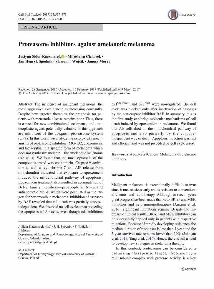
ORIGINAL ARTICLE Proteasome inhibitors against amelanotic melanoma Justyna Sidor-Kaczmarek & Mirosława Cichorek & Jan Henryk Spodnik & Sławomir Wójcik & Janusz Moryś Received: 28 September 2016 /Accepted: 15 February 2017 /Published online: 9 March 2017 # The Author(s) 2017. This article is published with open access at Springerlink.com Abstract The incidence of malignant melanoma, the most aggressive skin cancer, is increasing constantly. Despite new targeted therapies, the prognosis for pa- tients with metastatic disease remains poor. Thus, there is a need for new combinational treatments, and anti- neoplastic agents potentially valuable in this approach are inhibitors of the ubiquitin-proteasome system (UPS). In this work, we analyze the cytotoxicity mech- anisms of proteasome inhibitors (MG-132, epoxomicin, and lactacystin) in a specific form of melanoma which does not synthesize melanin—the amelanotic melanoma (Ab cells). We found that the most cytotoxic of the compounds tested was epoxomicin. Caspase-9 activa- tion as well as cytochrome C and AIF release from mitochondria indicated that exposure to epoxomicin induced the mitochondrial pathway of apoptosis. Epoxomicin treatment also resulted in accumulation of Bcl-2 family members —proapoptotic Noxa and antiapoptotic Mcl-1, which were postulated as the tar- gets for bortezomib in melanoma. Inhibition of caspases by BAF revealed that cell death was partially caspase- independent. We observed no cell cycle arrest preceding the apoptosis of Ab cells, even though cdk inhibitors p21 Cip1/Waf1 and p27 Kip1 were up-regulated. The cell cycle was blocked only after inactivation of caspases by the pan-caspase inhibitor BAF. In summary, this is the first study exploring molecular mechanisms of cell death induced by epoxomicin in melanoma. We found that Ab cells died on the mitochondrial pathway of apoptosis and also partially by the caspase- independent way of death. Apoptosis induction was fast and efficient and was not preceded by cell cycle arrest. Keywords Apoptosis . Cancer . Melanoma . Proteasome inhibitors Introduction Malignant melanoma is exceptionally difficult to treat since it metastasizes early and is resistant to convention- al chemo- and radiotherapy. Although in recent years great progress has been made thanks to BRAF and MEK inhibitors and new immunotherapies (Amann et al. 2016), significant limitations remain. Despite the im- pressive clinical results, BRAF and MEK inhibitors can be successfully applied only in patients with respective mutations. Because of rapidly developing resistance, the median duration of responses is less than 1 year and the 5-year survival rate remains lower than 10% (Johnson et al. 2015; Tang et al. 2016). Hence, there is still a need to develop new strategies in melanoma therapy. In this context, proteasome can be considered a promising therapeutic target. Proteasome, a multisubunit complex with protease activity, is a key Cell Biol Toxicol (2017) 33:557–573 DOI 10.1007/s10565-017-9390-0 J. Sidor-Kaczmarek (*) : J. H. Spodnik : S. Wójcik : J. Moryś Department of Anatomy and Neurobiology, Medical University of Gdansk, Gdansk, Poland e-mail: [email protected] M. Cichorek Department of Embryology, Medical University of Gdansk, Gdansk, Poland
Transcript of Proteasome inhibitors against amelanotic … ARTICLE Proteasome inhibitors against amelanotic...
ORIGINAL ARTICLE
Proteasome inhibitors against amelanotic melanoma
Justyna Sidor-Kaczmarek & Mirosława Cichorek &
Jan Henryk Spodnik & Sławomir Wójcik & Janusz Moryś
Received: 28 September 2016 /Accepted: 15 February 2017 /Published online: 9 March 2017# The Author(s) 2017. This article is published with open access at Springerlink.com
Abstract The incidence of malignant melanoma, themost aggressive skin cancer, is increasing constantly.Despite new targeted therapies, the prognosis for pa-tients with metastatic disease remains poor. Thus, thereis a need for new combinational treatments, and anti-neoplastic agents potentially valuable in this approachare inhibitors of the ubiquitin-proteasome system(UPS). In this work, we analyze the cytotoxicity mech-anisms of proteasome inhibitors (MG-132, epoxomicin,and lactacystin) in a specific form of melanoma whichdoes not synthesizemelanin—the amelanotic melanoma(Ab cells). We found that the most cytotoxic of thecompounds tested was epoxomicin. Caspase-9 activa-tion as well as cytochrome C and AIF release frommitochondria indicated that exposure to epoxomicininduced the mitochondrial pathway of apoptosis.Epoxomicin treatment also resulted in accumulation ofBcl-2 family members—proapoptotic Noxa andantiapoptotic Mcl-1, which were postulated as the tar-gets for bortezomib in melanoma. Inhibition of caspasesby BAF revealed that cell death was partially caspase-independent. We observed no cell cycle arrest precedingthe apoptosis of Ab cells, even though cdk inhibitors
p21Cip1/Waf1 and p27Kip1 were up-regulated. The cellcycle was blocked only after inactivation of caspasesby the pan-caspase inhibitor BAF. In summary, this isthe first study exploring molecular mechanisms of celldeath induced by epoxomicin in melanoma. We foundthat Ab cells died on the mitochondrial pathway ofapoptosis and also partially by the caspase-independent way of death. Apoptosis induction was fastand efficient and was not preceded by cell cycle arrest.
Keywords Apoptosis .Cancer .Melanoma .Proteasomeinhibitors
Introduction
Malignant melanoma is exceptionally difficult to treatsince it metastasizes early and is resistant to convention-al chemo- and radiotherapy. Although in recent yearsgreat progress has beenmade thanks to BRAF andMEKinhibitors and new immunotherapies (Amann et al.2016), significant limitations remain. Despite the im-pressive clinical results, BRAF and MEK inhibitors canbe successfully applied only in patients with respectivemutations. Because of rapidly developing resistance, themedian duration of responses is less than 1 year and the5-year survival rate remains lower than 10% (Johnsonet al. 2015; Tang et al. 2016). Hence, there is still a needto develop new strategies in melanoma therapy.
In this context, proteasome can be considered apromising therapeutic target. Proteasome, amultisubunit complex with protease activity, is a key
Cell Biol Toxicol (2017) 33:557–573DOI 10.1007/s10565-017-9390-0
J. Sidor-Kaczmarek (*) : J. H. Spodnik : S. Wójcik :J. MoryśDepartment of Anatomy and Neurobiology, Medical University ofGdansk, Gdansk, Polande-mail: [email protected]
M. CichorekDepartment of Embryology, Medical University of Gdansk,Gdansk, Poland
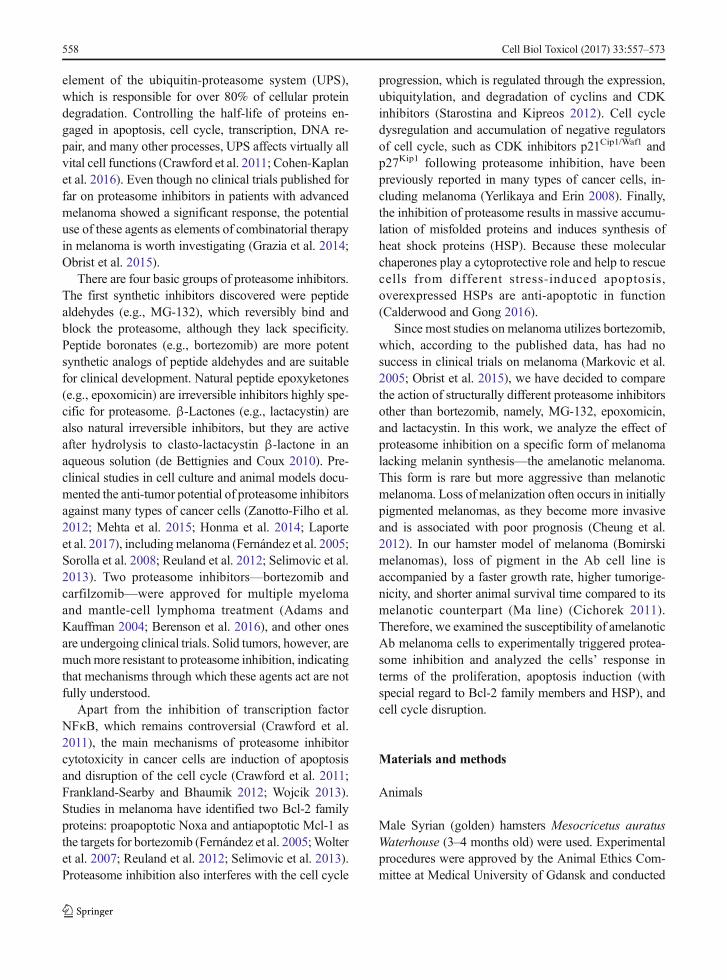
element of the ubiquitin-proteasome system (UPS),which is responsible for over 80% of cellular proteindegradation. Controlling the half-life of proteins en-gaged in apoptosis, cell cycle, transcription, DNA re-pair, and many other processes, UPS affects virtually allvital cell functions (Crawford et al. 2011; Cohen-Kaplanet al. 2016). Even though no clinical trials published forfar on proteasome inhibitors in patients with advancedmelanoma showed a significant response, the potentialuse of these agents as elements of combinatorial therapyin melanoma is worth investigating (Grazia et al. 2014;Obrist et al. 2015).
There are four basic groups of proteasome inhibitors.The first synthetic inhibitors discovered were peptidealdehydes (e.g., MG-132), which reversibly bind andblock the proteasome, although they lack specificity.Peptide boronates (e.g., bortezomib) are more potentsynthetic analogs of peptide aldehydes and are suitablefor clinical development. Natural peptide epoxyketones(e.g., epoxomicin) are irreversible inhibitors highly spe-cific for proteasome. β-Lactones (e.g., lactacystin) arealso natural irreversible inhibitors, but they are activeafter hydrolysis to clasto-lactacystin β-lactone in anaqueous solution (de Bettignies and Coux 2010). Pre-clinical studies in cell culture and animal models docu-mented the anti-tumor potential of proteasome inhibitorsagainst many types of cancer cells (Zanotto-Filho et al.2012; Mehta et al. 2015; Honma et al. 2014; Laporteet al. 2017), includingmelanoma (Fernández et al. 2005;Sorolla et al. 2008; Reuland et al. 2012; Selimovic et al.2013). Two proteasome inhibitors—bortezomib andcarfilzomib—were approved for multiple myelomaand mantle-cell lymphoma treatment (Adams andKauffman 2004; Berenson et al. 2016), and other onesare undergoing clinical trials. Solid tumors, however, aremuchmore resistant to proteasome inhibition, indicatingthat mechanisms through which these agents act are notfully understood.
Apart from the inhibition of transcription factorNFκB, which remains controversial (Crawford et al.2011), the main mechanisms of proteasome inhibitorcytotoxicity in cancer cells are induction of apoptosisand disruption of the cell cycle (Crawford et al. 2011;Frankland-Searby and Bhaumik 2012; Wojcik 2013).Studies in melanoma have identified two Bcl-2 familyproteins: proapoptotic Noxa and antiapoptotic Mcl-1 asthe targets for bortezomib (Fernández et al. 2005;Wolteret al. 2007; Reuland et al. 2012; Selimovic et al. 2013).Proteasome inhibition also interferes with the cell cycle
progression, which is regulated through the expression,ubiquitylation, and degradation of cyclins and CDKinhibitors (Starostina and Kipreos 2012). Cell cycledysregulation and accumulation of negative regulatorsof cell cycle, such as CDK inhibitors p21Cip1/Waf1 andp27Kip1 following proteasome inhibition, have beenpreviously reported in many types of cancer cells, in-cluding melanoma (Yerlikaya and Erin 2008). Finally,the inhibition of proteasome results in massive accumu-lation of misfolded proteins and induces synthesis ofheat shock proteins (HSP). Because these molecularchaperones play a cytoprotective role and help to rescuecells from different stress-induced apoptosis,overexpressed HSPs are anti-apoptotic in function(Calderwood and Gong 2016).
Since most studies on melanoma utilizes bortezomib,which, according to the published data, has had nosuccess in clinical trials on melanoma (Markovic et al.2005; Obrist et al. 2015), we have decided to comparethe action of structurally different proteasome inhibitorsother than bortezomib, namely, MG-132, epoxomicin,and lactacystin. In this work, we analyze the effect ofproteasome inhibition on a specific form of melanomalacking melanin synthesis—the amelanotic melanoma.This form is rare but more aggressive than melanoticmelanoma. Loss of melanization often occurs in initiallypigmented melanomas, as they become more invasiveand is associated with poor prognosis (Cheung et al.2012). In our hamster model of melanoma (Bomirskimelanomas), loss of pigment in the Ab cell line isaccompanied by a faster growth rate, higher tumorige-nicity, and shorter animal survival time compared to itsmelanotic counterpart (Ma line) (Cichorek 2011).Therefore, we examined the susceptibility of amelanoticAb melanoma cells to experimentally triggered protea-some inhibition and analyzed the cells’ response interms of the proliferation, apoptosis induction (withspecial regard to Bcl-2 family members and HSP), andcell cycle disruption.
Materials and methods
Animals
Male Syrian (golden) hamsters Mesocricetus auratusWaterhouse (3–4 months old) were used. Experimentalprocedures were approved by the Animal Ethics Com-mittee at Medical University of Gdansk and conducted
558 Cell Biol Toxicol (2017) 33:557–573
in accordance with National Health and Medical Re-search Council’s guide for the care and use of laboratoryanimals.
Transplantable hamster’s melanomas
Original transplantable melanotic melanoma (Ma) hasbeen derived from a spontaneous melanoma of the skinthat appeared in a bred of golden hamster in 1959.Amelanotic melanoma line (Ab) originated from theMa form by a spontaneous alteration (Bomirski et al.1988). Once established, amelanotic line is maintainedin vivo by consecutive, subcutaneous injection of tumorcells every 11 days. This melanoma model is known asBomirski hamster’s melanoma.
Isolation of amelanotic melanoma cells (Ab cells)
Ab cells were isolated from the solid tumors by a non-enzymatic method reported previously (Cichorek et al.2007). Cell suspension contained 95–98% of viablecells as estimated by trypan blue exclusion assay. Afterisolation, the cells were cultured for 24 h under standardconditions (RPMI 1640, 10% fetal bovine serum (FBS),antibiotics; 37 °C at 5% CO2) before subsequent exper-iments. Each independent experiment was performed onthe cells isolated from a single tumor.
Proteasome and caspase inhibitor treatment
Ab cells were incubated with proteasome inhibitors:epoxomicin, MG-132, or lactacystin at a concentrationranging from 0.1 to 10 μM for 6 to 72 h at the standardconditions. Untreated (control) samples were treatedwith solvent only: DMSO for epoxomicin and MG-132 and ddH20 for lactacystin. When using pan-caspase inhibitor BAF (Boc-D-FMK) (Calbiochem,USA), Ab cells were preincubated with BAF for 2 hbefore exposure to proteasome inhibitors.
Cell viability assay
Cell viability was determined by XTT assay (Roche,Germany), which measures ability of the cells to reducetetrazolium salt XTT (2,3-bis-(2-methoxy-4-nitro-5-sulfophenyl)-2h-tetrazolium-5-carboxanilide) to water-soluble formazan product. Ab melanoma cells wereseeded in 96-well plates at a concentration 2 × 10⁴ perwell and 5 h before indicated time XTT solution was
added. Orange-colored formazan product was quanti-fied at 450 nm using a microplate reader Multiscan FC(Thermoscientific, USA).
Chymotrypsin-like proteasome activity assay
Chymotrypsin-like (ChT-L) proteasome activity wasanalyzed by luminescent assay (Promega, USA). Ac-cording to the manufacturer’s instruction, Ab cellswere seeded in 96-well plates at a concentration 104
per well and incubated for 2 h. Fifteen minutes beforethe measurement, buffer containing luminogenicChT-L p r o t e a s ome s u b s t r a t e Su c -LLVY-aminoluciferin (final concentration 20 μM) and lu-ciferase was added. A luminescent signal generatedby cleavage of proteasome substrate was detectedusing a microplate luminometer Fluoroscan FC(ThermoScientific, USA).
Cell cycle analysis
Cell cycle distribution was determined by flow cytom-etry method based on the DNA content in cells’ nuclei,as described earlier (Cichorek 2011). Ethanol-fixed1 × 10 6 Ab cells were suspended in 1 ml of the stainingsolution containing 40 μg/ml propidium iodide (PI,Sigma Chemicals, USA) and 100 μg/ml RNaze A andincubated 30 min at 37 °C. Fluorescence was measuredusing a FACS Calibur flow cytometer (Becton Dickin-son Immunocytometry Systems, USA; Department ofPathophysiology, Medical University of Gdansk). Atotal of 20,000 events were stored from each stainedsample and analyzed off-line using WinMDI2.6 soft-ware (obtained from J. Trotter, The Scripps Institute, LaJolla, USA). Cells in S and G2/M phases of the cellcycle were regarded as dividing cells and analyzedtogether.
Hoechst staining
Apoptotic morphology of cell nuclei was analyzed byHoechst 33342 (Invitrogen, USA) staining. Ab cellswere incubated with 3 μg/ml Hoechst 33342 for30 min and assessed on a reversed microscope NikonEclipse TE300 (Nikon, Japan) under ×10 and ×40 ob-jective lenses.
Cell Biol Toxicol (2017) 33:557–573 559
Phosphatidylserine externalization assay
Early apoptotic change of plasma membrane structure,phosphatidylserine (PS) externalization, was deter-mined by Annexin V-FITC and PI staining (BDPharmingen, USA) according to the manufacturer’s in-structions. Cells were seeded in 12-well plates at aconcentration 1 × 10⁶ per well. Staining allows to de-termine three populations of cells: viable (An−/PI−),early apoptotic (An+/PI−), and late apoptotic (An+/PI+). Fluorescence intensity was measured by a FACSCalibur f low cytometer (Becton DickinsonImmunocytometry Systems, USA; Department of Path-ophysiology, Medical University of Gdansk). A total of20,000 events were stored from each stained sample andanalyzed off-line using WinMDI2.6 software.
Immunoblotting
Total cell lysates were obtained by incubation of 3 × 10⁶
cells in lysis buffer (10 mM Tris-HCl pH 8.0, 140 mMNaCl, 2% TX-100) with addition of the protease inhib-itors (1 mMAEBSF, 0.8 μM aprotinin, 50 μM bestatin,15 μM E-64, 20 μM leupeptin, 10 μM pepstatin A) for1 h on ice and cenrifugation for 15 min at 14,000 rpm.Supernatants were collected and stored at −70 °C untilfurther processing. Total protein amount was quantifiedby Lowry assay (Bio-Rad, USA). Sixty micrograms oflysate was subjected to electrophoresis in 10%, 12% orgradient 4–15% SDS gels under reducing conditionsand transferred to nitrocellulose membrane (Bio-Rad,USA). After 2-h blocking in 4% non-fat milk, mem-branes were probed overnight with primary antibodies:rabbit polyclonal anti-caspase 3 (Santa Cruz Biotech-nology, USA; 1:250), mouse monoclonal anti-caspase 9(Stressgen, USA; 1:500), rabbit polyclonal anti-HSP27(Stressgen, USA; 1:1000), mouse monoclonal anti-HSP70 (StressMarq, USA; 1:5000), mouse monoclonalanti-p21 (BD Pharmingen, USA; 1:250), mouse mono-clonal anti-p27 (BD Pharmingen, USA; 1:1000), mousemonoclonal anti-Noxa (Abcam, UK; 1:200), and rabbitmonoclonal anti-Mcl 1 (Abcam, UK; 1:500). Anti-β-actin mouse monoclonal antibody (Sigma Chemicals,USA; 1:15,000) was used as an equal protein loadingcontrol. After washing, membranes were next incubatedfor 2 h with horseradish peroxidase-conjugated second-ary antibody (Pierce, USA; 1:50,000). Chemilumines-cent signal was developed using the SuperSignal WestPico system (Pierce, USA) and visualized onX-ray film.
Band intensity was semiquantified using ImageJ 1.38software (National Institutes of Health, USA) and wasshown as a ratio to actin.
Cytochrome C and AIF immunofluorescent staining
For immunofluorescent detection of cytochrome C andapoptosis-inducing factor (AIF), cells were cultured oncoverslips with proteasome inhibitors for 6 h. Doublestaining of mitochondria and cytochrome C was per-formed to analyze cytochromeC release frommitochon-dria to cytoplasm. First, the mitochondria in living cellswere stained with 250 nM MitoTracker Red CMXRos(Invitrogen, USA) for 30 min. Next, the cells were10min fixed (3.7% formaldehyde), 5 min permeabilized(90% methanol), PBS rinsed, 30 min blocked in 3%FBS, and probed for 1 h with mouse monoclonal anti-cytochrome C antibody (clone 6H2.B4, BDPharmingen, USA; 1:400). After PBS rinsing, the cellswere incubated with AlexaFluor 488-conjugated goatanti-mouse secondary antibody (Invitrogen, USA;1:400) for 1 h.
To determine AIF translocation, double staining ofcell nuclei and AIF was performed. The cells were10 min fixed (4% paraformaldehyde), 10 min perme-abilized (0.2% Triton), PBS rinsed, blocked in 2% bo-vine serum albumin (BSA) for 30 min. Next, the cellswere incubated overnight in 4 °C with rabbit monoclo-nal anti-AIF antibody (clone E20, Abcam, UK; 1:500).After PBS rinsing, the cells were incubated withAlexaFluor 488-conjugated goat anti-rabbit secondaryantibodies (Invitrogen, USA; 1:400) for 1 h. To removeRNA before staining of cell nuclei by PI, the cells weretreated by 0.1 mg/ml RNase A (Sigma Chemical, USA)in 37 °C for 30 min, PBS rinsed, and then stained in2.5 μg/ml PI solution in PBS for 30 min. Finally, thestained cells on coverslips were mounted in PermaFluor
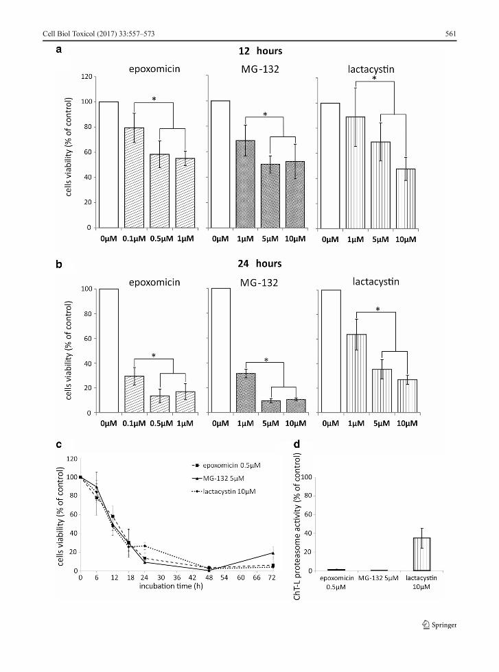
�Fig. 1 Cytotoxic effect of proteasome inhibitors on Abmelanomacells. Cell viability was determined byXTTassay and expressed asa percentage of the viability of untreated cells. Cells were exposedto increasing concentrations of epoxomicin, MG-132, orlactacystin for 12 (a) and 24 (b) hours or to 0.5 μM epoxomicin,5μMMG-132, or 10μM lactacystin for up to 72 h (c). An asterisk(*) indicates a statistically significant difference between variousproteasome inhibitor concentrations; p < 0.05, Kruskal-Wallis test.d Inhibition of chymotrypsin-like (ChT-L) proteasome activity inAb melanoma cells. Cells were exposed to 0.5 μM epoxomicin,5 μM MG-132, or 10 μM lactacystin for 2 h, and proteasomeactivity was measured using a luminescent assay. The datarepresent means ± SD of three independent experiments
560 Cell Biol Toxicol (2017) 33:557–573
Cell Biol Toxicol (2017) 33:557–573 561
(Shanon Lipshaw, USA) as the microscope slides. Im-munofluorescent staining was assessed using a confocallaser scanning microscopy system (Radiance 2100, Bio-Rad UK) mounted on a microscope Eclipse 600 (Nikon,Japan). Images were obtained with ×60 oil immersionobjective lenses and analyzed with the LaserSharp 2000and LaserPix v. 2.0 software (both Bio-Rad, UK).
Statistical analysis
The statistical analysis was performed using the dataanalysis software system STATISTICA version 12,StatSoft Inc. (2014). Nonparametric Mann-Whitney Utest, Kruskall-Wallis, and Dunn tests were used. p ≤ 0.05was considered statistically significant. Data are displayedas arithmetical mean ± standard deviation (SD).
Results
Proteasome inhibition decreases viability of Abmelanoma cells
Ab cells were exposed to increasing concentrations ofthree structurally different proteasome inhibitors:epoxomicin, MG-132, and lactacystin for up to 72 hand cell viability changes were analyzed by XTT reduc-tion. All the compounds tested diminished Ab cell via-bility in a dose-dependent manner (Fig. 1a, b). Duringthe initial 6 h of treatment, only minor changes wereobserved, since neither of the inhibitors reduced the cellviability more than 20% of the control value (data notshown). After 12 h, the lowest concentrations of inhib-itors (0.1 μM epoxomicin, 1 μM MG-132, and 1 μMlactacystin) decreased Ab cell viability to 79, 69, and89% of the control, respectively (Fig. 1a). The fivetimes’ higher doses reduced the cell viability a further20% (Fig. 1a; p < 0.05). Twenty-four-hour treatmentwith the lowest concentration of the inhibitors decreasedcell viability to 29, 31, and 63%, respectively (Fig. 1b).Again, five times higher doses were significantly morecytotoxic and left only 13, 9, and 35% of viable cells(Fig. 1b). A further increase in the inhibitor concentra-tion did not change their cytotoxicity significantly(Fig. 1a, b). At all time points, epoxomicin was the mostpotent of the three proteasome inhibitors tested. At aconcentration of 0.5 μM, its effect was comparable withthe action of a ten times higher dose of MG-132 (5 μM)or a 20 times higher dose of lactacystin (10 μM)
(Fig. 1c). The least effective of the proteasome inhibitorswas lactacystin. Twenty-four-hour incubation with its10 μM concentration left 27% cells alive, and similarviability level was observed at that time for 1 μM MG-132 or 0.1 μM epoxomicin (Fig. 1b). Finally, 48-hexposure, regardless of the proteasome inhibitor used,resulted in the death of over 95% of the cells (Fig. 1c).
It is worth noting that 2-h exposure to 0.5 μMepoxomicin and 5 μM MG-132 totally blockedchymotrypsin-like (ChT-L) proteasome activity in Abcells (Fig. 1d), while lactacystin, even in dose of 10 μM,did not inhibit this activity completely, but decreased itto 35% of the control value. Higher concentrations oflactacystin were not tested because of the possible un-specific inhibition of the other cellular proteases.
Because epoxomicin exerted the strongest cytotoxiceffect on Ab melanoma cells, it was chosen for furtherinvestigations concerning the molecular mechanisms ofcell death induced by proteasome inhibition.Epoxomicin was used in 0.5 μM concentration, whichwas the lowest dose that totally blocked ChT-L protea-some activity (Fig. 1d).
Epoxomicin induces apoptosis in Ab melanoma cells
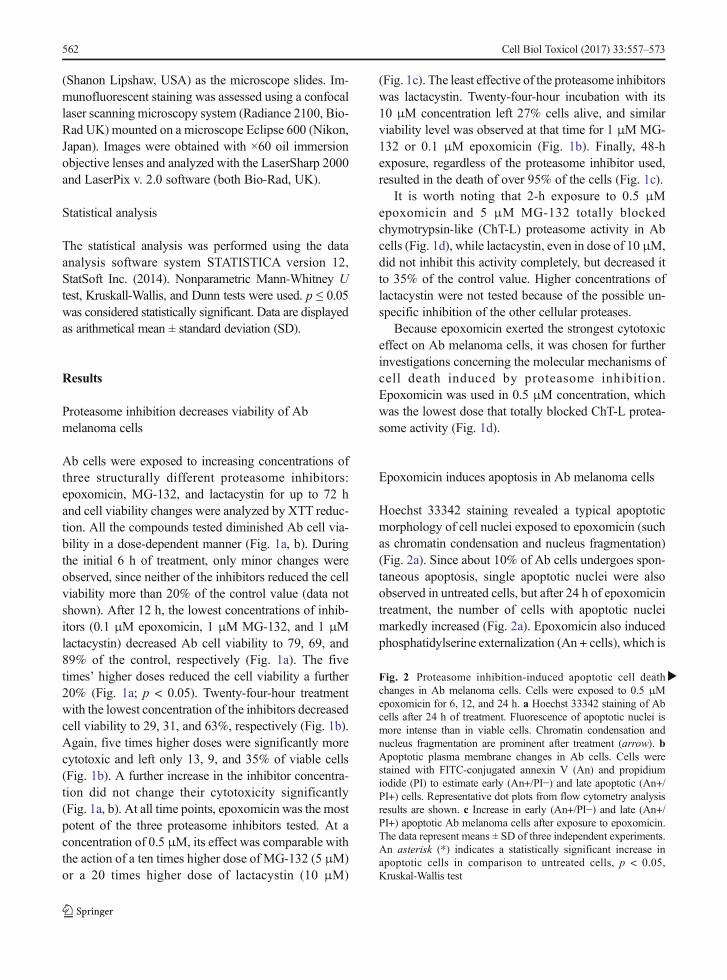
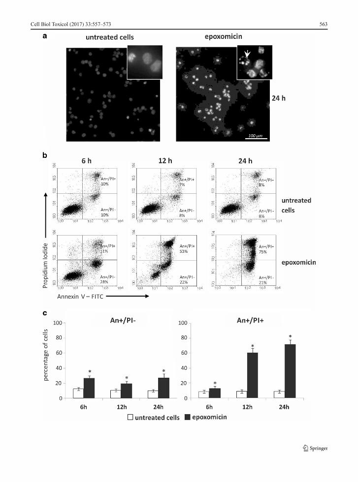
Hoechst 33342 staining revealed a typical apoptoticmorphology of cell nuclei exposed to epoxomicin (suchas chromatin condensation and nucleus fragmentation)(Fig. 2a). Since about 10% of Ab cells undergoes spon-taneous apoptosis, single apoptotic nuclei were alsoobserved in untreated cells, but after 24 h of epoxomicintreatment, the number of cells with apoptotic nucleimarkedly increased (Fig. 2a). Epoxomicin also inducedphosphatidylserine externalization (An + cells), which is
�Fig. 2 Proteasome inhibition-induced apoptotic cell deathchanges in Ab melanoma cells. Cells were exposed to 0.5 μMepoxomicin for 6, 12, and 24 h. a Hoechst 33342 staining of Abcells after 24 h of treatment. Fluorescence of apoptotic nuclei ismore intense than in viable cells. Chromatin condensation andnucleus fragmentation are prominent after treatment (arrow). bApoptotic plasma membrane changes in Ab cells. Cells werestained with FITC-conjugated annexin V (An) and propidiumiodide (PI) to estimate early (An+/PI−) and late apoptotic (An+/PI+) cells. Representative dot plots from flow cytometry analysisresults are shown. c Increase in early (An+/PI−) and late (An+/PI+) apoptotic Ab melanoma cells after exposure to epoxomicin.The data represent means ± SD of three independent experiments.An asterisk (*) indicates a statistically significant increase inapoptotic cells in comparison to untreated cells, p < 0.05,Kruskal-Wallis test
562 Cell Biol Toxicol (2017) 33:557–573
Cell Biol Toxicol (2017) 33:557–573 563
the marker of apoptotic changes in the plasma mem-brane (Fig. 2b, c). In untreated cells, the spontaneousearly (An+/PI−) and late (An+/PI+) apoptotic cells com-prised about 10% each. A statistically significant re-sponse to epoxomicin was detected as early as after6 h when the number of early apoptotic cells (An+/PI−) rose to over 20% (Fig. 2c, p < 0.05) and remained atthis level until 24 h. The number of late apoptotic cells(An+/PI+) under epoxomicin treatment increased sig-nificantly to 60% after 12 h and 70% after 24 h (Fig. 2c).
The apoptotic death of Ab cells exposed toepoxomicin was further confirmed by caspase 3 activa-tion. Procaspase 3 activation (detected by the presenceof 17 kDa cleaved subunit) started at 6 h and lastedthroughout the duration of the experiment, as shown byimmunoblot studies (Fig. 3a). Classic apoptotic cellmorphology was also observed in cytochrome C immu-nofluorescent staining, where membrane blebbing wasevident (Fig. 3b, arrow indication).
Epoxomicin activates the mitochondrial apoptoticpathway in Ab melanoma cells
Immunoblot analysis of initiator caspase 9 revealed itsactivation, estimated by the presence of the active sub-units 35/37 kDa, after 6 h of exposure to epoxomicin.The procaspase 9 (46 kDa) protein level gradually de-clined with time up to 24 h (Fig. 3a). Induction of themitochondrial pathway of apoptosis was confirmed byimmunofluorescent detection of cytochrome C and AIFtranslocation from mitochondria (Fig. 3b). In untreatedcells, cytochrome C and AIF were localized in mito-chondria, but after 6 h of epoxomicin exposure, bothproteins were released to cytosol (Fig. 3b).
Ab cells express antiapoptotic factors such assurvivin or Bcl-2 (unpublished data). In this work, weexamined two other proteins of Bcl-2 family:proapoptotic Noxa and antiapoptotic Mcl-1. Immuno-blot analysis presented in Fig. 3a showed that exposureto epoxomicin induced Noxa protein expression and itsaccumulation with time. Mcl-1, as the prosurvival pro-tein, was constitutively expressed in untreated Ab cells,and proteasome inhibition caused its transient increaseat 6 h of treatment (Fig. 3a).
Furthermore, proteasome inhibition resulted in theaccumulation of anti-apoptotic proteins from the HSPfamily: HSP70 and HSP27. We found no constitutiveexpression of HSP70 and a very low level of HSP27 inuntreatedAb cells (Fig. 3a). Proteasome inhibition led to
rapid and massive HSP70 up-regulation, which started6 h after epoxomicin treatment. Induction of HSP27expression was weaker and became apparent after 12 h.
Epoxomicin induces apoptosis in bothcaspase-dependent and caspase-independent way
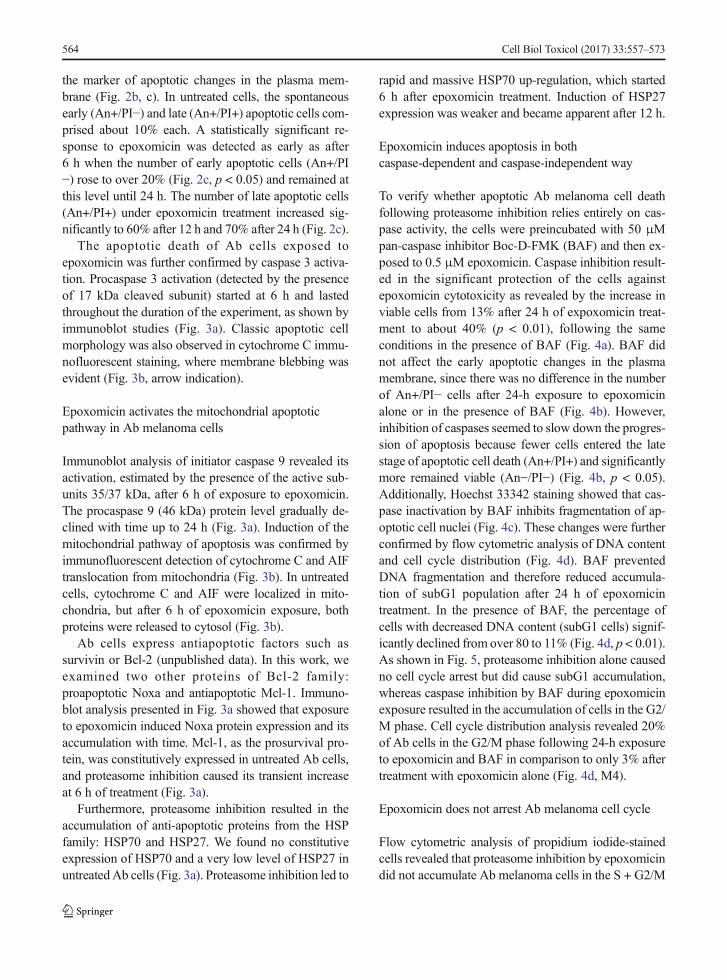
To verify whether apoptotic Ab melanoma cell deathfollowing proteasome inhibition relies entirely on cas-pase activity, the cells were preincubated with 50 μMpan-caspase inhibitor Boc-D-FMK (BAF) and then ex-posed to 0.5 μM epoxomicin. Caspase inhibition result-ed in the significant protection of the cells againstepoxomicin cytotoxicity as revealed by the increase inviable cells from 13% after 24 h of expoxomicin treat-ment to about 40% (p < 0.01), following the sameconditions in the presence of BAF (Fig. 4a). BAF didnot affect the early apoptotic changes in the plasmamembrane, since there was no difference in the numberof An+/PI− cells after 24-h exposure to epoxomicinalone or in the presence of BAF (Fig. 4b). However,inhibition of caspases seemed to slow down the progres-sion of apoptosis because fewer cells entered the latestage of apoptotic cell death (An+/PI+) and significantlymore remained viable (An−/PI−) (Fig. 4b, p < 0.05).Additionally, Hoechst 33342 staining showed that cas-pase inactivation by BAF inhibits fragmentation of ap-optotic cell nuclei (Fig. 4c). These changes were furtherconfirmed by flow cytometric analysis of DNA contentand cell cycle distribution (Fig. 4d). BAF preventedDNA fragmentation and therefore reduced accumula-tion of subG1 population after 24 h of epoxomicintreatment. In the presence of BAF, the percentage ofcells with decreased DNA content (subG1 cells) signif-icantly declined from over 80 to 11% (Fig. 4d, p < 0.01).As shown in Fig. 5, proteasome inhibition alone causedno cell cycle arrest but did cause subG1 accumulation,whereas caspase inhibition by BAF during epoxomicinexposure resulted in the accumulation of cells in the G2/M phase. Cell cycle distribution analysis revealed 20%of Ab cells in the G2/M phase following 24-h exposureto epoxomicin and BAF in comparison to only 3% aftertreatment with epoxomicin alone (Fig. 4d, M4).
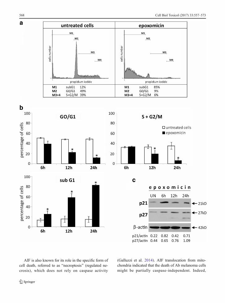
Epoxomicin does not arrest Ab melanoma cell cycle
Flow cytometric analysis of propidium iodide-stainedcells revealed that proteasome inhibition by epoxomicindid not accumulate Ab melanoma cells in the S + G2/M
564 Cell Biol Toxicol (2017) 33:557–573

phases (Fig. 5a; M3, M4). On the contrary, even thoughat 6 h the number of cells in the S + G2/M phases didnot change markedly, remaining over 30%, later it
began to decrease and finally declined to 6% after 24 h(Fig. 5b; p < 0.05). With time, epoxomicin also mark-edly reduced the G0/G1 cell content (Fig. 5a, b).
Fig. 3 Epoxomicin-induced mitochondrial pathway of apoptosisand HSP expression in Ab melanoma cells. Cells were exposed to0.5 μM epoxomicin for 6, 12, and 24 h. a Immunoblot analysis ofcaspase 3 and 9 activation and expression of Mcl-1, Noxa, andHSP proteins in Ab cells in response to epoxomicin. β-Actin wasused as a control of the equal protein loading. UN untreated cells.The densitometric ratio of band intensity is shown. b Double-
labeled immunofluorescent staining revealing cytochrome C andAIF release from mitochondria in Ab cells treated withepoxomicin for 6 h. Upper panel cytochrome C (green) andmitochondria (red), an arrow indicates the membrane blebbingspecific for apoptotic cell death; lower panel: AIF (green) and cellnucleus (red)
Cell Biol Toxicol (2017) 33:557–573 565
566 Cell Biol Toxicol (2017) 33:557–573
Reduction of cells in G0/G1 and S + G2/M phases wasaccompanied by the accumulation of the subG1 popu-lation, which comprises the cells with reduced DNAcontent: cells with fragmented nuclei and apoptotic bod-ies (Fig. 5a, M1). Epoxomicin treatment significantlyincreased subG1 population from about 10% in untreat-ed cells to about 80% after 24-h exposure (Fig. 5a, b).Accumulation of subG1 population corresponds withdecreased cell viability measured by XTT reduction(Fig. 1b) and apoptotic changes in cell nuclei revealedby Hoechst 33342 staining (Fig. 2a).
Immunoblot analysis of the inhibitors of cycle-dependent kinases, p21Cip1/Waf1 and p27Kip1, re-vealed the transient accumulation of both proteins(Fig. 5c).
Discussion
In this work, we examine the cytotoxic effect of threestructurally different proteasome inhibitors: MG-132,epoxomicin, and lactacystin on amelanotic melanomaof hamster origin (Ab cells). We also explore molecularmechanisms of apoptosis induction and cell cycle dis-ruption elicited by epoxomicin, which we found to bethe most potent proteasome inhibitor against amelanoticmelanoma.
Sensitivity of Ab melanoma cells is different for variousproteasome inhibitors
Among the proteasome inhibitors examined, the mosteffective was epoxomicin, while the action oflactacystin was the weakest. Similar differences in cellsensitivity were shown previously in human melanomacell lines, which weremore sensitive to epoxomicin thanto MG-132 or to MG-132 than lactacystin (Sorolla et al.2008; Qin et al. 2005). It seems that the cytotoxicpotential of epoxomicin is related to its high specificityand irreversible binding to proteasome (de Bettigniesand Coux 2010). The relative resistance of Ab cells tolactacystin probably results from the incomplete inhibi-tion of proteasome activity, which in our study was notachieved even for 10 μM drug concentration. We mayonly hypothesize that this is due to the specific chemicalstructure of β-lactone. Lactacystin is not active itself,but in an aqueous solutions, it undergoes spontaneoushydrolysis to clasto-lactacystin β-lactone, which is anactive but less stable compound (de Bettignies and Coux2010). Furthermore, hydrolysis requires neutral pH,while amelanotic melanoma cells are known for theirability to acidify the medium (Halaban 2002).
Epoxomicin induces apoptosis in Ab melanoma cells
Because of its cytotoxic potency against Ab melanomacells, we have chosen epoxomicin for the further inves-tigation of the mechanisms underlying the cell’s re-sponse. Many studies have proved that the principalway of cancer cell death elicited by proteasome inhibi-tors is apoptosis. However, most studies on melanomarelied on bortezomib (Fernández et al. 2005; Qin et al.2005; Wolter et al. 2007; Yerlikaya and Erin 2008;Amschler et al. 2010; Reuland et al. 2012; Selimovicet al. 2013), and little is known about the mechanisms ofcell death induced by other proteasome inhibitors, suchas epoxomicin. Only Sorolla et al. (2008) reported cas-pase activation after epoxomicin treatment in primaryhuman melanoma cell lines. We found that in Ab cells,epoxomicin decreased viability mainly through apopto-sis induction, as indicated by typical apoptotic hall-marks, e.g., chromatin condensation, externalization ofphosphatidylserine, or caspase-3 activation. Thecaspase-9 activation as well as cytochrome C and AIFrelease from mitochondria showed the induction of themitochondrial pathway of apoptosis.
�Fig. 4 Epoxomicin-induced apoptosis of Abmelanoma cells onlypartially relies on caspase activation. Ab cells were treated with0.5 μM epoxomicin alone or in the presence of 50 μM pan-caspase inhibitor BAF for 24 h. a Cell viability was measured byan XTT assay and calculated as a percentage of the viability ofuntreated cells. b Cells were stained with FITC-conjugatedannexin V (An) and propidium iodide (PI) to estimate live cells(An−/PI−), early (An+/PI−) and late apoptotic (An+/PI+) cells. Ina and b, the data represent the means ± SD of three independentexperiments; asterisk (*) indicates statistically significantdifference between BAF 0 μM and BAF 50 μM, Mann-WhitneyU test, p < 0.01 (a) or p < 0.05 (b). c Ab cells were treated withepoxomicin alone or in the presence of BAF and stained withHoechst 33342. Arrows indicate the nucleus fragmentation in theabsence of BAF (upper panel, BAF 0 μM) and chromatincondensation, but no nucleus fragmentation in the presence ofBAF (lower panel, BAF 50 μM). d Cell cycle distribution wasdetermined in PI-stained Ab cells by flow cytometry.Representative histograms are shown. The percentage of cellswith fragmented DNA (subG1, M1) and in the G2/M (M4)phase of the cell cycle are indicated below the correspondinghistograms
Cell Biol Toxicol (2017) 33:557–573 567
AIF is also known for its role in the specific form ofcell death, referred to as Bnecroptosis^ (regulated ne-crosis), which does not rely on caspase activity
(Galluzzi et al. 2014). AIF translocation from mito-chondria indicated that the death of Ab melanoma cellsmight be partially caspase-independent. Indeed,
568 Cell Biol Toxicol (2017) 33:557–573
inactivation of caspases did not prevent the cytotoxiceffect of epoxomicin on Ab cells, but reduced it by half.This is in agreement with other studies, whose authorsobserved partial protection of melanoma cells by cas-pase inhibitors against cytotoxic effect of bortezomib(Qin et al. 2005; Sorolla et al. 2008; Selimovic et al.2013). In our study, inhibition of caspases had littleimpact on the early stages of cell death, such as changesin plasma membrane, but it completely blocked the lateevents of cell demise, e.g., nucleus fragmentation oraccumulation of cells with fragmented DNA (subG1fraction). We also observed cell cycle arrest in G2/Mphase, which was not present following epoxomicintreatment alone.
Epoxomicin changes the expression of Bcl-2 and HSPproteins
It has been postulated that the key elements ofbortezomib-induced apoptosis in melanoma cells areBcl-2 family members—proapoptotic Noxa andantiapoptotic Mcl-1. Bortezomib has been found totrigger substantial accumulation of Noxa after 18 or24 h in numerous melanoma cell lines (Fernándezet al. 2005; Qin et al. 2005; Wolter et al. 2007;Reuland et al. 2012; Selimovic et al. 2013). We ob-served a similar effect in our model, where Noxa accu-mulation started after 6 h of epoxomicin treatment andcontinued throughout the time of the experiment (24 h).Stabilization of Noxa probably facilitated cytochrome Cand AIF release from mitochondria in Ab cells. Accu-mulation of Noxa was also reported after MG-132
treatment in A375 amelanotic melanoma cells (Milleret al. 2009).
Induction of Noxa expression may be a way forproteasome inhibitors to overcome relatively highlevels of the antiapoptotic proteins present in mela-noma. Ab melanoma cells constitutively expresssurvivin, Bcl-2, and Bcl-XL proteins (unpublisheddata). In this study, we also found the constitutiveexpression of Mcl-1, the prosurvival member of Bcl-2 family preferentially bounded by Noxa, and itstransient accumulation after epoxomicin treatment.Our results are in accordance with studies on thebortezomib effect in different melanoma cell lines,which revealed a rapid increase in Mcl-1 expressionat first and then its reduction to or below basal levels(Wolter et al. 2007). Mcl-1 accumulation was alsofound in melanoma cell line A375 after 24-h treat-ment with MG-132 (Miller et al. 2009).
Othe r an t i apop to t i c p ro t e ins commonlyoverexpressed in many types of cancers includingmelanoma are HSP70 and HSP27 (Shipp et al.2013; Calderwood and Gong 2016). As molecularchaperones, HSP70 and HSP27 are strongly up-regulated in response to proteasome inhibition invarious types of cancer cells (Kim et al. 2011; Shahet al. 2016). In Ab cells, we found that proteinaccumulation after epoxomicin treatment was partic-ularly massive in the case of HSP70 and this proteinseemed not to be constitutively expressed in thesecells. HSP27 was present in untreated Ab cells at avery low level and its accumulation was slower.HSP70 and HSP27 status is different in variousmelanoma models. BML, A375, SK-Mel-19, andSK-Mel-103 cells showed no constitutive expressionof HSP70, but it very efficient induction bybortezomib (Fernández et al. 2005; Selimovic et al.2013). In another study, HSP70 was found to beconstitutively expressed in B16F10 cells and onlyweakly up-regulated by bortezomib (Yerlikaya andErin 2008). There is no data on HSP27 changes inmelanoma following proteasome inhibition, butstudies on the other cancer cell lines revealed eitherno changes, as in HL-60 and K562 cells (Klikovaet al. 2015) or its accumulation in response tobortezomib or MG-132, in the case of PC-3 cells(Kumano et al. 2012). In our model, the overexpres-sion of both HSP70 and HSP27 was apparently notsufficient to rescue Ab melanoma cells fromepoxomicin-induced apoptosis.
�Fig. 5 Ab melanoma cell cycle analysis and changes in thecdk (cell cycle-dependent kinases) inhibitors p21Cip1/Waf1 andp27Kip1 after epoxomicin treatment. Cells were exposed to0.5 μM epoxomicin for up to 24 h. a Representativehistograms of cell cycle analysis of cells exposed toepoxomicin for 24 h. DNA content was estimated bypropidium iodide staining, and cell cycle distribution wasassessed by flow cytometry. b Time-dependent decrease inG0/G1 and S + G2/M phases and accumulation of destroyedcells in subG1. Data represent means ± SD of threeindependent experiments. Asterisks (*) indicate statisticallysignificant difference between untreated and epoxomicin-treated cells (p < 0.05), Kruskal-Wallis test. c Immunoblotanalysis of p21Cip1/Waf1 and p27Kip1 up-regulation in Ab cellstreated with 0.5 μM epoxomicin. β-actin was used as a controlof the equal protein loading. UN-untreated cells. Thedensitometric ratio of band intensity is shown
Cell Biol Toxicol (2017) 33:557–573 569
Epoxomicin does not arrest the Ab melanoma cell cycle
Proteasome inhibition can disrupt the cell cycle andconsequently induce apoptosis also by targeting thecell cycle control factors (Crawford et al. 2011;Frankland-Searby and Bhaumik 2012). Various protea-some inhibitors, such as MG-132, bortezomib, andcarfilzomib, have been proven to stop the cell cycle
of different tumor cells (Zanotto-Filho et al. 2012;Mehta et al. 2015), including melanoma (Fernándezet al. 2005; Amschler et al. 2010), in a phases S andG2/M. In our study, however, there was no accumula-tion of Ab cells in the S + G2/M phase afterepoxomicin treatment. Instead, even as early as 6 hafter exposure, we noticed a significant accumulationof the subG1 population, which comprises cells with a
570 Cell Biol Toxicol (2017) 33:557–573
reduced DNA content. The cell cycle was blockedonly after inactivation of caspases which blocked latestages of apoptosis (nucleus fragmentation and apopto-tic body formation). Consequently, caspase inhibitionduring epoxomicin treatment resulted in a subG1 frac-tion decrease and cell cycle arrest in the G2/M phase.The same effect of caspase inactivation was reportedby Sorolla et al. (2008) in bortezomib-treated melano-ma cells.
To better understand the lack of cell cycle arrestfollowing exposure to epoxomicin alone, we esti-mated the level of key cdk inhibitors p21Cip1/Waf1
and p27Kip1 in Ab cells. The up-regulation of theseproteins has been shown to accompany cell cyclearrest induced by bortezomib or MG-132 in differenttypes of cancer cells (Zanotto-Filho et al. 2012; Luet al. 2008; Yang et al. 2012) including melanoma(Yerlikaya and Erin 2008). We found p21Cip1/Waf1
and p27Kip1 accumulation in Ab cells afterepoxomicin treatment, even though their cell cyclewas not arres ted. The cytotoxic act ion ofepoxomicin is probably potent enough to overcomestabilization of p21Cip1/Waf1 and p27Kip1 proteins andinduce apoptosis without cell cycle disruption.
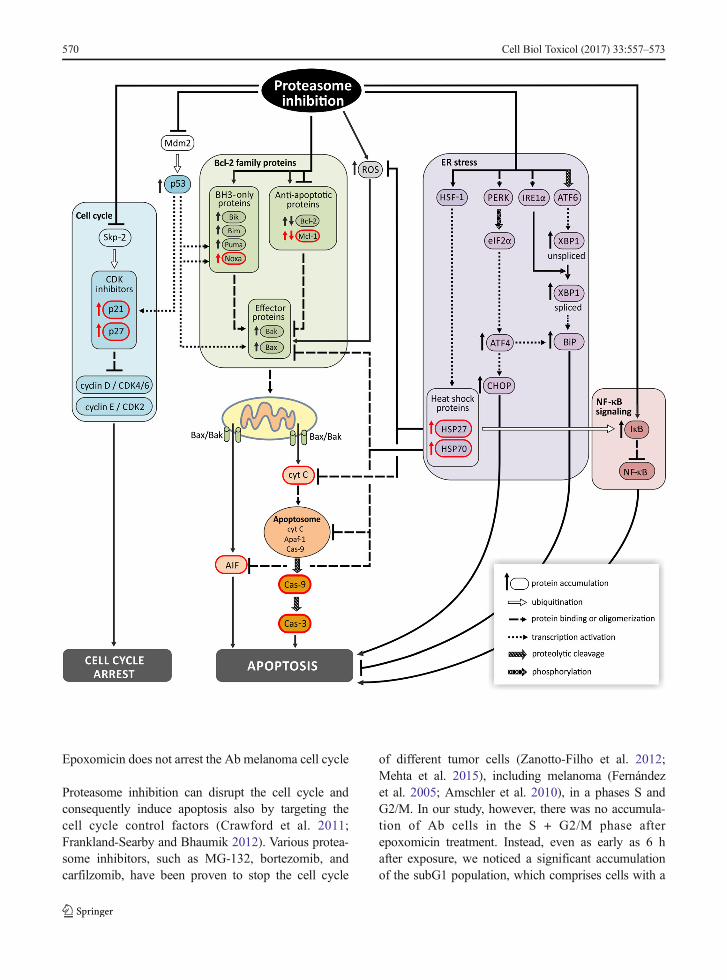
In summary, our study has revealed the differentialsensitivity of amelanotic Ab melanoma cells to threeproteasome inhibitors of different chemical structures,among which epoxomicin—a member of peptideepoxyketone natural product family from an Actinomy-cetes strain Q996-17—was especially effective. Theantitumor activity of epoxomicin was first discoveredin studies against amelanotic melanoma (B16F10 cellline) (Hanada et al. 1992); the authors, however, onlyreported its cytotoxic effect on cell viability without anyanalysis of its action. In this study, we explored the celldeath mechanisms elicited by epoxomicin in melanomafor the first time.We have demonstrated that Ab cells dieon the mitochondrial pathway of apoptosis but alsopartially by a caspase-independent way of death. Apo-ptotic signaling is so potent that cell death is not pre-ceded by cell cycle arrest despite p21Cip1/Waf1 andp27Kip1 stabilization. Finally, even HSP accumulationdoes not protect the Ab melanoma cells againstepoxomicin cytotoxicity. Thus, this special sensitivityof amelanotic melanoma to epoxomicin needs furtherinvestigation. In Fig. 6, we summarize mechanisms ofaction of proteasome inhibitors in cancer cells, includingmelanoma, with elements examined in the present studyshown in red.
�Fig. 6 Principal mechanisms of action of proteasome inhibitors incancer cells includingmelanoma. Principal molecular mechanismsunderlying cytotoxic effect of proteasome inhibition involve thefollowing: (i) up-regulation of pro-apoptotic Bcl-2 family proteins,(ii) stabilization of CDK inhibitors, (iii) p53 stabilization, (iv)endoplasmic reticulum (ER) stress, (v) decreased NF-κBsignaling, and (vi) oxidative stress (accumulation of ROS).These changes result in cell cycle arrest and apoptosis in cancercells. Inhibition of proteasome activity increases expression ofpro-apoptotic BH3-only members of Bcl-2 family, includingNoxa, and in some models down-regulates anti-apoptotic factorssuch as Bcl-2 and Mcl-1. Enhanced pro-apoptotic signalingpromotes the assembly of Bax-Bak oligomers in mitochondrialouter membrane, cytochrome C and AIF release to cytosol,apoptosome formation and finally activation of caspases.Apoptotic signals are augmented by p53, which is activated byROS-induced DNA damage and further stabilized by blocking itsproteasomal degradation after ubiquitination by Mdm2.Accumulated p53 activates expression of its target genes: Puma,Noxa, and Bax. p53 also arrests cell cycle by inducing expressionof CDK inhibitor p21. CDK inhibitors p21 and p27 are alsodirectly up-regulated by proteasome inhibitors, which preventtheir proteasomal degradation after ubiquitination by Skp-2.Another mechanism promoting apoptosis is down-regulation ofNF-κB signaling. Inhibition of proteasome prevents degradationof inhibitory protein-IκB and subsequent activation andtranslocation of NF-κB to the nucleus. Aggregation of misfoldedproteins resulting from proteasome inhibition triggers ER stressand defense mechanism called unfolded protein response (UPR).Aggregated proteins activate ER transmembrane proteins PERK,IRE1α, and transcription factor ATF6 by their homodimerizationand proteolytical processing. ER stress also triggers HSF-1trimerization and binding to HSP promoters. This mechanisminitially protects the cell by increased expression of proteinchaperones HSP70, HSP27, and BiP/Grp78. OverexpressedHSP70 and HSP27 are inhibitors of apoptosis. HSP70 inhibitsassembly of Bax oligomers, prevents recruitment of procaspase-9to apoptosome, and blocks nuclear translocation of AIF andchromatin condensation. HSP27 associates with cyt C andinhibits the formation of apoptosome, decreases ROS content,and targets IκB for ubiquitination. When overwhelmed, UPRtriggers apoptosis by activation of CHOP. Elements examined inthe present study are shown in red.→ activation, ⊣ inhibition. AIFapoptosis-inducing factor, Apaf-1 apoptotic protease activatingfactor 1, ATF activating transcription factor, Bak Bcl-2antagonist/killer-1, Bax Bcl-2-associated X protein, Bcl-2 B celllymphoma 2, Bik BCL2 interacting killer, Bim Bcl-2-like protein11, BiP/Grp78 binding immunoglobulin protein/78 kDa glucose-regulated protein, CDK cyclin-dependent kinase, CHOP CCAAT/enhancer-binding protein homologous protein, eIF2a eukaryoticinitiation factor 2, IκB inhibitory protein κB, HSF-1 heat-shocktranscription factor 1, IRE1α inositol-requiring enzyme 1, Mcl-1myeloid cell leukemia 1, NF-κB nuclear factor κB, PERK ER-resident protein kinase, Puma p53-up-regulated modulator ofapoptosis, ROS reactive oxygen species, Skp-2 S-phase kinase-associated protein 2, XBP1 X-box binding protein 1
Cell Biol Toxicol (2017) 33:557–573 571
Acknowledgments This work was supported by research grantsfrom Medical University of Gdańsk (MN-157) and the PolishMinistry of Science and Higher Education (N N401 005735).
Open Access This article is distributed under the terms of theCreative Commons Attribution 4.0 International License (http://creativecommons.org/licenses/by/4.0/), which permits unrestrict-ed use, distribution, and reproduction in any medium, providedyou give appropriate credit to the original author(s) and the source,provide a link to the Creative Commons license, and indicate ifchanges were made.
References
Adams J, Kauffman M. Development of the proteasome inhibitorVelcade (bortezomib). Cancer Investig. 2004;22:304–11.
AmannVC, Ramelyte E, Thurneysen S, Pitocco R, Bentele-JabergN, Goldinger SM, et al. Developments in targeted therapy inmelanoma. Eur J Surg Oncol. 2016:1–13.
Amschler K, Schön MP, Pletz N, Wallbrecht K, Erpenbeck L,Schön M. NF-kappa B inhibition through proteasome inhi-bition or IKKbeta blockade increases the susceptibility ofmelanoma cells to cytostatic treatment through distinct path-ways. J Invest Dermatol. 2010;130:1073–86.
Berenson A, Vardanyan S, David M, Wang J, Harutyunyan NM,Gottlieb J, et al. Outcomes of multiple myeloma patientsreceiving bortezomib, lenalidomide, and carfilzomib. AnnHematol. 2016 (ahead of print)
Bomirski A, Słominski A, Bigda J. The natural history of a familyof transplantable melanomas in hamsters. Cancer MetastasisRev. 1988;7:95–118.
Calderwood SK, Gong J. Heat shock proteins promote cancer: It’sa protection racket. Trends Biochem Sci. 2016;41:311–23.
Cheung WL, Patel RR, Leonard A, Firoz B, Meehan SA.Amelanotic melanoma: a detailed morphologic analysis withclinicopathologic correlation of 75 cases. J Cutan Pathol.2012;39:33–9.
Cichorek M. Camptothecin-induced death of amelanotic and mel-anotic melanoma cells in different phases of cell cycle.Neoplasma. 2011;58:227–34.
CichorekM, Kozłowska K, Bryl E. Mitochondrial transmembranepotential in spontaneous and camptothecin-induced apopto-sis of melanotic and amelanotic melanoma cells. Neoplasma.2007;54:29–36.
Cohen-Kaplan V, Livneh I, Avni N, Cohen-Rosenzweig C,Ciechanover A. The ubiquitin-proteasome system and au-tophagy: coordinated and independent activities. Int JBiochem Cell Biol. 2016;79:403–18.
Crawford LJ, Walker B, Irvine AE. Proteasome inhibitors incancer therapy. J Cell Commun Signal. 2011;5:101–10.
de Bettignies G, Coux O. Proteasome inhibitors: dozens of mole-cules and still counting. Biochimie. 2010;92:1530–45.
Fernández Y, Verhaegen M, Miller TP, Rush JL, Steiner P, OpipariAW, et al. Differential regulation of noxa in normal melano-cytes and melanoma cells by proteasome inhibition: thera-peutic implications. Cancer Res. 2005;65:6294–304.
Frankland-Searby S, Bhaumik SR. The 26S proteasome complex:an attractive target for cancer therapy. Biochim BiophysActa. 2012;1825:64–76.
Galluzzi L, Kepp O, Krautwald S, Kroemer G, Linkermann A.Molecular mechanisms of regulated necrosis. Semin CellDev Biol. 2014 Nov;35:24–32.
Grazia G, Penna I, Perotti V, Anichini A, Tassi E. Towards com-binatorial targeted therapy in melanoma: from pre-clinicalevidence to clinical application (review). Int J Oncol.2014;45:929–49.
Halaban R. Pigmentation in melanomas: changes manifestingunderlying oncogenic and metabolic activities. Oncol Res.2002;13:3–8.
Hanada M, Sugawara K, Kaneta K, Toda S, Nishiyama Y, TomitaK, et al. Epoxomicin, a new antitumor agent of microbialorigin. J Antibiot (Tokyo). 1992;45:1746–52.
Honma Y, Shimizu S, Takehara T, Harada M. Sorafenib enhancesproteasome inhibitor-induced cell death via inactivation ofAkt and stress-activated protein kinases. J Gastroenterol.2014;49:517–26.
Johnson DB, Menzies AM, Zimmer L, Eroglu Z, Ye F, Zhao S,et al. Acquired BRAF inhibitor resistance: a multicentermeta-analysis of the spectrum and frequencies, clinical be-haviour, and phenotypic associations of resistance mecha-nisms. Eur J Cancer. 2015;51:2792–9.
Kim HJ, Joo HJ, Kim YH, Ahn S, Chang J, Hwang KB, et al.Systemic analysis of heat shock response induced by heatshock and a proteasome inhibitor MG132. PLoS One.2011;6:e20252.
Klikova K, Stefanikova A, Pilchova I, Hata J, Chudy P, Chudej J,et al. Differential impact of bortezomib on HL-60 and K562cells. Gen Physiol Biophys. 2015;34:33–42.
KumanoM, Furukawa J, Shiota M, Zardan A, Zhang F, Beraldi E,et al. Cotargeting stress-activated Hsp27 and autophagy as acombinatorial strategy to amplify endoplasmic reticular stressin prostate cancer. Mol Cancer Ther. 2012;11:1661–71.
Laporte AN, Barrott JJ, Yao RJ, Poulin NM, Brodin BA, JonesKB, et al. HDAC and proteasome inhibitors synergize toactivate pro-apoptotic factors in synovial sarcoma. PLoSOne. 2017;12:e0169407.
Lu G, Punj V, Chaudhary PM. Proteasome inhibitor bortezomibinduces cell cycle arrest and apoptosis in cell lines derivedfrom Ewing’s sarcoma family of tumors and synergizes withTRAIL. Cancer Biol Ther. 2008;7:603–8.
Markovic SN, Geyer SM, Dawkins F, Sharfman W, Albertini M,Maples W, et al. A phase II study of bortezomib in thetreatment of metastatic malignant melanoma. Cancer.2005;103:2584–9.
Mehta A, Zhang L, Boufraqech M, Zhang YQ, Patel D, Shen M,et al. Carfilzomib is an effective anticancer agent in anaplasticthyroid cancer. Endocr Relat Cancer. 2015;22:319–29.
Miller LA, Goldstein NB, Johannes WU, Walton CH, Fujita M,Norris DA, et al. BH3 mimetic ABT-737 and a proteasomeinhibitor synergistically kill melanomas through noxa-dependent apoptosis. J Invest Dermatol. 2009;129:964–71.
Obrist F, Manic G, Kroemer G, Vitale I, Galluzzi L. Trial watch:proteasomal inhibitors for anticancer therapy. Mol CellOncol. 2015;2:e974463.
Qin JZ, Ziffra J, Stennett L, Bodner B, Bonish BK, Chaturvedi V,et al. Proteasome inhibitors trigger NOXA-mediated
572 Cell Biol Toxicol (2017) 33:557–573
apoptosis in melanoma and myeloma cells. Cancer Res.2005;65:6282–93.
Reuland SN, Goldstein NB, Partyka KA, Smith S, Luo Y, FujitaM, et al. ABT-737 synergizes with bortezomib to kill mela-noma cells. Biol Open. 2012;1:92–100.
Selimovic D, Porzig BB, El-Khattouti A, Badura HE, Ahmad M,Ghanjati F, et al. Bortezomib/proteasome inhibitor triggersboth apoptosis and autophagy-dependent pathways in mela-noma cells. Cell Signal. 2013;25:308–18.
Shah SP, Nooka AK, Jaye DL, Bahlis NJ, Lonial S, Boise LH.Bortezomib-induced heat shock response protects multiplemyeloma cells and is activated by heat shock factor 1 serine326 phosphorylation. Oncotarget. 2016;7:59727–41.
Shipp C, Weide B, Derhovanessian E, Pawelec G. Hsps are up-regulated in melanoma tissue and correlate with patient clin-ical parameters. Cell Stress Chaperones. 2013;18:145–54.
Sorolla A, Yeramian A, Dolcet X, Pérez de Santos AM, Llobet D,Schoenenberger JA, et al. Effect of proteasome inhibitors onproliferation and apoptosis of human cutaneous melanoma-derived cell lines. Br J Dermatol. 2008;158:496–504.
Starostina NG, Kipreos ET. Multiple degradation pathways regu-late versatile CIP/KIP CDK inhibitors. Trends Cell Biol.2012;22:33–41.
Tang T, Eldabaje R, Yang L. Current status of biological therapiesfor the treatment of metastatic melanoma. Anticancer Res.2016;36:3229–41.
Wojcik S. Crosstalk between autophagy and proteasome proteindegradation systems: possible implications for cancer thera-py. Folia Histochem Cytobiol. 2013;51:249–64.
Wolter KG, Verhaegen M, Fernández Y, Nikolovska-Coleska Z,Riblett M, de la Vega CM, et al. Therapeutic window formelanoma treatment provided by selective effects of theproteasome on Bcl-2 proteins. Cell Death Differ. 2007;14:1605–16.
Yang F, Jove V, Chang S, Hedvat M, Liu L, Buettner R, et al.Bortezomib induces apoptosis and growth suppression inhuman medulloblastoma cells, associated with inhibition ofAKTand NF kappa B signaling, and synergizes with an ERKinhibitor. Cancer Biology & Therapy. 2012;13:349–57.
Yerlikaya A, Erin N. Differential sensitivity of breast cancer andmelanoma cells to proteasome inhibitor Velcade. Int J MolMed. 2008;22:817–23.
Zanotto-Filho A, Braganhol E, Battastini AM, Moreira JC.Proteasome inhibitor MG132 induces selective apoptosis inglioblastoma cells through inhibition of PI3K/Akt andNFkappaB pathways, mitochondrial dysfunction, and activa-tion of p38-JNK1/2 signaling. Investig New Drugs. 2012;30:2252–62.
Cell Biol Toxicol (2017) 33:557–573 573