The Phosphoinositide 3-Kinase a Selective Inhibitor BYL719 ... · The Phosphoinositide 3-Kinase a...
12
Small Molecule Therapeutics The Phosphoinositide 3-Kinase a Selective Inhibitor BYL719 Enhances the Effect of the Protein Kinase C Inhibitor AEB071 in GNAQ/GNA11-Mutant Uveal Melanoma Cells Elgilda Musi, Grazia Ambrosini, Elisa de Stanchina, and Gary K. Schwartz Abstract G-protein mutations are one of the most common mutations occurring in uveal melanoma activating the protein kinase C (PKC)/mitogen-activated protein kinase and phosphoinositide 3-kinase (PI3K)/AKT pathways. In this study, we described the effect of dual pathway inhibition in uveal melanoma harboring GNAQ and GNA11 mutations via PKC inhibition with AEB071 (sotrastaurin) and PI3K/AKT inhibition with BYL719, a selective PI3Ka inhibitor. Growth inhibition was observed in GNAQ/GNA11-mutant cells with AEB071 versus no activity in wild-type cells. In the GNAQ-mutant cells, AEB071 decreased phosphorylation of myristoylated alanine-rich C-kinase substrate, a substrate of PKC, along with ERK1/2 and ribosomal S6, but persistent AKT activation was present. BYL719 had minimal antiproliferative activity in all uveal melanoma cell lines, and inhibited phosphorylation of AKT in most cell lines. In the GNA11-mutant cell line, similar effects were observed with ERK1/2 inhibition, mostly inhibited by BYL719. With the combination treatment, both GNAQ- and GNA11-mutant cell lines showed synergistic inhibition of cell proliferation and apoptotic cell death. In vivo studies correlated with in vitro findings showing reduced xenograft tumor growth with the combination therapy in a GNAQ-mutant model. These findings suggest a new therapy treatment option for G-protein–mutant uveal melanoma with a focus on specific targeting of multiple downstream pathways as part of combination therapy. Mol Cancer Ther; 13(5); 1044–53. Ó2014 AACR. Introduction Uveal melanoma is one of the most common intraocular malignancies of the adult eye, affecting 6 individual per million per year. Most cases of uveal melanoma metastasize to the liver and have a poor survival rate after initial diagnosis. Few treatment options for this fatal disease exist (1). Chemotherapy is ineffective. There has been recent interest in targeting the mitogen-activated protein kinase (MAPK) pathway (2). MAPK activation is found to be present among 80% of uveal melanoma, although it rarely occurs through mutations such as BRAF (3, 4). Activation of MAPK in uveal melanoma can be attributed to mutations in GNA11 or GNAQ, an early event of malignant transfor- mation (5, 6). Collectively these genetic alterations are present in 85% of diagnosed cases (5, 7). These genes encode members of the q class of G-protein a subunits involved in mediating signals between G-protein–coupled receptors (GPCR) and downstream effectors. Activation is through stimulation of phospholipase C-b (PLCb), which cleaves phosphatidylinositol (4,5)-bisphosphate (PIP2) to inositol triphosphate (IP3) and diacylglycerol (DAG), which activates protein kinase C (PKC) further activating the MAPK pathway (2, 8). To date, most experimental therapies used to treat GNAQ- and GNA11-mutant uveal melanoma have focused on the targeting of the MEK/ERK pathway. How- ever, these mutations have been shown to activate other signaling pathways upstream of MEK, including PKC family members that are involved in cell proliferation and apoptosis. The PKC family is a group of serine/threonine kinases composed of different isoforms that are divided into (1) the classical (or conventional) PKC isoforms a,bI,b II,g (2), the novel PKC isoforms d,e,h,m, and (3) the atypical PKC isoforms l and z each having different roles and functions in cancer (9). Myristoylated alanine-rich C-kinase substrate (MARCKS) is a major PKC substrate expressed in many different cell types. MARCKS, when phosphorylated by PKC, has such roles as calcium/calmodulin binding and actin–membrane interactions (10–12). PI3K/AKT signaling pathways are also activated in the setting of G-protein mutations in uveal melanoma (2). There are also signaling interactions involved with GNAQ and some PI3K isoforms (13). The PI3K/AKT pathway has been implicated in uveal melanoma, as well as in Authors' Affiliation: Jennifer Goodman Linn Laboratory of New Drug Development in Sarcoma and Rare Cancers, Memorial Sloan-Kettering Cancer Center, New York, New York Note: Supplementary data for this article are available at Molecular Cancer Therapeutics Online (http://mct.aacrjournals.org/). Corresponding Author: Elgilda Musi, Herbert Irving Comprehensive Can- cer Center, Columbia University Medical Center, 1130 Saint Nicholas Avenue, ICRC 207, New York, New York 10032. Phone: 212-851-4903; Email: [email protected] doi: 10.1158/1535-7163.MCT-13-0550 Ó2014 American Association for Cancer Research. Molecular Cancer Therapeutics Mol Cancer Ther; 13(5) May 2014 1044 on March 29, 2020. © 2014 American Association for Cancer Research. mct.aacrjournals.org Downloaded from Published OnlineFirst February 21, 2014; DOI: 10.1158/1535-7163.MCT-13-0550 on March 29, 2020. © 2014 American Association for Cancer Research. mct.aacrjournals.org Downloaded from Published OnlineFirst February 21, 2014; DOI: 10.1158/1535-7163.MCT-13-0550 on March 29, 2020. © 2014 American Association for Cancer Research. mct.aacrjournals.org Downloaded from Published OnlineFirst February 21, 2014; DOI: 10.1158/1535-7163.MCT-13-0550
Transcript of The Phosphoinositide 3-Kinase a Selective Inhibitor BYL719 ... · The Phosphoinositide 3-Kinase a...

Small Molecule Therapeutics
The Phosphoinositide 3-Kinase a Selective Inhibitor BYL719Enhances the Effect of the Protein Kinase C InhibitorAEB071 in GNAQ/GNA11-Mutant Uveal Melanoma Cells
Elgilda Musi, Grazia Ambrosini, Elisa de Stanchina, and Gary K. Schwartz
AbstractG-protein mutations are one of the most common mutations occurring in uveal melanoma activating the
protein kinase C (PKC)/mitogen-activated protein kinase and phosphoinositide 3-kinase (PI3K)/AKT
pathways. In this study, we described the effect of dual pathway inhibition in uveal melanoma harboring
GNAQandGNA11mutations via PKC inhibitionwithAEB071 (sotrastaurin) andPI3K/AKT inhibitionwith
BYL719, a selective PI3Ka inhibitor. Growth inhibition was observed in GNAQ/GNA11-mutant cells with
AEB071 versus no activity inwild-type cells. In theGNAQ-mutant cells, AEB071 decreased phosphorylation
of myristoylated alanine-rich C-kinase substrate, a substrate of PKC, along with ERK1/2 and ribosomal S6,
but persistent AKT activation was present. BYL719 had minimal antiproliferative activity in all uveal
melanoma cell lines, and inhibited phosphorylation of AKT inmost cell lines. In theGNA11-mutant cell line,
similar effects were observed with ERK1/2 inhibition, mostly inhibited by BYL719. With the combination
treatment, both GNAQ- andGNA11-mutant cell lines showed synergistic inhibition of cell proliferation and
apoptotic cell death. In vivo studies correlated with in vitro findings showing reduced xenograft tumor
growth with the combination therapy in a GNAQ-mutant model. These findings suggest a new therapy
treatment option for G-protein–mutant uveal melanoma with a focus on specific targeting of multiple
downstream pathways as part of combination therapy. Mol Cancer Ther; 13(5); 1044–53. �2014 AACR.
IntroductionUveal melanoma is one of the most common intraocular
malignancies of the adult eye, affecting 6 individual permillionperyear.Most casesofuvealmelanomametastasizeto the liver and have a poor survival rate after initialdiagnosis. Few treatment options for this fatal disease exist(1). Chemotherapy is ineffective. There has been recentinterest in targeting the mitogen-activated protein kinase(MAPK) pathway (2). MAPK activation is found to bepresent among 80% of uveal melanoma, although it rarelyoccurs throughmutations suchasBRAF(3, 4).ActivationofMAPKinuvealmelanomacanbeattributed tomutations inGNA11 or GNAQ, an early event of malignant transfor-mation (5, 6). Collectively these genetic alterations arepresent in 85% of diagnosed cases (5, 7). These genesencode members of the q class of G-protein a subunits
involved in mediating signals between G-protein–coupledreceptors (GPCR) and downstream effectors. Activation isthrough stimulation of phospholipase C-b (PLCb), whichcleaves phosphatidylinositol (4,5)-bisphosphate (PIP2) toinositol triphosphate (IP3) and diacylglycerol (DAG),which activates protein kinase C (PKC) further activatingthe MAPK pathway (2, 8).
To date, most experimental therapies used to treatGNAQ- and GNA11-mutant uveal melanoma havefocused on the targeting of theMEK/ERK pathway. How-ever, these mutations have been shown to activate othersignaling pathways upstream of MEK, including PKCfamily members that are involved in cell proliferation andapoptosis. The PKC family is a group of serine/threoninekinases composed of different isoforms that are dividedinto (1) the classical (or conventional) PKC isoforms a,bI,bII,g (2), the novel PKC isoforms d,e,h,m, and (3) the atypicalPKC isoforms l and z each having different roles andfunctions in cancer (9).Myristoylated alanine-richC-kinasesubstrate (MARCKS) is amajor PKC substrate expressed inmanydifferent cell types.MARCKS,whenphosphorylatedby PKC, has such roles as calcium/calmodulin bindingand actin–membrane interactions (10–12).
PI3K/AKT signaling pathways are also activated in thesetting of G-protein mutations in uveal melanoma (2).There are also signaling interactions involvedwithGNAQand some PI3K isoforms (13). The PI3K/AKT pathwayhas been implicated in uveal melanoma, as well as in
Authors' Affiliation: Jennifer Goodman Linn Laboratory of New DrugDevelopment in Sarcoma and Rare Cancers, Memorial Sloan-KetteringCancer Center, New York, New York
Note: Supplementary data for this article are available at Molecular CancerTherapeutics Online (http://mct.aacrjournals.org/).
Corresponding Author: Elgilda Musi, Herbert Irving Comprehensive Can-cer Center, Columbia University Medical Center, 1130 Saint NicholasAvenue, ICRC 207, New York, New York 10032. Phone: 212-851-4903;Email: [email protected]
doi: 10.1158/1535-7163.MCT-13-0550
�2014 American Association for Cancer Research.
MolecularCancer
Therapeutics
Mol Cancer Ther; 13(5) May 20141044
on March 29, 2020. © 2014 American Association for Cancer Research. mct.aacrjournals.org Downloaded from
Published OnlineFirst February 21, 2014; DOI: 10.1158/1535-7163.MCT-13-0550
on March 29, 2020. © 2014 American Association for Cancer Research. mct.aacrjournals.org Downloaded from
Published OnlineFirst February 21, 2014; DOI: 10.1158/1535-7163.MCT-13-0550
on March 29, 2020. © 2014 American Association for Cancer Research. mct.aacrjournals.org Downloaded from
Published OnlineFirst February 21, 2014; DOI: 10.1158/1535-7163.MCT-13-0550

many other cancers, to drive cell survival and cell migra-tion (14, 15). PI3K mediates phosphorylation of PIP2 tophosphatidylinositol (3,4,5)-tris-phosphate (PIP3), whichactivatesAKT topromote tumorgrowthandproliferation.The PI3Ks are classified as class IA and IB. Class IA consistof heterodimers with an inhibitory adaptor unit (p85) aswell as a catalytic subunit (p110). There are 3 knownisoforms of the class IA p110 subunits, which includep110a, p110b, and p110d. Class IB consists of p110g anda regulatory subunit, p101 (16). p110a (PI3Ka) has beenlinked to activation of AKT in cell lines. It has been shownthat siRNA knockdown of p110a in C2C12 myoblastsreduced phosphorylation of AKT stimulated by insulin-like growth factor-I whereas silencing of p110b did notaffect AKT (17). AlthoughAKT phosphorylation has beenshown to be increased in uveal melanoma (18, 19), itremains to be determined whether a pan-PI3K or AKTinhibitor can be clinically developed in this or other dis-eases because of concerns for systemic toxicity. Rather, aninhibitor of PI3Ka, by providing isoform specificity for thepathway, may avoid toxicity and off target effects thus farassociated with this class of drugs (20). This will beparticularly relevant in uveal melanomawhere activationof multiple downstream pathways, including PI3K/AKT,MEK, and PKC, drive tumor growth. Combination ther-apies will ultimately be absolutely essential to maximizeeffective growth inhibition.Toward this end, we elected to evaluate dual target
inhibition of PI3Ka and PKC in GNAQ- and GNA11-mutant uveal melanoma cells. It has been previouslyreported that PKC inhibitors have antiproliferative activ-ity in vitro against uveal melanoma cells carrying GNAQmutations (21, 22). AEB071 (sotrastaurin) is a potentinhibitor of many forms of PKC shown to have anti-proliferative activity in other cell types as well (23). It iscurrently being evaluated in a phase I clinical trial forpatientswithmetastatic uvealmelanoma (NCT01430416).BYL719 is a next generation PI3Ka-specific inhibitor,which has been tested in a broad cancer cell line panel(24) and currently under clinical evaluation to asses effi-cacy in PI3Ka-driven tumors (25). We now show that thecombination of the PKC inhibitor AEB071 with BYL719, aselective PI3Ka inhibitor, results in an enhanced antitu-mor effect because of inhibition of both PKC and PI3K/AKT signaling inG-protein–mutant uvealmelanoma cellsin vitro aswell as in vivo. These findingswould support theclinical development of this drug combination such thatbothof thesepathways are inhibited inpatientswithuvealmelanoma.
Materials and MethodsCell cultureAll cell lines were maintained in RPMI 1640 supple-
mented with heat inactivated 10% FBS supplementedwith 100 units/mL penicillin and 100 mg/mL streptomy-cin and maintained at 37�C and 5% CO2. 92.1 cells wereprovided by Dr. W. Harbour (Washington University, St.Louis, MO). Omm1.3 and Mel270 have been provided by
Dr. B. Ksander (Harvard Medical School, Boston, MA).Omm1was kindly provided by Dr. B. Bastian (UniversityCalifornia of San Francisco, San Francisco, CA). Mel290was from David Folberg (University of Illinois, Chicago,IL). C918 were obtained from Dr. David H. Abramson(Memorial SloanKetteringCancerCenter,NewYork,NY)originally from David Folberg. As previously reported,uveal melanoma cell lines have been sequenced for thepresence of activating mutations in codons 209 (exon 5)and 183 (exon 4) of GNAQandGNA11. Two cell lines hadQ209L mutation (92.1, Mel202), whereas Omm1.3 andMel270 had Q209P mutation (26). Omm1 (GNA11) cellline had a Q209L mutation. A karyotype test was alsoperformed for each cell line in 2012, confirming theauthenticity of the cell lines as reported previously (27).
ChemicalsAEB071 (sotrastaurin) and BYL719 were supplied by
Novartis and dissolved in dimethyl sulfoxide (DMSO)at 10 mmol/L concentration and stored aliquoted at�20�C.
Cell viability assays and combination index analysisCells were plated in a 96-well plate and treated with
AEB071, BYL719, orDMSOat indicated concentrations fora period of 5 days. Viability was assessed using CellCounting Kit from Dojindo Molecular Technologies asper manufacturer’s instructions. The combination index(CI) valueswere calculated using theCompuSyn software(Combosyn) as developed by Chou and colleagues (28).Briefly explained, the plots generated by the CompuSynsoftware demonstrate the Y-axis CI values, where CI <1,¼1, and >1 indicate synergism, additive effect, and antag-onism, respectively. The X-axis represents the fractionalactivity, which reflects the fraction of cells inhibited by thetreatments relative to vehicle control. For combinationindex studies, the concentrations tested included AEB071(0, 125, 250, 500, and 1,000 nmol/L) and BYL719 (0, 250,500, 1,000, and 2,000 nmol/L).
Gene silencingExperiments with siRNAs were performed as previ-
ously described (29). Briefly, the cells were transfectedusing Liptofectamine RNAiMax (Invitrogen) according tomanufacturer’s instructions. Next day, cells were treatedwith drug for an additional 24 hours. Cells were thenharvested and lysates were collected for Western Blotanalysis. siRNA for human PI3Ka was purchased fromThermo Scientific, On Target plus SMARTpool (L-003018-00), and control siRNA (sc-37007) was from Santa CruzBiotechnology.
Western blotsCells were lysed in radioimmunoprecipitation assay
(RIPA) buffer supplemented with protease inhibitorcocktail tablet (Roche Diagnostics) and 1 mmol/L Na3VO4. Equal amounts of protein were loaded and sepa-rated on a 4% to 12% PAGE gel (Invitrogen). Proteins
PI3Ka and PKC Inhibition Are Synergistic in Uveal Melanoma
www.aacrjournals.org Mol Cancer Ther; 13(5) May 2014 1045
on March 29, 2020. © 2014 American Association for Cancer Research. mct.aacrjournals.org Downloaded from
Published OnlineFirst February 21, 2014; DOI: 10.1158/1535-7163.MCT-13-0550
were transferred to polyvinylidene difluoride mem-branes, which were blocked in 5% nonfat dried milk.Membranes were then incubated with primary andsecondary antibody and developed by ECL. Antibodiesused to probe were p-AKT (Ser473), Pan AKT, p-S6(S240/244), S6 ribosomal protein, p-MARCKS (S152/156), MARCKS, a-tubulin, ERK1/2, cleaved PARP (CellSignaling), and p-ERK1/2 (Y204) (Santa Cruz Bio-technology). For time course evaluation, media fromplated cells was aspirated and freshly prepared drug inmedia was added to the plates. Cells were then har-vested and lysed in RIPA buffer at 2, 6, and 24 hours.
Flow cytometryCell-cycle analysis was performed as previously
described (30, 31). Briefly, cell-cycle analysis was per-formed after 72 hours of treatment. Cells were ethanolfixed, stained with propidium iodide and MPM-2 anti-body, and analyzed by flow cytometry.
Xenograft studiesSix- to eight-week SCID female mice bearing subcuta-
neously injected 92.1 tumors (7 mice/group) of 100 mm3
diameter were treated with vehicle, AEB071 (80 mg/kg/d) 3 times a day and/or BYL719 orally (50 mg/kg/d)every day as single agents and in combination, 5 days/week for 2 weeks. After 2 weeks, 2 animals from eachgroup were sacrificed and tumors were collected to ana-lyze forWesternblot analysis. ForOmm1xenogratfs, 6 to 8weeks athymic female mice bearing subcutaneouslyinjected Omm1 tumors (7 mice/group) of 100 mm3 diam-eter were treated with vehicle, AEB071 (80 mg/kg/d) 3times a day and/or BYL719 orally (50 mg/kg/d) everyday as single agents and in combination, 5 days/week for3 weeks. Tumors were homogenizedwith grinding resinskits from GE Healthcare as per manufacturer’s instruc-tions. Tumors were collected to analyze for hematoxylinand eosin, and terminal deoxynucleotidyl transferasedUTP nick end labeling (TUNEL) staining as previouslydescribed (31). Tumors were measured every 2 to 3 dayswith calipers, and tumor volumes were calculated by theformula 4/3 � r3, where [r ¼ (larger diameter þ smallerdiameter)/4. Toxicity was monitored by weight loss.Experiments were carried out under institutional guide-lines addressing the proper and humane use of animals.The Memorial Sloan- Kettering Cancer Center AnimalCare and Use Committee and Research Animal Resourcecenter approved this study. The study is also in accor-dance of the Principles of Laboratory Animal Care (NIHPublication No. 85-23, released 1985).
Statistical analysisAll in vitro experiments were carried out at least 2 to 3
times. SE was calculated as the SD divided by thesquare root of number of samples. Statistical analysisfor in vivo studies was determined by the 2-sided t test.We chose P ¼ 0.05 as statistically significant in indi-vidual comparisons.
ResultsAEB071 inhibits cell proliferation inGNAQ/GNA11-mutant uveal melanoma cell lines with inhibition ofthe PKC/ERK1/2 pathway
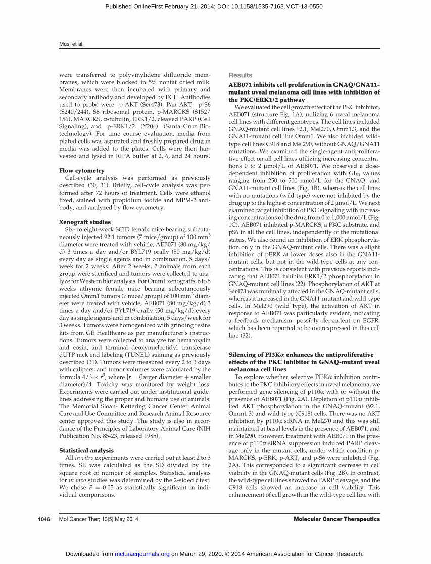
Weevaluated the cell growth effect of thePKC inhibitor,AEB071 (structure Fig. 1A), utilizing 6 uveal melanomacell lines with different genotypes. The cell lines includedGNAQ-mutant cell lines 92.1, Mel270, Omm1.3, and theGNA11-mutant cell line Omm1. We also included wild-type cell lines C918 andMel290, without GNAQ/GNA11mutations. We examined the single-agent antiprolifera-tive effect on all cell lines utilizing increasing concentra-tions 0 to 2 mmol/L of AEB071. We observed a dose-dependent inhibition of proliferation with GI50 valuesranging from 250 to 500 nmol/L for the GNAQ- andGNA11-mutant cell lines (Fig. 1B), whereas the cell lineswith no mutations (wild type) were not inhibited by thedrug up to the highest concentration of 2mmol/L.Wenextexamined target inhibition of PKC signalingwith increas-ing concentrations of thedrug from0 to 1,000nmol/L (Fig.1C). AEB071 inhibited p-MARCKS, a PKC substrate, andpS6 in all the cell lines, independently of the mutationalstatus. We also found an inhibition of ERK phosphoryla-tion only in the GNAQ-mutant cells. There was a slightinhibition of pERK at lower doses also in the GNA11-mutant cells, but not in the wild-type cells at any con-centrations. This is consistent with previous reports indi-cating that AEB071 inhibits ERK1/2 phosphorylation inGNAQ-mutant cell lines (22). Phosphorylation of AKT atSer473wasminimally affected in theGNAQ-mutant cells,whereas it increased in the GNA11-mutant andwild-typecells. In Mel290 (wild type), the activation of AKT inresponse to AEB071 was particularly evident, indicatinga feedback mechanism, possibly dependent on EGFR,which has been reported to be overexpressed in this cellline (32).
Silencing of PI3Ka enhances the antiproliferativeeffects of the PKC inhibitor in GNAQ-mutant uvealmelanoma cell lines
To explore whether selective PI3Ka inhibition contri-butes to the PKC inhibitory effects in uvealmelanoma,weperformed gene silencing of p110a with or without thepresence of AEB071 (Fig. 2A). Depletion of p110a inhib-ited AKT phosphorylation in the GNAQ-mutant (92.1,Omm1.3) and wild-type (C918) cells. There was no AKTinhibition by p110a siRNA in Mel270 and this was stillmaintained at basal levels in the presence of AEB071, andin Mel290. However, treatment with AEB071 in the pres-ence of p110a siRNA suppression induced PARP cleav-age only in the mutant cells, under which condition p-MARCKS, p-ERK, p-AKT, and p-S6 were inhibited (Fig.2A). This corresponded to a significant decrease in cellviability in the GNAQ-mutant cells (Fig. 2B). In contrast,thewild-type cell lines showednoPARPcleavage, and theC918 cells showed an increase in cell viability. Thisenhancement of cell growth in the wild-type cell line with
Musi et al.
Mol Cancer Ther; 13(5) May 2014 Molecular Cancer Therapeutics1046
on March 29, 2020. © 2014 American Association for Cancer Research. mct.aacrjournals.org Downloaded from
Published OnlineFirst February 21, 2014; DOI: 10.1158/1535-7163.MCT-13-0550

PI3Ka suppression and AEB071 may be attributed to theabsence of ERK1/2 decrease as seen with the GNAQ-mutant cells (Fig. 2A).We next examined the effect of single-agent BYL719
(structure in Fig. 2C) on the same cell lines, utilizingconcentrations ranging from 0 to 2 mmol/L (Fig. 2D). Weobserved inhibition of phosphorylation of AKT (Ser473)up to 1 mmol/L in most cell lines (Fig. 2D), even thoughtherewas reactivation at higher doses inOmm1.3,Mel270,and Mel290. Some of the cell lines, such as Omm1.3,Omm1, and Mel290 showed inhibition of p-ERK1/2 byBYL719. There is recent evidence supporting the inhibi-tion of ERK1/2 phosphorylation by the selective PI3Kainhibitor BYL719 (33). There was also significant inhibi-tion of S6 phosphorylation in theOmm1.3 andMel290 celllines. However, PI3Ka/pAKT inhibition in itself wasclearly not sufficient to have an antiproliferative effect inany cell line (Fig. 2E). The exception to thiswasOmm1, theGNA11 cell line, in which cell growth inhibition wasobserved at 1,000 nmol/L, a concentration at which therewas inhibition of p-ERK.
The combination AEB071/BYL719 induces asynergistic decrease in cell viability in GNAQ/GNA11-mutant uveal melanoma cell lines
To examine the effects of the AEB071/BYL719 combi-nation on cell viability, we performed combination indexanalysis utilizing the Chou–Talay method to determinewhether the effects are synergistic (28). The combinationof AEB071 and BYL719 at different doses exhibited acombination index (CI) <1, indicative of synergy and afractional activity of more than 50% [(Fa) > 0.5] in allGNAQ/GNA11-mutant cell lines (Supplementary Fig.S1). CI values for wild-type cell lines C918 and Mel290ranged from >1 to 30 with a (Fa) < 0.2, showing that thecombination was in fact antagonistic in cells that are wildtype for G proteins.
We next investigated the occurrences of moleculareventswith the combination treatment (Fig. 3).We treated2 GNAQ (92.1, Mel270) and 2 wild-type (C918, Mel290)cell lines with concentrations of 500 nmol/L of AEB071and 1 mmol/L of BYL719 respectively and examined theeffect on downstream signaling at 2, 6, and 24 hours. In
Figure 1. AEB071 reduces cell viability in G-protein–mutant cell lines with minimal impact on the AKT pathway. A, structure of AEB071. B, AEB071 as singleagent selectively inhibits cell proliferation of GNAQ/GNA11-mutant cells. Cell lines were treated with 0, 62.5, 125, 250, 500, 1,000, and 2,000 nmol/L ofAEB071 for 5 days. Results represent the mean of 3 independent experiments. C, AEB071 inhibits PKC and mTOR pathways but not AKT. Western blotanalysis of MARCKS, ERK, ribosomal S6, and AKT phosphorylation following drug treatments for 24 hours. a-Tubulin was used as a loading control.
PI3Ka and PKC Inhibition Are Synergistic in Uveal Melanoma
www.aacrjournals.org Mol Cancer Ther; 13(5) May 2014 1047
on March 29, 2020. © 2014 American Association for Cancer Research. mct.aacrjournals.org Downloaded from
Published OnlineFirst February 21, 2014; DOI: 10.1158/1535-7163.MCT-13-0550

siRNA Control
siRNA PI3kaAEB071 (500 nmol/L)
PI3K a
p-AKT (S473)
P-ERK1/2 (Y204)
p-S6 (S240/244)
p-MARCKS
Cleaved Parp
Ku70
92.1 (Gq) MeI270 (Gq) Omm1.3 (Gq) Mel290 (WT)C918 (WT)
+ ++ +
++ +
+ ++ +
+ + +++
++ + +
++ +
+
+ ++ +
++
- ---
- -- -- -
- -- -- -
- -- -- -
- ---
-- -
-
BYL719 (nmol/L)
P-AKT (S473)
P-ERK1/2 (Y204)
P-S6 (S240/244)
PAN AKT
Total ERK1/2
Total S6
a-Tubulin
92.1 (Gq) Mel270 (Gq) Omm1.3 (Gq) Omm1 (G11) C918 (WT) Mel290 (WT)
CF3
NH2
N
NN
N
H
SO
O
NVP-BYL719
Via
bil
ity
(%
)
BYL719 (nmol/L)
120
100
80
60
40
20
0
0 10 100 1,000
92.1 (Gq mutant)
Mel270 (Gq mutant)
Omm1.3 (Gq mutant)
Omm1 (GNA11 mutant)
Mel290 (WT)
C918 (WT)
Via
bil
ity
(%
)V
iab
ilit
y (
%)
Via
bil
ity
(%
)V
iab
ilit
y (
%)
120
100
80
60
40
20
0
120
100
80
60
40
20
0
120
100
80
60
40
20
0
300
250
200
150
100
50
0
92.1 (Gq) Mel270
Mel290 C918 (WT)
A
B C
D
E
S. Control+ND S. Control+500 nmol/L AEB071 SiRNA Pi3ka+ND SiRNA Pi3ka+500 nmol/L AEB071
Figure 2. Selective inhibition of PI3Ka enhances AEB071 antiproliferative effect in GNAQ-mutant cells. A, PI3Ka siRNA inhibits AKT phosphorylation in uvealmelanoma cell lines. siRNA knockdown of p110a isoform was performed with or without AEB071 in 92.1, Mel270, Omm1.3 (GNAQ mutant), and Mel290,C918 (wild type) cell lines. A nonspecific siRNA was used as control. Western blots of PI3Ka, AKT, MARCKS, ERK, S6, and cleaved PARP were thenperformed. The nuclear protein Ku70was used as a loading control. B, PI3Ka and PKC inhibition reduces cell proliferation in GNAQ-mutant uveal melanoma.Cell viability was assessed after transfection, with or without AEB071 for 5 days. C, structure of BYL719. D, BYL719 inhibits AKT (Ser473) phosphorylation,and/or p-ERK in some cell lines. Cells were treated with indicated concentrations of BYL719 for 24 hours, and analyzed for pAKT, pERK, pS6, and therespective total proteins. a-Tubulin was used as a loading control. E, BYL719 as single agent hasminimal to no effect on cell viability in uveal melanoma after5 days of exposure. Concentrations tested include 0, 10, 100, and 1,000 nmol/L. Results represent the mean of 3 independent experiments.
Musi et al.
Mol Cancer Ther; 13(5) May 2014 Molecular Cancer Therapeutics1048
on March 29, 2020. © 2014 American Association for Cancer Research. mct.aacrjournals.org Downloaded from
Published OnlineFirst February 21, 2014; DOI: 10.1158/1535-7163.MCT-13-0550

contrast to single-agent AEB071 or BYL719, the combina-tion therapy resulted in inhibition of p-MARCKS and p-AKT over time and all downstream signaling pathwayssuch as p-ERK and p-S6 in the GNAQ-mutant cell lines.Single-agent BYL719 caused a slight activation of p-ERKthatwasdetectable as early as 2hours afterdrug exposure,especially in the 92.1 cells, but this activation was sup-pressed with the combination treatment. This would infact suggest that the reactivation of p-ERK in this settingmaybemediated through PKC. The reactivation of p-ERKat 6 hours, especially in the Mel270 cell line, even inuntreated controls, may reflect a DMSO effect. In theMel290 wild-type cell line, there was activation of AKTto both AEB071 and BYL719 along with reactivation of p-MARCKS at later time points, suggesting that this cell linemay have a dependency on other survival pathways suchas EGFR (32). For the wild-type C918 cell line, p-MARCKS, p-AKT, and p-S6 are inhibited over time butminimal p-ERK inhibition occurs. We also tested theGNA11-mutant cell line, Omm1, with the combinationtherapy (Supplementary Fig. S2A). In these cells, AEB071inhibited p-MARCKS and p-S6 but induced activation
of p-AKT over time, which was inhibited by BYL719 inthe combination therapy. Interestingly, BYL719 alsoinhibitedpERK in this cell line. Thus, these results indicatethat inhibition of PKC and PI3Ka together achieves asynergistic effect in GNAQ and GNA11 cells with inhi-bition of downstream survival pathways that is notachievable with single-agent therapy alone.
AEB071/BYL719 combination induces apoptosisWenext elected to determinewhether this inhibition of
cell growth was because of growth arrest or induction ofapoptosis. We assessed cell-cycle effects of the drugcombination with bi-parameter flow cytometry analysisfor DNA content (detected with propidium iodide) andMPM-2, a mitotic marker, as previously described (31).Apoptosiswas assessed by the detection of a sub-G1 peakand of PARP cleavage. The flow cytometry data indicatedessentiallyminimal growth arrest (data not shown) withAEB071, but there was evidence of a sub-G1 peak withthe combination therapy only in the GNAQ-mutantcell lines (Fig. 4A). Furthermore, the combination treat-ment induced PARP cleavage, an early apoptotic
Figure 3. The combination AEB071/BYL719 synergistically reduces tumor cell viability in GNAQ-mutant cells because of cooperative inhibition of PKC/ERKand PI3Ka/AKT pathways. Cells were treated with indicated AEB071/BYL719 concentrations for 2, 6, and 24 hours andWestern blots were then performed.Suppression of PKC/ERK and PI3Ka/AKT pathways was observed with the combination treatment in the GNAQ-mutant cells, as evidenced by inhibitionof MARCKS, ERK, AKT (Ser473) and S6 phosphorylation. a-Tubulin was used as a loading control.
PI3Ka and PKC Inhibition Are Synergistic in Uveal Melanoma
www.aacrjournals.org Mol Cancer Ther; 13(5) May 2014 1049
on March 29, 2020. © 2014 American Association for Cancer Research. mct.aacrjournals.org Downloaded from
Published OnlineFirst February 21, 2014; DOI: 10.1158/1535-7163.MCT-13-0550

marker, in GNAQ-mutant cells, whereas there was noevidence of PARP cleavage in the wild-type cell lines(Fig. 4B).
AEB071/BYL719 combination inhibits tumor growthin vivo in a GNAQ-mutant xenograft model
In view of the synergistic effects of the combinationtherapy observed in vitro, we elected to determine wheth-er the combination therapy would be superior to single-agent therapy in a GNAQ-mutant xenograft mouse mod-el. As shown in Fig. 5A, both single agents had a modesteffect on inhibiting tumor growth at their respectivesingle-agent MTDs. The combination therapy resulted ina significantly enhanced reduction in tumor volumewhencompared with either AEB071 or BYL719 alone (P¼ 0.049vs. BYL719 and P ¼ 0.022 vs. AEB071 at day 26). Therewas even a greater effect when compared with vehiclecontrol (P ¼ 0.016). Examination of the tumor lysatesfollowing drug therapy (day #26) confirmed the in vitrodata indicating that the combination therapy resulted inconcomitant inhibition of PKC and PI3K signaling withinhibition p-MARCKS, p-AKT, p-ERK, and p-S6 that wasnot observed with either single-agent alone. Toxicity wasmeasured alongwith tumor volumebyweight loss,whichwas less than 14.7% for all treatments (Fig. 5A, right).Examination of apoptosis by TUNEL indicates the pres-ence of apoptotic cells only with the combination therapy(Supplementary Fig. S3).
The Omm1 (GNA11)-mutant xenograft mouse modelwas also performedwith both single agents having an effecton inhibiting tumor growth at their respective single-agentMTDs, which seemed greater than that observed in theGNAQ xenograft. Furthermore, the combination therapydid not result in a significant reduction in tumor volume
when compared with either AEB071 or BYL719 alone,although a trend favored the combination therapy (Supple-mentary Fig. S2B). For unclear reasons, the animals bearingtheGNA11xenografts experienceda23.9%weight losswithcombination therapy (Supplementary Fig. S2B, right).
DiscussionPKC, MAPK, and PI3K/AKT pathways are highly acti-
vated in uvealmelanoma (3, 4, 18, 34).MEK/ERK signalinghas been considered as a major target in uveal melanomaand clinical trials are underway targeting this pathway (35).Alternatively, dual-targeted therapy is now being consid-ered as a treatment option as further studies elucidate theimportance of targeting activated signal pathways. Previ-ous studies have focused on combining inhibitors of MEKwithmTORandPI3K/AKTpathways (26, 36, 37). Targetedtherapy directed against PI3K isoforms has usually beeneffective in cancers harboring mutations of the isoform butPI3Kmutations havenot been reported inuvealmelanoma.Clinically, the benefit of targeting a specific isoform of PI3Khas led to more complete target inhibition at lower dosesresulting in less adverse effects (38). The blocking of AKTactivation in other cell types through PI3Ka has beenwidely studied (17, 39). The role of PI3Ka isoform in itsactivestate allows forcell survival andmigration, leading totumor formation and metastases (40). We postulated thatpreventing this activationwith a selective PI3Ka inhibitorwouldprovide ameans to inhibitAKT in these cells aspartof a combinatorial approach. The G-protein oncogenicmutations have been shown to activate PKC. In fact,GNAQ mutations rely mostly on PKC and MAPK path-ways for survival (26, 41). This present study demon-strates that inhibiting PKC and ERK1/2 signaling with
Su
b-G
1 %
cells
10
9
8
7
6
5
4
3
2
1
092.1 Mel270 Omm1.3 Mel290C918
GNAQ Mutant WT
Vehicle AEB071 BYL719 Combination
92.1 Mel270 Omm1.3 C918 Mel290
Cleaved Parp
a-Tubulin
-- -
- -- -
- -- -
- -- -
- -- -
-+++
+ + ++ +
++
++
++ +
+ ++ +
+ AEB071 (500 nmol/L)
BYL719 (1,000 nmol/L)
A
B
Figure 4. The combinationAEB071/BYL719 inducesapoptosis in GNAQ-mutant cells.A, cells were treated withAEB071/BYL719 for 72 hours andlabeled with MPM-2, stained withpropidium iodide, and analyzed byflow cytometry. Sub-G1 fractionswere quantified using FlowJo.Results represent the mean of3 independent experiments. B,uveal melanoma cell lines weretreated with drug for 24 hours.Western blots were performed forcleaved PARP. a-Tubulin wasused as a loading control.
Musi et al.
Mol Cancer Ther; 13(5) May 2014 Molecular Cancer Therapeutics1050
on March 29, 2020. © 2014 American Association for Cancer Research. mct.aacrjournals.org Downloaded from
Published OnlineFirst February 21, 2014; DOI: 10.1158/1535-7163.MCT-13-0550

AEB071 togetherwith thePI3Ka inhibitor, BYL719, resultsin a synergistic antitumor effect in uveal melanoma har-boring GNAQ and GNA11 mutations in vitro. Previousreports have shown that PKC inhibition has a negativeregulatory effect on PI3K/AKT by stimulating Akt/PKBphosphorylation at Ser473 (42). This may explain theminimal single-agent effect ofAEB071on inhibitingpAKTin theGNAQ- andGNA11-mutant cell lines. Interestingly,our current results also show inhibition of ribosomal S6phosphorylation with PKC inhibition and combinationtreatment. S6 overexpression has been reported as a prog-nostic marker for uveal melanoma and mTOR signalingmay similarly provide a growth advantage to these cells(19). This could explain why inhibition of this pathwayalso enhances the antitumor effect we have observed inthese cells. In the setting of GNAQ/GNA11 mutations,the inhibition of MARCKS, ERK, AKT, and S6 phos-
phorylation results in apoptosis by the combination treat-ment. Similar pathway targeting in pancreatic cells haveshown an apoptotic outcome by sulforaphane treatmentbecause of synergistic inhibition of PI3K/AKT andMEK/ERK (43).
The effects of this combination treatment demonstratethe importance of targeting parallel survival and growthpathways that are downstream of GNAQ and GNA11 inuveal melanoma. However, simply inhibiting several ofthese pathways may not be sufficient to inhibit tumorgrowth inhibition in vivo. For example, we previouslyreported that blockade of Torc1 and Torc2 with AZD8055in combination with the MEK inhibitor selumetinib(AZD6244) in GNAQ-mutant cells is insufficient to sup-press tumor growth in vivo (36). In contrast, we haveshown that inhibition of p-AKT with MK2206, an alloste-ric inhibitor of AKT, in combination with selumetinib
Tum
or
volu
me (
mm
3)
Tum
or
weig
ht
(gm
)
Day of drug treatment
Day of drug treatment
1,400.00
1,200.00
1,000.00
800.00
600.00
400.00
200.00
0.0010 15 20 25 30
40.00
35.0030.00
25.00
20.0015.00
10.005.00
0.0010 15 20 25 30
Vehicle
AEB071 80 mg/kg/TID
AEB071+BYL719
BYL719 50 mg/kg/QD
1 2 3 4 5 6 7 8
pAKT (473)
P-ERK1/2 (Y204)
P-S6 (S240/244)
Total MARCKS
p MARCKS (S152/156)
a- Tubulin
1,2 Vehicle
3,4 AEB071 80 mg/kg TID5,6 BYL719 50 mg/kg QD
7,8 BYL719 50 mg/kg QD AEB071 80 mg/kg TID
A
B
Figure 5. AEB071/BYL719 combination inhibits in vivo tumor growth in a GNAQ-mutant xenograft model. A, AEB071/BYL719 inhibited tumor growthin a xenograft model with the GNAQ-mutant 92.1 cell line. Six- to eight-week SCID female mice were subcutaneously injected with 92.1 cells.Drug treatments began after tumors reached 100 mm3. Mice with tumors were treated 3 times daily with AEB071 (80 mg/kg/d) or once daily with BYL719(50 mg/kg/d) or in combination for 5 days each week for a total of 2 weeks. Tumors were measured with calipers every 2 to 3 days. Tumor volumewas compared between groups of mice at various time points. P value when compared with either AEB071 or BYL719 alone P ¼ 0.049 versusBYL719 and P ¼ 0.022 versus AEB071 at day 26. Toxicity was measured by weight loss (right). B, 2 animals from each cohort were sacrificed andtumors were collected on day 26. Tumors were lysed with RIPA buffer and Western blots were performed for pAKT, pERK, pS6, pMARCKS, anda-tubulin. QD, every day; TID; 3 times a day.
PI3Ka and PKC Inhibition Are Synergistic in Uveal Melanoma
www.aacrjournals.org Mol Cancer Ther; 13(5) May 2014 1051
on March 29, 2020. © 2014 American Association for Cancer Research. mct.aacrjournals.org Downloaded from
Published OnlineFirst February 21, 2014; DOI: 10.1158/1535-7163.MCT-13-0550

results in enhanced antitumor effects (26). This wouldsuggest that the development of combination therapy forthe treatment of GNAQ uveal melanoma will dependlargely on the selective targeting of pathways that arecritical for tumor growth. Based on our MK2206 andselumetinib data, inhibition of p-ERK in combinationwithinhibition of p-AKT seems to be the minimal requirementnecessary to promote an antitumor effect with combina-tion therapy. As AEB071 results in inhibition of PKC aswell as p-ERK, this dual pathway inhibition, when com-binedwith inhibition of PI3Ka/AKT by BYL719, seems toprovide a means to broaden pathway inhibition aboveand beyond MEK and AKT alone for cell growth inhibi-tion of GNAQ/GNA11-mutant cells.
All together our findings indicate that the targeting ofPKC/ERK, alongwith a selective PI3Ka isoform inhibitor,cooperates in inhibiting growth in GNAQ-mutant uvealmelanoma cells. In the GNA11 cell line, this same effect isultimately achieved, although it is BYL719, the PI3Kainhibitor, rather than AEB071, the PKC inhibitor, thatcontributes to the inhibition of p-ERK. We also show atranslatable GNAQ xenograft model of uveal melanomademonstrating effective PKC/ERK and PI3K/AKT path-way inhibition, which results in enhanced in vivo tumorgrowth inhibition with the combination therapy. Thecombination effects in vivo on the GNA11 xenograft wereless definitive despite synergy in vitro. There was unex-pected weight loss with the combination therapy, whichmay have been because of the difference inmouse species(nudemice rather than SCIDmice) needed to establish theGNA11 xenograft.
The combination of AEB071 and MEK162, the MEKinhibitor, has recently been reported in uveal melanomaxenografts (41). Our studies indicate that both AEB071
andBYL719 can inhibit p-ERKas single agents or aspart ofcombination therapy. However, only BYL719 can inhibitp-AKT, which seems critical to the synergistic effect. Inconclusion, our results suggest an effective new therapyfor the treatment of uveal melanoma, and targeting thesepathways with AEB071 and BYL719 could have thera-peutic implications for the future treatment of patientswith this disease. Based on these results, a phase Ib/IIclinical trial of this drug combination in patients withmetastatic uveal melanoma is now planned.
Disclosure of Potential Conflicts of InterestG.K. Schwartz is a consultant/advisory board member for Novartis.
No potential conflicts of interest were disclosed by the other authors.
Authors' ContributionsConception and design: E. Musi, E. de Stanchina, G.K. SchwartzDevelopment of methodology: E. Musi, E. de Stanchina, G.K. SchwartzAcquisition of data (provided animals, acquired and managed patients,provided facilities, etc.): E. Musi, E. de Stanchina, G.K. SchwartzAnalysis and interpretation of data (e.g., statistical analysis, biostatis-tics, computational analysis): E. Musi, G.K. SchwartzWriting, review, and/or revision of the manuscript: E. Musi, G. Ambro-sini, G.K. SchwartzAdministrative, technical, or material support (i.e., reporting or orga-nizing data, constructing databases): E. Musi, G.K. SchwartzStudy supervision: G. Ambrosini, E. de Stanchina, G.K. Schwartz
AcknowledgmentsThe authors thankV.K. Rajasekhar for assistingwith our in vivo studies.
Grant SupportE.Musi, G. Ambrosini, and G.K. Schwartz received grant support from
Cycle for Survival (Philanthropy). E. de Stanchina received grant supportfrom NIH grant # P30 CA 008748.
Received July 10, 2013; revised January 29, 2014; accepted February 17,2014; published OnlineFirst February 21, 2014.
References1. Augsburger JJ, Correa ZM, Shaikh AH. Effectiveness of treatments for
metastatic uveal melanoma. Am J Ophthalmol 2009;148:119–27.2. PatelM,SmythE,ChapmanPB,Wolchok JD,SchwartzGK,Abramson
DH, et al. Therapeutic implications of the emerging molecular biologyof uveal melanoma. Clin Cancer Res 2011;17:2087–100.
3. Weber A, HenggeUR, Urbanik D,Markwart A,MirmohammadsaeghA,Reichel MB, et al. Absence of mutations of the BRAF gene andconstitutive activation of extracellular-regulated kinase in malignantmelanomas of the uvea. Lab Invest 2003;83:1771–6.
4. Zuidervaart W, van Nieuwpoort F, Stark M, Dijkman R, Packer L,Borgstein AM, et al. Activation of the MAPK pathway is a commonevent in uvealmelanomas although it rarely occurs throughmutation ofBRAF or RAS. Br J Cancer 2005;92:2032–8.
5. Van Raamsdonk CD, Bezrookove V, Green G, Bauer J, Gaugler L,O'Brien JM, et al. Frequent somatic mutations of GNAQ in uvealmelanoma and blue naevi. Nature 2009;457:599–602.
6. Onken MD, Worley LA, Long MD, Duan S, Council ML, Bowcock AM,et al. Oncogenic mutations in GNAQ occur early in uveal melanoma.Invest Ophthalmol Vis Sci 2008;49:5230–4.
7. Van Raamsdonk CD, Griewank KG, Crosby MB, Garrido MC, VemulaS, Wiesner T, et al. Mutations in GNA11 in uveal melanoma. N Engl JMed 2010;363:2191–9.
8. Rozengurt E. Mitogenic signaling pathways induced by G protein-coupled receptors. J Cell Physiol 2007;213:589–602.
9. Martiny-Baron G. Classical PKC isoforms in cancer. Pharmacol Res2007;55:477.
10. Thelen M, Rosen A, Nairn AC, Aderem A. Regulation by phos-phorylation of reversible association of a myristoylated proteinkinase C substrate with the plasma membrane. Nature 1991;351:320–2.
11. Hartwig JH, Thelen M, Rosen A, Janmey PA, Nairn AC, Aderem A.MARCKS is an actin filament crosslinking protein regulated by proteinkinase C and calcium-calmodulin. Nature 1992;356:618–22.
12. Graff JM, Young TN, Johnson JD, Blackshear PJ. Phosphorylation-regulated calmodulin binding to a prominent cellular substrate forprotein kinase C. J Biol Chem 1989;264:21818–23.
13. Ballou LM, Chattopadhyay M, Li Y, Scarlata S, Lin RZ. Galphaq bindsto p110a/p85a phosphoinositide 3-kinase and displaces Ras. Bio-chem J 2006;394:557–62.
14. Ye M, Hu D, Tu L, Zhou X, Lu F, Wen B, et al. Involvement of PI3K/Akt signaling pathway in hepatocyte growth factor-induced migra-tion of uveal melanoma cells. Invest Ophthalmol Vis Sci 2008;49:497–504.
15. Stephens L, Williams R, Hawkins P. Phosphoinositide 3-kinases asdrug targets in cancer. Curr Opin Pharmacol 2005;5:357–65.
16. Hennessy BT, Smith DL, Ram PT, Lu Y, Mills GB. Exploiting the PI3K/AKT pathway for cancer drug discovery. Nat Rev Drug Discov 2005;4:988–1004.
Musi et al.
Mol Cancer Ther; 13(5) May 2014 Molecular Cancer Therapeutics1052
on March 29, 2020. © 2014 American Association for Cancer Research. mct.aacrjournals.org Downloaded from
Published OnlineFirst February 21, 2014; DOI: 10.1158/1535-7163.MCT-13-0550

17. Matheny RW Jr., Adamo ML. PI3K p110a and p110b have differentialeffects on Akt activation and protection against oxidative stress-induced apoptosis in myoblasts. Cell Death Differ 2010;17:677–88.
18. Saraiva VS, Caissie AL, Segal L, Edelstein C, Burnier MNJr. Immuno-histochemical expression of phospho-Akt in uveal melanoma. Mela-noma Res 2005;15:245–50.
19. Populo H, Soares P, Rocha AS, Silva P, Lopes JM. Evaluation of themTORpathway in ocular (uvea andconjunctiva)melanoma.MelanomaRes 2010;20:107–17.
20. Brana I, Siu LL. Clinical development of phosphatidylinositol 3-kinaseinhibitors for cancer treatment. BMC Med 2012;10:161.
21. Wu X, Zhu M, Fletcher JA, Giobbie-Hurder A, Hodi FS. The proteinkinase C inhibitor enzastaurin exhibits antitumor activity against uvealmelanoma. PLoS One 2012;7:e29622.
22. Wu X, Li J, Zhu M, Fletcher JA, Hodi FS. Protein kinase C inhibitorAEB071 targets ocular melanoma harboring GNAQ mutations viaeffects on the PKC/Erk1/2 and PKC/NF-kB pathways. Mol CancerTher 2012;11:1905–14.
23. Naylor TL, Tang H, Ratsch BA, Enns A, Loo A, Chen L, et al. Proteinkinase C inhibitor sotrastaurin selectively inhibits the growth of CD79mutant diffuse large B-cell lymphomas. Cancer Res 2011;71:2643–53.
24. Huang A FC, Wilson C, Reddy A, Liu M, Lehar J, et al. Single agentactivity of PIK3CA inhibitor BYL719 in a broad cancer cell line panel. In:Proceedings of the 103rd Annual Meeting of the American Associationfor Cancer Research; 2012 Mar 31-Apr 4; Chicago, IL. Philadelphia(PA): AACR; Cancer Res 2012;72(8 Suppl):Abstract nr 3749.
25. Furet P, Guagnano V, Fairhurst RA, Imbach-Weese P, Bruce I, KnappM, et al. Discovery of NVP-BYL719 a potent and selective phospha-tidylinositol-3 kinase a inhibitor selected for clinical evaluation. BioorgMed Chem Lett 2013;23:3741–8.
26. AmbrosiniG,Musi E, HoAL, deStanchina E, SchwartzGK. Inhibition ofmutant GNAQ signaling in uvealmelanoma induces AMPK-dependentautophagic cell death. Mol Cancer Ther 2013;12:768–76.
27. Griewank KG, Yu X, Khalili J, SozenMM, Stempke-Hale K, BernatchezC, et al. Genetic andmolecular characterization of uvealmelanoma celllines. Pigment Cell Melanoma Res 2012;25:182–7.
28. Chou TC. Drug combination studies and their synergy quantificationusing the Chou-Talalay method. Cancer Res 2010;70:440–6.
29. Ambrosini G, Cheema HS, Seelman S, Teed A, Sambol EB, Singer S,et al. Sorafenib inhibits growth and mitogen-activated protein kinasesignaling in malignant peripheral nerve sheath cells. Mol Cancer Ther2008;7:890–6.
30. Motwani M, Delohery TM, Schwartz GK. Sequential dependentenhancement of caspase activation and apoptosis by flavopiridol onpaclitaxel-treated human gastric and breast cancer cells. Clin CancerRes 1999;5:1876–83.
31. Nair JS, de Stanchina E, Schwartz GK. The topoisomerase I PoisonCPT-11 enhances the effect of the aurora B kinase inhibitor AZD1152both in vitro and in vivo. Clin Cancer Res 2009;15:2022–30.
32. Amaro A, Mirisola V, Angelini G, Musso A, Tosetti F, Esposito AI,et al. Evidence of epidermal growth factor receptor expression inuveal melanoma: inhibition of epidermal growth factor-mediatedsignalling by Gefitinib and Cetuximab triggered antibody-depen-dent cellular cytotoxicity. Eur J Cancer (Oxford, England: 1990)2013;49:3353–65
33. Brady SW, Zhang J, Seok D,WangH, YuD. Enhanced PI3K p110alphasignaling confers acquired lapatinib resistance which can be effec-tively reversed by a p110alpha-selective PI3K inhibitor. Mol CancerTher 2014;13:60–70.
34. Babchia N, Calipel A, Mouriaux F, Faussat AM,Mascarelli F. The PI3K/Akt andmTOR/P70S6K signaling pathways in human uvealmelanomacells: interaction with B-Raf/ERK. Invest Ophthalmol Vis Sci 2010;51:421–9.
35. Carvajal RD A, Wolchok JD, Chapman PB, Dickson MA, D'Angelo SP,et al. Pharmacodynamic activity of selumetinib to predict radiographicresponse in advanced uveal melanoma. J Clin Oncol 2012;30:15s(suppl; abstr 8598).
36. HoAL,Musi E, Ambrosini G, Nair JS, Deraje Vasudeva S, de StanchinaE, et al. Impact of combined mTOR and MEK inhibition in uvealmelanoma is driven by tumor genotype. PLoS One 2012;7:e40439.
37. Khalili JS, Yu X, Wang J, Hayes BC, Davies MA, Lizee G, et al.Combination small molecule MEK and PI3K inhibition enhances uvealmelanoma cell death in a mutant GNAQ- and GNA11-dependentmanner. Clin Cancer Res 2012;18:4345–55.
38. Courtney KD, Corcoran RB, Engelman JA. The PI3K pathway as drugtarget in human cancer. J Clin Oncol 2010;28:1075–83.
39. JamiesonS, FlanaganJU,KolekarS, BuchananC,Kendall JD, LeeWJ,et al. A drug targeting only p110alpha can block phosphoinositide 3-kinase signalling and tumour growth in certain cell types. The Bio-chemical journal 2011;438:53–62.
40. Marone R, Cmiljanovic V, Giese B, Wymann MP. Targeting phosphoi-nositide 3-kinase—moving towards therapy. Biochim Biophys Acta2008;1784:159–85.
41. Chen X, Wu Q, Tan L, Porter D, Jager MJ, Emery C, et al. CombinedPKC and MEK inhibition in uveal melanoma with GNAQ and GNA11mutations. Oncogene. Epub 2013 Oct 21.
42. Wen HC, HuangWC, Ali A, Woodgett JR, LinWW. Negative regulationof phosphatidylinositol 3-kinase and Akt signalling pathway by PKC.Cell Signal 2003;15:37–45.
43. Roy S, Srivastava R, Shankar S. Inhibition of PI3K/AKT and MAPK/ERKpathways causes activation of FOXO transcription factor, leading to cellcycle arrest and apoptosis in pancreatic cancer. J Mol Signal 2010;5:10.
www.aacrjournals.org Mol Cancer Ther; 13(5) May 2014 1053
PI3Ka and PKC Inhibition Are Synergistic in Uveal Melanoma
on March 29, 2020. © 2014 American Association for Cancer Research. mct.aacrjournals.org Downloaded from
Published OnlineFirst February 21, 2014; DOI: 10.1158/1535-7163.MCT-13-0550

Correction
Correction: The Phosphoinositide 3-Kinase a
Selective Inhibitor BYL719 Enhances the Effectof the Protein Kinase C Inhibitor AEB071 inGNAQ/GNA11-Mutant Uveal Melanoma Cells
In this article (Mol Cancer Ther 2014;13:1044–53), whichwas published in theMay 2014 issue ofMolecular Cancer Therapeutics (1), the authors regret that they-axis of the small graph (right) in Fig. 5A is incorrectly labeled "tumorweight(gm)." It should instead read "weight (gm)." The corrected Fig. 5 is shownbelow.
In addition, in Supplementary Fig. S2B, the y-axis of the large graph (left) inSupplementary Fig. S2B was incorrectly labeled "tumor weight (gm)." Itshould instead read "tumor volume (mm3)." The online version of Supple-mentary Fig. S2B has been replaced with a corrected version.
Reference1. Musi E, Ambrosini G, de Stanchina E, Schwartz GK. The phosphoinositide 3-kinase a selective
inhibitor BYL719 enhances the effect of the protein kinase C inhibitor AEB071 in GNAQ/GNA11-mutant uveal melanoma cells. Mol Can Ther 2014;13:1044–53.
Published online August 15, 2014.doi: 10.1158/1535-7163.MCT-14-0586�2014 American Association for Cancer Research.
1400.00
1200.00
1000.00
800.00
600.00
400.00
200.00
0.00
40.0035.0030.0025.0020.0015.0010.00
5.000.00
10 15 20
Day of drug treatment25 30
Weig
ht (g
m)
Vehicle
A
B
10 15 20
Day of drug treatment25
1
pAKT (473)
P-ERK1/2 (Y204)
P-S6 (S240/244)
pMARCKS (S152/156)
Total MARCKS
α-Tubulin
2 3 4 5 6 7 8
30
AEB071 80 mg/kg/TID
AEB071+BYL719
BYL719 50 mg/kg/QD
1,2 Vehicle3,4 AEB071 80 mg/kg TID
7,8 BYL719 50 mg/kg/QD AEB071 80 mg/kg TID5,6 BYL719 50 mg/kg QD
Tum
or
volu
me (
mm
3)
Figure 5.
MolecularCancer
Therapeutics
Mol Cancer Ther; 13(9) September 20142250

2014;13:1044-1053. Published OnlineFirst February 21, 2014.Mol Cancer Ther Elgilda Musi, Grazia Ambrosini, Elisa de Stanchina, et al. GNAQ/GNA11-Mutant Uveal Melanoma CellsEnhances the Effect of the Protein Kinase C Inhibitor AEB071 in
Selective Inhibitor BYL719αThe Phosphoinositide 3-Kinase
Updated version
10.1158/1535-7163.MCT-13-0550doi:
Access the most recent version of this article at:
Material
Supplementary
http://mct.aacrjournals.org/content/suppl/2014/02/21/1535-7163.MCT-13-0550.DC1
Access the most recent supplemental material at:
Cited articles
http://mct.aacrjournals.org/content/13/5/1044.full#ref-list-1
This article cites 41 articles, 16 of which you can access for free at:
Citing articles
http://mct.aacrjournals.org/content/13/5/1044.full#related-urls
This article has been cited by 3 HighWire-hosted articles. Access the articles at:
E-mail alerts related to this article or journal.Sign up to receive free email-alerts
Subscriptions
Reprints and
To order reprints of this article or to subscribe to the journal, contact the AACR Publications Department at
Permissions
Rightslink site. Click on "Request Permissions" which will take you to the Copyright Clearance Center's (CCC)
.http://mct.aacrjournals.org/content/13/5/1044To request permission to re-use all or part of this article, use this link
on March 29, 2020. © 2014 American Association for Cancer Research. mct.aacrjournals.org Downloaded from
Published OnlineFirst February 21, 2014; DOI: 10.1158/1535-7163.MCT-13-0550