kinase delta (PI3K delta) #2907 inhibitor with ex-vivo ...
1
Log concentration (nM) Viability (% control) 0 1 2 3 4 5 0 20 40 60 80 100 120 CAL-101 PWT143 PCI-32765 Log concentration (nM) Viability (% control) 1 2 3 4 0 20 40 60 80 100 120 CAL-101 PWT143 PCI-32765 Preclinical characterization of PWT143, a novel selective and potent phosphatidylinositol 3‑kinase delta (PI3K delta) inhibitor with ex-vivo activity in hematologic malignancies #2907 PI3K delta is a validated target Table 4 A-B). Compounds were profiled against primary hematological malignancy samples; Blood was collected from patients with appropriate consent, and ficoll enriched cells were cultured in serum containing medium +/- compounds, assayed for viability using an MTS assay at 72 hr, as described (Tyner et al., 2012). IC50 values calculated from 7-point dose response curves are shown, color coded from green (<100 nM) to red (>5000 nM). B) Only AML and CLL, classified as sensitive or resistant based on IC50< 100 nM to > 5uM respectively. Data for CAL-101 are consistent with published data (Lanutti et al, 2011). Table 1. In vitro kinase IC50 values were determined using Kinase-Glo (alpha, delta), ADP-Glo (beta, gamma) assays, and mean values are shown. Cellular IC50 values were determined by in cell or capture ELISA measurement of pAKT as follows: Delta; pAKT T308 in anti-IgM stimulated Raji, Alpha; pAKT T308 in EGF stimulated MDA-MB-453, Beta; pAKT S473 in LPA stimulated PC3, Gamma; pAKT S473 in C5a stimulated Raw264.7. Table 3. IC50 values for inhibition of proliferation were determined by Cell Titer Glo or MTS assays, with cell lines cultured in the presence of serum for 48 or 72 hours. A dose response for each compound was performed, and data are representative of several experiments. IC 50 (nM) Delta Alpha Beta Gamma In vitro kinase assays for PI3K isoforms PWT143 5.0 5022 208 2137 CAL-101 7.1 1122 485 89 Cellular phosphorylation assays (pAKT T308) PWT143 0.6 1372 18 428 CAL-101 5.0 24799 310 1880 IC 50 (nM) Raji pAKT T308 Raji TNF alpha Whole blood basophil CD63 Whole blood basophil CD63 SAmulus AnAIgM AnAIgM AnAIgE fMLP PWT143 0.6 0.6 1.6 >3000 CAL101 5.0 3.9 77 >5000 PCI32765 0.9 1.4 19 >10000 Log concentration (nM) CD63 positive cells (% control) -2 -1 0 1 2 3 4 0 20 40 60 80 100 120 CAL-101 PWT143 PCI-32765 PWT143 has no activity against unrelated kinases Log concentration (nM) pAKT T308 (% control) -2 -1 0 1 2 3 0 20 40 60 80 100 120 CAL-101 PWT143 PCI-32765 Log concentration (nM) TNFalpha (% control) -2 -1 0 1 2 3 0 20 40 60 80 100 120 CAL-101 PWT143 PCI-32765 Delta Cell IC50 (nM) PWT143 CAL-101 0 50 100 150 2h 4h 2+2h 2+4h Washout Continuous GeoMean Standard Inhibits proliferation across leukemia and lymphoma cell lines PWT143 is more active than CAL-101 or PCI-32765 in primary AML and CLL samples to date • PI3K delta is selectively expressed in immune cells, and involved in cell survival, proliferation, activation, adhesion and migration • PI3K delta signals downstream of the B cell receptor, and is clinically validated by CAL-101 (GS-1101) in several B cell malignancies (CLL, MCL and iNHL) • PCI-32765 (ibrutinib) which inhibits BTK has also demonstrated clinical proof of concept in CLL, MCL and FL, with some clinical responses also observed in DLBCL • Activity of CAL-101 and ibrutinib in CLL is partly via inhibition of chemokine signaling networks, associated with lymphocytosis due to release of cells from lymph nodes • PWT143 is a novel selective inhibitor of PI3K delta, with low nM potency, and improved cellular activity when compared to CAL-101 or PCI-32765 Table 2. IC50 values were determined as shown in Figure 2. IC50 (nM) on fresh primary cells from patients (MTS assay) Diagnosis PWT143 CAL-101 PCI-32765 Myelodysplastic syndrome, unclassifiable 9 28 >10000 AML, NOS 14 >10000 >10000 CLL 26 1350 10000 AML, NOS 37 >10000 7944 CLL 51 >10000 >10000 Polycythaemia vera 87 2345 >10000 CLL 94 >10000 >10000 AML without maturation 132 6608 >10000 B-cell prolymphocytic leukaemia 283 3468 >10000 AML, NOS 706 3716 >10000 Mantle cell lymphoma 892 3389 >10000 Chronic myelomonocytic leukaemia 971 7944 >10000 Acute erythroid leukaemia 1425 >10000 >10000 AML, NOS 1584 913 >10000 Chronic myelomonocytic leukaemia 4593 >10000 >10000 T lymphoblastic leukaemia/lymphoma >5000 >10000 >10000 Mantle cell lymphoma >5000 >10000 >10000 CLL >5000 >10000 >10000 Acute monoblastic & monocytic leukaemia >5000 4572 742 Therapy-related myeloid neoplasms >5000 B lymphoblastic leukaemia/lymphoma >5000 >10000 >10000 B lymphoblastic leukaemia/lymphoma >5000 PWT143 potently inhibits PI3K delta Figure 1. PWT143 was tested in the Kinomescan platform at 1 uM against approximately 450 kinases. This assay measures competitive binding to each kinase active site. Lowest % control refers to highest binding. PWT143 has low nM potency in PI3K delta driven assays Protein kinases Atypical kinases Lipid kinases Figure 2. Compounds were tested for activity in several PI3K delta-mediated assays. A) Anti-IgM stimulated pAKT in Raji cells, B) Anti-IgM stimulated TNF alpha release in Raji cells C) Anti-IgE stimulated CD63 expression on basophils by FACS on normal donor human blood D) fMLP stimulated CD63 expression on basophils (not mediated by PI3K delta). All data are calculated as % vehicle control. Representative dose response experiments are shown. IC50 values are shown in Table 2. • PWT143 potently inhibits PI3K delta, with selectivity of 2200-, 30- and 700-fold against the alpha, beta and gamma isoforms of PI3K in cellular phosphorylation assays. • PWT143 inhibits PI3K delta mediated cellular activity in activated immune cells (Raji cell line, primary basophils) with low nM IC50, even in the presence of whole blood. • PWT143 broadly inhibits proliferation across a panel of hematological malignancy cell lines, with exquisite pM potency in several DLBCL cell lines. PWT143 is not generally cytotoxic, tested in the BioMap panel of 10 different primary human cell culture models (data not shown). • Approximately 30 primary hematological malignancy samples have been profiled, including CLL and AML, and PWT143 consistently exhibits higher potency than CAL-101 or PCI-32765. PWT143 also exhibits prolonged duration of inhibition of pAKT than CAL-101. • Increased direct cell death with PWT143 in CLL could result in decreased lymphocytosis and/or better duration of response and bone marrow response than observed with CAL-101. • PWT143 is a compelling PI3K delta inhibitor for clinical development in hematological malignancies. Summary Persistent pAKT inhibition Figure 4. Wash-out studies were conducted to compare duration of inhibition of pAKT T308 by PWT143 and CAL-101. pAKT T308 levels were measured after stimulation of Raji cells with anti-IgM, removal of compound by washing and harvesting at 2 or 4 hours later. Colored dots refer to washout studies and black dots refer to standard protocol. PWT143 shows more prolonged duration of pAKT inhibition than CAL-101. Figure 3. A) SU-DHL-5 B) WSU-NHL Marie O’Farrell, Richard Ventura, Albert Tai, Jeffrey W. Tyner 1 , Marc M. Loriaux 1 , Daruka Mahadevan 2 , Carla Morales 2 , S. David Brown, David J. Matthews. Pathway Therapeutics, San Francisco, CA., 1 Oregon Health and Science University Knight Cancer Institute, Portland, Oregon. 2 Arizona Cancer Center, Tucson, AZ (current address The West Clinic/University of Tennessee Health Sciences Center, Memphis,TN). A) B) A) B) B) D) A) C) Figure 5. Primary CLL and AML samples Table 5. Compounds were profiled against additional primary CLL or AML samples, viably frozen and commercially obtained. IC50 values for inhibition of proliferation were determined by Cell Titer Glo, after culture in the presence of serum for 48 hours. IC50 values calculated from 8 point dose response curves are shown. Cell Titer- Glo assay IC50 (nM) in previously frozen patient samples CLL #1 CLL #2 CLL #3 AML #1 PWT143 3115 1876 1790 437 CAL-101 > 30000 > 10000 > 10000 > 3333 PCI-32765 14200 14146 7406 1028 Sensitive Resistant IC50 uM <0.1 0.1-1 1-5 >5 !"# !"#$%& ()*+$,$ !(-+&./01 121 $21 ,21 !"" !"#$%& ()*+$,$ !(-+&./01 &2% $2% ,2% Raji, pAKT Anti-IgM Raji, TNF alpha Anti-IgM Basophil, CD63 Anti-IgE Basophil, CD63 fMLP PWT143 has 1 nM potency in whole blood assay Log concentration (nM) Proliferation (% Control) -3 -2 -1 0 1 2 3 4 5 0 20 40 60 80 100 120 CAL-101 PWT143 PCI-32765 Log concentration (nM) Proliferation (% control) -3 -2 -1 0 1 2 3 4 5 0 20 40 60 80 100 120 CAL-101 PWT143 PCI-32765 AML#1 CLL#2 Log concentration (nM) CD63 positive cells (% control) -2 -1 0 1 2 3 4 5 0 20 40 60 80 100 120 CAL-101 PWT143 PCI-32765 Cell Line Tumor type IC50 (uM) in proliferation assays PWT143 CAL-101 PCI-32765 Daudi Burkitt’s Lymphoma 0.47 0.45 0.32 Ramos Burkitt’s Lymphoma 0.91 0.91 1.01 Farage DLBCL (GCB) 0.03 0.25 0.25 SU-DHL-5 DLBCL (GCB) 0.003 0.06 0.30 WSU-NHL DLBCL (GCB) or FL 0.00003 0.01 0.36 RL DLBCL (GCB) 1.42 8.22 2.41 SU-DHL-10 DLBCL (GCB) 1.06 17.3 no data U-2932 DLBCL (ABC) 1.64 13.6 no data SR Lymphoma (indeterminate origin) 1.46 >10 7.67 GRANTA-519 Mantle cell lymphoma 1.72 2.6 ND HL-60 Promyelocytic leukemia 1.11 5.37 4.82 CCRF-CEM T-cell lymphoblastic leukemia 1.53 8.05 9.53 MOLT-4 T-cell lymphoblastic leukemia 1.40 >10 4.15 CCRF-SB B-cell lymphoblastic leukemia 4.2 13 24 sensitive sensitive A pdf of this poster is available at pathwaytx.com. Acknowledgements: Thanks to Chempartner, Southern Research Institute and DiscoverX for services provided. References: Lannutti et al., 2011, Blood 117: 591, Tyner et al., 2012, Cancer Res. Oct 18 (epub). CD19 CD79B CD79A LYN !"#$% BCL10 AKT mTOR NF-kB activation BCR SYK BTK PIP3 DAG PI3K delta PLC! IP3 PKC Ca++ PIP2 CARD11 Inside-out signaling PWT143 CAL-101 Ibrutinib BCR signaling regulates cell survival, proliferation, migration and adhesion
Transcript of kinase delta (PI3K delta) #2907 inhibitor with ex-vivo ...
Log concentration (nM)
Via
bilit
y (%
con
trol)
0 1 2 3 4 50
20
40
60
80
100
120
CAL-101PWT143PCI-32765
Log concentration (nM)
Via
bilit
y (%
con
trol)
1 2 3 40
20
40
60
80
100
120
CAL-101PWT143PCI-32765
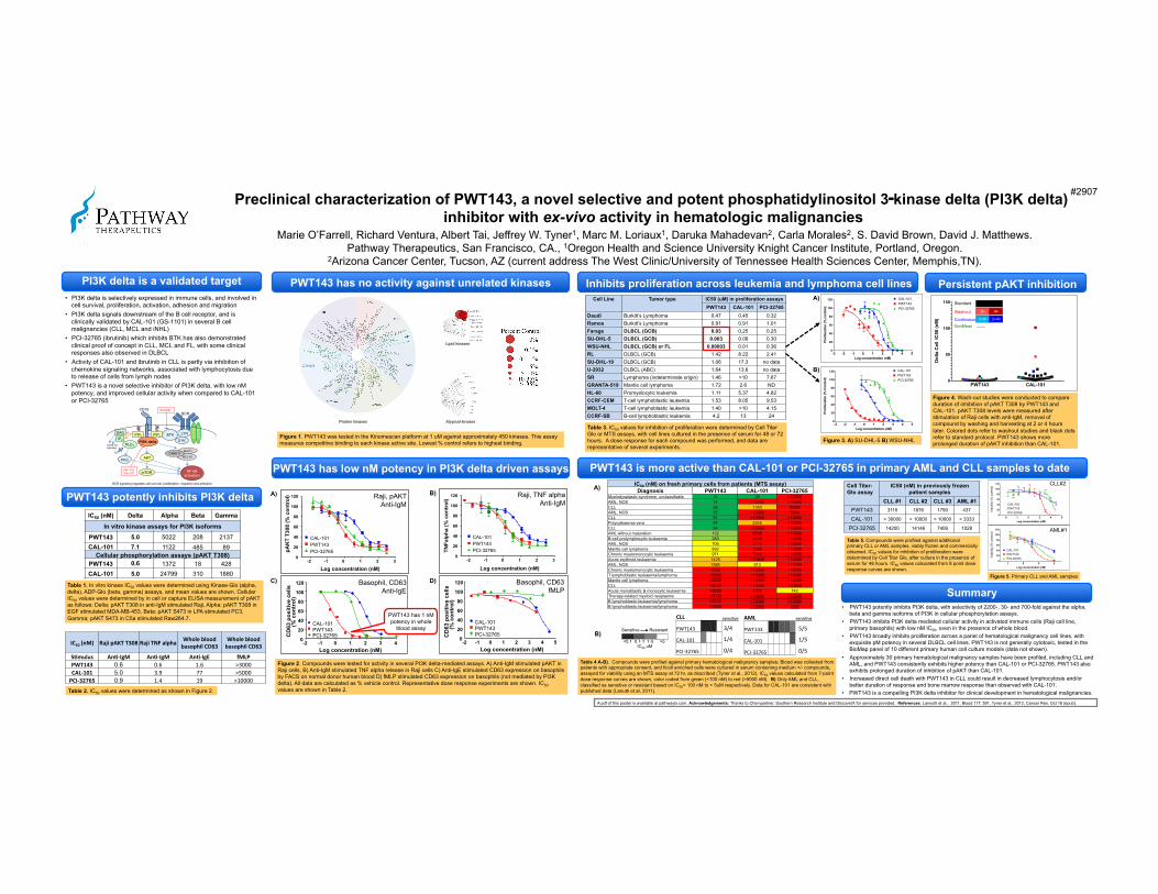
Preclinical characterization of PWT143, a novel selective and potent phosphatidylinositol 3‑kinase delta (PI3K delta) inhibitor with ex-vivo activity in hematologic malignancies
#2907
PI3K delta is a validated target
Table 4 A-B). Compounds were profiled against primary hematological malignancy samples; Blood was collected from patients with appropriate consent, and ficoll enriched cells were cultured in serum containing medium +/- compounds, assayed for viability using an MTS assay at 72 hr, as described (Tyner et al., 2012). IC50 values calculated from 7-point dose response curves are shown, color coded from green (<100 nM) to red (>5000 nM). B) Only AML and CLL, classified as sensitive or resistant based on IC50< 100 nM to > 5uM respectively. Data for CAL-101 are consistent with published data (Lanutti et al, 2011).
Table 1. In vitro kinase IC50 values were determined using Kinase-Glo (alpha, delta), ADP-Glo (beta, gamma) assays, and mean values are shown. Cellular IC50 values were determined by in cell or capture ELISA measurement of pAKT as follows: Delta; pAKT T308 in anti-IgM stimulated Raji, Alpha; pAKT T308 in EGF stimulated MDA-MB-453, Beta; pAKT S473 in LPA stimulated PC3, Gamma; pAKT S473 in C5a stimulated Raw264.7.
Table 3. IC50 values for inhibition of proliferation were determined by Cell Titer Glo or MTS assays, with cell lines cultured in the presence of serum for 48 or 72 hours. A dose response for each compound was performed, and data are representative of several experiments.
IC50 (nM) Delta Alpha Beta Gamma
In vitro kinase assays for PI3K isoforms
PWT143 5.0 5022 208 2137 CAL-101 7.1 1122 485 89
Cellular phosphorylation assays (pAKT T308) PWT143 0.6 1372 18 428 CAL-101 5.0 24799 310 1880
IC50 (nM) Raji pAKT T308 Raji TNF alpha Whole blood basophil CD63
Whole blood basophil CD63
SAmulus AnA-‐IgM AnA-‐IgM AnA-‐IgE fMLP PWT143 0.6 0.6 1.6 >3000 CAL-‐101 5.0 3.9 77 >5000 PCI-‐32765 0.9 1.4 19 >10000
Log concentration (nM)
CD
63 p
ositi
ve c
ells
(% c
ontr
ol)
-2 -1 0 1 2 3 4 0 20 40 60 80
100 120
CAL-101 PWT143 PCI-32765
PWT143 has no activity against unrelated kinases
Log concentration (nM)
pAK
T T3
08 (%
con
trol
)
-2 -1 0 1 2 3 0 20 40 60 80
100 120
CAL-101 PWT143 PCI-32765
Log concentration (nM)
TNFa
lpha
(% c
ontr
ol)
-2 -1 0 1 2 3 0 20 40 60 80
100 120
CAL-101 PWT143 PCI-32765
Del
ta C
ell
IC50
(nM
)
PWT143 CAL-1010
50
100
150
2h 4h
2+2h 2+4h
Washout
Continuous
GeoMean
Standard
Inhibits proliferation across leukemia and lymphoma cell lines
PWT143 is more active than CAL-101 or PCI-32765 in primary AML and CLL samples to date
• PI3K delta is selectively expressed in immune cells, and involved in cell survival, proliferation, activation, adhesion and migration
• PI3K delta signals downstream of the B cell receptor, and is clinically validated by CAL-101 (GS-1101) in several B cell malignancies (CLL, MCL and iNHL)
• PCI-32765 (ibrutinib) which inhibits BTK has also demonstrated clinical proof of concept in CLL, MCL and FL, with some clinical responses also observed in DLBCL
• Activity of CAL-101 and ibrutinib in CLL is partly via inhibition of chemokine signaling networks, associated with lymphocytosis due to release of cells from lymph nodes
• PWT143 is a novel selective inhibitor of PI3K delta, with low nM potency, and improved cellular activity when compared to CAL-101 or PCI-32765
Table 2. IC50 values were determined as shown in Figure 2.
IC50 (nM) on fresh primary cells from patients (MTS assay) Diagnosis PWT143 CAL-101 PCI-32765
Myelodysplastic syndrome, unclassifiable 9 28 >10000 AML, NOS 14 >10000 >10000 CLL 26 1350 10000 AML, NOS 37 >10000 7944 CLL 51 >10000 >10000 Polycythaemia vera 87 2345 >10000 CLL 94 >10000 >10000 AML without maturation 132 6608 >10000 B-cell prolymphocytic leukaemia 283 3468 >10000 AML, NOS 706 3716 >10000 Mantle cell lymphoma 892 3389 >10000 Chronic myelomonocytic leukaemia 971 7944 >10000 Acute erythroid leukaemia 1425 >10000 >10000 AML, NOS 1584 913 >10000 Chronic myelomonocytic leukaemia 4593 >10000 >10000 T lymphoblastic leukaemia/lymphoma >5000 >10000 >10000 Mantle cell lymphoma >5000 >10000 >10000 CLL >5000 >10000 >10000 Acute monoblastic & monocytic leukaemia >5000 4572 742 Therapy-related myeloid neoplasms >5000 >10000 >10000 B lymphoblastic leukaemia/lymphoma >5000 >10000 >10000 B lymphoblastic leukaemia/lymphoma >5000 >10000 3237
PWT143 potently inhibits PI3K delta
Figure 1. PWT143 was tested in the Kinomescan platform at 1 uM against approximately 450 kinases. This assay measures competitive binding to each kinase active site. Lowest % control refers to highest binding.
PWT143 has low nM potency in PI3K delta driven assays
Protein kinases Atypical kinases
Lipid kinases
Figure 2. Compounds were tested for activity in several PI3K delta-mediated assays. A) Anti-IgM stimulated pAKT in Raji cells, B) Anti-IgM stimulated TNF alpha release in Raji cells C) Anti-IgE stimulated CD63 expression on basophils by FACS on normal donor human blood D) fMLP stimulated CD63 expression on basophils (not mediated by PI3K delta). All data are calculated as % vehicle control. Representative dose response experiments are shown. IC50 values are shown in Table 2.
• PWT143 potently inhibits PI3K delta, with selectivity of 2200-, 30- and 700-fold against the alpha, beta and gamma isoforms of PI3K in cellular phosphorylation assays.
• PWT143 inhibits PI3K delta mediated cellular activity in activated immune cells (Raji cell line, primary basophils) with low nM IC50, even in the presence of whole blood.
• PWT143 broadly inhibits proliferation across a panel of hematological malignancy cell lines, with exquisite pM potency in several DLBCL cell lines. PWT143 is not generally cytotoxic, tested in the BioMap panel of 10 different primary human cell culture models (data not shown).
• Approximately 30 primary hematological malignancy samples have been profiled, including CLL and AML, and PWT143 consistently exhibits higher potency than CAL-101 or PCI-32765. PWT143 also exhibits prolonged duration of inhibition of pAKT than CAL-101.
• Increased direct cell death with PWT143 in CLL could result in decreased lymphocytosis and/or better duration of response and bone marrow response than observed with CAL-101.
• PWT143 is a compelling PI3K delta inhibitor for clinical development in hematological malignancies.
Summary
Persistent pAKT inhibition
Figure 4. Wash-out studies were conducted to compare duration of inhibition of pAKT T308 by PWT143 and CAL-101. pAKT T308 levels were measured after stimulation of Raji cells with anti-IgM, removal of compound by washing and harvesting at 2 or 4 hours later. Colored dots refer to washout studies and black dots refer to standard protocol. PWT143 shows more prolonged duration of pAKT inhibition than CAL-101. Figure 3. A) SU-DHL-5 B) WSU-NHL
Marie O’Farrell, Richard Ventura, Albert Tai, Jeffrey W. Tyner1, Marc M. Loriaux1, Daruka Mahadevan2, Carla Morales2, S. David Brown, David J. Matthews. Pathway Therapeutics, San Francisco, CA., 1Oregon Health and Science University Knight Cancer Institute, Portland, Oregon.
2Arizona Cancer Center, Tucson, AZ (current address The West Clinic/University of Tennessee Health Sciences Center, Memphis,TN).
A)
B)
A)
B)
B)
D)
A)
C) Figure 5. Primary CLL and AML samples
Table 5. Compounds were profiled against additional primary CLL or AML samples, viably frozen and commercially obtained. IC50 values for inhibition of proliferation were determined by Cell Titer Glo, after culture in the presence of serum for 48 hours. IC50 values calculated from 8 point dose response curves are shown.
Cell Titer-Glo assay
IC50 (nM) in previously frozen patient samples
CLL #1 CLL #2 CLL #3 AML #1 PWT143 3115 1876 1790 437
CAL-101 > 30000 > 10000 > 10000 > 3333
PCI-32765 14200 14146 7406 1028
Sensitive Resistant !! !! !! !!
IC50 uM <0.1 0.1-1 1-5 >5
!"#$
!"#$%&' '' '' '' '' ''
()*+$,$' '' '' '' '' ''
!(-+&./01' '' '' '' '' ''
121'
$21'
,21'
!""#
!"#$%&' '' '' '' ''
()*+$,$' '' '' '' ''
!(-+&./01' '' '' '' ''
&2%'
$2%'
,2%'
Raji, pAKT Anti-IgM
Raji, TNF alpha Anti-IgM
Basophil, CD63 Anti-IgE
Basophil, CD63 fMLP
PWT143 has 1 nM potency in whole
blood assay
Log concentration (nM)
Prol
ifera
tion
(% C
ontro
l)
-3 -2 -1 0 1 2 3 4 50
20
40
60
80
100
120 CAL-101PWT143PCI-32765
Log concentration (nM)
Prol
ifera
tion
(% c
ontro
l)
-3 -2 -1 0 1 2 3 4 50
20
40
60
80
100
120 CAL-101PWT143PCI-32765
AML#1
CLL#2
Log concentration (nM)
CD
63 p
ositi
ve c
ells
(%
con
trol
)
-2 -1 0 1 2 3 4 5 0 20 40 60 80
100 120
CAL-101 PWT143 PCI-32765
Cell Line Tumor type IC50 (uM) in proliferation assays PWT143 CAL-101 PCI-32765
Daudi Burkitt’s Lymphoma 0.47 0.45 0.32 Ramos Burkitt’s Lymphoma 0.91 0.91 1.01 Farage DLBCL (GCB) 0.03 0.25 0.25 SU-DHL-5 DLBCL (GCB) 0.003 0.06 0.30 WSU-NHL DLBCL (GCB) or FL 0.00003 0.01 0.36 RL DLBCL (GCB) 1.42 8.22 2.41 SU-DHL-10 DLBCL (GCB) 1.06 17.3 no data U-2932 DLBCL (ABC) 1.64 13.6 no data SR Lymphoma (indeterminate origin) 1.46 >10 7.67 GRANTA-519 Mantle cell lymphoma 1.72 2.6 ND HL-60 Promyelocytic leukemia 1.11 5.37 4.82 CCRF-CEM T-cell lymphoblastic leukemia 1.53 8.05 9.53 MOLT-4 T-cell lymphoblastic leukemia 1.40 >10 4.15 CCRF-SB B-cell lymphoblastic leukemia 4.2 13 24
sensitive sensitive
A pdf of this poster is available at pathwaytx.com. Acknowledgements: Thanks to Chempartner, Southern Research Institute and DiscoverX for services provided. References: Lannutti et al., 2011, Blood 117: 591, Tyner et al., 2012, Cancer Res. Oct 18 (epub).
CD19
CD
79B
C
D79
A
LYN!
!"#$%&
BCL10!AKT!
mTOR! NF-kB activation
BCR
SYK!BTK!PIP3!
DAG!
PI3K delta!PLC!"
IP3!
PKC!
Ca++
PIP2!
CARD11!
Inside-out signaling
PWT143 CAL-101
Ibrutinib
BCR signaling regulates cell survival, proliferation, migration and adhesion