The Cyclin-Dependent Kinase Inhibitor KRP6 Induces Mitosis and ...
Upload
nguyenthienCategory
view
220download
0
Cancer Therapy: Preclinical
The Novel Pan-PIM Kinase Inhibitor, PIM447,Displays Dual Antimyeloma and Bone-ProtectiveEffects, and Potently Synergizes with CurrentStandards of CareTeresa Paíno1,2, Antonio Garcia-Gomez1,2, Lorena Gonz�alez-M�endez1,2,Laura San-Segundo1,2, Susana Hern�andez-García1,2, Ana-Alicia L�opez-Iglesias1,2,Esperanza M. Algarín1,2, Montserrat Martín-S�anchez1,2, David Corbacho3,Carlos Ortiz-de-Solorzano3, Luis A. Corchete1,2, Norma C. Guti�errez1,2,María-Victoria Maetos1,2, Mercedes Garayoa1,2, and Enrique M. Ocio1,2
Abstract
Purpose: PIM kinases are a family of serine/threonine kinasesrecently proposed as therapeutic targets in oncology. In thepresent work, we have investigated the effects of the novel pan-PIM kinase inhibitor, PIM447, on myeloma cells and myeloma-associated bone disease using different preclinical models.
Experimental Design: In vitro/ex vivo cytotoxicity of PIM447was evaluated on myeloma cell lines and patient samples. Syn-ergistic combinations with standard treatments were analyzedwith Calcusyn Software. PIM447 effects on bone cells wereassessed on osteogenic and osteoclastogenic cultures. Themechanisms of PIM447 were explored by immunoblotting,qPCR, and immunofluorescence. A murine model of disseminat-ed multiple myeloma was employed for in vivo studies.
Results: PIM447 is cytotoxic formyeloma cells due to cell-cycledisruption and induction of apoptosis mediated by a decrease inphospho-Bad (Ser112) and c-Myc levels and the inhibition of
mTORC1 pathway. Importantly, PIM447 demonstrates a verystrong synergy with different standard treatments such as borte-zomib þ dexamethasone (combination index, CI ¼ 0.002),lenalidomide þ dexamethasone (CI ¼ 0.065), and pomalido-mideþdexamethasone (CI¼0.077). PIM447 also inhibits in vitroosteoclast formation and resorption, downregulates key mole-cules involved in these processes, and partially disrupts the F-actinring, while increasing osteoblast activity and mineralization.Finally, PIM447 significantly reduced the tumor burden andprevented tumor-associated bone loss in a disseminated murinemodel of human myeloma.
Conclusions: Our results demonstrate dual antitumoral andbone-protective effects of PIM447. This fact, togetherwith the verystrong synergy exhibited with standard-of-care treatments, sup-ports the future clinical development of this drug in multiplemyeloma. Clin Cancer Res; 23(1); 225–38. �2016 AACR.
IntroductionMultiple myeloma is characterized by a proliferation of
malignant plasma cells in the bone marrow (BM) that secretesa monoclonal immunoglobulin (1). It is typically associatedwith osteolytic lesions, due to an increase in the number and
bone-resorptive activity of osteoclasts (OC) together with oste-oblast (OB) inhibition (2). Over the last decade, the introduc-tion of new drugs, such as proteasome inhibitors and immu-nomodulatory agents, has improved the outcome of patientswith multiple myeloma (3, 4); however, multiple myeloma isstill considered incurable mainly due to the developmentof drug resistance. Therefore, the identification of new targetsand the investigation of novel drugs against such targets areextremely important for the discovery of more effective treat-ments (5).
Recently, PIM kinases have been proposed as being newtherapeutic targets for treating hematologic cancers (6). Theseare a family of serine/threonine kinases comprising threemembers (PIM1, PIM2, and PIM3) that regulate oncogenesis,survival pathways, drug resistance, and migration, amongother functions (7). Several features make PIM inhibition anattractive therapeutic strategy, particularly against myelomacells: (i) PIM2 is among the most highly overexpressed genesin multiple myeloma cells (8), (ii) this protein is required formaintaining multiple myeloma cell growth (9), and (iii) theexpression of PIM2 in multiple myeloma cells is enhanced bymesenchymal stromal cells (MSC) and OCs as a survival and
1Centro de Investigaci�on del C�ancer-IBMCC (CSIC-Universidad de Salamanca),Salamanca, Spain. 2Complejo Asistencial Universitario de Salamanca-IBSAL,Salamanca, Spain. 3Program in Solid Tumors and Biomarkers, Centro deInvestigaci�onM�edica Aplicada (CIMA), UniversidaddeNavarra; Navarra's HealthResearch Institute (IDISNA), Pamplona, Spain.
Note: Supplementary data for this article are available at Clinical CancerResearch Online (http://clincancerres.aacrjournals.org/).
T. Paíno and A. Garcia-Gomez contributed equally to this article.
M. Garayoa and E.M. Ocio contributed equally to this article.
Corresponding Author: Enrique M. Ocio, Hospital Universitario de Salamanca,Paseo de San Vicente, 58-182, 37007 Salamanca, Spain. Phone: 34-923-291384;Fax: 34-923-294624; E-mail: [email protected]
doi: 10.1158/1078-0432.CCR-16-0230
�2016 American Association for Cancer Research.
ClinicalCancerResearch
www.aacrjournals.org 225
on November 15, 2018. © 2017 American Association for Cancer Research.clincancerres.aacrjournals.org Downloaded from
Published OnlineFirst July 20, 2016; DOI: 10.1158/1078-0432.CCR-16-0230

drug resistance mechanism (10). In addition, recent studieshave also demonstrated the importance of PIM kinases in thebiology of bone-remodeling cells. Accordingly, Kim and col-leagues found that PIM1 positively regulates the ligand of thereceptor activator of NF-kB (RANKL)–induced osteoclastogen-esis (11). More recently, it has been shown that PIM2 acts as anegative regulator of osteoblastogenesis (12). Based on allthese data, it is tempting to hypothesize that the inhibitionof these proteins would result not only in an antitumoractivity, but may also have a beneficial effect on multiplemyeloma patients' bone disease.
PIM447 (N-(4-((1R,3S,5S)-3-amino-5-methylcyclohexyl)pyri-din-3-yl)-6-(2,6-difluorophenyl)-5-fluoropicolinamide) is apotent and selective pan-PIM kinase inhibitor, derived from thetool compound LGB321 (6, 9), with pharmacokinetic and drug-like properties suitable for clinical development (13). In fact,preliminary results from a phase I clinical trial with PIM447 inpatients with relapsed and/or refractory multiple myeloma havedemonstrated single-agent activity and a tolerable safety profile(14). Although the preclinical efficacy of other PIM inhibitorshas been studied in several hematologic malignancies includ-ing multiple myeloma (12, 15–18), only two of them, SGI-1776 and AZD1208, have reached clinical trials but none inmultiple myeloma.
In the present work, we have investigated through the use ofpreclinical models, the activity and mechanism of action ofPIM447 in myeloma cells, its potential synergism with stan-dard-of-care treatments, and its effects on myeloma-associatedbone disease. To the best of our knowledge, this is the first clinicalpan-PIM-inhibitor that demonstrates dual antimyeloma andbone-protective effects, and this fact, together with the very strongsynergy that PIM447 exhibits with standard-of-care treatmentsand the preliminary favorable clinical data, supports the futureclinical development of this drug for the treatment of patientswith multiple myeloma.
Materials and MethodsDrugs
PIM447 was provided by Novartis Pharmaceuticals, Inc. Bor-tezomib was purchased from LC Laboratories, lenalidomide and
pomalidomide from Selleckchem, and dexamethasone fromSigma-Aldrich.
Cell lines, primary samples, and culturesThe human myeloma cell lines, MM1S, MM1R, U266, and
NCI-H929, were purchased from the ATCC, whereas RPMI-8226 and OPM-2 were obtained from DSMZ. The humanmyeloma cell line MM144 was a generous gift from Dr. S.Rudikoff (National Cancer Institute, National Institutes ofHealth, Bethesda, MD). The source of the human myelomacell lines RPMI-LR5, U266-Dox4, and U266-LR7 has beenpreviously described (19). The origin of MM1S-luc andRPMI-8226-luc cells (luciferase-expressing) was previouslyexplained (20). All myeloma cell lines were cultured aspreviously described (19). The MM1S-luc cocultures witheither the hMSC–TERT cell line (obtained from Dr. D.Campana, St. Jude Children's Research Hospital, Memphis,TN) or OCs were performed in the absence or presence ofdifferent concentrations of PIM447 as previously explained(20). Cell line identities have been tested and authenticatedby short tandem repeat (STR) analysis with a PowerPlex 16HS System kit (Promega) and online STR matching analysis(DSMZ institute; available from: http://www.dsmz.de/fp/register.php).
All primary BM and peripheral blood samples were obtainedafter the approval of the Complejo Asistencial Universitario deSalamanca Review Board and with the written informed consentof all participating subjects.
Cell viability, cell cycle, and apoptosis assaysCell viability of myeloma cells was evaluated by the MTT
method (19). The IC50 of PIM447was calculated using SigmaPlotgraphing software. The cell-cycle profile and apoptosis inductionwere evaluated as previously described (21).
Ex vivo analysis of apoptosis in freshly isolated patient cellsBM samples from patients with multiple myeloma were
lysed and cultured as previously described (21), in the absenceor presence of PIM447. The percentage of Annexin-V–positivecells was analyzed by flow cytometry on myelomatous plasmacells (CD38þbright, CD45�/low, SSClow/intermediate, CD56�/þ)and normal lymphocytes (CD45þþ, SSClow) populations.
Evaluation of the potential synergism of PIM447 with otherantimyeloma agents
MM1S or RPMI-8226 cells were treated for 48 to 72 hours withdouble and triple combinations of PIM447 and other antimye-loma agents (bortezomib, lenalidomide, pomalidomide, anddexamethasone). Cell viability was analyzed by MTT assays. Thepotency of each combination was quantified with Calcusynsoftware (Biosoft), which is based on the Chou–Talalay method,yielding a combination index (CI) with the following interpre-tation:CI>1, antagonistic effect; CI¼1, additive effect; andCI<1,synergistic effect.
In vitro OC formation, resorption pits, and integrity of F-actinring
Peripheral blood mononuclear cells (PBMCs) fromhealthy donors were differentiated in osteoclastogenic medi-um (containing 25 ng/mL M-CSF and 50 ng/mL RANKL,
Translational Relevance
PIMkinases have been recently proposed asnew therapeutictargets in oncology. PIM447 is the only pan-PIM kinaseinhibitor that has reached clinical development in multiplemyeloma with promising preliminary efficacy results. In thiswork, we demonstrate through the use of preclinical modelsthe potent antimyeloma and bone-protective effects ofPIM447, and the mechanisms responsible of such effects. Inaddition, we show the very strong synergy exhibited by thisdrug in combination with different standard-of-care treat-ments. The preclinical efficacy of PIM447 demonstrated inthe present work together with the preliminary single-agentefficacy and tolerable safety profile observed in the clinicalsetting, reinforces PIM inhibition as a promising therapeuticstrategy in myeloma, and opens the door to new clinical trialswith this drug.
Paíno et al.
Clin Cancer Res; 23(1) January 1, 2017 Clinical Cancer Research226
on November 15, 2018. © 2017 American Association for Cancer Research.clincancerres.aacrjournals.org Downloaded from
Published OnlineFirst July 20, 2016; DOI: 10.1158/1078-0432.CCR-16-0230

both from Peprotech) as previously described (22). Assaysrelated to OCs formation and function included: F-actinring formation (pre-OCs, 14 days differentiation), resorp-
tion capacity (17 days differentiation), and OC formation(21 days differentiation), and were performed as previouslyreported (23).
Figure 1.
Multiple myeloma (MM) cells express PIM kinases and are sensitive to the antiproliferative effect of the pan-PIM kinase inhibitor, PIM447. A, Basal protein levelsof PIM1, PIM2, and PIM3were analyzed byWestern blot in 10multiplemyeloma cell lines.a-Tubulinwas used as a loading control. B,Basal gene expression of PIM1, PIM2,and PIM3, assessed by the Human gene 1.0 ST array (Affymetrix) in CD138þ myeloma cells isolated from 41 patients with multiple myeloma. Normalizedexpression intensity was calculated as described in Material and Methods. An outlier at a distance of greater than 1.5x the interquartile range from the box is plottedindividually as a dot. Global differences amonggroupswereevaluatedby theKruskal–Wallis test; significantpair-wise differenceswere identifiedby theMann–WhitneyUtest. All the P values were adjusted for multiple comparisons using the FDR method (��� , P < 0.001). C, The indicated multiple myeloma cell lines were incubatedwith increasing concentrations of PIM447 for 24, 48, and 72 hours, and cell viability was analyzed by MTT assay. The average absorbance values of controluntreated sampleswere taken as 100%. Data are summarized as themean� SD (n¼ 3).D, IC50 values for PIM447 at 48 hourswere plotted against PIM1, PIM2, and PIM3expression as measured by Western blot and normalized with a-tubulin using the image processing package Fiji. r ¼ Pearson correlation coefficient; p ¼ P value.
Preclinical Activity of PIM447 in Multiple Myeloma
www.aacrjournals.org Clin Cancer Res; 23(1) January 1, 2017 227
on November 15, 2018. © 2017 American Association for Cancer Research.clincancerres.aacrjournals.org Downloaded from
Published OnlineFirst July 20, 2016; DOI: 10.1158/1078-0432.CCR-16-0230

Paíno et al.
Clin Cancer Res; 23(1) January 1, 2017 Clinical Cancer Research228
on November 15, 2018. © 2017 American Association for Cancer Research.clincancerres.aacrjournals.org Downloaded from
Published OnlineFirst July 20, 2016; DOI: 10.1158/1078-0432.CCR-16-0230

In vitro OB differentiation, alkaline phosphatase activity, andmineralization assays
OBs were generated and assayed as previously described(20). Briefly, primary MSCs (passages 2–3) were cultured inosteogenic medium (containing 5 mmol/L b-glycerophosphateand 50 mg/mL ascorbic acid) and exposed to PIM447. After 11days, alkaline phosphatase (ALP) activity was quantified byhydrolysis of p-nitrophenylphosphate into p-nitrophenol (Sig-ma-Aldrich), whereas mineralization was assessed by AlizarinRed staining of calcium deposits at day 21.
Gene expression data: Source, processing, and analysisAll gene expression microarray datasets were retrieved from
Gene Expression Omnibus (http://www.ncbi.nlm.nih.gov/geo/,accession number: GSE47552; ref. 24). CEL files were back-ground-corrected and normalized using the Robust Multi-Array(RMA) algorithm implemented in the Affymetrix ExpressionConsole, to estimate the log2-normalized expression values forPIM1, PIM2, and PIM3.
ImmunoblottingWestern blot was performed following standard procedures
(25). Phospho-TSC2 levels were examined with a specific phos-pho-Akt substrate antibody (Cell Signaling Technology) followingTSC2 immunoprecipitation, aspreviouslydescribed(9). Theoriginof primary antibodies was as follows: anti–Bcl-2, anti–Mcl-1, anti–p-Erk1/2, anti-NFATc1, and anti–cathepsin K from Santa CruzBiotechnology; a-tubulin from Calbiochem; and all other anti-bodies from Cell Signaling Technology. The horseradish peroxi-dase–conjugated secondary antibodies were from GE Healthcare.
Reverse transcription real-time PCR analysesTotal RNA was isolated using TRIzol reagent (Invitrogen).
TaqMan Gene Expression Assays (Applied Biosystems) were per-formed according to the manufacturer's instructions: CA2(Hs00163869_m1), ATP6V1A (Hs01097169_m1), and MMP9(Hs00234579_m1). Normalized gene expression was calculatedas 2�DCt, being DCt ¼ Ct (gene) � Ct (GAPDH).
Mouse model of BM-disseminated human multiple myelomaAnimal experiments were conducted according to institutional
guidelines for the use of laboratory animals, and after grantedpermission from the University of Salamanca Animal EthicalCommittee for animal experimentation. RPMI-8226-luc cells
(8 � 106) were injected intravenously into 6-week-old femaleNOD-SCID-IL-2Rg�/� (NSG) mice (Charles River Laboratories),and tumor development was monitored by noninvasive biolu-minescence imaging (BLI), as previously described (20). After4 weeks, animals were randomized into two groups (n ¼ 12/group), one receiving vehicle (5 times/week by oral gavage), theother receiving PIM447 (100 mg/kg, 5 times/week by oralgavage). Serum levels of human Igl (secreted by RPMI-8226-luccells) were determined by ELISA (Bethyl Laboratories). Formicro-computed tomography (microCT) analysis, one femur of eachanimal was fixed in 10% formalin and scanned using a microCTsystem (MicroCATII; Siemens) as described previously (22).The trabecular microarchitecture in the distal femur was ana-lyzed using BoneJ (26). Carboxy-terminal telopeptide collagencross-links (CTX) and N-terminal propeptide of type I procol-lagen (P1NP) were measured in mice sera by ELISA (Immu-nodiagnostic Systems).
Statistical analysesStatistical analyses were performed using SPSS-v21.0 software
(IBM Corp.).
ResultsPIM kinases are expressed in multiple myeloma cell lines andmyeloma cells from patients
We initially evaluated the basal levels of PIM kinases inmultiple myeloma cell lines and primary myeloma cells. Asshown in Fig. 1A, the three isoforms (PIM1, PIM2, and PIM3)were expressed in the 10 multiple myeloma cell lines analyzed.Notably, the level of expression of PIM2 was higher than theother two PIM kinases. Consistent with these results, we alsoobserved in a series of 41 patients with newly diagnosedmultiple myeloma that PIM2 levels were significantly higherthan PIM1 and PIM3 in CD138þ myeloma cells (Fig. 1B). Inaddition, PIM1 levels were significantly higher than PIM3levels (Fig. 1B).
PIM447 is cytotoxic for multiple myeloma cells and overcomesthe resistance conferred by MSCs and OCs
Because PIM kinases are highly expressed inmultiple myelomacells, we evaluated the antimyeloma effect of the new pan-PIMkinase inhibitor, PIM447. Different multiple myeloma cell lineswere treated with increasing concentrations of PIM447 (0.05–
Figure 2.PIM447 induces apoptosis and cell-cycle arrest in multiple myeloma (MM) cells and overcomes the resistance conferred by MSCs and OCs. A, MM1S, NCI-H929,RPMI-8226, OPM-2, and RPMI-LR5 cells were treated with increasing doses (0.1–10 mmol/L) of PIM447 for 24 and 48 hours, and the induction of apoptosiswas analyzed by flow cytometry after Annexin-V staining. Data represent the percentage of apoptotic cells for each condition after subtracting the basal percentageof apoptotic cells for each cell line. B, MM1S cells were treated with 10 mmol/L PIM447 for the indicated times, and the induction of apoptosis was analyzedby flow cytometry. Data are summarized as the mean � SEM (n ¼ 3). Statistically significant differences between control (0 hour) and different time points arerepresented as ��� , P < 0.001; �� , P < 0.01; and � , P < 0.05 (Student t test). C, MM1S cells were treated with 10 mmol/L PIM447 for the indicated times, andthe expression of caspase 8, caspase 9, caspase 3, caspase 7, and PARP was analyzed byWestern blot using a-tubulin as loading control. D,MM1S and OPM-2 cellswere incubated with increasing concentrations of PIM447 for 48 hours, and the cell-cycle profile was examined by flow cytometry, as shown byrepresentative histograms. Tables refer to one experiment that was repeated twice with similar results in the two cell lines and indicate the percentage ofcells in subG0 (apoptotic cells) and the percentages in G0–G1, S and G2–M from the non-subG0 population. E, Freshly isolated BM cells obtained from 10 patientswith multiple myeloma were treated ex vivo with increasing concentrations of PIM447 for 48 hours. After the incubation period, cells were stained with thecombination of Annexin V–FITC and three monoclonal antibodies (CD56-PE, CD45-PerCP-Cy5.5, and CD38-APC) for the analysis of apoptosis in myelomacells and lymphocytes. Outliers further than 1.5x or 3x the interquartile range from thebox are plotted individually as dots and asterisks, respectively.F,MM1S-luc cellswere cocultured with the hMSC-TERT cell line for 2 days or pre-OCs derived from healthy donors for 5 days with the indicated concentrations of PIM447 andunder low-serum conditions. After the coculture period, MM1S-luc growth was assessed by luciferase bioluminescence measurement. Graphs illustratethe mean � SD (n ¼ 3).
Preclinical Activity of PIM447 in Multiple Myeloma
www.aacrjournals.org Clin Cancer Res; 23(1) January 1, 2017 229
on November 15, 2018. © 2017 American Association for Cancer Research.clincancerres.aacrjournals.org Downloaded from
Published OnlineFirst July 20, 2016; DOI: 10.1158/1078-0432.CCR-16-0230

10 mmol/L) for 24, 48, and 72 hours. The dose–response curvesobtained by MTT assay revealed two patterns of sensitivity:sensitive cell lines with IC50 values at 48 hours ranging from0.2 to 3.3 mmol/L (MM1S, MM1R, RPMI-8226, MM144, U266,and NCI-H929) and less sensitive cell lines with IC50 values at 48hours >7 mmol/L (OPM-2, RPMI-LR5, U266-Dox4, and U266-LR7; Fig. 1C). A lack of correlationwas foundbetween the levels ofPIM kinases and the IC50 values for PIM447 (P > 0.05; Fig. 1D). Toexplore the induction of apoptosis, three of the sensitive cell lines(MM1S, NCI-H929, and RPMI-8226) and two of the less sensitivecell lines (OPM-2 and RPMI-LR5) were treated with increasingdoses of PIM447 for 24 and 48 hours (Fig. 2A), revealing a cleardose-response: while low doses of the drug (0.1–1 mmol/L) didnot induce important levels of apoptosis in any of the cell lines
tested, PIM447 at 5 mmol/L substantially increased Annexin-Vlevels (about 30%) in sensitive cell lines but not in OPM-2 andRPMI-LR5. In addition, the highest dose (10 mmol/L) inducedapoptosis in all the cell lines but to a lesser extent in OPM-2 andRPMI-LR5. When MM1S cells were treated with 10 mmol/LPIM447 for different times, we observed a time-dependentincrease in apoptotic cells (Fig. 2B). Moreover, treatment ofMM1S cells with PIM447 promoted the cleavage of initiatorcaspases, such as caspases 8 and 9, and also the cleavage of theeffector caspases 3 and 7, together with PARP cleavage (Fig.2C). Similar results were found in RPMI-8226 and NCI-H929cells (Supplementary Fig. S1A and S1B). To assess the potentialeffect of PIM447 on cell cycle, MM1S and OPM-2 cells wereincubated with increasing concentrations (0.1–10 mmol/L) of
Figure 3.
PIM447 synergizes with different antimyeloma agents in the MM1S cell line. MM1S cells were treated with the indicated double or triple combinations ofPIM447, dexamethasone, and either (A) bortezomib for 48 hours, (B) lenalidomide for 72 hours, or (C) pomalidomide for 72 hours. Cell viability was analyzedby MTT assay as represented in the graphs.
Paíno et al.
Clin Cancer Res; 23(1) January 1, 2017 Clinical Cancer Research230
on November 15, 2018. © 2017 American Association for Cancer Research.clincancerres.aacrjournals.org Downloaded from
Published OnlineFirst July 20, 2016; DOI: 10.1158/1078-0432.CCR-16-0230

Figure 4.
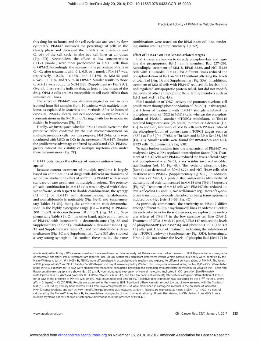
Effect of PIM447 on PIM kinase–related targets. MM1S or OPM-2 cells were treated with 10 mmol/L PIM447 for the indicated times, and the expression ofdifferent proteins was analyzed by Western blot. Alpha-tubulin was used as a loading control. A, Protein levels of phospho-Bad (Ser112), total Bad, Bcl-2, Bcl-xl,and Mcl-1 in MM1S. B, The levels of phospho-TSC2 in MM1S were analyzed by immunoprecipitation of TSC2 with an anti-TSC2 antibody and subsequentimmunoblotting with a phospho-Akt substrate antibody. The expression of total TSC2, phospho PRAS40 (Thr246), total PRAS40, phospho 4EBP1 (Thr 37/46), total4EBP1, phospho P70S6 (Thr 389), total P70S6, phospho S6RP (Ser 235/236), and total S6RPwas analyzed byWestern blot.C,Protein levels of total c-Myc, phospho-c-Myc (Ser 62), Mad-1, cyclin D2, and cyclin E1 in MM1S. D, Protein levels of phospho-Bad (Ser112), total Bad, c-Myc, and phospho-c-Myc (Ser 62) in OPM-2. E and F,IC50 values for PIM447 at 48 hours were plotted against Bad and phospho-Bad expression, respectively, as measured by Western blot and normalized witha-tubulin using the image processing package Fiji. r ¼ Pearson correlation coefficient; p ¼ P value.
Preclinical Activity of PIM447 in Multiple Myeloma
www.aacrjournals.org Clin Cancer Res; 23(1) January 1, 2017 231
on November 15, 2018. © 2017 American Association for Cancer Research.clincancerres.aacrjournals.org Downloaded from
Published OnlineFirst July 20, 2016; DOI: 10.1158/1078-0432.CCR-16-0230

Figure 5.
PIM447 inhibits in vitro OC formation and resorption and increases OB differentiation and activity. A, PBMCs were differentiated in osteoclastogenic mediumfor the indicated times. PIM1, PIM2, and PIM3 expression was analyzed by Western blot. B, OC formation was evaluated by the mean number of TRAPþ
multinucleated cells (�3 nuclei) per well after PIM447 treatment of osteoclastogenic cultures for 21 days. IC50 of PIM447 was calculated using SigmaPlotgraphing software. Data are summarized as the mean� SEM. Representative micrographs of TRAPþOCs are shown. Bar, 50 mm. C, To test the effect of PIM447 onOC resorption, inhibition of matrix mineralization was assessed by PBMCs cultured on calcium-coated slices in the presence of osteoclastogenic medium andPIM447 treatment as indicated. (Continued on the following page.)
Paíno et al.
Clin Cancer Res; 23(1) January 1, 2017 Clinical Cancer Research232
on November 15, 2018. © 2017 American Association for Cancer Research.clincancerres.aacrjournals.org Downloaded from
Published OnlineFirst July 20, 2016; DOI: 10.1158/1078-0432.CCR-16-0230
this drug for 48 hours, and the cell cycle was analyzed by flowcytometry. PIM447 increased the percentage of cells in theG0–G1 phase and decreased the proliferative phases (S andG2–M) of the cell cycle, in the two cell lines at all doses(Fig. 2D). Nevertheless, the effects at low concentrations(0.1–1 mmol/L) were more pronounced in MM1S cells thanin OPM-2. Accordingly, the increase in the percentage of cells inG0–G1 after treatment with 0.1, 0.5, or 1 mmol/L PIM447 was,respectively, 10.2%, 16.66%, and 19.18% in MM1S; and6.54%, 11.09%, and 9.51% in OPM-2. Similar results to thoseof MM1S were found in NCI-H929 (Supplementary Fig. S1C).Overall, these results indicate that, at least at low doses of thedrug, OPM-2 cells are less susceptible to cell-cycle effects thansensitive cell lines.
The effect of PIM447 was also investigated ex vivo in cellsisolated from BM samples from 10 patients with multiple mye-loma, as explained in Materials and Methods. After 48 hours ofexposure, PIM447 clearly induced apoptosis in myeloma cells(concentrations in the 5–10 mmol/L range) with low to moderatetoxicity in lymphocytes (Fig. 2E).
Finally, we investigated whether PIM447 could overcome theprotective effect conferred by the BM microenvironment onmultiple myeloma cells. For this purpose, MM1S-luc cells werecocultured with MSCs or OCs in the presence of PIM447. Despitethe proliferative advantage conferred by MSCs and OCs, PIM447greatly reduced the viability of multiple myeloma cells underthese circumstances (Fig. 2F).
PIM447 potentiates the efficacy of various antimyelomaagents
Because current treatment of multiple myeloma is largelybased on combinations of drugs with different mechanisms ofaction, we studied the effect of combining PIM447 with severalstandard-of-care treatments in multiple myeloma. The potencyof each combination in MM1S cells was analyzed with Calcu-syn software. With respect to double combinations, the synergy(CI < 1) of PIM447 with dexamethasone, lenalidomide,and pomalidomide is noticeable (Fig. 3A–C and Supplemen-tary Tables S1–S3), being the combination with dexametha-sone in the highly synergistic range (CI ¼ 0.096) at PIM447200 nmol/L þ dexamethasone 10 nmol/L (Fig. 3A and Sup-plementary Table S1). On the other hand, triple combinationsof PIM447 with bortezomib þ dexamethasone (Fig. 3A andSupplementary Table S1), lenalidomideþ dexamethasone (Fig.3B and Supplementary Table S2), and pomalidomide þ dexa-methasone (Fig. 3C and Supplementary Table S3) also showeda very strong synergism. To confirm these results, the same
combinations were tested on the RPMI-8226 cell line, render-ing similar results (Supplementary Fig. S2).
Effect of PIM447 on PIM kinase–related targetsPIM kinases are known to directly phosphorylate and regu-
late the proapoptotic Bcl-2 family member, Bad (27–29).Accordingly, treatment of MM1S, RPMI-8226, and NCI-H929cells with 10 mmol/L PIM447 for different times reduced thephosphorylation of Bad on Ser112 without affecting the levelsof total Bad (Fig. 4A and Supplementary Fig. S3A). In addition,treatment of MM1S cells with PIM447 reduced the levels of theBad-regulated antiapoptotic protein Bcl-xl, but did not modifythe levels of other antiapoptotic Bcl-2 family members such asBcl-2 and Mcl-1 (Fig. 4A).
PIM2modulatesmTORC1 activity and promotesmyeloma cellproliferation throughphosphorylation of TSC2 (9). In this regard,just 1 hour of treatment with PIM447 strongly inhibited thephosphorylation of TSC2 in MM1S cells, whereas the phosphor-ylation of PRAS40, another mTORC1 modulator, at Thr246required longer exposure (24 hours) to produce a decrease (Fig.4B). In addition, treatment of MM1S cells with PIM447 reducedthe phosphorylation of downstream mTORC1 targets such as4EBP1 at Thr 37/46, P70S6 at Thr 389, and S6RP at Ser 235/236(Fig. 4B). Similar results were found for RPMI-8226 and NCI-H929 cells (Supplementary Fig. S3B).
To gain further insights into the mechanism of PIM447, weanalyzed c-Myc, a PIM-regulated transcription factor (30). Treat-ment ofMM1S cells with PIM447 reduced the levels of total c-Mycand phospho-c-Myc at Ser62, a key residue involved in c-Mycstabilization (ref. 30; Fig. 4C). The levels of phospho-c-Myc(Ser62) also decreased in RPMI-8226 and NCI-H929 cells aftertreatment with PIM447 (Supplementary Fig. S3C). In addition,the levels of Mad-1, a protein that antagonizes Myc-mediatedtranscriptional activity, increased inMM1Safter PIM447 exposure(Fig. 4C). Treatment ofMM1S cells with PIM447 also reduced thelevels of cyclins D2 and E1, two well-known regulators of G1-to-Sphase transition, previously described as being transcriptionallyinduced by c-Myc (refs. 31–33; Fig. 4C).
As previously commented, the sensitivity to PIM447 differsamong differentmultiplemyeloma cell lines. In order to elucidatethe molecular basis for these differences, we explored the molec-ular effects of PIM447 in the low sensitive cell line OPM-2.Treatment of OPM-2 with 10 mmol/L PIM447 reduced the levelsof phospho-S6RP (Ser 235/236) and phospho-4EBP1 (Thr 37/46) after just 1 hour of treatment, indicating the inhibition ofthe mTORC1 pathway (Supplementary Fig. S3D). Interestingly,PIM447 did not reduce the levels of phospho-Bad (Ser112) in
(Continued.) After 17 days, OCs were removed and the area of resorbed lacunae analyzed. Data are summarized as the mean � SEM. Representative micrographsof resorptive pits after PIM447 treatment are reported. Bar, 50 mm. Statistically significant differences versus vehicle control in B and C were identified by theMann–Whitney U test (� , P < 0.05). D, PBMCs were differentiated in osteoclastogenic medium and exposed to different concentrations of PIM447. The levelsof PU.1, phospho Erk1/2, andNFATc1 at day 7 andCathepsin K at day 14were analyzed byWestern blot, usinga-tubulin as a loading control. E, Pre-OCs differentiatedunder PIM447 exposure for 14 days were stained with rhodamine-conjugated phalloidin and examined by fluorescence microscopy to visualize the F-actin ring.Representative micrographs are shown. Bar, 50 mm. F, Normalized gene expression of several molecules implicated in OC resorption [MMP9 (matrixmetalloproteinase 9), ATP6V1A (vacuolar-Hþ-ATPase catalytic subunit A1), and CA2 (carbonic anhydrase II)] after osteoclastogenic differentiation of PBMCsfor 21 days in the presence of PIM447 (2.5 mmol/L) was assessed by real-time RT-PCR. Relative gene expression was calculated by the 2�DCt method, whereDCt ¼ Ct (gene) � Ct (GAPDH). Results are expressed as the mean � SEM. Significant differences with respect to control were assessed with the Student ttest (� , P < 0.05). G, Primary bone marrow MSCs from myeloma patients (n ¼ 5) were maintained in osteogenic medium in the presence of indicatedPIM447 concentrations, and ALP activity (nmol/L/min/mg protein) was measured at day 11. Results are expressed as mean � SEM (�� , P < 0.01 vs. control,calculated by the Mann–Whitney test). H, Representative micrographs of matrix mineralization by Alizarin Red staining in OBs derived from MSCs from amultiple myeloma patient (21 days of osteogenic differentiation in the presence of PIM447).
www.aacrjournals.org Clin Cancer Res; 23(1) January 1, 2017 233
Preclinical Activity of PIM447 in Multiple Myeloma
on November 15, 2018. © 2017 American Association for Cancer Research.clincancerres.aacrjournals.org Downloaded from
Published OnlineFirst July 20, 2016; DOI: 10.1158/1078-0432.CCR-16-0230

Paíno et al.
Clin Cancer Res; 23(1) January 1, 2017 Clinical Cancer Research234
on November 15, 2018. © 2017 American Association for Cancer Research.clincancerres.aacrjournals.org Downloaded from
Published OnlineFirst July 20, 2016; DOI: 10.1158/1078-0432.CCR-16-0230

OPM-2, whereas c-Myc and phospho-c-Myc (Ser62) onlydecreased after 24hours of exposure to the drug (Fig. 4D). Becausethe effects of PIM447 on these proteins were detected earlier insensitive cell lines, we then analyzed the potential correlationbetween their basal levels in multiple myeloma cell lines and thesensitivity to PIM447. The analysis showed a strong positivecorrelation between basal levels of Bad and the IC50 for PIM447(r ¼ 0.8880; P ¼ 0.0006; Fig. 4E). A positive correlation was alsofound between basal levels of phospho-Bad and IC50 values,although in this case, the correlation was not so strong (r ¼0.6044; P ¼ 0.0642; Fig. 4F). Finally, sensitivity to PIM447 doesnot seem to be associated with basal levels of c-Myc (r ¼ 0.4222;P ¼ 0.2242) or phospho-c-Myc (r ¼ 0.4270; P ¼ 0.2184; Sup-plementary Fig. S4A and S4B).
PIM447 inhibits osteoclastogenesis and bone resorption whileincreasing OB activity and mineralization in vitro
Because PIM1 is known to be involved in murine RANKL-induced osteoclastogenesis (11), we investigated the effect ofPIM447 on the formation and function of OCs from humanorigin. First of all, we observed an upregulation of PIM1 andPIM3 during OC differentiation (Fig. 5A). To evaluate the effectof PIM447 on OC formation, human PBMCs were exposed toseveral concentrations of the drug during OC differentiation.PIM447 reduced the number of TRAPþ multinucleated cells(IC50 ¼ 2 mmol/L) derived from PBMCs of healthy donors (Fig.5B). Moreover, representative micrographs in Fig. 5B illustratethat cell densities were not significantly reduced in culturesexposed to up to 5 mmol/L PIM447, suggesting a selective effectof PIM447 on inhibition of OC formation rather than on cellviability.
To evaluate changes in OC functionality, we examined theeffect of PIM447 on osteoclastogenic cultures established oncalcium substrate–coated slides. A dose-dependent reduction ofthe area of resorptive pits was observed with PIM447 treatment(Fig. 5C). Of note, PIM447 inhibited OC-mediated resorption atdoses clearly lower than those required to inhibit OC formation,suggesting that this drug not only inhibits osteoclastogenesis butalso directly affects OC functionality.
To gain insight into the mechanisms mediating the aforemen-tioned effects, we tested the effect of PIM447 on different tran-scription factors/molecules involved in OC differentiation andactivity. Although levels of PU.1 and p-ERK1/2 were not signif-icantlymodified, PIM447 treatment reduced the levels ofNFATc1,a master transcription factor in the differentiation of OCs, andcathepsin K, a protease responsible for degradation of organic
bone matrix (Fig. 5D). We also observed that pre-OCs differen-tiated under PIM447 exposure were associated with partial dis-ruption of the F-actin ring (Fig. 5E). Expression of moleculesinvolved in matrix resorption, such as the matrix metalloprotei-nase 9 (MMP9) and the vacuolar-Hþ-ATPase catalytic subunit A1(ATP6V1A), was significantly diminished after PIM447 treatment(Fig. 5F), therefore stating the role of PIM447 in preventing boneresorption. Expression of carbonic anhydrase II (CA2) alsodecreased although not reaching statistical significance.
Because PIM2 has recently been identified as a negative regu-lator for osteoblastogenesis in multiple myeloma and as animportant target for treatment in myeloma bone disease (12),we also evaluated whether PIM447 was capable of promoting invitroOBdifferentiation and activity. PrimaryMSCs frommyelomapatients (n ¼ 5) were maintained in osteogenic medium in thepresence of different PIM447 concentrations, andALP activitywasmeasured at day 11 as a surrogate marker of early OB function. Asseen in Fig. 5G, a significant increment in ALP activity wasobserved after treatment with the pan-PIM kinase inhibitor. Wealso observed a modest increase in matrix mineralization at theend of the osteogenic differentiation period (day 21) as assessedby Alizarin Red staining of calcium deposits (Fig. 5H).
PIM447 reduces tumor burden and prevents myeloma-associated bone loss in a mouse model of disseminatedmultiple myeloma
We examined whether the in vitro effects of PIM447 were alsopresent in vivo in a disseminated murine model of human mye-loma. Compared with the vehicle group, PIM447 clearly con-trolled tumor progression as measured by bioluminescence(Fig. 6A) and serum levels of hIgl secreted by RPMI-8226-luccells (Fig. 6B). Importantly, PIM447 was well tolerated, as thebody weight of mice did not decrease by more than 10% (Sup-plementary Fig. S5).
Representative microCT images at the metaphysis of distalfemurs showed tumor-associated bone loss in vehicle mice com-pared with normal bone (mice not injected with myeloma cells);in contrast, PIM447-treated animals showed a trabecular micro-architecture similar to that of normal bone (Fig. 6C). Moreover,the analysis of bone morphometric parameters indicated thatPIM447 increased bone volume density and trabecular numberand reduced trabecular separation relative to vehicle control (Fig.6D). In accordance with these findings, the serum levels of thebone resorption marker CTX were significantly diminished inPIM447-treated mice (Fig. 6E). Although there was a trend forPIM447 augmenting serum levels of the bone formation marker
Figure 6.Oral administration of PIM447 decreased tumor burden and prevented myeloma-associated bone loss in a mouse model of disseminated multiple myeloma.RPMI-8226-luc cells (8 � 106) were intravenously injected into NSG mice. After 4 weeks, mice were randomized into two groups (receiving vehicle or PIM447;n¼ 12/group), and treated with dosing and regimen schedules as specified. Orally delivered PIM447 effectively reduced tumor burden as measured by BLI (A) andserum levels of human Igl secreted by RPMI-8226-luc cells (B). Data (A and B) are summarized as the mean � SEM. In A, � (P < 0.05) represents the firstday in which differences with respect to vehicle were statistically significant. In B, ��� , P < 0.001 versus vehicle. C, Representative microCT cross-sections at themetaphysis of distal femurs show loss of trabecular architecture in vehicle mice with multiple myeloma, whereas PIM447-treated mice retain a trabecularmicroarchitecture similar to that of normal bone (mice not injected with myeloma cells; n ¼ 4). D, PIM447 significantly increased bone parameters of bonevolume density and trabecular number, whereas trabecular separation was decreased. Results are summarized as the mean � SEM. Global differences among thethree groups were evaluated with the Kruskal–Wallis test, whereas pairwise comparisons were evaluated with the Mann–Whitney U test. All the P values wereadjusted for multiple comparisons using the FDR method: � , P < 0.05; �� , P < 0.01; and n.s. ¼ not significant. E, PIM447 significantly reduced serum levelsof the bone resorption marker CTX. � , P < 0.05 versus vehicle (Student t test). F, PIM447 also augmented serum levels of the bone formation marker P1NPwith respect to vehicle-treated mice, although this change was not found to be significant (P ¼ 0.06, calculated with the Student t test). Data are expressedas the mean � SD (E and F).
Preclinical Activity of PIM447 in Multiple Myeloma
www.aacrjournals.org Clin Cancer Res; 23(1) January 1, 2017 235
on November 15, 2018. © 2017 American Association for Cancer Research.clincancerres.aacrjournals.org Downloaded from
Published OnlineFirst July 20, 2016; DOI: 10.1158/1078-0432.CCR-16-0230

P1NP with respect to the vehicle-treated group, this was in thelimit of statistical significance (P ¼ 0.06; Fig. 6F).
DiscussionThe search for new targets is of utmost importance in multiple
myeloma because the disease is still incurable and the therapeuticoptions currently available are limited. PIM kinases are a family ofserine/threonine kinases composed of three members (PIM1,PIM2, and PIM3) that have recently been proposed as newtherapeutic targets in multiple myeloma (6). Specifically, PIM2is one of the most highly overexpressed genes in multiple mye-loma (8), and its role as an antiapoptotic and cell growthmediatorin myeloma cells has been described (9, 10). In line with thesedata, our results demonstrate that the level of expression ofPIM2 in myeloma cells is higher than the expression of PIM1and PIM3, suggesting that PIM2 has amajor role in the biology ofmultiple myeloma. Nevertheless, the three PIM kinases haveshown some functional redundancy in transgenic Em-myc mice(34, 35). Because the functional overlap of PIM isoforms is alsolikely to occur in myeloma, it is reasonable to hypothesize thatthe use of pan-PIM inhibitors may be more effective than target-ing individual isoforms. In this work, we have evaluated inpreclinical models the efficacy of the novel pan-PIM kinaseinhibitor, PIM447, in myeloma cells and bone disease.
PIM447 shows antimyeloma activity in primary myeloma cellsand multiple myeloma cell lines, with different degrees of sen-sitivity. It is of note that these differences do not depend exclu-sively on basal levels of PIM kinases, because we found a lack ofcorrelation between the expression of these proteins and the IC50
values for PIM447. On the other hand, interactions betweenmyeloma cells and their microenvironment are a crucial factorin myeloma growth and drug resistance (36). Here, we haveshown in vitro that PIM447 reduces myeloma cell viability evenin the presence of MSCs and OCs, suggesting that this drug isable to overcome microenvironment-mediated drug resistance.Moreover, the clear antitumoral effect of PIM447 observed inthe in vivo murine model of disseminated multiple myelomademonstrates the efficacy of this agent in the context of the BMmicroenvironment.
PIM kinases phosphorylate and regulate multiple targetsinvolved in different functions such as cell cycle, apoptosis, andmetastasis (7). Our results with multiple myeloma cell lines andpatient samples indicate that the cytotoxic effects of PIM447 aremediated through cell-cycle disruption and induction of apopto-sis. The effects at low doses of the drug aremainly due to cell-cyclearrest rather than apoptosis induction, what is concordant withthe clinical data, as many of the patients treated with the drug inmonotherapy achieved stabilization of the disease, being some ofthem quite durable (14). Treatment of myeloma cells withPIM447 provokes an increase of cell-cycle G0–G1 phase and adecrease of S phase, suggesting a cell-cycle blockade at the G1-to-Stransition. Moreover, PIM447 also downregulates the expressionof cyclins D2 and E1, two fundamental regulators of G1-to-Sphase progression (37, 38). In addition, PIM447 reduced thelevels of the transcription factor c-Myc, a direct target of PIMkinases (7), and increased the expression Mad-1, a protein thatantagonizes Myc-mediated transcriptional activity (39). Becausecyclins D2 and E1 have been described in fibroblast models to betranscriptionally induced by c-Myc (31–33), it is reasonable tospeculate that the reduced expression of these cyclins after treat-
ment with PIM447 may be a consequence of the reduction ofc-Myc levels.
In addition to cell-cycle blockade, themechanismof PIM447 inmyeloma involves the induction of apoptosis, as indicated by theincrease of Annexin-V–positive cells after treatment, and thecleavage of caspases and their substrate, PARP. It should be noted,however, that the levels of apoptosis are quite moderate in lesssensitive versus sensitive cell lines, even at high doses and longtimes of exposure to the drug. It is well established that PIMkinases phosphorylate Bad at Ser112 as an antiapoptotic mech-anism (27, 28). Accordingly, treatment of sensitive multiplemyeloma cell lines with PIM447 reduced phospho-Bad (Ser112)levels, suggesting that apoptosis induction is mediated, at least inpart, by dephosphorylated Bad which is known to bind andthereby inactivate the antiapoptotic proteins Bcl-2 and Bcl-xl(40). In addition, treatment with PIM447 also reduced Bcl-xllevels in MM1S, but not Mcl-1 which has been shown to bedownregulated by PIM inhibition in CLL and MCL (15, 41). Onthe contrary, the levels of phospho-Bad (Ser112) did not decreaseafter treatment with PIM447 in less sensitive cells, suggesting apotential mechanism for apoptosis resistance. Moreover, ourresults indicate that basal levels of Bad and phospho-Bad couldpredict the sensitivity to PIM447, because a positive correlationwas found between basal levels of these proteins and the IC50
values. Although there is no clinical information validating thiscorrelation, the results presented here strongly support the studyof changes induced in the phosphorylation of Bad as a potentialbiomarker of response to this family of agents.
Together with the described mechanisms, our results alsoindicate that PIM447 represses mTORC1 signaling in myelomacells. In this regard, treatment with PIM447 reduces the phos-phorylation of TSC2 and PRAS40, two mTORC1 inhibitors intheir unphosphorylated state (42, 43). The effect on phospho-TSC2 was detected earlier than the effect on phospho-PRAS40,suggesting that PIM447 primarily modulates mTORC1 activity inmyeloma cells through TSC2 (9). As a result of mTORC1 inhi-bition, there was a decrease in phospho-4EBP1 and phospho-P70S6, two of its downstream targets implicated in the translationof proteins involved in survival, cell-cycle progression, and thetranslation machinery itself (42, 43). These results indicate thatthe inhibition of protein translationwould contribute to PIM447-induced cell death, as previously observed with other PIM inhi-bitors (15–17, 44).
A second major finding of our work is the beneficial effect ofPIM447 on myeloma bone disease. It is known that the interac-tion between myeloma cells and their microenvironment con-tributes to bone disease as a consequence of increased osteoclas-togenesis and suppressed osteoblastogenesis (45). Moreover,osteolytic lesions are the most common complications of mye-loma, developing in more than 80% of patients, and associatedwith decreased overall survival (46). This highlights the impor-tance of finding treatments not only targeting malignant plasmacells but also having a beneficial effect on bone disease. On theone hand, it has been reported that PIM1 positively regulatesRANKL-inducedmurine osteoclastogenesis viaNFATc1 induction(11). In this work, we have shown that both PIM1 and PIM3expressions increase during OC differentiation from humanPBMCs, and, consequently, our in vitro experiments show thatPIM447 inhibits OC formation and resorptive activity. Mecha-nistically, these effects seem to be mediated, at least in part, bydisruption of the F-actin ring and downregulation of several
Paíno et al.
Clin Cancer Res; 23(1) January 1, 2017 Clinical Cancer Research236
on November 15, 2018. © 2017 American Association for Cancer Research.clincancerres.aacrjournals.org Downloaded from
Published OnlineFirst July 20, 2016; DOI: 10.1158/1078-0432.CCR-16-0230

molecules involved inOCdifferentiation and function, includingNFATc1, the major transcription factor integrating RANKL signal-ing in terminal OC differentiation. On the other hand, PIM2expressionhas been found tobe upregulated inBMMSCs andpre-OBs from patients with myeloma in the presence of inhibitoryfactors of osteoblastogenesis in myeloma (i.e., IL3, IL7, TNFa,TGFb, activin A) or after coculture with multiple myeloma cells,and PIM2 inhibition was able to resume in vitro osteoblastogen-esis (12). In accordancewith the later results, PIM447 treatment ofMSCs from myeloma patients also significantly increased ALPactivity and augmented mineralization in in vitro assays. These invitro effects on bone had their correlate in our mouse model ofdisseminated multiple myeloma. As observed in the microCTimages, the bone trabecular architecture at the metaphyses ofdistal femurs in PIM447-treated animals was similar to that ofnormal bone. In fact, these results match with significantly aug-mented bone parameters of bone volume density and trabecularnumber and reduced trabecular separation.Moreover, significant-ly lower serum levels of CTX (bone resorption marker) wereobserved in PIM447-treated animals than in the vehicle control,being indicative of an in vivo effect of this drug in reducing OCresorptive activity. PIM447 also augmented levels of the boneformation marker P1NP with respect to those mice treated withvehicle, although differences did not reach statistical significance.Taken together, our data seem to be indicative of PIM447 havingantimyeloma activity and preventing bone loss, by both antire-sorptive and bone-anabolic effects. This is in line with previousreports showing that PIM2 kinase is an important target oftreatment for tumor progression and bone loss in myeloma (12).
One final important point is the very strong synergistic effectobserved when PIM447 is combined with standard treatmentssuch as dexamethasone, bortezomib þ dexamethasone, lenali-domide þ dexamethasone, and pomalidomide þ dexametha-sone. This makes this drug attractive to try to improve the efficacyof these standards of care, particularly with oral agents, as it wouldresult in effective all-oral combinations.
Our preclinical results demonstrate the relevance of the newpan-PIM-kinase inhibitor, PIM447, in multiple myeloma asshown by the dual antitumoral and bone-protective effects dis-played by this drug. Moreover, in addition to these beneficialeffects, PIM447 strongly synergizes in vitro with standard-of-care
treatments. In summary, the present work supports the use ofPIM447 in patients with multiple myeloma, particularly in com-bination with the current standards of care.
Disclosure of Potential Conflicts of InterestM.V. Maetos is a consultant/advisory board member for Novartis. E.M. Ocio
reports receiving a commercial research grant from and is a consultant/advisoryboard member for Novartis Pharmaceuticals. No potential conflicts of interestwere disclosed by the other authors.
Authors' ContributionsConception and design: T. Paíno, A. Garcia-Gomez, M. Garayoa, E.M. OcioDevelopment of methodology: T. Paíno, A. Garcia-Gomez, L. Gonz�alez-M�endez, L. San-Segundo, S. Hern�andez-García, E.M. Algarín, M. Martín-S�anchezAcquisition of data (provided animals, acquired and managed patients,provided facilities, etc.): L. Gonz�alez-M�endez, L. San-Segundo, D. Corba-cho-Gonz�alez, C. Ortiz-de-Solorzano, M.-V. MaetosAnalysis and interpretation of data (e.g., statistical analysis, biostatistics,computational analysis): T. Paíno, A. Garcia-Gomez, L. Gonz�alez-M�endez,L. San-Segundo, S. Hern�andez-García, A.-A. L�opez-Iglesias, E.M. Algarín,M. Martín-S�anchez, D. Corbacho, C. Ortiz-de-Solorzano, L.A. Corchete,N.C. Guti�errez, M. Garayoa, E.M. OcioWriting, review, and/or revisionof themanuscript: T. Paíno, A.Garcia-Gomez,C. Ortiz-de-Solorzano, N.C. Guti�errez, M. Garayoa, E.M. OcioStudy supervision: T. Paíno, M. Garayoa, E.M. Ocio
AcknowledgmentsThe authors thank Phil Mason for his help in reviewing English grammar.
Grant SupportThis work was supported by funding from the RTICC-Hematology Group
(RD12/0036/0058), Spanish FIS (PI11/01465 and PI15/02156) and FEDERFunds, Ministerio de Economía y Competitividad grants DPI2012-38090-C03-02 and DPI2015-64221-C2-2, AECC (GCB120981SAN), Junta de Castilla yLe�on, Consejerías de Sanidad (GRS 862/A/13, GRS 1175/A/15 and BIO/SA05/14) y Educaci�on (FIC335U14) and the Centro en Red deMedicina Regenerativay Terapia Celular de Castilla y Le�on.
The costs of publication of this article were defrayed in part by the paymentof page charges. This article must therefore be hereby marked advertisementin accordance with 18 U.S.C. Section 1734 solely to indicate this fact.
Received January 29, 2016; revised June 28, 2016; accepted July 1, 2016;published OnlineFirst July 20, 2016.
References1. Rollig C, Knop S, Bornhauser M. Multiple myeloma. Lancet 2015;385:
2197–208.2. Raje N, Roodman GD. Advances in the biology and treatment of bone
disease in multiple myeloma. Clin Cancer Res 2011;17:1278–86.3. Kumar SK, Rajkumar SV, Dispenzieri A, Lacy MQ, Hayman SR, Buadi FK,
et al. Improved survival in multiple myeloma and the impact of noveltherapies. Blood 2008;111:2516–20.
4. Kumar SK, Dispenzieri A, Lacy MQ, Gertz MA, Buadi FK, Pandey S, et al.Continued improvement in survival in multiple myeloma: Changes inearlymortality andoutcomes inolder patients. Leukemia2014;28:1122–8.
5. Ocio EM, Richardson PG, Rajkumar SV, Palumbo A, Mateos MV, OrlowskiR, et al.Newdrugs andnovelmechanismsof action inmultiplemyeloma in2013: A report from the International Myeloma Working Group (IMWG).Leukemia 2014;28:525–42.
6. Garcia PD, Langowski JL, Wang Y, Chen M, Castillo J, Fanton C, et al. Pan-PIM kinase inhibition provides a novel therapy for treating hematologiccancers. Clin Cancer Res 2014;20:1834–45.
7. Blanco-AparicioC,CarneroA. Pimkinases in cancer: diagnostic, prognosticand treatment opportunities. Biochem Pharmacol 2013;85:629–43.
8. Claudio JO, Masih-Khan E, Tang H, Goncalves J, Voralia M, Li ZH, et al. Amolecular compendium of genes expressed in multiple myeloma. Blood2002;100:2175–86.
9. Lu J, Zavorotinskaya T, Dai Y, Niu XH, Castillo J, Sim J, et al. Pim2 isrequired for maintaining multiple myeloma cell growth through modu-lating TSC2 phosphorylation. Blood 2013;122:1610–20.
10. Asano J, Nakano A, Oda A, AmouH, HiasaM, Takeuchi K, et al. The serine/threonine kinase Pim-2 is a novel anti-apoptotic mediator in myelomacells. Leukemia 2011;25:1182–8.
11. Kim K, Kim JH, Youn BU, Jin HM, KimN. Pim-1 regulates RANKL-inducedosteoclastogenesis via NF-kappaB activation and NFATc1 induction. JImmunol 2010;185:7460–6.
12. HiasaM, Teramachi J,OdaA, Amachi R,Harada T,Nakamura S, et al. Pim-2kinase is an important target of treatment for tumor progression and boneloss in myeloma. Leukemia 2015;29:207–17.
13. Burger MT, Nishiguchi G, Han W, Lan J, Simmons R, Atallah G, et al.Identification of N-(4-((1R,3S,5S)-3-Amino-5-methylcyclohexyl)pyridin-3-yl)-6-(2,6-difluorophenyl)- 5-fluoropicolinamide (PIM447), a potentand selective proviral insertion site of moloney murine leukemia (PIM)
Preclinical Activity of PIM447 in Multiple Myeloma
www.aacrjournals.org Clin Cancer Res; 23(1) January 1, 2017 237
on November 15, 2018. © 2017 American Association for Cancer Research.clincancerres.aacrjournals.org Downloaded from
Published OnlineFirst July 20, 2016; DOI: 10.1158/1078-0432.CCR-16-0230

1, 2, and3kinase inhibitor in clinical trials for hematologicalmalignancies.J Med Chem 2015;58:8373–86.
14. RaabMS, Ocio EM, Thomas SK, G€unther A, Goh YT, Lebovic D, et al. Phase1 study update of the novel Pan-Pim kinase inhibitor LGH447 in patientswith relapsed/refractory multiple myeloma. ASH 2014 Annual MeetingAbstract 2014;301.
15. Yang Q, Chen LS, Neelapu SS, Miranda RN, Medeiros LJ, Gandhi V.Transcription and translation are primary targets of Pim kinase inhibitorSGI-1776 in mantle cell lymphoma. Blood 2012;120:3491–500.
16. Cervantes-Gomez F, Chen LS, Orlowski RZ, Gandhi V. Biological effects ofthe Pimkinase inhibitor, SGI-1776, inmultiplemyeloma.Clin LymphomaMyeloma Leuk 2013;13Suppl 2:S317–29.
17. Keeton EK, McEachern K, Dillman KS, Palakurthi S, Cao Y, Grondine MR,et al. AZD1208, a potent and selective pan-Pim kinase inhibitor, demon-strates efficacy in preclinical models of acute myeloid leukemia. Blood2014;123:905–13.
18. Meja K, Stengel C, Sellar R, Huszar D, Davies BR, Gale RE, et al. PIM andAKT kinase inhibitors show synergistic cytotoxicity in acute myeloidleukaemia that is associated with convergence on mTOR and MCL1pathways. Br J Haematol 2014;167:69–79.
19. Paino T, Sarasquete ME, Paiva B, Krzeminski P, San-Segundo L, CorcheteLA, et al. Phenotypic, genomic and functional characterization reveals nodifferences between CD138þþ and CD138low subpopulations in multi-ple myeloma cell lines. PLoS One 2014;9:e92378.
20. Garcia-Gomez A, Quwaider D, Canavese M, Ocio EM, Tian Z, Blanco JF,et al. Preclinical activity of the oral proteasome inhibitor MLN9708 inMyeloma bone disease. Clin Cancer Res 2014;20:1542–54.
21. Ocio EM,Maiso P, Chen X, GarayoaM, Alvarez-Fernandez S, San-SegundoL, et al. Zalypsis: A novel marine-derived compound with potent anti-myeloma activity that reveals high sensitivity of malignant plasma cells toDNA double-strand breaks. Blood 2009;113:3781–91.
22. Garcia-Gomez A, Ocio EM, Crusoe E, Santamaria C, Hernandez-Campo P,Blanco JF, et al. Dasatinib as a bone-modifying agent: Anabolic and anti-resorptive effects. PLoS One 2012;7:e34914.
23. HurchlaMA, Garcia-Gomez A, HornickMC,Ocio EM, Li A, Blanco JF, et al.The epoxyketone-based proteasome inhibitors carfilzomib and orallybioavailable oprozomib have anti-resorptive and bone-anabolic activityin addition to anti-myeloma effects. Leukemia 2013;27:430–40.
24. Lopez-Corral L, Corchete LA, Sarasquete ME, Mateos MV, Garcia-Sanz R,Ferminan E, et al. Transcriptome analysis reveals molecular profiles asso-ciated with evolving steps of monoclonal gammopathies. Haematologica2014;99:1365–72.
25. Maiso P, Carvajal-Vergara X, Ocio EM, Lopez-Perez R, Mateo G, GutierrezN, et al. The histone deacetylase inhibitor LBH589 is a potent antimyelomaagent that overcomes drug resistance. Cancer Res 2006;66:5781–9.
26. Doube M, Klosowski MM, Arganda-Carreras I, Cordelieres FP, DoughertyRP, Jackson JS, et al. BoneJ: Free and extensible bone image analysis inImageJ. Bone 2010;47:1076–9.
27. Yan B, ZemskovaM,Holder S, Chin V, Kraft A, Koskinen PJ, et al. The PIM-2kinase phosphorylates BAD on serine 112 and reverses BAD-induced celldeath. J Biol Chem 2003;278:45358–67.
28. Aho TL, Sandholm J, Peltola KJ,MankonenHP, LillyM, Koskinen PJ. Pim-1kinase promotes inactivation of the pro-apoptotic Bad protein by phos-phorylating it on the Ser112 gatekeeper site. FEBS Lett 2004;571:43–9.
29. Amaravadi R, Thompson CB. The survival kinases Akt and Pim as potentialpharmacological targets. J Clin Invest 2005;115:2618–24.
30. Zhang Y, Wang Z, Li X, Magnuson NS. Pim kinase-dependent inhibition ofc-Myc degradation. Oncogene 2008;27:4809–19.
31. CollerHA,GrandoriC, TamayoP,Colbert T, Lander ES, EisenmanRN, et al.Expression analysis with oligonucleotide microarrays reveals that MYCregulates genes involved in growth, cell cycle, signaling, and adhesion. ProcNatl Acad Sci U S A 2000;97:3260–5.
32. Bouchard C, Thieke K,Maier A, Saffrich R, Hanley-Hyde J, AnsorgeW, et al.Direct induction of cyclin D2 by Myc contributes to cell cycle progressionand sequestration of p27. EMBO J 1999;18:5321–33.
33. Perez-Roger I, Solomon DL, Sewing A, Land H. Myc activation of cyclin E/Cdk2 kinase involves induction of cyclin E gene transcription and inhibi-tion of p27(Kip1) binding to newly formed complexes. Oncogene1997;14:2373–81.
34. van der Lugt NM, Domen J, Verhoeven E, Linders K, van der Gulden H,Allen J, et al. Proviral tagging in Emu-myc transgenicmice lacking the Pim-1 proto-oncogene leads to compensatory activation of Pim-2. EMBO J1995;14:2536–44.
35. Mikkers H, Allen J, Knipscheer P, Romeijn L, Hart A, Vink E, et al. High-throughput retroviral tagging to identify components of specific signalingpathways in cancer. Nat Genet 2002;32:153–9.
36. Abe M. Targeting the interplay between myeloma cells and the bonemarrow microenvironment in myeloma. Int J Hematol 2011;94:334–43.
37. Chiles TC. Regulation and function of cyclin D2 in B lymphocyte subsets. JImmunol 2004;173:2901–7.
38. OhtsuboM, Theodoras AM, Schumacher J, Roberts JM, PaganoM. Humancyclin E, a nuclear protein essential for the G1-to-S phase transition. MolCell Biol 1995;15:2612–24.
39. Baudino TA, Cleveland JL. The Max network gone mad. Mol Cell Biol2001;21:691–702.
40. Danial NN. BAD: Undertaker by night, candyman by day. Oncogene2008;27Suppl 1:S53–70.
41. Chen LS, Redkar S, Bearss D, Wierda WG, Gandhi V. Pim kinase inhibitor,SGI-1776, induces apoptosis in chronic lymphocytic leukemia cells. Blood2009;114:4150–7.
42. Hay N, Sonenberg N. Upstream and downstream of mTOR. Genes Dev2004;18:1926–45.
43. Ma XM, Blenis J. Molecular mechanisms of mTOR-mediated translationalcontrol. Nat Rev Mol Cell Biol 2009;10:307–18.
44. Chen LS, Redkar S, Taverna P, Cortes JE, Gandhi V. Mechanisms ofcytotoxicity to Pim kinase inhibitor, SGI-1776, in acutemyeloid leukemia.Blood 2011;118:693–702.
45. Yaccoby S. Advances in the understanding of myeloma bone disease andtumour growth. Br J Haematol 2010;149:311–21.
46. SonmezM, Akagun T, TopbasM, Cobanoglu U, Sonmez B, YilmazM, et al.Effect of pathologic fractures on survival in multiple myeloma patients: acase control study. J Exp Clin Cancer Res 2008;27:11.
Clin Cancer Res; 23(1) January 1, 2017 Clinical Cancer Research238
Paíno et al.
on November 15, 2018. © 2017 American Association for Cancer Research.clincancerres.aacrjournals.org Downloaded from
Published OnlineFirst July 20, 2016; DOI: 10.1158/1078-0432.CCR-16-0230

2017;23:225-238. Published OnlineFirst July 20, 2016.Clin Cancer Res Teresa Paíno, Antonio Garcia-Gomez, Lorena González-Méndez, et al. with Current Standards of Care
SynergizesAntimyeloma and Bone-Protective Effects, and Potently The Novel Pan-PIM Kinase Inhibitor, PIM447, Displays Dual
Updated version
10.1158/1078-0432.CCR-16-0230doi:
Access the most recent version of this article at:
Material
Supplementary
http://clincancerres.aacrjournals.org/content/suppl/2016/07/20/1078-0432.CCR-16-0230.DC1
Access the most recent supplemental material at:
Cited articles
http://clincancerres.aacrjournals.org/content/23/1/225.full#ref-list-1
This article cites 45 articles, 21 of which you can access for free at:
Citing articles
http://clincancerres.aacrjournals.org/content/23/1/225.full#related-urls
This article has been cited by 2 HighWire-hosted articles. Access the articles at:
E-mail alerts related to this article or journal.Sign up to receive free email-alerts
Subscriptions
Reprints and
To order reprints of this article or to subscribe to the journal, contact the AACR Publications Department at
Permissions
Rightslink site. Click on "Request Permissions" which will take you to the Copyright Clearance Center's (CCC)
.http://clincancerres.aacrjournals.org/content/23/1/225To request permission to re-use all or part of this article, use this link
on November 15, 2018. © 2017 American Association for Cancer Research.clincancerres.aacrjournals.org Downloaded from
Published OnlineFirst July 20, 2016; DOI: 10.1158/1078-0432.CCR-16-0230