THE OF CHEMISTRY Vol. 263, 32, 15, pp. 16610 …. 263, No. 32, Ienue of November 15, pp....
9
THE JOURNAL OF BIOLOGICAL CHEMISTRY 0 1988 by The American Society for Biochemistry and Molecular Biology, Inc. Vol. 263, No. 32, Ienue of November 15, pp. 16610-16618,1988 Printed in U.S.A. Thyroidal Regulation of Rat Renal and Hepatic Na,K-ATPase Gene Expression* (Received for publication, April 4, 1988) Gregory G. GickS, Faramarz Ismail-BeigiQ, and Isidore S. Edelman From the Departments of Biochemistry and Molecular Biophysics and of §Medicine, College of Physicians and Surgeons of Columbia University, New York, New York 10032 Na,K-ATPase activity, Na,K-ATPase a- and &sub- unit mRNA abundance (mRNA, and mRNAB), and gene transcription rates were determined in kidney cortex and liver of hypothyroid and triiodothyronine (Ts)- treated rats. In hypothyroid rats, Na,K-ATPase activ- ity (expressed per unit of DNA) was 3.6-fold greater in kidneycortex than liver, and the abundance of mRNA, and mRNAB in kidney cortexexceeded that of liver by 2.8- and 6.2-fold, respectively. In vitro nu- clear run-on analysis revealed similar rates of Na,K- ATPase a and @ gene transcription in nuclei isolated from either kidney cortex or liver. Administration of T3 for 72 h elicited a 2.3-fold stimulation of renal Na,K-ATPase activity that was associated with a 3.1- and 2.6-fold increase of mRNA, and mRNAB content, respectively. In contrast, T3 induced a 1.3-fold stimu- lation of liver Na,K-ATPase activity accompanied by a 7.3-fold increase in mRNA, and no change in mRNAB abundance. Transcription rates of a and @ genes (as- sayed by nuclear run-on) in renal cortex were both stimulated 1.8-fold in response to T3 injection. Simi- larly in liver nuclei, T3 treatment produced a 1.4- and 1.3-fold stimulation in the rate of a! and 6 gene tran- scription, respectively. These results indicate that sig- nificant discrepancies exist in the quantitative rela- tionships between control and Ts-induced changes in renal and hepatic enzyme activity, mRNA abundance and rate of gene transcription, and imply that the Ts- induced increase in Na,K-ATPase abundance is me- diated at both transcriptional and post-transcriptional steps. The sodium-potassium pump (Na,K-ATPase,’ EC 3.6.1.3) is an intrinsic plasma membrane oligomer that transports Na+ out of and K+ into the intracellular compartment, coupled * This work was supported by National Institutes of Health Grant CA22376. A preliminary report of some of these findings is currently in press (Gick, G. G., Ismail-Beigi, F., and Edelman, I. S. (1988) in Na,K-ATPase (Liss, A., ed) Alan R. Liss, Inc., New York, NY). The costs of publication of this article were defrayed in part by the payment of page charges. This article must therefore be hereby marked “advertisement” in accordance with 18 U.S.C. Section 1734 solely to indicate this fact. $ . To whom correspondence should be addressed Dept. of Biochem- istry and Molecular Biophysics, College of Physicians and Surgeons, Columbia University, 630 West 168th St., New York, NY 10032. The abbreviations used are: Na,K-ATPase, (sodium and potas- sium ion)-activated ATP phosphohydrolase; T3, triiodothyronine; EGTA, [ethylenebis(oxyethylenenitrilo)]tetraacetic acid; Hepes, 4- (2-hydroxyethyl)-l-piperazineethanesulfonic acid; SDS, sodium do- decyl sulfate; Pipes, 1,4-piperazinediethanesulfonic acid; mRN%, Na,K-ATPase a-subunit mRNA; mRNAB, Na,K-ATPase @-subunit mRNA cDNA,, Na,K-ATPase a-subunit cDNA, cDNAB, Na,K- ATPase 8-subunit cDNA. to the hydrolysis of ATP. The Na,K-pump consists of two noncovalently linked, dissimilar subunits a and j3 (1). The larger a-subunit (molecular mass of 112,000 daltons) is re- sponsible for catalysis and contains sites for ATP binding and phosphorylation (2, 3). Elucidation of the primary structure of the a-subunit by cDNA sequence analysis revealed a strik- ing degree of amino acid sequence homology in several species (4-8). In contrast to the known functions of the a-subunit, the role of the glycosylated @-subunit (molecular mass of 55,000 daltons) remains to be established. @-Peptides,how- ever, are present in equimolar amounts with a-subunits in tissues in which Na,K-ATPase has been purified to near- homogeneity (1). Na,K-ATPase activity is stimulated by thyroid hormone in a variety of mammalian target tissues (9, 10). In contrast to the action of short-term regulators of Na,K-pump activity (11, 12), thyroid hormone elicits an increase in Na,K-pump content, as evidenced by a stimulation in the maximum ve- locity of the enzyme and concomitant increments in the number of ouabain-binding and sodium-dependent phosphor- ylation sites/mg of membrane protein (13-18). In rat renal cortex, increased a- and @-subunit synthesis accounted quan- titatively for the T3-induced increase in Na,K-ATPase activ- ity (19, 20). We recently demonstrated,furthermore, that stimulation of Na,K-ATPase activity in rat renal cortex and ventricular myocardium by T3 is associated with augmenta- tion in theabundance of mRN& (21). In the present studies, Na,K-ATPase a- and @-cDNAs were used to assess the quan- titative relationships between enzyme activity, mRNA abun- dances, and gene transcription rates in renal cortex and liver of hypo- and hyperthyroid rats. The results indicate unex- pected degrees of complexity in the regulation of the bioge- nesis of Na,K-pumps in both thyroid states. EXPERIMENTAL PROCEDURES Materials-The Remington low-iodide rat chow was purchased from ICN Biochemicals. [Y-~’P]ATP (30 Ci/mmol), [L~-~’P]TTP (3000 Ci/mmol and [a-”P]UTP (3000 Ci/mmol) were obtained from Amer- sham Corp. and BA-85 nitrocellulose filters from Schleicher & Schuell. Proteinase Kand RNase A were purchased from Boehringer Mannheim, RNasin ribonuclease inhibitor from Promega-Biotech, and RNase-free DNase and nick translation kits from Bethesda Research Laboratories. Analytical grade, standard compounds, and reagents were purchased from Sigma. Animals-To induce hypothyroidism, male Sprague-Dawley rats (200-250 g) were maintained on a low-iodide diet with 0.5% sodium perchlorate in the drinking water for 4-5 weeks, as previously de- scribed (22). Hypothyroid rats were injected intraperitoneally with either 100 pg of T&OO g body weight or diluent (100 pl of 1 X lo“ M NaOH) daily for 3 days. Animals were maintained on the specified diet and decapitated 72 h after the first injection. The treatment schedule was chosen to produce maximal hypo- and hyperthyroidism Tissue Protein, DNA, and RNA Content-Portions of kidney cortex and liver were excised and rapidly frozen in liquid nitrogen. Tissue (22-24). 16610
Transcript of THE OF CHEMISTRY Vol. 263, 32, 15, pp. 16610 …. 263, No. 32, Ienue of November 15, pp....

THE JOURNAL OF BIOLOGICAL CHEMISTRY 0 1988 by The American Society for Biochemistry and Molecular Biology, Inc. Vol. 263, No. 32, Ienue of November 15, pp. 16610-16618,1988
Printed in U.S.A.
Thyroidal Regulation of Rat Renal and Hepatic Na,K-ATPase Gene Expression*
(Received for publication, April 4, 1988)
Gregory G. GickS, Faramarz Ismail-BeigiQ, and Isidore S. Edelman From the Departments of Biochemistry and Molecular Biophysics and of §Medicine, College of Physicians and Surgeons of Columbia University, New York, New York 10032
Na,K-ATPase activity, Na,K-ATPase a- and &sub- unit mRNA abundance (mRNA, and mRNAB), and gene transcription rates were determined in kidney cortex and liver of hypothyroid and triiodothyronine (Ts)- treated rats. In hypothyroid rats, Na,K-ATPase activ- ity (expressed per unit of DNA) was 3.6-fold greater in kidney cortex than liver, and the abundance of mRNA, and mRNAB in kidney cortex exceeded that of liver by 2.8- and 6.2-fold, respectively. In vitro nu- clear run-on analysis revealed similar rates of Na,K- ATPase a and @ gene transcription in nuclei isolated from either kidney cortex or liver. Administration of T3 for 72 h elicited a 2.3-fold stimulation of renal Na,K-ATPase activity that was associated with a 3.1- and 2.6-fold increase of mRNA, and mRNAB content, respectively. In contrast, T3 induced a 1.3-fold stimu- lation of liver Na,K-ATPase activity accompanied by a 7.3-fold increase in mRNA, and no change in mRNAB abundance. Transcription rates of a and @ genes (as- sayed by nuclear run-on) in renal cortex were both stimulated 1.8-fold in response to T3 injection. Simi- larly in liver nuclei, T3 treatment produced a 1.4- and 1.3-fold stimulation in the rate of a! and 6 gene tran- scription, respectively. These results indicate that sig- nificant discrepancies exist in the quantitative rela- tionships between control and Ts-induced changes in renal and hepatic enzyme activity, mRNA abundance and rate of gene transcription, and imply that the Ts- induced increase in Na,K-ATPase abundance is me- diated at both transcriptional and post-transcriptional steps.
The sodium-potassium pump (Na,K-ATPase,’ EC 3.6.1.3) is an intrinsic plasma membrane oligomer that transports Na+ out of and K+ into the intracellular compartment, coupled
* This work was supported by National Institutes of Health Grant CA22376. A preliminary report of some of these findings is currently in press (Gick, G. G., Ismail-Beigi, F., and Edelman, I. S. (1988) in Na,K-ATPase (Liss, A., ed) Alan R. Liss, Inc., New York, NY). The costs of publication of this article were defrayed in part by the payment of page charges. This article must therefore be hereby marked “advertisement” in accordance with 18 U.S.C. Section 1734 solely to indicate this fact.
$. To whom correspondence should be addressed Dept. of Biochem- istry and Molecular Biophysics, College of Physicians and Surgeons, Columbia University, 630 West 168th St., New York, NY 10032.
The abbreviations used are: Na,K-ATPase, (sodium and potas- sium ion)-activated ATP phosphohydrolase; T3, triiodothyronine; EGTA, [ethylenebis(oxyethylenenitrilo)]tetraacetic acid; Hepes, 4- (2-hydroxyethyl)-l-piperazineethanesulfonic acid; SDS, sodium do- decyl sulfate; Pipes, 1,4-piperazinediethanesulfonic acid; mRN%, Na,K-ATPase a-subunit mRNA; mRNAB, Na,K-ATPase @-subunit mRNA cDNA,, Na,K-ATPase a-subunit cDNA, cDNAB, Na,K- ATPase 8-subunit cDNA.
to the hydrolysis of ATP. The Na,K-pump consists of two noncovalently linked, dissimilar subunits a and j3 (1). The larger a-subunit (molecular mass of 112,000 daltons) is re- sponsible for catalysis and contains sites for ATP binding and phosphorylation (2, 3). Elucidation of the primary structure of the a-subunit by cDNA sequence analysis revealed a strik- ing degree of amino acid sequence homology in several species (4-8). In contrast to the known functions of the a-subunit, the role of the glycosylated @-subunit (molecular mass of 55,000 daltons) remains to be established. @-Peptides, how- ever, are present in equimolar amounts with a-subunits in tissues in which Na,K-ATPase has been purified to near- homogeneity (1).
Na,K-ATPase activity is stimulated by thyroid hormone in a variety of mammalian target tissues (9, 10). In contrast to the action of short-term regulators of Na,K-pump activity (11, 12), thyroid hormone elicits an increase in Na,K-pump content, as evidenced by a stimulation in the maximum ve- locity of the enzyme and concomitant increments in the number of ouabain-binding and sodium-dependent phosphor- ylation sites/mg of membrane protein (13-18). In rat renal cortex, increased a- and @-subunit synthesis accounted quan- titatively for the T3-induced increase in Na,K-ATPase activ- ity (19, 20). We recently demonstrated, furthermore, that stimulation of Na,K-ATPase activity in rat renal cortex and ventricular myocardium by T3 is associated with augmenta- tion in the abundance of mRN& (21). In the present studies, Na,K-ATPase a- and @-cDNAs were used to assess the quan- titative relationships between enzyme activity, mRNA abun- dances, and gene transcription rates in renal cortex and liver of hypo- and hyperthyroid rats. The results indicate unex- pected degrees of complexity in the regulation of the bioge- nesis of Na,K-pumps in both thyroid states.
EXPERIMENTAL PROCEDURES
Materials-The Remington low-iodide rat chow was purchased from ICN Biochemicals. [Y-~’P]ATP (30 Ci/mmol), [L~-~’P]TTP (3000 Ci/mmol and [a-”P]UTP (3000 Ci/mmol) were obtained from Amer- sham Corp. and BA-85 nitrocellulose filters from Schleicher & Schuell. Proteinase K and RNase A were purchased from Boehringer Mannheim, RNasin ribonuclease inhibitor from Promega-Biotech, and RNase-free DNase and nick translation kits from Bethesda Research Laboratories. Analytical grade, standard compounds, and reagents were purchased from Sigma.
Animals-To induce hypothyroidism, male Sprague-Dawley rats (200-250 g) were maintained on a low-iodide diet with 0.5% sodium perchlorate in the drinking water for 4-5 weeks, as previously de- scribed (22). Hypothyroid rats were injected intraperitoneally with either 100 pg of T&OO g body weight or diluent (100 pl of 1 X lo“ M NaOH) daily for 3 days. Animals were maintained on the specified diet and decapitated 72 h after the first injection. The treatment schedule was chosen to produce maximal hypo- and hyperthyroidism
Tissue Protein, DNA, and RNA Content-Portions of kidney cortex and liver were excised and rapidly frozen in liquid nitrogen. Tissue
(22-24).
16610

Thyroidal Regulation of Na,K-ATPase 16611
samples were homogenized ( l : l l , wt/vol) in 50 mM Tris, 0.25 M sucrose, and 0.5 mM EGTA adjusted to pH 7.5 (STE buffer), and analyzed for protein, DNA, and RNA contents. The samples were suspended in and washed twice with ice-cold 0.25 M perchloric acid; the resultant pellets were dissolved in 0.3 M NaOH at 37 "C for 1 h and aliquots assayed for protein by the method of Lowry et al. (25). DNA was precipitated by addition of perchloric acid to the NaOH solution to a final concentration of 0.25 M, and the resulting super- natants were analyzed for RNA by spectroscopy as described by Munro and Fleck (26). DNA in the precipitates was extracted with 0.5 M perchloric acid by the method of Burton (27) and quantified in the resulting extracts by the method of Giles and Meyers (28).
Na,K-ATPase and Mg-ATPase Activity Assays-Tissue samples were frozen in liquid nitrogen and stored for up to 2 weeks at -80 "C prior to homogenization (1:11) in STE buffer. Na,K-ATPase activity of the crude homogenates was determined by measuring the rate of [-p3'P]ATP hydrolysis in the absence and presence of 3 mM ouabain as previously described (29). The homogenates were incubated with 1 mg/ml deoxycholate for 30 min prior to dilution in STE buffer and ATPase assay. Preliminary experiments utilizing various concentra- tions of the detergent indicated that 1 mg/ml deoxycholate stimulated Na,K-ATPase activity maximally in both kidney cortex and liver homogenates by 10-23%. The ouabain-inhibitable and insensitive portions of the ATP hydrolytic activity were taken to represent Na,K- ATPase and Mg-ATPase activities, respectively.
Na,K-ATPase mRNA, and mRNAB Abundance-Tissues were fro- zen in liquid nitrogen, and total RNA was isolated by homogenization in guanidine thiocyanate and centrifugation through cesium chloride (30), and quantified by the method of Munro and Fleck (26). The relative abundances of Na,K-ATPase mRNA, and mRNAB in hypo- thyroid kidney cortex and liver were assayed by Northern blot analy- sis (31). Preliminary Northern assays in which equal amounts of RNA were analyzed indicated significant tissue-specific differences in the content of Na,K-ATPase mRNAs. Linearity in the densito- metric quantitation of mRN& and mRNAB abundances was obtained by loading unequal amounts of liver and kidney cortex total RNA (30 and 3 pg, respectively). These samples were electrophoresed in adja- cent lanes on a 1% agarose gel containing 6% formamide and trans- ferred to nitrocellulose paper (21). To ensure quantitative loading, electrophoresis, and transfer of RNA samples, the intensities of ethidium bromide-stained 28 S and 18 S rRNAs were routinely monitored. Blots were prehybridized as described by Chaudhury et al. (21). Equal amounts (0.1 pg) of either rat brain 1200-bp cDN& (32) or 900-bp cDNAB (33) labeled with [~u-~'P]TTP by nick transla- tion to approximately the same specific activity (5 X 10' cpm/pg cDNA) were added to the hybridization mixture and incubated for 3 days at 42 "C. Blots were then washed four times for 15 min at 50 "C in 250 ml of 0.1 X SSC containing 0.1% SDS (1 X SSC contains 0.15 M NaCl, 0.015 M sodium citrate, pH 7.0). Autoradiography was performed at -70 "C using XAR-5 film and two intensifying screens. The resultant autoradiographs were analyzed by scanning densitom- etry. The optical density units were divided by the tissue RNA/DNA ratio to yield a normalized mRNA abundance/unit DNA.
To quantify the effect of T3 on Na,K-ATPase mRNAs, replicate samples of total RNA isolated from either kidney cortex or liver of hypo- and hyperthyroid rats were analyzed by Northern blotting with subunit-specific cDNAs, as described above, and mRNA abundance (expressed/unit DNA) was determined by scanning densitometry, as previously described (21). In all cases, RNA samples from hypothyroid and T3-treated rats were analyzed simultaneously on the same nitro- cellulose filter.
@-Actin and a',-Globulin mRNA Content-Replicate Northern blots of kidney cortex and liver total RNA were hybridized with either full-length chicken p-actin cDNA (34) or rat 22,-globulin cDNA (35) and washed as described above. The resultant autoradiographs re- vealed the characteristic @-actin and a%-globulin mRNA bands, thereby providing the rationale for quantification of TB effects by RNA dot blot analysis. Total RNA isolated from hypo- and hyper- thyroid rats was serially diluted in 15 X SSC and replicate dot blots prepared as previously described (21). Blots were prehybridized as detailed above for Northern analysis, and hybridized with 2-4 X IO6 cpm/ml of either &actin or aZu-globulin cDNA. Dot blots were washed and filters exposed to x-ray film as described for Northern blots. The relative intensities of p-actin and a%-globulin dots were determined by densitometry and normalized/unit of DNA as described above. The optical density value obtained for each hyperthyroid tissue sample was normalized to the mean hypothyroid control value of that tissue.
In Vitro Nuclear Run-on Assays-Nuclei were purified from rat liver and kidney cortex by homogenization in 15 mM Hepes, pH 7.5, 0.3 M sucrose, 60 mM KCl, 15 mM NaC1,0.15 mM spermine, 0.5 mM spermidine, 14 mM @-mercaptoethanol, 0.5 mM EGTA, 2 mM EDTA buffer and ultracentrifugation through 2.0 M sucrose as described by Schibler et ul. (36). Duplicate aliquots of nuclei were assayed for DNA by the method of Giles and Myers (28). In uitro elongation reactions were carried out by a modification of methods described by McKnight and Palmiter (37). Preliminary experiments indicated that nuclei could be stored at -80 "C for several weeks with no loss of either total or gene-specific transcription activity. Frozen liver and kidney cortex nuclei were thawed at 4 "C and incubated at 26 "C in a reaction mixture (200 pl) containing 30 mM Hepes, pH 7.9, 140 mM KC1, 5 mM MgC12, 3 mM MnC12, 1 mM each of ATP, CTP, and GTP, 5 p M UTP, 3 mM dithiothreitol, 25% glycerol, 2 mM creatine phosphate, 35 pg/ml creatine phosphokinase, 150 units/ml RNasin, and [a-"PI UTP (3000 pCi/mmol) (see "Gene Specific Transcription").
RNA Polymerase Aetiuity-Transcription reactions demonstrated near-linear incorporation of [a-32P]UTP up to a duration of 12 min. To ensure linearity, nuclei were subsequently incubated with the reaction mixture for 10 min. To determine the effect of TI on RNA polymerase activity, kidney cortex ( 5 pg of DNA) and liver (10 pg of DNA) nuclei were preincubated at 4 "C for 5 min with 2 pg/ml a- amanitin prior to incubation in the above reaction mixture containing 8-15 pCi of [a-"PIUTP for 10 min at 26 "C. Reactions were stopped with the addition of 900 p1 of 3% trichloroacetic acid containing 10 g/liter sodium pyrophosphate, Precipitated RNA was collected on nitrocellulose filters and washed five times with 10 ml of the above acid solution and hydrolyzed with 40 mM NaOH. Incorporation of [a-3ZP]UTP in the solubilized RNA was measured by liquid scintil- lation counting. RNA polymerase I1 and RNA polymerase (I + 111) activity was measured as the a-amanitin (2 pg/ml)-inhibitable and insensitive components of the total transcription, respectively (38).
Gene-specific Transcription-To assay gene-specific transcription (i.e. the rate of elongation of the primary transcripts), liver (50 pg of DNA), and kidney cortex (20 pg of DNA) nuclei were incubated for 10 min as described above (see "In Vitro Nuclear Run-on Assays") with 170-220 pCi of [cY-~'P]UTP and RNA isolated by a modification of established methods (39). After the initial 10-min reaction, 20 pg/ ml RNase-free DNase and 75 units/ml RNasin were added to the reaction mix and incubated at 26 "C for an additional 10 min. Proteinase K and yeast RNA were added at final concentrations of 100 and 200 pglml, respectively, and the mixture incubated at 45 "C for 30 min. Following extraction with 500 pl of phenol-chloroform (1:1), the aqueous phase was precipitated at 4 "C by the addition of saturated sodium pyrophosphate and trichloroacetic acid to a final concentration of 5% (w/v). The precipitates were collected on nitro- cellulose filters and washed five times with 10 ml of the above acid solution. Filters were transferred to sterile glass scintillation vials and RNA eluted by incubation at 60 "C for 15 min in 450 pl of 1 X SET (10 mM Tris-C1, pH 7.5, 5 mM EDTA, 1% SDS). The solution was adjusted to 0.3 M NaCl and RNA precipitated with 2 volumes of ethanol at -20 "C. The average length of the in uitro labeled RNA product was 200-400 nucleotides as estimated by agarose gel electro- phoresis (data not shown).
To determine Na,K-ATPase gene transcription, in uitro labeled nuclear RNA was hybridized to plasmids containing full-length sheep kidney cDNA, and cDNA0 (4, 40). To enhance the specific signal in transcriptional analysis, full-length sheep kidney cDN& and cDNAB, rather than the partial rat brain cDNAs available at the time of execution of these experiments, were utilized in run-on assays. The use of a heterologous hybridization system should provide quantita- tion of specific a and /3 gene transcription, based on the high degree of nucleic acid sequence homologies (-90%) between the rat and sheep subunit mRNAs (4, 8, 32,33). In preliminary experiments, the yields of specific transcripts under high stringency conditions were nearly 3-fold greater with the full-length sheep probes, as compared with the nearly one-third length rat cDNAs (data not shown).
The conditions needed to ensure that DNA bound to filters was in excess by at least 4-fold were defined for cDNA,, cDNAB, cDNA p- actin (34), and genomic DNA fragments for a%-globulin (35) and 18 S rRNA (41). At in uitro labeled RNA inputs in 2-fold excess of those used in any of our transcription assays, 5 pg of filter-bound DNA yielded maximal binding. Accordingly, 10 pg of filter-bound DNA was used in all assays. The in uitro labeled RNA was hybridized to linearized DNA immobilized on nitrocellulose paper by a modification of previously described methods (42). Briefly, linearized DNA was adjusted to 2.0 M NaCl and 0.3 M NHIOH, boiled for 3 min and

16612 Thyroidal Regulation of Na,K-ATPase
chilled on ice. Aliquots containing 10 pg of the appropriate DNA were directly applied with low vacuum to nitrocellulose paper presoaked in 6 x SSC on a dot blot template manifold. Filters were baked at 80 "C for 2 h and stored under vacuum. The immobilized plasmid DNAs were prehybridized overnight at 45 "C in a single glass scintillation vial in a final volume of 500 pl containing 50 mM Pipes, pH 7.0, 0.5 M NaCl, 33% formamide, 0.4% SDS, and 2 mM EDTA. In vitro labeled RNA from a single transcription reaction was collected by centrifugation, dried at 45 "C, and dissolved in 200 pl of 0.1 X SET. An aliquot of 3H-labeled cRNA (1000 cpm) encoding full-length sheep kidney aI-subunit derived from an SP 64/65 transcription system was added to the in vitro labeled RNA (43). A hybridization mix was added to yield the same constituents as the prehybridization solution and the mixture incubated at 60 "C for 10 min. Hybridization was carried out with gentle shaking at 45 "C for 3-4 days. In the analysis of 18 S rRNA gene transcription, in vitro labeled RNA was diluted 800-fold and hybridized as above in a separate vial with nitrocellulose filters containing pBR322 and 18 S rRNA plasmids. Following hy- bridization, the filters were washed twice in 2 X SSC containing 0.1% SDS at 45 "C for 30 min, and then washed for 30 min in 2 X SSC at room temperature. RNase A (10 pg/ml) in 2 X SSC was added and filters incubated at 37 "C for 30 min with vigorous agitation. Filters were washed in 2 X SSC containing 0.1% SDS at 45 "C for an additional 30 min. Hybridized RNA was digested with 250 pl of 40 mM NaOH, neutralized with 100 p1 of 0.1 N acetic acid and radioac- tivity determined by liquid scintillation counting for 10-20 min. Radioactivity associated with pBR322 filters was subtracted from gene-specific filters to yield a determination of in vitro transcription. Transcription results were expressed as specific cpm/unit DNA rather than the conventional ppm, since previous studies (as well as our own results, see below) indicated that T, exerted significant effects on total in vitro transcription activity (44). The inclusion of a-amanitin at 2 pg/ml in both kidney cortex and liver nuclear in vitro elongation reactions significantly reduced gene-specific transcription by 49-95%, whereas 18 S rRNA gene transcription was invariant. The hybridi- zation efficiency of the sheep kidney a1 3H-cRNA was 57 f 2% and 58 +. 2% in renal cortical nuclear RNA derived from 12 control and 12 T3-untreated animals, respectively. The mean efficiency of a1 3H- cRNA hybridization in transcription assays of liver nuclei derived from 12 hypo- and 12 T3-treated rats was 62 f 2% and 63 f 2%, respectively. Since hybridization efficiency of sheep kidney a1 3H- cRNA was invariant with thyroid status, it was not used as a correc- tion factor in calculations of rates of gene-specific transcription, in either kidney or liver nuclear assays.
Statistical Analysis-Data are expressed as mean f S.E. Non- paired, two-tailed student's t test was used to examine differences between groups and a p value < 0.05 was considered statistically significant.
RESULTS
To establish a reference standard for the effect of T3 on Na,K-ATPase activity or expression of the a and ,8 genes in rat kidney cortex and liver, we evaluated the effect of T3 on tissue protein, DNA, and RNA content (Table I). In kidney cortex, T3 elicited 1.5- and 1.7-fold stimulations of protein and RNA content (expressed/mg of DNA), respectively. In contrast, in the liver, TS elicited a small decrease in the protein/DNA ratio and a small increase in RNA/DNA ratio,
but neither was statistically significant. In both tissues, the increases in protein and RNA content/unit of tissue weight were significant and ranged from 1.2- to 1.6-fold. In liver, DNA content/unit of tissue weight rose and in kidney cortex fell in response to T3. In view of these variations in tissue composition, multiple standards of reference were used in evaluating the responses to T3.
Enzyme Actiuities-In confirmation of previous results, Na,K-ATPase activity (expressed/mg of protein) was aug- mented 1.6- and 1.4-fold in renal cortex and liver homoge- nates, respectively, in response to T3 (Table 11) (10, 14, 22). Mg-ATPase activity in both tissues (per mg of protein) was invariant with thyroid status. When standardized/unit of DNA, the stimulatory effect on renal and liver Na,K-ATPase activity was 2.3- and 1.3-fold, respectively. Mg-ATPase activ- ity (per mg of DNA) was stimulated 1.3-fold in kidney cortex and was unchanged in liver.
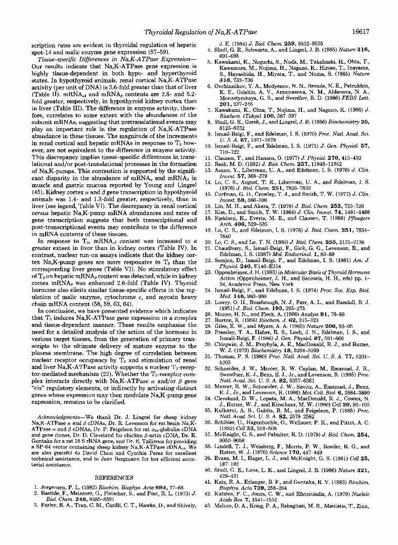
Na,K-ATPase mRNA, and mRNAB Contents-To assess the qualitative pattern of hybridization of rat kidney cortex and liver RNA to the corresponding rat cDNA, and cDNAB probes, total RNA was isolated from hypo- and hyperthyroid tissues and analyzed by Northern blotting (Fig. 1). In kidney cortex, 32P-labeled cDNA, hybridized to a single RNA species migrating at 26-27 S, as noted earlier (21). Hybridization of a replicate kidney cortex RNA blot with 32P-labeled rat brain cDNA0 revealed a predominate and a minor RNA band mi- grating at 22 S and 18 S, respectively. These mRNA bands are consistent with previous descriptions of tissue-specific expression of multiple forms of mRNAB (45). The character- istic 26-27 S mRNA, band was also evident in liver RNA. Replicate Northern blot analysis of total liver RNA with cDNAB revealed two doublet hybridization bands, the first doublet migrating at 22 S and 20 S, and the second at 18 S and 17 S (Fig. 1). Administration of T3 did not alter the migration of either kidney cortex or liver mRNAs (a or /3), nor did hormone treatment elicit the appearance of any ad- ditional bands.
The abundance of renal cortical and hepatic mRNA, and mRNAB in hypothyroid rats was assessed by quantitative Northern blots (Table 111). Liver mRNA, content (per pg of DNA) was 3.9-fold more abundant than liver mRNAB. Simi- larly, in kidney cortex, mRNA, was in greater abundance (2.1- fold) than mRNAB, and mRNA, and mRNAB contents in kidney cortex were 2.8- and 5.2-fold more abundant than the corresponding liver mRNAs, respectively. A comparison of the relative mRNA abundances and respective Na,K-ATPase activities in kidney cortex and liver reveals an approximate correlation (ie. a 3.6-fold greater enzyme activity in kidney cortex than in liver (cf . Tables I and 11).
The effects of T3 on kidney cortex and liver mRNA, and mRNAB content (analyzed by quantitative Northern blots and
TABLE I Effect of T3 on protein, DNA and RNA content of rat renal cortex and liver
Hypothyroid rats were injected daily with either diluent or T3 (100 pg of T3/100 g body weight) and tissues analyzed for protein, DNA and RNA at 72 h of treatment; n = 6 each for diluent-injected rats (Hypo) and the 72 h T3 groups (Hypo + T3). Results are expressed as means f S.E.
Tissue Thyroid status Protein RNA DNA ProteinIDNA RNAPNA
m g k mglg m g k m g l w %/mg Kidney cortex Hypo 120 f 4 3.35 f 0.13 3.95 f 0.19 30.4 f 1.1 0.85 f 0.02
Liver Hypo 169 f 2 6.03 f 0.18 1.79 f 0.15 94.4 f 9.0 3.37 & 0.27 H.WO + TS 195 f 2' 9.76 f 0.11' 2.30 f 0.11" 84.8 f 6.7 4.24 f 0.29
Hypo + T3 138 f 6" 4.43 f 0.26' 3.12 f 0.14' 44.2 f 2.1' 1.42 f 0.04'
" p < 0.05. ' p < 0.01. ' p e 0.001.

Thyroidal Regulation of Na,K-ATPase 16613 TABLE I1
Effect of T3 on Na,K-ATPase and Mg-ATPase activity Hypothyroid rats were treated as described in Table I and enzyme activities determined as described under
"Experimental Procedures;" n = 6 each for diluent-injected (Hypo) and 72 h T3 groups (Hypo + T3). Results are expressed as means f S.E.
Tissue Thyroid status Na,K-ATPase Mg-ATPase
rmlfmg rmllmg rmllmg rmllmg proternlh DNAIh proteinlh DNAIh
Kidney cortex HYPO 15.3 f 1.3 465 f 38 10.2 f 0.8 309 f 25
Liver HYPO 1.4 f 0.1 129 f 3 1.9 f 0.1 178 f 8 Hypo + T3 24.3 f 1.1" 1070 f 50" 9.0 f 0.3 396 f Ilb
Hypo + T3 2.0 f 0.1" 166 f 6" 2.0 f 0.1 172 f 4 "p e 0.001. bp e 0.01.
Kidney Cortex Liver
a P ' Q P ' f
A I A
28s- - 28s + +
18s- -18s
dil. T3 dil. T3 dil. T3 dil. T3
FIG. 1. Northern blot analysis of kidney cortex and liver Na.K-ATPase mRNA and mRNAB in hypothyroid and Ts- treated rats. Northern blots of total RNA isolated from hypothyroid (diluent-treated) and hyperthyroid (T3-treated) kidney cortex (20 pg) and liver (25 pg) were prepared as described under "Experimental Procedures." Blots were hybridized for 3 days with rat brain Na,K- ATPase cDNA, and cDNAB labeled to approximately the same spe- cific activity by nick translation. Kidney cortex a and @ blots and liver a blots were hybridized with lo7 cpm of 32P-labeled cDNA. 3 X lo7 cpm of nick-translated cDNA8 was used to probe liver RNA blots. Following washing in 0.1 X SSC at 50 "C, blots were exposed to x- ray film for the following periods; kidney cortex-a, 1.5 h; kidney cortex-& 5 h; liver-a, 5 h; and liver-& 3 days. The positions of 28 S and 18 S rRNA are indicated as standards, and the arrows denote the positions of mRNA8 bands migrating at 22 S, 20 S, 18 S, and 17 S.
TABLE I11 Relative abundance of Na,K-ATPase rnRNA, and mRNAB in
hypothyroid rat kidney cortex and liver Total RNA was isolated from hypothyroid rat kidney cortex and
liver. mRNA,, and mRNA8 content was determined by Northern blotting as in Fig. 1. A RNA/DNA ratio of 0.85 and 3.37 for hypothy- roid kidney cortex and liver, respectively, was used to express mRNA content/unit DNA (see Table I). The resultant mRNA content values were then normalized to the mean value of liver mRNA8. Results are expressed as means f S.E.; n = 6 hypothyroid rats.
Tissue mRNA abundance (per DNA)
a B Kidney cortex 11.1 f 0.9 5.2 f 0.3 Liver 3.9 f 0.4 1.0 f 0.1
expressed/unit of DNA) are summarized in Table IV. Admin- istration of T3 elicited 3.1- and 2.6-fold increases in kidney cortex mRNA, and mRNAp content, respectively. The relative abundances of the 22 S as compared to the 18 S mRNAB forms were unchanged with thyroid status (ie. both were increased 2.6-fold by T3). The magnitude of the increases in mRNA, and mRNAB abundance were similar to the 2.3-fold increment in Na,K-ATPase activity in renal cortex (Tables I1 and IV). In liver, T3 elicited a 7.3-fold increase in mRNA, content while mRNAB abundance decreased minimally (sta-
TABLE IV Effect of T3 on Na,K-ATPase nRNA abundances
Hypothyroid rats were treated with diluent (Hypo) or T3 for 72 h (Hypo + T3) (see Table I), and mRNA., and mRNAB contents were assessed by Northern blots. To account for the effect of T3 on tissue RNA content, the results were corrected for the T3-induced changes in RNA/DNA ratio. A RNA/DNA ratio of 1.67 and 1.26, respectively, for kidney cortex and liver was utilized (see Table I). In each tissue and for each mRNA species, the values obtained for the hypothyroid tissue was normalized to 1.0 and the effect of T3 is expressed as a function of the hypothyroid value. Results are expressed as means f S.E.; n = 12 each for Hypo and Hypo + T3 groups.
mRNA abundance Tissue Thyroid status (per DNA)
a B
Kidney cortex Hypo 1.0 f 0.1 1.0 & 0.1 Hypo + T3 3.1 f 0.5" 2.6 f 0.3"
Liver Hypo 1.0 f 0.1 1.0 f 0.1 Hypo + TJ 7.3 f 0.9" 0.8 f 0.1
" p < 0.001.
tistically insignificant), and Na,K-ATPase activity increased by 1.3-fold (Tables I1 and IV). RNA dot blot analysis by the method of Chaudhury et al. (21) confirmed the T3-induced increments in kidney cortex mRNA, and mRNAB content and in liver mRN& abundance (data not shown). In addition, the lack of an effect of T3 on liver mRNAB content eliminates the possibility that the increase in mRNA, abundance, in the same sample, reflected differences in RNA yield rather than in content.
Effects of T3 on RNA Polymerase Activity-RNA polymer- ase (I + 111) and RNA polymerase I1 activities were assessed in transcription reactions containing 2 pg/ml a-amanitin (38). T3 produced 2.1- and 1.6-fold increases in total RNA polym- erase activity in renal cortex and liver nuclei, respectively (Table V). RNA polymerase (I + 111) activity was augmented 3.1- and 2.2-fold in kidney cortex and liver nuclei, respectively. T3 elicited a 1.6-fold stimulation of RNA polymerase I1 activ- ity in kidney cortex nuclei but had no significant effect in liver nuclei.
Na,K-ATPase a and p Gene Transcription-To examine the effect of thyroid status on Na,K-ATPase a and @ gene transcription, nascent nuclear RNA transcripts were elon- gated in uitro in the presence of [u-~*P]UTP and identified by hybridization to cDN& and cDNAB (Table VI). In all instances, the yield of specific counts was at least 95 cprn above the pBR322 background. In kidney cortex nuclei iso- lated from hypothyroid rats, the transcription rates were 4.7 zk 0.4 and 6.0 & 0.3 cpm 32P incorporated/lO min/pg DNA, for a and 8, respectively (see legend, Table VI). Administra- tion of T3 elicited a 1.8-fold stimulation of these rates. Liver

16614 Thyroidal Regulation of Na,K-ATPase
TABLE V Effects of T3 o n R N A polymerase activity of rat renal cortex and liver nuclei
Kidney cortex and liver nuclei were isolated from hypo- and hyperthyroid rats and total transcription activity was quantified as described under “Experimental Procedures.” To distinguish between RNA polymerase (I + 111) and RNA polymerase I1 activity, transcription assays were performed in the presence and absence of 2 pg/ml of a-amanitin. Liver (10 pg of DNA) and kidney cortex nuclei (5 pg of DNA) were incubated for 10 min in the transcription reaction mixture with approximately 8 and 15 pCi of [a-32P]UTP, respectively. Results are expressed as means f S.E.; n = 12 each for Hypo and Hypo + T3 (72 h) groups.
Tissue Thyroid status Transcription
Total (RNA Polvmerase I + 111) (RNA Polvmerase 11) ~~ ~ ~ ~ ~~ ~ ~ ~ ~ ~
cprn X
Kidney cortex Hypo 14.1 f 1.2 4.8 f 0.4 9.3 f 1.0
Liver Hypo 27.4 f 3.3 11.7 f 1.7 15.7 f 1.7 Hypo + T3 29.5 & 2.7” 14.7 f 1.4“ 14.8 f 1.5b
HVUO + Ta 43.2 f 5.7‘ 25.5 f 3.2“ 17.7 f 3.4 “ p < 0.001. b p < 0.05.
TABLE VI Na,K-ATPase LY and p gene transcription i n rat renu1 cortex and
liver i n response to T3 (nuclear run-on assays) Nuclei isolated from hypo- and hyperthyroid kidney cortex and
liver were analyzed for 01 and p gene transcription by nuclear run-on assays as described under “Experimental Procedures.” Incubation of kidney cortex nuclei (20 pg of DNA) isolated from hypothyroid rats with the reaction mixture containing 170 to 220 pCi [LY-~’P]UTP for 10 min yielded an average of lo6 cpm of 32P-labeled RNA. Following hybridization and washing, 144 f 11 and 170 & 9 cpm were detected on a and p filters, respectively. Nonspecific hybridization to pBR322
of DNA) isolated from hypothyroid rats yielded an average of 3.0 X averaged 50 f 4 cpm. Under the same conditions, liver nuclei (50 pg
lo6 cpm of total in vitro 32P-labeled RNA. Following hybridization of hypothyroid liver 32P-RNA and washing, 246 f 19 and 297 f 15 cpm were detected on a and filters, respectively, while nonspecific hybridization to pBR322 filters averaged 74 f 9 cpm. The absolute cpm hybridized to a and /3 filters represents an average of three separate experiments, each conducted with 170 to 220 pCi of [01-~’Pl UTP and 3 to 4 days of hybridization. The gene transcription values from hypothyroid rats were averaged and normalized to 1.0 for each subunit. The mean of hypothyroid values was calculated in each separate experiment, and individual hypothyroid values were then expressed relative to the mean hypothyroid value. In addition, the ratio of each value obtained from hyperthyroid rats to the mean hypothyroid value of each experiment was calculated. Finally, a composite mean and deviation representing the pooled results derived from three independent experiments with a total of 12 diluent- injected hypothyroid rats (Hypo) and 12 rats treated for 72 h with TI (Hypo + T3) was determined. The final normalized values are ex- Dressed as means f S.E. ~
Transcription
a P Tissue Thyroid status
Kidney cortex Hypo 1.00 f 0.04 1.00 f 0.04 Hypo + T3 1.76 f 0.16“ 1.77 f 0.11“
Liver Hypo 1.00 f 0.05 1.00 f 0.05 H.ypo + T3 1.36 k 0.10’ 1.28 k 0.07b
“ p < 0.001. bp < 0.01.
nuclei isolated from hypothyroid rats incorporated 3.4 f 0.3 and 4.5 f 0.2 cpm 32P/10 min/pg DNA, for LY and p, respec- tively (see legend, Table VI), and T3 elicited 1.4- and 1.3-fold increases in these rates. Thus, basal rates of a and p tran- scription were almost the same in kidney and liver nuclei and were equally induced by T3.
To investigate the selectivity of T3-induced changes in Na,K-ATPase gene activity, the effects of TB on two addi- tional genes transcribed by RNA polymerase I1 (/?-actin and azu-globulin) and a gene transcribed by RNA polymerase I (18 S rRNA) were examined (Table VII). In both kidney
cortex and liver, T3 elicited 2.3-fold increases in /?-actin mRNA content and stimulated @-actin gene transcription 1.5- 1.7-fold. T3 induced a 300-fold increase in hepatic azu-globulin mRNA content, but the in vitro transcription rate was stim- ulated only 1.4-fold. Transcription of the 18 S rRNA gene increased 3.0- and 2.4-fold in kidney cortex and liver nuclei, respectively, values that are nearly identical to the T3-induced increments in the RNA polymerase (I + 111) activity (Table W.
DISCUSSION
These studies were designed to evaluate the possibility of proportionate increases in response to T3 in gene transcrip- tion, mRNA abundance, and enzyme abundance in the Na,K- ATPase system. This possibility deserved evaluation in that T3 regulates the expression of many genes in a variety of organs and species (23). Accordingly, we analyzed Na,K- ATPase activity, mRNA, and mRNA6 abundances, and rates of LY and p gene transcription (ie. nuclear run-on assays) in kidney cortex and liver derived from hypo- and hyperthyroid rats. An analysis of the quantitative relationships between these levels of Na,K-pump gene expression is simplified when the system is in the steady state. The dietary regimen em- ployed in the present study to induce hypothyroidism has been demonstrated to rapidly reduce serum T3 and Tq concen- trations and following 3-4 weeks of treatment results in a new hypothyroid steady state (22). Na,K-ATPase activity is maximally induced following treatment of hypothyroid rats for 48-72 h with a daily dose of T3 sufficient to ensure near saturation of nuclear T3 receptors (22-24). At this time, Na,K- ATPase activity has apparently attained a hyperthyroid steady state. To minimize potential complications arising from prolonged exposure to high doses of T3, administration of T3 for 72 h was selected to assess Na,K-ATPase gene expression.
To ensure quantification of maximal Na,K-ATPase activ- ity, tissue homogenates were incubated with deoxycholate. Previous studies demonstrated a quantitative (i.e. a 1:l) re- lationship between enzyme activity and Na,K-pump content in response to T3 (14-16). Quantitative Northern blot analysis of total RNA was used to assess the abundance of Na,K- ATPase mRNAs. In these studies, we did not distinguish between cytoplasmic mature mRNAs and nuclear mRNA precursors. Nuclear run-on assays were used to quantify Na,K-ATPase LY and @ gene transcription. The underlying assumptions in the use of nuclear run-on assays as synony- mous with rates of specific gene transcription are as follows: 1) the preparation of nuclear fractions recovers all or an

Thyroidal Regulation of Na,K-ATPase TABLE VI1
Effects of T3 on &Actin, a2,-globulin and 18 S rRNA gene expression
16615
The effect of T3 on kidney cortex and liver 0-actin, cYz,-globulin, and 18 S rRNA gene expression was quantified by RNA dot blot and nuclear run-on transcription analysis as described under “Experimental Procedures.” mRNA content was expressed/unit DNA (see Table IV); n = 12 each for diluent-injected (Hypo) and the Tdreated (Hypo + T3) groups analyzed for kidney cortex @-actin and liver aa,-globulin mRNA content, n = 9 each for Hypo and Hypo + T3 groups assayed for liver 0-actin mRNA abundance. Hybridization of in vitro 32P-labeled RNA derived from hypothyroid kidney cortex and liver nuclei to nitrocellulose filters containing 0-actin cDNA yielded 98 -C 7 and 172 f 16 cpm, respectively. Similarly, hybridization of hypothyroid liver-derived in vitro transcription products to filters containing a rat a2,-globulin genomic DNA yielded 1,100 f 156 cpm. When the 800-fold dilution factor was used to correct the cpm obtained in hybridization to filters containing a rat 18 S rRNA gene, then 104,000 f 16,000 and 316,000 f 21,000 cpm were detected in transcription products derived from hypothyroid renal cortex and liver, respectively. Since each transcription result was a composite of three separate experiments conducted with slightly different conditions, the results were normalized as in Table VI; n = 12 each for diluent-injected hypothyroid rats (Hypo) and the 72 h T3 groups (Hypo + T3). Results are expressed as means f S.E.
&Actin az,-Globulin Thyroid 18 S rRNA
Tissue status mRNA Transcription mRNA ~~~~~~~i~~~~~ Transcription content content
Kidney cortex Hypo 1.0 f 0.1 1.0 f 0.1 ND“ ND 1.0 k 0.1
Liver Hypo 1.0 f 0.1 1.0 f 0.1 1.0 f 0.2 1.0 f 0.1 1.0 k 0.1 Hypo + T3
Hypo + T3
2.3 f 0.4b 1.7 f 0.1‘ ND ND 3.0 f 0.3‘
2.3 f 0.2‘ 1.5 f 0.1‘ 301 f 100b 1.4 f O . l b 2.4 k 0.2‘
ND, not determined. bp c.o.01. ‘ p c 0.001.
appropriate proportion of the trans-acting factors that may be involved in the action of the hormone. 2) The microenvi- ronment used in the assay (e.g. specific ions, pH, etc.) does not alter the basic processes involved in transcription. 3) Template activity is preserved and reflects the in vivo properties. Since these assumptions have not been tested in our system, the nuclear run-on assays are considered to be indices of gene transcription, rather than a direct measure thereof.
Na,K-ATPase Isoforms-Na,K-ATPase a peptide isoforms were first described, and designated a and a(+) by Sweadner (46). Subsequently Shull et al. (8) isolated three rat brain isoform cDNAs, designated a, a(+), and aIII. The existence and characterization of these isoforms have been confirmed by additional cDNA and gene cloning and by chromosomal localization (47-50). As a result of the use of alternative notations by various authors, the identification of the iso- forms in various tissues of various species may be confused. For our purposes, we have adopted the following notation: a1 = a; a11 = a(+); a111 = aIII. The notation “a” is used to designate the entire class of isoforms. In our studies of mRNA, abundance, we made use of a rat brain-derived cDN& which encodes 1200 bp of the coding region spanning the active site of the a1 isoform (32). We previously demon- strated that this cDNA a1 hybridized to mRNAmI in Northern blots of total RNA isolated from brain and myocardium (21), tissues which express multiple a isoform mRNAs (45). This finding coupled with the reported low abundance of a11 and a111 isoform mRNAs in kidney and liver, suggests that the hybridization and wash conditions used in our current studies ensure the specific detection of mRNAaI (45,473.
In contrast to a-subunit isoforms, heterogeneity of p pep- tides has not been reported although the extent of glycosyla- tion varies (1). mRNAp of various lengths have been identi- fied, and this finding has been confirmed in our study (Fig. 1) (33, 45, 51). Heterogeneity of mRNAB, however, has been attributed to transcription of a single gene with differential utilization of 5’ initiation and 3’ polyadenylation sites (51). These findings predict that the amino acid sequence of the mature peptide backbone will be invariant in any one species.
Regulation of Renal Cortical Na,K-ATPase-Earlier studies
reported coordinate regulation of the synthesis of renal cor- tical a and p peptides in various thyroid states (19, 20). In view of the expected equimolar ratio of a:@ in the mature oligomer and if the coordinate control of subunit synthesis was a consequence of pretranslational regulation, one would predict equal abundances of mRNA, and mRNAp. In hypo- thyroid renal cortex, mRNAOI is 2.1-fold more abundant that mRNAB, suggesting that differential regulation exists at trans- lational and/or post-translational levels (Table 111). In con- trast to the discrepancies in the relative abundances of the mRNAs, p gene transcription rate slightly exceeds that of a, implying that processing and/or degradation of a and /3 tran- scripts may not proceed at equivalent rates (see legend, Table VI).
Daily administration of 100 pg of T3/100 g body weight ensures continual, near saturation of specific nuclear recep- tors (23) and, after 48-72 h of treatment, maximal stimulation of Na,K-ATPase activity in responsive tissues (22, 24). In confirmation of previous studies, kidney cortex Na,K-ATPase was augmented by T3 while Mg-ATPase activity was unal- tered (Table 11) (9, 21). As noted earlier, renal cortical mRNAaI content (per unit of DNA) increased 3.1-fold in response to T3 (Table IV) (21). T s also elicited a 2.6-fold stimulation of both the predominate 22 S and the minor 18 S form of kidney cortex mRNAp (Fig. 1, Table IV). The mag- nitude of the T3-induced increments in mRNA,r and mRNAp contents are similar to the corresponding increase in Na,K- ATPase activity (2.3-fold) (Fig. 2, upperpanel). These results are consistent with previous studies which indicated. a T3- induced 2-%fold stimulation of guinea pig renal Na,K- ATPase mRNA, translational activity in reticulocyte lysates (52,53). Lo et al. (14) and Lo and Lo (20) found the maximal doses of T3 elicited -1.6-fold increases in the rate of synthesis of the a and p peptides in rat renal cortex. The increases in the abundances of mRNA,I and mRNAp are sufficient to account for the increments in Na,K-pump content of renal cortex. Translational or post-translational regulatory events, however, cannot be ruled out since T3-induced increases in mRNAaI and mRNAp tend to be somewhat greater than the corresponding increases in Na,K-ATPase content. This issue

16616
4
3
2
I
2
I
h
Thyroidal Regulation of Na,K-ATPase
A Kidney I Cortex T
11 K-ATPase
k t i v i t y
i 1 h " _ """"_ " -
a B a b mRNA
Content Transcription Gene
FIG. 2. Effects of Ts on Na,K-ATPase activity, mRNA abun- dance, and gene transcription. The effects of TI on renal cortical and hepatic Na,K-ATPase activity, mRNA content, and gene tran- scription rate are expressed/unit of DNA and represent a compilation of data from Tables 11, IV, and VI. Statistical significance is denoted by *, p < 0.001, and **, p < 0.01. All results are expressed as the ratio of each parameter in the Ts-treated (72 h) group versus the hypothy- roid control group. The S. E. (vertical lines) are computed as described in the appropriate Table legends.
is more dramatically evident in the hepatic response to T3 (see below).
In kidney cortex, T3 induced equivalent 1.8-fold increases in the rate of CY and @ gene transcription (Table VI). If the nuclear run-on assays are an accurate index of in vivo tran- scription rates, the discrepancy between the magnitude of the T3-induced increments in mRNA abundances and rates of gene transcription suggests the existence of post-transcrip- tional regulatory processes (Fig. 2, upper panel). In addition to regulation of Na,K-pump gene transcription, T3 may alter the processing of nuclear RNA precursors, change the rate of delivery of mRNA to the cytoplasm, or effect the degradation of either nuclear or cytoplasmic mRNAs. The apparent dual action of TS is not unique to Na,K-ATPase gene expression since thyroid hormone regulates the transcription of the genes for growth hormone and the CY and @ subunits of thyrotropin (54-57), while exerting post-transcriptional regulation of spot-14 and malic enzyme gene expression (57-59).
Thyroidal activation of Na,K-pump CY and @ gene transcrip- tion may reflect enhancement of a fairly large set of consti- tutively expressed genes (60). In our studies, T3 elicited a 1.7- fold increase in renal @-actin gene transcription, a 1.6-fold stimulation of RNA polymerase I1 activity, and a 3.1-fold stimulation of RNA polymerase (I + 111) activity (Tables V and VII). The latter correlated with a 3.0-fold increase in 18 S rRNA gene transcription rate (Table VII). These results are consistent with increased abundance of rRNA content in target tissues and are in accord with the growth-promoting action of T3 summarized by Towle et al. (60).
Regulation of Hepatic Na,K-ATPase-In hypothyroid liver, mRNA,I is present at a 3.9-fold excess relative to mRNAp (Table 111). An even greater disparity in the relative abun- dances of mRNA, and mRNAp content was recently noted in a clonal rat liver cell line (61). Our observation that the @
gene transcription rate is slightly greater than CY in hypothy- roid rat liver nuclei implies an additional level of complexity in the regulation of liver Na,K-pump mRNA abundances (see legend, Table VI). The discrepancies in the quantitative re- lationships between hepatic Na,K-pump mRNA abundances and rates of gene transcription in the thyroid-deficient state raises interesting further problems, including the possibility of differential post-transcriptional regulation of these mRNAs, differential translatability, and subunit-specific post-translational processing of the nascent peptides. Other possibilities include the existence of an as yet undetected isoform or of a mature Na,K-pump with an alternative subunit composition. For example, Hubert et al. (62) failed to detect immunoprecipitable @-subunits in rat liver and suggested that the hepatic enzyme may not have a @-subunit, or has a highly divergent @ peptide. If so, the rat brain cDNAp used in our studies may have failed to detect such a variant hepatic isoform mRNAp. The presence of mRNAp bands seen in Fig. 1 could represent mRNAp expressed in some liver cells (e.g. Kupfer cells) that are unresponsive to T3. In an attempt to detect the existence of a divergent liver mRNAB isoform, we hybridized Northern blots of hepatic RNA with cDNAp and washed the blots under graded, reduced stringency conditions. An additional band migrating at -28 S was detected following a low stringency wash at 25" in 0.1 x SSC (data not shown). The identity of this hepatic 28 S mRNA in relation to mRNAB, however, remains to be determined. Hepatic nuclear run-on experiments yielded nearly equivalent rates of transcription of the CY and @ genes (see legend, Table VI). This finding suggests the transcription of a liver gene product with a high degree of homology to mRNAp found in other tissues. Until either liver Na,K-ATPase is purified to homogeneity or a liver isoform cDNAp is cloned and sequenced, we can not exclude the possibility that the hepatic Na,K-pump contains a highly divergent @ peptide.
In agreement with earlier studies, administration of T3 elicited a 1.4- and 1.3-fold stimulation of liver Na,K-ATPase activity expressed/unit protein or per unit DNA, respectively (Table 11) (9, 10). T3 had a markedly unequal effect on liver Na,K-ATPase mRNAs (Table IV). mRNA,I abundance was increased 7.3-fold, while mRNAp content was marginally de- creased. If the subunit composition of liver Na,K-ATPase is indeed identical to renal Na,K-ATPase, then T3 must exert a substantial degree of translational and/or post-translational control in the biogenesis of the mature subunits of the hepatic Na,K-pump.
Administration of T3 produced 1.4- and 1.3-fold increases in hepatic Na,K-ATPase CY and @ gene transcription, without a concomitant increment in RNA polymerase I1 activity (Ta- ble V and VI). The discrepancy between the 1.4-fold stimu- lation of gene transcription and the corresponding 7.3-fold increase in mRNA,I abundance suggests that TS may either increase the nuclear to cytoplasmic processing or decrease the degradation of mRNA,I (Fig. 2, lowerpanel). If post-transcrip- tional effects of T3 are the explanation for this discrepancy, it may not be specific to Na,K-ATPase transcripts since the magnitude of T3-induced stimulation of @-actin and az,-glob- ulin gene transcription were not equivalent to their respective increments in mRNA content (Table VII). The 1.3-fold in- crease in hepatic @ gene transcription, without a correspond- ing increase in mRNAp abundance, suggests additional post- transcriptional complexities in the response. Alternatively, of course, some of these discrepancies may reflect limitations in the use of nuclear run-on assays as a quantitative index of gene transcription in intact cells. Similar discrepancies in the magnitude of changes in mRNA abundance and gene tran-
Thyroidal Regulatioi
scription rates are evident in thyroidal regulation of hepatic spot-14 and malic enzyme gene expression (57-59).
Tissue-specific Differences in Na,K-ATPase Expresswn- Our results indicate that Na,K-ATPase gene expression is highly tissue-dependent in both hypo- and hyperthyroid states. In hypothyroid animals, renal cortical Na,K-ATPase activity (per unit of DNA) is 3.6-fold greater than that of liver (Table 11). mRNAer and mRNAB contents are 2.8- and 5.2- fold greater, respectively, in hypothyroid kidney cortex than in liver (Table 111). The difference in enzyme activity, there- fore, correlates to some extent with the abundances of the subunit mRNAs, suggesting that pretranslational events may play an important role in the regulation of Na,K-ATPase abundance in these tissues. The magnitude of the increments in renal cortical and hepatic mRNAs in response to T3 how- ever, are not equivalent to the difference in enzyme activity. This discrepancy implies tissue-specific differences in trans- lational and/or post-translational processes in the formation of Na,K-pumps. This contention is supported by the signifi- cant disparity in the abundance of mRNA, and mRNAp in muscle and gastric mucosa reported by Young and Lingrel (45). Kidney cortex a and p gene transcription in hypothyroid animals was 1.4- and 1.3-fold greater, respectively, than in liver (see legend, Table VI). The discrepancy in renal cortical uersus hepatic Na,K-pump mRNA abundances and rates of gene transcription suggests that both transcriptional and post-transcriptional events may contribute to the difference in mRNA contents of these tissues.
In response to T3, mRNA,I content was increased to a greater extent in liver than in kidney cortex (Table IV). In contrast, nuclear run-on assays indicate that the kidney cor- tex Na,K-pump genes are more responsive to T3 than the corresponding liver genes (Table VI). No stimulatory effect of T3 on hepatic mRNAB content was detected, while in kidney cortex mRNAp was enhanced 2.6-fold (Table IV). Thyroid hormone also elicits similar tissue-specific effects in the reg- ulation of malic enzyme, cytochrome c , and myosin heavy chain mRNA content (58, 59,63, 64).
In conclusion, we have presented evidence which indicates that TS induces Na,K-ATPase gene expression in a complex and tissue-dependent manner. These results emphasize the need for a detailed analysis of the action of the hormone in various target tissues, from the generation of primary tran- scripts to the ultimate delivery of mature enzyme to the plasma membrane. The high degree of correlation between nuclear receptor occupancy by T3 and stimulation of renal and liver Na,K-ATPase activity supports a nuclear T3-recep- tor-mediated mechanism (22). Whether the T3-receptor com- plex interacts directly with Na,K-ATPase a and/or ,8 gene “cis” regulatory elements, or indirectly by activating distinct genes whose expression may then modulate Na,K-pump gene expression, remains to be clarified.
Acknowledgments-We thank Dr. J. Lingrel for sheep kidney Na,K-ATPase a and @ cDNAs, Dr. R. Levenson for rat brain Na,K- ATPase a and (3 cDNAs, Dr. P. Feigelson for rat m,-globulin cDNA and gene clones, Dr. D. Cleveland for chicken p-actin cDNA, Dr. R. Guntaka for a rat 18 S rRNA gene, and Dr. E. Tzilianos for providing a SP-64 vector containing sheep kidney Na,K-ATPase CDNA,~. We are also grateful to David Chon and Cynthia Perez for excellent technical assistance, and to Jean Bergmann for her efficient secre- tarial assistance.
REFERENCES 1. Jorgensen, P. L. (1982) Biochirn. Biophys. Acta 6 9 4 , 27-68 2. Bastide, F., Meissner, G., Fleischer, S., and Post, R. L. (1973) J.
3. Farley, R. A., Tran, C. M., Carilli, C. T., Hawke, D., and Shively, Biol. Chem. 248,8385-8391
r2. of Nu, K-A TPase 16617
J. E. (1984) J. Biol. Chem. 269 , 9532-9535 4. Shull, G. E., Schwartz, A., and Lingrel, J. B. (1985) Nature 3 1 6 ,
5. Kawakami, K., Noguchi, S., Noda, M., Takahashi, H., Ohta, T., Kawamura, M., Nojima, H., Nagano, K., Hirose, T., Inayama, S., Hayashida, H., Miyata, T., and Numa, S. (1985) Nature
6. Ovchinnikov, Y. A., Modyanov, N. N., Broude, N. E., Petrukhin, K. E., Grishin, A. V., Arzamazova, N. M., Aldanova, N. A., Monastyrskaya, G. S., and Sverdlov, E. D. (1986) FEBS Lett.
7. Kawakami, K., Ohta, T., Nojima, H., and Nagano, K. (1986) J.
8. Shull, G. E., Greeb, J., and Lingrel, J. B. (1986) Biochemistry 25 ,
9. Ismail-Beiai, F., and Edelman, I. S. (1970) Proc. Natl. Acad. Sci.
691-695
316 , 733-736
201,237-245
Biochem. (Tokyo) 100,387-397
8125-8232
u. s. A. Si, ion-1078 10. Ismail-Beiei. F.. and Edelman. I. S. (1971) J. Gen. Phvsiol. 57. . .
710-722- ’ ’
11. Clausen, T., and Hansen, 0. (1977) J. Physiol. 270,415-430 12. Resh, M. D. (1982) J. Biol. Chem. 257,11946-11952 13. Asano, Y., Liberman, U. A., and Edelman, I. S. (1976) J. Clin.
14. Lo, C. S., August, T. R., Liberman, U. A., and Edelman, I. S.
15. Curfman, G. D., Crowley, T. J., and Smith, T. W. (1977) J. Clin.
16. Lin, M. H., and Akera, T. (1978) J. Biol. Chem. 2 5 3 , 723-726 17. Kim, D., and Smith, T. W. (1984) J. Clin. Invest. 7 4 , 1481-1488 18. Kjeldsen, K., Everts, M. E., and Clausen, T. (1986) Pfluegers
19. Lo, C. S., and Edelman, I. S. (1976) J. Biol. Chem. 251 , 7834-
20. Lo, C. S., and Lo, T. N. (1980) J. Biol. Chem. 255,2131-2136 21. Chaudhury, S., Ismail-Beigi, F., Gick, G. G., Levenson, R., and
22. Somjen, D., Ismail-Beigi, F., and Edelman, I. S. (1981) Am. J.
23. Oppenheimer, J. H. (1983) in Molecular Basis of Thyroid Hormone Action (Oppenheimer, J. H., and Samuels, H. H., eds) pp. 1- 34, Academic Press, New York
24. Ismail-Beigi, F., and Edelman, I. S. (1974) Proc. SOC. Exp. Biol. Med. 146,983-988
25. Lowry, 0. H., Rosebrough, N. J., Farr, A. L., and Randall, R. J. (1951) J. Biol. Chem. 193, 265-275
26. Munro, H. N., and Fleck, A. (1966) Analyst 9 1 , 78-88 27. Burton, K. (1956) Biochem. J. 62,315-323 28. Giles, K. W., and Myers, A. A. (1965) Nature 206,93-95 29. Pressley, T. A., Haber, R. S., Loeb, J. N., Edelman, I. S., and
Ismail-Beigi, F. (1986) J. Gen. Physiol. 8 7 , 591-606 30. Chirgwin, J. M., Przybyla, A. E., MacDonald, R. J., and Rutter,
W. J. (1979) Biochemistry 18 , 5294-5299 31. Thomas, P. S. (1980) Proc. Natl. Acad. Sci. U. S. A. 7 7 , 5201-
5205 32. Schneider, J. W., Mercer, R. W. Caplan, M., Emanuel, J. R.,
Natl. Acad. Sci. U. S. A. 8 2 , 6357-6361 Sweadner, K. J., Benz, E. J., Jr., and Levenson, R. (1985) Proc.
33. Mercer, R. W., Schneider, J. W., Savitz, A., Emanuel, J., Benz, E. J., Jr., and Levenson, R. (1986) Mol. Cell. Biol. 6,3884-3890
34. Cleveland, D. W., Lopata, M. A., MacDonald, R. J., Cowan, N. J., Rutter, W. J., and Kirschner, M. W. (1980) Cell 20, 95-105
35. Kulkarni, A. B., Gubits, R. M., and Feigelson, P. (1985) Proc. Natl. Acad. Sci. U. S. A. 82,2579-2582
36. Schibler, U., Hagenbuchle, O., Wellauer, P. K., and Pittet, A. C. (1983) Cell 33 , 501-508
37. McKnight, G. S., and Palmiter, R. D. (1979) J. Biol. Chem. 2 5 4 ,
38. Lindell, T. J., Weinberg, F., Morris, P. W., Roeder, R. G., and
39. Evans, M. I., Hager, L. J., and McKnight, G. S. (1981) Cell 2 5 ,
40. Shull, G. E., Lane, L. K., and Lingrel, J. B. (1986) Nature 321 ,
41. Katz, R. A., Erlanger, B. F., and Guntaka, R. V. (1983) Biochim.
42. Kafatos, F. C., Jones, C. W., and Efstratiadis, A. (1979) Nucleic
43. Melton, D. A., Krieg, P. A., Rebagliati, M. R., Maniatis, T., Zinn,
Znuest. 57,368-379
(1976) J. Biol. Chem. 251 , 7826-7833
Invest. 59,586-590
Arch. 406,529-535
7840
Edelman, I. S. (1987) Mol. Endocrinol. 1,83-89
Physiol. 240, E146-E154
9050-9058
Rutter, W. J. (1970) Science 170,447-449
187-193
429-431
Biophys. Acta 739 , 258-264
Acids Res. 7 , 1541-1552

16618 Thyroidal Regulation of Na,K-ATPase K., and Green, M. R. (1984) Nucleic Acids Res. 12,7035-7056.
44. Tata, J. R., and Widnell, C. C. (1966) Biochem. J. 98,604-620 45. Young, R. M., and Lingrel, J. B. (1987) Biochem. Biophys. Res.
46. Sweadner, K. J. (1979) J. Biol. Chem. 264,6060-6067 47. Herrera, V. L. M., Emanuel, J. R., Ruiz-Opazo, N., Levenson, R.,
and Nadal-Ginard, B. (1987) J. Cell Biol. 106 , 1855-1865 48. Shull, M. M., and Linpel, J. B. (1987) Proc. Natl. Acad. Sci. U.
49. Sverdiov, E. D., Monastyrskaya, G. S., Broude, N. E., Ushkaryov, Yu.A., Allikmets, R. L., Melkov, A. M., Smirnov, Yu.A., Ma- lyshev, I. V., Dulobova, I. E., Petrukhin, K. E., Grishin, A. V., Kijatkin, N. I., Kostina, M. B., Sverdlov, V. E., Modyanova, N. N., and Ovchnikova, Y. A. (1987) FEBS Lett. 217,275-278
50. Kent, R. B., Fallows, D. A., Geissler, E., Glaser, T., Emanuel, J. R., Lalley, P. A., Levenson, R., and Housman, D. E. (1987) Proc. Natl. Acad. Sci. U. S. A. 84,5369-5373
51. Young, R. M., Shull, G. E., and Lingrel, J. B. (1987) J. Bwl.
52. Edelman, I. S., Pressley, T. A., and Hiatt, A. (1985) in The Sodium Pump (Glynn, I., and Ellory, C., eds) pp. 153-159, Company of Biologists, Ltd, Cambridge, England
53. McDonough (1985) in The Sodium Pump (Glynn, I., and Ellory,
Commun. 146,52-58
S. A. 84, 4039-4043
Chem. 262,4905-4910
54.
55.
56.
57.
58.
59.
60.
61.
62.
63.
64.
C., eds) pp. 161-163, Company of Biologists, Ltd., Cambridge, England
Yaffe, B. M., and Samuels, H. H. (1984) J. Biol. Chem. 269,
Spindler, S. R., Mellon, S. H., and Baxter, J. D. (1982) J. Biol.
Shupnik, M. A., Chin, W. W., Habener, J. F., and Ridgway, E. C.
Narayan, P., and Towle, H. C. (1985) Mol. Cell. Bwl. 6, 2642-
Back, D. W., Wilson, S. B., Morris, S. M., Jr., and Goodridge, A.
Dozin, B., Magnuson, M. A., and Nikodem, V. M. (1986) J. Bwl.
Towle, H. C., Dillmann, W. H., and Oppenheimer, J. H. (1979)
Pressley, T. A., Ismail-Beigi, F., Gick, G. G., and Edelman, I. S.
Hubert, J. J., Schenk, D. B., Skelly, H., and Leffert, H. L. (1986)
Scarpulla, R. C., Kilar, M. C., and Scarpulla, K. M. (1986) J.
Izumo, S., Nadal-Ginard, B., and Mahdavi, V. (1986) Science
6284-6291
Chem. 267,11627-11632
(1985) J. Biol. Chem. 2 6 0 , 2900-2903
2646
G. (1986) J. Biol. Chem. 261,12555-12561
Chem. 261,10290-10292
J . Bwl. Chem. 264,2250-2257
(1988) Am. J. Physwl. 266 , C252-C260
Biochemistry 26,4156-4163
Bwl. Chem. 261,4660-4662
231,597-600