THE No. CHEMISTRY 17, 8318-8326,1988 OF 263, pp. 15, … file0 1988 by The American Society for...
8
THE JOURNAL OF BIOLOGICAL CHEMISTRY 0 1988 by The American Society for Biochemistry and Molecular Biology, Inc. Vol. 263, No. 17, Issue of June 15, pp. 8318-8326,1988 Printed in U.S.A. A High Yield Purification of the Human Transferrin Receptor and Properties of Its Major Extracellular Fragment* (Received for publication, November 9, 1987) Aaron P. TurkewitzSO, James F. AmatrudaSlT, David BorhaniS, Stephen C. Harrison$)), and Alan L. Schwartz**$$ From the $Department of Biochemistry, Harvard University, Cambridge, Massachusetts 02138 and the **Department of Pediatrics and Pharmncology, Washington University School of Medicine, Division of Pediatric Hemtology/Oncology, Children’s Hospital, St. Louis, Missouri 63110 Human transferrin receptor is a disulfide-linked homodimer of 90-kDa glycoprotein subunits, capable of binding two transferrins. We report a new high yield affinity purification protocol for transferrin receptor from placenta which produces 3-4 mg of highly puri- fied protein. Trypsin cleaves the protein at arginine- 121, producing a stable fragment that contains 95% of the extracytoplasmic sequence; similar fragments are produced by several other proteases. The tryptic frag- ment is a nondisulfide-linked dimer in solution and binds two transferrin molecules. The dimensions of both the dimer fragment and its complex with trans- ferrin are estimated by gel filtration. With an equilibrium between two oxidation states that is sensitive to both pH and ligation, iron is a critical element in many biological molecules involved in redox processes (1). In warm blooded organisms, these include the cytochromes and hemoglobin, and tissues inwhich their synthesis isactive must be reliably supplied with iron from serum. But because iron forms highly insoluble Fe(OH)3 under physiologicalcon- ditions (I), specific iron transport systems have evolved to facilitate delivery. Iron chelation in the human circulatory system is accomplished by transferrin, a glycoprotein which binds two iron atoms tightly and in a pH-sensitive fashion (2-4) and which is present at concentrations of 2-3 mg/ml in serum (5, 6). The need for selective iron absorption by distinct tissues implies differential receptivity to iron-loadedtransferrin. This requirement is fulfilled by an integral membrane glycoprotein, transferrin receptor (7). First identified on reticulocytes (8- ll), it is present on a large number of cell types (12-16) including placental trophoblasts (17). Immunoprecipitation of radiolabeled transferrin receptor showed it to be a disulfide- linked dimer of approximately 90-kDa subunits (15, 18). The protein is initially synthesized as a 79-kDa polypeptide, which subsequently undergoes carbohydrate addition and trimming in the endoplasmic reticulum and Golgi (18). Transferrin receptor undergoes both post-translational fatty acylation DMB85-02920 and National Institutes of Health Grant CA-13202 (to * This work was supported by National Science Foundation Grant S. C. H.). The costs of publication of this article were defrayed in part by the payment of page charges. This article must therefore be hereby marked “advertisement” in accordance with 18 U.S.C. Section 1734 solely to indicate this fact. $ National Science Foundation predoctoral fellow. ll Current address: Washington University School of Medicine. 1) To whom correspondence should be addressed. $4 Established Investigator of the American Heart Association (GM 38284). (19-21) and serine- and threonine-linked phosphorylation (15,22,23). Transferrin receptor on the cell membrane or in a detergent lysate can be cleaved with trypsin to yield a 70- kDa fragment which contains the transferrin-binding site (15, 19, 24).Purification of transferrin receptor, first reported by Seligman et al. (17) and subsequently by others (6,16,22,25- 29), has permitted determination both of its size and binding stoichiometrywith transferrin, using centrifugation in sucrose gradients and cross-linking (15, 26). Cloning of the gene and nucleotide sequencing supported the predictions of these stud- ies showing that the receptor is comprised of a 61-residue cytoplasmic segment, including the NH2 terminus, followed by a 28-residue hydrophobicmembrane-spanning region and that the bulk of the protein is outside of the cell (30,31). The cellular function of transferrin receptor and its role in iron uptake has been studied in tissue culture cells (24, 32- 42). Transferrin receptor, at physiologic pH, preferentially binds to transferrin bearing two iron atoms (diferric transfer- rin) andwith progressively lower affinities to monoferric and apotransferrin (43,44). The receptor is endocytosed via clath- rin-coated pits (45-49) and thereby enters the intracellular membrane compartment network which includes coated ves- icles, endosomes, and tubular arrays (14,47,50). The exposure of ferrotransferrin to the acidic pH of these compartments leads to rapid dissociation of iron (32, 34, 35, 39, 51), which by some undiscovered mechanism is transported to the cell cytoplasm either for hemesynthesis or to be stored in ferritin (40, 52). The resulting apotransferrin still remains bound to transferrin receptor, but that complex israpidly recycled to a neutral pH compartment, whichmaybe the cell exterior, where rapid dissociation of apotransferrin from transferrin receptor occurs (35,36,39). The unoccupied receptor is then free to ferry another iron-loaded transferrin molecule, and an individualtransferrin receptor may make as many as 300 such trips prior to its eventual degradation. We here report a rapid efficient high yield purification of human transferrin receptor from placentas. The protein is uniform and shows tight binding to transferrin. We have reproduced the trypsin cleavage of transferrin receptor, using purified protein, and show similar results with three other proteases. Trypsin generates a fragment, with an apparent mass of 70 kDa under denaturing conditions, which is stable in theabsence of detergent and lacks the interchaindisulfide bonds. The fragment produced is a stable dimer in solution and binds transferrin, as shown by gel filtration studies and chemical cross-linking. NH2-terminalsequencing locates the position of the trypsin cleavage site to residue 121. Gelfiltra- tion of both the transferrin receptor fragment and its complex with transferrin allows estimation of the dimensions of these species.
Transcript of THE No. CHEMISTRY 17, 8318-8326,1988 OF 263, pp. 15, … file0 1988 by The American Society for...

THE JOURNAL OF BIOLOGICAL CHEMISTRY 0 1988 by The American Society for Biochemistry and Molecular Biology, Inc.
Vol. 263, No. 17, Issue of June 15, pp. 8318-8326,1988 Printed in U.S.A.
A High Yield Purification of the Human Transferrin Receptor and Properties of Its Major Extracellular Fragment*
(Received for publication, November 9, 1987)
Aaron P. TurkewitzSO, James F. AmatrudaSlT, David BorhaniS, Stephen C. Harrison$)), and Alan L. Schwartz**$$ From the $Department of Biochemistry, Harvard University, Cambridge, Massachusetts 02138 and the **Department of Pediatrics and Pharmncology, Washington University School of Medicine, Division of Pediatric Hemtology/Oncology, Children’s Hospital, St. Louis, Missouri 63110
Human transferrin receptor is a disulfide-linked homodimer of 90-kDa glycoprotein subunits, capable of binding two transferrins. We report a new high yield affinity purification protocol for transferrin receptor from placenta which produces 3-4 mg of highly puri- fied protein. Trypsin cleaves the protein at arginine- 121, producing a stable fragment that contains 95% of the extracytoplasmic sequence; similar fragments are produced by several other proteases. The tryptic frag- ment is a nondisulfide-linked dimer in solution and binds two transferrin molecules. The dimensions of both the dimer fragment and its complex with trans- ferrin are estimated by gel filtration.
With an equilibrium between two oxidation states that is sensitive to both pH and ligation, iron is a critical element in many biological molecules involved in redox processes (1). In warm blooded organisms, these include the cytochromes and hemoglobin, and tissues in which their synthesis is active must be reliably supplied with iron from serum. But because iron forms highly insoluble Fe(OH)3 under physiological con- ditions (I), specific iron transport systems have evolved to facilitate delivery. Iron chelation in the human circulatory system is accomplished by transferrin, a glycoprotein which binds two iron atoms tightly and in a pH-sensitive fashion (2-4) and which is present at concentrations of 2-3 mg/ml in serum (5, 6).
The need for selective iron absorption by distinct tissues implies differential receptivity to iron-loaded transferrin. This requirement is fulfilled by an integral membrane glycoprotein, transferrin receptor (7). First identified on reticulocytes (8- ll), it is present on a large number of cell types (12-16) including placental trophoblasts (17). Immunoprecipitation of radiolabeled transferrin receptor showed it to be a disulfide- linked dimer of approximately 90-kDa subunits (15, 18). The protein is initially synthesized as a 79-kDa polypeptide, which subsequently undergoes carbohydrate addition and trimming in the endoplasmic reticulum and Golgi (18). Transferrin receptor undergoes both post-translational fatty acylation
DMB85-02920 and National Institutes of Health Grant CA-13202 (to * This work was supported by National Science Foundation Grant
S. C. H.). The costs of publication of this article were defrayed in part by the payment of page charges. This article must therefore be hereby marked “advertisement” in accordance with 18 U.S.C. Section 1734 solely to indicate this fact.
$ National Science Foundation predoctoral fellow. ll Current address: Washington University School of Medicine. 1) To whom correspondence should be addressed. $4 Established Investigator of the American Heart Association
(GM 38284).
(19-21) and serine- and threonine-linked phosphorylation (15,22,23). Transferrin receptor on the cell membrane or in a detergent lysate can be cleaved with trypsin to yield a 70- kDa fragment which contains the transferrin-binding site (15, 19, 24). Purification of transferrin receptor, first reported by Seligman et al. (17) and subsequently by others (6,16,22,25- 29), has permitted determination both of its size and binding stoichiometry with transferrin, using centrifugation in sucrose gradients and cross-linking (15, 26). Cloning of the gene and nucleotide sequencing supported the predictions of these stud- ies showing that the receptor is comprised of a 61-residue cytoplasmic segment, including the NH2 terminus, followed by a 28-residue hydrophobic membrane-spanning region and that the bulk of the protein is outside of the cell (30,31).
The cellular function of transferrin receptor and its role in iron uptake has been studied in tissue culture cells (24, 32- 42). Transferrin receptor, at physiologic pH, preferentially binds to transferrin bearing two iron atoms (diferric transfer- rin) and with progressively lower affinities to monoferric and apotransferrin (43,44). The receptor is endocytosed via clath- rin-coated pits (45-49) and thereby enters the intracellular membrane compartment network which includes coated ves- icles, endosomes, and tubular arrays (14,47,50). The exposure of ferrotransferrin to the acidic pH of these compartments leads to rapid dissociation of iron (32, 34, 35, 39, 51), which by some undiscovered mechanism is transported to the cell cytoplasm either for heme synthesis or to be stored in ferritin (40, 52). The resulting apotransferrin still remains bound to transferrin receptor, but that complex is rapidly recycled to a neutral pH compartment, which may be the cell exterior, where rapid dissociation of apotransferrin from transferrin receptor occurs (35,36,39). The unoccupied receptor is then free to ferry another iron-loaded transferrin molecule, and an individual transferrin receptor may make as many as 300 such trips prior to its eventual degradation.
We here report a rapid efficient high yield purification of human transferrin receptor from placentas. The protein is uniform and shows tight binding to transferrin. We have reproduced the trypsin cleavage of transferrin receptor, using purified protein, and show similar results with three other proteases. Trypsin generates a fragment, with an apparent mass of 70 kDa under denaturing conditions, which is stable in the absence of detergent and lacks the interchain disulfide bonds. The fragment produced is a stable dimer in solution and binds transferrin, as shown by gel filtration studies and chemical cross-linking. NH2-terminal sequencing locates the position of the trypsin cleavage site to residue 121. Gel filtra- tion of both the transferrin receptor fragment and its complex with transferrin allows estimation of the dimensions of these species.
Purification of Homogeneous Active Transferrin Receptor 8319
MATERIALS AND METHODS
Phenylmethanesulfonyl fluoride, dithiothreitol, Triton X-100, and CHAPS' were obtained from Sigma; sodium deoxycholate from Behr- ing Diagnostics; desferrioxamine from Ciba Pharmaceuticals, Inc.; trypsin and other proteases from Worthington; human transferrin from Behring Diagnostics; rabbit anti-human transferrin IgG and horseradish peroxidase-conjugated goat anti-rabbit IgG from Accu- rate; prestained high molecular weight standards from Bethesda Research Laboratories; gel electrophoresis molecular weight stand- ards and horseradish peroxidase color development reagent from Bio-Rad; cyanogen bromide-activated Sepharose 4B, Sepharose CL-4B, Sephacryl S-300 Superfine, and gel filtration molecular weight standards from Pharmacia, LKB Biotechnology Inc.; bis(sulfosuccinimidy1) suberate from Pierce Chemical Co.
An outline of the purification scheme appears in Fig. 1. Early steps in this protocol are similar to those devised by Seligman et al. (17).
Preparation of Placental Homogenate-Human placentas were ob- tained within 1 h of delivery and placed immediately on ice. All subsequent operations were performed at 4 "C. After removal of the umbilical cord and membranes, the placenta was cut into pieces of approximately 50 g which were homogenized in a total volume of approximately 1 liter of KPi/NaCl (10 mM potassium phosphate, pH 7.5, 150 mM NaC1; equilibration buffer of Ref. 17). All buffers during the preparation contained 0.5 mM phenylmethanesulfonyl fluoride, 0.1 mM dithiothreitol, and 0.02% sodium azide, except where noted. The homogenate was centrifuged at 17,000 X g for 90 min, after which the supernatant was discarded and the pellets resuspended in KPi/NaCl to roughly 800 ml.
Detergent Solubilization-A 20% solution of Triton X-100 (v/v in KPi/NaCl) was added to the resuspension from step 1 to a final detergent concentration of 1% (v/v), and the lysate was stirred for 30 min. It was then centrifuged at 17,000 X g for 35 min, and the pellets discarded.
Iron Chelation-The pH of the detergent supernatant was reduced to pH 5.0 with 1 N HCl, and the iron chelator desferrioxamine was added to a final concentration of approximately 0.2 mM. After 15 min, the pH was raised to 8.0 with 1 N NaOH, and the solution was incubated for an additional 15 min.
Ammoniu.m Sulfate Precipitation-The solution was made 40% with ammonium sulfate (w/v), stirred for 30 min, and Centrifuged at 17,000 X g for 20 min. The presence of Triton X-100 caused the pellets to float. The supernatant was gently poured off and the pellets resuspended in 300 ml of KPi/NaCl. The solution was then stirred for a minimum of 3 h and thereafter dialyzed against KPi/NaCl. Considerable precipitate occasionally formed in the dialysis sacs; this did not affect the final yield. The dialysate was centrifuged at 17,000 X g for 40 min; the supernatant was adjusted to pH 7.5 and applied to a Sepharose-transferrin affinity column.
Affinity Chromatography-Human transferrin (10 mg/ml in phos- phate-buffered saline, pH 7.3) was charged with iron according to Karin and Mintz (32). Afterwards, the protein was dialyzed against 100 mM NaHC03, pH 8.3, 0.5 M NaCl prior to coupling. Transferrin was coupled to CNBr-activated Sepharose 4B at a concentration of 5 mg of protein/ml of Sepharose according to the manufacturer's in- structions. The final suggested low pH rinse was omitted, as it would have removed the iron from the transferrin. The dialysate superna- tant was applied serially to two columns at a flow rate of 20-30 ml/ h. The first contained 4 ml of Sepharose CL-4B to filter any large aggregates. The second contained 2 ml of Sepharose-transferrin. After
was washed rapidly with 500 ml of KPi/NaCl, 0.2% Triton X-100 loading was completed, the first column was discarded; the second
Removal of Triton X-100"It was frequently useful to remove the Triton X-100 in exchange for a detergent that could be readily dialyzed. In these cases, the column was washed rapidly with 500 ml of KPi/NaCl and then slowly with 10 ml of KPi/NaCl, 10 mM CHAPS.
Remoual of Bound Transferrin-10 ml of 50 mM sodium citrate, pH 4.9, 100 mM NaC1, 10 mM CHAPS, 1 mM desferrioxamine were passed through the column at a rate of 2 ml/min. During this step the iron release from the column-bound transferrin could be readily
(1-2 h).
The abbreviations used are: CHAPS, 3-[(3-cholamidopro- pyl)dimethylammonio]-1-propanesulfonate; HEPES, N-2-hydroxy- ethylpiperazine-N'-2-ethanesulfonic acid; SDS, sodium dodecyl sul- fate; PAGE, polyacrylamide gel electrophoresis; MES, P-(N-morpho- 1ino)ethanesulfonic acid; FPLC, fast protein liquid chromatography; BS3, bis(sulfosuccinimidy1) suberate.
washed with 10 ml of 50 mM HEPES, pH 7.5,lOO mM NaC1,lO mM seen as a color change. Immediately afterwards, the column was
CHAPS. Elution of Transferrin Receptor-Transferrin receptor was slowly
eluted (10 min) with 2 column volumes of 0.5 M NaSCN, 50 mM HEPES, pH 7.5, 10 mM CHAPS. No dithiothreitol was added to this buffer. Alternatively, the receptor could be eluted using 4 column volumes of 2 M KCl, 50 mM HEPES, pH 7.5, 10 mM CHAPS (see "Results"). Sepharose-transferrin columns were used only a single time, since it was found that on repeated use the purified protein contained unidentified contaminants.
The eluate was divided into 2-ml fractions which were immediately applied to Pharmacia PD-10 (Sephadex G-25M) columns pre-equili- brated with KPi/NaCl.
Rebinding Assay-50 pl of solution containing 10-30 pg of purified transferrin receptor, dialyzed from the elution agent, was incubated with gentle agitation for 1 h at 4 "C in a 1.5-ml Eppendorf tube containing 10 pl of Sepharose-transferrin, prewashed in KPJNaCl with the appropriate detergent. A small hole was bored into the bottom of the tube, which was fit into a 13 X 100-mm glass tube and centrifuged for approximately 30 s in a clinical centrifuge at high speed. The pellet was washed with an additional 200 p1 of buffer. The Eppendorf tube was transferred to a second glass tube, and the pellet was washed with 0.5% SDS. The contents of both tubes were then assayed by either optical absorbance (unless the samples contained Triton X-loo), BCA protein assay, or SDS-PAGE. Controls included Sepharose CL-4B and Sepharose-bovine serum albumin to assay ligand-independent binding to the matrix. Such nonspecific retention was undetectable using this assay.
Protein Determination-Protein concentrations were determined in the presence of detergent using the Pierce BCA assay. Otherwise, the optical density was read at 280 nm.
Proteolysis-Proteolysis of transferrin receptor was carried out at pH 7.5 in KPi/NaC1 with detergent for 30 min at 0 "C, except where noted. Proteolysis was terminated by the addition of phenylmeth- anesulfonyl fluoride in 100-fold molar excess relative to the protease concentration.
Gel Filtration-Samples (1-5 ml) were applied to a 1.6 X 57-cm column of Sephacryl S-300 (superfine) equilibrated with KPi/NaC1. The void volume and the included volume were determined with blue dextran 2000 (Pharmacia LKB Biotechnology Inc.) and K3Fe(CN)*, respectively. The column was calibrated with ferritin, catalase, aldo- lase, and bovine albumin.
FPLC--200-pl samples were chromatographed on a Pharmacia LKB Biotechnology Inc. Superose 6 column at a flow rate of 0.4 ml/ min, and 0.75-ml fractions were collected. The column was calibrated with thyroglobulin, ferritin, catalase, and transferrin.
for 30 min at 22 "C in KPi/NaCl. BS3 was freshly dissolved in KPi/ Chemical Cross-linking-Cross-linking of proteins was carried out
NaCl at twice the final concentration (0.25-4 mM) and added to an equal volume of protein solution. Cross-linking reactions were quenched by addition of 2-aminoethanol in a 4-fold molar excess relative to the cross-linker concentration. SDS-PAGE-Electrophoresis in 1.5-mm gels was after Laemmli
(53). Reduced samples were prepared by SDS-PAGE by boiling for 3 min in sample buffer containing 1% SDS and 5% 2-mercaptoethanol. Cross-linked samples were instead heated for 30 min at 37 "C. Elec- trophoresis was generally at a constant current of 20 mA during stacking and 30 mA thereafter. Gels were stained with Coomassie Brilliant Blue to detect protein or with the Du Pont-New England Nuclear silver staining kit.
jected to SDS-PAGE under nonreducing conditions. The 180-kDa Generation of Antibodies-Purified transferrin receptor was sub-
transferrin receptor band was identified using Coomassie Blue, ex- cised, and destained. Protein was electroeluted, and rabbits were injected with 150-200 pg, initially in Freund's complete adjuvant and 2 weeks later in the incomplete adjuvant. Antibody titers were deter- mined using enzylhe-linked immunosorbent assays. The hybridoma OKT9 (12,54) was obtained from the American Type Culture Collec- tion and grown in ascites.
Antisera Hybridizations-After SDS-PAGE, proteins were trans- ferred to nitrocellulose and visualized using a horseradish peroxidase- coupled antibody as described in Ref. 55. Rabbit anti-human trans- ferrin receptor serum was used at 1:100, rabbit anti-human transferrin IgG at 1:500.
NHz-terminal Sequencing-NHz-terminal sequences were deter- mined on an Applied Biosystems 470A Protein Sequencer equipped with a 120A on-line phenylthiohydantoin analyzer. We thank William
8320 Purification of Homogeneous Active Transferrin Receptor
S. Lane of the Harvard Microchemistry Facility for performing the sequencing.
Estimation of Molecular Dimensions (after T i m 0 et al. (56))”The frictional coefficient f of a sphere is related to its radius r by Stokes law,
f = 6aqr (1)
where 7 is the viscosity in g/(cm. 8).
partial specific volume ijz by the equation For an unsolvated spherical macromolecule, r is related to the
where M is the molecular weight and No is Avogadro’s number. The frictional coefficient fo of a solvated sphere can be calculated from fmin using the relation
where 8, is the solvation in g of HzO/g of protein, up is the specific volume of the solvent in cm3/g, and fmin is the frictional coefficient based on rc.lc, the radius based on the volume of the unsolvated species.
The axial ratio a/b of a prolate ellipsoid of revolution is related to the quantity f/fo, where f is the experimentally determined frictional coefficient, by the Perrin equations. Various values of f/fo for given values of a /b are available in tabulated form. Using the calculated fo and the f from Equation 1, where r is the experimental Stokes radius, and calculating a value for ijz of 0.73 cm3/g from the known amino acid composition of the fragment, it is possible to determine f/fo and hence estimate a/b.
For the purposes of this paper, the following assumptions are made.
q = 0.01 g/(cm.s)
up = 1.00 cm3/g
61 = 0.35 g of HtO/g of protein
The dry mass of the 70-kDa fragment was calculated based on the trypsin cleavage site at residue 121 (see “Results”). Based on the amino acid composition, the 640-residue fragment has a molecular mass of 71.4 kDa. Two high-mannose and one complex oligosaccha- rides are covalently attached to the fragment (7), so the mass is augmented by 5.3 kDa, yielding a total molecular mass of 76.7 kDa for the fragment and 313 kDa for the complex.
RESULTS
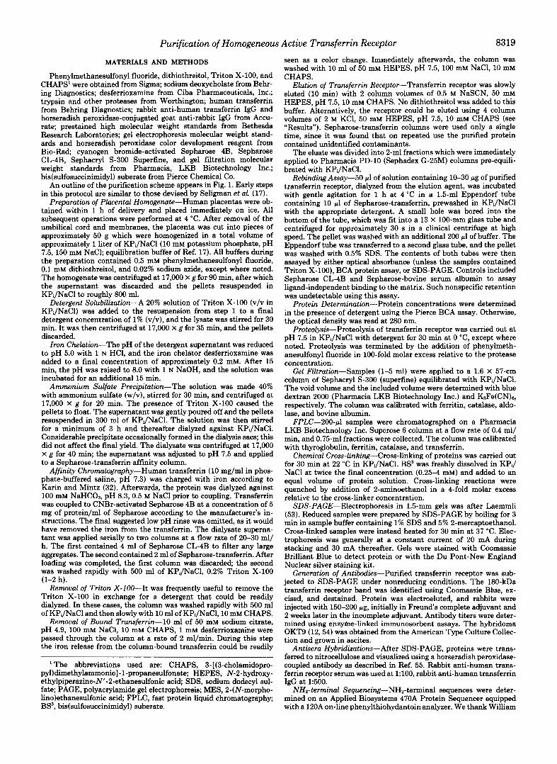
Purification of Transferrin Receptor-The purification pro- tocol produces 3-4 mg of transferrin receptor from a single placenta (-200 g of pellet after first centrifugation). This protein appears as a single major species by SDS-PAGE, with an apparent mass of 90 kDa under reducing conditions and approximately 180 kDa when unreduced (Fig. 1, lunes 2 and 3 and 5 and 6). These frequently appear as slightly resolved doublets, and occasionally triplets, on lightly loaded gels (not shown). The protein cross-reacts with the monoclonal anti- transferrin receptor antibody OKT9 in Western blots (not shown). Minor species appearing in heavily loaded gel lanes appear largely to be degradation products of transferrin recep- tor, based on their cross-reactivity with a rabbit antiserum against a single gel-purified band. Chief among these is a 45- kDa species, which becomes more abundant in preparations which are stored for several weeks at 4 “C. When we attempted to determine the NH2-terminal sequence of gel-purified ma- terial the sample behaved as though it were completely blocked.
NaSCN was found to be an efficient reversible elution agent. 1 M NaSCN dissociates transferrin receptor from di- ferric transferrin, while 0.5 M is adequate if the transferrin has first been stripped of its iron by a pH 5 wash. We have used the latter treatment because the low pH wash, followed
1 2 3 4 5 6 7 FIG. 1. SDS-PAGE of transferrin receptor and trypsin-di-
gested transferrin receptor. Transferrin receptor was purified as described under “Materials and Methods.” The samples in lanes 2,3, 5, and 6 were eluted directly from Sepharose-transferrin with 0.5% SDS. The samples in lows 4 and 7 were eluted with 0.5 M NaSCN, 10 mM CHAPS, desalted on a PD-10 column, digested with trypsin a t a 1:10 molar ratio (trypsin:transferrin receptor), concentrated to 5 mg/ml using a Centricon 30 (Amicon) apparatus, and chromato- graphed on an FPLC Superose 6 column. Samples were electropho- resed on a 5-15% polyacrylamide gel and stained with Coomassie Blue to visualize protein. Samples in lanes 1-4 contained 5% 2- mercaptoethanol; those in lanes 5-7 contained no reducing agent. Lane I , molecular mass standards: phosphorylase b (97.4 kDa); bovine albumin (66.2 kDa); ovalbumin (42.7 kDa). Lane 2, 9 pg of transferrin receptor. Lane 3, 4.5 pg of transferrin receptor. Lane 4, 10 pg of trypsin-cleaved transferrin receptor. Lane 5,9 pg of transferrin recep- tor. Lane 6, 4.5 pg of transferrin receptor. hne 7, 10 pg of trypsin- cleaved transferrin receptor.
by a wash at pH 7.5, removes some small amount of transfer- rin which is otherwise found in the final receptor preparation. This transferrin is not being released from the column matrix itself, since no transferrin is released by such a treatment before the cell lysate is applied, and probably represents transferrin that is bound to one of the two sites of a transferrin receptor dimer whose other binding site is engaged to a Sepharose-coupled ligand. 0.5 M NaSCN elutes at least 90% of the receptor released by boiling in 1% SDS.
Using the rebinding assay described under “Materials and Methods,” we have examined a number of other elution con- ditions. Elution with 0.5 or 1.0 M NaSCN yields transferrin receptor that rebinds to its ligand in Triton X-100 or CHAPS. Urea, guanidine, and SDS will efficiently but irreversibly elute receptor from Sepharose-transferrin. Seligman et al. (17) re- port using a buffer containing 50 mM glycine, pH 10, 1 M NaCl, 1% Triton X-100, but under our conditions this elutes only a small fraction of the bound receptor. Anderson et al. (6) reported the use of 1 M KC1 for elution of transferrin receptor from Sepharose-transferrin. We have reproduced this but find that rapid and efficient elution is more easily achieved using 2 M KCl. Quantitative NaSCN elution can be accom- plished in approximately one-half the volume necessary for KC1 elution, but otherwise the yields of the two methods are comparable (not shown). NaSCN elution may produce a frac- tion of the eluted receptor which is binding competent but aggregates during gel filtration. We have not seen this in the KC1-eluted material.
The eluted receptor rebinds to Sepharose-transferrin when it is solubilized in Triton X-100 or CHAPS (in KPi/NaCl) and shows slightly diminished activity in sodium deoxycholate (in 50 mM HEPES, pH 8.0,lOO mM NaCl) (data not shown).

Purification of Homogeneous Active Transferrin Receptor 8321
Gel filtration of the receptor in Triton X-100 demonstrates that it is chiefly a single species (Fig. 2, solid line). The minor peak, with an apparent mass of 420,000 Da, may be a trans- ferrin receptor tetramer, that is a dimer of dimers. When the purified receptor is incubated with transferrin prior to gel filtration, the complex migrates as a single peak, and the free transferrin peak follows (Fig. 2, broken line and inset).
Controlled Proteolysis of the Transferrin Receptor-Purified transferrin receptor was incubated with a variety of proteases with different amino acid specificities. As shown in Fig. 3, digestion with trypsin, subtilisin, thermolysin, or chymotryp- sin all results in the generation of a fragment with an apparent molecular weight of approximately 70,000, which migrates as a single species by gel filtration (not shown). The unchanged mobility of the 70-kDa fragment under reducing versus non- reducing conditions with SDS-PAGE (Fig. 1, lanes 4 and 7) demonstrates that it lacks the interchain disulfide bonds present in the intact receptor, as others have shown (15, 19). Minor postdigest species, which may be seen in heavily loaded lanes, are primarily bands with apparent molecular masses between 60 and 70 kDa (Fig. 1, lane 4 ) . NHz-terminal sequenc-
. . -d-
UImI" I , -. ma
ing of the FPLC-purified 70-kDa tryptic fragment yielded the following sequence.
90% Arg-Leu-Tyr-Trp-Asp-Asp-Leu-Lys-Arg-Lys
10% Leu-Tyr-Trp-Asp-Asp-Leu-Lys-Arg-Lys
Comparison with the known amino acid sequence (30, 31) establishes the major trypsin cleavage site as residue 121.
The 70-kDa tryptic fragment retains transferrin binding activity, and migrates in a complex with transferrin when assayed by gel filtration (Fig. 4A). The Stokes radii and masses of both the tryptic fragment and its complex with transferrin may be calculated from their gel filtration behavior by comparison with known standards (Fig. 4B). The following Stokes radii and masses are obtained for the 70-kDa trans- ferrin receptor fragment, 4.52 + 0.11 nm and 151,000 Da; for the 70-kDa fragment-transferrin complex, 5.65 + 0.05 nm and 331,000 Da. These suggest that the tryptic fragment exists as a stable noncovalent dimer and that it binds 2 transferrins. Consistent with the former, the mobility of the fragment in a nondenaturing polyacrylamide gel is intermediate between
0 C D
I 1 1
1 I I I I I l I I I I I I 1 I I I I I I I I I I I l I I I l
5 10 15 20 25
fraction number
FIG. 2. Gel filtration of transferrin receptor and transferrin receptor-transferrin complex. Trans- ferrin receptor was purified according to "Materials and Methods," eluted using 2 M KC1 and 0.5% Triton X-100, and concentrated using a Centricon 30 (Amicon) apparatus. Transferrin was saturated with iron according to "Materials and Methods." FPLC was performed in 10 mM MES, pH 6.5, 100 mM KCI, 0.5 mM M@&, 0.1 mM dithiothreitol, 0.5% Triton X-100. Solid line, 50 pg of transferrin receptor. Broken line, 25 pg of transferrin receptor + 100 pg of transferrin, incubated together for 1 h before gel filtration. Inset, SDS-PAGE on column fractions from transferrin receptor-transferrin sample (broken l ine) . 50-pl aliquots of each fraction were withdrawn and electrophoresed on a 7.5% polyacrylamide gel under nonreducing conditions and visualized by silver staining. Gel filtration standards are indicated by arrows. A, thyroglobulin; B, ferritin; C, catalase; D, transferrin; E, fully included volume of column.

8322 Purification of Homogeneous Active Transferrin Receptor
""- -
1 2 3 4 5 6 7 8 FIG. 3. Purified transferrin receptor in RPi/NaCl, 10 mM
CHAPS was digested for 30 min at 4 "C except as noted. Lanes 1-6 each contain 15 pg of transferrin receptor; lanes 7 and 8 contain 6 pg of transferrin receptor. 5 mM calcium was added to thermolysin digestions. Protease/transferrin receptor ratios (molar) are indicated. Proteolysis was terminated by the addition of phenylmethanesulfonyl fluoride, and samples were prepared for SDS-PAGE (reducing con- ditions) as described under "Materials and Methods." After electro- phoresis, protein was visualized with Coomassie Blue. Lane 1, no protease; lane 2, trypsin 1:l; lane 3, trypsin 1:lO; lane 4, trypsin 1:50; lane 5, subtilisin 1:50; lane 6, chymotrypsin 1:l; lane 7, thermolysin 1:l (37 " C ) ; lane 8, thermolysin 1 : lO (37 T).
the mobilities of transferrin and the transferrin-fragment complex (not shown).
Further proof of this stoichiometry is provided by chemical cross-linking experiments using the bifunctional succinimidyl ester BS3. As shown in Fig. 5, cross-linking of the 70-kDa fragment with bound transferrin (80 kDa) generates three major species with approximate apparent masses of 141,272, and 342 kDa. All three of these bands cross-react with anti- bodies against transferrin receptor and transferrin (not shown), suggesting that the 141-kDa species represents a cross-link between the tryptic fragment and transferrin. Higher concentrations of BS3 cross-link the dimer subunits, leading to species containing three and four subunits. The pattern suggests that the 272-kDa species represents either transferrin receptor2-transferrin or transferrin receptor- transferrin,, while the 342-kDa species is transferrin receptor2-transferrin2. The high molecular masses may be due to anomalous migration of the cross-linked complexes. The disappearance of the lower molecular mass species demon- strates that the receptor fragment is uniformly bound to transferrin.
Estimation of Molecular Dimensions-Using the experi- mental Stokes radius of 4.52 nm and the calculated molecular mass of 153.4 kDa for the transferrin receptor fragment dimer,
f = 8.52 X lo-* g/s
rdc = 3.54 X 10" cm
fmin = 6.67 X lo-' g/s
fo = 7.60 X lo-' g/s
f / f o = 1.12
sions of the protein by calculating the volume V of the prolate ellipsoid using a = 3.lb.
v = 4/3nab2
= (5) 02.
Thus, we obtain b = 24.3 A and a = 75.2 A. Similar calculations using the experimental Stokes radius of 3.7 nm and the known molecular weight of 80,000 for transferrin give b = 19.0 A and a = 64.3 A. For the complex, b = 30.8 A and a = 92.5 A. These radii are doubled to give the overall dimen- sions for each species.
DISCUSSION
Wide interest in iron metabolism and transferrin receptor has prompted numerous studies on its physiologic role and cell biology. While its activity was established more than 20 years ago (57,58), purification was first reported in 1979 (17, 25) and subsequently by a number of groups. These purifica- tions have relied largely on multiple ligand binding or immune affinity columns, and the purified protein was obtained in a denatured state or without clearly established activity. The reported yields of those protocols using large amounts of tissue are dramatically lower than that reported here.
More recently Anderson et al. (6) have reported a simplified purification of human transferrin receptor, relying on re- peated batch adsorption of solubilized transferrin receptor to immobilized transferrin. The purification relies on depletion of endogenous nonbound transferrin by entensive rinsing of tissue prior to homogenization. We avoid this problem with an iron chelation procedure which renders the endogenous transferrin noncompetitive with the immobilized diferric transferrin. Our protocol has a higher yield and appears to be more efficient in that it requires far less affinity matrix, and receptor absorption occurs during a single passage through a column.
Our purification protocol is designed to maximize the effi- ciency of transferrin receptor binding to a ligand affinity column by specifically depleting the placental homogenate not only of free transferrin but also of ferrotransferrin that may remain bound to receptor. By lowering the pH of the detergent lysate to 5 in the presence of desferrioxamine, we chelate iron from the endogenous transferrin. After neutrali- zation, the resulting apotransferrin will probably dissociate to some extent from its receptor because of the reduced binding constant for apotransferrin at neutral pH (35,40,59). We found a decrease in yield when the iron chelation was omitted. The ammonium sulfate precipitation leaves unbound transfemn in the supernatant, as well as reducing the volume of lysate applied to the affinity column. On the Sepharose- transfemn column, receptor should dissociate from any re- maining endogenous apotransferrin in favor of the diferric transfemn available, aided by the rapid half-time of dissocia- tion of apotransferrin and receptor a t neutral pH (39). This is supported by our observation that if the flow-through from the first affinity column is applied to a second equivalent column, the final yield is increased by less than 10% (data not shown).
The buffers and elution conditions in our purification pro- tocol were screened to avoid denaturation of transferrin recep- tor, as defined initially by our Sepharose-transferrin rebinding assay. This rebinding is demonstrated by the complete ab- sence of a free transferrin receptor peak in all gel filtration experiments done in the presence of excess transferrin. The tight binding so observed is consistent with the reported
giving an axial ratio a/b of 3.1. We can estimate the dimen- association constant for these species at neutral pH (Kd cz

Purification of Homogeneous Active Transferrin Receptor
A -
8323
FIG. 4. Gel filtration and deter- mination of Stokes radii. A, gel filtra- tion of transferrin receptor 70-kDa frag- ment-transferrin complex. Transferrin receptor was purified as described under “Materials and Methods” and eluted with 0.5 M NaSCNl 10 mM CHAPS. Thiocyanate was removed using a PD- 10 column, and the protein-bearing frac- tions were pooled and digested with tryp- sin a t 4 “C for 30 min at a molar ratio of 1:10 (trypsin/transferrin receptor). CHAPS was removed by dialysis, and the sample was centrifuged for 1 h at 100,OOO X g to remove hydrophobic ag- gregates. 2 ml of supernatant containing 0.6 mg of 70-kDa fragment were incu- bated for 4 h at 4 “C with a 2-fold molar excess of transferrin. The sample was filtered using Sephacryl S-300 (Super- fine) a5 described under “Materials and Methods,” and the absorbance of each fraction at 280 nm was measured. SDS- PAGE confirmed that the first peak con- tained both the 70-kDa fragment and transferrin in bands of apparent equal intensity, while the second peak con- tained only transferrin (not shown). B, determination of Stokes radii by gel fil- tration. Samples (1-3 ml) were filtered using Sephacryl S-300 (Superfine) as de- scribed under “Materials and Methods.” Arrows indicate the elution volumes of the 70-kDa fragment and of the 70-kDa fragment-transferrin complex, based on two measurements of each species.
A5 -
:: P 0 N
.03 -
.Ol -
.07 -
5.0 -
a B 3j
4.2 -
lo-’ (33, 36, 39). The receptor-transferrin complex remains stable when the pH is reduced to pH 5.5, as shown by gel filtration under these conditions (not shown).
Trypsin treatment of transferrin receptor has previously been described for protein on the cell surface and in a deter- gent lysate (15, 19). We have reproduced this with purified protein and found that the transferrin receptor extracyto- plasmic sequence contains a domain with an apparent molec- ular mass of 70 kDa by SDS-PAGE that is extremely resistant
to proteolysis and thus is readily obtained as a digestion product using any of a number of proteases. The 70-kDa fragment is a dimer in solution and binds transferrin in a 1:l stoichiometry, as shown by gel filtration and chemical cross- linking. This complex remains stable at pH 5.5 (not shown). Our measured Stokes radii of 4.52 and 5.65 nm for the transferrin receptor 70-kDa fragment and its complex with transferrin, respectively, are comparable to the values of 4.6 and 6.2 nm obtained by velocity sedimentation for intact

8324 Purification of Homogeneous Active Transferrin Receptor
-200kD
-97
-66
4 3 2 1 FIG. 5. Cross-linking of the transferrin receptor 70-kDa
fragment with transferrin. Gel filtration purified complex (see Fig. 4.4, first peak) was concentrated using a Centricon 30 apparatus to approximately 1 mg/ml. Four aliquots, each containing 12 pg of complex, were cross-linked using BS3 as described under “Materials and Methods.” Samples were analyzed on a 5-15% SDS-polyacryl- amide gel (reducing conditions), and protein was visualized using Coomassie Blue. Mass standards are indicated. Lune I , no cross- linker; lane 2, BS3, 0.25 mg/ml; lane 3, BS3, 1 mg/ml; lane 4, BS3, 4 mg/ml.
transferrin receptor and transferrin receptor-transferrin by Enns and Sussman (26). We have estimated the dimensions of the 70-kDa fragment dimer and its complex with transfer- rin using gel filtration and obtain dimensions of 49 X 150 A for the former and 62 x 185 A for the latter. Dimensions calculated in this manner must be considered as rough esti- mates; compare our calculated long axis dimension of trans- ferrin, 129 A, to the dimension obtained from the crystallo- graphic structure of related lactoferrin, approximately 98 A (60). They do, however, suggest that transferrin is unlikely to bind to transferrin receptor in an end-to-end manner. Our cross-linking data on the complex suggests that the first cross- link is established between the receptor and bound transfer- rin, and higher cross-linker concentrations result in linkage of the dimer pair. We do not know whether the two bound transferrin molecules are in contact, though there is no evi- dence of a cross-linked transferrin dimer.
We have identified the major trypsin cleavage site as residue 121, and the cleavage sites of thermolysin, subtilisin, and chymotrypsin must lie relatively nearby based on the similar size of the fragments produced. Trypsin makes multiple cleav- ages between the NH, terminus and residue 121 based on the absence of any discernible distinct cleavage product in the mass range of 10,000-20,000 Da (data not shown). The 61- residue cytoplasmic sequence can also be tryptically cleaved in intact vesicles in which the extracytoplasmic sequence faces the vesicle lumen (not shown). The cytoplasmic sequence could form a stably folded structure, since it is longer than the minimum length of known stable domains, but the cyto- plasmic domains of some other membrane receptors (61) are shorter than any known sequence that folds into a determi- nate structure. Based on the wide susceptibility of the extra- cytoplasmic domain near arginine 121, which is 32 residues
removed from the putative membrane spanning border at cysteine 89, we expect a relatively extended structure until the beginning of the large stable region that includes the transferrin-binding site. The region between residues 89 and 121 includes the interchain disulfide bonds, as shown by the failure of the 70-kDa fragment to dimerize when analyzed by nonreducing SDS-PAGE. These disulfide bonds have recently been reported to involve cysteine residues 89 and 98 (62). Since the 70-kDa tryptic fragments remain stably dimerized in the absence of covalent attachment, this region is not crucial for maintaining dimer contacts, though it is possible that disulfide formation in newly synthesized protein brings the COOH-terminal portions of two polypeptides into prox- imity and facilitates folding. The conservation of cysteines 89 and 98 in the murine transferrin receptor supports their functional significance (63). Jing and Trowbridge (62) have used site-directed mutagenesis to express an altered transfer- rin receptor lacking the disulfide bonds in this region. The mutated protein is expressed on the cell surface, but neither the kinetics nor efficiency of this expression has been re- ported. Alternatively, the extended and therefore flexible region may be necessary to allow flexible interaction between the transferrin binding domains. Such subunit interactions in multimeric receptors are a plausible mechanism for signal transduction to the cytoplasm. There is no evidence that transferrin binding on the cell surface leads to signal trans- duction, but such quaternary changes may occur during the receptor’s itinerations of intracellular compartments with widely varying pH values. Analysis of these questions requires knowledge of the three-dimensional structure of transferrin receptor and of its disposition in the membrane. The availa- bility of large amounts of highly purified transferrin receptor, as well as the hydrophilic tryptic fragment, makes it possible to consider crystallographic analysis of the molecule, which is currently being attempted in this laboratory.
Acknowledgments-We would like to acknowledge Stacey Merritt, Phil Breitfeld, and Charles Simmons for their help with antisera, Marina Babyonyshev for technical assistance, and Michael Brand for helpful discussions.
1. 2.
3. 4.
5.
6.
7.
8.
9.
10. 11. 12.
13.
14.
REFERENCES Bergeron, R. J. (1986) Trends Biochem. Sci. 11,133-136 Baker, E. N., Rumball, S. V., and Anderson, B. F.0987) Trends
Princiotto, J. V., and Zapolski, E. S. (1975) Nature 266.87-88 Aisen, P., and Listowsky, I. (1980) Annu. Rev. Biochem. 49,357-
393 Hanover, J. A., and Dickson, R. B. (1985) in Receptor-mediated
Endocytosis, pp. 341-387, Plenum Publishing Corp., New York Anderson, G. J., Mackerras, A., Powell, L. W., and Halliday, J.
W. (1986) Biochim. Biophys. Acta 884,225-233 Newman, R., Schneider, C., Sutherland, R., Vodinelich, L., and
Greaves, M. (1982) Trends Biochem. Sci. 397-400 Fielding, J., and Speyer, B. E. (1974) Biochim. Biophys. Acta 363 ,
Hemmaplardh, D., and Morgan, E. H. (1974) Exp. Cell Res. 87,
Sullivan, A. L., and Weintraub, L. R. (1978) Blood 52,436-446 Hu, H.-Y., and Aisen, P. (1978) J. Supramol. S t r u t . 8,349-360 Sutherland, R., Delia, D., Schneider, C., Newman, R., Kemshead,
J., and Greaves, M. (1981) Proc. Natl. Acad. Sci. U. S. A. 7 8 ,
Stein, B. S., and Sussman, H. H. (1983) J. Biol. Chem. 268,
Hopkins, C. R. (1983) Cell 35.321-330
Biochem. Sci. 12,350-353
387-396
207-212
4515-4519
2668-2673
15. Schneider, C., Sutherland, R., Newman, R., and Greaves, M.
16. Fernandez-Pol, J. A., and Klos, D. J. (1980) Biochemistry 19 , (1982) J. Bwl. Chem. 257,8516-8522
3904-3912

Purification of Homogeneous Active Transferrin Receptor 8325
17. Seligman, P. A., Schleicher, R. B., and Allen, R. H. (1979) J. Biol. 40. Klausner, R. D., Van Renswoude, J., Ashwell, G., Kempf, C., Chem. 254,9943-9946 Schechter, A. N., Dean, A., and Bridges, K. R. (1983) J. Bwl.
18. Omary, M. B., and Trowbridge, I. S. (1981) J. Biol. Chem. 256, Chem. 258,4715-4724 12888-12892 41. Hopkins, C. R., and Trowbridge, I. S. (1983) J. Cell Biol. 9 7 ,
19. Omary, M. B., and Trowbridge, I. S. (1981) J. Biol. Chem. 2 5 6 , 508-521
20. Adam, M., Rodriguez, A., Turbide, C., Larrick, J., Meighen, E., 83,6445-6449
21. Sefton, B. M., Trowbridge, I. S., and Cooper, J. A. (1982) Cell 44- Young, s. p.9 Bornford, A.7 and Williams, R. (lgU) Biochem. J.
4715-4718 42. Ajioka, R. S., and Kaplan, J. (1986) Proc. Natl. Acad. Sci. U. S. A.
and Johnstone, R. M. (1984) J. Biol. Chem. 2 5 9 , 15460-15463 43. Kornfeld, s. (1969) Biochim. BwPhYs. Acta 194 , 25-33
31,465-474 219,505-510
259,9944-9952 93 22. Hunt, R. c., Ruffin, R., and Yang, y.-s. (1984) J. Bwl. Cbm. 45. Wall, D. A*, Wilson, G.9 and Hubbard, A. L. (1980) cell 2 1 , 79-
23. May, W. S., Jacobs, S., and Cuatrecasas, P. (1984) Proc. Natl. 46. Booth, A. G.* and M. J. (lg81) Biochem. J. lg6* 355-
24. ~ l ~ i l , J. D., and Bretscher, M. s. (1982) E M B o J. 1 , 351-355 47. Geuze, H. JV Slot, J. w.7 StroUs, G. J. A. M.9 Lodish, H. FV and 25. Wada, H. G., Hass, P. E., and Sussman, H. H. (1979) J. Biol. 48. Hopkins, C. R. (1985) Cell 4 0 , 199-208 26. Enns, C. A., and Sussman, H. H. (1981) J. Bwl. Chem. 2 5 6 , 49. c. (1985) J. 'Oo9 633-637 50. Willingham, M. C., Hanover, J. A., Dickson, R. B., and Pastan, 27. Brown, P. J., and Johnson, P. M. (1981) Placenta 2,1-10 28. Escarot-Charrier, B., Grey, V. L., Wilczynska, A., and Schulman, 51. Yamashiro, D. J., Tycko, B., Fluss, S. R., and Maxfield, F. R.
29. Van Driel, I. R., Stearne, P. A., Grego, B., Simpson, R. J., and 52. Octave, J.-N., Schneider, Y.-J., Hoffmann, P., Trouet, A., and
30. Schneider, C., Owen, M. J., Banville, D., and Williams, J. G. 54. E. L., K ~ ~ ~ , p. c., ~ ~ l b ~ ~ i ~ , G., L ~ ~ ~ ~ , R. H., and 53. Laemmli, U. K. (1970) Nature 227,680-685
31. McClelland, A., Kuhn, L. C., and Ruddle, F. H. (1984) Cell 3 9 , Schlossman, S. F. (1980) Proc. Natl. Acad. Sci. U. S. A. 7 7 , 1588-1592
32. Karin, M., and Mintz, B. (1981) J. Biol. Chem. 256,3245-3252 55. Schwartz, A. L., Ciechanover, A., Merritt, S., and Turkewitz, A.
33. van Renswoude, J. K., Bridges, K. R., Harford, J. B., and Klaus- 56. ~ i ~ ~ ~ ~ , I., Sauer, K., and Wang, J. c. (1978) Physical Chemistry, (1986) J. Biol. Chem. 2 6 1 , 15225-15232
ner, R. D. (1982) Proc. Natl. Acad. Sci. U. S. A. 79,6186-6190 34. Lamb, J. E., Ray, F., Ward, J. H., Kushner, J. p., and Kaplan, J. 57. Jandl, J. H., Inman, J. K., Simmons, R. L., and Allen, D. W.
pp. 219-234, Prentice-Hall, Englewood Cliffs, NJ
(1983) J. Bwl. Chem. 258,8751-8758 35. Ciechanover, A., Schwartz, A. L., Dautry-Varsat, A., and Lodish, 58. Jandl, J. H., and Katz, J. H. (1963) J. Clin. Invest. 42,314-326
(1959) J. Clin. Znuest. 3 8 , 161-185
H. F. (1983) J. Bwl. Chem. 258,9681-9689 36. Dautry-Varsat, A., Ciechanover, A., and Lodish, H. F. (1983)
59. Harding, C., and Stahl, P. (1983) Biochem. Biophys. Res. Com-
Proc. Natl. Acad. Sci. U. S. A. 80,2258-2262 mun. 113,650-658
60. Anderson, B. F., Baker, H. M., Dodson, E. J., Norris, G. E., 37. Harding, C., Heuser, J., and Stahl, P. (1983) J. Cell Bwl. 97, Rumball, S. V., Waters, J. M., and Baker, E. N. (1978) Proc.
38. Hopkins, c. R., and Trowbridge, I. s. (1983) J. Cell Bid. 9 7 , 61. Drickamer, K., Mamon, J. F., Binns, G., and hung, J. 0. (1984)
39. Klausner, R. D., Ashwell, G., van Renswoude, J., Harford, J. B., 62. Jing, S., and Trowbridge, I. S. (1987) EMBO J. 6,327-331
Acad. Sci. U. S. A. 81,2016-2020 362
Schwartz, A. L. (1983) Cell 3 2 , 277-287 Chem. 254,12629-12635
9820-9823 I. (1984) Proc. Natl. Acad. Sci. U. S. A. 8 1 , 175-179
(1984) Cell 37,789-800
Crichton, R. R. (1982) Eur. J. Biochem. 123,235-240 H. M. (1980) Can. J. Biochem. 58,418-426
Goding, J. W. (1984) J. Zmmuml. 133,3220-3224
(1984) Nature 3 1 1 , 675-678
267-274
329-339 Natl. Acad. Sci. U. S. A. 8 4 , 1769-1773
508-521 J. Biol. Chem. 259,770-778
and Bridges, K. R. (1983) Proc. Natl. Acad. Sci. U. S. A. 80, 63. Stearne, P. A., Pietersz, G. A., and Goding, J. W. (1985) J. 2263-2266 Immuml. 134,3474-3479


![Index [] · 3 Anna Margaretha [6960] *1884 .....446 Arij [8318] .....53 Cornelis Ariensz.](https://static.fdocuments.in/doc/165x107/6147e417a830d0442101ba8a/index-3-anna-margaretha-6960-1884-446-arij-8318-53-cornelis-ariensz.jpg)