Micro-solvation of tyrosine-kinase inhibitor AG1478 ......Micro-solvation of tyrosine-kinase...
11
Micro-solvation of tyrosine-kinase inhibitor AG1478 explored with fluorescence spectroscopy and computational chemistry† Muhammad Khattab, a Feng Wang * b and Andrew H. A. Clayton * a Tyrosine kinase inhibitors (TKI) are an important class of molecules. Specific interactions of TKI with water are of interest since they appear in high resolution X-ray structures of TKI–protein complexes and are thought to be important determinants of drug efficacy. Methods for determining the specific interactions of TKI with water molecules in solution are therefore highly desirable. Recently, we revealed that the TKI, AG1478, exhibits absorbance and fluorescence spectra which are sensitive to the conformation of the molecule and the polarity of the surrounding environment. In the present work, we investigated the potential hydrogen bond binding sites of AG1478 using spectroscopic measurements of acetonitrile– water solutions. UV-Vis absorbance spectroscopy of AG1478 revealed H-bond interactions between water molecules and AG1478 in the ground state, as evidenced by changes in spectral shape and optical density with increases in water fraction. The fluorescence spectra of AG1478 in acetonitrile were also greatly influenced by water interactions, revealing fluorescence quenching (by 80%) with the addition of 2% by volume of water. The AG1478 fluorescence quantum yield decreased with increasing temperature in neat acetonitrile but revealed an unorthodox increase with increasing temperature in acetonitrile– water solution. Taken together, these changes are consistent with a specific complex or complexes formed between AG1478 and water molecules. Potential AG1478–water clusters were investigated using ab initio calculations. The effects of explicit hydrogen bonding on vertical excitation, topology and electronic configuration of AG1478 were examined computationally. 1. Introduction Several theories and models have been developed to describe the complex process of solvation of solutes in binary mixtures. 1–4 Solutes can be preferentially solvated by either solvent, nevertheless the solvent–solvent interactions can signicantly inuence solute–solvent interactions (sol- vatochromic parameters). 5 Preferential solvation occurs when a solute molecule has, at its vicinity, more of one solvent than in the bulk environment. Hence the understanding of this phenomenon plays a role in unravelling the spectroscopic, kinetic and thermodynamic behaviour of solute molecules. 6 Binary mixtures containing water as a cosolvent have been successfully deployed to quantify the number of H-bonding interactions between solute and water molecules. This is accomplished by application of various spectroscopic experi- ments and/or theoretical calculations using explicit water models. 7–11 Hence the organic solvents which are miscible with water are ultimately used for these kind of experiments. Acetonitrile is characterized by good UV transparency, aqueous miscibility, low viscosity and low chemical reactivity. In addition, it has a relatively high dielectric constant and a small autoprotolysis constant. 5 Water can solubilize ionic and dipolar solutes and its colligative properties have a large bearing on vast number of biological and chemical systems through its ability to form intermolecular hydrogen bonds. 12,13 It has been reported that the structure of pure water is signi- cantly altered upon mixing with other solvents generally and with acetonitrile specically. 13–17 Therefore, binary mixtures of acetonitrile with water are solvents of choice in physical organic chemistry. 5,13 A number of studies have attempted to address the impact of microhydration on the geometrical and electronic properties of solute molecules by dissolving them in acetoni- trile–water solutions. For instance, the excited state H-bond dynamics of coumarin 102 were investigated in acetonitrile–water binary mixtures by Wells et al. 7 The experimental and simulation data revealed that two water molecules, acting as H-bond donors, can form H- bonds at the carbonyl site of coumarin 102. 7 This nding gave a Centre for Micro-Photonics, Faculty of Science, Engineering and Technology, Swinburne University of Technology, Melbourne, Victoria 3122, Australia. E-mail: [email protected] b Molecular Model Discovery Laboratory, Department of Chemistry and Biotechnology, School of Science, Faculty of Science, Engineering and Technology, Swinburne University of Technology, Melbourne, Victoria 3122, Australia. E-mail: fwang@swin. edu.au † Electronic supplementary information (ESI) available. See DOI: 10.1039/c7ra04435f Cite this: RSC Adv. , 2017, 7, 31725 Received 20th April 2017 Accepted 13th June 2017 DOI: 10.1039/c7ra04435f rsc.li/rsc-advances This journal is © The Royal Society of Chemistry 2017 RSC Adv. , 2017, 7, 31725–31735 | 31725 RSC Advances PAPER Open Access Article. Published on 21 June 2017. Downloaded on 21/07/2017 03:02:57. This article is licensed under a Creative Commons Attribution 3.0 Unported Licence. View Article Online View Journal | View Issue
Transcript of Micro-solvation of tyrosine-kinase inhibitor AG1478 ......Micro-solvation of tyrosine-kinase...
RSC Advances
PAPER
Ope
n A
cces
s A
rtic
le. P
ublis
hed
on 2
1 Ju
ne 2
017.
Dow
nloa
ded
on 2
1/07
/201
7 03
:02:
57.
Thi
s ar
ticle
is li
cens
ed u
nder
a C
reat
ive
Com
mon
s A
ttrib
utio
n 3.
0 U
npor
ted
Lic
ence
.
View Article OnlineView Journal | View Issue
Micro-solvation
aCentre for Micro-Photonics, Faculty of
Swinburne University of Technology, Melb
[email protected] Model Discovery Laboratory, Dep
School of Science, Faculty of Science, E
University of Technology, Melbourne, Victor
edu.au
† Electronic supplementary informa10.1039/c7ra04435f
Cite this: RSC Adv., 2017, 7, 31725
Received 20th April 2017Accepted 13th June 2017
DOI: 10.1039/c7ra04435f
rsc.li/rsc-advances
This journal is © The Royal Society of C
of tyrosine-kinase inhibitorAG1478 explored with fluorescence spectroscopyand computational chemistry†
Muhammad Khattab, a Feng Wang *b and Andrew H. A. Clayton *a
Tyrosine kinase inhibitors (TKI) are an important class of molecules. Specific interactions of TKI with water
are of interest since they appear in high resolution X-ray structures of TKI–protein complexes and are
thought to be important determinants of drug efficacy. Methods for determining the specific interactions
of TKI with water molecules in solution are therefore highly desirable. Recently, we revealed that the TKI,
AG1478, exhibits absorbance and fluorescence spectra which are sensitive to the conformation of the
molecule and the polarity of the surrounding environment. In the present work, we investigated the
potential hydrogen bond binding sites of AG1478 using spectroscopic measurements of acetonitrile–
water solutions. UV-Vis absorbance spectroscopy of AG1478 revealed H-bond interactions between
water molecules and AG1478 in the ground state, as evidenced by changes in spectral shape and optical
density with increases in water fraction. The fluorescence spectra of AG1478 in acetonitrile were also
greatly influenced by water interactions, revealing fluorescence quenching (by 80%) with the addition of
2% by volume of water. The AG1478 fluorescence quantum yield decreased with increasing temperature
in neat acetonitrile but revealed an unorthodox increase with increasing temperature in acetonitrile–
water solution. Taken together, these changes are consistent with a specific complex or complexes
formed between AG1478 and water molecules. Potential AG1478–water clusters were investigated using
ab initio calculations. The effects of explicit hydrogen bonding on vertical excitation, topology and
electronic configuration of AG1478 were examined computationally.
1. Introduction
Several theories and models have been developed to describethe complex process of solvation of solutes in binarymixtures.1–4 Solutes can be preferentially solvated by eithersolvent, nevertheless the solvent–solvent interactions cansignicantly inuence solute–solvent interactions (sol-vatochromic parameters).5 Preferential solvation occurs whena solute molecule has, at its vicinity, more of one solvent than inthe bulk environment. Hence the understanding of thisphenomenon plays a role in unravelling the spectroscopic,kinetic and thermodynamic behaviour of solute molecules.6
Binary mixtures containing water as a cosolvent have beensuccessfully deployed to quantify the number of H-bondinginteractions between solute and water molecules. This is
Science, Engineering and Technology,
ourne, Victoria 3122, Australia. E-mail:
artment of Chemistry and Biotechnology,
ngineering and Technology, Swinburne
ia 3122, Australia. E-mail: fwang@swin.
tion (ESI) available. See DOI:
hemistry 2017
accomplished by application of various spectroscopic experi-ments and/or theoretical calculations using explicit watermodels.7–11 Hence the organic solvents which are miscible withwater are ultimately used for these kind of experiments.
Acetonitrile is characterized by good UV transparency,aqueous miscibility, low viscosity and low chemical reactivity.In addition, it has a relatively high dielectric constant anda small autoprotolysis constant.5 Water can solubilize ionic anddipolar solutes and its colligative properties have a largebearing on vast number of biological and chemical systemsthrough its ability to form intermolecular hydrogen bonds.12,13
It has been reported that the structure of pure water is signi-cantly altered upon mixing with other solvents generally andwith acetonitrile specically.13–17 Therefore, binary mixtures ofacetonitrile with water are solvents of choice in physical organicchemistry.5,13 A number of studies have attempted to addressthe impact of microhydration on the geometrical and electronicproperties of solute molecules by dissolving them in acetoni-trile–water solutions.
For instance, the excited state H-bond dynamics of coumarin102 were investigated in acetonitrile–water binary mixtures byWells et al.7 The experimental and simulation data revealed thattwo water molecules, acting as H-bond donors, can form H-bonds at the carbonyl site of coumarin 102.7 This nding gave
RSC Adv., 2017, 7, 31725–31735 | 31725
RSC Advances Paper
Ope
n A
cces
s A
rtic
le. P
ublis
hed
on 2
1 Ju
ne 2
017.
Dow
nloa
ded
on 2
1/07
/201
7 03
:02:
57.
Thi
s ar
ticle
is li
cens
ed u
nder
a C
reat
ive
Com
mon
s A
ttrib
utio
n 3.
0 U
npor
ted
Lic
ence
.View Article Online
a more robust clue for estimating hydrogen bond strength of C102 molecule. It was found that clustered water molecules canalso turn on the bright emission of a molecule and the lifetimeof the excited state was dependent on the number of watermolecules in rst solvation shell. Studies conducted on theadenine analogue, 2-aminopurine, have also led to similarconclusions.18,19 Studies of Lobsiger and co-workers showedthat the lifetime of 2-aminopurine increased systematically withthe number of water molecules. They also found out thatposition of bound water molecules was important. The excited-state lifetime was much longer when one water moleculeinteracted with the NH2 group of 2-aminopurine.18
In general, spectroscopic measurements of drugs inaqueous–organic binary mixtures provide a controllable modelsystem to investigate the interaction of water molecules withdrugs. Water molecules are highly translocated at proteinbinding sites and found essential for drug–protein intermo-lecular interactions and stability. Therefore, binding interac-tions between explicit water molecules and drugs affects thenature and diversity of drug chemical structures and properties.Mastering the gain in protein–drug binding affinity can beachieved by targeting or neglecting H-bonding with clusteredwaters.20
AG1478 is a tyrosine kinase inhibitor,21–23 synthesized tomimic the purine ring of adenosine triphosphate (ATP)cofactor.24 It possesses ve H-acceptor moieties and one H-donor group, acting as a potential H-bonding target. Giventhat it can bind to the protein hinge region of tyrosine kinasesthrough one to three H-bonds,25 it is therefore important toexplore the impacts of H-bonding statics and dynamics on theelectronic and geometric properties of this TKI.
Our earlier experimental26 and computational27 studiesshowed that the absorbance of AG1478 was sensitive to themolecular conformation (twisted versus planar), while theuorescence was inuenced by the polarity and hydrogen-bonding power of the solvent.26 We have also observed exten-sive uorescence quenching of aqueous AG1478 solutioncompared to other non-aqueous AG1478 solutions.26 In the lastfew decades, spectroscopic studies have been performed onprobes in different media such as binary solvent mixtures,micelles, reverse micelles and ionic liquids.28–31 When studies inpure water are not feasible, solvent–water binary mixturesprovide a controllable environment in which to probe theinuence of water against a moderately polar host solvent.32,33
Therefore, we used the binary mixture approach to investigatethe intrinsic and extrinsic H-bonding interactions of AG1478 inthe ground and excited states.
In this article, the steady-state UV-Vis spectroscopy ofAG1478 in acetonitrile solution containing water as a co-solvent,was investigated to reveal the spectral properties of AG1478 inthe presence of water molecules. We also performedtemperature-dependent uorescence experiments to delineatethe inuence of dynamic versus static quenching betweenAG1478 and water molecules. In addition, time-dependantdensity functional theory (TD-DFT) was utilized to examinepossible complexes of AG1478 with different numbers of watermolecules.
31726 | RSC Adv., 2017, 7, 31725–31735
2. Experimental2.1. Materials
Spectroscopic grade acetonitrile was purchased from ThermoFisher Scientic Inc. and was used without further purication.Deionized water (Millipore) was used to prepare the aqueoussolutions. Tyrphostin AG1478 was purchased from SapphireBioscience Pty Ltd and used as received. A matched pair of quartzcuvettes with a path length of 1 cm was obtained from Starna PtyLtd.
2.2. Methods
2.2.1. UV-vis spectroscopy. The absorption spectra ofAG1478 were scanned using a Shimadzu Recording Spectropho-tometer UV-1601. Excitation and emission measurements wereperformed with a Perkin Elmer LS55 Fluorescence Spectrometer.The temperature (25–70 �C) was regulated using Perkin ElmerPeltier Temperature Programmer PTP-1. The background absor-bance, scatter and uorescence were corrected using blanksamples.
Solutions containing acetonitrile and water were prepared byadding a small volume of water to acetonitrile (in the range 0–16%v/v). Dilute solutions of AG1478 were prepared for spectroscopicmeasurements (AG1478 concentration was 10�5 M (absorbance)and 10�6 M (uorescence)). The optical density of 300–360 nmband was adjusted to be above 0.1 for absorption experiments andwas lower than 0.05 for excitation and emission measurements tominimize inner lter effect. All spectra are averages of nine scans.
2.2.2. Computational details. The geometries of ground andexcited state structures were optimized by DFT and TD-DFTcalculations, respectively, at the B3LYP/6-311+G* level of theory.No imaginary frequencies at the optimized structures were ob-tained, indicating that the corresponding geometries are true localminima. The Becke's three-parameter hybrid exchange-correlationfunctional (B3LYP)34,35with 6-311+G* basis set was employed in allcalculations. Implicit solvent effect was considered in our calcu-lations. Hence, the conductor-like polarizable continuum model(CPCM)36 with the dielectric constant of 3 ¼ 37 was used (which isassumed to be the dielectric constant of acetonitrile watermixture). All calculations were performed using GAUSSIAN 09Revision C.01 (ref. 37) on Swinburne supercomputing facilities.
First, we screened for the best possible position(s) (giving moststable complex) for binding of n¼ 1–7 water molecules to AG1478.That means an explicit water molecule(s) was included at prox-imity (1.7 A) to different H-bond acceptor sites of AG1478.Secondly, we determined the most stable geometry for eachcomplex and performed single point energy calculations indielectric continuum with the dielectric constant of 37 using theCPCM model. We also computed the corresponding vertical exci-tation energy including state-specic solvation correction. Here-aer, the planar structure complexed with water molecules werereferred as P-nw, where n ¼ 1–7, and the twisted rotamer exhib-iting intermolecular H-bond with watermolecules were referred asT-nw, where n ¼ 1–7. Full geometric coordinates, structuralproperties and energies are contained in the ESI Tables S1–S5 andFig. S1–S4.†
This journal is © The Royal Society of Chemistry 2017
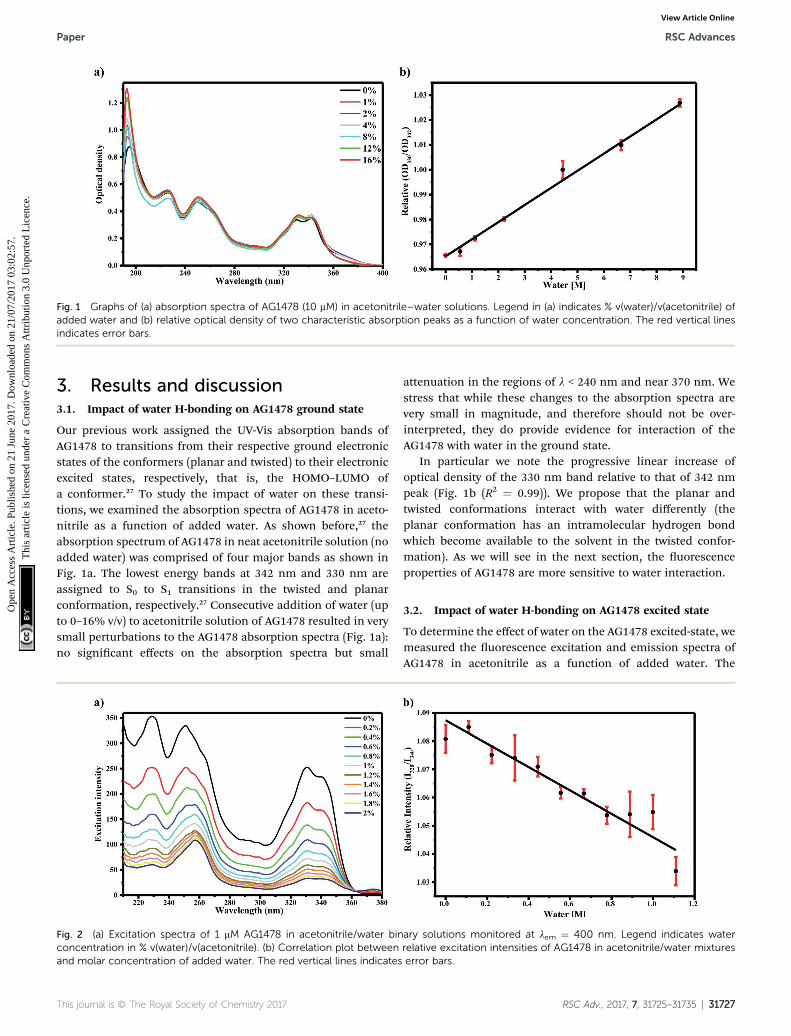
Fig. 1 Graphs of (a) absorption spectra of AG1478 (10 mM) in acetonitrile–water solutions. Legend in (a) indicates % v(water)/v(acetonitrile) ofadded water and (b) relative optical density of two characteristic absorption peaks as a function of water concentration. The red vertical linesindicates error bars.
Paper RSC Advances
Ope
n A
cces
s A
rtic
le. P
ublis
hed
on 2
1 Ju
ne 2
017.
Dow
nloa
ded
on 2
1/07
/201
7 03
:02:
57.
Thi
s ar
ticle
is li
cens
ed u
nder
a C
reat
ive
Com
mon
s A
ttrib
utio
n 3.
0 U
npor
ted
Lic
ence
.View Article Online
3. Results and discussion3.1. Impact of water H-bonding on AG1478 ground state
Our previous work assigned the UV-Vis absorption bands ofAG1478 to transitions from their respective ground electronicstates of the conformers (planar and twisted) to their electronicexcited states, respectively, that is, the HOMO–LUMO ofa conformer.27 To study the impact of water on these transi-tions, we examined the absorption spectra of AG1478 in aceto-nitrile as a function of added water. As shown before,27 theabsorption spectrum of AG1478 in neat acetonitrile solution (noadded water) was comprised of four major bands as shown inFig. 1a. The lowest energy bands at 342 nm and 330 nm areassigned to S0 to S1 transitions in the twisted and planarconformation, respectively.27 Consecutive addition of water (upto 0–16% v/v) to acetonitrile solution of AG1478 resulted in verysmall perturbations to the AG1478 absorption spectra (Fig. 1a):no signicant effects on the absorption spectra but small
Fig. 2 (a) Excitation spectra of 1 mM AG1478 in acetonitrile/water binconcentration in % v(water)/v(acetonitrile). (b) Correlation plot betweenand molar concentration of added water. The red vertical lines indicates
This journal is © The Royal Society of Chemistry 2017
attenuation in the regions of l < 240 nm and near 370 nm. Westress that while these changes to the absorption spectra arevery small in magnitude, and therefore should not be over-interpreted, they do provide evidence for interaction of theAG1478 with water in the ground state.
In particular we note the progressive linear increase ofoptical density of the 330 nm band relative to that of 342 nmpeak (Fig. 1b (R2 ¼ 0.99)). We propose that the planar andtwisted conformations interact with water differently (theplanar conformation has an intramolecular hydrogen bondwhich become available to the solvent in the twisted confor-mation). As we will see in the next section, the uorescenceproperties of AG1478 are more sensitive to water interaction.
3.2. Impact of water H-bonding on AG1478 excited state
To determine the effect of water on the AG1478 excited-state, wemeasured the uorescence excitation and emission spectra ofAG1478 in acetonitrile as a function of added water. The
ary solutions monitored at lem ¼ 400 nm. Legend indicates waterrelative excitation intensities of AG1478 in acetonitrile/water mixtureserror bars.
RSC Adv., 2017, 7, 31725–31735 | 31727
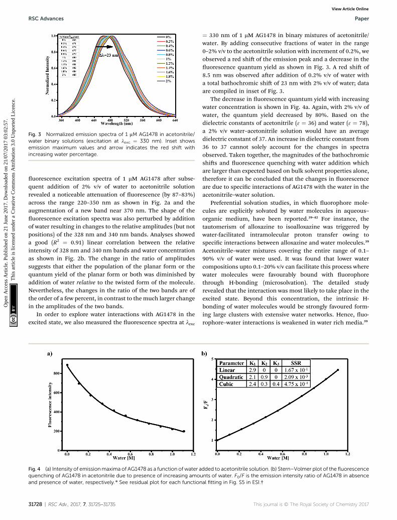
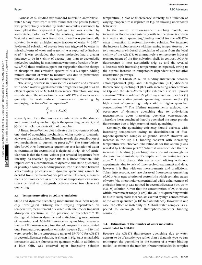
Fig. 3 Normalized emission spectra of 1 mM AG1478 in acetonitrile/water binary solutions (excitation at lexc ¼ 330 nm). Inset showsemission maximum values and arrow indicates the red shift withincreasing water percentage.
RSC Advances Paper
Ope
n A
cces
s A
rtic
le. P
ublis
hed
on 2
1 Ju
ne 2
017.
Dow
nloa
ded
on 2
1/07
/201
7 03
:02:
57.
Thi
s ar
ticle
is li
cens
ed u
nder
a C
reat
ive
Com
mon
s A
ttrib
utio
n 3.
0 U
npor
ted
Lic
ence
.View Article Online
uorescence excitation spectra of 1 mM AG1478 aer subse-quent addition of 2% v/v of water to acetonitrile solutionrevealed a noticeable attenuation of uorescence (by 87–83%)across the range 220–350 nm as shown in Fig. 2a and theaugmentation of a new band near 370 nm. The shape of theuorescence excitation spectra was also perturbed by additionof water resulting in changes to the relative amplitudes (but notpositions) of the 328 nm and 340 nm bands. Analyses showeda good (R2 ¼ 0.91) linear correlation between the relativeintensity of 328 nm and 340 nm bands and water concentrationas shown in Fig. 2b. The change in the ratio of amplitudessuggests that either the population of the planar form or thequantum yield of the planar form or both was diminished byaddition of water relative to the twisted form of the molecule.Nevertheless, the changes in the ratio of the two bands are ofthe order of a few percent, in contrast to the much larger changein the amplitudes of the two bands.
In order to explore water interactions with AG1478 in theexcited state, we also measured the uorescence spectra at lexc
Fig. 4 (a) Intensity of emissionmaxima of AG1478 as a function of water aquenching of AG1478 in acetonitrile due to presence of increasing amouand presence of water, respectively.* See residual plot for each function
31728 | RSC Adv., 2017, 7, 31725–31735
¼ 330 nm of 1 mM AG1478 in binary mixtures of acetonitrile/water. By adding consecutive fractions of water in the range0–2% v/v to the acetonitrile solution with increment of 0.2%, weobserved a red shi of the emission peak and a decrease in theuorescence quantum yield as shown in Fig. 3. A red shi of8.5 nm was observed aer addition of 0.2% v/v of water witha total bathochromic shi of 23 nm with 2% v/v of water; dataare compiled in inset of Fig. 3.
The decrease in uorescence quantum yield with increasingwater concentration is shown in Fig. 4a. Again, with 2% v/v ofwater, the quantum yield decreased by 80%. Based on thedielectric constants of acetonitrile (3 ¼ 36) and water (3 ¼ 78),a 2% v/v water–acetonitrile solution would have an averagedielectric constant of 37. An increase in dielectric constant from36 to 37 cannot solely account for the changes in spectraobserved. Taken together, the magnitudes of the bathochromicshis and uorescence quenching with water addition whichare larger than expected based on bulk solvent properties alone,therefore it can be concluded that the changes in uorescenceare due to specic interactions of AG1478 with the water in theacetonitrile–water solution.
Preferential solvation studies, in which uorophore mole-cules are explicitly solvated by water molecules in aqueous–organic medium, have been reported.39–42 For instance, thetautomerism of alloxazine to isoalloxazine was triggered bywater-facilitated intramolecular proton transfer owing tospecic interactions between alloxazine and water molecules.39
Acetonitrile–water mixtures covering the entire range of 0.1–90% v/v of water were used. It was found that lower watercompositions upto 0.1–20% v/v can facilitate this process wherewater molecules were favourably bound with uorophorethrough H-bonding (microsolvation). The detailed studyrevealed that the interaction was most likely to take place in theexcited state. Beyond this concentration, the intrinsic H-bonding of water molecules would be strongly favoured form-ing large clusters with extensive water networks. Hence, uo-rophore–water interactions is weakened in water rich media.39
dded to acetonitrile solution. (b) Stern–Volmer plot of the fluorescencents of water. F0/F is the emission intensity ratio of AG1478 in absenceal fitting in Fig. S5 in ESI.†
This journal is © The Royal Society of Chemistry 2017
Paper RSC Advances
Ope
n A
cces
s A
rtic
le. P
ublis
hed
on 2
1 Ju
ne 2
017.
Dow
nloa
ded
on 2
1/07
/201
7 03
:02:
57.
Thi
s ar
ticle
is li
cens
ed u
nder
a C
reat
ive
Com
mon
s A
ttrib
utio
n 3.
0 U
npor
ted
Lic
ence
.View Article Online
Barbosa et al. studied ve standard buffers in acetonitrile–water binary mixtures.43 It was found that the proton (solute)was preferentially solvated by water molecules evidenced bylower pH(s) than expected if hydrogen ion was solvated byacetonitrile molecules.43 On the contrary, studies done byWakisaka and coworkers found that phenol was preferentiallysolvated by water at higher mole fraction of water $ 0.85.42
Preferential solvation of acetate ions was triggered by water inmixed solvents of water and acetonitrile as reported by Barbosaet al.41 It was concluded that water molecules had a greatertendency to be in vicinity of acetate ions than to acetonitrilemolecules reaching its maximum at water mole fraction of 0.20–0.25.41 All these studies support our suggestion that the changein absorption and emission spectra of AG1478 upon addingminute amount of water to medium was due to preferentialmicrosolvation of AG1478 by water molecule.
The strong decrease in uorescence excitation and emissionwith added water suggests that water might be thought of as anefficient quencher of AG1478 uorescence. Therefore, one wayto understand the interaction between AG1478 and water was toquantify the water-induced uorescence quenching byemploying the Stern–Volmer equation44
F0
F¼ 1þ KSV Q½ � (1)
where F0 and F are the uorescence intensities in the absenceand presence of quencher, KSV is the quenching constant, and[Q] is the molar concentration of quencher.
A linear Stern–Volmer plot indicates the involvement of onlyone kind of quenching mechanism, either static or dynamic.However the deviation from linearity reveals the contribution oftwo mechanisms to quenching process.45,46 The Stern–Volmerplot for AG1478 uorescence quenching as a function of waterconcentration (in acetonitrile) is depicted in Fig. 4b. Of partic-ular note is that the Stern–Volmer plot revealed departures fromlinearity, as revealed by poor ts to a linear function. Thisimplies either a combination of dynamic and static quenchingor possibly a complex binding process. The distinction betweenstatic/binding processes and dynamic quenching cannot bedecided from the Stern–Volmer plot alone. However, measure-ments of uorescence as a function of temperature can some-times be used to distinguish between these two classes ofquenching.
3.3. Temperature effect on AG1478 emission
Static and dynamic quenching mechanisms have been report-edly investigated utilizing their varying dependence ontemperature, measurement of excited state lifetime or transientabsorption spectrum in the presence of quencher.47,48 Todistinguish between dynamic and static/binding mechanismsof water-induced AG1478 uorescence quenching, measure-ments of uorescence as a function of temperature were carriedout. Temperature-dependant emission spectra (lexc ¼ 330 nm)were recorded in the temperature range of 25–70 �C for AG1478in acetonitrile/water solution, as shown in Fig. 5a. A remarkableincrease in AG1478 uorescence quantum yield, in addition toa blue shi, was observed upon increasing solution
This journal is © The Royal Society of Chemistry 2017
temperature. A plot of uorescence intensity as a function ofvarying temperature is depicted in Fig. 5b showing unorthodoxbehaviour.
In the context of uorescence quenching models, anincrease in uorescence intensity with temperature is consis-tent with a static quenching/binding model for the AG1478–water interaction in acetonitrile–water solution. We interpretthe increase in uorescence with increasing temperature as dueto a temperature-induced dissociation of water from the localvicinity of the AG1478, or alternatively a temperature inducedrearrangement of the rst solvation shell. In contrast, AG1478uorescence in neat acetonitrile (Fig. 5c and d), revealeda decrease with increasing temperature, as expected, owing tothe normal increase in temperature-dependent non-radiativedeactivation pathways.
Studies of Ghosh et al. on binding interaction betweenchloramphenicol (Clp) and b-lactoglobulin (bLG) discloseduorescence quenching of bLG with increasing concentrationof Clp and the Stern–Volmer plot exhibited also an upwardcurvature.49 The non-linear SV plot may arise due to either (1)simultaneous static–dynamic quenching process and/or (2)high extent of quenching (only static) at higher quencherconcentration.45,48 The lifetime measurements excluded theoccurrence of dynamic quenching due to undeviatingmeasurements upon increasing quencher concentration.Therefore it was concluded that Clp quenched the target proteinuorescence due to high extent of static quenching.
Generally, the quenching efficiency should decrease withincreasing temperature owing to destabilization of uo-rophore–quencher complex at ground state.48 However anincrease in the Clp–bLG binding constant with increasingtemperature was observed. The rationale for this anomaly wasrevealed by Arrhenius plot.50,51 Where it was concluded that theincrease in binding (quenching) rate constant overweigh itsdecrease due to instability of complex with increasing temper-ature.49 At rst glance, this seems contradictory with ourexperiments, due to lack of time-resolved equipment and data,however it is line with our measurements and predictions.Taken into account, we have observed uorescence quenchingof AG1478 in neat solution of acetonitrile which contains tracesof water (viz. micromolar concentration) while enhancement ofemission intensity was noticed in acetonitrile/water (1% v/v ¼0.55 M) solution. Given that the concentration of AG1478 waswithin micromolar range (1 mM), the AG1478 quenching wouldbe due to solely static mechanism exerted by high concentrationof the water quencher (x106 fold abundance). However in ourcase, the effect of instability of AG1478–water complex is ex-pected to overweigh the uorophore–quencher bindingconstant.
3.4. Estimation of the number of water moleculescoordinated to AG1478
Because the AG1478 uorescence quenching due to waterappears to be of a static-type rather than a dynamic-type we canreinterpret the quenching in the context of a water bindingmodel. To estimate the number of water molecules in complex
RSC Adv., 2017, 7, 31725–31735 | 31729
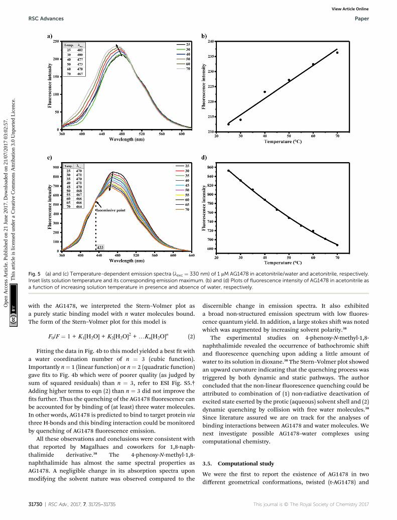
Fig. 5 (a) and (c) Temperature-dependent emission spectra (lexc ¼ 330 nm) of 1 mM AG1478 in acetonitrile/water and acetonitrile, respectively.Inset lists solution temperature and its corresponding emission maximum. (b) and (d) Plots of fluorescence intensity of AG1478 in acetonitrile asa function of increasing solution temperature in presence and absence of water, respectively.
RSC Advances Paper
Ope
n A
cces
s A
rtic
le. P
ublis
hed
on 2
1 Ju
ne 2
017.
Dow
nloa
ded
on 2
1/07
/201
7 03
:02:
57.
Thi
s ar
ticle
is li
cens
ed u
nder
a C
reat
ive
Com
mon
s A
ttrib
utio
n 3.
0 U
npor
ted
Lic
ence
.View Article Online
with the AG1478, we interpreted the Stern–Volmer plot asa purely static binding model with n water molecules bound.The form of the Stern–Volmer plot for this model is
F0/F ¼ 1 + K1[H2O] + K2[H2O]2 + .Kn[H2O]n (2)
Fitting the data in Fig. 4b to this model yielded a best t witha water coordination number of n ¼ 3 (cubic function).Importantly n¼ 1 (linear function) or n¼ 2 (quadratic function)gave ts to Fig. 4b which were of poorer quality (as judged bysum of squared residuals) than n ¼ 3, refer to ESI Fig. S5.†Adding higher terms to eqn (2) than n ¼ 3 did not improve thets further. Thus the quenching of the AG1478 uorescence canbe accounted for by binding of (at least) three water molecules.In other words, AG1478 is predicted to bind to target protein viathree H-bonds and this binding interaction could be monitoredby quenching of AG1478 uorescence emission.
All these observations and conclusions were consistent withthat reported by Magalhaes and coworkers for 1,8-naph-thalimide derivative.38 The 4-phenoxy-N-methyl-1,8-naphthalimide has almost the same spectral properties asAG1478. A negligible change in its absorption spectra uponmodifying the solvent nature was observed compared to the
31730 | RSC Adv., 2017, 7, 31725–31735
discernible change in emission spectra. It also exhibiteda broad non-structured emission spectrum with low uores-cence quantum yield. In addition, a large stokes shi was notedwhich was augmented by increasing solvent polarity.38
The experimental studies on 4-phenoxy-N-methyl-1,8-naphthalimide revealed the occurrence of bathochromic shiand uorescence quenching upon adding a little amount ofwater to its solution in dioxane.38 The Stern–Volmer plot showedan upward curvature indicating that the quenching process wastriggered by both dynamic and static pathways. The authorconcluded that the non-linear uorescence quenching could beattributed to combination of (1) non-radiative deactivation ofexcited state exerted by the protic (aqueous) solvent shell and (2)dynamic quenching by collision with free water molecules.38
Since literature assured we are on track for the analyses ofbinding interactions between AG1478 and water molecules. Wenext investigate possible AG1478–water complexes usingcomputational chemistry.
3.5. Computational study
We were the rst to report the existence of AG1478 in twodifferent geometrical conformations, twisted (t-AG1478) and
This journal is © The Royal Society of Chemistry 2017
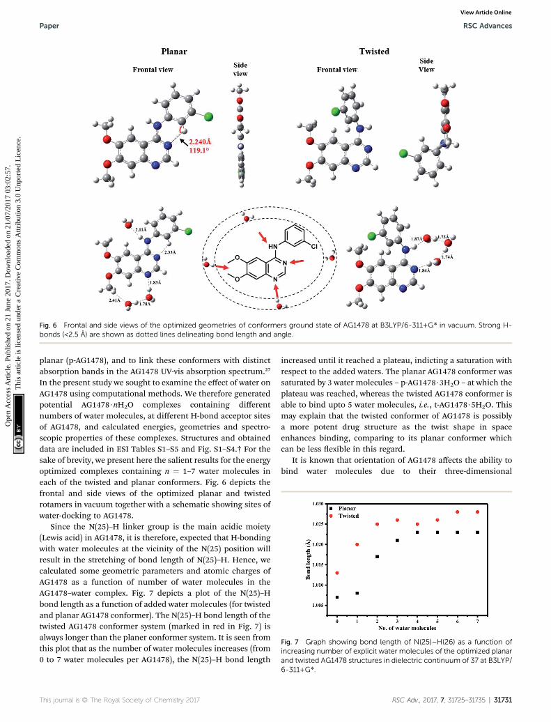
Fig. 6 Frontal and side views of the optimized geometries of conformers ground state of AG1478 at B3LYP/6-311+G* in vacuum. Strong H-bonds (<2.5 A) are shown as dotted lines delineating bond length and angle.
Fig. 7 Graph showing bond length of N(25)–H(26) as a function ofincreasing number of explicit water molecules of the optimized planarand twisted AG1478 structures in dielectric continuum of 37 at B3LYP/
Paper RSC Advances
Ope
n A
cces
s A
rtic
le. P
ublis
hed
on 2
1 Ju
ne 2
017.
Dow
nloa
ded
on 2
1/07
/201
7 03
:02:
57.
Thi
s ar
ticle
is li
cens
ed u
nder
a C
reat
ive
Com
mon
s A
ttrib
utio
n 3.
0 U
npor
ted
Lic
ence
.View Article Online
planar (p-AG1478), and to link these conformers with distinctabsorption bands in the AG1478 UV-vis absorption spectrum.27
In the present study we sought to examine the effect of water onAG1478 using computational methods. We therefore generatedpotential AG1478$nH2O complexes containing differentnumbers of water molecules, at different H-bond acceptor sitesof AG1478, and calculated energies, geometries and spectro-scopic properties of these complexes. Structures and obtaineddata are included in ESI Tables S1–S5 and Fig. S1–S4.† For thesake of brevity, we present here the salient results for the energyoptimized complexes containing n ¼ 1–7 water molecules ineach of the twisted and planar conformers. Fig. 6 depicts thefrontal and side views of the optimized planar and twistedrotamers in vacuum together with a schematic showing sites ofwater-docking to AG1478.
Since the N(25)–H linker group is the main acidic moiety(Lewis acid) in AG1478, it is therefore, expected that H-bondingwith water molecules at the vicinity of the N(25) position willresult in the stretching of bond length of N(25)–H. Hence, wecalculated some geometric parameters and atomic charges ofAG1478 as a function of number of water molecules in theAG1478–water complex. Fig. 7 depicts a plot of the N(25)–Hbond length as a function of added water molecules (for twistedand planar AG1478 conformer). The N(25)–H bond length of thetwisted AG1478 conformer system (marked in red in Fig. 7) isalways longer than the planer conformer system. It is seen fromthis plot that as the number of water molecules increases (from0 to 7 water molecules per AG1478), the N(25)–H bond length
This journal is © The Royal Society of Chemistry 2017
increased until it reached a plateau, indicting a saturation withrespect to the added waters. The planar AG1478 conformer wassaturated by 3 water molecules – p-AG1478$3H2O – at which theplateau was reached, whereas the twisted AG1478 conformer isable to bind upto 5 water molecules, i.e., t-AG1478$5H2O. Thismay explain that the twisted conformer of AG1478 is possiblya more potent drug structure as the twist shape in spaceenhances binding, comparing to its planar conformer whichcan be less exible in this regard.
It is known that orientation of AG1478 affects the ability tobind water molecules due to their three-dimensional
6-311+G*.
RSC Adv., 2017, 7, 31725–31735 | 31731
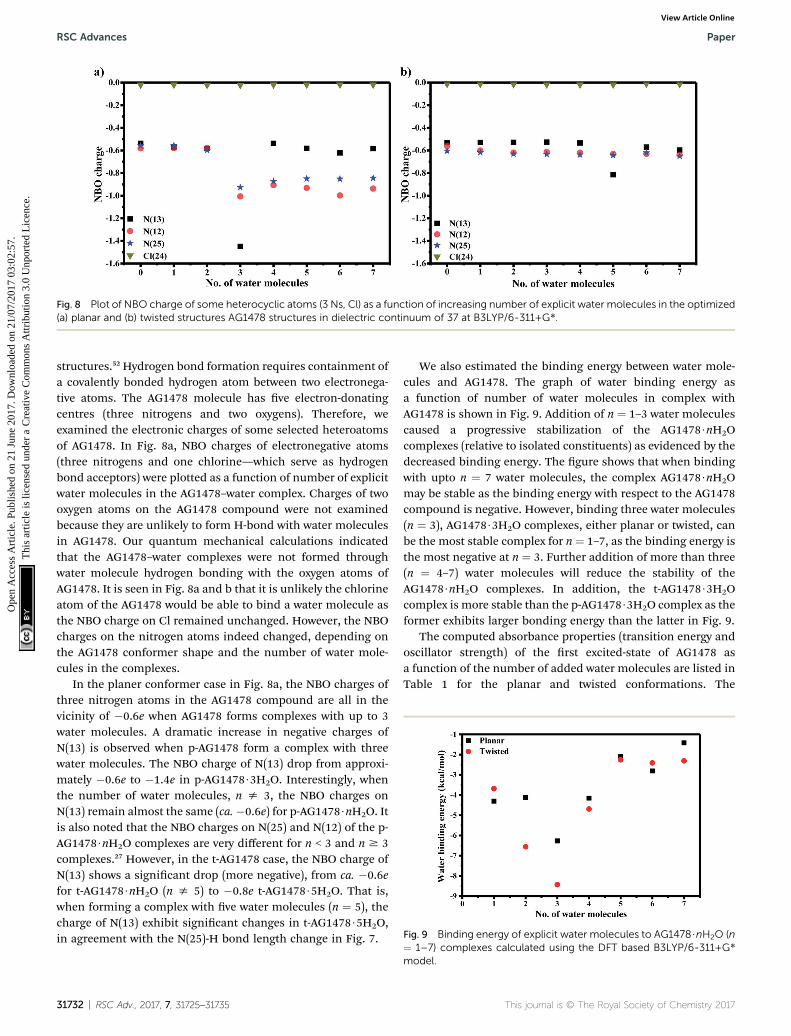
Fig. 8 Plot of NBO charge of some heterocyclic atoms (3 Ns, Cl) as a function of increasing number of explicit water molecules in the optimized(a) planar and (b) twisted structures AG1478 structures in dielectric continuum of 37 at B3LYP/6-311+G*.
Fig. 9 Binding energy of explicit water molecules to AG1478$nH2O (n
RSC Advances Paper
Ope
n A
cces
s A
rtic
le. P
ublis
hed
on 2
1 Ju
ne 2
017.
Dow
nloa
ded
on 2
1/07
/201
7 03
:02:
57.
Thi
s ar
ticle
is li
cens
ed u
nder
a C
reat
ive
Com
mon
s A
ttrib
utio
n 3.
0 U
npor
ted
Lic
ence
.View Article Online
structures.52 Hydrogen bond formation requires containment ofa covalently bonded hydrogen atom between two electronega-tive atoms. The AG1478 molecule has ve electron-donatingcentres (three nitrogens and two oxygens). Therefore, weexamined the electronic charges of some selected heteroatomsof AG1478. In Fig. 8a, NBO charges of electronegative atoms(three nitrogens and one chlorine—which serve as hydrogenbond acceptors) were plotted as a function of number of explicitwater molecules in the AG1478–water complex. Charges of twooxygen atoms on the AG1478 compound were not examinedbecause they are unlikely to form H-bond with water moleculesin AG1478. Our quantum mechanical calculations indicatedthat the AG1478–water complexes were not formed throughwater molecule hydrogen bonding with the oxygen atoms ofAG1478. It is seen in Fig. 8a and b that it is unlikely the chlorineatom of the AG1478 would be able to bind a water molecule asthe NBO charge on Cl remained unchanged. However, the NBOcharges on the nitrogen atoms indeed changed, depending onthe AG1478 conformer shape and the number of water mole-cules in the complexes.
In the planer conformer case in Fig. 8a, the NBO charges ofthree nitrogen atoms in the AG1478 compound are all in thevicinity of �0.6e when AG1478 forms complexes with up to 3water molecules. A dramatic increase in negative charges ofN(13) is observed when p-AG1478 form a complex with threewater molecules. The NBO charge of N(13) drop from approxi-mately �0.6e to �1.4e in p-AG1478$3H2O. Interestingly, whenthe number of water molecules, n s 3, the NBO charges onN(13) remain almost the same (ca.�0.6e) for p-AG1478$nH2O. Itis also noted that the NBO charges on N(25) and N(12) of the p-AG1478$nH2O complexes are very different for n < 3 and n $ 3complexes.27 However, in the t-AG1478 case, the NBO charge ofN(13) shows a signicant drop (more negative), from ca. �0.6efor t-AG1478$nH2O (n s 5) to �0.8e t-AG1478$5H2O. That is,when forming a complex with ve water molecules (n ¼ 5), thecharge of N(13) exhibit signicant changes in t-AG1478$5H2O,in agreement with the N(25)-H bond length change in Fig. 7.
31732 | RSC Adv., 2017, 7, 31725–31735
We also estimated the binding energy between water mole-cules and AG1478. The graph of water binding energy asa function of number of water molecules in complex withAG1478 is shown in Fig. 9. Addition of n ¼ 1–3 water moleculescaused a progressive stabilization of the AG1478$nH2Ocomplexes (relative to isolated constituents) as evidenced by thedecreased binding energy. The gure shows that when bindingwith upto n ¼ 7 water molecules, the complex AG1478$nH2Omay be stable as the binding energy with respect to the AG1478compound is negative. However, binding three water molecules(n ¼ 3), AG1478$3H2O complexes, either planar or twisted, canbe the most stable complex for n ¼ 1–7, as the binding energy isthe most negative at n ¼ 3. Further addition of more than three(n ¼ 4–7) water molecules will reduce the stability of theAG1478$nH2O complexes. In addition, the t-AG1478$3H2Ocomplex is more stable than the p-AG1478$3H2O complex as theformer exhibits larger bonding energy than the latter in Fig. 9.
The computed absorbance properties (transition energy andoscillator strength) of the rst excited-state of AG1478 asa function of the number of added water molecules are listed inTable 1 for the planar and twisted conformations. The
¼ 1–7) complexes calculated using the DFT based B3LYP/6-311+G*model.
This journal is © The Royal Society of Chemistry 2017
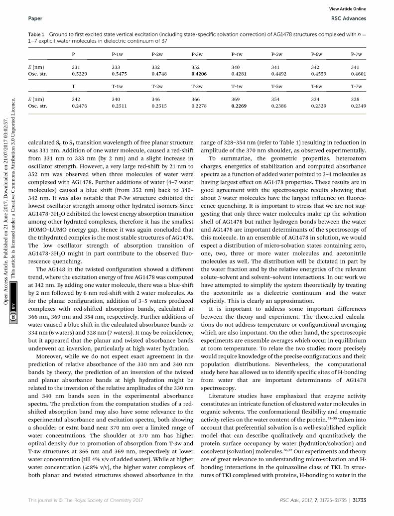
Table 1 Ground to first excited state vertical excitation (including state-specific solvation correction) of AG1478 structures complexed with n ¼1–7 explicit water molecules in dielectric continuum of 37
P P-1w P-2w P-3w P-4w P-5w P-6w P-7w
E (nm) 331 333 332 352 340 341 342 341Osc. str. 0.5229 0.5475 0.4748 0.4206 0.4281 0.4492 0.4559 0.4601
T T-1w T-2w T-3w T-4w T-5w T-6w T-7w
E (nm) 342 340 346 366 369 354 334 328Osc. str. 0.2476 0.2511 0.2515 0.2278 0.2269 0.2386 0.2329 0.2349
Paper RSC Advances
Ope
n A
cces
s A
rtic
le. P
ublis
hed
on 2
1 Ju
ne 2
017.
Dow
nloa
ded
on 2
1/07
/201
7 03
:02:
57.
Thi
s ar
ticle
is li
cens
ed u
nder
a C
reat
ive
Com
mon
s A
ttrib
utio
n 3.
0 U
npor
ted
Lic
ence
.View Article Online
calculated S0 to S1 transition wavelength of free planar structurewas 331 nm. Addition of one water molecule, caused a red-shifrom 331 nm to 333 nm (by 2 nm) and a slight increase inoscillator strength. However, a very large red-shi by 21 nm to352 nm was observed when three molecules of water werecomplexed with AG1478. Further additions of water (4–7 watermolecules) caused a blue shi (from 352 nm) back to 340–342 nm. It was also notable that P-3w structure exhibited thelowest oscillator strength among other hydrated isomers SinceAG1478$3H2O exhibited the lowest energy absorption transitionamong other hydrated complexes, therefore it has the smallestHOMO–LUMO energy gap. Hence it was again concluded thatthe trihydrated complex is the most stable structures of AG1478.The low oscillator strength of absorption transition ofAG1478$3H2O might in part contribute to the observed uo-rescence quenching.
The AG148 in the twisted conguration showed a differenttrend, where the excitation energy of free AG1478 was computedat 342 nm. By adding one water molecule, there was a blue-shiby 2 nm followed by 6 nm red-shi with 2 water molecules. Asfor the planar conguration, addition of 3–5 waters producedcomplexes with red-shied absorption bands, calculated at366 nm, 369 nm and 354 nm, respectively. Further additions ofwater caused a blue shi in the calculated absorbance bands to334 nm (6 waters) and 328 nm (7 waters). It may be coincidence,but it appeared that the planar and twisted absorbance bandsunderwent an inversion, particularly at high water hydration.
Moreover, while we do not expect exact agreement in theprediction of relative absorbance of the 330 nm and 340 nmbands by theory, the prediction of an inversion of the twistedand planar absorbance bands at high hydration might berelated to the inversion of the relative amplitudes of the 330 nmand 340 nm bands seen in the experimental absorbancespectra. The prediction from the computation studies of a red-shied absorption band may also have some relevance to theexperimental absorbance and excitation spectra, both showinga shoulder or extra band near 370 nm over a limited range ofwater concentrations. The shoulder at 370 nm has higheroptical density due to promotion of absorption from T-3w andT-4w structures at 366 nm and 369 nm, respectively at lowerwater concentration (till 4% v/v of added water). While at higherwater concentration ($8% v/v), the higher water complexes ofboth planar and twisted structures showed absorbance in the
This journal is © The Royal Society of Chemistry 2017
range of 328–354 nm (refer to Table 1) resulting in reduction inamplitude of the 370 nm shoulder, as observed experimentally.
To summarize, the geometric properties, heteroatomcharges, energetics of stabilization and computed absorbancespectra as a function of added water pointed to 3–4molecules ashaving largest effect on AG1478 properties. These results are ingood agreement with the spectroscopic results showing thatabout 3 water molecules have the largest inuence on uores-cence quenching. It is important to stress that we are not sug-gesting that only three water molecules make up the solvationshell of AG1478 but rather hydrogen bonds between the waterand AG1478 are important determinants of the spectroscopy ofthis molecule. In an ensemble of AG1478 in solution, we wouldexpect a distribution of micro-solvation states containing zero,one, two, three or more water molecules and acetonitrilemolecules as well. The distribution will be dictated in part bythe water fraction and by the relative energetics of the relevantsolute–solvent and solvent–solvent interactions. In our work wehave attempted to simplify the system theoretically by treatingthe acetonitrile as a dielectric continuum and the waterexplicitly. This is clearly an approximation.
It is important to address some important differencesbetween the theory and experiment. The theoretical calcula-tions do not address temperature or congurational averagingwhich are also important. On the other hand, the spectroscopicexperiments are ensemble averages which occur in equilibriumat room temperature. To relate the two studies more preciselywould require knowledge of the precise congurations and theirpopulation distributions. Nevertheless, the computationalstudy here has allowed us to identify specic sites of H-bondingfrom water that are important determinants of AG1478spectroscopy.
Literature studies have emphasized that enzyme activityconstitutes an intricate function of clustered water molecules inorganic solvents. The conformational exibility and enzymaticactivity relies on the water content of the protein.53–55 Taken intoaccount that preferential solvation is a well-established explicitmodel that can describe qualitatively and quantitatively theprotein surface occupancy by water (hydration/solvation) andcosolvent (solvation) molecules.56,57 Our experiments and theoryare of great relevance to understanding micro-solvation and H-bonding interactions in the quinazoline class of TKI. In struc-tures of TKI complexed with proteins, H-bonding to water in the
RSC Adv., 2017, 7, 31725–31735 | 31733
RSC Advances Paper
Ope
n A
cces
s A
rtic
le. P
ublis
hed
on 2
1 Ju
ne 2
017.
Dow
nloa
ded
on 2
1/07
/201
7 03
:02:
57.
Thi
s ar
ticle
is li
cens
ed u
nder
a C
reat
ive
Com
mon
s A
ttrib
utio
n 3.
0 U
npor
ted
Lic
ence
.View Article Online
protein binding site and to amino acids inside the protein aretypically observed.58,59 Given the high sensitivity of AG1478uorescence to hydration (as observed here from the acetoni-trile–water mixtures), we would predict that uorescence will bea useful probe of water–TKI interactions in protein bindingsites. More detailed study of micro-solvation of AG1478 can berevealed using molecular dynamics (MD) which is out of thescope of this study.
4. Conclusion
Spectroscopic analysis of AG1478 in acetonitrile–water binarymixtures was a successful strategy to quantify sites available forH-bonding of AG1478. Analysis of uorescence quenchingusing a binding model disclosed three potential sites forforming H-bonds between AG1478 and water molecules. Thetheoretical study was consistent with experimental results andrevealed that AG1478 might exist in equilibrium of planar andtwisted structures bound to varying number (1–7) of watermolecules. All of the AG1478 : water complexes exhibitedcalculated excitation energies within the experimentallyobserved values. The hydrated twisted structures were predictedto be more energetically favoured than hydrated planar coun-terparts, in contrast to dehydrated AG1478. In this regard, wepredict that the extend of hydration of the drug binding site inthe protein may be an important determinant of whether planaror twisted drug conformation is populated in the drug–proteincomplex. The studies conducted here show that AG1478 can beused as a micro-solvation probe and lay the ground work forfuture studies of AG1478 in more complex environments suchas proteins.
Acknowledgements
M. Khattab acknowledges Swinburne University PostgraduateResearch Award (SUPRA). FW acknowledges supercomputersupport from Swinburne University of Technology.
References
1 M. Jabbari, N. Khosravi, M. Feizabadi and D. Ajloo, RSC Adv.,2017, 7, 14776–14789.
2 V. T. B. Pham, H. M. Hoang, G. Grampp and D. R. Kattnig, J.Phys. Chem. B, 2017, 121, 2677–2683.
3 M. Daneri and C. J. Abelt, J. Photochem. Photobiol., A, 2015,310, 106–112.
4 A. Duereh, Y. Sato, R. L. Smith and H. Inomata, J. Phys. Chem.B, 2015, 119, 14738–14749.
5 S. Sanli, J. Solution Chem., 2013, 42, 967–978.6 D. C. da Silva, I. Ricken, M. A. D. Silva and V. G. Machado, J.Phys. Org. Chem., 2002, 15, 420–427.
7 N. P. Wells, M. J. McGrath, J. I. Siepmann, D. F. Underwoodand D. A. Blank, J. Phys. Chem. A, 2008, 112, 2511–2514.
8 K. Bhaskaran-Nair, M. Valiev, S. H. M. Deng, W. A. Shelton,K. Kowalski and X. B. Wang, J. Chem. Phys., 2015, 143,224301.
31734 | RSC Adv., 2017, 7, 31725–31735
9 D. Ghosh, O. Isayev, L. V. Slipchenko and A. I. Krylov, J. Phys.Chem. A, 2011, 115, 6028–6038.
10 J. W. Ho, H. C. Yen, H. Q. Shi, L. H. Cheng, C. N. Weng,W. K. Chou, C. C. Chiu and P. Y. Cheng, Angew. Chem., Int.Ed., 2015, 54, 14772–14776.
11 V. B. Pacheco and P. Chaudhuri, J. Phys. Chem. A, 2013, 117,5675–5684.
12 D. S. Venables and C. A. Schmuttenmaer, J. Chem. Phys.,2000, 113, 11222–11236.
13 J. Catalan, C. Diaz and F. Garcia-Blanco, Org. Biomol. Chem.,2003, 1, 575–580.
14 P. Petong, R. Pottel and U. Kaatze, J. Phys. Chem. A, 2000, 104,7420–7428.
15 D. B. Wong, K. P. Sokolowsky, M. I. El-Barghouthi,E. E. Fenn, C. H. Giammanco, A. L. Sturlaugson andM. D. Fayer, J. Phys. Chem. B, 2012, 116, 5479–5490.
16 H. Tanaka, J. Walsh and K. E. Gubbins, Mol. Phys., 1992, 76,1221–1233.
17 G. Matisz, A. M. Kelterer, W. M. F. Fabian and S. Kunsagi-Mate, Phys. Chem. Chem. Phys., 2015, 17, 8467–8479.
18 S. Lobsiger, S. Blaser, R. K. Sinha, H. M. Frey andS. Leutwyler, Nat. Chem., 2014, 6, 989–993.
19 M. Barbatti and H. Lischka, Phys. Chem. Chem. Phys., 2015,17, 15452–15459.
20 A. T. Garcia-Sosa, J. Chem. Inf. Model., 2013, 53, 1388–1405.21 A. Gazit, J. Chen, H. App, G. McMahon, P. Hirth, I. Chen and
A. Levitzki, Bioorg. Med. Chem., 1996, 4, 1203–1207.22 Z. Shi, A. K. Tiwari, S. Shukla, R. W. Robey, I. W. Kim,
S. Parmar, S. E. Bates, Q. S. Si, C. S. Goldblatt, I. Abraham,L. W. Fu, S. V. Ambudkar and Z. S. Chen, Biochem.Pharmacol., 2009, 77, 781–793.
23 L. Caja, P. Sancho, E. Bertran, C. Ortiz, J. S. Campbell,N. Fausto and I. Fabregat, Biochem. Pharmacol., 2011, 82,1583–1592.
24 F. A. Al-Obeidi and K. S. Lam, Oncogene, 2000, 19, 5690–5701.25 Y. Liu and N. S. Gray, Nat. Chem. Biol., 2006, 2, 358–364.26 M. Khattab, F. Wang and A. H. Clayton, Spectrochim. Acta,
Part A, 2016, 164, 128–132.27 M. Khattab, S. Chatterjee, A. H. A. Clayton and F. Wang, New
J. Chem., 2016, 40, 8296–8304.28 D. Banerjee and S. K. Pal, J. Phys. Chem. A, 2008, 112, 7314–
7320.29 F. Gao, X. J. Ye, H. R. Li, X. L. Zhong and Q. Wang,
ChemPhysChem, 2012, 13, 1313–1324.30 M. K. Nayak, J. Photochem. Photobiol., A, 2012, 241, 26–37.31 K. Suda, M. Terazima and Y. Kimura, Chem. Phys. Lett., 2012,
531, 70–74.32 T. S. Choi, J. W. Lee, K. S. Jin and H. I. Kim, Biophys. J., 2014,
107, 1939–1949.33 M. T. Sonoda, N. H. Moreira, L. Martinez, F. W. Favero,
S. M. Vechi, L. R. Martins and M. S. Skaf, Braz. J. Phys.,2004, 34, 3–16.
34 A. D. Becke, J. Chem. Phys., 1993, 98, 1372–1377.35 A. D. Becke, J. Chem. Phys., 1993, 98, 5648–5652.36 M. Cossi, N. Rega, G. Scalmani and V. Barone, J. Comput.
Chem., 2003, 24, 669–681.
This journal is © The Royal Society of Chemistry 2017
Paper RSC Advances
Ope
n A
cces
s A
rtic
le. P
ublis
hed
on 2
1 Ju
ne 2
017.
Dow
nloa
ded
on 2
1/07
/201
7 03
:02:
57.
Thi
s ar
ticle
is li
cens
ed u
nder
a C
reat
ive
Com
mon
s A
ttrib
utio
n 3.
0 U
npor
ted
Lic
ence
.View Article Online
37 G. W. T. M. J. Frisch, H. B. Schlegel, G. E. Scuseria,M. A. Robb, J. R. Cheeseman, G. Scalmani, V. Barone,B. Mennucci, G. A. Petersson, H. Nakatsuji, M. Caricato,X. Li, H. P. Hratchian, A. F. Izmaylov, J. Bloino, G. Zheng,J. L. Sonnenberg, M. Hada, M. Ehara, K. Toyota,R. Fukuda, J. Hasegawa, M. Ishida, T. Nakajima, Y. Honda,O. Kitao, H. Nakai, T. Vreven, J. A. Montgomery Jr,J. E. Peralta, F. Ogliaro, M. Bearpark, J. J. Heyd,E. Brothers, K. N. Kudin, V. N. Staroverov, R. Kobayashi,J. Normand, K. Raghavachari, A. Rendell, J. C. Burant,S. S. Iyengar, J. Tomasi, M. Cossi, N. Rega, J. M. Millam,M. Klene, J. E. Knox, J. B. Cross, V. Bakken, C. Adamo,J. Jaramillo, R. Gomperts, R. E. Stratmann, O. Yazyev,A. J. Austin, R. Cammi, C. Pomelli, J. W. Ochterski,R. L. Martin, K. Morokuma, V. G. Zakrzewski, G. A. Voth,P. Salvador, J. J. Dannenberg, S. Dapprich, A. D. Daniels,O. Farkas, J. B. Foresman, J. V. Ortiz, J. Cioslowski, andD. J. Fox, Gaussian 09, Revision C.01, Gaussian, Inc.,Wallingford CT, 2009.
38 J. L. Magalhaes, R. V. Pereira, E. R. Triboni, P. Berci,M. H. Gehlen and F. C. Nart, J. Photochem. Photobiol., A,2006, 183, 165–170.
39 S. D. Choudhury and H. Pal, J. Phys. Chem. B, 2016, 120,11970–11977.
40 V. A. Sirotkin and A. A. Kuchierskaya, J. Phys. Chem. B, 2017,121, 4422–4430.
41 J. Barbosa, D. Barron, R. Berges, V. SanzNebot and I. Toro, J.Chem. Soc., Faraday Trans., 1997, 93, 1915–1920.
42 A. Wakisaka, S. Takahashi and N. Nishi, J. Chem. Soc.,Faraday Trans., 1995, 91, 4063–4069.
43 J. Barbosa and V. Sanznebot, J. Chem. Soc., Faraday Trans.,1994, 90, 3287–3292.
44 H. Boaz and G. K. Rollefson, J. Am. Chem. Soc., 1950, 72,3435–3443.
This journal is © The Royal Society of Chemistry 2017
45 B. K. Paul, N. Ghosh and S. Mukherjee, Langmuir, 2014, 30,5921–5929.
46 B. K. Paul, D. Ray and N. Guchhait, Phys. Chem. Chem. Phys.,2013, 15, 1275–1287.
47 I. Y. S. Lee and H. Suzuki, J. Photochem. Photobiol., A, 2008,195, 254–260.
48 J. R. Lakowicz, Principles of uorescence spectroscopy,Springer Science & Business Media, 2013.
49 N. Ghosh, R. Mondal and S. Mukherjee, Langmuir, 2015, 31,8074–8080.
50 F. F. Tian, F. L. Jiang, X. L. Han, C. Xiang, Y. S. Ge, J. H. Li,Y. Zhang, R. Li, X. L. Ding and Y. Liu, J. Phys. Chem. B, 2010,114, 14842–14853.
51 J. Q. Tong, F. F. Tian, Q. Li, L. L. Li, C. Xiang, Y. Liu, J. Daiand F. L. Jiang, Photochem. Photobiol. Sci., 2012, 11, 1868–1879.
52 A. Ganesan, J. Dreyer, F. Wang, J. Akola and J. Larrucea, J.Mol. Graphics Modell., 2013, 45, 180–191.
53 T. Kijima, S. Yamamoto and H. Kise, Enzyme Microb.Technol., 1996, 18, 2–6.
54 K. Griebenow and A. M. Klibanov, J. Am. Chem. Soc., 1996,118, 11695–11700.
55 Y. L. Khmelnitsky, V. V. Mozhaev, A. B. Belova,M. V. Sergeeva and K. Martinek, Eur. J. Biochem., 1991, 198,31–41.
56 S. N. Timasheff, Proc. Natl. Acad. Sci. U. S. A., 2002, 99, 9721–9726.
57 T. Arakawa, Y. Kita and S. N. Timasheff, Biophys. Chem.,2007, 131, 62–70.
58 N. M. Levinson and S. G. Boxer, Nat. Chem. Biol., 2014, 10,127–132.
59 E. De Moliner, N. R. Brown and L. N. Johnson, Eur. J.Biochem., 2003, 270, 3174–3181.
RSC Adv., 2017, 7, 31725–31735 | 31735