The Metalloprotease ADAMTS8 Displays Antitumor Properties through Antagonizing...
12
Chromatin, Gene, and RNA Regulation The Metalloprotease ADAMTS8 Displays Antitumor Properties through Antagonizing EGFR–MEK–ERK Signaling and Is Silenced in Carcinomas by CpG Methylation Gigi C.G. Choi 1 , Jisheng Li 1,2 , Yajun Wang 1 , Lili Li 1 , Lan Zhong 1 , Brigette Ma 1 , Xianwei Su 1 , Jianming Ying 1,3 , Tingxiu Xiang 4 , Sun Young Rha 5 , Jun Yu 6 , Joseph J.Y. Sung 6 , Sai Wah Tsao 7 , Anthony T.C. Chan 1 , and Qian Tao 1 Abstract A disintegrins and metalloproteinases with thrombospondin motifs (ADAMTS) family members have been reported dysregulated in various cancers. Through refining a loss of heterozygosity locus at 11q25 by array-CGH, we identified ADAMTS8 as a novel candidate tumor suppressor gene. Although ADAMTS8 downregulation has been reported in several tumors, its biologic function and underlying mechanism remain largely unknown. Here, we found that ADAMTS8 is broadly expressed in normal tissues but frequently downregulated or silenced by promoter methylation in common carcinoma cell lines, including nasopharyngeal, esophageal squamous cell, gastric, and colorectal carcino- mas. Pharmacologic or genetic demethylation restored ADAMTS8 expression, indicating that promoter methylation mediates its silencing. Aberrant methylation of ADAMTS8 was also detected in several types of primary tumors but rarely in normal tissues. Further functional studies showed that restoring ADAMTS8 expression suppressed tumor cell clonogenicity through inducing apoptosis. ADAMTS8 as a secreted protease inhibited epidermal growth factor receptor (EGFR) signaling along with decreased levels of phosphorylated MEK and ERK. We further found that ADAMTS8 disrupted actin stress fiber organization and inhibited tumor cell motility. Thus, our data demonstrate that ADAMTS8 metalloprotease acts as a functional tumor suppressor through antagonizing EGFR–MEK–ERK signaling, in addition to its previously reported anti-angiogenesis function, and is frequently methylated in common tumors. Implications: This study uncovers the tumor suppressive function of ADAMTS8, one of the ADAMTS family members, and its frequent methylation in certain tumors could be developed as a potential biomarker. Mol Cancer Res; 12(2); 228–38. Ó2013 AACR. Introduction Metalloproteinases are initially regarded to facilitate tumor progression by promoting angiogenesis and metastatic dis- semination of cancer cells through extracellular matrix (ECM) degradation (1). However, this concept is challenged by recent findings of several members showing to possess tumor suppressive functions (2–6). A disintegrins and metal- loproteinases with thrombospondin motifs (ADAMTS), a family of extracellular metalloproteases, is structurally and functionally similar to matrix metalloproteinases (MMP) and ADAMs (7). Unlike ADAMs mainly as trans-membrane proteins, ADAMTSs are secreted proteinases binding to ECM (8). Dysregulated expression of ADAMTSs, such as ADAMTS1 (2), ADAMTS8, ADAMTS9 (5), ADAMTS12 (3), ADAMTS15 (4), ADAMTS18 (6), and ADAMTS20 (9, 10), have been detected in diverse types of malignancies including lung, brain, breast, gastric, prostate, pancreatic cancers, and glioblastoma (8). Various ADAMTSs have been shown to regulate cell proliferation, adhesion, migration, angiogenesis, and intracellular signaling (8, 11), thus involved in multiple tumor pathogenesis (1, 6). ADAMTS8 is 1 of the 3 ADAMTS members with anti- angiogenic property, indicating its potential as a tumor suppressor (5, 12, 13). Downregulation of ADAMTS8 has been found in some tumors, such as brain tumors (14), breast carcinoma (9), non–small cell lung carcinoma (15), head and neck squamous cell carcinoma (16), and pancreatic Authors' Affiliations: 1 Cancer Epigenetics Laboratory, Department of Clinical Oncology, State Key Laboratory of Oncology in South China, Sir YK Pao Center for Cancer and Li Ka Shing Institute of Health Sciences, The Chinese of University of Hong Kong, Hong Kong; 2 Department of Chemo- therapy, Cancer Center, Qilu Hospital, Shandong University, Jinan, Shan- dong, China; 3 Department of Pathology, Cancer Hospital, Peking Union Medical College & Chinese Academy of Medical Sciences, Beijing, China; 4 The First Affiliated Hospital of Chongqing Medical University, Chongqing, China; 5 Department of Internal Medicine, Yonsei University College of Medicine, Korea; 6 State Key Laboratory of Digestive Diseases, Department of Medicine and Therapeutics, The Chinese University of Hong Kong; 7 Departments of Anatomy, University of Hong Kong, Hong Kong Note: Supplementary data for this article are available at Molecular Cancer Research Online (http://mcr.aacrjournals.org/). G.C.G. Choi, J. Li, and Y. Wang contributed equally to this work. Corresponding Author: Qian Tao, Room 315, Cancer Center, PWH, The Chinese University of Hong Kong, Shatin, Hong Kong. Phone: 852-2632- 1340; Fax: 852-2648-8842; E-mail: [email protected] doi: 10.1158/1541-7786.MCR-13-0195 Ó2013 American Association for Cancer Research. Molecular Cancer Research Mol Cancer Res; 12(2) February 2014 228 on June 8, 2018. © 2014 American Association for Cancer Research. mcr.aacrjournals.org Downloaded from Published OnlineFirst November 1, 2013; DOI: 10.1158/1541-7786.MCR-13-0195
Transcript of The Metalloprotease ADAMTS8 Displays Antitumor Properties through Antagonizing...

Chromatin, Gene, and RNA Regulation
The Metalloprotease ADAMTS8 Displays AntitumorProperties through Antagonizing EGFR–MEK–ERK Signalingand Is Silenced in Carcinomas by CpG Methylation
Gigi C.G. Choi1, Jisheng Li1,2, Yajun Wang1, Lili Li1, Lan Zhong1, Brigette Ma1, Xianwei Su1,Jianming Ying1,3, Tingxiu Xiang4, Sun Young Rha5, Jun Yu6, Joseph J.Y. Sung6, Sai Wah Tsao7,Anthony T.C. Chan1, and Qian Tao1
AbstractAdisintegrins andmetalloproteinases with thrombospondinmotifs (ADAMTS) familymembers have been reported
dysregulated in various cancers. Through refining a loss of heterozygosity locus at 11q25 by array-CGH, we identifiedADAMTS8 as a novel candidate tumor suppressor gene. Although ADAMTS8 downregulation has been reported inseveral tumors, its biologic function and underlying mechanism remain largely unknown. Here, we found thatADAMTS8 is broadly expressed in normal tissues but frequently downregulated or silenced by promoter methylationin common carcinoma cell lines, including nasopharyngeal, esophageal squamous cell, gastric, and colorectal carcino-mas. Pharmacologic or genetic demethylation restored ADAMTS8 expression, indicating that promoter methylationmediates its silencing. Aberrant methylation of ADAMTS8 was also detected in several types of primary tumors butrarely in normal tissues. Further functional studies showed that restoring ADAMTS8 expression suppressed tumor cellclonogenicity through inducing apoptosis. ADAMTS8 as a secreted protease inhibited epidermal growth factor receptor(EGFR) signaling along with decreased levels of phosphorylated MEK and ERK. We further found that ADAMTS8disrupted actin stress fiber organization and inhibited tumor cell motility. Thus, our data demonstrate that ADAMTS8metalloprotease acts as a functional tumor suppressor through antagonizing EGFR–MEK–ERK signaling, in additionto its previously reported anti-angiogenesis function, and is frequently methylated in common tumors.
Implications: This study uncovers the tumor suppressive function of ADAMTS8, one of the ADAMTS familymembers, and its frequent methylation in certain tumors could be developed as a potential biomarker.Mol CancerRes; 12(2); 228–38. �2013 AACR.
IntroductionMetalloproteinases are initially regarded to facilitate tumor
progression by promoting angiogenesis and metastatic dis-semination of cancer cells through extracellular matrix
(ECM) degradation (1). However, this concept is challengedby recent findings of several members showing to possesstumor suppressive functions (2–6). A disintegrins andmetal-loproteinases with thrombospondin motifs (ADAMTS), afamily of extracellular metalloproteases, is structurally andfunctionally similar to matrix metalloproteinases (MMP)and ADAMs (7).Unlike ADAMsmainly as trans-membraneproteins, ADAMTSs are secreted proteinases binding toECM (8). Dysregulated expression of ADAMTSs, such asADAMTS1 (2), ADAMTS8, ADAMTS9 (5), ADAMTS12(3), ADAMTS15 (4), ADAMTS18 (6), and ADAMTS20(9, 10), have been detected in diverse types of malignanciesincluding lung, brain, breast, gastric, prostate, pancreaticcancers, and glioblastoma (8). Various ADAMTSs have beenshown to regulate cell proliferation, adhesion, migration,angiogenesis, and intracellular signaling (8, 11), thusinvolved in multiple tumor pathogenesis (1, 6).ADAMTS8 is 1 of the 3 ADAMTS members with anti-
angiogenic property, indicating its potential as a tumorsuppressor (5, 12, 13). Downregulation of ADAMTS8 hasbeen found in some tumors, such as brain tumors (14),breast carcinoma (9), non–small cell lung carcinoma (15),head and neck squamous cell carcinoma (16), and pancreatic
Authors' Affiliations: 1Cancer Epigenetics Laboratory, Department ofClinicalOncology, State Key Laboratory ofOncology inSouthChina, Sir YKPao Center for Cancer and Li Ka Shing Institute of Health Sciences, TheChinese of University of Hong Kong, Hong Kong; 2Department of Chemo-therapy, Cancer Center, Qilu Hospital, Shandong University, Jinan, Shan-dong, China; 3Department of Pathology, Cancer Hospital, Peking UnionMedical College & Chinese Academy of Medical Sciences, Beijing, China;4The First Affiliated Hospital of Chongqing Medical University, Chongqing,China; 5Department of Internal Medicine, Yonsei University College ofMedicine, Korea; 6State Key Laboratory of DigestiveDiseases, Departmentof Medicine and Therapeutics, The Chinese University of Hong Kong;7Departments of Anatomy, University of Hong Kong, Hong Kong
Note: Supplementary data for this article are available at Molecular CancerResearch Online (http://mcr.aacrjournals.org/).
G.C.G. Choi, J. Li, and Y. Wang contributed equally to this work.
Corresponding Author: Qian Tao, Room 315, Cancer Center, PWH, TheChinese University of Hong Kong, Shatin, Hong Kong. Phone: 852-2632-1340; Fax: 852-2648-8842; E-mail: [email protected]
doi: 10.1158/1541-7786.MCR-13-0195
�2013 American Association for Cancer Research.
MolecularCancer
Research
Mol Cancer Res; 12(2) February 2014228
on June 8, 2018. © 2014 American Association for Cancer Research. mcr.aacrjournals.org Downloaded from
Published OnlineFirst November 1, 2013; DOI: 10.1158/1541-7786.MCR-13-0195
cancer (17), whereas its expression in other common solidtumors including nasopharyngeal (NPC), esophageal squa-mous cell (ESCC), gastric, colorectal (CRC), renal (RCC),and cervical carcinomas remains unclear.Genetic and epigenetic alterations especially promoter
methylation and histone modifications play a crucial rolein tumor initiation and progression, although leading toactivation of oncogene and inactivation of tumor suppressorgene (TSG). Promoter methylation of ADAMTS8 has beendetected in brain tumors (14), non–small cell lung carcino-ma (15) and thyroid cancer (18), which also could serve asone of the signatures for primary thyroid cancer (18),suggesting that epigenetic silencing of ADAMTS8 may beinvolved in tumorigenesis. However, its tumor suppressivefunctions and underlying mechanisms in tumor pathogen-esis are largely unknown.Through refining an LOH locus at 11q25 by 1-Mb
array comparative genomic hybridization (array-CGH)and expression profiling of the affected genes, we iden-tified ADAMTS8 as a candidate TSG. In this study, weexamined the expression and methylation of ADAMTS8in common solid carcinomas and further explored itstumor suppressive functions and relevant mechanisms intumorigenesis.
Materials and MethodsCell lines, tumors, and normal tissue samplesMultiple cell lines of NPC, ESCC, gastric, and CRC,
hepatocellular (HCC), lung, breast, RCC, and cervicalcarcinomas and several immortalized normal epithelialcell lines were used (19, 20). Cell lines were obtainedeither from the American Type Culture Collection or ourcollaborators. HCT116-DKO cell line with doubleknockout of DNA methyltransferases DNMT1 andDNMT3B was also used (gifts of Bert Vogelstein, JohnsHopkins University, Baltimore, MD). Cell lines weretreated with 10 mmol/L 5-aza-20-deoxycytidine (Aza;Sigma-Aldrich) for 3 days or further treated with 100nmol/L trichostatin A (TSA; Cayman Chemical Co.) foradditional �16 hours as described previously (21).Human normal adult and fetal tissue RNA samples were
purchased commercially (Stratagene or Millipore-Chemi-con; refs. 20 and 22). Genomic DNA samples of normalnasopharyngeal and gastric tissues, as well as primary tumortissues of nasopharyngeal carcinoma, gastric cancer, andcolorectal carcinoma, have been described previously(19, 23–25). Clinical information was available for themajority of gastric cancer samples.
Semiquantitative reverse transcription-PCR andquantitative real-time RT-PCRSemiquantitative reverse transcription-PCR (RT-PCR)
and quantitative real-time PCR (qPCR) were performedas described previously (21, 26). RT-PCR was performedfor 32 cycles using Go-Taq Flexi DNA polymerase (Pro-mega). SYBR Green master mix (Applied Biosystems)was used for real-time PCR analysis. GAPDH was used as
an internal control. Primers are listed in SupplementaryTable S1.
Methylation-specific PCR and bisulfite genomicsequencingBisulfite treatment of genomic DNA, methylation-spe-
cific PCR (MSP), and bisulfite genomic sequencing (BGS)were performed as described previously (22, 27). Briefly, 1mm of bisulfite-treated DNA (around 50 ng) was used forMSP amplified by using 0.625 U of AmpliTaq Goldpolymerase (Applied Biosystems) with 2.0 mmol/LMgCl2 and 0.2 mmol/L dNTP in a 25 mL reaction volume.PCR was performed at 95�C for 10 minutes, followed by 40cycles consisting of 94�C for 30 seconds, annealing at 60�C(methylation detection) or 58�C (unmethylation detection)for 30 seconds, and 72�C for 30 seconds, and a finalextension at 72�C for 5 minutes. PCR products wereanalyzed on a 1.8% agarose gel. For BGS, bisulfite-treatedDNA was amplified for using BGS primers, and the PCRproducts were cloned into the PCR4-TOPO vector (Invi-trogen). In all, 6 to 10 colonies were randomly chosen andsequenced. MSP and BGS primers are shown in Supple-mentary Table S1.
ADAMTS8 deletion analysisHomozygous deletion of ADAMTS8 coding exon 1 and
exon 2was examined usingmultiplex genomic DNAPCR aspreviously described (20, 23). A 301-bp fragment and a 349-bp fragment of the ADAMTS8 were amplified along with a134-bp fragment of APRT as internal control. The PCR wasperformed in a 12.5 mL reaction mixture consisting of 0.4mmol/L of primers located onADAMTS8 exon 1 and exon 2,0.2 mmol/L of APRT primers, 0.2 mmol/L of dNTP, 2.0mmol/L of MgCl2, 1� PCR Buffer II, 0.3125 U of Ampli-Taq Gold (Applied Biosystems), and 50 ng of templateDNA. PCR was conducted as 95�C for 10 minutes, then 35cycles (94�C, 30 seconds; 55�C, 30 seconds; 72�C, 30seconds), followed by 72�C for 10 minutes. PCR productswere analyzed on 1.8% agarose gels. Primers are listed inSupplementary Table S1.
Construction of ADAMTS8-expressing vectorsThe full-length open reading frame of ADAMTS8 was
cloned from normal adult larynx cDNA library into pCR4-TOPO vector (Invitrogen). A Flag tag was fused to theC-terminal of ADAMTS8 by PCR amplification andthe coding sequence of ADAMTS8-Flag was subclonedinto pcDNA3.1(þ) with BamHI and XhoI to generatepcDNA3.1(þ)-ADAMTS8-Flag. ADAMTS8 was furthersubcloned into the pEGFP-N1 vector to generate a pEGFP-N1-ADAMTS8. All PCR reactions were performed withAccuPrime polymerase (Invitrogen) and all cloned frag-ments were validated by sequencing.
ImmunofluorescenceCells grown on coverslips were stained by immunofluo-
rescence as described previously (19, 20). In brief, HONE1or KYSE150 cells were transfected with pcDNA3.1
Methylation of ADAMTS8 in Carcinomas
www.aacrjournals.org Mol Cancer Res; 12(2) February 2014 229
on June 8, 2018. © 2014 American Association for Cancer Research. mcr.aacrjournals.org Downloaded from
Published OnlineFirst November 1, 2013; DOI: 10.1158/1541-7786.MCR-13-0195
(þ)-ADAMTS8-Flag or pEGFP-N1-ADAMTS8 plasmid.At 48-hour posttransfection, cells were fixed with 4% (w/v)paraformaldehyde before staining with primary and FITC-conjugated secondary antibodies (F313; Dako) for half anhour at 37�C. For actin staining, cells were stained withrhodamine-conjugated phalloidin for 1 hour at 37�C. Cellswere counterstained with 40,6-diamidino-2-phenylindole(DAPI) before analysis using Olympus BX51 microscope(OlympusCorporation) and Leica TCS SP5 confocalmicro-scope (Leica Microsystems CMS GmbH).
Colony formation assayClonogenicity was determined by measuring colonies
growing in monolayer culture as described previously(6, 21). HONE1 and KYSE150 cells (1 � 105 per well)were seeded in a 12-well plate and were transiently trans-fected with pcDNA3.1(þ)-ADAMTS8-Flag plasmid or thepcDNA3.1 vector alone, using FuGENE 6 (Roche). At 48-hours posttransfection, cells were collected and plated atappropriate density in a 6-well plate underG418 (0.4mg/mL)selection for 2 to 3 weeks. Cell colonies were fixed andstained with Gentian Violet (ICM Pharma) before countingof surviving colonies (>50 cells per colony). Statisticalanalysis was performed with Student t test, P < 0.05 wasconsidered as statistically significant difference.
Cell-proliferation assayMTS (Promega) assay was performed according to the
manufacturer's instruction. Cells were seeded in 96-wellplates at a density of 1 to 2� 103, 15 mLMTS solution wasadded into each well at indicated time points and thenincubated for 3 hours at 37�C. Effect of cell number onabsorbance at 490 nm was measured. The experiments wereperformed in triplicate and repeated 3 times.
Apoptosis assayHONE1 and KYSE150 cells were seeded on glass
coverslips and fixed in 4% paraformaldehyde. Cells werepermeabilized with 0.1% Triton X-100 in PBS for 5minutes on ice. Apoptoic cells with strand breaks in DNAwere stained using the In Situ Cell Death Detection Kit,Fluorescein (Roche) according to Manufacturer's proto-col. The labeling reaction was performed by incubatingeach sample with terminal deoxynucleotidyl transferase–mediated dUTP nick end labeling (TUNEL) reactionmixture containing terminal deoxynucleotidyl transferaseand fluorescein-labeled dUTP at 37�C for 1 hour. DAPIwas used to stain total nuclei. Coverslips were mounted onglass slides and analyzed under a fluorescence microscope.Apoptotic cells with condensed or fragmented nuclei werealso examined by DAPI staining.
Western blot analysisHONE1 and KYSE150 cells were transiently transfected
with pcDNA3.1(þ)-ADAMTS8-Flag plasmid or thepcDNA3.1 vector alone using FuGENE 6 (Roche). At48-hour posttransfection, cells were harvested and lysed inlysis buffer [10 mmol/L Tris-HCl (pH 7.4), 1% SDS, 10%
glycerol, 5 mmol/L MgCl2, 1 mmol/L phenylmethylsulfo-nyl fluoride, 1 mmol/L sodium orthovandate, 5 mg/mLleupeptin, and 21 mg/mL aprotinin]. Protein samples wereincubated for 30 minutes on ice and followed by centrifu-gation to remove cell debris. Supernatant containing 30 mgof total protein lysate from each sample was subjected toSDS-PAGE and transferred onto a polyvinylidene difluoridemembranes which were probed with anti-MEK1/2, anti-ERK1/2, anti-phospho-MEK1/2 (Ser217/Ser221), anti-phos-pho-ERK1/2 (Thr202/Tyr204; Cell Signaling Technology);anti-epidermal growth factor receptor (EGFR; BD Trans-duction Laboratories); anti-phosphor-EGFR (Tyr1086; Invi-trogen); anti-Flag (Sigma-Aldrich), or anti-tubulin (LabVision Corporation) primary antibodies. Protein bands werevisualized by enhanced chemiluminescence detection system(GE Healthcare Bio-Sciences).
Conditioned mediumConditioned medium containing secreted ADAMTS8
was collected from pcDNA3.1(þ)-ADAMTS8-Flag-trans-fected KYSE150 and HONE1 cells cultured in RPMI 1640with 3% FBS for indicated times after centrifuged at 1,000 gfor 30 minutes. KYSE150 cells were treated with condi-tioned medium containing ADAMTS8 for 24 hours andcollected for further study.
Luciferase reporter assayReporter activity of the serum response element (SRE)-
luc, AP-1 plasmids (Stratagene) was determined in HONE1and KYSE150 cells as described previously (19, 28). Sub-confluent cells in 24-well plates were transiently cotrans-fected with pSRE-luc, pcDNA3.1(þ)-ADAMTS8-Flag andpRL-SV40. After 48 hours, cells were harvested for luciferaseactivity measurement using Dual-Luciferase Reporter AssaySystem (Promega). Activity of the firefly luciferase reporter(SRE)-luc and AP-1-luc were normalized with the activity ofthe renilla luciferase pRL-SV40 as an internal control tocorrect the differences in transfection efficiency. Each exper-iment was performed in triplicates and was repeated 3 times.
Wound healing assayCell motility of HONE1 and KYSE150 cells transfected
with pEGFP-N1-ADAMTS8 or control pEGFP-N1 vectorwas assessed using a scratch wound assay. Cells were culturedin 6-well dishes until confluent before using sterile tips toscratch a wound. After rinsing with PBS, cells were incu-bated with fresh medium and images of wounds were takenunder a phase contrast microscope at 0, 24, and 48 hoursafter wounding. The experiments were performed intriplicate.
ResultsIdentification of ADAMTS8 as a downregulated gene at11q24.2-25A deletion at 11q24.2-25 was frequently detected in a
panel of NPC and ESCC tumor cell lines by 1-Mb aCGH(Fig. 1A). Expression profiling of all the 34 genes within thisdeletion was further analyzed by semiquantitative RT-PCR
Choi et al.
Mol Cancer Res; 12(2) February 2014 Molecular Cancer Research230
on June 8, 2018. © 2014 American Association for Cancer Research. mcr.aacrjournals.org Downloaded from
Published OnlineFirst November 1, 2013; DOI: 10.1158/1541-7786.MCR-13-0195

(data not shown). ADAMTS8 was found to be silenced invirtually all the NPC cell lines studied, but readily expressedin immortalized normal nasopharyngeal epithelial cell lineand normal tissues of larynx and trachea as well as othernormal adult and fetal tissues with varying expression levelsexcept for bone marrow (Figs. 1B and 2A). In addition,frequent silencing or downregulation of ADAMTS8 wasalso observed in multiple other carcinoma cell lines, includ-ing 6 of 6 NPC (100%), 12 of 16 ESCC (75%), 14 of 16gastric (88%) and 4 of 5 CRC (80%), 11 of 13HCC (85%),3 of 5 Lung (60%), 7 of 9 breast (78%), 5 of 7 RCC (71%),and 3 of 4 cervical (75%) cancers (Fig. 2A and B andSupplementary Fig. S1 and Table S2). We further assessedADAMTS8 expression in representative nasopharyngeal,esophageal, and colon carcinoma cell lines by qPCR. Asexpected, we confirmed that ADAMTS8 was frequentlydownregulated in tumor cell lines, but readily expressed innormal tissues, consistent with the semiquantitativeRT-PCR results. These data suggest that ADAMTS8 is acandidate tumor suppressor.
Promoter methylation of ADAMTS8 contributes to itssilencingTo investigate the mechanism of ADAMTS8 downregu-
lation in tumors, we first evaluated its genetic alterations.Multiplex differential genomic DNA-PCR for ADAMTS8and APRT was performed to detect ADAMTS8 deletion in aregion spanning exon 1 and exon 2. Results showed thathomozygous or hemizygous deletion was detected in severaltumor cell lines with or without silenced ADAMTS8 (Sup-
plementary Fig. S2), suggesting that genetic alterations areone of the mechanisms for ADAMTS8 silencing. Moreover,analysis of ADAMTS8 mutations using online database(Wellcome Trust Cancer Genome Project, http://www.sanger.ac.uk) revealed only one missense somatic mutationof ADAMTS8 reported in glioblastoma (29), indicating thatits genetic sequence mutation is uncommon in tumors.A typical CpG Island (CGI) spanning the exon 1 of
ADAMTS8 is predicted by CpG Island Searcher (http://cpgislands.usc.edu/; Fig. 1A). Thus, promoter methylationofADAMTS8was further examined byMSP and found to befrequently detected in multiple carcinoma cell lines, includ-ing 100% of NPC, 44% of ESCC, 56% of gastric, 78% ofCRC, 8% of HCC, 22% of breast, 43% of RCC, and 29%of cervical cancers (Fig. 2A and Supplementary Fig. S1 andTable S2), but seldom in lung cancer cell lines and not innormal epithelial cell lines (Fig. 2A). MSP results werefurther confirmed using high-resolution BGS of 63 CpGsites spanning the ADAMTS8CGI (Fig. 2B). We also notedthat no methylation was detected in several cell lines withsilenced or reduced ADAMTS8, including ESCC cell lines:EC1, EC18, HKESC1, HKESC2, KYSE140, KYSE180,KYSE510, and KYSE520 and gastric tumor cell lines:SNU16, YCC1, YCC7, and YCC16, indicating that otherregulatory mechanisms such as histone modifications mightalso be involved.We further found that ADAMTS8 expression was
restored after treatment with DNA methyltransferaseinhibitor Aza, or in combination with TSA, accompaniedby concomitant decrease of methylated alleles and
Figure 1. ADAMTS8 is a candidateTSG located at 11q25. A,schematic diagram showing therelative gene locus of ADAMTS8 in11q25 and the exon/intronstructure of its transcript. Theposition of ADAMTS8 inchromosome 11 is labeledaccording to Ensemble(http://www.ensembl.org/). Theforward and reverse RT-PCRprimers are indicated with arrows.Other genes located adjacent tothe ADAMTS8 locus are alsoshown. Structure of the predictedCGI spanning ADAMTS8 promoterand exon 1. Each vertical linerepresents one CpG site.Respective positions of MSP andBGS primers, and the regionanalyzed by BGS are indicated byarrows. B, semiquantitativeRT-PCR shows ADAMTS8 isbroadly expressed in normal adulttissues and fetal tissues except forbone marrow, with GAPDH as aninternal control. Sk. M, skeletalmuscle; B.M., bone marrow;L.N., lymph node.
Methylation of ADAMTS8 in Carcinomas
www.aacrjournals.org Mol Cancer Res; 12(2) February 2014 231
on June 8, 2018. © 2014 American Association for Cancer Research. mcr.aacrjournals.org Downloaded from
Published OnlineFirst November 1, 2013; DOI: 10.1158/1541-7786.MCR-13-0195

increased unmethylated alleles in silenced tumor cells (Fig.3A and C). Reactivation of ADAMTS8 and completedemethylation were also observed in a genetic demethyl-ation model using colorectal cell line (HCT116) withgenetic double knockout of both DNA methyltransferaseDNMT1 and DNMT3B (DKO; Fig. 3B and C). Theseresults suggest that ADAMTS8 is an epigenetic-regulatedTSG, and promoter methylation is a major mechanismmediating ADAMTS8 silencing in tumors.
Frequent ADAMTS8 methylation detected in primarycarcinomasWe next examined ADAMTS8 methylation in primary
tumors. ADAMTS8 methylation was detected in 88% (36/41) of primary NPC, 58% (69/119) of gastric, 27% (3/11)of CRC, 22% (8/36) of ESCC, and 6% (3/47) of HCCtumor samples (Fig. 3E and Supplementary Table S2), but
rarely observed in normal nasopharyngeal tissues (2/11,18%), and normal gastric tissues (3/18, 17%; Fig. 3D andSupplementary Table S2). Thus, ADAMTS8methylation isfrequent during tumor pathogenesis.Although the frequency of ADAMTS8 methylation in
gastric cancer was high, further investigation on patientswith gastric cancer showed no correlations between theADAMTS8 methylation status and clinical parametersincluding gender, Helicobacter pylori infection, TNMstage, Lauren type, and tumor differentiation (data notshown).
ADAMTS8 is a secreted protease inhibiting tumor cellclonogenicity by inducing apoptosisADAMTS8 is a secreted protease with a signal peptide at
its N-terminus according to bioinformatics analysis (pTAR-GET, http://bioapps.rit.albany.edu/; Fig. 4A). We next
Figure 2. Epigenetic inactivation ofADAMTS8 in multiple tumor celllines. A, analyses of ADAMTS8expression and promotermethylation in tumor cell lines andnormal controls. Immortalizednormal epithelial cells areunderlined. M, methylated;U, unmethylated; NPC,nasopharyngeal carcinoma;ESCC, esophageal squamous cellcarcinoma; CRC, colorectalcarcinoma; Ca, carcinoma. B,ADAMTS8 expression wasdetected in representative tumorcell lines and normal tissues byquantitative RT-PCR. Theexpression level of each samplewas normalized to internal controlGAPDH. Fold change ofADAMTS8expression was calculated relativeto that of normal tissue. C,representative BGS analyses ofADAMTS8 promotermethylation intumor cells and immortalizednormal epithelial cells. TheADAMTS8 transcription start site isindicated with a bent arrow.Circles, CpG sites analyzed; row ofcircles, an individual promoterallele that was cloned, randomlyselected, and sequenced; filledcircle, methylated CpG site; opencircle, unmethylated CpG site.
Choi et al.
Mol Cancer Res; 12(2) February 2014 Molecular Cancer Research232
on June 8, 2018. © 2014 American Association for Cancer Research. mcr.aacrjournals.org Downloaded from
Published OnlineFirst November 1, 2013; DOI: 10.1158/1541-7786.MCR-13-0195

selected NPC cell line HONE1 and ESCC cell lineKYSE150 with silenced and methylated ADAMTS8 astumor model for following functional and mechanicalstudies. Subcellular localization of ADAMTS8 by confocalmicroscopy showed that ADAMTS8 was mainly localizedto the cell membrane (Fig. 4B).Culturing media from ADAMTS8-transfected KYSE150
cells and HONE1 cells were collected and subjected toTCA protein precipitation for the detection of ADAMTS8expression by Western blot analysis. ADAMTS8 proteinwas detected in both the medium and cell lysates of thetransfected KYSE150 cells, with much higher expressionin the medium (Fig. 4C). We also examined ADAMTS8expression in the medium of ADAMTS8-transfectedHONE1 cells collected at 24, 48, and 72 hours, whichall showed good growth status as measured by cell-prolif-eration assay (Supplementary Fig. S3A). We found thatADAMTS8 protein level was gradually decreased in atime-dependent manner, with no expression of a-tubulindetected, a marker of total cell lysate (SupplementaryFig. S3B). These results reveal that ADAMTS8 could besecreted into the culturing media, thus as a secretedprotease.
We further evaluated the impact ofADAMTS8 expressionon growth inhibition and apoptosis of tumor cells. Ectopicexpression of ADAMTS8 in HONE1 and KYSE150 cellsresulted in significant reduction of colony numbers, com-pared with controls (Fig. 4D; P < 0.05). As reduction oftumor cell clonogenicity could be attributed to the inductionof apoptosis (30), TUNEL assay was performed, increasedTUNEL-positive cells were observed in ADAMTS8 trans-fected-KYSE150 andHONE1 cells, together with increasedapoptotic marker cleaved PARP (Fig. 4E). In addition,ADAMTS8-expressing HONE1 and KYSE150 cells under-went obvious cell shrinkage, DNA condensation, and frag-mentation, which are distinct hallmarks of apoptosis (Sup-plementary Fig. S3C and S3D). These data demonstrate thatADAMTS8 exerts tumor suppressive function throughinducing apoptosis and inhibiting cell growth.
ADAMTS8 negatively modulates the EGFR–MEK–ERKsignaling pathwayAs metalloproteases have been shown involved in tumor-
igenesis through regulating EGFR signaling (31, 32), weinvestigated the impact of ADAMTS8 on the EGFR sig-naling pathway. Culturing medium from KYSE150 cells
Figure 3. Reactivation of ADAMTS8by pharmacologic and geneticdemethylation. A, restoration ofADAMTS8 expression bypharmacologic demethylationusing Aza (A) and TSA (T). B,genetic demethylation alsoactivated ADAMTS8 expression inHCT116 cell line with doubleknockout of DNMT1 and DNMT 3B(DKO). ADAMTS8mRNA level wasmeasure by semiquantitativeRT-PCR, and itsmethylation statuswas detected by MSP. C, detailedBGS analysis confirmed promoterdemethylation of ADAMTS8 withpharmacologic and geneticdemethylation. The ADAMTS8transcription start site is indicatedusing a bent arrow. Circles, CpGsites analyzed; row of circles, anindividual promoter allele that wascloned, randomly selected, andsequenced; filled circle,methylated CpG site; open circle,unmethylated CpG. D, E,representative analyses ofADAMTS8 methylation in certainprimary tumors and normal tissuesby MSP. M, methylated;U, unmethylated; NPC,nasopharyngeal carcinoma;GsCa, gastric carcinoma; CRC,colorectal carcinoma.
Methylation of ADAMTS8 in Carcinomas
www.aacrjournals.org Mol Cancer Res; 12(2) February 2014 233
on June 8, 2018. © 2014 American Association for Cancer Research. mcr.aacrjournals.org Downloaded from
Published OnlineFirst November 1, 2013; DOI: 10.1158/1541-7786.MCR-13-0195
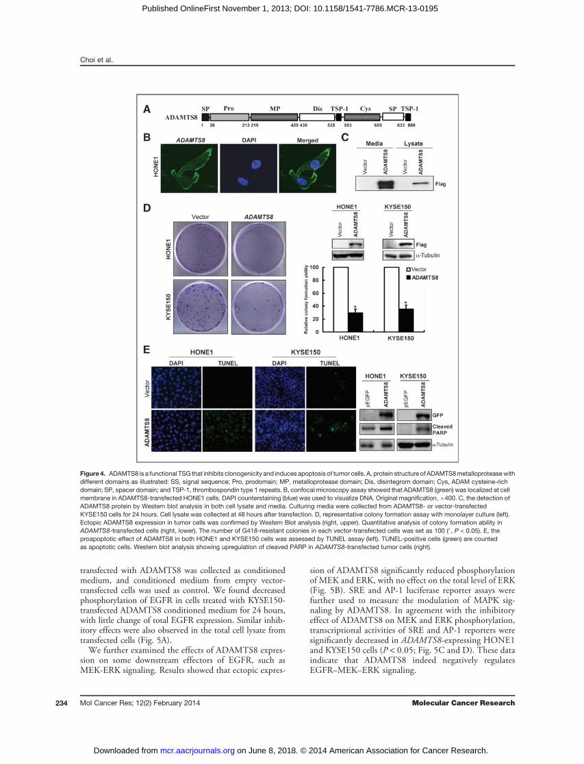
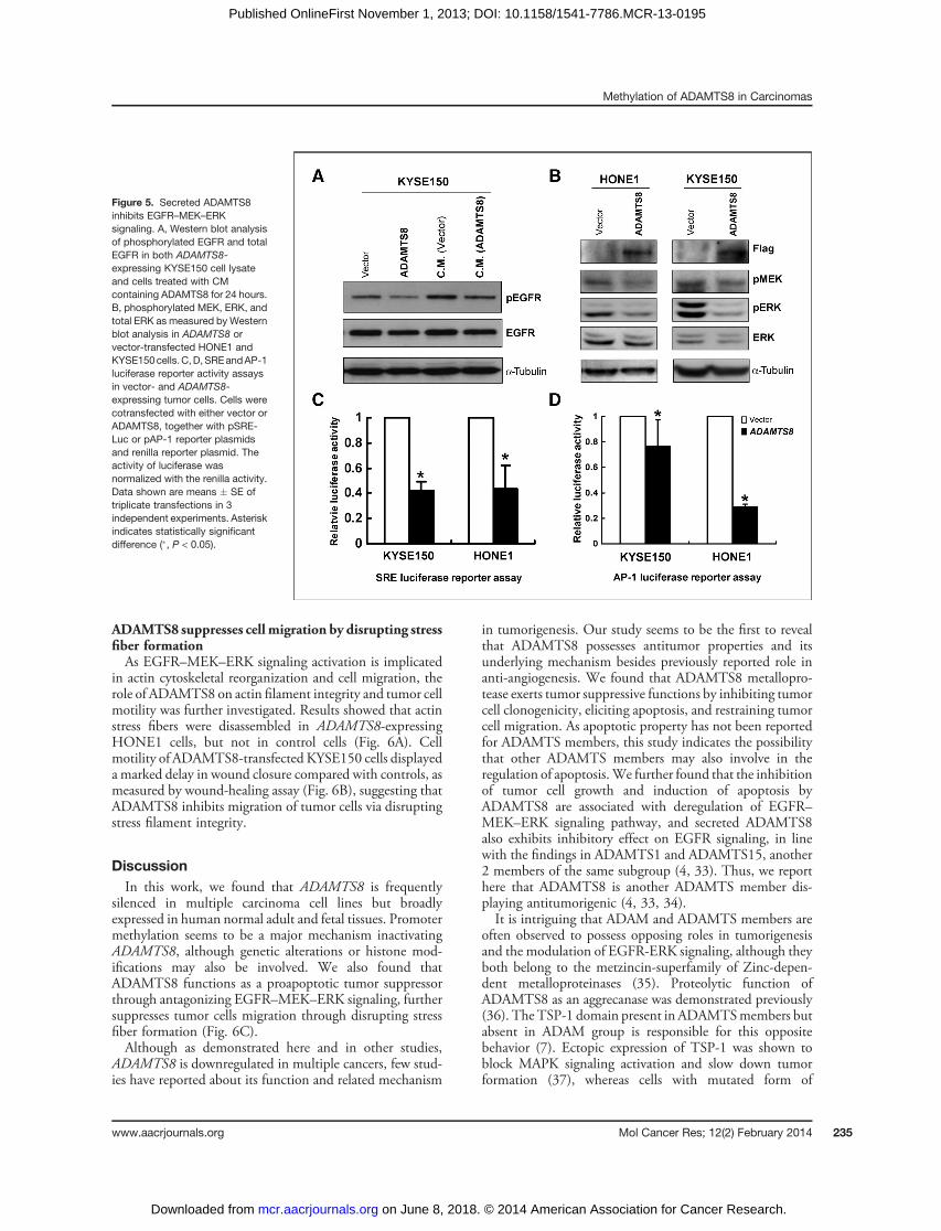
transfected with ADAMTS8 was collected as conditionedmedium, and conditioned medium from empty vector-transfected cells was used as control. We found decreasedphosphorylation of EGFR in cells treated with KYSE150-transfected ADAMTS8 conditioned medium for 24 hours,with little change of total EGFR expression. Similar inhib-itory effects were also observed in the total cell lysate fromtransfected cells (Fig. 5A).We further examined the effects of ADAMTS8 expres-
sion on some downstream effectors of EGFR, such asMEK-ERK signaling. Results showed that ectopic expres-
sion of ADAMTS8 significantly reduced phosphorylationof MEK and ERK, with no effect on the total level of ERK(Fig. 5B). SRE and AP-1 luciferase reporter assays werefurther used to measure the modulation of MAPK sig-naling by ADAMTS8. In agreement with the inhibitoryeffect of ADAMTS8 on MEK and ERK phosphorylation,transcriptional activities of SRE and AP-1 reporters weresignificantly decreased in ADAMTS8-expressing HONE1and KYSE150 cells (P < 0.05; Fig. 5C and D). These dataindicate that ADAMTS8 indeed negatively regulatesEGFR–MEK–ERK signaling.
Figure 4. ADAMTS8 is a functional TSG that inhibits clonogenicity and induces apoptosis of tumor cells. A, protein structure of ADAMTS8metalloproteasewithdifferent domains as illustrated: SS, signal sequence; Pro, prodomain; MP, metalloprotease domain; Dis, disintegrom domain; Cys, ADAM cysteine-richdomain; SP, spacer domain; and TSP-1, thrombospondin type 1 repeats. B, confocal microscopy assay showed that ADAMTS8 (green) was localized at cellmembrane in ADAMTS8-transfected HONE1 cells. DAPI counterstaining (blue) was used to visualize DNA. Original magnification, �400. C, the detection ofADAMTS8 protein by Western blot analysis in both cell lysate and media. Culturing media were collected from ADAMTS8- or vector-transfectedKYSE150 cells for 24 hours. Cell lysate was collected at 48 hours after transfection. D, representative colony formation assay with monolayer culture (left).Ectopic ADAMTS8 expression in tumor cells was confirmed by Western Blot analysis (right, upper). Quantitative analysis of colony formation ability inADAMTS8-transfected cells (right, lower). The number of G418-resistant colonies in each vector-transfected cells was set as 100 (�, P < 0.05). E, theproapoptotic effect of ADAMTS8 in both HONE1 and KYSE150 cells was assessed by TUNEL assay (left). TUNEL-positive cells (green) are countedas apoptotic cells. Western blot analysis showing upregulation of cleaved PARP in ADAMTS8-transfected tumor cells (right).
Choi et al.
Mol Cancer Res; 12(2) February 2014 Molecular Cancer Research234
on June 8, 2018. © 2014 American Association for Cancer Research. mcr.aacrjournals.org Downloaded from
Published OnlineFirst November 1, 2013; DOI: 10.1158/1541-7786.MCR-13-0195
ADAMTS8 suppresses cell migration by disrupting stressfiber formationAs EGFR–MEK–ERK signaling activation is implicated
in actin cytoskeletal reorganization and cell migration, therole of ADAMTS8 on actin filament integrity and tumor cellmotility was further investigated. Results showed that actinstress fibers were disassembled in ADAMTS8-expressingHONE1 cells, but not in control cells (Fig. 6A). Cellmotility of ADAMTS8-transfected KYSE150 cells displayeda marked delay in wound closure compared with controls, asmeasured by wound-healing assay (Fig. 6B), suggesting thatADAMTS8 inhibits migration of tumor cells via disruptingstress filament integrity.
DiscussionIn this work, we found that ADAMTS8 is frequently
silenced in multiple carcinoma cell lines but broadlyexpressed in human normal adult and fetal tissues. Promotermethylation seems to be a major mechanism inactivatingADAMTS8, although genetic alterations or histone mod-ifications may also be involved. We also found thatADAMTS8 functions as a proapoptotic tumor suppressorthrough antagonizing EGFR–MEK–ERK signaling, furthersuppresses tumor cells migration through disrupting stressfiber formation (Fig. 6C).Although as demonstrated here and in other studies,
ADAMTS8 is downregulated in multiple cancers, few stud-ies have reported about its function and related mechanism
in tumorigenesis. Our study seems to be the first to revealthat ADAMTS8 possesses antitumor properties and itsunderlying mechanism besides previously reported role inanti-angiogenesis. We found that ADAMTS8 metallopro-tease exerts tumor suppressive functions by inhibiting tumorcell clonogenicity, eliciting apoptosis, and restraining tumorcell migration. As apoptotic property has not been reportedfor ADAMTS members, this study indicates the possibilitythat other ADAMTS members may also involve in theregulation of apoptosis.We further found that the inhibitionof tumor cell growth and induction of apoptosis byADAMTS8 are associated with deregulation of EGFR–MEK–ERK signaling pathway, and secreted ADAMTS8also exhibits inhibitory effect on EGFR signaling, in linewith the findings in ADAMTS1 and ADAMTS15, another2 members of the same subgroup (4, 33). Thus, we reporthere that ADAMTS8 is another ADAMTS member dis-playing antitumorigenic (4, 33, 34).It is intriguing that ADAM and ADAMTS members are
often observed to possess opposing roles in tumorigenesisand the modulation of EGFR-ERK signaling, although theyboth belong to the metzincin-superfamily of Zinc-depen-dent metalloproteinases (35). Proteolytic function ofADAMTS8 as an aggrecanase was demonstrated previously(36). The TSP-1 domain present in ADAMTSmembers butabsent in ADAM group is responsible for this oppositebehavior (7). Ectopic expression of TSP-1 was shown toblock MAPK signaling activation and slow down tumorformation (37), whereas cells with mutated form of
Figure 5. Secreted ADAMTS8inhibits EGFR–MEK–ERKsignaling. A, Western blot analysisof phosphorylated EGFR and totalEGFR in both ADAMTS8-expressing KYSE150 cell lysateand cells treated with CMcontaining ADAMTS8 for 24 hours.B, phosphorylated MEK, ERK, andtotal ERK as measured by Westernblot analysis in ADAMTS8 orvector-transfected HONE1 andKYSE150cells.C,D,SREandAP-1luciferase reporter activity assaysin vector- and ADAMTS8-expressing tumor cells. Cells werecotransfected with either vector orADAMTS8, together with pSRE-Luc or pAP-1 reporter plasmidsand renilla reporter plasmid. Theactivity of luciferase wasnormalized with the renilla activity.Data shown are means � SE oftriplicate transfections in 3independent experiments. Asteriskindicates statistically significantdifference (�, P < 0.05).
Methylation of ADAMTS8 in Carcinomas
www.aacrjournals.org Mol Cancer Res; 12(2) February 2014 235
on June 8, 2018. © 2014 American Association for Cancer Research. mcr.aacrjournals.org Downloaded from
Published OnlineFirst November 1, 2013; DOI: 10.1158/1541-7786.MCR-13-0195

ADAMTS15 lacking 2 TSP-1 motifs had elevated ERKphosphorylation level compared with cells harboring wild-type ADAMTS15 (4). In addition, TSP-1 in auto-proteo-lytic cleaved ADAMTS1 is required for inhibiting ERK (2).Therefore, the TSP-1 domain in ADAMTS members islikely involved in suppressing the activated MEK-ERKsignaling in cancer cells.Most metalloproteinases are regarded as key regulators
of tumor cell invasiveness, because they possess proteolyticactivities to destruct the ECM and alter the cell–cellattachments and cell–matrix attachments, which are inte-gral to tumor cell migration (38). However, little is knownabout the involvement of ADAMTS members in cellinvasion except for ADAMTS1 and ADAMTS5. Full-length ADAMTS1 and ADAMTS5 are reported to pro-mote metastasis whereas fragment of ADAMTS1 wasshown to be antimetastatic (2, 39). Here, we also iden-tified that ADAMTS8 suppressed tumor cell migration bydisrupting actin polymerization. Further study ofADAMTS8 on cell epithelial–mesenchymal transition isneeded.
In summary, our study shows that ADAMTS8 func-tions as a proapoptotic TSG through suppressing EGFR–MEK–ERK signaling, which is frequently silenced bypromoter CpG methylation in common carcinomas.However, further investigations are needed to show exactfeature and function of ADAMTS8 in connection tocellular homeostasis.
Disclosure of Potential Conflicts of InterestNo potential conflicts of interest were disclosed.
Authors' ContributionsConception and design: G.C.G. Choi, Q. TaoDevelopment of methodology: G.C.G. Choi, S.W. Tsao, Q. TaoAcquisition of data (provided animals, acquired and managed patients, providedfacilities, etc.):G.C.G. Choi, J. Li, Y.Wang, L. Li, X. Su, T. Xiang, S.Y. Rha, Q. TaoAnalysis and interpretation of data (e.g., statistical analysis, biostatistics, compu-tational analysis): G.C.G. Choi, J. Li, Y. Wang, L. Li, L. Zhong, Q. TaoWriting, review, and/or revision of the manuscript: G.C.G. Choi, Y. Wang, L. Li,S.Y. Rha, A.T.C. Chan, Q. TaoAdministrative, technical, or material support (i.e., reporting or organizing data,constructing databases): L. Li, X. Su, J. Ying, J. Yu, A.T.C. Chan, Q. TaoStudy supervision: B. Ma, J.J.Y. Sung, A.T.C. Chan, Q. Tao
Figure 6. Ectopic expression ofADAMTS8 inhibits cancer cellmigration. A, phalloidin staining ofcells shows that expression ofADAMTS8 disrupts actin stressfiber formation. B, effect ofADAMTS8 on cell migration wasassessed by wound-healingmotility assay. Confluentmonolayers of vector- andADAMTS8-transfected KYSE150tumor cells were scratched 48hours after transfection. Phase-contrast microscopy photos ofwound margins were taken at 24and 48 hours after scratching. C,proposed mechanism of the tumorsuppressive function of ADAMTS8through suppressing the EGFR–MEK–ERK signaling pathway.
Choi et al.
Mol Cancer Res; 12(2) February 2014 Molecular Cancer Research236
on June 8, 2018. © 2014 American Association for Cancer Research. mcr.aacrjournals.org Downloaded from
Published OnlineFirst November 1, 2013; DOI: 10.1158/1541-7786.MCR-13-0195
AcknowledgmentsThe authors thank Dr. B. Vogelstein for HCT116 cells with knockout of
DNMTs, and DSMZ (German Collection of Microorganisms & Cell Cultures)for the KYSE cell lines (Shimada et al., 1992).
Grant SupportThis study was supported by grants from Hong Kong RGC (GRF
#475009), National Natural Science Foundation (#81172582 and
#81201934), and the Group Research Schemes of The Chinese University ofHong Kong.
The costs of publication of this article were defrayed in part by the paymentof page charges. This article must therefore be hereby marked advertisement inaccordance with 18 U.S.C. Section 1734 solely to indicate this fact.
Received April 22, 2013; revised September 17, 2013; accepted September 30,2013; published OnlineFirst November 1, 2013.
References1. Lopez-Otin C, Matrisian LM. Emerging roles of proteases in tumour
suppression. Nat Rev Cancer 2007;7:800–8.2. Liu YJ, Xu Y, Yu Q. Full-length ADAMTS-1 and the ADAMTS-1 frag-
ments display pro- andantimetastatic activity, respectively. Oncogene2006;25:2452–67.
3. Moncada-Pazos A, Obaya AJ, Fraga MF, Viloria CG, Capell�a G,Gausachs M, et al. The ADAMTS12 metalloprotease gene is epige-netically silenced in tumor cells and transcriptionally activated in thestroma during progression of colon cancer. J Cell Sci 2009;122(Pt16):2906–13.
4. Viloria CG, Obaya AJ, Moncada-Pazos A, Llamazares M, Astudillo A,Capell�aG, et al. Genetic inactivation of ADAMTS15metalloprotease inhuman colorectal cancer. Cancer Res 2009;69:4926–34.
5. Lo PH, Lung HL, Cheung AK, Apte SS, Chan KW, Kwong FM, et al.Extracellular protease ADAMTS9 suppresses esophageal and naso-pharyngeal carcinoma tumor formation by inhibiting angiogenesis.Cancer Res 2010;70:5567–76.
6. Jin H, Wang X, Ying J, Wong AH, Li H, Lee KY, et al. Epigeneticidentification of ADAMTS18 as a novel 16q23.1 tumor suppressorfrequently silenced in esophageal, nasopharyngeal and multiple othercarcinomas. Oncogene 2007;26:7490–8.
7. Tang BL. ADAMTS: a novel family of extracellular matrix proteases. IntJ Biochem Cell Biol 2001;33:33–44.
8. Rocks N, Paulissen G, El Hour M, Quesada F, Crahay C, Gueders M,et al. Emerging roles of ADAM and ADAMTS metalloproteinases incancer. Biochimie 2008;90:369–79.
9. Porter S, Scott SD, Sassoon EM, Williams MR, Jones JL, Girling AC,et al. Dysregulated expression of adamalysin-thrombospondingenes in human breast carcinoma. Clin Cancer Res 2004;10:2429–40.
10. Llamazares M, Cal S, Quesada V, Lopez-Otin C. Identificationand characterization of ADAMTS-20 defines a novel subfamily ofmetalloproteinases-disintegrins with multiple thrombospondin-1repeats and a unique GON domain. J Biol Chem 2003;278:13382–9.
11. Cauwe B, Van den Steen PE, Opdenakker G. The biochemical, bio-logical, and pathological kaleidoscope of cell surface substratesprocessed by matrix metalloproteinases. Crit Rev Biochem Mol Biol2007;42:113–85.
12. Vazquez F,HastingsG,OrtegaMA, Lane TF, OikemusS, LombardoM,et al. METH-1, a human ortholog of ADAMTS-1, and METH-2 aremembers of a new family of proteins with angio-inhibitory activity.J Biol Chem 1999;274:23349–57.
13. KooBH,CoeDM,DixonLJ,Somerville RP,NelsonCM,WangLW, et al.ADAMTS9 is a cell-autonomously acting, anti-angiogenic metallopro-tease expressed by microvascular endothelial cells. Am J Pathol2010;176:1494–504.
14. Dunn JR, Reed JE, du Plessis DG, Shaw EJ, Reeves P, Gee AL, et al.Expression of ADAMTS-8, a secreted protease with antiangiogenicproperties, is downregulated in brain tumours. Br J Cancer 2006;94:1186–93.
15. Dunn JR, Panutsopulos D, Shaw MW, Heighway J, Dormer R, SalmoEN, et al.METH-2 silencing andpromoter hypermethylation inNSCLC.Br J Cancer 2004;91:1149–54.
16. Demircan K, Gunduz E, Gunduz M, Beder LB, Hirohata S, NagatsukaH, et al. Increased mRNA expression of ADAMTS metalloproteinasesin metastatic foci of head and neck cancer. Head Neck 2009;31:793–801.
17. Masui T, Hosotani R, Tsuji S, Miyamoto Y, Yasuda S, Ida J, et al.Expression of METH-1 andMETH-2 in pancreatic cancer. Clin CancerRes 2001;7:3437–43.
18. Rodriguez-Rodero S, Fernandez AF, Fernandez-Morera JL, Castro-Santos P, Bayon GF, Ferrero C, et al. DNA methylation signaturesidentify biologically distinct thyroid cancer subtypes. J Clin EndocrinolMetab 2013;98:2811–21.
19. ChengY,GengH,ChengSH, LiangP,Bai Y, Li J, et al. KRABzincfingerprotein ZNF382 is a proapoptotic tumor suppressor that repressesmultiple oncogenes and is commonly silenced in multiple carcinomas.Cancer Res 2010;70:6516–26.
20. Li L, Ying J, Li H, ZhangY, Shu X, FanY, et al. The human cadherin 11 isa pro-apoptotic tumor suppressor modulating cell stemness throughWnt/b-catenin signaling and silenced in common carcinomas. Onco-gene 2012;31:3901–12.
21. Ying J, Li H, Seng TJ, Langford C, Srivastava G, Tsao SW, et al.Functional epigenetics identifies a protocadherin PCDH10 as a can-didate tumor suppressor for nasopharyngeal, esophageal andmultipleother carcinomas with frequent methylation. Oncogene 2006;25:1070–80.
22. Tao Q, Huang H, Geiman TM, Lim CY, Fu L, Qiu GH, et al. Defective denovomethylation of viral and cellular DNA sequences in ICF syndromecells. Hum Mol Genet 2002;11:2091–102.
23. Qiu GH, Tan LK, Loh KS, Lim CY, Srivastava G, Tsai ST, et al. Thecandidate tumor suppressor gene BLU, located at the commonlydeleted region 3p21.3, is an E2F-regulated, stress-responsive geneand inactivated by both epigenetic and genetic mechanisms in naso-pharyngeal carcinoma. Oncogene 2004;23:4793–806.
24. Li L, Tao Q, Jin H, van Hasselt A, Poon FF, Wang X, et al. The tumorsuppressor UCHL1 forms a complex with p53/MDM2/ARF to promotep53 signaling and is frequently silenced in nasopharyngeal carcinoma.Clin Cancer Res 2010;16:2949–58.
25. Xu L, Li X, Chu ES, ZhaoG, GoMY, TaoQ, et al. Epigenetic inactivationof BCL6B, a novel functional tumour suppressor for gastric cancer, isassociated with poor survival. Gut 2011;61:977–85.
26. Shu XS, Li L, Ji M, Cheng Y, Ying J, Fan Y, et al. FEZF2, a novel 3p14tumor suppressor gene, represses oncogene EZH2 and MDM2expressionand is frequentlymethylated in nasopharyngeal carcinoma.Carcinogenesis 2013;34:1984–93.
27. TaoQ,SwinnenLJ,Yang J, SrivastavaG,RobertsonKD,AmbinderRF.Methylation status of the Epstein-Barr virus major latent promoter C iniatrogenic B cell lymphoproliferative disease. Application of PCR-based analysis. Am J Pathol 1999;155:619–25.
28. Ying J, Li H, Yu J, Ng KM, Poon FF, Wong SC, et al. WNT5A exhibitstumor-suppressive activity through antagonizing the Wnt/b-cateninsignaling, and is frequently methylated in colorectal cancer. ClinCancer Res 2008;14:55–61.
29. Parsons DW, Jones S, Zhang X, Lin JC, Leary RJ, Angenendt P, et al.An integrated genomic analysis of human glioblastoma multiforme.Science 2008;321:1807–12.
30. Ballif BA, Blenis J. Molecular mechanisms mediating mammalianmitogen-activated protein kinase (MAPK) kinase (MEK)-MAPK cellsurvival signals. Cell Growth Differ 2001;12:397–408.
31. Marcoux N, Vuori K. EGF receptor activity is essential for adhesion-induced stress fiber formation and cofilin phosphorylation. Cell Signal2005;17:1449–55.
32. Toral C, Solano-AgamaC,Reyes-MarquezB, SabaneroM, Talam�asP,Gonz�alez del PliegoM, et al. Role of extracellularmatrix-cell interaction
Methylation of ADAMTS8 in Carcinomas
www.aacrjournals.org Mol Cancer Res; 12(2) February 2014 237
on June 8, 2018. © 2014 American Association for Cancer Research. mcr.aacrjournals.org Downloaded from
Published OnlineFirst November 1, 2013; DOI: 10.1158/1541-7786.MCR-13-0195

and epidermal growth factor (EGF) on EGF-receptors and actin cyto-skeleton arrangement in infantile pituitary cells. Cell Tissue Res 2007;327:143–53.
33. Suga A, Hikasa H, Taira M. Xenopus ADAMTS1 negatively modulatesFGF signaling independent of its metalloprotease activity. Dev Biol2006;295:26–39.
34. LlamazaresM,ObayaAJ,Moncada-PazosA,HeljasvaaraR, Espada J,L�opez-Otín C, et al. The ADAMTS12 metalloproteinase exhibits anti-tumorigenic properties throughmodulation of theRas-dependent ERKsignalling pathway. J Cell Sci 2007;120(Pt 20):3544–52.
35. Duffy MJ, McKiernan E, O'Donovan N, McGowan PM. Role of ADAMsin cancer formation and progression. Clin Cancer Res 2009;15:1140–4.
36. Collins-Racie LA, Flannery CR, Zeng W, Corcoran C, Annis-FreemanB, Agostino MJ, et al. ADAMTS-8 exhibits aggrecanase activity and isexpressed in human articular cartilage. Matrix Biol 2004;23:219–30.
37. Zhang YW, Su Y, Volpert OV, Vande Woude GF. Hepatocyte growthfactor/scatter factor mediates angiogenesis through positive VEGFand negative thrombospondin 1 regulation. Proc Natl Acad Sci U S A2003;100:12718–23.
38. Yoon SO, Park SJ, Yun CH, Chung AS. Roles of matrix metallopro-teinases in tumor metastasis and angiogenesis. J Biochem Mol Biol2003;36:128–37.
39. NakadaM,Miyamori H, Kita D, Takahashi T, Yamashita J, Sato H, et al.Human glioblastomas overexpress ADAMTS-5 that degrades brevi-can. Acta Neuropathol 2005;110:239–46.
Choi et al.
Mol Cancer Res; 12(2) February 2014 Molecular Cancer Research238
on June 8, 2018. © 2014 American Association for Cancer Research. mcr.aacrjournals.org Downloaded from
Published OnlineFirst November 1, 2013; DOI: 10.1158/1541-7786.MCR-13-0195

2014;12:228-238. Published OnlineFirst November 1, 2013.Mol Cancer Res Gigi C.G. Choi, Jisheng Li, Yajun Wang, et al. in Carcinomas by CpG Methylation
ERK Signaling and Is Silenced−MEK−through Antagonizing EGFR The Metalloprotease ADAMTS8 Displays Antitumor Properties
Updated version
10.1158/1541-7786.MCR-13-0195doi:
Access the most recent version of this article at:
Cited articles
http://mcr.aacrjournals.org/content/12/2/228.full#ref-list-1
This article cites 39 articles, 15 of which you can access for free at:
Citing articles
http://mcr.aacrjournals.org/content/12/2/228.full#related-urls
This article has been cited by 1 HighWire-hosted articles. Access the articles at:
E-mail alerts related to this article or journal.Sign up to receive free email-alerts
Subscriptions
Reprints and
To order reprints of this article or to subscribe to the journal, contact the AACR Publications Department at
Permissions
Rightslink site. Click on "Request Permissions" which will take you to the Copyright Clearance Center's (CCC)
.http://mcr.aacrjournals.org/content/12/2/228To request permission to re-use all or part of this article, use this link
on June 8, 2018. © 2014 American Association for Cancer Research. mcr.aacrjournals.org Downloaded from
Published OnlineFirst November 1, 2013; DOI: 10.1158/1541-7786.MCR-13-0195