The Effect of Mechanical Vibration on Human PDL Cell ...
56
Marquee University e-Publications@Marquee Master's eses (2009 -) Dissertations, eses, and Professional Projects e Effect of Mechanical Vibration on Human PDL Cell Differentiation and Response to Inflammation Megan DesRoches Marquee University Recommended Citation DesRoches, Megan, "e Effect of Mechanical Vibration on Human PDL Cell Differentiation and Response to Inflammation" (2016). Master's eses (2009 -). Paper 374. hp://epublications.marquee.edu/theses_open/374
Transcript of The Effect of Mechanical Vibration on Human PDL Cell ...

Marquette Universitye-Publications@Marquette
Master's Theses (2009 -) Dissertations, Theses, and Professional Projects
The Effect of Mechanical Vibration on HumanPDL Cell Differentiation and Response toInflammationMegan DesRochesMarquette University
Recommended CitationDesRoches, Megan, "The Effect of Mechanical Vibration on Human PDL Cell Differentiation and Response to Inflammation" (2016).Master's Theses (2009 -). Paper 374.http://epublications.marquette.edu/theses_open/374

THE EFFECT OF MECHANICAL VIBRATION ON HUMAN PDL CELL
DIFFERENTIATION AND RESPONSE TO INFLAMMATION
by
Megan S. DesRoches, D.D.S.
A Thesis submitted to the Faculty of the Graduate School,
Marquette University,
in Partial Fulfillment of the Requirements for
the Degree of Master of Science
Milwaukee, Wisconsin
August 2016

ABSTRACT
THE EFFECT OF MECHANICAL VIBRATION ON HUMAN PDL CELL
DIFFERENTIATION AND RESPONSE TO INFLAMMATION
Megan S. DesRoches, D.D.S.
Marquette University, 2016
Low magnitude mechanical vibration is a therapeutic adjunct being investigated
to alter bone remodeling and inflammation in areas such as osteoporosis, bone fracture
healing, and muscle soreness after exercise. In orthodontics a device named AcceleDent
has been marketed that claims to increase the rate of tooth movement and decrease pain.
However evidence for these claims is lacking. In this study we looked at two potential
cellular mechanisms for these claims: periodontal ligament (PDL) cell differentiation and
inflammation under an orthodontic model of strain (IL-1β).
Increased PDL cell differentiation into osteogenic cells could be an avenue of
increasing orthodontic tooth movement. To test for cell differentiation, cultured human
PDL cells were observed for calcification by Alizarin Red staining. Also the gene
expression of periostin, a cytokine with roles in bone formation, was analyzed using
qPCR. To test for inflammation, the gene expression of MMP-13, an inflammatory
cytokine was tested. The cells were treated under conditions of +/- Il-1β (10 ng/ml) and
+/- 0.3g/30Hz vibration.
PDL cell differentiation was decreased with addition of IL-1β (1 ng/ml), as
expected under an inflammatory environment. However vibration exhibited no
observable effect. Gene expressions of periostin and MMP-13 under all conditions
showed no statistically significant results. However, a general trend was noted that
vibration may decrease inflammation/MMP-13 production. These findings warrant
further investigation.

i
ACKNOWLEDGEMENTS
Megan S. DesRoches
I would like to thank all the people who helped with this project including Dr.
Dawei Liu, the project visionary; Dr. Khadijah Makky, the pragmatic cellular expert; Drs.
Fei Wang and Yuan Gao for their extraordinary hard work and perseverance; Dr. Gerard
Bradley for his oversight; and Dr. Brad Gauthier for traveling with me during this
journey.
Also I would like to thank my family for their support and unceasing
encouragement, as I would not have been able to succeed without them.

ii
TABLE OF CONTENTS
ACKNOWLEDGEMENTS……………………………………………….…….………..i
TABLE OF CONTENTS…………………………………………………….…………...ii
LIST OF TABLES……………………………………………………………………….iii
LIST OF FIGURES…………………………………………………………………..….iv
CHAPTER
I. INTRODUCTION ………………………………………..…..….…..................1
II. LITERATURE REVIEW…………………..…………………...……………..3
A. ORTHODONTIC TOOTH MOVEMENT……………………………3
B. NEGATIVE SIDE EFFECTS OF OTM……………………………….9
C. LOW MAGNITUDE VIBRATION THERAPY…………………….12
D. STUDY AIMS……………………………………………………..…16
III. MATERIALS AND METHODS ……………………………....….………..20
IV. RESULTS ……………………………………………….…...……………...26
A. DIFFERENTIATION ASSAY..…………………………………….26
B. GENE EXPRESSION…………………………………………..…...30
V. DISCUSSION ……………………………………………......………………35
BIBLIOGRAPHY……………………………………………………….……………….43

iii
LIST OF TABLES
Table 1) Cell Differentiation Experimental Groups……………………………….23
Table 2) Gene Expression Experimental Groups……………….…………………24
Table 3) Sequences of Primers utilized……………………………………………25
Table 4) MMP-13 and GAPDH CT values in Periodontal Ligament cells………..31
Table 5) MMP-13 expression in PDL cells………………………………………..31
Table 6) MMP-13 ANOVA……………………………………………………….32
Table 7) Periostin and GAPDH CT values in Periodontal Ligament Cells……….33
Table 8) Periostin expression in PDL cells ………………………………………..33
Table 9) Periostin ANOVA………………………………………………………..34

iv
LIST OF FIGURES
Figure 1) Effect of orthodontic force on blood flow in the periodontal ligament…5
Figure 2) Cellular effects of PDL compression……………………………………8
Figure 3) Cellular effects of PDL tension…………………………………………9
Figure 4) Study Design…………………………………………………………….19
Figure 5) Human Periodontal Ligament Cells……………………………………..20
Figure 6) Mechanical Vibration Set-up Diagram…………………………………..21
Figure 7) Mechanical Vibration Set-up…………………………………………….22
Figure 8) Staining of PDL cells with Alizarin Red …………….………………….27
Figure 9) Qualitative Analysis of PDL Differentiation…………………………….28
Figure 10) Control vs 30Hz Vibration PDL Differentiation………………………...29
Figure 11) Quality of primers; GAPDH, MMP-13, Periostin…………………….....30
Figure 12) MMP-13 expression in PDL cells subjected to IL-1β and 30Hz Vibration….32
Figure 13) Periostin expression in PDL cells subjected to IL-1β and 30Hz Vibration…..34
1
CHAPTER I
INTRODUCTION
In the field of orthodontics, many people would benefit from treatment. Dentists
estimate 35% of individuals as having normal occlusion, and recommend approximately
55% of people to seek orthodontic care, while 10% could benefit but have only mild
malocclusion. Even the general public sees 35% of individuals as needing care.
However, less than 25% of people pursue treatment (Proffit, 2007). There are many
deterrents to care, including financial constraints, the extended treatment time needed for
care, pain associated, and esthetics concerns of appliances. Of interest to this study is the
time needed to provide orthodontic care and associated pain.
Over the last several years, there has been a focus on clinical adjuncts that can
help speed orthodontic tooth movement. Surgical adjuncts including open flap
procedures to remove the bony cortex with either conventional mechanical
instrumentation or piezoelectrics have seen increased tooth movement, leading to shorter
overall orthodontic treatment time (Wu, 2015). However the surgical nature of this
adjunct makes it unappealing to patients. This has led to more conservative procedures
such as cortical micro-perforations without a gingival flap, low energy laser radiation,
low magnitude vibration, pulsed electromagnetic waves, and various pharmacological
methods (El-Angbawi, 2015).
Of these adjuncts, a device called AcceleDent claims that the low magnitude
vibration it provides speeds orthodontic tooth movement and can decrease pain.
AcceleDent’s relative ease of use and patient acceptance is appealing to both practitioners
and patients. AcceleDent is used in the comfort of the home and patients are instructed to

2
gently hold the mouthpiece-shaped wafer between their teeth for 20 minutes a day while
in active orthodontic treatment. It delivers a low magnitude vibration stimulus to the
teeth of 0.3g/30Hz while its hands free and low maintenance style allow a patient to
continue to address other aspects of their busy day. The company, OrthoAccel
Technologies, claims that AcceleDent is a “Leader in Accelerated Orthodontics®, is an
FDA-cleared, class II medical device clinically proven to move teeth up to 50% faster.
Simple-to-use and hands-free, AcceleDent is clinically shown to reduce discomfort that
may be associated with orthodontic treatment. AcceleDent is the fast, safe and gentle way
to speed orthodontic treatment” (retrieved June 20, 2016 from http://acceledent.com/how-
it-works/introducing-acceledent). However, literature backing these claims is of
controversy. A recent Cochrane Collaboration review of non-surgical adjuncts for
accelerating tooth movement concluded that “Although there have been claims that there
may be a positive effect of light vibrational forces, the results of the current studies do
not reach either statistical or clinical significance and are at high risk of bias” (El-
Angbawi, 2015). The aims of this study are to determine if there are molecular and
cellular mechanisms associated with low magnitude vibrational therapy to periodontium
cells in regards to accelerated tooth movement or decreased pain. Periodontal ligament
cells were chosen due to their key role in the orthodontic tooth movement process.
3
Chapter II
Literature Review
A. Orthodontic Tooth Movement
Pressure-Tension Theory of Orthodontic Tooth Movement
Several theories exist in regards to orthodontic tooth movement. One of the most
prominent theories is the pressure-tension theory. When orthodontic forces are applied to
a tooth, areas within the periodontal ligament (PDL) are compressed in the direction of
tooth movement on the pressure side. Conversely on the opposite side, tension exists as
orthodontic force moves the tooth away from the alveolar bone and the PDL is stretched.
The stretching and compression of the periodontal ligament causes a cascade of
molecular and cellular events and is dependent on the degree of force applied.
The cellular events that happen on the pressure side of orthodontic tooth
movement differ based on the length and quantity of force applied. If heavy or light
pressure is applied but is less than one second in length, the internal pressure in the PDL
causes the tissue to be incompressible and any tooth movement occurs within the
surrounding alveolar bone. However when a longer duration of force is applied, changes
occur. A force of 1-2 seconds will cause a tooth to move within the socket and PDL fluid
to be expressed from the tissue. If a force continues but is light in nature (20-25g/cm2 of
root surface) after 2 seconds the capillary bed located within the PDL is partially
occluded and PDL fibers and cells are mechanically distorted. Within minutes blood
flow is altered, oxygen levels change and inflammatory mediators such as prostaglandins
and cytokines are released. Within several hours of prolonged force these chemical
mediators affect cellular activity, differentiation of cells within the PDL and tooth

4
movement begins as alveolar bone is remodeled by osteoclasts and osteoblasts
approximately 2 days after initial consistent force. However if forces are heavy the PDL
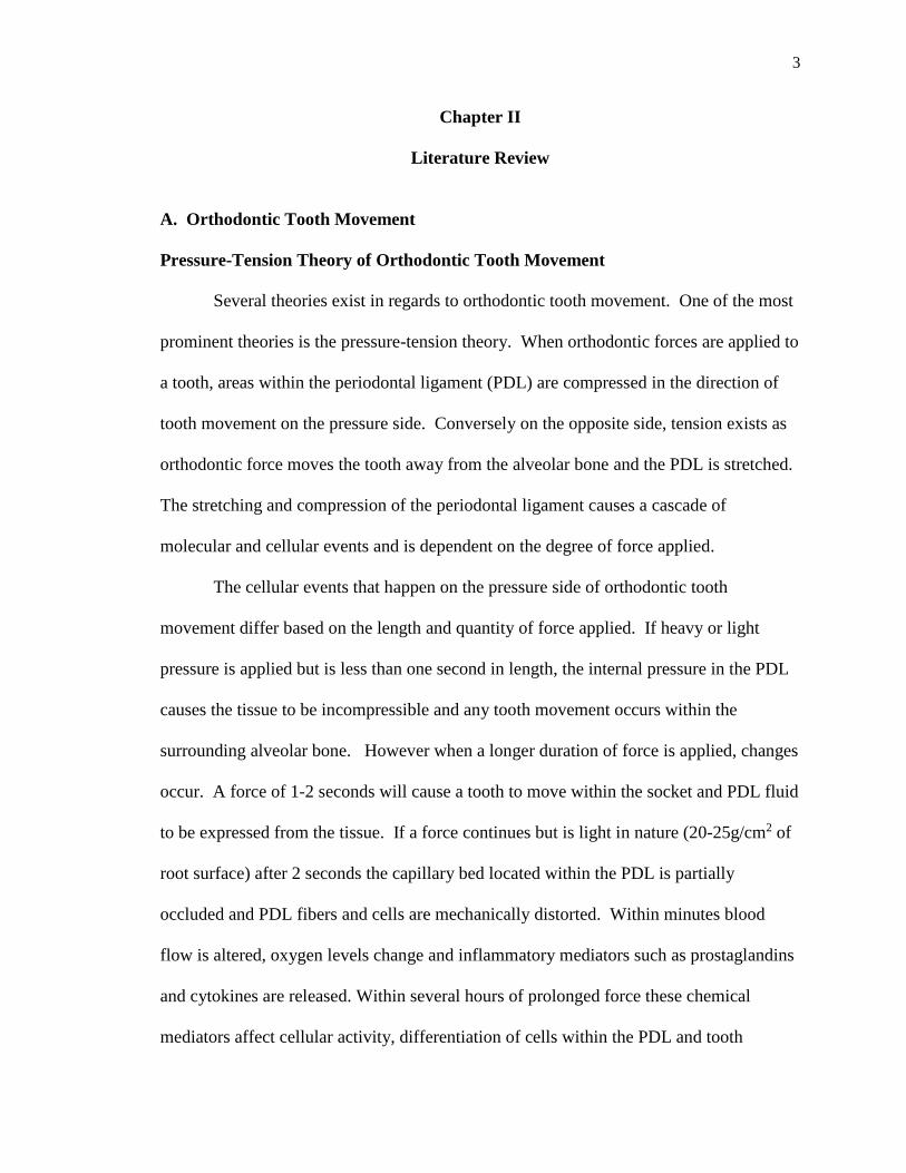
capillary bed is occluded fully within seconds as seen below in figure 1 adapted from
Proffit, 2007. Within hours tissue necrosis occurs as cells are cut off from oxygen and
nutrients. Physical contact between the tooth and bone can occur with great forces. This
necrosis results in undermining resorption or hyalinization of adjacent marrow spaces as
cells are not viable within the PDL to help with remodeling of bone in a physiologic
fashion. During this process, inflammatory cell mediators recruit cells such as
macrophages, foreign body giant cells and osteoclasts which resorb bone and dead tissue
adjacent to the PDL over a process of days. Tooth movement eventually occurs once
enough undermining resorption occurs usually after 7-14 days. While light forces within
orthodontics are preferable and have a better physiologic response, in clinical situations a
mix of light, physiologic and heavy, hyalinized undermining resorption will likely occur
in different areas of PDL (Krishnan, 2006 and Proffit, 2007).

5
Effect of orthodontic force on blood flow in the periodontal ligament
Figure 1: Representation on how increasing force occludes capillaries within the PDL.
A. normal periodontal ligament, attached to tooth surface on right and alveolar bone, left.
B. light orthodontic forces begin to compress PDL space. C. As forces increase, blood
vessels are compressed. D. Heavy forces fully occlude blood vessels. Adapted
from Contemporary Orthodontics (p337), by W.R. Proffit, 2007, St. Louis, MO: Mosby
Elsevier. Copyright 2007 by Mosby Elsevier. Reprinted with permission.
Conversely on the tension side of orthodontic tooth movement, PDL fibers are
stretched between the moving tooth and alveolar bone. This causes blood flow to be
maintained or even increased. This alteration, just as in the pressure side of tooth
movement, causes a change in metabolites, chemical signaling, and cellular
differentiation. These changes allow new periodontal tissue, such as bone, ligament
fibers and cementum to be produced (Proffit, 2007).
Orthodontic Tooth Movement Phases
In all, orthodontic tooth movement can be summarized into three phases; an initial
phase, lag phase and a post-lag phase. The initial phase lasts approximately 1-2 days
where initial forces displace the tooth within the alveolar socket. Here some movement
6
occurs and blood flow is altered leading to a cascade of cellular and tissue reactions.
Following the initial phase, a lag phase occurs when areas of hyalinization arise as a
result of heavy forces. No tooth movement occurs during this time and depending on the
magnitude of force this phase can last 4-40 days. During this time, cells are busy
removing any necrotic tissue to allow for tooth movement to continue. Cells such as
macrophages, foreign body giant cells and osteoclasts are recruited, while on the tension
side pre-osteoblasts migrate towards the socket wall and fibroblasts remodel PDL tissues.
After enough necrotic tissue is removed, active tooth movement can occur in a post-lag
period. Here osteoclasts continue to resorb bone on areas in pressure while newly
differentiated osteoblasts lay down new bone on the tension side.
Although phases are a good way to understand the biologic sequence of events
occurring within the PDL, tooth movement in reality is a dynamic process occurring over
the entire tooth surface. There can be simultaneous areas of physiologic tooth movement
in areas subjected to light pressure as well as hylanization due to heavy force application.
(Krishnan, 2006).
Cytokines in Orthodontic Tooth Movement
Of particular interest is the role of cytokines in orthodontic tooth movement.
Cytokines are signaling molecules that are produced in connective tissue cells such as
fibroblasts and osteoblasts and have roles in bone remodeling and hence orthodontic
tooth movement. They work as autocrine or paracrine signaling molecules and there are
numerous families and functions for each molecule, some of which are not fully
understood (Meikle, 2006). Interleukin 1, interleukin 2, interleukin 3, interleukin 6,

7
interleukin 8, tumor necrosis factor alpha, gamma interferon, and osteoclast
differentiation factor all have roles in orthodontic tooth movement, with IL-1 having the
most important role as it stimulates osteoclast function (Krishnan, 2006). IL-1, with its
two isoforms IL-1α and IL-1β, is a pro inflammatory cytokine and has roles in the
regulation of feeding, sleep, temperature, and bone remodeling. However overproduction
can lead to acute and chronic inflammation as well as autoimmune disorders and pain
(Ren, 2009). In tooth movement, IL-1β regulates bone resorption and bone formation by
mechanical stress (Davidovitch, 1988 and Preiss, 1994). Il-1β can stimulate other
cytokines such as adhesion molecules, RANKL and matrix metalloproteinases (MMPs)
(Meikle, 2006, and Nogueira, 2013). Together these chemical signaling molecules
regulate the cellular changes in the PDL needed to organize clearing out necrotic tissue,
and remodeling the bone, connective tissue, and vascular elements under orthodontic
strain. Leukocytes are attracted to clear necrotic tissue, fibroblasts are stimulated to
produce connective tissue, osteoclasts to resorb bone on the pressure areas and
osteoblasts to build new bone on the tension sites (Krishnan, 2006). Together these
changes allow orthodontic tooth movement to occur. However, these molecular
processes are still being researched and interactions between cytokines and their intended
receptors are complex. Below are figures by Meikle of hypothesized pathways for these
cytokines on the pressure and tension sides of orthodontic tooth movement. The
diagrams show the complexity of cytokine and cellular interactions and illustrate that
molecular pathways under pressure and tension are different.

8
Cellular effects of PDL compression
Figure 2 Hypothetical PDL model of the remodeling of the periodontium: compression
side. “PDL cells under compressive strain synthesize interleukin-1 (IL-1) and IL-6 (1); IL-
1 and IL-6 act in an autocrine and paracrine manner to up-regulate receptor activator of
nuclear factor κ B ligand (RANKL) (2) and matrix metalloproteinase (MMP) (3)
expression by PDL cells and osteoblasts. Osteoblast-derived MMPs degrade the non-
mineralized surface osteoid layer of bone, while MMPs produced by PDL cells degrade
their extracellular matrix; (4) RANKL stimulates the formation and function of osteoclasts
from mononuclear precursor cells which access the bone surface and degrade the
mineralized matrix; (5) deformation of the alveolar bone up-regulates MMPs expression
by osteocytes adjacent to the bone surface.” Reprinted from “The tissue, cellular, and
molecular regulation of orthodontic tooth movement: 100 years after Carl Sandstedt,” by
M.C. Meikle, 2006, European Journal of Orthodontics, 28, 221-240. Copyright 2006 by
the Oxford University Press. Reprinted with permission.

9
Cellular effects of PDL tension
Figure 3 Hypothetical PDL model of the remodeling of the periodontium: tension side.
“PDL fibroblasts under tensile strain synthesize cytokines such as interleukin-1 (IL-1) and
IL-6 (1); IL-1 and IL-6 in turn stimulate matrix metalloproteinase (MMP) and inhibit tissue
inhibitor of metalloproteinase (TIMP) synthesis by PDL cells via autocrine and paracrine
mechanisms (2); vascular endothelial growth factor (VEGF) produced by mechanically
activated fibroblasts promotes angiogenesis (3). Degradation of the extracellular matrix by
MMPs facilitates cell proliferation and capillary growth; PDL cells (4), osteoblasts, and
bone-lining cells (5) enter a biosynthetic phase with the synthesis of structural and other
matrix molecules.” Reprinted from “The tissue, cellular, and molecular regulation of
orthodontic tooth movement: 100 years after Carl Sandstedt,” by M.C. Meikle, 2006,
European Journal of Orthodontics, 28, 221-240. Copyright 2006 by the Oxford University
Press. Reprinted with permission.
B. Negative side effects of OTM
While the goal of orthodontic tooth movement is to align teeth, create better
occlusion, esthetics and health, there are negative side effects. Inflammation plays a key
role within orthodontic tooth movement as cellular necrosis and release of various

10
inflammatory molecules such as IL-1β and prostaglandins occur. Although helpful in
recruitment and differentiation of cells to move teeth, inflammation can also lead to
unwanted consequences such as pain, root resorption, and periodontal breakdown.
Acute inflammation and pain
Acute inflammation occurs during the initial phase of orthodontic tooth
movement and results in plasma and leukocytes leaving capillaries that are under strain.
Molecularly, the gingival crevicular fluid expressed from the periodontal ligament
contains high concentrations of inflammatory mediators such as cytokines and
prostaglandins. One of the hallmark symptoms from this acute period is pain and this
discomfort occurs shortly after orthodontic appliances are activated. (Proffit, 2007 and
Krishnan, 2006). Pain itself is a very individual experience and depends on such things
as age, gender, individual pain threshold, magnitude of force applied, present emotional
state and stress, cultural differences and previous pain experiences (Krishnan, 2007).
Patient surveys have shown that pain is a strong deterrent of treatment, and one survey by
O’Connor showed that pain was the greatest dislike of treatment and rated fourth for fear
prior to start of treatment (O’Connor, 2000). Pain during orthodontic treatment causes a
decrease in quality of life and can influence several aspects in a patient’s life including
sleep and diet, causing patients to take analgesic medication (Krishnan, 2007). Pain
generally starts as early as 4 hours after activation of an orthodontic appliance and peaks
at 24 hours (Jones, 1984). The most intense period of orthodontic pain typically lasts 2-4
days and in most patients will cease within 7 days (Jones, 1992). This pain can interfere
with treatment progress as patients may have decreased compliance with treatment

11
recommendations, such as wearing elastics or headgear due to experienced pain (Egolf,
1990). This can lead to compromised orthodontic treatment or an increase in total
treatment time.
The mechanism for orthodontic pain, however, is not fully understood. A linkage
with initial inflammatory mediators released and a resultant rise in hyperalgesia of the
PDL are the best known pathways for pain. Changes in blood flow in the pulp release
many molecules including substance P, IL-1β, histamine, encephalin, dopamine,
serotonin, glycine, glutamate gamma-amino butyric acid, prostaglandins, leukotrienes
and cytokines. An increase in these mediators is associated with hyperalgesia (Krishnan,
2007 and Ren, 2009). Orthodontic patients are recommended to take anti-inflammatory
medications to offset pain, with short-term Ibuprofen showing the most efficacy. This
leads support to orthodontic pain as a primarily inflammatory response (Ngan, 1994).
Chronic Inflammation, Root Resorption, and Periodontal Breakdown
Within a couple of days after orthodontic activation a chronic inflammatory
response takes over where leukocytes, fibroblasts, endothelial cells, osteoblasts and
alveolar bone marrow cells differentiate and proliferate. They migrate into the
surrounding periodontal tissues as the remodeling process begins (Krishnan, 2006). This
allows for the desirable effect of tooth movement to occur as bone is resorbed and new
bone constructed. However, deleterious side effects of chronic inflammation can also
occur including root resorption, and periodontal breakdown.
Like the orthodontic tooth movement process, remodeling of the root surface also
occurs. However, unlike the bony resorption of the alveolar bone in orthodontic tooth

12
movement, root resorption is an unwanted side effect. Root resorption has been
theorized to be induced by inflammation (Brezniak, 2002) and has the tendency to occur
with increased force levels (Proffit, 2007). Thus having a physiologic level of force and
inflammation while avoiding high force and unhealthy levels of inflammation is
important.
Similarly, periodontal breakdown can be worsened by orthodontic tooth
movement if a patient has periodontal disease. Periodontal disease is caused by infective
bacteria and the body’s inflammatory reaction towards these bacterium. It is important
for clinicians to treat any pre-existing periodontal disease and ensure that a patient is
stable prior to start of orthodontics. If active periodontal disease is in progress, then the
inflammatory nature of orthodontics can increase the amount of gingival and periodontal
inflammation (Proffit, 2007). This in turn can worsen alveolar bone loss (Cardaropoli,
2014). Since root resorption and periodontal breakdown can be worsened by increased
levels of inflammation, keeping inflammation low while still allowing orthodontic tooth
movement to occur, is also imperative.
C. Low magnitude Vibration Therapy
Vibration therapy within medicine
Application of low magnitude vibration to human tissue is a novel idea and
research is ongoing into potential therapeutic uses. In medicine, full body vibration helps
in bone formation, which could be useful to treat osteoporosis or fractures (Thompson,
2014). Two biologic mechanisms for increased bone formation have been proposed.
First, mechanical loading by low magnitude vibration lowers osteoclast formation by

13
preventing differentiation of osteoclast precursor cells (Lau, 2010, and Kulkarni, 2013).
Second, dynamic loads to bone have shown to increase bone formation. This is
illustrated by the fact that static loads, such as external force from orthodontic appliances
or growth from tumors, cause resorption of bone. However, dynamic loads such as
muscles pulling on the skeleton during exercise increase bone formation, with skeletons
of more active individuals having increased density of bone. Similarly, vibration acts as
a dynamic load, stimulating the bone formative processes of the skeleton (Thompson,
2014). Thus, low magnitude vibration promotes bone formation by its abilities to
decrease osteoclast formation and resultant bone resorption and by increasing bone
formation in stimulating anabolic capacity (Thompson, 2014).
Vibration is also being investigated as it relates to pain perception, especially
within the field of exercise science. After intensive exercise, muscular soreness occurs
with pain, stiffness, reduced range of motion and decreased muscle force being key
symptoms. This muscular soreness is a result of microscopic muscle tears (Veqar,
2014). Several studies have found that low magnitude vibration applied post exercise
reduce post exercise soreness, increasing the blood flow under the skin and increasing a
patient’s range of motion (Veqar, 2014). This has led to the suggestion that whole body
low magnitude vibration can be used as a post exercise recovery therapy and a way to
decrease pain (Wheeler, 2013).
Vibration therapy within orthodontics
Recently in the field of orthodontics, various adjuncts to treatment have been
introduced with aims to reduce the treatment time of orthodontic tooth movement. One

14
adjunct is AcceleDent, a device that provides a low magnitude vibrational force of
0.3g/30Hz to the occlusal surfaces of teeth. Patients in orthodontic treatment are directed
to gently bite on the AcceleDent mouthguard for 20 minutes a day. The product claims
two major benefits, decreased treatment time of orthodontics (faster orthodontic tooth
movement) and decreased discomfort or associated orthodontic pain. However studies of
these claims are mixed (El-Aghbawi, 2015).
The idea that vibration could be utilized in orthodontics to increase bone turnover,
thus increasing the frequency of orthodontic tooth movement is an attractive one.
Patients continually wish to have decreased treatment times and being able to speed tooth
movement could lead to several positive effects. First, orthodontic treatment could be
more convenient within a patient’s everyday life. Second, negative effects such as root
resorption, decreased patient compliance and white spot, decalcified lesions all increase
with extended treatment times. Preserving the health of tissues during treatment by
shortening treatment time is a desirable goal. Pavlin et al showed a 48% greater tooth
movement in clinical human based experiment, similar to Kau et al who showed 2-3mm
of tooth movement per month with AcceleDent versus an average of 1mm in control
populations (Kau, 2010, and Pavlin 2015). These studies are very exciting regarding the
advantages of AcceleDent, however upon closer review the scientific merit of these
studies needs to be questioned as neither is published into a peer reviewed journal. In
fact more support can be found against vibration’s effect on orthodontic tooth movement.
Two animal based studies showed no or decreased tooth movement (Kalajzic, 2014, and
Yadav, 2015), and Woodhouse et al found in a clinical prospective randomized clinical
trial no evidence that supplemental vibration force could significantly increase the rate of

15
initial tooth movement or reduce treatment times (Woodhouse, 2015). Together current
research led to a 2015 Cochrane review on the subject that concluded there is limited
nonbiased published research available to support the claim that vibration increases the
rate orthodontic tooth movement (El-Angbawi, 2015).
Similarly, the theory that vibration can decrease pain in orthodontic treatment is
also alluring. Even prior to AcceleDent, cyclical forces on teeth were discussed as an
option to alleviate orthodontic tooth movement associated pain. Proffit suggests that pain
associated with orthodontic activation occurs as a result of blood flow being cut off in the
PDL. To relieve the pain, he recommended patients engage in repetitive chewing of
gum, on a plastic wafer or any other material during the first 8 hours after orthodontic
activation. This is to allow the tooth to be engaged and moved enough to allow some
blood through the compressed areas, keeping cytokines and other inflammatory
mediators from building up, while allowing nutrients access to compressed areas (Proffit,
2007). Also prior to AcceleDent, a study from 1982 looked at vibration on the pain
threshold of human teeth. They found that vibration of 100Hz and 10Hz could increase
the pain threshold 1.2-1.8 times the control, respectively. Interestingly, while a smaller
frequency took longer to change the pain threshold, surprisingly a larger increase in pain
threshold occurred. However, the rise in pain threshold was short lived and only lasted 0-
20 min after vibration ceased (Ekblom, 1982). Since the development of adjunctive
vibration aids into the field of orthodontics has occurred, more recent studies have looked
at AcceleDent or a similar device the Tooth Masseuse in their abilities to decrease
orthodontic pain. Lobre et al showed significant reductions of pain with AcceleDent in
both overall pain and pain associated with biting (Lobre, 2015). However, studies

16
completed by Miles et al with the Tooth Masseuse and Woodhouse et al with AcceleDent
showed no statistically significant advantage to these devices for pain control (Miles,
2012 and Woodhouse, 2015).
D. Study Aims
AcceleDent, a low magnitude vibration therapeutic adjunct, claims to increase the
rate of orthodontic tooth movement as well as decrease pain. It is important to
understand the molecular and cellular mechanisms behind these claims. If vibration can
indeed alter these aspects of orthodontic tooth movement there could be several benefits.
First, if vibration can increase the rate of orthodontic tooth movement, more people may
be willing to seek treatment as lowered time commitment would be needed. For those
already seeking active treatment, lowered treatment times could decrease the risk for
treatment length-dependent risks such as root resorption and caries. Looking on a
broader field, if vibration positively effects bone remodeling this could have implications
for osteoporosis and fracture healing. Second if vibration could decrease pain, a key
deterrent and negative side effect for orthodontic treatment could be managed. Also if
pain relief from vibration could be translated to other conditions, patients suffering from
other acute or chronic pain conditions could benefit from a better quality of life and less
morbidity. While there are many potential cellular mechanisms by which vibration could
affect orthodontic tooth movement and pain, this study will focus on two possible
mechanisms: 1) periodontal ligament cell differentiation and 2) inflammation.

17
PDL Differentiation
Within the orthodontic tooth movement process, the PDL is at the front lines.
PDL cells are instantly compressed by the orthodontic force placed on teeth, with blood
supply compromised. As orthodontic tooth movement occurs, PDL cellular
differentiation plays an important role. Differentiation of PDL cells into osteogenic cells
capable of mineralization occurs when activated by chemical messengers (Wolf, 2013).
Vibration has already been shown to alter this differentiation process. A previous study
by Zhang et al, showed that human PDL stem cells had increased markers for
osteogenesis when vibration of 40-120Hz was applied, with 50Hz being the peak
differentiation frequency (Zhang, 2012). However, we are interested in finding if
increased osteogenic differentiation of PDL cells also occurs in orthodontically stressed
cells and at the current commercially available frequency. Thus, we will study, in vitro,
the osteogenic differentiation of PDL cells when vibration of 0.3g/30Hz is applied with
the addition of IL-1β as a model of orthodontic inflammation or strain. Our study will
focus on two ways to measure differentiation. First, Alzarin red stain will determine if
any calcification occurs in PDL cells signaling osteogenesis. The second will be to look
at the gene expression of periostin.
Periostin is part of the fascilin-1 family of proteins which are known to function
in wound repair, bone and tooth morphogenesis and remodeling (Cobo, 2016). Periostin
is sensitive to mechanical force (Conway, 2013), however seems to have different
expression based on cell type and degree of mechanical stress (Cobo, 2016). Periostin is
highly expressed in PDL cells and has a role in maintaining the integrity of periodontal
tissues (Rangiani, 2015). It is involved in tissue remodeling; increasing the proliferation
18
and migration rates of fibroblasts (Cobo, 2016). Higher levels of periostin increase
osteoblast proliferation and differentiation (Zhu, 2009). However, periostin is complex
and has many regulatory roles. Even within orthodontic tooth movement, periostin has
differing roles as Wilde found greater expression in compression sites and decreased in
tension areas compared to control sites in rats (Wilde, 2003). However periostin was
chosen in this study as it responds to mechanical stress, is found during the bone
formative period within the PDL and can signal increased osteoblast differentiation. In
short, periostin is important for maintaining healthy alveolar bone, preserving bone mass
and promoting bone remodeling (Conway, 2013) and in this study will help us gain
insight into the orthodontic tooth movement process.
Inflammation
In the second part of this study we will look at inflammation and PDL cells,
where the cells will be subjected to vibration and IL-1β as our model of orthodontic
strain. Pain is intimately connected to inflammation and with AcceleDent’s claim of
reducing pain we expect to see a similar decrease in the expression of inflammation
markers. The gene expression of MMP-13, an inflammatory cytokine will be measured.
MMP-13 is from a family of proteins known as matrix metalloproteinases which
are zinc-ion-dependent proteolytic enzymes that cells produce in a variety of situations.
They have functions in development, inflammation, degenerative articular diseases,
tumor invasion and wound healing (Takahashi, 2003). MMPs are also key inflammatory
mediators in the orthodontic tooth movement process (Capell, 2011). There are various
subgroups, which MMP-13 is a collagenase. MMP-13 is regulated by inflammatory
19
cytokines, such as prostaglandins and interleukins as well as mechanical strain. MMP-13
is known to be expressed in PDL cells during tooth movement and plays a role in the
remodeling of periodontal tissues (Takahashi, 2003). MMP-13 is integrally involved in
inflammation pathways. From this study we hope to determine if vibration can alter
inflammation and provide a potential molecular reason that vibration could decrease pain
perception.
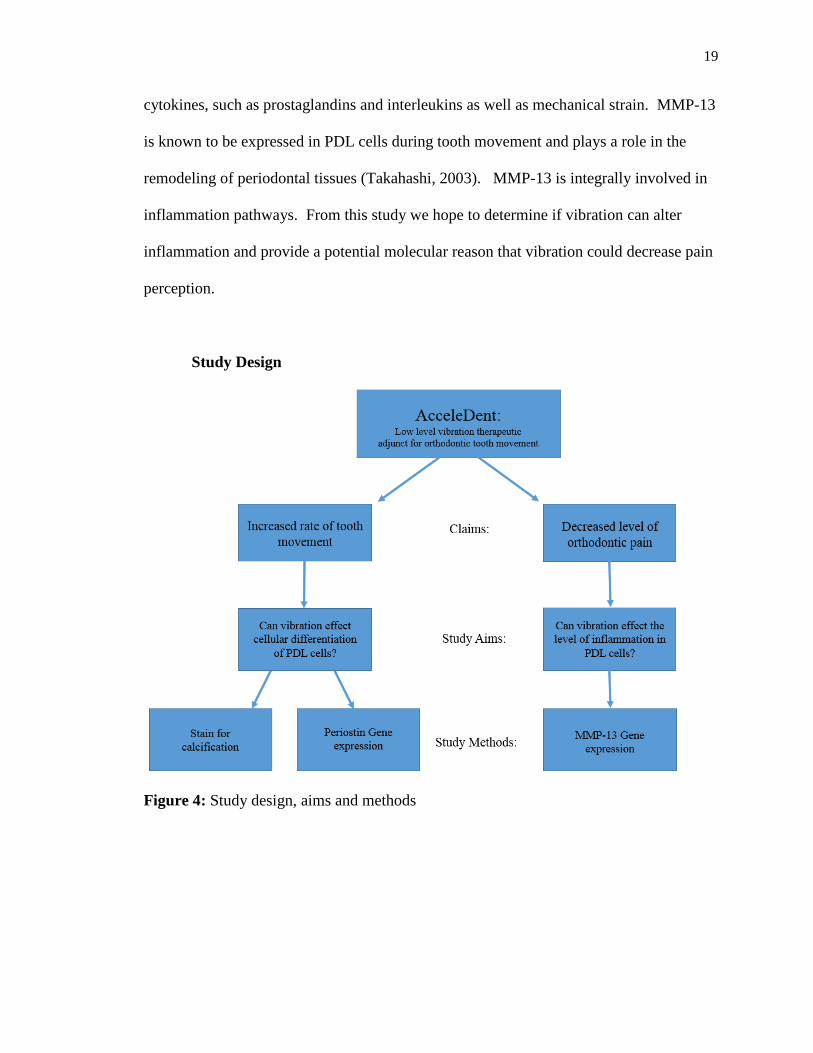
Study Design
Figure 4: Study design, aims and methods

20
CHAPTER III
MATERIALS AND METHODS
Human Periodontal Ligament Fibroblasts
Human periodontal ligament cells (#2630) from ScienCell Research Laboratories
were chosen for this study. The periodontal ligament is important in the maintenance and
regeneration of periodontal tissue, and plays a key role in orthodontic tooth movement.
The cells of the PDL are primarily fibroblasts but can be multipotent or composed of
heterogeneous cell populations that differ in function. Below is a picture of human
periodontal ligament fibroblasts #2630 from ScienCell Research Laboratories.
Human Periodontal Ligament Cells
Figure 5: Human periodontal ligament fibroblast cells from ScienCell Research
Laboratories seen under 200x magnification. Reprinted from ScienCell Research
Laboratories. (http://www.sciencellonline.com/human-periodontal-ligament-
fibroblasts.html)

21
Mechanical vibration setup
Plated cells were placed on a rigid platform. A base capable of delivering
measured amounts of vertical vibration was generated by a modular piezoelectric device.
The vibration frequency of 30Hz was controlled by a function generator (Instek: Model
FG 8015G). A current amplifier (Advanced Motion controls, Camarillo CA, Model
Brush Type PWM Servo Amplifier) delivered 0.3 g of acceleration to the vibration plate.
To verify the amount of vibration cells received an accelerometer was connected to the
cell plates (Endevco). The mechanical setup was housed in a thermos box of 37oC. A
similar setup, used by Kulkarni et al, 2013 is shown in the figure below.
Mechanical Vibration Set-up Diagram
Figure 6: Mechanical vibration system composed of an enclosed 37oC heated box,
vibration generator and platform for plated cells. Vibration is controlled by a piezo
amplifier and controller. An accelerometer measures output. Reprinted from “Mechanical
vibration inhibits osteoclast formation by reducing DC-STAMP receptor expression in
osteoclast precursor cells,” by R.N. Kulkami, P.A Voglewede and D. Liu, 2013, Bone, 57,
493-498. Copyright 2013 by Elsevier. Reprinted with permission.

22
Mechanical Vibration Set-up
Figure 7: Enclosed thermal box at 370C. Platform delivering vibration to plate of cells.
Accelerometer attached on top of plate.
Experimental setup, groups and procedure
1) Differentiation Assay
Human PDL cells were cultured at 37 °C and 5% CO2. A control group was
cultured in DMEM supplemented with 10 % Fetal Bovine Serum (FBS) and 1%
Penicillin/Streptomycin. The experimental group was cultured in an osteogenic medium
containing β-glycerophosphate (10 mM), ascorbic acid (50 µg/ml) and dexamethasone
(0.1 µM). After 2 days, the cells were seeded on 24-well culture plates at a density of
5x10⁵/well, and assigned within the plate to the study groups shown below (Table 1). IL-
1β working concentration was 1ng/ml.

23
Cell Differentiation Experimental Groups
Control medium Control medium + IL-1β
Differentiation medium Differentiation medium + IL-1β
Table 1: Experimental groups of PDL differentiation.
After confluence, the cells were either subjected to 0.3g/30Hz mechanical vibration or
treated as sham control (0g/0Hz) for 1 hour per day over 21 consecutive days. Culture
medium was refreshed every 3 days.
The level of differentiation of the PDL cells was examined using Alizarin Red
staining. In details, the cells were washed with PBS and fixed with 10% formalin for 15
minutes at room temperature. Following, the cells were washed twice with distilled
water and incubated at room temperature for 20 min with gentle shaking and addition of
10 % Alizarin (adjusted to 4.1-4.3pH with 0.5% NaOH). After incubation, the stained
cells were washed four times with distilled water. The results of staining were observed
under a reverse microscope and scanned into .tiff files by using Photoshop software
(version CS6, 13.0.1). The stains were qualitatively analyzed.
2) Gene expression
Human PDL cells were maintained in 6-well plates with DMEM supplemented
with 10 % Fetal Bovine Serum (FBS) and 1% Penicillin/Streptomycin at 37 °C and 5%
CO2. The cells were used in gene expression experiments after confluence,
approximately 1-2 days after seeding. The four experimental groups, all seeded in
separate plates, are shown below (Table2).
24
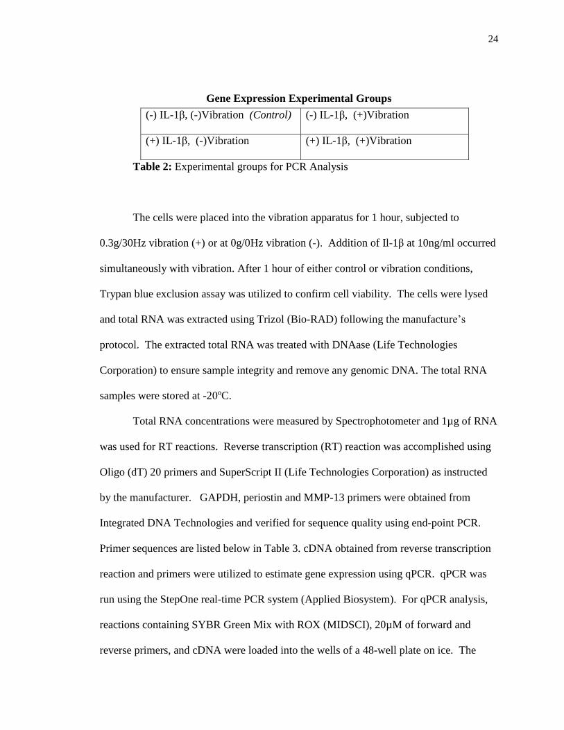
Gene Expression Experimental Groups
Table 2: Experimental groups for PCR Analysis
The cells were placed into the vibration apparatus for 1 hour, subjected to
0.3g/30Hz vibration (+) or at 0g/0Hz vibration (-). Addition of Il-1β at 10ng/ml occurred
simultaneously with vibration. After 1 hour of either control or vibration conditions,
Trypan blue exclusion assay was utilized to confirm cell viability. The cells were lysed
and total RNA was extracted using Trizol (Bio-RAD) following the manufacture’s
protocol. The extracted total RNA was treated with DNAase (Life Technologies
Corporation) to ensure sample integrity and remove any genomic DNA. The total RNA
samples were stored at -20oC.
Total RNA concentrations were measured by Spectrophotometer and 1µg of RNA
was used for RT reactions. Reverse transcription (RT) reaction was accomplished using
Oligo (dT) 20 primers and SuperScript II (Life Technologies Corporation) as instructed
by the manufacturer. GAPDH, periostin and MMP-13 primers were obtained from
Integrated DNA Technologies and verified for sequence quality using end-point PCR.
Primer sequences are listed below in Table 3. cDNA obtained from reverse transcription
reaction and primers were utilized to estimate gene expression using qPCR. qPCR was
run using the StepOne real-time PCR system (Applied Biosystem). For qPCR analysis,
reactions containing SYBR Green Mix with ROX (MIDSCI), 20µM of forward and
reverse primers, and cDNA were loaded into the wells of a 48-well plate on ice. The
(-) IL-1β, (-)Vibration (Control) (-) IL-1β, (+)Vibration
(+) IL-1β, (-)Vibration (+) IL-1β, (+)Vibration
25
cycling parameters were 95°C for 10 minutes followed by 40 cycles of 95°C (15s), and
60°C (60s). A melting curve was observed to ensure amplification of a single product.
All reactions were performed in triplicate and the mean threshold cycle (ct) for each gene
product for each sample was used for analysis. The mRNA levels of target genes were
normalized to that of the housekeeping gene, GAPDH. The entire experiment was
repeated three times. Statistical analysis of ANOVA was performed.
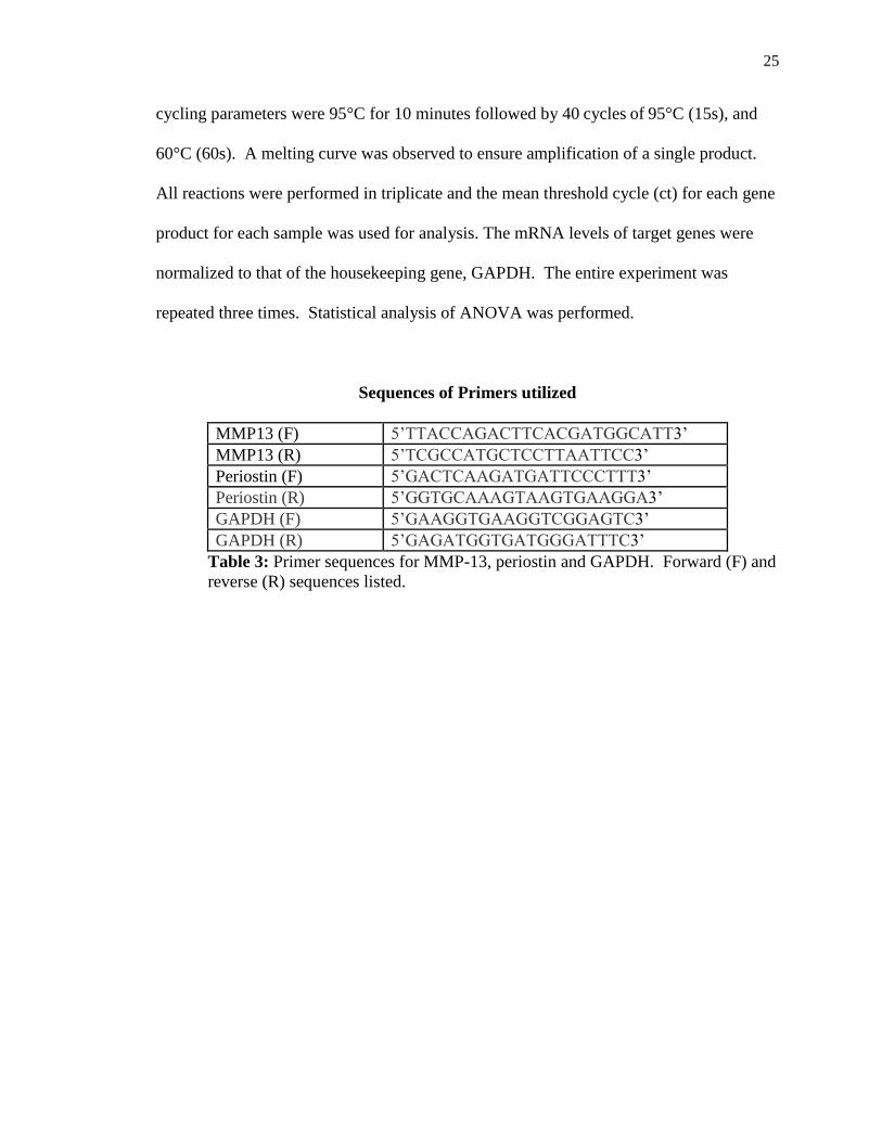
Sequences of Primers utilized
MMP13 (F) 5’TTACCAGACTTCACGATGGCATT3’
MMP13 (R) 5’TCGCCATGCTCCTTAATTCC3’
Periostin (F) 5’GACTCAAGATGATTCCCTTT3’
Periostin (R) 5’GGTGCAAAGTAAGTGAAGGA3’
GAPDH (F) 5’GAAGGTGAAGGTCGGAGTC3’
GAPDH (R) 5’GAGATGGTGATGGGATTTC3’
Table 3: Primer sequences for MMP-13, periostin and GAPDH. Forward (F) and
reverse (R) sequences listed.

26
CHAPTER IV
RESULTS
A) Differentiation Assay
Results of Alizarin Red staining of the PDL cells subjected to IL-1β and vibration
are shown in Figure 8. PDL cells cultured in the control medium, DMEM with 10% FBS
and 1% Penicillin/Streptomycin, produced no calcification in any treatment conditions.
Whereas Alizarin Red staining shows high calcification in the cells cultured in osteogenic
medium (differentiation medium). Addition of IL-1β markedly decreased calcification
seen in the cells cultured in osteogenic medium, however did not fully return to zero
levels as seen in control medium cultured cells. Vibration at 0.3g/30Hz had no effect on
the levels of calcification seen in all treatment groups, as seen in Figure 8.

27
Staining of PDL cells with Alizarin Red
Figure 8: Picture of plated cells stained with Alizarin Red. Stained cells show
calcification.

28
Qualitative analysis was performed to compare samples, with a 4 degrees scale of
(-) no calcification noted, (+) slight staining, (++) moderate staining, and (+++) complete
staining of sample. The qualitative analysis is shown in Figure 9 and results summarized
in a bar graph in Figure 10. The cells subjected to osteogenic medium had moderate
calcification, reduced to slight levels with addition of IL-1β. Vibration samples mirrored
these levels. If 0.3g/30Hz vibration positively affects the cellular differentiation of PDL
cells, an increase in calcification would be expected. However, no differences in
calcification between PDL cells treated with 0.3g/30Hz vibration and without could be
determined.
Qualitative Analysis of PDL Differentiation
Figure 9: Qualitative Analysis of PDL differentiation, with listed scale

29
Figure 10: Qualitative analysis of PDL Differentiation

30
2) Gene Expression Results
In this experiment, we wanted to see the gene expressions of MMP-13 and
periostin compared with the housekeeping gene GAPDH in PDL cells subjected to IL-1β
with or without vibration. Prior to utilization within the experiment, the quality of the
primers, GAPDH, MMP-13 and periostin was verified. End-point PCR using TAQ
polymerase enzyme showed distinct bands of product at expected size, as shown in
Figure 11. End-point PCR also showed some contamination, so the samples were treated
using DNAse prior to qPCR.
Quality of primers; GAPDH, MMP-13, Periostin
Figure 11: End-point PCR analysis of the PCR primers for GAPDH, MMP13, periostin.
All three sets of intense bands at expected sizes confirmed the primers’ reliability.
31
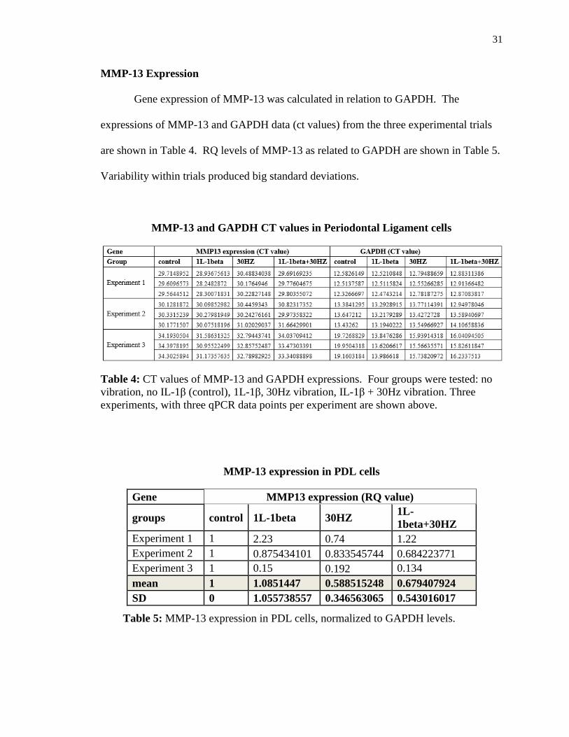
MMP-13 Expression
Gene expression of MMP-13 was calculated in relation to GAPDH. The
expressions of MMP-13 and GAPDH data (ct values) from the three experimental trials
are shown in Table 4. RQ levels of MMP-13 as related to GAPDH are shown in Table 5.
Variability within trials produced big standard deviations.
MMP-13 and GAPDH CT values in Periodontal Ligament cells
Table 4: CT values of MMP-13 and GAPDH expressions. Four groups were tested: no
vibration, no IL-1β (control), 1L-1β, 30Hz vibration, IL-1β + 30Hz vibration. Three
experiments, with three qPCR data points per experiment are shown above.
MMP-13 expression in PDL cells
Table 5: MMP-13 expression in PDL cells, normalized to GAPDH levels.
Gene MMP13 expression (RQ value)
groups control 1L-1beta 30HZ 1L-
1beta+30HZ
Experiment 1 1 2.23 0.74 1.22
Experiment 2 1 0.875434101 0.833545744 0.684223771
Experiment 3 1 0.15 0.192 0.134
mean 1 1.0851447 0.588515248 0.679407924
SD 0 1.055738557 0.346563065 0.543016017

32
Looking at data trends, IL-1β increased MMP-13 expression but was similar to
the control group, only 0.1 fold higher. With addition of vibration, MMP-13 expression
decreased in both conditions. Without IL-1β, a decrease of 0.4 fold and with IL-1β, 0.3
fold. This is summarized in graphical format in Figure 12. Due to variability within the
trials, ANOVA analysis led to no statistical significance and a p value of 0.72, Table 6.
MMP-13 expression in PDL cells subjected to IL-1β and 30Hz Vibration
Figure 12: Data Trends, MMP-13 expression in PDL cells, normalized to GAPDH.
MMP-13 ANOVA
Table 6: MMP-13 ANOVA analysis. No statistically significant findings, p =.720 (≤0.5)

33
Periostin Expression
Gene expression of periostin was also compared in relation to GAPDH.
Expressions of periostin and GAPDH data from the three experimental trials are shown in
Table 7. RQ levels of periostin as related to GAPDH are shown in Table 8.
Periostin and GAPDH CT values in Periodontal Ligament Cells
Table 7: CT values of Periostin and GAPDH expressions. Four groups were tested: no
vibration, no IL-1β (control), 1L-1β, 30Hz vibration, IL-1β + 30Hz vibration. Three
experiments, with three qPCR data points per experiment are shown above.
Periostin expression in PDL cells
Gene Periostin expression (RQ value)
group control 1L-1beta 30HZ 1L-
1beta+30HZ
Experiment 1 1 1.87 1.19 1.05
Experiment 2 1 0.916033447 1.278030753 1.425864577
Experiment 3 1 0.669 0.668 0.659
mean 1 1.151677816 1.045343584 1.044954859
SD 0 0.634229021 0.329740032 0.383457182
Table 8: Periostin expression in PDL cells, normalized to GAPDH levels.

34
Periostin expression in all groups was very similar. Compared with control,
periostin expression in the presence of IL-1β slightly increased by 0.15 fold. With
addition of vibration a 0.04 fold increase occurred with and without IL-1β. This is
summarized in graphical format in Figure 13. As stated only slight difference in periostin
expression was found across all groups and ANOVA analysis confirmed no statistical
significance and a p value of 0.971, Table 9.
Periostin expression in PDL cells subjected to IL-1β and 30Hz Vibration
Figure 13: Periostin expression in PDL cells, normalized to GAPDH levels.
Periostin ANOVA
Table 9: Periostin ANOVA analysis. No statistically significant findings, p=.971 (≤0.5)

35
CHAPTER V
DISCUSSION
Experimental Setup
The study goal was to see the effects of vibration on cellular differentiation and
inflammation under orthodontic tooth movement. Periodontal ligament cells (PDL) were
chosen due to their essential role in response to mechanical force and capacity for
renewal and repair during orthodontic tooth movement (Lekic, 1996). As an important
player in the events of alveolar bone remodeling, PDL cells are an ideal population of
cells to study. Periodontal ligament cells are primarily fibroblasts but also can include
osteoblasts, osteoclasts, epithelial cell rests of Malassez, macrophages, undifferentiated
mesenchymal cells, neural elements, cementoblasts and endothelial cells (Lekic, 1996).
Previous studies of differentiation, however, utilized PDL cells from animals, or PDL
stem cell populations only, which can be limitations. Hence our study utilized cells
obtained from ScienCell Laboratories which were human PDL in origin with a
heterogeneous mix of cells as seen within an in vivo periodontal ligament.
Other aspects of our study also sought to provide the most pertinent view of
orthodontics. IL-1β was utilized as our model of orthodontic stress, as IL-1β is a key
cytokine that is released early to response to mechanical stress (Krishnan, 2006).
Vibration frequency of 30Hz and magnitude of 0.3g paralleled the output of AcceleDent,
the FDA approved vibration treatment adjunct currently on the orthodontic market. To
study the mRNA changes of cytokines i.e. MMP-13 and periostin, a short treatment time
of 1 hour was chosen to see immediate and pronounced molecular effects of vibration.
Whereas, a longer treatment time for the differentiation experiment of 21 days was

36
utilized to gain insight on how cells react to extended treatment, as occlusal vibration in
orthodontics is also extended, usually 20 min a day over the course of an entire treatment,
usually 12-24 months.
Cellular Staining Results
The differentiation aspect of our study confirmed that the human PDL cells can be
differentiated, capable of calcification and osteogenesis, as seen when PDL cells were
cultured in osteogenic medium. This is consistent with the findings from Wolf et al who
showed transplanted human PDL cells into mice can show osteogenic and cementogenic
differentiation. The transplanted cells in their study showed osteogenic markers for
alkaline phosphatase, osteopontin, PTH-receptor 1 and osteoclacin as well as early
mineralization by calcium and phosphate staining (Wolf, 2013). Lin et al, went further
into detail about how this process occurs. After tooth extraction in rats, Lin et al
monitored PDL fibroblasts and their role in osteogenesis. After extraction PDL cells
began to proliferate highly, peaking at 24 hours. Between 1-2 days, cells began to
migrate into the coagulum and by 3 days were producing collagen fibers and dense
connective tissue. By day 4-5 some PDL fibroblasts had differentiated into osteoblasts
and began to lay down new bone (Lin 1994).
With the addition of IL-1β as our model of orthodontic tooth movement, we see a
marked decrease of calcification. A significant decrease of calcification was seen in the
sham control plate when IL-1β was added and similar decrease was seen in the 30Hz
vibration group. This shows that IL-1β inhibits differentiation. This is to be expected as
inflammation factors, such as IL-1β, have global catabolic effects. While IL-1β is
37
important for bone resorption, it inhibits bone formation (Iwasaki, 2001). By lowering
the differentiation potential of PDL cells, IL-1β concentrations could decrease the rate of
bone formation. Any intervention to offset this potential could lead to an avenue to
increase the rate of orthodontic tooth movement.
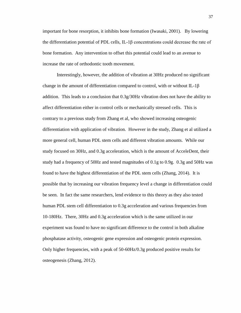
Interestingly, however, the addition of vibration at 30Hz produced no significant
change in the amount of differentiation compared to control, with or without IL-1β
addition. This leads to a conclusion that 0.3g/30Hz vibration does not have the ability to
affect differentiation either in control cells or mechanically stressed cells. This is
contrary to a previous study from Zhang et al, who showed increasing osteogenic
differentiation with application of vibration. However in the study, Zhang et al utilized a
more general cell, human PDL stem cells and different vibration amounts. While our
study focused on 30Hz, and 0.3g acceleration, which is the amount of AcceleDent, their
study had a frequency of 50Hz and tested magnitudes of 0.1g to 0.9g. 0.3g and 50Hz was
found to have the highest differentiation of the PDL stem cells (Zhang, 2014). It is
possible that by increasing our vibration frequency level a change in differentiation could
be seen. In fact the same researchers, lend evidence to this theory as they also tested
human PDL stem cell differentiation to 0.3g acceleration and various frequencies from
10-180Hz. There, 30Hz and 0.3g acceleration which is the same utilized in our
experiment was found to have no significant difference to the control in both alkaline
phosphatase activity, osteogenic gene expression and osteogenic protein expression.
Only higher frequencies, with a peak of 50-60Hz/0.3g produced positive results for
osteogenesis (Zhang, 2012).

38
Although Zhang et al has studied PDL stem cell’s differentiation responses to
vibration, it is worth note that they did not investigate whether a change would be seen in
an inflammatory environment, as seen with orthodontic tooth movement. With addition
of IL-1β, an orthodontic model of stress, our intent was to see if a change in
differentiation could be seen in an inflammatory environment. However our results
indicate no change of PDL differentiation with or without IL-1β. This could indicate that
vibration of 0.3g/30Hz has no measurable impact on healthy or inflammatory-stressed
PDL cells.
Gene Expression Results
Orthodontic tooth movement relies on recruitment of cells within the PDL to
respond to mechanical stress and differentiate into cells capable of tissue remodeling.
Cytokine mediators within orthodontic tooth movement help orchestrate these cellular
responses to tooth movement. These cytokines have many regulatory and signaling
functions. We were interested in two specific cytokines, periostin and MMP-13.
Periostin has roles in the bone remodeling and differentiation of cells during tooth
movement. Periostin preserves bone mass and promotes bone remodeling (Conway,
2013). Higher levels of periostin increase osteoblast proliferation, and differentiation
(Zhu, 2009). Since one of our aims is to determine if vibration has an effect on
differentiation of PDL cells, periostin was chosen.
MMP-13 is a collagenase and an inflammatory cytokine. By measuring MMP-13
mRNA expression within the cell, we hoped to see if vibration had an effect on overall
cellular inflammation. While inflammation cytokines are needed to facilitate orthodontic
39
tooth movement, higher than physiologic levels of inflammation can bring up side effects
including pain, root resorption and exasperate periodontal disease. Within orthodontics,
it would be positive if vibration therapy could increase the rate of orthodontic tooth
movement while simultaneously reducing the overall level of inflammation.
As can be seen, the qPCR analyses of MMP-13 and periostin produced large
variations within the three experiments which limited our results and consequently
produced no statistically significant conclusions. However, general trends can be seen to
which future study can be oriented towards. Analysis of MMP-13 using PDL cells
showed similar expression in both control and IL-1β treated cells. These similar levels
could be due to a lack of quality, concentration or treatment time of IL-1β. With addition
of vibration, a parallel decrease is seen with and without addition of IL-1β. This may
show that vibration can lower cellular inflammation. These findings parallel other
studies. Xu et al looked at cyclic tensile strain on chondrocytes and found that low
vibration of 0.5 Hz acted as an antagonist of IL-1β, decreasing inflammation (Xu, 2000).
Similarly, Lavagnino et al showed decrease of MMP-1, interstitial collagenases
expression in tendon cells when subjected to 0.17 Hz to 1.0 Hz (Lavagnino 2003). Our
preliminary findings that vibration may decrease MMP-13 production in PDL cells are
interesting and further studies are warranted.
The qPCR results of periostin expression in this study are more complicated to
explain. In our experiments, a slight increase in periostin is seen with addition of IL-1β,
however similar to control values are seen with addition of vibration with and without IL-
1β treatment. No values produced statistically significant results and no trend could be
determined. This could be related to the following factors. First, periostin is related to

40
the differentiation of cells during tooth movement. As seen previously, 0.3g/30Hz
vibration did not produce any change in PDL osteogenic differentiation when stained
with Alizarin Red. Since there were no significant results nor any general trends seen
within periostin expression, it could once again be evidence that vibration at this level is
not capable of producing differentiation of PDL cells. Secondly, periostin is a complex
molecule, with many different roles of which many are not fully understood. Cobo states
that periostin seems to have different expression based on cell type and degree of
mechanical stress (Cobo, 2016) and as stated previously even within tooth movement
differing amounts of periostin occur within different areas of the PDL (Wilde, 2003).
Thus perhaps our experimental model is not adequate to study periostin expression in
orthodontic tooth movement. Continued studies may benefit from utilizing a different
cytokine to focus on.
Limitations of the Study
During this study there are limitations that could have affected our results. First,
orthodontic tooth movement was simulated in vitro by utilizing IL-1β. While this model
is warranted, as IL-1β is produced during initial orthodontic tooth movement and has
been shown to regulate bone resorption and bone formation by mechanical stress
(Davidovitch, 1988 and Preiss, 1994), orthodontic tooth movement is complex and many
factors, cytokines and cells are actively involved. In such, IL-1β is a simplistic model,
although mimicking all conditions that occur in vivo is not possible. Secondly, IL-1β
was utilized in concentrations of 1 ng/ml for differentiation and 10 ng/ml for qPCR
analysis. There is limited research available to determine in vivo concentrations of IL-1β
41
during orthodontic tooth movement and studies that exist attempt to measure gingival
crevicular fluid (GCF), a process that is inexact. Preiss reported that the concentration of
IL-1β in healthy patients GCF to be within the range of 22.8 ng/ml – 150 ng/ml and 85.8
ng/ml - 882.2 ng/ml in periodontitis patients (Preiss 1994). This would signal to us that
our concentration of IL-1β was too low. However a study of orthodontic tooth movement
on IL-1β in canines showed a lower concentration. In this study teeth showed a
concentration of 0.10 ng/ml of IL-1β in the GCF of orthodontically moved teeth
(Leethanakul, 2016). Different concentrations of IL-1β in vivo reported in literature,
leads to confusion on how much to utilize as a study model. Future studies may utilize
higher concentrations or longer treatment time of IL-1β on the PDL cells.
The nature of our qPCR results was also a limiting factor of this study. Variations
within our qPCR results was seen. This could have been from many factors and due to
the numerous steps from cell culture to qPCR analysis, any variability or inconsistency is
of great detriment. This variations led to big standard deviations and no statistically
significant conclusions. Such large end stage variability may point to errors in the
beginning of cell culture. Future experiments under close laboratory control are needed
to obtain a larger sample size with more consistency.
Clinical Implications and Conclusions
Low magnitude, high frequency vibration is an interesting therapeutic adjunct or
treatment that many medical fields are studying. There are studies that show the positive
results of vibration in treating osteoporosis, maintaining strength when a patient is
disabled, during facture healing, and for tendon and muscle healing (Edwards 2015).

42
Low magnitude, high frequency vibration in the field of orthodontics may have benefits
such as increasing osteogenesis and decreasing inflammation. The implications of these
could help many people who seek orthodontic care. The negative aspects of orthodontics
such as increased caries and gingival hypertrophy due to poor oral hygiene combined
with extended treatment times, and inflammation modulated side effects such as pain,
root resorption and periodontal breakdown could be reduced if the rate of orthodontic
tooth movement could be increased.
Our study focused specifically on the magnitude and frequency of the most
common vibration treatment adjunct available in orthodontics, AcceleDent at 30Hz, 0.3g.
AcceleDent claims to increase the rate orthodontic tooth movement and decrease pain.
While other studies focus on different frequencies and magnitudes, this study sought to
limit focus to this commercially available treatment adjunct to make findings clinically
relevant. In our study, the addition of vibration at 0.3g/30Hz did not change the
differentiation of PDL cells, both with and without an inflammatory environment. Thus
if AcceleDent does indeed produce faster orthodontic tooth movement, this study implies
that PDL differentiation is not the mechanism. Also our gene expression results indicate
that inflammation in PDL cells may be reduced with vibration, however our data trends
were not statistically significant. Hence, these data alone do not support reduction in
inflammation as a mechanism that pain could be decreased. However further
experiments focusing on reducing experimental variability are warranted at investigating
vibration’s effect on PDL inflammation.

43
BIBLIOGRAPHY
Alhashimi, N., Frithiof, L., Brudvik, P., & Bakhiet, M. (2001). Orthodontic tooth
movement and de novo synthesis of proinflammatory cytokines. American Journal
of Orthodontics and Dentofacial Orthopedics, 119(3), 307-312.
Alikhani, M., Khoo, E., Alyami, B., Raptis, M., Salgueiro, J. M., Oliveira, S. M., et al.
(2012). Osteogenic effect of high-frequency acceleration on alveolar bone. Journal
of Dental Research, 91(4), 413-419.
Alikhani, M., Lopez, J. A., Alabdullah, H., Vongthongleur, T., Sangsuwon, C., Alikhani,
M., et al. (2016). High-frequency acceleration: Therapeutic tool to preserve bone
following tooth extractions. Journal of Dental Research, 95(3), 311-318.
Bollen, A. M., Cunha-Cruz, J., Bakko, D. W., Huang, G. J., & Hujoel, P. P. (2008). The
effects of orthodontic therapy on periodontal health: A systematic review of
controlled evidence. Journal of the American Dental Association, 139(4), 413-422.
Brezniak, N., & Wasserstein, A. (2002). Orthodontically induced inflammatory root
resorption. part I: The basic science aspects. The Angle Orthodontist, 72(2), 175-179.
Brown, D. F., & Moerenhout, R. G. (1991). The pain experience and psychological
adjustment to orthodontic treatment of preadolescents, adolescents, and adults.
American Journal of Orthodontics and Dentofacial Orthopedics, 100(4), 349-356.
Capelli, J.,Jr, Kantarci, A., Haffajee, A., Teles, R. P., Fidel, R.,Jr, & Figueredo, C. M.
(2011). Matrix metalloproteinases and chemokines in the gingival crevicular fluid
during orthodontic tooth movement. European Journal of Orthodontics, 33(6), 705-
711.
Cardaropoli, D., Gaveglio, L., Abou-Arraj, R.V. (2014). Orthodontic movement and
periodontal bone defects: Rationale, timing and clinical implications. Seminars in
Orthodontics, 20(3), 177-187.
Chien, H. H., Lin, W. L., & Cho, M. I. (1999). Interleukin-1beta-induced release of matrix
proteins into culture media causes inhibition of mineralization of nodules formed by
periodontal ligament cells in vitro. Calcified Tissue International, 64(5), 402-413.
Cobo, T., Viloria, C. G., Solares, L., Fontanil, T., Gonzalez-Chamorro, E., De Carlos, F.,
et al. (2016). Role of periostin in adhesion and migration of bone remodeling cells.
PloS One, 11(1), e0147837.
Conway, S. J., Izuhara, K., Kudo, Y., Litvin, J., Markwald, R., Ouyang, G., et al. (2014).
The role of periostin in tissue remodeling across health and disease. Cellular and
Molecular Life Sciences, 71(7), 1279-1288.

44
Davidovitch, Z., Nicolay, O. F., Ngan, P. W., & Shanfeld, J. L. (1988). Neurotransmitters,
cytokines, and the control of alveolar bone remodeling in orthodontics. Dental
Clinics of North America, 32(3), 411-435.
Diercke, K., Sen, S., Kohl, A., Lux, C. J., & Erber, R. (2011). Compression-dependent up-
regulation of ephrin-A2 in PDL fibroblasts attenuates osteogenesis. Journal of
Dental Research, 90(9), 1108-1115.
Edwards, J. H., & Reilly, G. C. (2015). Vibration stimuli and the differentiation of
musculoskeletal progenitor cells: Review of results in vitro and in vivo. World
Journal of Stem Cells, 7(3), 568-582.
Egolf, R. J., BeGole, E. A., & Upshaw, H. S. (1990). Factors associated with orthodontic
patient compliance with intraoral elastic and headgear wear. American Journal of
Orthodontics and Dentofacial Orthopedic, 97(4), 336-348.
Ekblom, A., & Hansson, P. (1982). Effects of conditioning vibratory stimulation on pain
threshold of the human tooth. Acta Physiologica Scandinavica, 114(4), 601-604.
El-Angbawi, A., McIntyre, G. T., Fleming, P. S., & Bearn, D. R. (2015). Non-surgical
adjunctive interventions for accelerating tooth movement in patients undergoing
fixed orthodontic treatment. The Cochrane Database of Systematic Reviews, 11,
CD010887.
Huang, H., Williams, R. C., & Kyrkanides, S. (2014). Accelerated orthodontic tooth
movement: Molecular mechanisms. American Journal of Orthodontics and
Dentofacial Orthopedics, 146(5), 620-632.
Human Periodontal Ligament Fibroblasts, Catalog #2630. (2016, June 20). Retrieved
from http://www.sciencellonline.com/human-periodontal-ligament-fibroblasts.html
Introducing AcceleDent. (2016, June 20). Retrieved from http://acceledent.com/how-it-
works/introducing-acceledent/
Ito, Y., Kimura, T., Nam, K., Katoh, A., Masuzawa, T., & Kishida, A. (2011). Effects of
vibration on differentiation of cultured PC12 cells. Biotechnology and
Bioengineering, 108(3), 592-599.
Iwasaki, L. R., Haack, J. E., Nickel, J. C., Reinhardt, R. A., & Petro, T. M. (2001). Human
interleukin-1 beta and interleukin-1 receptor antagonist secretion and velocity of
tooth movement. Archives of Oral Biology, 46(2), 185-189.
Jones, M., & Chan, C. (1992). The pain and discomfort experienced during orthodontic
treatment: A randomized controlled clinical trial of two initial aligning arch wires.
American Journal of Orthodontics and Dentofacial Orthopedics, 102(4), 373-381.
Jones, M. L., & Richmond, S. (1985). Initial tooth movement: Force application and pain-
-a relationship? American Journal of Orthodontics, 88(2), 111-116.

45
Kalajzic, Z., Peluso, E. B., Utreja, A., Dyment, N., Nihara, J., Xu, M., et al. (2014). Effect
of cyclical forces on the periodontal ligament and alveolar bone remodeling during
orthodontic tooth movement. The Angle Orthodontist, 84(2), 297-303.
Kapoor, P., Kharbanda, O. P., Monga, N., Miglani, R., & Kapila, S. (2014). Effect of
orthodontic forces on cytokine and receptor levels in gingival crevicular fluid: A
systematic review. Progress in Orthodontics, 15, 65-014-0065-6.
Kau, C.H., Nguyen, J.T., English, J.D. (2010). The clinical evaluation of a novel cyclical
force generating device in orthodontics. Orthodontic Practice US, 1(1), 10-15.
Koyama, Y., Mitsui, N., Suzuki, N., Yanagisawa, M., Sanuki, R., Isokawa, K., et al.
(2008). Effect of compressive force on the expression of inflammatory cytokines and
their receptors in osteoblastic saos-2 cells. Archives of Oral Biology, 53(5), 488-496.
Krishnan, V. (2007). Orthodontic pain: From causes to management--a review. European
Journal of Orthodontics, 29(2), 170-179.
Krishnan, V., & Davidovitch, Z. (2006). Cellular, molecular, and tissue-level reactions to
orthodontic force. American Journal of Orthodontics and Dentofacial Orthopedics,
129(4), 469.e1-469.32.
Kulkarni, R. N., Voglewede, P. A., & Liu, D. (2013). Mechanical vibration inhibits
osteoclast formation by reducing DC-STAMP receptor expression in osteoclast
precursor cells. Bone, 57(2), 493-498.
Kusano, K., Miyaura, C., Inada, M., Tamura, T., Ito, A., Nagase, H., et al. (1998).
Regulation of matrix metalloproteinases (MMP-2, -3, -9, and -13) by interleukin-1
and interleukin-6 in mouse calvaria: Association of MMP induction with bone
resorption. Endocrinology, 139(3), 1338-1345.
Lau, E., Al-Dujaili, S., Guenther, A., Liu, D., Wang, L., & You, L. (2010). Effect of low-
magnitude, high-frequency vibration on osteocytes in the regulation of osteoclasts.
Bone, 46(6), 1508-1515.
Lavagnino, M., Arnoczky, S. P., Tian, T., & Vaupel, Z. (2003). Effect of amplitude and
frequency of cyclic tensile strain on the inhibition of MMP-1 mRNA expression in
tendon cells: An in vitro study. Connective Tissue Research, 44(3-4), 181-187.
Leethanakul, C., Suamphan, S., Jitpukdeebodintra, S., Thongudomporn, U.,
Charoemratrote, C. (2016). Vibratory stimulation increases interleukin-1 beta
secretion during orthodontic tooth movement. The Angle Orthodontist, 86(1), 74-80.
Lekic, P., & McCulloch, C. A. (1996). Periodontal ligament cell population: The central
role of fibroblasts in creating a unique tissue. The Anatomical Record, 245(2), 327-
341.

46
Liang, Z. J., Zhuang, H., Wang, G. X., Li, Z., Zhang, H. T., Yu, T. Q., et al. (2012).
MiRNA-140 is a negative feedback regulator of MMP-13 in IL-1beta-stimulated
human articular chondrocyte C28/I2 cells. Inflammation Research, 61(5), 503-509.
Lin, W. L., McCulloch, C. A., & Cho, M. I. (1994). Differentiation of periodontal
ligament fibroblasts into osteoblasts during socket healing after tooth extraction in
the rat. The Anatomical Record, 240(4), 492-506.
Linden, R. W., & Millar, B. J. (1989). The effect of vibration on the discharge of
periodontal ligament mechanoreceptors to controlled loading of the cat canine tooth.
Archives of Oral Biology, 34(4), 275-281.
Lobre, W. D., Callegari, B. J., Gardner, G., Marsh, C. M., Bush, A. C., & Dunn, W. J.
(2015). Pain control in orthodontics using a micropulse vibration device: A
randomized clinical trial. The Angle Orthodontist,
Luppanapornlarp, S., Kajii, T. S., Surarit, R., & Iida, J. (2010). Interleukin-1beta levels,
pain intensity, and tooth movement using two different magnitudes of continuous
orthodontic force. European Journal of Orthodontics, 32(5), 596-601.
Meikle, M. C. (2006). The tissue, cellular, and molecular regulation of orthodontic tooth
movement: 100 years after Carl Sandstedt. European Journal of Orthodontics, 28(3),
221-240.
Miles, P., Smith, H., Weyant, R., & Rinchuse, D. J. (2012). The effects of a vibrational
appliance on tooth movement and patient discomfort: A prospective randomised
clinical trial. Australian Orthodontic Journal, 28(2), 213-218.
Mussig, E., Tomakidi, P., & Steinberg, T. (2005). Molecules contributing to the
maintenance of periodontal tissues, their possible association with orthodontic tooth
movement. Journal of Orofacial Orthopedics, 66(6), 422-433.
Ngan, P., Wilson, S., Shanfeld, J., & Amini, H. (1994). The effect of ibuprofen on the
level of discomfort in patients undergoing orthodontic treatment. American Journal
of Orthodontics and Dentofacial Orthopedics, 106(1), 88-95.
Nishimura, M., Chiba, M., Ohashi, T., Sato, M., Shimizu, Y., Igarashi, K., et al. (2008).
Periodontal tissue activation by vibration: Intermittent stimulation by resonance
vibration accelerates experimental tooth movement in rats. American Journal of
Orthodontics and Dentofacial Orthopedics, 133(4), 572-583.
Nogueira, A. V. B., Chaves de Souza, J. A., Kim, Y. J., Damiao de Sousa-Neto, M., Chan
Cirelli, C., & Cirelli, J. A. (2013). Orthodontic force increases interleukin-1beta and
tumor necrosis factor-alpha expression and alveolar bone loss in periodontitis.
Journal of Periodontology, 84(9), 1319-1326.

47
Nohutcu, R. M., McCauley, L. K., Koh, A. J., & Somerman, M. J. (1997). Expression of
extracellular matrix proteins in human periodontal ligament cells during
mineralization in vitro. Journal of Periodontology, 68(4), 320-327.
O' Connor, P. J. (2000). Patients' perceptions before, during, and after orthodontic
treatment. Journal of Clinical Orthodontics, 34(10), 591-592.
Pavlidis, D., Bourauel, C., Rahimi, A., Gotz, W., & Jager, A. (2009). Proliferation and
differentiation of periodontal ligament cells following short-term tooth movement in
the rat using different regimens of loading. European Journal of Orthodontics,
31(6), 565-571.
Pavlin, D., Anthony, R., Raj, V., Gakunga, P.T. (2015). Cyclic loading (vibration)
accelerates tooth movement in orthodontic patients: A double-blind, randomized
controlled trial. Seminars in Orthodontics, 21(3), 187-194.
Pavlin, D., & Gluhak-Heinrich, J. (2001). Effect of mechanical loading on periodontal
cells. Critical Reviews in Oral Biology and Medicine, 12(5), 414-424.
Preiss, D. S., & Meyle, J. (1994). Interleukin-1 beta concentration of gingival crevicular
fluid. Journal of Periodontology, 65(5), 423-428.
Proffit, W. R., Fields, H. W., & Sarver, D. M. (2007). Contemporary Orthodontics. St.
Louis, Mo: Mosby Elsevier.
Rangiani, A., Jing, Y., Ren, Y., Yadav, S., Taylor, R., & Feng, J. Q. (2015). Critical roles
of periostin in the process of orthodontic tooth movement. European Journal of
Orthodontics, pii: cjv071.
Ren, K., & Torres, R. (2009). Role of interleukin-1beta during pain and inflammation.
Brain Research Reviews, 60(1), 57-64.
Takahashi, I., Nishimura, M., Onodera, K., Bae, J. W., Mitani, H., Okazaki, M., et al.
(2003). Expression of MMP-8 and MMP-13 genes in the periodontal ligament
during tooth movement in rats. Journal of Dental Research, 82(8), 646-651.
Thompson, W. R., Yen, S. S., & Rubin, J. (2014). Vibration therapy: Clinical applications
in bone. Current Opinion in Endocrinology, Diabetes, and Obesity, 21(6), 447-453.
Veqar, Z., & Imtiyaz, S. (2014). Vibration therapy in management of delayed onset
muscle soreness. Journal of Clinical and Diagnostic Research, 8(6), LE01-4.
Wheeler, A. A., & Jacobson, B. H. (2013). Effect of whole-body vibration on delayed
onset muscular soreness, flexibility, and power. Journal of Strength and
Conditioning Research, 27(9), 2527-2532.

48
Wilde, J., Yokozeki, M., Terai, K., Kudo, A., & Moriyama, K. (2003). The divergent
expression of periostin mRNA in the periodontal ligament during experimental tooth
movement. Cell and Tissue Research, 312(3), 345-351.
Wolf, M., Lossdorfer, S., Abuduwali, N., Meyer, R., Kebir, S., Gotz, W., et al. (2013). In
vivo differentiation of human periodontal ligament cells leads to formation of dental
hard tissue. Journal of Orofacial Orthopedics, 74(6), 494-505.
Woodhouse N.R., DiBase A.T., Johnson, N., Slipper, C., Grant, J., Alsaleh, M., et al.
(2015). Supplemental vibration force during orthodontic alignment: a randomized
trial. Journal of Dental Research, 94(5), 682-689.
Woodhouse, N. R., DiBiase, A. T., Papageorgiou, S. N., Johnson, N., Slipper, C., Grant,
J., et al. (2015). Supplemental vibrational force does not reduce pain experience
during initial alignment with fixed orthodontic appliances: A multicenter
randomized clinical trial. Scientific Reports, 5, 17224.
Wu, J., Jiang, J. H., Xu, L., Liang, C., Bai, Y., & Zou, W. (2015). A pilot clinical study of
class III surgical patients facilitated by improved accelerated osteogenic orthodontic
treatments. The Angle Orthodontist, 85(4), 616-624.
Xu, Z., Buckley, M. J., Evans, C. H., & Agarwal, S. (2000). Cyclic tensile strain acts as an
antagonist of IL-1 beta actions in chondrocytes. Journal of Immunology, 165(1),
453-460.
Yadav, S., Dobie, T., Assefnia, A., Gupta, H., Kalajzic, Z., & Nanda, R. (2015). Effect of
low-frequency mechanical vibration on orthodontic tooth movement. American
Journal of Orthodontics and Dentofacial Orthopedics, 148(3), 440-449.
Yamamoto, T., Ugawa, Y., Yamashiro, K., Shimoe, M., Tomikawa, K., Hongo, S., et al.
(2014). Osteogenic differentiation regulated by rho-kinase in periodontal ligament
cells. Differentiation; Research in Biological Diversity, 88(2-3), 33-41.
Zainal Ariffin, S. H., Yamamoto, Z., Zainol Abidin, I. Z., Megat Abdul Wahab, R., &
Zainal Ariffin, Z. (2011). Cellular and molecular changes in orthodontic tooth
movement. The Scientific World Journal, 11, 1788-1803.
Zhang, C., Lu, Y., Zhang, L., Liu, Y., Zhou, Y., Chen, Y., et al. (2015). Influence of
different intensities of vibration on proliferation and differentiation of human
periodontal ligament stem cells. Archives of Medical Science, 11(3), 638-646.
Zhang, C., Li, J., Zhang, L., Zhou, Y., Hou, H., Quan, H., et al. (2012). Effects of
mechanical vibration on proliferation and osteogenic differentiation of human
periodontal ligament stem cells. Archives of Oral Biology, 57, 1395-1407.
Zheng, L., Chen, L., Chen, Y., Gui, J., Li, Q., Huang, Y., et al. (2016). The effects of fluid
shear stress on proliferation and osteogenesis of human periodontal ligament cells.
Journal of Biomechanics, 49(4), 572-579.

49
Zhu, S., Barbe, M. F., Liu, C., Hadjiargyrou, M., Popoff, S. N., Rani, S., et al. (2009).
Periostin-like-factor in osteogenesis. Journal of Cellular Physiology, 218(3), 584-
592.