The Cardiovascular System Your Heart and Stuff. The Cardiovascular System includes the heart, blood...
63
The Cardiovascular System • Your Heart and Stuff
-
Upload
eleanore-blankenship -
Category
Documents
-
view
229 -
download
0
Transcript of The Cardiovascular System Your Heart and Stuff. The Cardiovascular System includes the heart, blood...

The Cardiovascular System
•Your Heart and Stuff

The Cardiovascular SystemThe Cardiovascular System
includes the heart, blood includes the heart, blood vessels, and blood.vessels, and blood.
Cardiology- the study of the Cardiology- the study of the heart and the diseases heart and the diseases associated with it.associated with it.

FunctionsFunctions
1.1. to supply cells & tissues with to supply cells & tissues with oxygenoxygen
2.2. to circulate substances & to circulate substances & nutrientsnutrients
3.3. to to remove wastesremove wastes (CO (CO22 & urea) & urea)
from cells and tissues.from cells and tissues.

Heart CoveringsHeart Coverings
Pericardium – covers the heart, has 3 Pericardium – covers the heart, has 3 layerslayers
Fibrous pericardium - outermostFibrous pericardium - outermostParietal pericardium - middleParietal pericardium - middle (Pericardial cavity)(Pericardial cavity)Visceral pericardium (continuous with Visceral pericardium (continuous with
epicardium) - innermostepicardium) - innermost


The Heart Wall (3 layers):The Heart Wall (3 layers):
1.1. epicardiumepicardium (visceral pericardium) – (visceral pericardium) – reduces frictionreduces friction
2.2. myocardiummyocardium - cardiac muscle tissue - cardiac muscle tissue (bulk of heart)(bulk of heart)
3.3. endocardiumendocardium – smooth inner lining – smooth inner lining of heart chambers and valves.of heart chambers and valves.


Heart ChambersHeart Chambers The upper chambers – atria (atrium) blood flows here The upper chambers – atria (atrium) blood flows here
11stst. Pumps to ventricles. Pumps to ventricles The lower chambers – ventricles, pump blood out to The lower chambers – ventricles, pump blood out to
body or lungs.body or lungs. The right side of your heart receives blood (deoxy) from The right side of your heart receives blood (deoxy) from
the body and pumps it to the lungs. the body and pumps it to the lungs. The left side of the heart receives blood (oxy) from the The left side of the heart receives blood (oxy) from the
lungs and pumps it out to the body lungs and pumps it out to the body A solid wall-like A solid wall-like septumseptum separates the atrium and separates the atrium and
ventricle on the right from those on the left – so blood ventricle on the right from those on the left – so blood on one side never mixes with blood on the other sideon one side never mixes with blood on the other side

Why is this the right side? And this the left?

Heart ValvesHeart Valves
Tricuspid Valve –btwn RA and RVTricuspid Valve –btwn RA and RV Pulmonary Valve –allows blood to leave RVPulmonary Valve –allows blood to leave RV
– Pulmonary means lungs. Pulmonary means lungs. Bicuspid (mitral) Valve – btwn LA and LVBicuspid (mitral) Valve – btwn LA and LV Aortic Valve – allows blood to leave LVAortic Valve – allows blood to leave LV


What color is blood?What color is blood? Blood when oxygenated is redBlood when oxygenated is red However, deoxygenated is not blue as However, deoxygenated is not blue as
believed. It is actually a redish purple.believed. It is actually a redish purple. It appears blue because the color is It appears blue because the color is
diffused looking through the skindiffused looking through the skin This is also why veins typically appears This is also why veins typically appears
almost green in African-Americans. almost green in African-Americans.

Blood FlowBlood Flow Blood low on OBlood low on O22 (deoxygenated) enters (deoxygenated) enters
Right Atrium through the superior and Right Atrium through the superior and inferior venae cavae and coronary sinusinferior venae cavae and coronary sinus
Right Atrium wall contracts, and blood Right Atrium wall contracts, and blood passes thru tricuspid valve into Right passes thru tricuspid valve into Right Ventricle (only adds 30% of vol. to RV)Ventricle (only adds 30% of vol. to RV)
Right Ventricle contracts, and blood is Right Ventricle contracts, and blood is forced thru pulmonary valve into forced thru pulmonary valve into pulmonary trunkpulmonary trunk divides into divides into pulmonary arteries(left & right)pulmonary arteries(left & right)to lungsto lungs

pulmonary arteries take blood to lungs; pulmonary arteries take blood to lungs; gas exchange occurs between blood and gas exchange occurs between blood and air in alveoliair in alveoli
Carbon Dioxide is released. Oxygen is Carbon Dioxide is released. Oxygen is taken in. Blood goes from deoxygenated taken in. Blood goes from deoxygenated to oxygenatedto oxygenated
Freshly oxygenated blood returns to heart Freshly oxygenated blood returns to heart thru the pulmonary veins that lead to Left thru the pulmonary veins that lead to Left AtriumAtrium
Blood Flow cont.Blood Flow cont.

Left Atrium wall contracts, and blood moves Left Atrium wall contracts, and blood moves thru bicuspid valve into Left Ventriclethru bicuspid valve into Left Ventricle
Left Ventricle contracts, blood moves thru Left Ventricle contracts, blood moves thru aortic valve and into the aortaaortic valve and into the aortaaorta carries aorta carries oxygenated blood to tissuesoxygenated blood to tissues
Oxygen to tissues, Carbon Dioxide made in Oxygen to tissues, Carbon Dioxide made in tissues released into bloodtissues released into blood
Deoxygenated blood is sent back to heart Deoxygenated blood is sent back to heart thru superior and inferior vena cavathru superior and inferior vena cava
Blood Flow cont.Blood Flow cont.


Heart SoundsHeart Sounds Heart sounds are produced by vibrations in the Heart sounds are produced by vibrations in the
tissue associated with the closing of the valves.tissue associated with the closing of the valves. The first part of the heart sound (lubb) is heard The first part of the heart sound (lubb) is heard
during ventricular contraction when the valves during ventricular contraction when the valves between the Atrium & Ventricles closes.between the Atrium & Ventricles closes.
The closing causes the blood to stop flowing or The closing causes the blood to stop flowing or back up causing a sound almost like waves back up causing a sound almost like waves crashing on a beach. They do not make a crashing on a beach. They do not make a sound until they crash. Blood does not make a sound until they crash. Blood does not make a sound when flowing; only when it is stopped sound when flowing; only when it is stopped and crashes into heart valve. and crashes into heart valve.

Heart Sounds cont’dHeart Sounds cont’d The second part of the heart sound (dubb) The second part of the heart sound (dubb)
happens during ventricular relaxation when happens during ventricular relaxation when the pulmonary and aortic valves snap shutthe pulmonary and aortic valves snap shut
Heart sounds give doctors an indication of Heart sounds give doctors an indication of how well the valves are functioning (ex: how well the valves are functioning (ex: murmurs) murmurs)
Mitral Valve prolapse is usually identified by Mitral Valve prolapse is usually identified by lubb-dubb-squish. The squish is the valve lubb-dubb-squish. The squish is the valve closing improperly and some blood still is closing improperly and some blood still is leaking through. leaking through.

National geographic websiteNational geographic website
Here the heart Hit next twice

Cardiac Muscle fibersCardiac Muscle fibers Cardiac fibers are highly branched, Cardiac fibers are highly branched,
so when any part of the network is so when any part of the network is stimulated, the whole unit stimulated, the whole unit contracts (called a functional contracts (called a functional syncytium)syncytium)
There are 2 syncytia – the atrial There are 2 syncytia – the atrial syncytium and the ventricular syncytium and the ventricular syncytiumsyncytium

Conduction of a cardiac impulse:Conduction of a cardiac impulse: Starts at the sinoatrial (S-A) Starts at the sinoatrial (S-A)
node located in the RA node located in the RA The S-A node is self-exciting (no The S-A node is self-exciting (no
outside stimulation needed) and outside stimulation needed) and is rhythmic (initiates 70-80 is rhythmic (initiates 70-80 impulses/min. in an adult)impulses/min. in an adult)
Called the “pacemaker”Called the “pacemaker”

Conduction of a cardiac impulse:Conduction of a cardiac impulse:
impulse generated by the S-A node impulse generated by the S-A node causes the atrial syncytium to contractcauses the atrial syncytium to contract
impulse then travels to the impulse then travels to the atrioventricular (A-V) node located in atrioventricular (A-V) node located in the septum that separates the atriathe septum that separates the atria


Conduction of a cardiac impulse:Conduction of a cardiac impulse: impulse is delayed as it passes impulse is delayed as it passes
thru the A-V node, allowing time for thru the A-V node, allowing time for the atria to empty and the the atria to empty and the ventricles to fill with bloodventricles to fill with blood
impulse then travels thru a bundle impulse then travels thru a bundle of fibers called the bundle of His of fibers called the bundle of His located in the interventricular located in the interventricular septum.septum.

Conduction of a cardiac impulse:Conduction of a cardiac impulse: The bundle of His gives rise to Purkinje fibers The bundle of His gives rise to Purkinje fibers The Purkinje fibers extend down into the apex of The Purkinje fibers extend down into the apex of
the heart and curve upward thru the walls of the the heart and curve upward thru the walls of the ventriclesventricles
As impulse passes thru Purkinje fibers it As impulse passes thru Purkinje fibers it stimulates the ventricular syncytium to contractstimulates the ventricular syncytium to contract
Ventricles squeeze up from the bottom of V to Ventricles squeeze up from the bottom of V to squeeze blood out of heart. squeeze blood out of heart.


Blood vesselsBlood vessels ArteriesArteries
– carry blood carry blood away from heartaway from heart; strong, thick; ; strong, thick; carry blood under high pressure; composed carry blood under high pressure; composed of mainly smooth muscle tissueof mainly smooth muscle tissue
– Not always oxygenated bloodNot always oxygenated blood. Pulmonary . Pulmonary artery takes deoxygenated blood to lungs artery takes deoxygenated blood to lungs away from heart. away from heart.
– Are typically deeper than veinsAre typically deeper than veins
Arteries subdivide into smaller tubes called Arteries subdivide into smaller tubes called arterioles.arterioles.

Blood vessels cont’dBlood vessels cont’d Capillaries Capillaries
– are the smallest blood vessels. They connect the are the smallest blood vessels. They connect the arterioles with the venules.arterioles with the venules.
– Capillary walls are thin enough to allow Capillary walls are thin enough to allow substances to pass through such as O2 & CO2substances to pass through such as O2 & CO2
– Capillaries are microscopic and are only big Capillaries are microscopic and are only big enough for one red blood cell to go through at a enough for one red blood cell to go through at a time. If you can see it; it is not a capillary.time. If you can see it; it is not a capillary.
– 10-40 billion capillaries in your body10-40 billion capillaries in your body– No cell is 1/100 of cm from a capillaryNo cell is 1/100 of cm from a capillary

Blood vessels cont’dBlood vessels cont’d
VeinsVeins– Venules are small vessels that merge to Venules are small vessels that merge to
form veins; parallel to arteriolesform veins; parallel to arterioles– these vessels carry blood back to heart these vessels carry blood back to heart
and are and are not always deoxygenated. not always deoxygenated. – Venules and veins have thinner walls Venules and veins have thinner walls
than arteries because the blood pressure than arteries because the blood pressure is less. is less.


The Cardiac CycleThe Cardiac Cycle The series of events that constitute a The series of events that constitute a
heartbeatheartbeat– The atrial walls contract; the ventricle walls The atrial walls contract; the ventricle walls
are relaxedare relaxed– The ventricle walls contract; the atrial walls The ventricle walls contract; the atrial walls
relaxrelax– Both the atria and the ventricles relaxBoth the atria and the ventricles relax– Ventricle contractions control blood Ventricle contractions control blood
pressurepressure– Contracting – systoleContracting – systole– Relaxation - diastoleRelaxation - diastole


Blood PressureBlood Pressure
The force blood exerts against the inner walls of The force blood exerts against the inner walls of blood vesselsblood vessels
Usually refers to the pressure in the arteries Usually refers to the pressure in the arteries supplied by the aortasupplied by the aorta
When the ventricles contract blood moves into When the ventricles contract blood moves into the aorta and pulmonary trunk, increasing the aorta and pulmonary trunk, increasing pressurepressure
maximum pressure during ventricular maximum pressure during ventricular contraction is called the contraction is called the systolic pressuresystolic pressure

Blood PressureBlood Pressure
When ventricles relax, arterial pressure dropsWhen ventricles relax, arterial pressure drops The lowest pressure before the next ventricular The lowest pressure before the next ventricular
contraction is called the contraction is called the diastolic pressurediastolic pressure If there’s a drop in blood pressure, walls of If there’s a drop in blood pressure, walls of
veins constrict, helping to maintain blood veins constrict, helping to maintain blood pressure by returning more blood to heart. pressure by returning more blood to heart. (Less blood in veins if veins are smaller)(Less blood in veins if veins are smaller)
This ensures a nearly normal blood flow even This ensures a nearly normal blood flow even when as much as 25% of blood volume is lost.when as much as 25% of blood volume is lost.
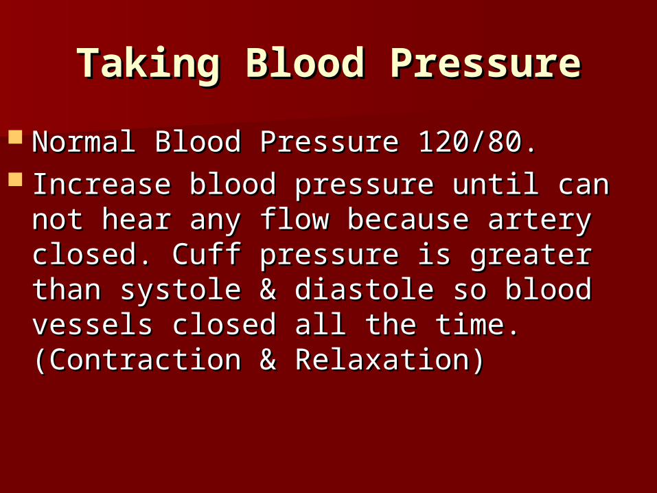
Taking Blood PressureTaking Blood Pressure
Normal Blood Pressure 120/80.Normal Blood Pressure 120/80. Increase blood pressure until can not hear any Increase blood pressure until can not hear any
flow because artery closed. Cuff pressure is flow because artery closed. Cuff pressure is greater than systole & diastole so blood vessels greater than systole & diastole so blood vessels closed all the time. (Contraction & Relaxation)closed all the time. (Contraction & Relaxation)

Taking Blood PressureTaking Blood Pressure
Let blood pressure come down until under Let blood pressure come down until under 120. During Systole (contraction), 120. During Systole (contraction), pressure in arteries is greater than cuff, so pressure in arteries is greater than cuff, so artery open only during systoleartery open only during systole
But during diastole (relaxation) pressure of But during diastole (relaxation) pressure of cuff is greater than pressure in blood cuff is greater than pressure in blood vessels so blood vessels are closed.vessels so blood vessels are closed.

Taking Blood PressureTaking Blood Pressure
The walls of the blood vessels go in and out The walls of the blood vessels go in and out causing turbulent flow of blood. causing turbulent flow of blood.
Can hear turbulent flow because walls of arteries Can hear turbulent flow because walls of arteries going in and out do to change in pressure.going in and out do to change in pressure.
Under 80 silent because cuff pressure less Under 80 silent because cuff pressure less diastole pressure so arteries stay open. diastole pressure so arteries stay open.
Blood pressure always recorded as Blood pressure always recorded as systolicsystolic = = 120 120
diastolic 80diastolic 80


Pulse RatePulse Rate
The pulse rate is equal to the rate at The pulse rate is equal to the rate at which the ventricles contract or equal which the ventricles contract or equal to heart rate. to heart rate.
The pulse is the alternate expanding The pulse is the alternate expanding and recoiling of the artery walls. and recoiling of the artery walls.

PulsePulse

Blood vessel disordersBlood vessel disorders ArteriosclerosisArteriosclerosis- degenerative - degenerative
disease in which the arteries loose disease in which the arteries loose elasticity; the vessels become elasticity; the vessels become brittle and can rupture easily; brittle and can rupture easily; associated with fatty diet, genetics, associated with fatty diet, genetics, lack of exercise, cigarette smoking, lack of exercise, cigarette smoking, etc.etc.
Go to Heart Attack Then Blocking the Artery to watch video

ArteriosclerosisArteriosclerosis

Blood vessel disordersBlood vessel disorders
AneurysmAneurysm- a bulge in a blood - a bulge in a blood vessel; this area of the blood vessel; this area of the blood vessel then weakens and may vessel then weakens and may burst; can result from trauma, high burst; can result from trauma, high blood pressure, infections, or blood pressure, infections, or genetic defectsgenetic defects
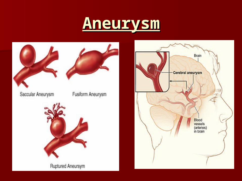
AneurysmAneurysm
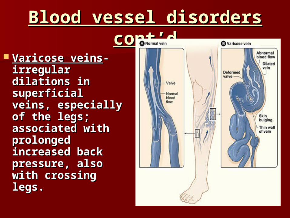
Blood vessel disorders cont’dBlood vessel disorders cont’d
Varicose veinsVaricose veins- - irregular dilations irregular dilations in superficial in superficial veins, especially veins, especially of the legs; of the legs; associated with associated with prolonged prolonged increased back increased back pressure, also pressure, also with crossing legs. with crossing legs.


Blood vessel disorders cont’dBlood vessel disorders cont’d HypertensionHypertension – high – high
b.p.; caused by kidney b.p.; caused by kidney disease, high Nadisease, high Na++ intake, obesity, stress, intake, obesity, stress, arteriosclerosis; left arteriosclerosis; left ventricle works ventricle works overtime so overtime so myocardium thickens, myocardium thickens, enlarging enlarging heartheartcoronary coronary vessels can’t feed vessels can’t feed overgrowth so parts of overgrowth so parts of heart dieheart die

Disorders, cont.Disorders, cont. AnemiaAnemia – condition in which the – condition in which the
oxygen carrying capacity of the blood oxygen carrying capacity of the blood is reducedis reduced; symptoms: fatigue, ; symptoms: fatigue, intolerance to cold, and paleness. intolerance to cold, and paleness. – Nutritional Anemia – inadequate diet, Nutritional Anemia – inadequate diet,
especially lacking in especially lacking in ironiron and vitamin and vitamin B12B12
– Sickle-Cell Anemia – abnormal kind of Sickle-Cell Anemia – abnormal kind of hemoglobin results in cells shaped like a hemoglobin results in cells shaped like a sickle (bent); they can rupture easily and sickle (bent); they can rupture easily and often get stuck together; (genetic)often get stuck together; (genetic)
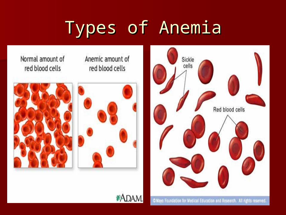
Types of AnemiaTypes of Anemia

PericarditisPericarditis Inflammation of the pericardium and Inflammation of the pericardium and
therefore an enlargement of the therefore an enlargement of the pericardial sac. pericardial sac.
This causes an increase in pressure on This causes an increase in pressure on the outside of the heart and causes the outside of the heart and causes the heart to have to work harderthe heart to have to work harder
Can cause heart attackCan cause heart attack Treatment – typically the fluid is Treatment – typically the fluid is
drained with a needle into the drained with a needle into the pericardial sac.pericardial sac.

PericarditisPericarditis

BloodBlood Connective tissue with liquid matrix.Connective tissue with liquid matrix. Carries oxygen, protects against infection, Carries oxygen, protects against infection,
promotes clotting, and carries other vital promotes clotting, and carries other vital substancessubstances
1.1. PlasmaPlasma clear, straw-colored clear, straw-colored (yellowish)(yellowish) mixture of water (95%), amino acids, proteins, carbs, mixture of water (95%), amino acids, proteins, carbs,
lipids, vitamins, hormones, electrolytes, and cellular lipids, vitamins, hormones, electrolytes, and cellular wasteswastes
2.2. Red Blood Cells (Erythrocytes)Red Blood Cells (Erythrocytes) contain hemoglobin (a protein that carries oxygen) contain hemoglobin (a protein that carries oxygen)
made of Iron – loves oxygen – what causes blood to made of Iron – loves oxygen – what causes blood to change colors. Red to purplishchange colors. Red to purplish
Also the chemical when changed which causes urine to Also the chemical when changed which causes urine to be yellow and feces to be brown. be yellow and feces to be brown.
formation of RBC (hematopoiesis) – in red marrowformation of RBC (hematopoiesis) – in red marrow

Blood continuedBlood continued3. White Blood Cells (Leukocytes)3. White Blood Cells (Leukocytes)
Can squeeze through vessel walls and move through Can squeeze through vessel walls and move through interstitial spaces via amoeboid movementinterstitial spaces via amoeboid movement
Many kinds of white blood cells; all have different jobs. Many kinds of white blood cells; all have different jobs. Protect against disease in 2 ways:Protect against disease in 2 ways:
– Phagocytize bacteria (eat up bacteria like pacaman)Phagocytize bacteria (eat up bacteria like pacaman)– Produce antibodies (proteins that destroy or disable Produce antibodies (proteins that destroy or disable
foreign particles)foreign particles)
4. Platelets (Thrombocytes)4. Platelets (Thrombocytes) Cell fragments that help close breaks in vessels and Cell fragments that help close breaks in vessels and
initiate formation of blood clots - (coagulation) initiate formation of blood clots - (coagulation) Causes scabs and stops bleeding.Causes scabs and stops bleeding.
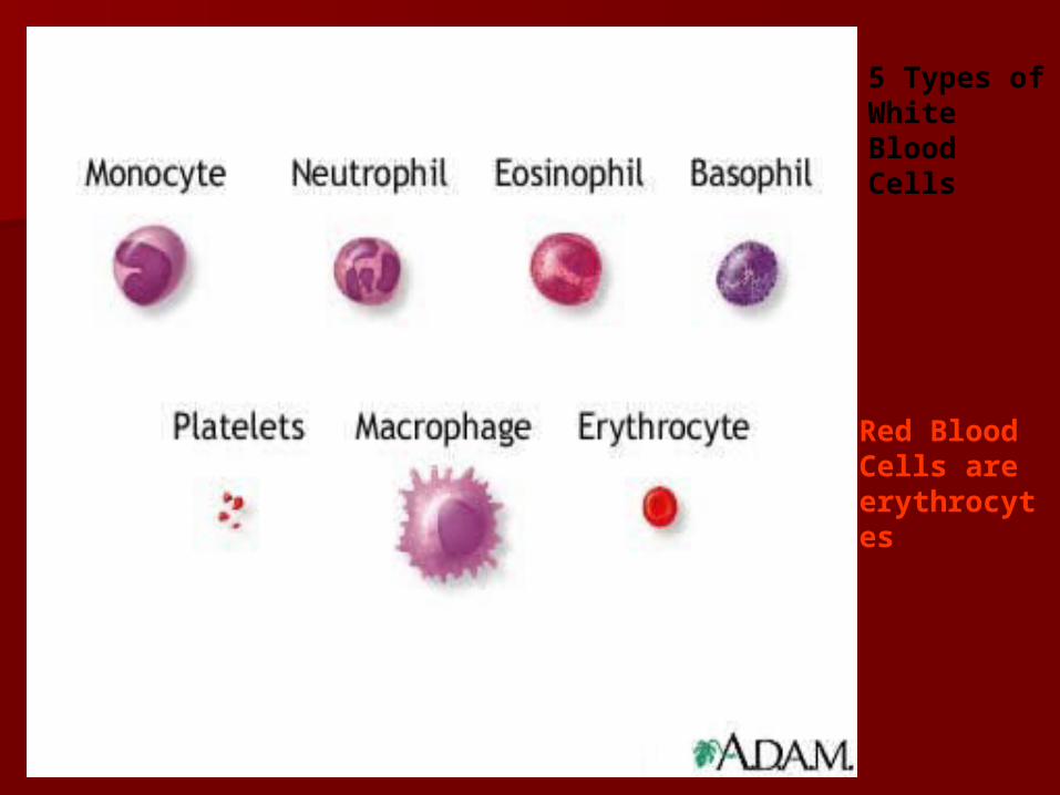
5 Types of White Blood Cells
Red Blood Cells are erythrocytes

ABO Blood TypesABO Blood Types Antigen- protein or carb on RBC Antigen- protein or carb on RBC
surface surface – Presence or absence of antigens Presence or absence of antigens
is an inherited traitis an inherited trait 2 major antigens: Antigen A and 2 major antigens: Antigen A and
Antigen B Antigen B 4 possible antigen combinations: 4 possible antigen combinations:
A only, B only, A and B, or neither A only, B only, A and B, or neither A nor BA nor B

ABO Blood TypesABO Blood Types
Antibodies- proteins in the plasma Antibodies- proteins in the plasma that destroy foreign substancesthat destroy foreign substances
Antibodies develop about 2-8 months Antibodies develop about 2-8 months after birthafter birth– If antigen A is absent – a person If antigen A is absent – a person
develops anti-A antibodydevelops anti-A antibody– If antigen B is absent – a person If antigen B is absent – a person
develops anti-B antibodydevelops anti-B antibody


ABO Blood Types, contABO Blood Types, cont
Blood Type Blood Type AntigenAntigen AntibodyAntibody
AA A A Anti-BAnti-B
BB BB Anti-AAnti-A
ABAB A and BA and B Neither Anti-A Neither Anti-A nor Anti-Bnor Anti-B
OONeither A nor Neither A nor BB
Both Anti-A Both Anti-A and Anti-Band Anti-B

ABO Blood Types, cont.ABO Blood Types, cont.
An antigen and an antibody of the An antigen and an antibody of the same type react to clump RBC – so same type react to clump RBC – so such combos must be avoidedsuch combos must be avoided
Type AB – universal recipients (lacks Type AB – universal recipients (lacks antibodiesantibodies anti-A and anti-B) anti-A and anti-B)
Type O – universal donors (lacks Type O – universal donors (lacks antigensantigens A and B) A and B)
However, the preferred donor is one However, the preferred donor is one with the matching blood typewith the matching blood type

Rh Blood TypeRh Blood Type Blood type – a person is either Blood type – a person is either
positive or negative.positive or negative. It takes only 1 gene to be positive. It takes only 1 gene to be positive. erythroblastosis fetaliserythroblastosis fetalis – When a – When a
mother is RH – and her baby is RH +. mother is RH – and her baby is RH +. During their first pregnancy some During their first pregnancy some blood is transferred from baby to blood is transferred from baby to mother. The mother then develops mother. The mother then develops antibodies against RH+ blood. antibodies against RH+ blood.

erythroblastosis fetaliserythroblastosis fetalis
Next pregnancy some of the blood Next pregnancy some of the blood from the mom gets in the baby and from the mom gets in the baby and the antibodies cause the blood to the antibodies cause the blood to agglutinate. Can cause fatality agglutinate. Can cause fatality because of lack of oxygen (severe because of lack of oxygen (severe anemia)anemia)
Treated by massive transfusions of Treated by massive transfusions of Rh+ blood for the baby and removal Rh+ blood for the baby and removal of blood containing Rh+ antibodies. of blood containing Rh+ antibodies.