THE BIOLOGY OF FRACTURE HEALING INLONG BONES€¦ · · 2017-02-03THE BIOLOGY OF FRACTURE HEALING...
13
150 THE JOURNAL OF BONE AND JOINT SURGERY THE BIOLOGY OF FRACTURE HEALING IN LONG BONES B. McKIBBIN Frotn the Department of Traumatic and Orthopaedic Surgery, Cardiff Royal infirmary The healing of a fracture is one of the most remarkable of all the repair processes in the body since it results, not in a scar, but in the actual reconstitution of the injured tissue in something very like its original form. It is not to be expected therefore that the mechanisms controlling such a process will be easily elucidated and indeed they involve problems of cellular homeostasis which are among the most fundamental in biology. If it is not quite the “cunning’st pattern of excelling nature” then it is something quite close to it and a great deal of that pattern at present stands unrevealed. However, this review is primarily concerned with those features which have direct clinical relevance and it is fortunately possible to treat fractures successfully without a complete understanding of the cellular mechanisms involved without at the same time relying entirely on empiricism. A number of factors influence the healing which can be identified from both clinical and experimental work and may be taken into consideration to put treatment on a more rational basis. It is with these observations that we shall be particularly concerned and cellular mechanisms will be discussed only if they appear to have clinical implications. Such an account must necessarily include details of the healing process as it is modified by contemporary methods of treatment but first it is necessary to consider the events that occur in the healing of a simple fracture in an unsplinted long bone. CHANGES ASSOCIATED WITH THE HEALING OF A FRACTURE These can conveniently be considered as a series of phases which occur in sequence but which of course also overlap to a certain extent. Immediate reaction (phase of inflammation) Initially there is bleeding from the damaged bone ends and from the associated soft tissues and a clot soon forms between the fragments. The soft parts in the region show the usual changes of acute inflammation with vasodilata- tion and the exudation of plasma and leucocytes (Wray 1964). Polymorphs, histiocytes and mast cells soon make their appearance and the process of clearing up of the debris begins (Lindholm et a!. 1969). The first evidence of increased cell division is to be found within about eight hours of the injury reaching a maximum in some twenty-four hours. This activity is first seen in the periosteum and tissues immediately around it and at first extends throughout the whole length of the injured bone. However, within a few days this generalised activity declines and eventually becomes confined to the area immediately adjacent to the fracture where it remains above normal levels for several weeks (Tonna and Cronkite 1961). Perhaps the most significant observation to be made at this stage is that the ends of the broken bones are not themselves participating in this proliferative activity but are in fact dead, as evidenced by the presence of empty osteocyte lacunae which extend for a variable distance away from the fracture. This phenomenon is explicable on the basis of the anatomical arrangements and anas- tomotic connections of the blood vessels of compact bone (Ham 1930) and its implications are profound. Although it is almost instinctive to think that the ends of broken bone unite directly to one another it is evident that on the contrary they play, at best, only a passive role in what is an essentially bridging process between the more distant regions of living bone (Fig. 1). This concept is fundamental to much of what is to follow. The role ofthe periosteum. A long-standing controversy exists as to the osteogenic potential of the periosteum which was heightened by the authoritative statement by MacEwen (1912) that it was nothing more than a limiting membrane and that the cells which were responsible for the production of new bone belonged properly to the surface of the bone. However, Ham and Harris (1971) have pointed out that this issue is largely semantic. The periosteum is now regarded as consisting of at least two layers, the outer fibrous and the inner cambial, and it is only in the latter that the cells in question lie. These are rather insignificant spindle- shaped cells which are indistinguishable from normal fibroblasts and have been termed osteoprogenitor cells (Young 1962) to distinguish them from the osteoblasts to which they give rise, since these latter have lost the power of cell division. In fact these cells are to be found on all free bone surfaces, endosteal as well as periosteal, so that when the periosteum is removed the bone is by no means deprived of its osteogenic potential. It would Professor B. McKibbin, M.D., F.R.C.S., Department ofTraumatic and Orthopaedic Surgery, Cardiff Royal Infirmary, Cardiff CF2 1 SZ, Wales.
Transcript of THE BIOLOGY OF FRACTURE HEALING INLONG BONES€¦ · · 2017-02-03THE BIOLOGY OF FRACTURE HEALING...
150 THE JOURNAL OF BONE AND JOINT SURGERY
THE BIOLOGY OF FRACTURE HEALING IN LONG BONES
B. McKIBBIN
Frotn the Department of Traumatic and Orthopaedic Surgery, Cardiff Royal infirmary
The healing of a fracture is one of the most
remarkable of all the repair processes in the body since it
results, not in a scar, but in the actual reconstitution of
the injured tissue in something very like its original
form. It is not to be expected therefore that the
mechanisms controlling such a process will be easily
elucidated and indeed they involve problems of cellular
homeostasis which are among the most fundamental in
biology. If it is not quite the “cunning’st pattern of
excelling nature” then it is something quite close to it
and a great deal of that pattern at present stands
unrevealed.
However, this review is primarily concerned with
those features which have direct clinical relevance and it
is fortunately possible to treat fractures successfully
without a complete understanding of the cellular
mechanisms involved without at the same time relying
entirely on empiricism. A number of factors influence
the healing which can be identified from both clinical
and experimental work and may be taken into
consideration to put treatment on a more rational basis.
It is with these observations that we shall be particularly
concerned and cellular mechanisms will be discussed
only if they appear to have clinical implications.
Such an account must necessarily include details of
the healing process as it is modified by contemporary
methods of treatment but first it is necessary to consider
the events that occur in the healing of a simple fracture in
an unsplinted long bone.
CHANGES ASSOCIATED WITH THE
HEALING OF A FRACTURE
These can conveniently be considered as a series of
phases which occur in sequence but which of course also
overlap to a certain extent.
Immediate reaction (phase of inflammation)
Initially there is bleeding from the damaged bone ends
and from the associated soft tissues and a clot soon forms
between the fragments. The soft parts in the region show
the usual changes of acute inflammation with vasodilata-
tion and the exudation of plasma and leucocytes (Wray
1964). Polymorphs, histiocytes and mast cells soon
make their appearance and the process of clearing up of
the debris begins (Lindholm et a!. 1969).
The first evidence of increased cell division is to be
found within about eight hours of the injury reaching a
maximum in some twenty-four hours. This activity is
first seen in the periosteum and tissues immediately
around it and at first extends throughout the whole
length of the injured bone. However, within a few days
this generalised activity declines and eventually
becomes confined to the area immediately adjacent to
the fracture where it remains above normal levels for
several weeks (Tonna and Cronkite 1961).
Perhaps the most significant observation to be made
at this stage is that the ends of the broken bones are not
themselves participating in this proliferative activity but
are in fact dead, as evidenced by the presence of empty
osteocyte lacunae which extend for a variable distance
away from the fracture. This phenomenon is explicable
on the basis of the anatomical arrangements and anas-
tomotic connections of the blood vessels of compact
bone (Ham 1930) and its implications are profound.
Although it is almost instinctive to think that the ends of
broken bone unite directly to one another it is evident
that on the contrary they play, at best, only a passive role
in what is an essentially bridging process between the
more distant regions of living bone (Fig. 1). This concept
is fundamental to much of what is to follow.
The role ofthe periosteum. A long-standing controversy
exists as to the osteogenic potential of the periosteum
which was heightened by the authoritative statement by
MacEwen (1912) that it was nothing more than a
limiting membrane and that the cells which were
responsible for the production of new bone belonged
properly to the surface of the bone. However, Ham and
Harris (1971) have pointed out that this issue is largely
semantic. The periosteum is now regarded as consisting
of at least two layers, the outer fibrous and the inner
cambial, and it is only in the latter that the cells in
question lie. These are rather insignificant spindle-
shaped cells which are indistinguishable from normal
fibroblasts and have been termed osteoprogenitor cells
(Young 1962) to distinguish them from the osteoblasts
to which they give rise, since these latter have lost the
power of cell division. In fact these cells are to be found
on all free bone surfaces, endosteal as well as periosteal,
so that when the periosteum is removed the bone is by no
means deprived of its osteogenic potential. It would
Professor B. McKibbin, M.D., F.R.C.S., Department ofTraumatic and Orthopaedic Surgery, Cardiff Royal Infirmary, Cardiff CF2 1 SZ, Wales.
a
b
C
-�
(=-�-�
�t?-��.� �
,� �-� -�
DeadBone
THE BIOLOGY OF FRACTURE HEALING IN LONG BONES 151
VOL. 60-B, No. 2. MAY 1978
seem therefore that although an alternative terminology
has prevailed, MacEwen’s view of the perioSteum as a
separate non-osteogenic fibrous layer is less confusing.
Nevertheless, it must be borne in mind that when
periosteum is stripped from the bone it carries some of
these osteogenic cells with it so that if a segment of bone
is excised and the periosteal tube remains this may
successfully regenerate a new bone (Mulholland and
Pritchard 1959; McClements, Templeton and Pritchard
1961).
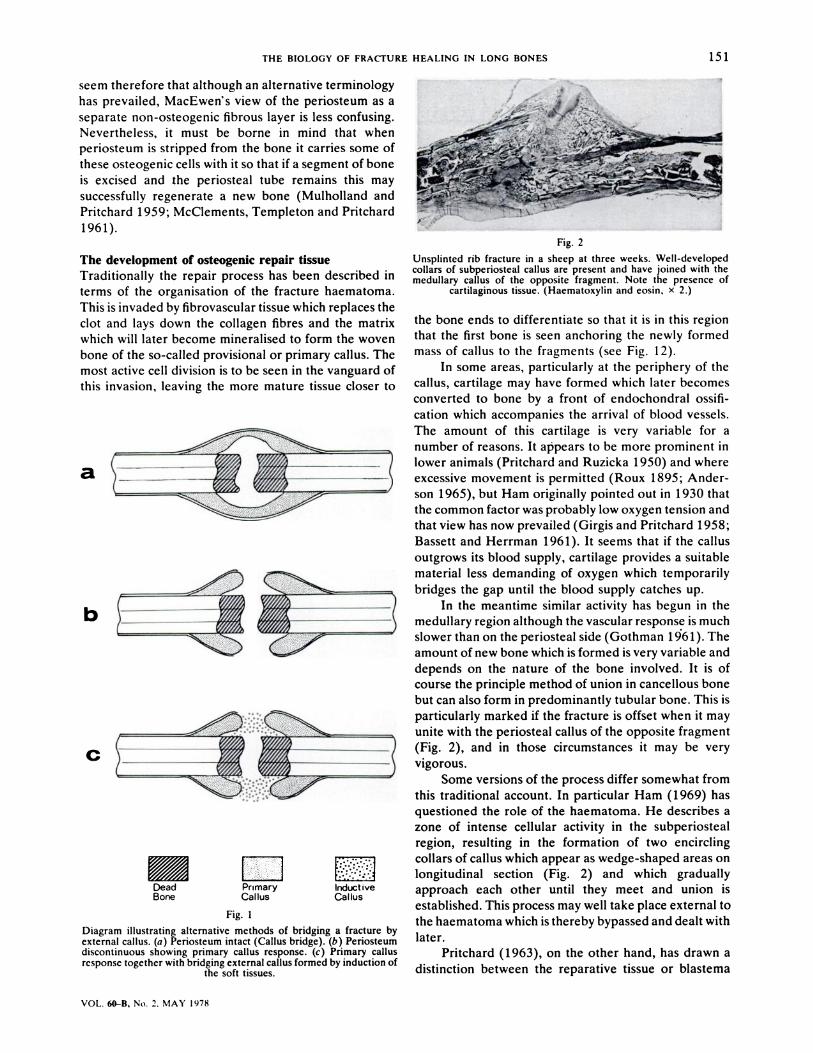
The development of osteogenic repair tissue
Traditionally the repair process has been described in
terms of the organisation of the fracture haematoma.
This is invaded by fibrovascular tissue which replaces the
clot and lays down the collagen fibres and the matrix
which will later become mineralised to form the woven
bone of the so-called provisional or primary callus. The
most active cell division is to be seen in the vanguard of
this invasion, leaving the more mature tissue closer to
Induct EveCallus
[ I:� __PrimaryCallus
Fig. 1
Diagram illustrating alternative methods of bridging a fracture byexternal callus. (a) Periosteum intact (Callus bridge). (b) Periosteumdiscontinuous showing primary callus response. (c) Primary callusresponse together with bridging external callus formed by induction of
the soft tissues.
Fig. 2
Unsplinted rib fracture in a sheep at three weeks. Well-developedcollars of subperiosteal callus are present and have joined with themedullary callus of the opposite fragment. Note the presence of
cartilaginous tissue. (Haematoxylin and eosin, x 2.)
the bone ends to differentiate so that it is in this region
that the first bone is seen anchoring the newly formed
mass of callus to the fragments (see Fig. 1 2).
In some areas, particularly at the periphery of the
callus, cartilage may have formed which later becomes
converted to bone by a front of endochondral ossifi-
cation which accompanies the arrival of blood vessels.
The amount of this cartilage is very variable for a
number of reasons. It appears to be more prominent in
lower animals (Pritchard and Ruzicka 1 950) and where
excessive movement is permitted (Roux 1895; Ander-
son 1965), but Ham originally pointed out in 1930 that
the common factor was probably low oxygen tension and
that view has now prevailed (Girgis and Pritchard 1958;
Bassett and Herrman 1961). It seems that if the callus
outgrows its blood supply, cartilage provides a suitable
material less demanding of oxygen which temporarily
bridges the gap until the blood supply catches up.
In the meantime similar activity has begun in the
medullary region although the vascular response is much
slower than on the periosteal side (Gothman 1961). The
amount of new bone which is formed is very variable and
depends on the nature of the bone involved. It is of
course the principle method of union in cancellous bone
but can also form in predominantly tubular bone. This is
particularly marked if the fracture is offset when it may
unite with the periosteal callus of the opposite fragment
(Fig. 2), and in those circumstances it may be very
vigorous.
Some versions of the process differ somewhat from
this traditional account. In particular Ham (1969) has
questioned the role of the haematoma. He describes a
zone of intense cellular activity in the subperiosteal
region, resulting in the formation of two encircling
collars of callus which appear as wedge-shaped areas on
longitudinal section (Fig. 2) and which gradually
approach each other until they meet and union is
established. This process may well take place external to
the haematoma which is thereby bypassed and dealt with
later.
Pritchard (1963), on the other hand, has drawn a
distinction between the reparative tissue or blastema

.1� -‘
Blood vessels in external callus originating from the soft tissues.(Reproduced, with permission, from Rhinelander 1974.)
152 B. MCKIBBIN
THE JOURNAL OF BONF. AND JOINT SURGERY
which arises from the outer fibrous layer of the
periosteum and that which emanates from the cambial
layer and the medullary cavity, which he terms the
osteogenic blastema. Normally the osteogenic blastema,
being more centrally placed, invades the haematoma
and produces the new bone which bridges the fragments,
while the more peripheral zones are more fibrous in
character and restore the continuity of the periosteum.
In some circumstances the fibrous tissue may invade the
fracture gap first, in which case the ingrowth of the
osteogenic blastema may be halted unless the fibrous
tissue is removed or converted to bone in some way
(Mulholland and Pritchard 1959). Clearly the factors
that influence this latter process are different from, and
indeed a good deal more uncertain than, those that
affect the direct growth of the osteogenic blastema, and
Pritchard saw the elucidation of these different
influences as central to the problem of non-union.
As we shall see, many of the differences of emphasis
that appear in these various accounts can probably be
explained by differences between the particular bone
and species studied, or sometimes by the mode of
production of the fracture, but a more fundamental
argument, which must be dealt with first, arises out of
differing concepts of the source of the osteogenic tissue.
Source of the osteogenic cells. There are two rival
theories. According to the first, the repair tissue arises
from specialised cells with a predetermined commitment
to bone formation. These are the osteoprogenitor cells
of which mention has already been made and which
occur only in close association with the bone surface or
the bone marrow (Owen 1970). As these cells
proliferate the fibrous periosteum becomes pushed away
from the bone to produce the two advancing collars of
callus described by Ham (1969), which eventually fuse
with one another.
The alternative view in its most extreme form was
originally put forward by Leriche and Policard (1928),
who suggested that repair tissue did not arise from
specialist cells but rather from the activity of previously
uncommitted fibroblasts which could develop the power
of osteogenesis if given the appropriate environmental
stimulus. Thus the reparative tissue does not arise from
the bone itself but from the surrounding soft tissues. This
distinction is obviously of considerable practical impor-
tance since it elevates the soft tissues to a new key role,
while the former theory regards them as being involved
only in so far as their damage may be related to
interference with the blood supply to the bones
themselves.
The phenomenon by which unassociated soft
tissues are recruited in this way is known as osteogenic
induction. This is a large topic and the reader is referred
to the comprehensive reviews by Ostrowski and
Wodarski (1971) and Chalmers, Gray and Rush (1975),
as well as the classical paper by Urist and McLean
( 1 952). For the present purpose it may be stated that the
formation of bone by non-specialised cells in extra-
skeletal sites is established beyond doubt and many
inducing agents both living and non-living have been
identified, but what is less certain is whether or not
osteogenic induction plays any part in the healing of a
fracture.
While it may legitimately be asked for what purpose
the induction mechanism does exist if it does not assist in
fracture healing, this merely begs the question, and it
must be admitted that the only supporting evidence is
purely circumstantial.
Fibroblasts iji the soft tissues cannot be disting-
uished morphologically from the osteoprogenitor cells
of bone and a clue to their origin is given by a
consideration of the vascular changes that accompany
fracture healing.
Normally the cortex is supplied largely through the
medullary system and the flow is centrifugal, the
periosteum at best making only a small arterial
contribution to the outer cortex (Brooks 1971).
Following a fracture, however, there develops very
rapidly an extraosseous blood supply (Rhinelander
1974) derived from the surrounding soft tissues.
Although this may help to revascularise any necrotic
cortex whose medullary supply has been affected, its
main purpose appears to be the supply ofexternal callus
(Fig. 3); Rhinelander has pointed out that it is
facultative and .transitory and in later stages of the
process regresses as its function is taken over by the
redeveloping medullary system.
It is therefore not difficult to believe that if the
vessels of external callus originate outside the bone, so
also do the cells themselves. Trueta (1968) adduced a
good deal of evidence that such cells did in fact arise
from vascular endothelium but a new possibility arises
from the work of Friedenstein (1968). It appears that
some of the cells with osteogenic potential in marrow
may have access to the circulation and it may be these
which are responsible for extraskeletal bone formation.
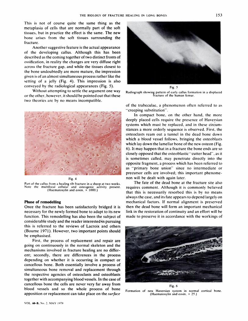
Fig. 5
Fig. 4
THE BIOLOGY OF FRACTURE HEAI.ING IN LONG BONES 153
VOL. 60-B. No. 2. MAY 1978
This is not of course quite the same thing as the
metaplasia of cells that are normally part of the soft
tissues, but in practice the effect is the same. The new
bone arises from the soft tissues surrounding the
fracture.
Another suggestive feature is the actual appearance
of the developing callus. Although this has been
described as the coming together of two distinct fronts of
ossification, in reality the changes are very diffuse right
across the fracture gap, and while the tissues closest to
the bone undoubtedly are more mature, the impression
given is of an almost simultaneous process rather like the
setting of a jelly (Fig. 4). This impression is also
conveyed by the radiological appearances (Fig. 5).
Without attempting to settle the argument one way
or the other, however, it should be pointed out that these
two theories are by no means incompatible.
Part of the callus from a healing rib fracture in a sheep at two weeks.Note the multifocal cellular and osteogenic activity present.
(Haematoxylin and eosin. X 1000.)
Phase of remodelling
Once the fracture has been satisfactorily bridged it is
necessary for the newly formed bone to adapt to its new
function. This remodelling has also been the subject of
considerable study and the reader interestedin pursuing
this is referred to the reviews of Lacroix and others
(Bourne 1971). However, two important points should
be emphasised.
First, the process of replacement and repair are
going on continuously in the normal skeleton and the
mechanisms involved in fracture healing are no differ-
ent; secondly, there are differences in the process
depending on whether it is occurring in compact or
cancellous bone. Both essentially involve a process of
simultaneous bone removal and replacement through
the respective agencies of osteoclasts and osteoblasts
together with accompanying blood vessels. In the case of
cancellous bone the cells are never very far away from
blood vessels and so the whole process of bone
apposition or replacement can take place on the surface
Radiograph showing pattern of early callus formation in a displacedfracture of the human femur.
of the trabeculae, a phenomenon often referred to as
“ creeping substitution”.
In compact bone, on the other hand, the more
deeply placed cells require the presence of Haversian
systems which must be replaced, and in these circum-
stances a more orderly sequence is observed. First, the
osteoclasts ream out a tunnel in the dead bone down
which a blood vessel follows, bringing the osteoblasts
which lay down the lamellar bone of the new osteon (Fig.
6). It may happen that in a fracture the bone ends are so
closely opposed that the osteoblastic “ cutter head” , as it
is sometimes called, may penetrate directly into the
opposite fragment, a process which has been referred to
as “ primary bone union” since no intermediate or
precursor cells are involved; this important phenome-
non will be dealt with again later.
The fate of the dead bone at the fracture site also
requires comment. Although it is commonly believed
that this is necessarily resorbed this is by no means
always the case, and its fate appears to depend largely on
mechanical factors. If normal alignment is preserved
then the dead bone will form an important mechanical
link in the restoration of continuity and an effort will be
made to preserve it in accordance with the workings of
Fig. 6
Formation of new Haversian system in normal cortical bone.(Haematoxylin and eosin. X 25.)
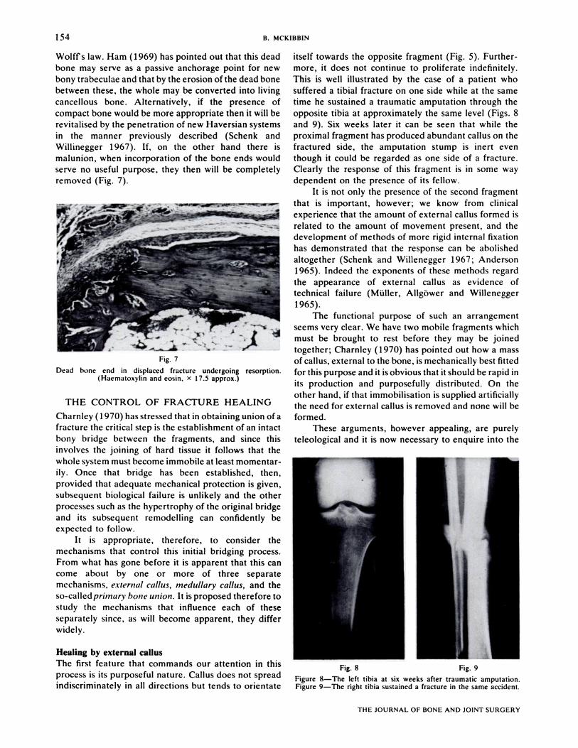
Fig. 7
Dead bone end in displaced fracture undergoing resorption.(Haematoxylin and eosin, x 17.5 approx.)
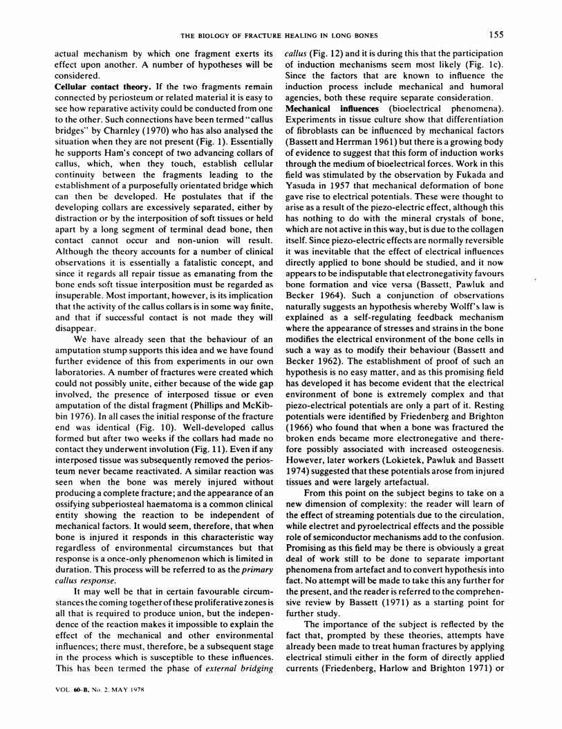
Fig. 8 Fig. 9
Figure 8-The left tibia at six weeks after traumatic amputation.Figure 9-The right tibia sustained a fracture in the same accident.
154 B. MCKIBBIN
THE JOURNAL OF BONE AND JOINT SURGERY
Wolffs law. Ham (1969) has pointed out that this dead
bone may serve as a passive anchorage point for new
bony trabeculae and that by the erosion of the dead bone
between these, the whole may be converted into living
cancellous bone. Alternatively, if the presence of
compact bone would be more appropriate then it will be
revitalised by the penetration of new Haversian systems
in the manner previously described (Schenk and
Willinegger 1967). If, on the other hand there is
malunion, when incorporation of the bone ends would
serve no useful purpose, they then will be completely
removed (Fig. 7).
THE CONTROL OF FRACTURE HEALING
Charnley (1 970) has stressed that in obtaining union of a
fracture the critical step is the establishment of an intact
bony bridge between the fragments, and since this
involves the joining of hard tissue it follows that the
whole system must become immobile at least momentar-
ily. Once that bridge has been established, then,
provided that adequate mechanical protection is given,
subsequent biological failure is unlikely and the other
processes such as the hypertrophy of the original bridge
and its subsequent remodelling can confidently be
expected to follow.
It is appropriate, therefore, to consider the
mechanisms that control this initial bridging process.
From what has gone before it is apparent that this can
come about by one or more of three separate
mechanisms, external callus, medullary callus, and the
so-calledpritnary bone union. It is proposed therefore to
study the mechanisms that influence each of these
separately since, as will become apparent, they differ
widely.
Healing by external callus
The first feature that commands our attention in this
process is its purposeful nature. Callus does not spread
indiscriminately in all directions but tends to orientate
itself towards the opposite fragment (Fig. 5). Further-
more, it does not continue to proliferate indefinitely.
This is well illustrated by the case of a patient who
suffered a tibial fracture on one side while at the same
time he sustained a traumatic amputation through the
opposite tibia at approximately the same level (Figs. 8
and 9). Six weeks later it can be seen that while the
proximal fragment has produced abundant callus on the
fractured side, the amputation stump is inert even
though it could be regarded as one side of a fracture.
Clearly the response of this fragment is in some way
dependent on the presence of its fellow.
It is not only the presence of the second fragment
that is important, however; we know from clinical
experience that the amount of external callus formed is
related to the amount of movement present, and the
development of methods of more rigid internal fixation
has demonstrated that the response can be abolished
altogether (Schenk and Willenegger 1967; Anderson
1965). Indeed the exponents of these methods regard
the appearance of external callus as evidence of
technical failure (Muller, Allgower and Willenegger
1965).The functional purpose of such an arrangement
seems very clear. We have two mobile fragments which
must be brought to rest before they may be joined
together; Charnley (1970) has pointed out how a mass
of callus, external to the bone, is mechanically best fitted
for this purpose and it is obvious that it should be rapid in
its production and purposefully distributed. On the
other hand, if that immobilisation is supplied artificially
the need for external callus is removed and none will be
formed.
These arguments, however appealing, are purely
teleological and it is now necessary to enquire into the
THE BIOLOGY OF FRACTURE HEALING IN LONG BONES 155
VOL. 60-B, No. 2. MAY 1978
actual mechanism by which one fragment exerts its
effect upon another. A number of hypotheses will be
considered.
Cellular contact theory. If the two fragments remain
connected by periosteum or related material it is easy to
see how reparative activity could be conducted from one
to the other. Such connections have been termed “ callus
bridges” by Charnley (1970) who has also analysed the
situation when they are not present (Fig. 1). Essentially
he supports Ham’s concept of two advancing collars of
callus, which, when they touch, establish cellular
continuity between the fragments leading to the
establishment of a purposefully orientated bridge which
can then be developed. He postulates that if the
developing collars are excessively separated, either by
distraction or by the interposition of soft tissues or held
apart by a long segment of terminal dead bone, then
contact cannot occur and non-union will result.
Although the theory accounts for a number of clinical
observations it is essentially a fatalistic concept, and
since it regards all repair tissue as emanating from the
bone ends soft tissue interposition must be regarded as
insuperable. Most important, however, is its implication
that the activity of the callus collars is in some way finite,
and that if successful contact is not made they will
disappear.
We have already seen that the behaviour of an
amputation stump supports this idea and we have found
further evidence of this from experiments in our own
laboratories. A number of fractures were created which
could not possibly unite, either because of the wide gap
involved, the presence of interposed tissue or even
amputation of the distal fragment (Phillips and McKib-
bin 1 976). In all cases the initial response of the fracture
end was identical (Fig. 10). Well-developed callus
formed but after two weeks if the collars had made no
contact they underwent involution (Fig. 1 1). Even if any
interposed tissue was subsequently removed the perios-
teum never became reactivated. A similar reaction was
seen when the bone was merely injured without
producing a complete fracture; and the appearance of an
ossifying subperiosteal haematoma is a common clinical
entity showing the reaction to be independent of
mechanical factors. It would seem, therefore, that when
bone is injured it responds in this characteristic way
regardless of environmental circumstances but that
response is a once-only phenomenon which is limited in
duration. This process will be referred to as the primary
callus response.
It may well be that in certain favourable circum-
stances the coming together ofthese proliferative zones is
all that is required to produce union, but the indepen-
dence of the reaction makes it impossible to explain the
effect of the mechanical and other environmental
influences; there must, therefore, be a subsequent stage
in the process which is susceptible to these influences.
This has been termed the phase of external bridging
callus (Fig. 1 2) and it is during this that the participation
of induction mechanisms seem most likely (Fig. ic).
Since the factors that are known to influence the
induction process include mechanical and humoral
agencies, both these require separate consideration.
Mechanical influences (bioelectrical phenomena).
Experiments in tissue culture show that differentiation
of fibroblasts can be influenced by mechanical factors
(Bassett and Herrman 1961) but there is a growing body
of evidence to suggest that this form of induction works
through the medium of bioelectrical forces. Work in this
field was stimulated by the observation by Fukada and
Yasuda in 1957 that mechanical deformation of bone
gave rise to electrical potentials. These were thought to
arise as a result of the piezo-electric effect, although this
has nothing to do with the mineral crystals of bone,
which are not active in this way, but is due to the collagen
itself. Since piezo-electric effects are normally reversible
it was inevitable that the effect of electrical influences
directly applied to bone should be studied, and it now
appears to be indisputable that electronegativity favours
bone formation and vice versa (Bassett, Pawluk and
Becker 1964). Such a conjunction of observations
naturally suggests an hypothesis whereby Wolff’s law is
explained as a self-regulating feedback mechanism
where the appearance of stresses and strains in the bone
modifies the electrical environment of the bone cells in
such a way as to modify their behaviour (Bassett and
Becker 1962). The establishment of proof of such an
hypothesis is no easy matter, and as this promising field
has developed it has become evident that the electrical
environment of bone is extremely complex and that
piezo-electrical potentials are only a part of it. Resting
potentials were identified by Friedenberg and Brighton
(1966) who found that when a bone was fractured the
broken ends became more electronegative and there-
fore possibly associated with increased osteogenesis.
However, later workers (Lokietek, Pawluk and Bassett
1974) suggested that these potentials arose from injured
tissues and were largely artefactual.
From this point on the subject begins to take on a
new dimension of complexity: the reader will learn of
the effect of streaming potentials due to the circulation,
while electret and pyroelectrical effects and the possible
role of semiconductor mechanisms add to the confusion.
Promising as this field may be there is obviously a great
deal of work still to be done to separate important
phenomena from artefact and to convert hypothesis into
fact. No attempt will be made to take this any further for
the present, and the reader is referred to the comprehen-
sive review by Bassett (1971) as a starting point for
further study.
The importance of the subject is reflected by the
fact that, prompted by these theories, attempts have
already been made to treat human fractures by applying
electrical stimuli either in the form of directly applied
currents (Friedenberg, Harlow and Brighton 1971) or

Fig. 10 Fig. 11 Fig. 12
Development and regression of suhperiosteal callus in rat metatarsals. (R#{225}lit’ Tetrachrome for osteoid and bone, X 50.) Figure10-Amputation stump at nine days showing the �primary callus” response. New woven bone is coloured blue. Figure 1 1 -Amputation stumpat seventeen days. Involution of the primary callus response has begun. Figure 12-Uniting fracture at seventeen days. Callus formation has
progressed leading to formation of bridging external callus’.
156 B. MCKIBBIN
THE JOIRNAE. OF BONE ANE) JOINt suR(;IR\
by the non-invasive use of electromagnetic fields
(Bassett et uI. I 974). Since the selection of these stimuli
has necessarily been arbitrary the results must be
assessed, as in any other empirical method, by clinical
criteria, and such an analysis is outside the scope of this
paper. It should be recalled, however, that fracture
bridging is a purposeful process and an increase in
osteogenesis of itself does not necessarily imply clinical
success.
Humoral theories. The role of mechanical factors is
obviously important in the induction phase of callus
formation hut this cannot be regarded as the only
stimulus since it does not account for two facts: attempts
to bridge the fracture by external callus are not
prolonged indefinitely, and the continued mobility of a
fracture is no guarantee of a continued callus response.
The result may be a local activity around the bone end,
as in the familiar “ elephant foot” radiographic appear-
ance, but the cells between the fragments may fail to
respond and bridging may not result. One explanation
may he that offered by Pritchard (1963) that the space
has now been invaded by non-ossifiable mature fibrous
tissue, but it could also be explained by the necessity for
some humoral inducing agent which is normally present
only for a limited period.
The possibility that fractured bone ends might
liberate an agent which influences the healing process
was suggested as early as 1920 by Bier. Since then the
search for this elusive “ wound hormone” has continued,
especially in fracture haematoma (Pritchard 1961 ; Urist
and McLean 1952)-but so far without success.
Nevertheless, in view of the fact that heterotopic bone
formation can be induced by dead tissue and even by
bone transplants separated from the host by a millipore
membrane (Goldhaber 1961), it seems inevitable that a
humoral inducing agent must exist although probably it
is effective only under very specific circumstances. Thus
Moss ( 1 960) was able to produce bone formation in the
skull of young rats with an extract of bovine bone
absorbed by Gelfoam. The exclusion of bone ends by
Silastic caps does not affect the formation of external
callus (Church and Young 1965) so that if a humoral
substance is involved at all in the formation of external
callus it must presumably be liberated, not from the
injured bone itself, but rather from the products of the
primary response. Since we know that this latter is
unaffected by mechanical factors and is subject to
temporal limitations, this would satisfactorily explain
the failure of prolonged mobility to continue the
inductive process. It would also account for the rapid
and almost simultaneous differentiation which occurs
right across the fracture gap in the early phases of
successful healing.
Although we have considered a number of
hypothesis none of these are mutually exclusive and
since each enjoys a measure of support it seems
reasonable to join them together. If the formation of
external callus can be considered as two separate but
interrelated phases many of the difficulties disappear.
Thus there is an initial primary callus response which is
largely independent of environmental circumstances
and which almost certainly arises from predetermined
cells in the bone tissues themselves. This may lead to the
subsequent phase ofbridging callusformation the speed
of which seems likely to depend on the recruitment of
cells from the surrounding tissues by a process of
induction. It is this phase of the process which is most
susceptible to environmental influences particularly
with regard to the mutual relationship and movement of
the fragments.
Medullary callusIn many accounts of healing in long bones medullary
callus is distinguished from external callus only by its
location although it has often been observed that
cartilage formation is much less prominent in medullary
callus. Nevertheless it appears to be assumed that it is
governed by the same influences. This may well be true
in the early stages of the healing process and we have
seen how in a displaced fracture it may unite directly
with external callus (Fig. 2). The reaction in these
circumstances is very like that of the primary callus
-� ...
.. � ..‘
*� - . ..
� �4a�,#{149}
�-. -� . � --�!i � � .
;�_ �. �
.,..
. .�*, ..� -‘
‘ � --.
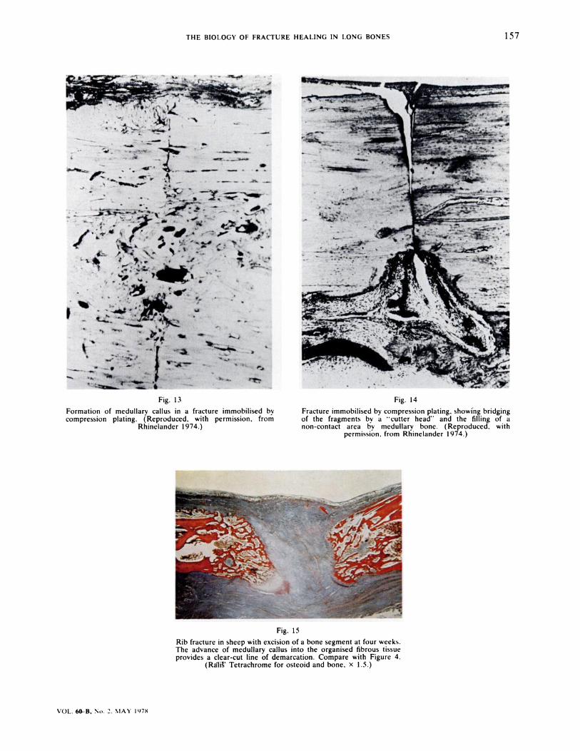
Formation of medullary callus in a fracture immobilised bycompression plating. (Reproduced, with permission, from
Rhinelander 1974.)
Fig. 14
Fracture immobilised by compression plating. showing bridgingof the fragments by a � cutter head” and the filling of anon-contact area by medullary bone. (Reproduced. with
permission. from Rhinelander 1974.)
Fig. 15
VOL. 60-B, � 2. MAY 1978
Fig. 13
THE BIOLOGY OF FRACTURE HEALING IN LONG BONES
Rib fracture in sheep with excision of a bone segment at four weeks.The advance of medullary callus into the organised fibrous tissueprovides a clear-cut line of demarcation. Compare with Figure 4.
(Ra�lit’ Tetrachrome for osteoid and bone. x 1.5.)
157
158 B. MCKIBBIN
THE JOURNAL OF BONE AND JOINT SURGERY
response of external callus but it seems that. just as a
second stage could be identified in that process so also is
it the case with medullary callus except that it appears
rather later in the healing process and the controlling
factors involved appear to be quite different.
The most obvious difference is the effect of
mechanical stability. Although this has an inhibitory
effect on external callus, medullary callus is unaffected
and often flourishes under these conditions (Fig. 13). In
intrinsically stable situations such as the healing of a burr
hole it is almost the sole method of repair (Johnson
1 927) while in fractures immobilised by rigid plating it is
often the earliest type of union (Rhinelander 1974;
Olerud and Dankwardt-Lilliestr#{246}m 1971). In these
circumstances it appears to form without any intermedi-
ary stage of fibrocartilage. One of its important functions
is to act as a replacing tissue; by filling up any gaps in the
fracture line with new woven bone it prepares the way
for the subsequent passage of new osteons from one
fragment to the other (Fig. 14).
At the same time it is obviously not dependent on
total immobility and it can be seen in a rather different
form in the later stages of the healing of an unsplinted
fracture where there is a large gap (Fig. 15). By this time
the external callus response has failed in its purpose and
faded away, leaving a well-defined ossifying front clearly
distinguishable from the fibrous tissue it is invading and
quite unlike the much more diffuse activity of the earlier
external callus. This now corresponds very well with the
“osteogenic blastema” described by Pritchard (1963).
Although the term “ medullary callus” is not ideal to
describe it since at least some of the activity comes from
the external surface of the bone, the term is preferred for
the present purpose because it is largely a central activity
and it serves to sharpen the many distinctions between it
and the earlier external bridging callus.
The appearances suggest much more strongly than
with external callus that the osteogenic material is
springing from the bone ends themselves and it has been
shown in the adult rat that the ability to bridge a fracture
gap is greatly diminished if these are lightly cauterised
(Mulholland and Pritchard 1959; Templeton 1960). At
this stage of the process the activity of the surrounding
soft tissue, far from being helpful, may actually interfere
with the bridging process by interposing fibrous tissue,
and if this can be excluded by joining the fracture ends
with a polythene tube much greater gaps can be bridged
(Mulholland and Pritchard 1959).
Nevertheless as Pritchard (1963) has pointed out,
even where there is fibrous tissue interposition union
may sometimes occur by a slow process of replacement,
as appears to be happening in Figure 15. This indicates
another important difference from external callus in that
the process is not an evanescent burst of activity but can
continue slowly and relentlessly for months in pursuit of
its goal.
Unfortunately we have very little idea about what
are the stimuli responsible for the long-continued
activity of this type of callus, which can by no means
always be relied on. Mechanical factors are obviously
not so important as in external callus and the sustained
production of a humoral agent for so long seems
unlikely. The presence of the opposite fragment is
obviously essential since such prolonged activity does
not occur in amputation stumps.
Once again, electrical phenomena, probably of the
non-mechanically generated type, seems the most likely
explanation but biochemical and even neurological
mechanisms may be involved. It would seem that this is a
part of the fracture healing process which has been
inadequately studied as a separate entity.
Primary bone healing
From the account which has been given of the mode of
healing of an unsplinted fracture the idea that direct
union between the bone ends may be achieved without
the assistance of bridging external callus appears to be
illogical. Charnley (1970) believed that there was
confusion with the situation in cancellous bone where
terminal bone death is minimal and union occurs largely
by the process of creeping substitution. Nevertheless,
the fact that clinical experience shows the process to be
an indisputable reality means that the scientific basis for
it must be reappraised.
The explanation was provided by the experiments
of Schenk and Willinegger (1967) in which rigid
compression plates were applied to the dog’s radius.
They found that, contrary to expectation, the dead ends
of the cortical bone were not resorbed but were
recanalised by new Haversian systems in the manner
already described, and where the fragments were in
direct contact these systems actually crossed from one
fragment into the other. In those areas where there were
small gaps on the opposite side from the plate the space
became filled by what they believed to be new bone
arising from the endosteum of the Haversian system.
This bone then provided the necessary bridge to conduct
new Haversian systems across the gap (Fig. 14). These
findings have since been confirmed by others (Olerud
and Dankwardt-Lilliestr#{246}m 1968; Rhinelander 1974;
Anderson 1 965). There does therefore appear to be a
third method of fracture bridging whose relationship to
mechanical factors is different again from the other two.
Obviously the process is not inhibited by stability, as in
external callus, and indeed even small degrees of
movement are inimical to the process, unlike medullary
callus.
This difference is relatively easily explained. It has
already been pointed out that the process is nothing
more than the mechanism of bone turnover which is
occurring all the time in the normal skeleton and there is
no reason why it should be inhibited by lack of
movement. On the contrary, Schenk and Willinegger
( 1 967) believed that the process was actually speeded up
THE BIOLOGY OF FRACTURE HEALING IN LONG BONES 159
VOL. 60-B, No. 2. MAY 1978
because of the need to replace the dead bone which
inevitably resulted from the fracture, but subsequent
work has not shown this supposition to be correct.
In the first place it seems that the very strength of
the fixation deprives the bone of the normal stresses
which maintain it and the net result may be an excess of
osteoclastic activity leading to some degree of atrophy
of the bone (Uhthoff and Dubuc 1971 ; Tonino et al.
1976). Furthermore in the experiments described the
circumstances were rather artificial: the fractures were
produced carefully with a fine saw and the amount of
bone necrosis was minimal. A more realistic model was
constructed by Olerud and Dankwardt-Lilliestr#{246}m
( 1971) who produced a segmental fracture, and after
ensuring that the middle fragment was entirely devoid of
vascular connections fixed the whole with a compression
plate as rigidly as possible. They found, as did Schenk
and Willinegger (1967), that the dead bone did not
reduced to a minimum and the mechanical stability is
enhanced, but the question arises as to whether there is
any direct biological effect due to the compression itself.
At first it was feared that compression might lead to
bone necrosis and further resorption but this has been
found not to be so by Perren et a!. (1969). Bassett and
Herrman (1961) showed that in tissue culture fibroblasts
could be induced to form bone under conditions of
compression, provided a high oxygen level was main-
tamed, while tension resulted in fibrous tissue. Bassett
and Becker (1962) did not think this was related to the
clinical effects of compression but did not say why. It
seems unlikely that such mechanically induced meta-
plasia would play much part in the healing of a fresh
fracture because of the blood supply to such cells is likely
to be inadequate, but it could conceivably be important
when compression is applied for the treatment of
established fibrous union. The problem is worth
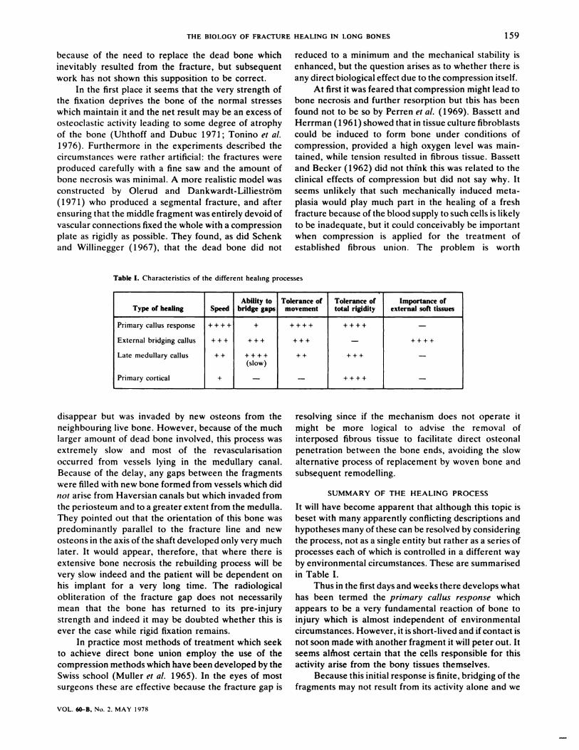
Table I. Characteristics of the different healing processes
Type of healing SpeedAbility to
bridge gapsTolerance of
movementTolerance oftotal rigidity
Importance ofexternal soft tissues
Primary callus response
External bridging callus
Late medullary callus
Primary cortical
+ + + +
+ + +
+ +
+
+
+ + +
+ + + +
(slow)
-
+ + + +
+ + +
+ +
-
+ + + +
-
+ + +
+ + + +
-
+ + + +
-
-
disappear but was invaded by new osteons from the
neighbouring live bone. However, because of the much
larger amount of dead bone involved, this process was
extremely slow and most of the revascularisation
occurred from vessels lying in the medullary canal.
Because of the delay, any gaps between the fragments
were filled with new bone formed from vessels which did
not arise from Haversian canals but which invaded from
the periosteum and to a greater extent from the medulla.
They pointed out that the orientation of this bone was
predominantly parallel to the fracture line and new
osteons in the axis of the shaft developed only very much
later. It would appear, therefore, that where there is
extensive bone necrosis the rebuilding process will be
very slow indeed and the patient will be dependent on
his implant for a very long time. The radiological
obliteration of the fracture gap does not necessarily
mean that the bone has returned to its pre-injury
strength and indeed it may be doubted whether this is
ever the case while rigid fixation remains.
In practice most methods of treatment which seek
to achieve direct bone union employ the use of the
compression methods which have been developed by the
Swiss school (Muller et a!. 1965). In the eyes of most
surgeons these are effective because the fracture gap is
resolving since if the mechanism does not operate it
might be more logical to advise the removal of
interposed fibrous tissue to facilitate direct osteonal
penetration between the bone ends, avoiding the slow
alternative process of replacement by woven bone and
subsequent remodelling.
SUMMARY OF THE HEALING PROCESS
It will have become apparent that although this topic is
beset with many apparently conflicting descriptions and
hypotheses many of these can be resolved by considering
the process, not as a single entity but rather as a series of
processes each of which is controlled in a different way
by environmental circumstances. These are summarised
in Table I.
Thus in the first days and weeks there develops what
has been termed the primary callus response which
appears to be a very fundamental reaction of bone to
injury which is almost independent of environmental
circumstances. However, it is short-lived and if contact is
not soon made with another fragment it will peter out. It
seems altnost certain that the cells responsible for this
activity arise from the bony tissues themselves.
Because this initial response is finite, bridging of the
fragments may not result from its activity alone and we
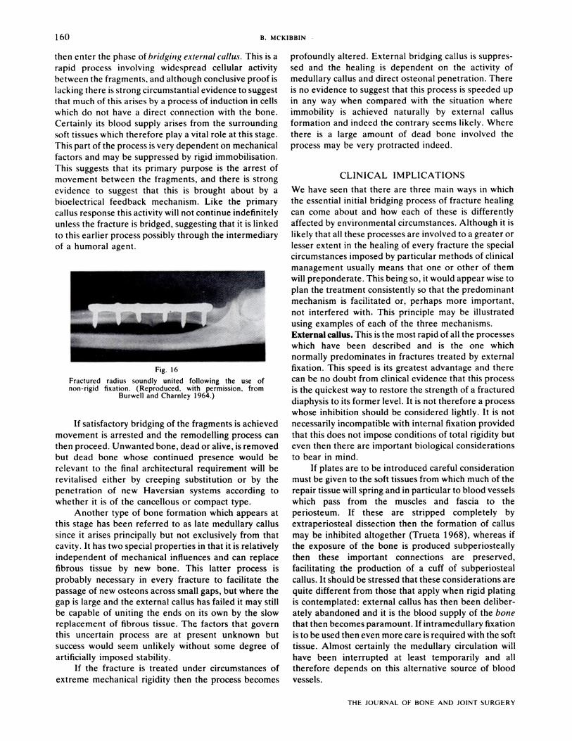
Fig. 16
I 60 B. MCKIBBIN
THE JOURNAL OF BONE AND JOINT SURGERY
then enter the phase of bridging external callus. This is a
rapid process involving widespread cellular activity
between the fragments, and although conclusive proof is
lacking there is strong circumstantial evidence to suggest
that much of this arises by a process of induction in cells
which do not have a direct connection with the bone.
Certainly its blood supply arises from the surrounding
soft tissues which therefore play a vital role at this stage.
This part of the process is very dependent on mechanical
factors and may be suppressed by rigid immobilisation.
This suggests that its primary purpose is the arrest of
movement between the fragments, and there is strong
evidence to suggest that this is brought about by a
bioelectrical feedback mechanism. Like the primary
callus response this activity will not continue indefinitely
unless the fracture is bridged, suggesting that it is linked
to this earlier process possibly through the intermediary
of a humoral agent.
Fractured radius soundly united following the use ofnon-rigid fixation. (Reproduced, with permission, from
Burwell and Charnley 1964.)
If satisfactory bridging of the fragments is achieved
movement is arrested and the remodelling process can
then proceed. Unwanted bone, dead or alive, is removed
but dead bone whose continued presence would be
relevant to the final architectural requirement will be
revitalised either by creeping substitution or by the
penetration of new Haversian systems according to
whether it is of the cancellous or compact type.
Another type of bone formation which appears at
this stage has been referred to as late medullary callus
since it arises principally but not exclusively from that
cavity. It has two special properties in that it is relatively
independent of mechanical influences and can replace
fibrous tissue by new bone. This latter process is
probably necessary in every fracture to facilitate the
passage of new osteons across small gaps, but where the
gap is large and the external callus has failed it may still
be capable of uniting the ends on its own by the slow
replacement of fibrous tissue. The factors that govern
this uncertain process are at present unknown but
success would seem unlikely without some degree of
artificially imposed stability.
If the fracture is treated under circumstances of
extreme mechanical rigidity then the process becomes
profoundly altered. External bridging callus is suppres-
sed and the healing is dependent on the activity of
medullary callus and direct osteonal penetration. There
is no evidence to suggest that this process is speeded up
in any way when compared with the situation where
immobility is achieved naturally by external callus
formation and indeed the contrary seems likely. Where
there is a large amount of dead bone involved the
process may be very protracted indeed.
CLINiCAL IMPLICATIONS
We have seen that there are three main ways in which
the essential initial bridging process of fracture healing
can come about and how each of these is differently
affected by environmental circumstances. Although it is
likely that all these processes are involved to a greater or
lesser extent in the healing of every fracture the special
circumstances imposed by particular methods of clinical
management usually means that one or other of them
will preponderate. This being so, it would appear wise to
plan the treatment consistently so that the predominant
mechanism is facilitated or, perhaps more important,
not interfered with. This principle may be illustrated
using examples of each of the three mechanisms.
External callus. This is the most rapid of all the processes
which have been described and is the one which
normally predominates in fractures treated by external
fixation. This speed is its greatest advantage and there
can be no doubt from clinical evidence that this process
is the quickest way to restore the strength of a fractured
diaphysis to its former level. It is not therefore a process
whose inhibition should be considered lightly. It is not
necessarily incompatible with internal fixation provided
that this does not impose conditions of total rigidity but
even then there are important biological considerations
to bear in mind.
If plates are to be introduced careful consideration
must be given to the soft tissues from which much of the
repair tissue will spring and in particular to blood vessels
which pass from the muscles and fascia to the
periosteum. If these are stripped completely by
extraperiosteal dissection then the formation of callus
may be inhibited altogether (Trueta 1968), whereas if
the exposure of the bone is produced subperiosteally
then these important connections are preserved,
facilitating the production of a cuff of subperiosteal
callus. It should be stressed that these considerations are
quite different from those that apply when rigid plating
is contemplated: external callus has then been deliber-
ately abandoned and it is the blood supply of the bone
that then becomes paramount. If intramedullary fixation
is to be used then even more care is required with the soft
tissue. Almost certainly the medullary circulation will
have been interrupted at least temporarily and all
therefore depends on this alternative source of blood
vessels.
THE BIOI.OGY OF FRACTURE HEAI.ING IN I.ONG BONES 161
VOL. 60-B, No. 2. MAY 1978
Late medullary callus. This method will predominate
when the external callus response has failed. It appears
to be assisted by immobilisation but since it will
inevitably be a slow process secure internal fixation
would appear to be most appropriate. If compression is
to be used then the only unanswered question is whether
the process can be speeded by the removal of the
intervening fibrous tissue or whether the compression
can be relied on to induce it to form bone, in which case it
can be left in place. This problem is worthy of further
study.
Primary bone union. The overwhelming disadvantage of
this method is its great slowness. It is not really a method
of union at all but a remodelling process which normally
occurs very late in the normal healing process, and
therefore artificial stability must be maintained for many
months and even years. Further delay is provided by the
fact that new osteons cannot cross gaps in fibrous tissue
and must await their preliminary replacement by
medullary callus. Where there are larger gaps it would
seem wise to supply that bridge with a bone graft but
there seems little point in applying cancellous grafts
external to the bone. This is normally done in order to
facilitate the formation of external callus and it is
scarcely logical to apply it in a situation where that
reaction has been deliberately inhibited.
The advantages of having the free use of the rigidly
fixed injured limb are nevertheless very great and
looking to the future it would seem that the ideal to be
pursued would be a system of secure internal fixation,
which at the same time did not sacrifice the rapid
assistance provided by external callus in the interests of
earlier consolidation. The radius illustrated in Figure 16
may represent a technical failure in the eyes of some
because of the presence of external callus but it may be
doubted if the patient saw it in that light.
Possibly this ideal can be achieved only by devising
fixation which can preserve security without necessarily
imposing total rigidity. As we have seen, however, there
are probably many other ways in which external callus is
controlled and we may learn to manipulate some of
these to our advantage to produce the same objective.
REFERENCES
Anderson, L. D. ( 1 965) Compression plate fixation and the effect of different types of internal fixation on fracture healing. Journal offlone (1,1(1Joint Surgery, 47-A, 191-208.
Bassett, C. A. L. (1971) Biophysical principles affecting bone structure. In The Biochemistrs’ and Physiology ofBone. Second edition. Vol. 3.Development and Growth, pp. 1-76. Edited by 0. H. Bourne. New York and London: Academic Press.
Bassett, C. A. L., and Becker, R. 0. (1962) Generation ofelectric potentials by bone in response to mechanical stress. Science, 137, 1063- 1064.
Bassett, C. A. L., and Herrman, I. (1961) Influence ofoxygen concentration and mechnical factorson differentiation ofconnective tissuein vitro.Nature, 190, 460-461.
Bassett, C. A. L., Pawluk, R. J., and Becker, R. 0. (1964) Effect of electric currents on bone in vivo. Nature, 204, 652-654.
Bassett, C. A. L., Pawluk, R. J., and Pilla, A. A. (1974) Acceleration of fracture repair by electromagnetic fields: a surgically non-invasivemethod. Annals ofthe New York Academy ofSciences, 238, 242-261.
Bier, H. (1920) Experimentelle Erfahrungen #{252}berPseudarthrosenbildung. M#{252}nchener medizinische Wochenschrift. 67, 22.
Bourne, G. H. (1971) The Biochemistry and Physiology ofBone. Second edition. Vol. 3. Development and Growth. New York and London:Academic Press.
Brookes, M. (1971) The Blood Supply ofBone; An Approach to Bone Biology. London: Butterworths.
Burwell, H. N., and Charnley, A. D. (1964) Treatment of forearm fractures in adults with particular reference to plate fixation.Journal of Boneand Joint Surgery, 46-B, 404-425.
Chalmers, J., Gray, D. H., and Rush, J. (1975) Observations on the induction of bone in soft tissues. Journal of Bone and Joint Surger�-.57-B, 36-45.
Charnley, J. (1970) The Closed Treatment ofCommon Fractures. Third edition. Edinburgh and London: E. & S. Livingstone Ltd.
Church, J., and Young, M. H. (1965) Unpublished data.
Friedenberg, Z. B., and Brighton, C. T. (1966) Bioelectrical potentials in bone. Journal of Bone and Joint Surgery. 48-A, 9 15-923.
Frledenberg, Z. B., Harlow, M. C., and Brighton, C. T. (1 971) Healing of nonunion of the medial malleolus by means of direct current: a casereport. Journal of Trauma, 11, 883-885.
Friedenstein, A. V. ( 1968) Induction of bone tissue by transitional epithelium. Clinical Orthopaedics and Related Research, 59, 21-37.
Fukada, E., and Yasuda, I. (1957) On the piezoelectric effect of bone. Journal of the Physical Society ofJapan. 12, 1 158- 1 162.
Girgis, F. G., and Pritchard, J. J. (1 958) Experimental production of cartilage during the repair of fractures of the skull vault in rats. Journal ofBone and Joint Surgery, 40-B, 274-28 1.
Goldhaber, P. (1961) Osteogenic induction across millipore filters in vivo. Science, 133, 2065-2067.
G#{246}thman, L. (1961) Vascular reactions in experimental fractures; microangiographic and radioisotope studies. Acta (‘hirurgica Scandinavica,Supplement 284.
Ham, A. W. (1930) A histological study of the early phase of bone repair. Journal of Bone and Joint Surgery, 12, 825-844.
Ham, A. W. (1969) Histology. Sixth edition. Philadelphia and Toronto: J. P. Lippincott Co.
Ham, A. W., and Harris, W. R. (1971) Repair and transplantation of bone. In The Biochemistry and Phvsiolog% ofBone. Second edition. Vol. 3.Development and Growth, pp. 337-399. Edited by G. H. Bourne. New York and London: Academic Press.
Johnson, R. W., Jun. (1927) A physiological study of the blood supply of the diaphysis. Journal of Bone and Joint Surgery, 9, 153-184.
Leriche, R., and Policard, A. (1928) The Norma/and Patho/ogica/Phs’sio/ogv ofBone: Its Problems. Translated by S. Moore and J. A. Key. StLouis: C. V. Mosby Co.
Lindholm, R., Lindholm, S., Liukko, P., Paasim#{225}ki, J., Lsok#{228}#{228}nt#{228},S., Rossi, R., Autlo, E., and Tamminen, E. (1969) The mast cell as acomponent of callus in healing fractures. Journal ofBone and Joint Surgery, 51-B, 148-155.
Lokietek, W., Pawluk, R. J. and Bassett, C. A. L. (1974) Muscle injury potentials; a source of voltage in the undeformed rabbit tibia. Journal ofBone and Joint Surgery, 56-B, 36 1-369.
162 B. MCKIBBIN
MacEwen, W. (1912) The Growth of Bone. Glasgow: Maclehose.
McClements, P., Templeton, R. W., and Pritchard, J. J. (1961) Repair of a bone gap. Journal ofAnatom%’, 95, 616.
Moss, M. L. (1960) Experimental induction of osteogenesis. In Calcification in Biological Systems, pp. 323-348. Edited by R. F. Sognnaes.Washington, D.C. : American Association for the Advancement of Science.
Mulholland, M. C., and Pritchard, J. J. (1959) The fracture gap. Journal ofAnatomy, 93, 590.
MUller, M. E., Allgower, M., and Willenegger, H. (1965) Technique ofinternal Fixation. Berlin, Heidelberg, New York: Springer-Verlag.
Olerud, S., and Dankwardt-Lilliestrbm, G. (1968) Fracture healing in compression osteosynthesis in the dog.JournalofBotieandJointSurgerv.50-B, 844-85 1.
Olerud, S., and Dankwardt.Lllliestr#{246}m, G. ( I 97 1 ) Fracture healing in compression osteosynthesis. Acta Orthopaedica Scandinavica,
Supplement 137.
Ostrowski, K., and Wodarskl, K. ( 1 97 1 ) Induction of heterotopic bone formation. In The Biochemistry and Physiology ofBone. Second edition.Vol. 3. Development and Growth, pp. 299-336. Edited by G. H. Bourne, New York and London: Academic Press.
Owen, M. (1970) The origin of bone cells. International Review of Cytology, 28, 213-238.
Perren, S. M., Huggler, A., Russenberger, M., Straumann, F., Muller, M. E., and Allgbwer, M. (1969) A method of measuring the change incompression applied to living cortical bone. Acta Orthopaedica Scandinavica, 125, 5-16.
Phillips, G., and McKibbln, B. (1 976) Unpublished data.
Pritchard, J. J. (1961) Hard tissues-bone and bones. In Recent Advances in Anatomy. Second series, pp. 204-236. Edited by F. Goldby and R.J. Harrison. London: J. & A. Churchill Ltd.
Pritchard, J. J. (1963) Bone healing. The Scientific Basis ofMedicine Annual Reviews, 286-301.
Pritchard, J. J., and Ruzicka, A. J. (1950) Comparison of fracture repair in the frog, lizard and the rat. Journal ofAnato�nv, 84, 236-26 1.
Rhinelander, F. W. (1974) Tibial blood supply in relation to healing. Clinical Orthopaedics and Related Research, 105, 34-81.
Roux, W. ( 1 895) Gesammelte Ahhand/uugen #{252}berEntwickelungsmechanik der Organismen. Leipzig: W. Englmann.
Schenk, R., and Willenegger, H. (1967) Morphological findings in primary fracture healing. Symposia Biotogica Hungarica, 8, 75-86.
Templeton, R. W. (1960) The Mode ofRepair ofa Fracture Gap. B.Sc. Thesis, Queens University, Belfast (cited by Pritchard, J. J. 1963).
Tonino, A. J., Davidson, C. L., Klopper, P. J., and Llnclau, L. A. (1976) Protection from stress in bone and its effects.Journa/ofBoneand JointSurgery, 58-B, 107-113.
Tonna, E. A., and Cronkite, E. P. (1961) Cellular response to fracture studied with tritiated thymidine. Journal ofBone and Joint Surgery.43-A, 352-362.
Trueta, J. ( 1 968) Studies of the Development and Decay of the Human Frame. London: William Heinemann Ltd.
Uhthoff, H. K., and Dubuc, F. L. (1971) Bone structure changes in the dog under rigid internal fixation. Clinical Orthopaedics and RelatedResearch, 81, 165-170.
Urist, M. R., and McLean, F. C. (1952) Osteogenetic potency and new-bone formation by induction in transplants to the anterior chamber of theeye. Journal of Bone and Joint Surgery, 34-A, 443-476.
Wray, J. B. ( 1 964) Acute changes in femoral arterial blood flow after closed tibial fracture in dogs. Journal of Bone and Joint Surgery,46-A, 1262-1268.
Young, R. W. (1962) Cell proliferation and specialization during endochondral osteogenesis in young rats.JournalofCell Biology, 14, 35 7-370.
THE JOURNAL OF BONE AND JOINT SURGERY