T Cell Receptor - Roswell Park Comprehensive Cancer Center · Both TCR and BCR have transmembrane...
32
T Cell Receptor Xuefang Cao, MD, PhD October 29, 2015
Transcript of T Cell Receptor - Roswell Park Comprehensive Cancer Center · Both TCR and BCR have transmembrane...
T Cell Receptor
Xuefang Cao, MD, PhD
October 29, 2015
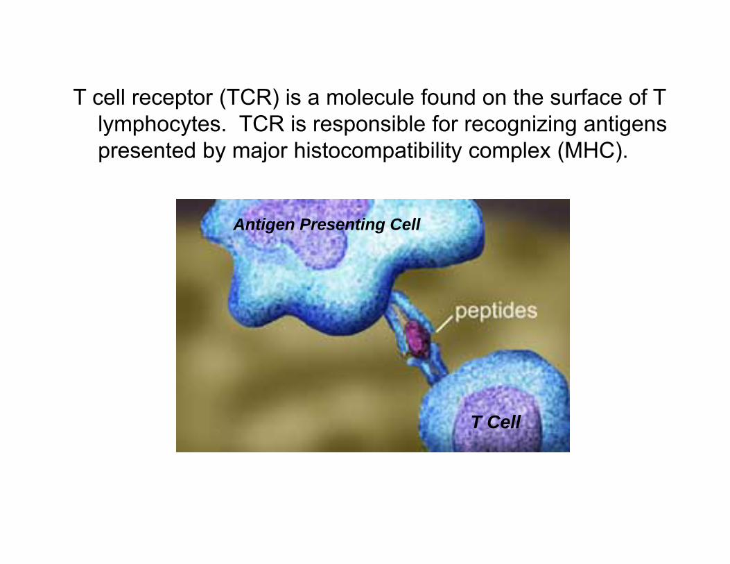
T Cell
Antigen Presenting Cell
T cell receptor (TCR) is a molecule found on the surface of T lymphocytes. TCR is responsible for recognizing antigens presented by major histocompatibility complex (MHC).
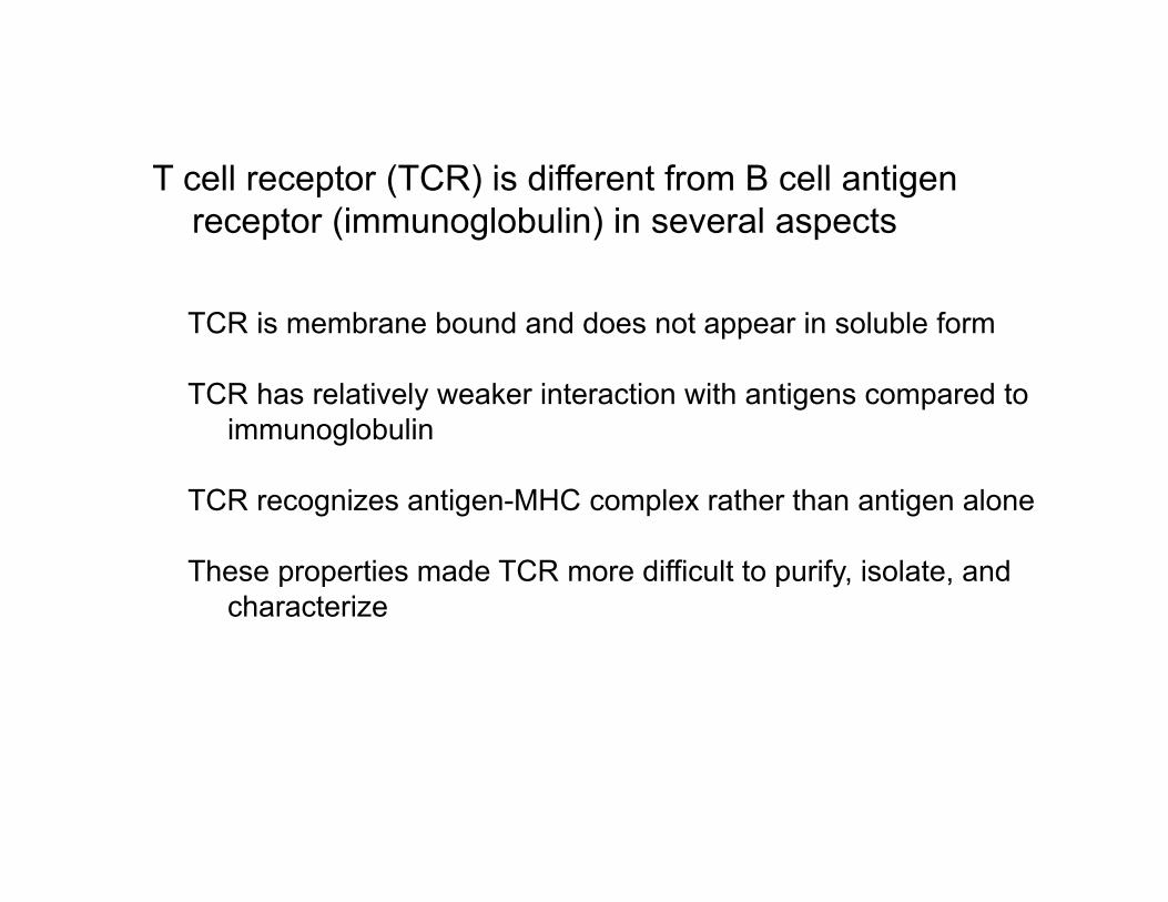
T cell receptor (TCR) is different from B cell antigen receptor (immunoglobulin) in several aspects
TCR is membrane bound and does not appear in soluble form
TCR has relatively weaker interaction with antigens compared to immunoglobulin
TCR recognizes antigen-MHC complex rather than antigen alone
These properties made TCR more difficult to purify, isolate, and characterize
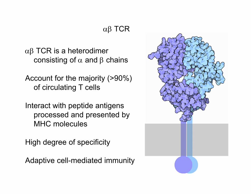
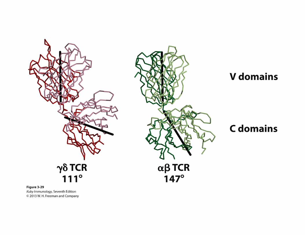
TCR is a heterodimer consisting of and chains
Account for the majority (>90%) of circulating T cells
Interact with peptide antigens processed and presented by MHC molecules
High degree of specificity
Adaptive cell-mediated immunity
TCR
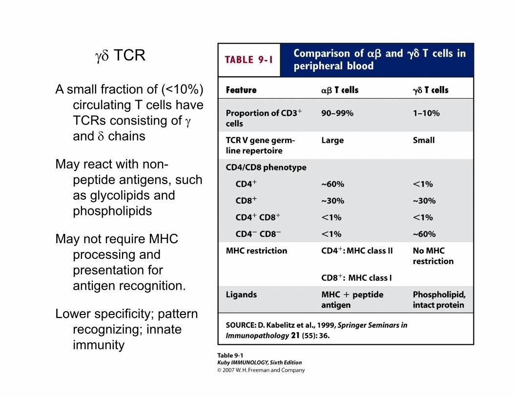
A small fraction of (<10%) circulating T cells have TCRs consisting of and chains
May react with non-peptide antigens, such as glycolipids and phospholipids
May not require MHC processing and presentation for antigen recognition.
Lower specificity; pattern recognizing; innate immunity
TCR
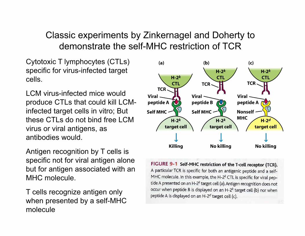
Cytotoxic T lymphocytes (CTLs) specific for virus-infected target cells.
LCM virus-infected mice would produce CTLs that could kill LCM-infected target cells in vitro; But these CTLs do not bind free LCM virus or viral antigens, as antibodies would.
Antigen recognition by T cells is specific not for viral antigen alone but for antigen associated with an MHC molecule.
T cells recognize antigen only when presented by a self-MHC molecule
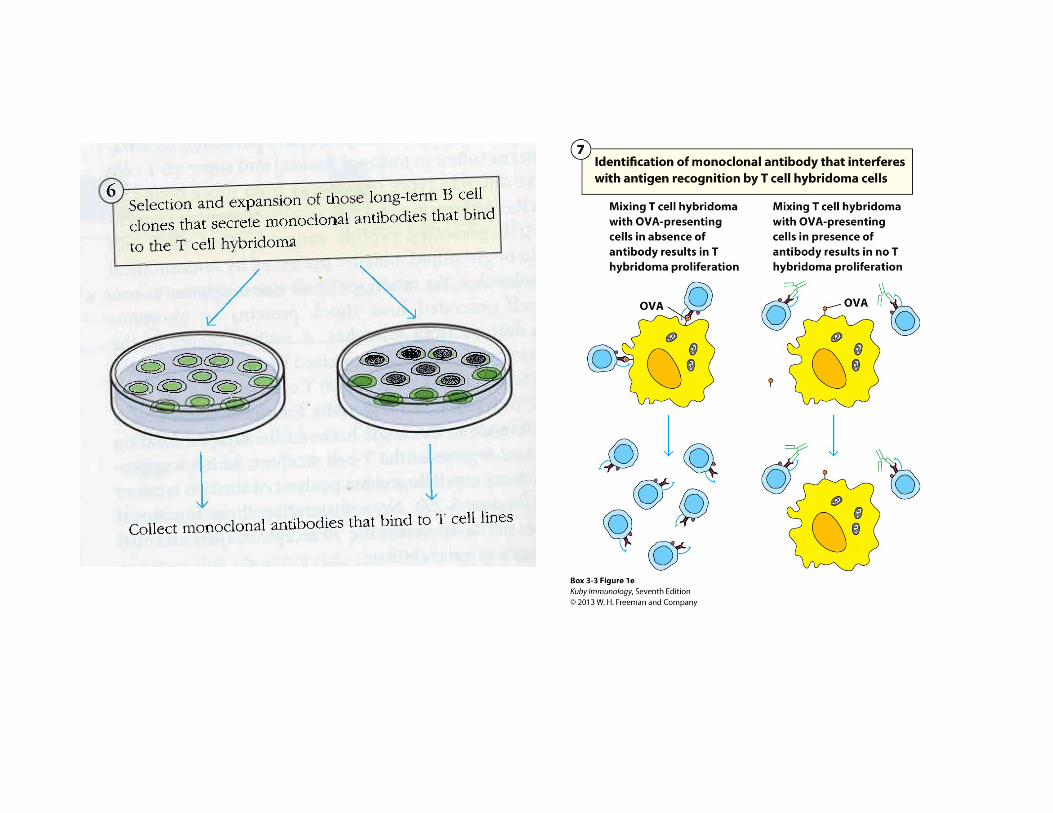
Classic experiments by Zinkernagel and Doherty to demonstrate the self-MHC restriction of TCR
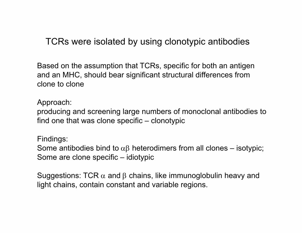
Based on the assumption that TCRs, specific for both an antigen and an MHC, should bear significant structural differences from clone to clone
Approach: producing and screening large numbers of monoclonal antibodies to find one that was clone specific – clonotypic
Findings: Some antibodies bind to heterodimers from all clones – isotypic; Some are clone specific – idiotypic
Suggestions: TCR and chains, like immunoglobulin heavy and light chains, contain constant and variable regions.
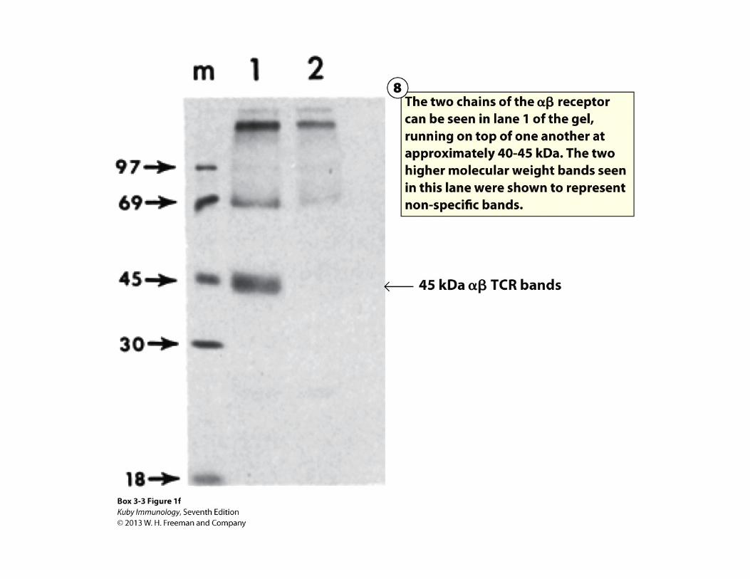
TCRs were isolated by using clonotypic antibodies
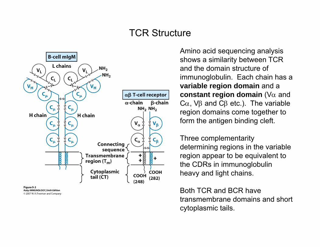
Amino acid sequencing analysis shows a similarity between TCR and the domain structure of immunoglobulin. Each chain has a variable region domain and a constant region domain (V and C, V and C etc.). The variable region domains come together to form the antigen binding cleft.
Three complementarity determining regions in the variable region appear to be equivalent to the CDRs in immunoglobulin heavy and light chains.
Both TCR and BCR have transmembrane domains and short cytoplasmic tails.
TCR Structure
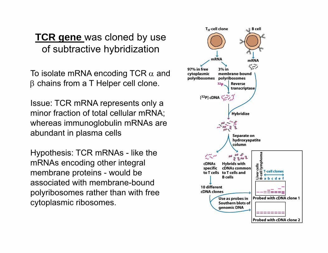
TCR gene was cloned by useof subtractive hybridization
To isolate mRNA encoding TCR and chains from a T Helper cell clone.
Issue: TCR mRNA represents only a minor fraction of total cellular mRNA; whereas immunoglobulin mRNAs are abundant in plasma cells
Hypothesis: TCR mRNAs - like the mRNAs encoding other integral membrane proteins - would be associated with membrane-bound polyribosomes rather than with free cytoplasmic ribosomes.
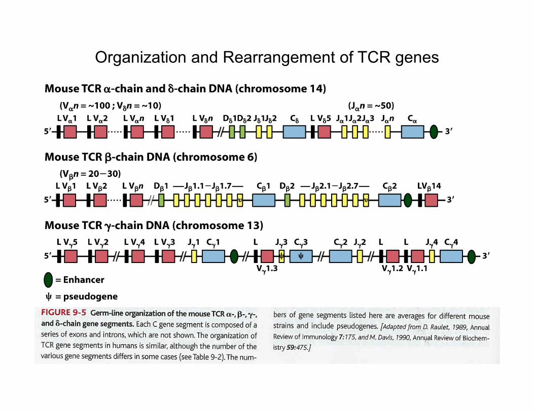
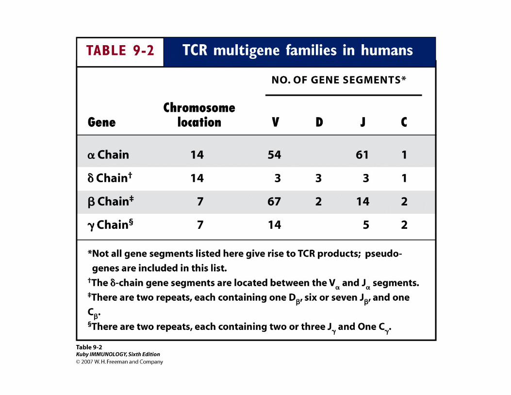
Organization and Rearrangement of TCR genes
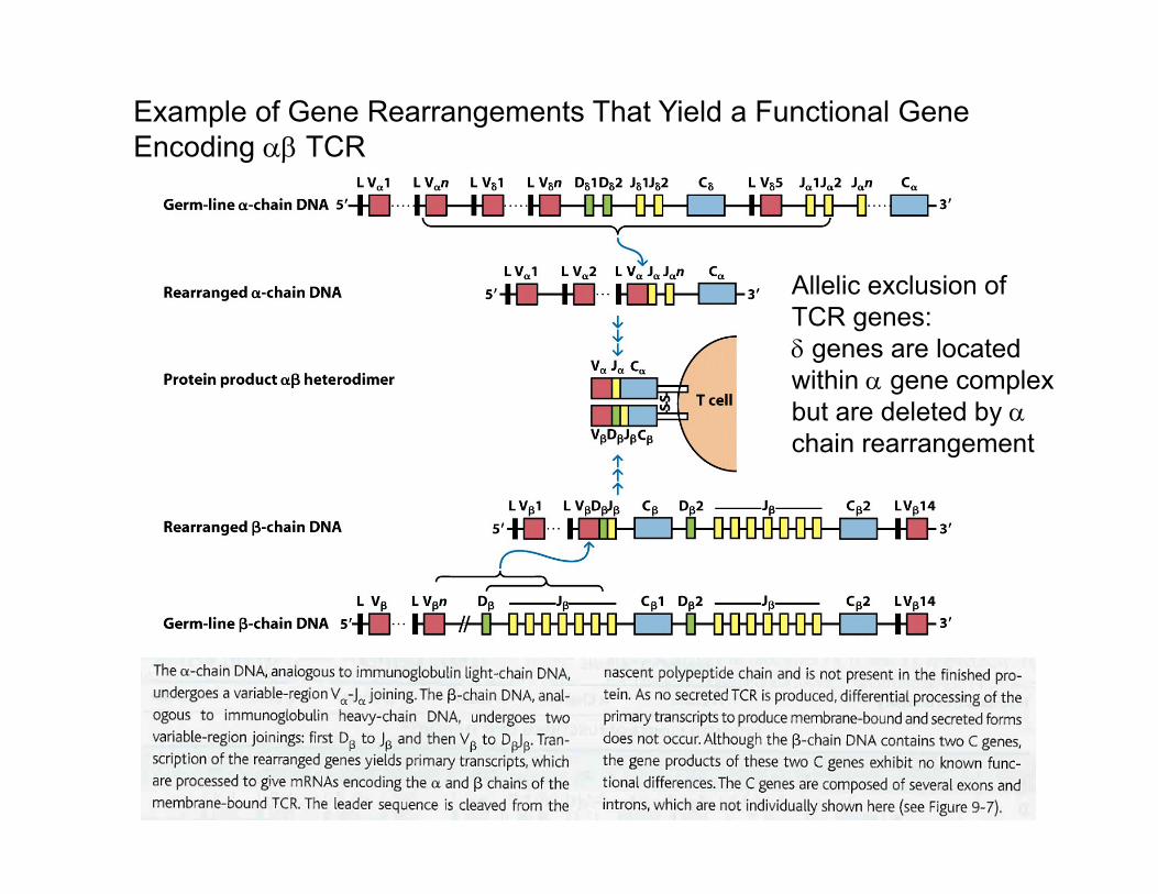
Example of Gene Rearrangements That Yield a Functional Gene Encoding TCR
Allelic exclusion of TCR genes: genes are located within gene complex but are deleted by chain rearrangement
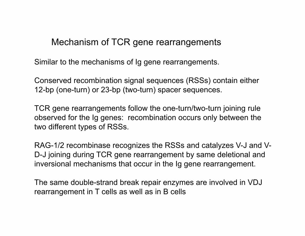
Mechanism of TCR gene rearrangements
Similar to the mechanisms of Ig gene rearrangements.
Conserved recombination signal sequences (RSSs) contain either 12-bp (one-turn) or 23-bp (two-turn) spacer sequences.
TCR gene rearrangements follow the one-turn/two-turn joining rule observed for the Ig genes: recombination occurs only between the two different types of RSSs.
RAG-1/2 recombinase recognizes the RSSs and catalyzes V-J and V-D-J joining during TCR gene rearrangement by same deletional and inversional mechanisms that occur in the Ig gene rearrangement.
The same double-strand break repair enzymes are involved in VDJ rearrangement in T cells as well as in B cells
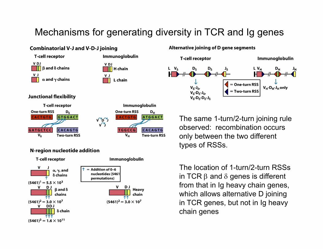
The same 1-turn/2-turn joining rule observed: recombination occurs only between the two different types of RSSs.
The location of 1-turn/2-turn RSSs in TCR and genes is different from that in Ig heavy chain genes, which allows alternative D joining in TCR genes, but not in Ig heavy chain genes
Mechanisms for generating diversity in TCR and Ig genes
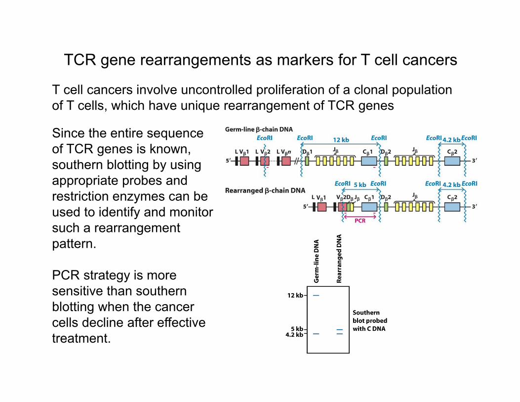
TCR gene rearrangements as markers for T cell cancers
Since the entire sequence of TCR genes is known, southern blotting by using appropriate probes and restriction enzymes can be used to identify and monitor such a rearrangement pattern.
PCR strategy is more sensitive than southern blotting when the cancer cells decline after effective treatment.
T cell cancers involve uncontrolled proliferation of a clonal population of T cells, which have unique rearrangement of TCR genes
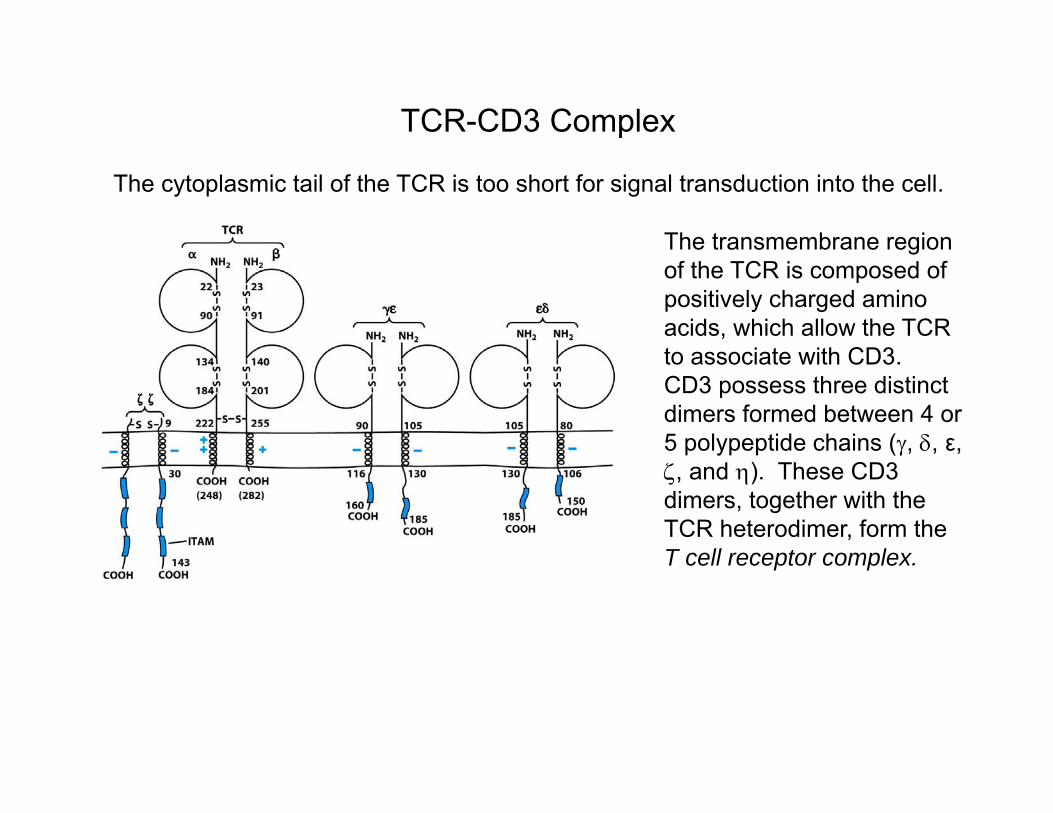
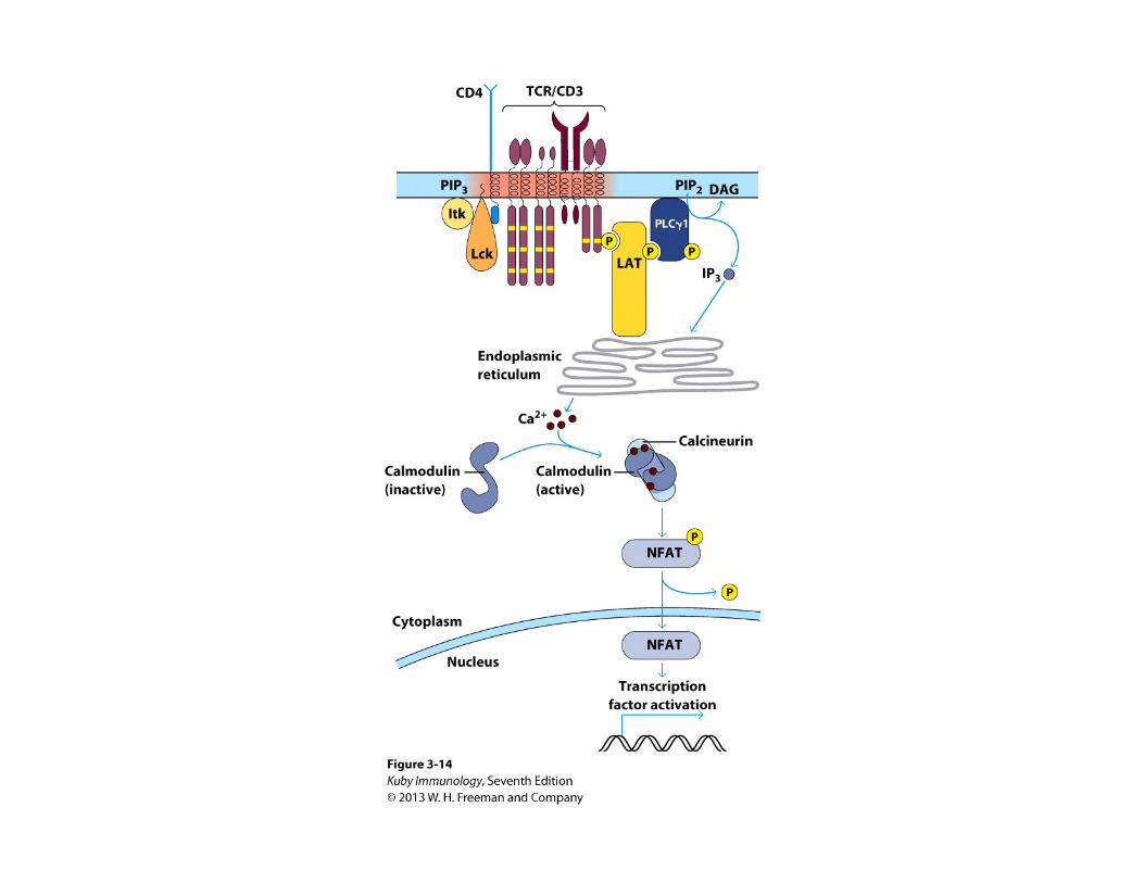
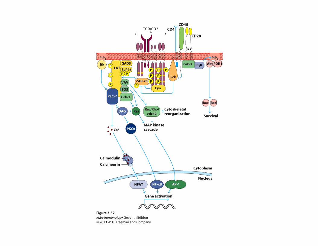
TCR-CD3 Complex
The cytoplasmic tail of the TCR is too short for signal transduction into the cell.
The transmembrane region of the TCR is composed of positively charged amino acids, which allow the TCR to associate with CD3. CD3 possess three distinct dimers formed between 4 or 5 polypeptide chains (, , ε, , and ). These CD3 dimers, together with the TCR heterodimer, form the T cell receptor complex.
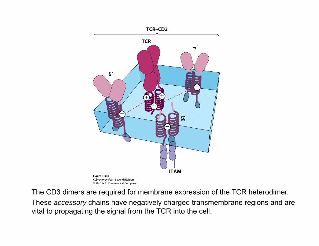
The CD3 dimers are required for membrane expression of the TCR heterodimer. These accessory chains have negatively charged transmembrane regions and are vital to propagating the signal from the TCR into the cell.
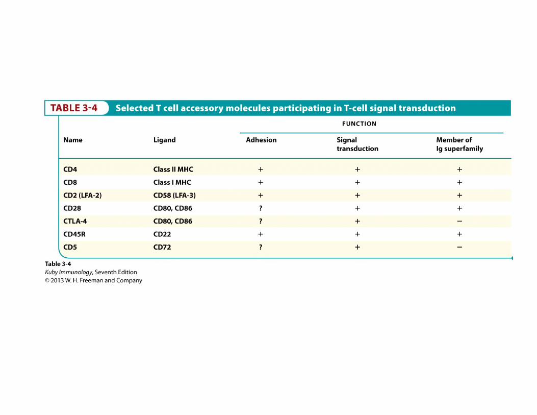
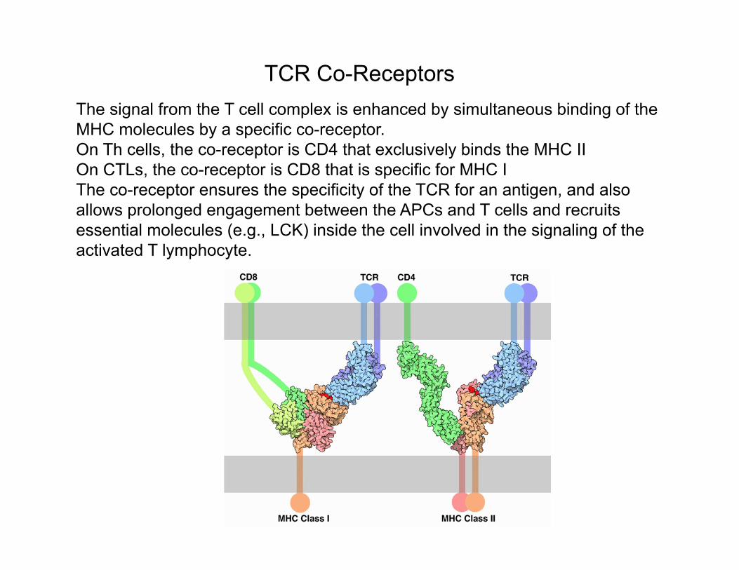
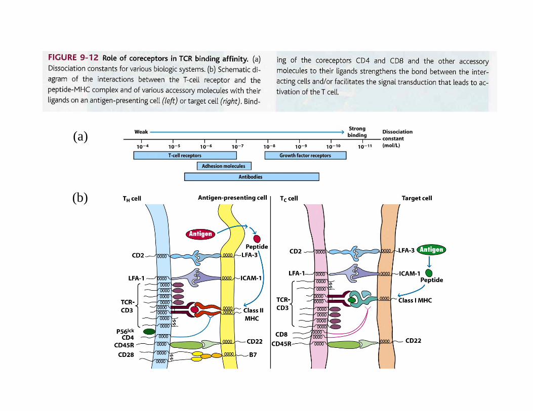
TCR Co-ReceptorsThe signal from the T cell complex is enhanced by simultaneous binding of the MHC molecules by a specific co-receptor.On Th cells, the co-receptor is CD4 that exclusively binds the MHC II On CTLs, the co-receptor is CD8 that is specific for MHC I The co-receptor ensures the specificity of the TCR for an antigen, and also allows prolonged engagement between the APCs and T cells and recruits essential molecules (e.g., LCK) inside the cell involved in the signaling of the activated T lymphocyte.
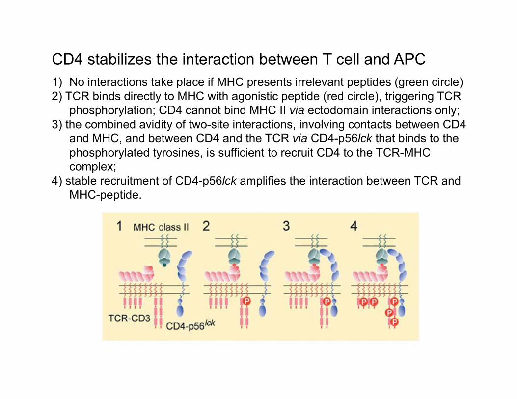
CD4 stabilizes the interaction between T cell and APC 1) No interactions take place if MHC presents irrelevant peptides (green circle)2) TCR binds directly to MHC with agonistic peptide (red circle), triggering TCR
phosphorylation; CD4 cannot bind MHC II via ectodomain interactions only; 3) the combined avidity of two-site interactions, involving contacts between CD4
and MHC, and between CD4 and the TCR via CD4-p56lck that binds to the phosphorylated tyrosines, is sufficient to recruit CD4 to the TCR-MHC complex;
4) stable recruitment of CD4-p56lck amplifies the interaction between TCR and MHC-peptide.
(a)
(b)
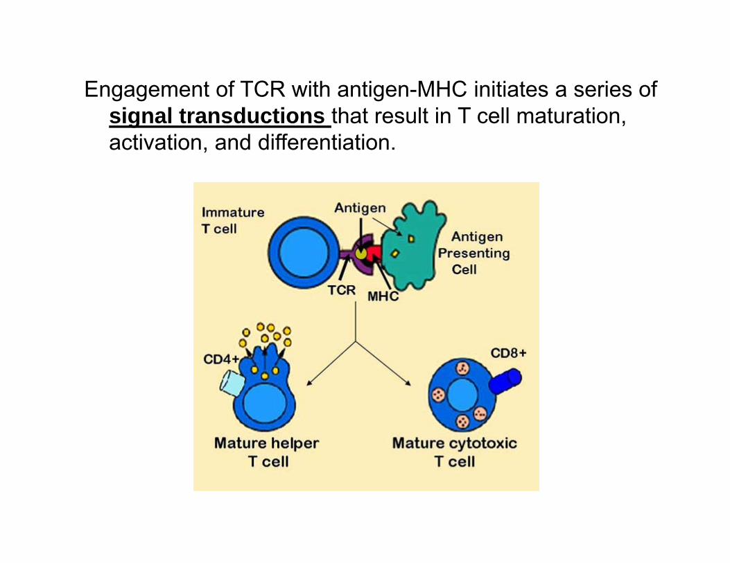
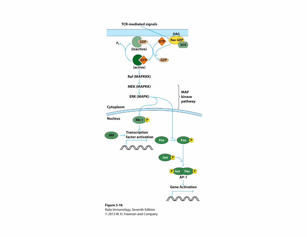
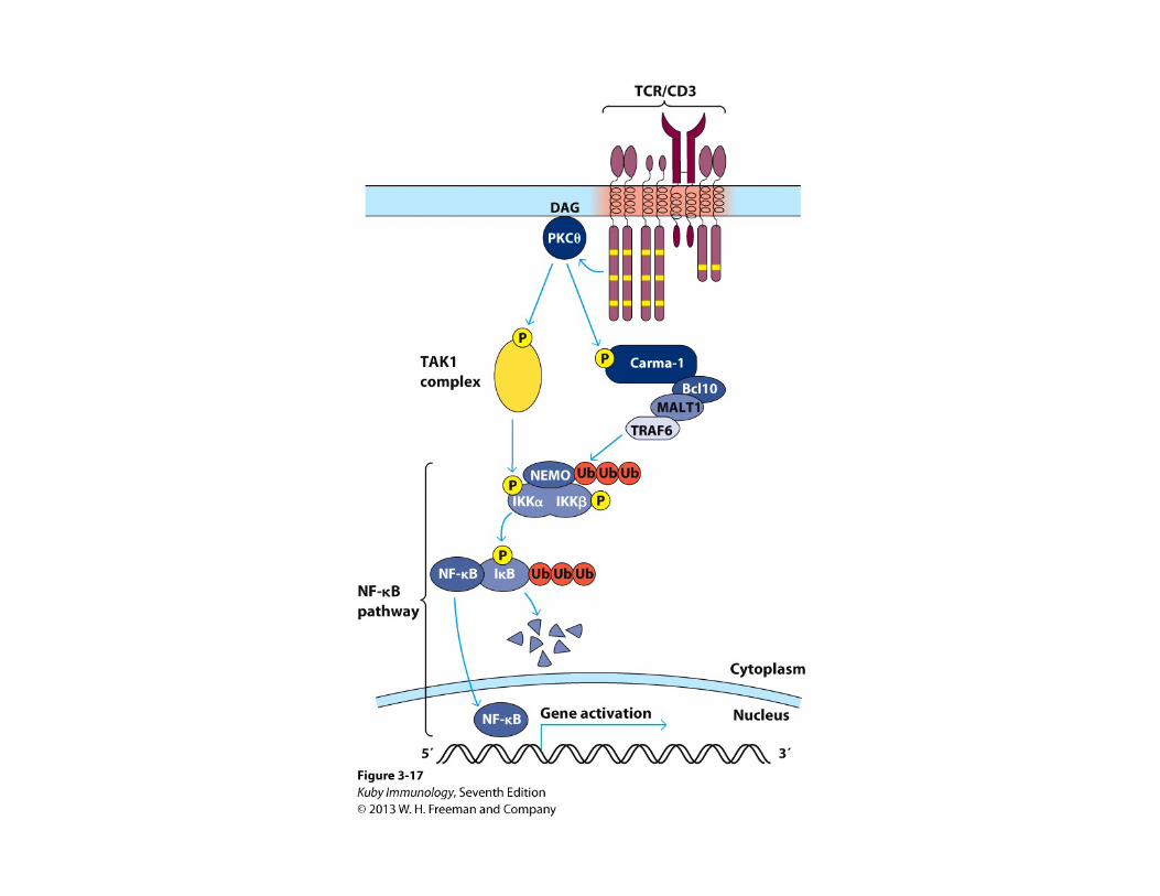
Engagement of TCR with antigen-MHC initiates a series of signal transductions that result in T cell maturation, activation, and differentiation.


![IMMUNOGLOBULINE E T CELL RECEPTOR T. Strachan e A.P. … · B cell antigen receptor tetramero [ IgH 2 + IgL 2 (Ig oppure Ig )] T cell receptor (TCR) eterodimero TCR /TCR TCR /TCR](https://static.fdocuments.in/doc/165x107/5c017b5c09d3f26f1e8cc6a0/immunoglobuline-e-t-cell-receptor-t-strachan-e-ap-b-cell-antigen-receptor.jpg)