Synthesis and Gene Silencing Activity of RNA Duplexes...
77
Synthesis and Gene Silencing Activity of RNA Duplexes Containing 3’-Deoxy-3’-Thiothymidine Siara Ruth Isaac Department of Chemistry McGill University Montreal, Canada July 2008 A thesis submitted to McGill University in partial fulfillment of the requirement for the degree of Master of Science © Siara Isaac, 2008
Transcript of Synthesis and Gene Silencing Activity of RNA Duplexes...

Synthesis and Gene Silencing Activity of RNA Duplexes Containing 3’-Deoxy-3’-Thiothymidine
Siara Ruth Isaac
Department of Chemistry McGill University Montreal, Canada
July 2008
A thesis submitted to McGill University in partial fulfillment of the requirement for the degree of
Master of Science
© Siara Isaac, 2008
Abstract
This thesis reports the first synthesis and biophysical characterization of
oligoribonucleotides (RNA) containing 3'-deoxy-3'-thiothymidine (3'-S-dT) units. Optimized
conditions for incorporating 3'-S-dT into 21-nt RNA strands is also reported. The impact of 3'-S-
dT substitutions on the structure and stability of RNA:RNA duplexes was investigated by circular
dichroism (CD) and UV/thermal (Tm) studies. Generally, replacement of the 3'-oxygen by a
sulphur atom resulted in a significant decrease in the thermal stability of the duplex and strong
positional effects were observed. No significant disruption of the A-form helical structure of the
duplexes was detected by CD, although in some cases 3'-S-dT inserts led to a reduction in base-
base stacking and/or local distortion of the duplex. Assays revealed that short interfering RNA
(siRNA) duplexes containing 3'-S-dT on the guide strand were not efficient at down-regulating
gene products, but those containing 3'-S-dT on the passenger strand OR on both strands exhibited
activities that often surpassed those of unmodified siRNA duplexes. Substitutions at the scissile
phosphate position of the passenger strand suggest that the non-bridging 3’-oxygen is not
involved in the coordination of a metal ion during the rate determining step in RISC-induced
RNA cleavage.
Résumé
Cette thèse présente la première synthèse et caractérisation biophysique
d’oligoribonucleotides (ARN) contenant des unités de 3'-deoxy-3'-thiothymidine (3'-S-dT). Des
conditions optimisées pour l’intégration de 3'-S-dT en 21-nt ARN sont aussi présentées. L’impact
de la substitution de 3'-S-dT sur la structure et la stabilité des duplex ARN:ARN fut recherché
systématiquement par l’entremise de dichroïsme circulaire (CD) et d’études UV/thermale (Tm).
Généralement, le remplacement du 3'-oxygène par un atome de sulfure résulta en une baisse
significative de la stabilité thermique du duplex, et des effets de positionnement important furent
observés. Aucune disruption significative de la structure hélicoïdale A-form du duplex ne fut
détectée par le CD, quoi que parfois des inserts de 3'-S-dT entraînèrent une réduction des
interactions base-base et/ou une distorsion locale du duplex. Des essaies révélèrent que des duplex
interférents d’ARN court (siRNA) contenant du 3'-S-dT sur le brin guide n’ont pas baissé la
production de gènes, mais que celles contenant du 3'-S-dT sur le brin complémentaire OU sur les
deux brins présentèrent des activités qui fréquemment surpassaient celles de duplex non-modifiés
de siRNA. Les substitutions à la position phosphate de rupture du brin complémentaire suggèrent
que l’étape déterminante du clivage par RISC n’implique pas le 3'-oxygène pour la coordination
d’un ion de métal.
2
Acknowledgments
It has been a privilege to work in Dr. Masad J. Damha’s lab. He is a great chemist and
teacher, who clearly cares about his students as individuals. He always encouraged me to continue
with my chemistry but he also supported me when my interests took me out of the lab. He
allowed me to take on teaching responsibilities, to take classes in education and to explore science
pedagogy. And through this, I have found my career. I am deeply grateful for his extended
patience and support.
I would also like to thank my colleagues in the Damha research group, particularly
Jonathan Watts and Robert Donga, who kept me company through long hours in the lab and
provided invaluable mentorship and discussions (some chemical, some political). Thanks are also
due to my team of editors: Jonathan Watts, Robert Donga, Alison Palmer, Jeremy Lackey, and Dr.
Anne Noronha.
I have benefited from significant financial support from the McGill University
Department of Chemistry, in the form of stipends and teaching assistantships. These last were also
an excellent opportunity to gain experience in university-level teaching.
I acknowledge and appreciate the RNAi experiments performed by Dr. Francis
Robert (Dr. Jerry Pelletier lab’s, Department of Biochemistry, McGill) and the MS
spectra obtained by Dr. Alain Tessier, Department of Chemistry and Biochemistry
(Concordia) of the oligoribonucleotides. I would like to thank Dr. Karine Auclair for the
use of the CD instrument, Dr. Nadim Saade for training and trouble shooting on the MS
machine I used for the nucleoside synthesis and Dr. Paul Xia for maintenance and
training on the NMR machines.
And finally, thank you to my friends and family for their support and
patience. Thanks particularly to my husband Jean-Philippe Roy, Emily Cranston and the
Third Floor Tea Club.
3
Table of Contents
page
Abstract (Résumé) 2
Acknowledgements 3
Table of Contents 4
List of Figures 6
List of Tables 7
Abbreviations 8
Chapter 1. Introduction
1.0 The Historical Context of Nucleic Acid Research 11
1.1 A Modern Understanding of Nucleic Acids 12
1.2 The Flow of Genetic Information 14
1.3 Furanose Conformation 14
1.4 Oligonucleotide-based Therapeutics 15
1.5 Antisense Gene Silencing 15
1.6 RNA interference 16
1.7 Chemical Modifications of Oligonucleotide for Therapeutics 18
1.8 Oligonucleotide Modifications in RNAi 18
1.9 Backbone Modifications 18
1.10 Sugar Modifications 19
1.11 Flexibility and Enzymatic Recognition 21
1.12 Thesis Objectives 22
1.13 References 22
Chapter 2. Experimental Materials and Methods
2.1 General Methods
2.1.1 General Reagents 28
2.1.2 Chromatography For Nucleosides And Intermediates 28
2.1.3 Instruments 28
2.2 Synthesis of 3’-Deoxy-3’-Thiothymidine Nucleoside and its 3’-S-
Phosphorothioamidite Derivative
2.2.1 5'-O-P-Monomethoxytritylthymidine (3.2) 29
2.2.2 3’-O-Methanesulphonyl-5’-(monomethoxytrityl)thymidine (3.3) 29
2.2.3 2,3’-dideoxy-5’-(monomethoxytrityl)anhydrothymidine (3.4) 30
2.2.4 3’-Acetylthio-3’-deoxy-5’-(monomethoxytrityl)thymidine (3.5) 30
2.2.5 3’-deoxy-3’-thio-5’-(monomethoxytrityl)thymidine (3.6) 30
2.2.6 3’-deoxy-5’-Monomethoxytritylthymidine-3’-S-
phosphorothioamidite (3.8) 31
2.3 Oligonucleotide Synthesis
2.3.1 General Reagents 31
2.3.2 Derivatization of Solid Support 32
2.3.3 General Automated Solid-Phase Synthesis of Oligonucleotides 32
2.4 Deprotection and Purification of Oligonucleotides
2.4.1 General Reagents 33
2.4.2 Cleavage from CPG and Deprotection of Oligomers 34
2.4.3 Analysis and Purification by Polyacrylamide Gel Electrophoresis 34
2.4.4 Reversed-Phase HPLC Analysis and Purification of 35
4

Oligonucleotides
2.4.5 Anion-Exchange HPLC Analysis of Oligonucleotides 35
2.4.6 Desalting of Oligonucleotides 36
2.5 Characterization of Oligonucleotides
2.5.1 Characterization of Oligonucleotides by Mass Spectrometry 36
2.5.2 UV-Thermal Denaturation of Oligonucleotides 36
2.5.3 Circular Dichroism Spectroscopy (CD) 37
2.6 Biological Studies
2.6.1 RNAi Transfection 37
2.6.2 Luciferase Activity Assay 38
2.6.3 Luciferase mRNA Quantitation 38
2.7 References 38
Chapter 3: Synthesis of 3’-Deoxy-3’-thiothymidine and Its Incorporation into
Oligonucleotides
3.1 Relevance of 3’,5’-Linked 3’-S-Phosphorothiolate Linkages 39
3.2 Synthesis of 3’-Deoxy-3’-thiothymidine 39
3.3 Synthesis of Oligonucleotides Containing 3’-Deoxy-3’-thiothymidine Inserts 42
3.4 Activators for 3’-Thio Coupling 43
3.5 Oxidants for the Preparation of dT12 Containing a Single 3’-S-dT Unit 44
3.6 Alternative Phosphitylation Conditions 47
3.7 Phosphorothioamidite Stability 48
3.8 Optimised Synthesis Conditions for Oligonucleotides Containing 3’-Deoxy-3’-
thiothymidine 49
3.9 References 49
Chapter 4: Physical and Biological Characterisation of Oligonucleotides
Containing 3’-S-Phosphorothiolate Linkages
4.1 Introduction 52
4.2 3’-Phosphorothiolate Linkages and Conformation 52
4.3 A Library of Modified RNA Duplexes Containing 3’-Thio Linkages 53
4.4 Conformation and Thermal Stability of Duplexes 57
4.5 Analysis of Oligonucleotide Conformation by Circular Dichroism (CD) 61
4.6 Evaluation of the Gene Silencing Activity of 3’-thio modified siRNA 63
4.7 Conclusion 67
4.8 References 68
Contribution to Knowledge 70
Appendix 1: NMR Spectra 71
Appendix 2: Tm & Thermodynamic Data 73
Appendix 3: CD Data 76
5

List of Figures
page
Chapter 1. Introduction
Figure 1.1: Structure of DNA and RNA Oligonucleotides 12
Figure 1.2: Helical Structure of Duplex DNA 12
Figure 1.3: Watson-Crick Base Pairing Specificity in DNA Nucleobases 13
Figure 1.4: Furanose Conformation Preferences in DNA and RNA 14
Figure 1.5: RNAi Pathway 16
Figure 1.6: Schematic of siRNA Sequences 17
Figure 1.7: Four Common Ribo Modifications 20
Figure 1.8: Four Common 2’-Deoxy Modifications 21
Figure 1.9: Butanediol and 2’,3’-Secouridine 22
Chapter 2: Experimental Materials and Methods
Figure 2.1: Schematic of DNA Synthesiser Solid-Phase Synthesis of
Oligonucleotides 33
Chapter 3: Synthesis of 3’-Deoxy-3’-thiothymidine and Its Incorporation into
Oligonucleotides
Figure 3.1: Cosstick’s Synthetic Scheme for 3’-Deoxy-3’-thiothymidine and its
Incorporation into a Dinucleotide 40
Figure 3.2: 3’-Deoxy-3’-(thioacetyl)thymidine Synthetic Scheme 40
Figure 3.3: Free Thiol and Disulfide Species Synthetic Scheme 41
Figure 3.4: 3’-S-Phosphorothioamidite Synthetic Scheme 42
Figure 3.5: Comparison of Oxidants for 3’-Thio Oligonucleotide Synthesis 44
Figure 3.6: Comparison of Oxidants Systems in the Preparation of dT12
Containing a Single 3’-S-Phosphorothiolate Linkage 45
Figure 3.7: HPLC trace of Crude Oligoribonucleotides 46
Figure 3.8: Schematic Illustration of Three Phosphitylation Methods 47
Figure 3.9: 31
P NMR of Phosphorothioamidite 48
Chapter 4: Physical and Biological Characterisation of Oligonucleotides
Containing 3’-S-Phosphorothiolate Linkages
Figure 4.1: Melting Temperature (Tm) Signatures of 5 Representative
RNA:RNA Duplexes 59
Figure 4.2: CD Spectra of Native and Modified RNA:RNA Duplexes 62
Figure 4.3: CD Spectra of Guide and Passenger Strand Modified RNA:RNA
Duplexes 63
Figure 4.4: RNAi Activity of Duplexes Modified Only on the Guide Strand 64
Figure 4.5: RNAi Activity of 3’-Thio Modified Passenger Strand with Native or
Modified Guide Strands 65
Figure 4.6: RNAi Activity of Modified Passenger Strand with Native or
Modified Guide Strands 67
6
List of Tables
page
Chapter 1. Introduction
Table 1.1: Characteristic Parameters Oligonucleotide Helices 13
Chapter 2: Experimental Materials and Methods
Table 2.1: Reagents for Solid-Phase Synthesis of Oligonucleotides 33
Chapter 3: Synthesis of 3’-Deoxy-3’-thiothymidine and Its Incorporation into
Oligonucleotides
Table 3.1: Prevention of Disulfide Formation with DTT 41
Table 3.2: A Comparison of the pKa and Structure of Relevant Activators 43
Table 3.3: Oxidants for the Preparation of Thymidylic Acid (12 mer)
Containing a Single 3’-S-dT Residue 45
Table 3.4: Comparison of Overall Coupling Efficacy for Mixed Sequence
21mers with 2 Oxidants 46
Table 3.5: Comparison of Coupling Efficacy for Alternative
Phosphitylation Methods with dT-12mers 48
Table 3.6: Optimised Conditions for 3’-S-Phosphorothioamidite Coupling 49
Chapter 4: Physical and Biological Characterisation of Oligonucleotides
Containing 3’-S-Phosphorothiolate Linkages
Table 4.1: Mass Spectral Characterization of Selected Oligonucleotide
Strands Used to Create the siRNA Duplex Library 54
Table 4.2: An Inventory of Duplexes with a Native RNA Passenger
Strand and a Modified RNA Guide Strand 55
Table 4.3: An Inventory of Duplexes with a Native RNA Guide Strand
and a Modified RNA Passenger Strand
56
Table 4.4: An Inventory of Duplexes with a 3’-Thio Modified RNA Guide
Strand and a Modified RNA Passenger Strand
57
Table 4.5: Melting Temperatures (Tm) of dT and 3’-S-dT Modified
Duplexes 60
Table 4.6: Melting Temperatures (Tm) of 3’-Thio and 2’-OMe Modified
Duplexes 60
Table 4.7: Melting Temperatures (Tm) of Guide Strand Mismatches 61
7

Abbreviations
A adenine
Å angstrom
LNA -D-Locked Nucleic Acid
ANA arabinonucleic acid
AON(s) antisense oligonucleotide(s)
APS ammonium persulfate
BPB bromophenol blue
BTT 5-benzylthio-1-H-tetrazole
BzCl benzoyl chloride
C cytosine
© copyright
CaH2 calcium hydride
CD Circular Dichroism
CE β-cyanoethyl
CH2Cl2 methylene chloride
CH3CN acetonitrile
CPG controlled pore glass
d d doublet (NMR)
dd doublet of doublet (NMR)
d6 six deuterium atoms
DCI 4,5-dicyanoimidazole
ddH2O double distilled water
ΔH enthalpy variation
ΔS entropy variation
DEPC diethyl pyrocarbonate
DIPEA N,N-diisopropylethylamine
DMAP 4-dimethylaminopyridine
DMSO dimethyl sulfoxide
DMT 4,4’- dimethoxytrityl
DNA deoxyribonucleic acid
ds double stranded
E. coli Escherichia coli
e.g. for example
260 molar extinction coefficients at 260
EDTA ethylene-diamine tetraacetate dihydrate
eq equivalent(s)
EtOH ethanol
2’-FANA 2’-deoxy-2’-fluoro-β-D-arabinonucleic acid
g gram
mg milligram
µg microgram
G guanine
h hour(s)
HBTU O-(benzotriazol-1-yl)-1,1,3,3-tetramethyluronium
hexafluorophosphate
HOAc acetic acid
HPLC anion exchange liquid phase chromatography
i.e that is
8
J coupling constant
KCl potassium chloride
l path length of a UV cell
mL milliliter
µL microliter
λ wavelength
LCAA succinyl-derivatized long chain alkyl amine
M molar
mM millimolar
M micromolar
m multiplet (NMR)
MeOH methanol
MgCl2 magnesium chloride
min minute(s)
MHz mega hertz
mmol millimole
µmol micromole
MMT monomethoxytrityl group
MMTCl p-anisylchlorodiphenylmethane
mol mole
m/z mass to charge ratio
NaClO4 sodium perchlorate
Na2HPO4 disodium hydrogen phosphate
NaHCO3 sodium bicarbonate
Na2SO4 anhydrous sodium sulphate
NH4OH ammonium hydroxide
nm nanometer
NMI N-methylimidazole
NMR nuclear magnetic resonance
OD optical density
2’-O-MOE 2’-O-methoxyethyl 32
P phosphorus 32
PAGE polyacrylamide gel electrophoresis
ppm parts per million
® registered trademark
Rf retention factor
RISC RNAi induced silencing complex
RNA ribonucleic acid
pre-mRNA precursor messenger RNA
mRNA RNA messenger
RNAi RNA interference
siRNA short interfering RNA
RNase H ribonuclease H
s s singulet (NMR)
t t triplet (NMR)
T thymine
TBAF tert-butylammoniumfluoride
TBDMS tert-butyldimethylsilane
TCA trichloroacetic acid
TEA triethylamine
TEMED N,N,N’,N’-tetramethylethylenediamine
9
THF tetrahydrofuran
TLC thin layer chromatography
Tm melting temperature
TREAT-HF triethylamine trihydrofluoride
Tris 2-Amino-2-(hydroxymethyl)-1,3-propanediol
™ trademark
U uracil
UV ultraviolet
UV-Vis ultraviolet visible
V volt(s)
v/v volume per volume
10
Chapter 1: Introduction
1.0 The Historical Context of Nucleic Acid Research
The last half-century of explosive advances in genetics and biology were initiated by 2
contemporaries, Gregor Johann Mendel and Charles Darwin. The latter was greatly interested in
mechanism of natural selection, first published in the On the Origin of Species in 1859,1 but he
did not attempt to explain how inheritance occurs. In contrast, Mendel studied the inheritance of
traits in pea plants, presenting his observations as a special case in 1865.2 These two great
scientists were peripherally aware of each other's work, but the molecular and biological
connections between their observations did not become apparent for some time.
As early as 1874, the necessity of a molecule to carry genetic information from one
generation to another had been identified,3 but proved difficult to characterise. Although 2‟-
deoxyribonucleic acid (DNA) was known, having been discovered and characterized along with
ribonucleic acid (RNA) by Friedrich Miescher in 1869,4, 5
it was thought to be too simple to carry
such complex information and the multitudinous variety of proteins was long thought to contain
the key substance.
The discovery of DNA's true role started when Fred Griffith, who was engaged in the
classification of streptococcal types, published his observations on the transformation of rough,
attenuated pneumococcus to the smooth, virulent form though exposure to heat-killed S.
pneumococci in 1928. This stimulated Oswald Avery and colleagues to examine the mechanism
of transformation and thus identify DNA as the carrier of genetic information in 1944.6
The four component nucleotide bases, adenine, cytosine, guanine and thymine, (A, C, G
and T) were readily identified but the structure of DNA was long obscured by the fact that most
higher organisms have essentially equal amounts each type of base. This coincidence was
circumvented in the late 1940s when Chargaff and his colleagues7 analysed the DNA from yeast
and tubercle bacilli and found the G + C content to range from 36 % to 70%, highlighting the
constant, near-unity ratios of A/T and G/C. The other essential piece that set the stage for
elucidation of the structure of DNA was the excellent crystallographic work of Rosalind Franklin
and co-workers in 1953.8
Shortly thereafter, James Watson and Francis Crick published their structure of DNA9 and
(cleverly) hypothesized how it might explain the chemical mechanism by which cells could
dependably pass on their characters to their daughter cells.9, 10
M. R. Pollock, in his 1970 address
to the General Meeting of the Society for General Microbiology, identified Watson and Crick‟s
finding as the “culminating point in one of the most fundamental and important discoveries in
11
biology of all time. This [is] because it not only shows how living systems replicate themselves
(and not something different) but has led directly to an understanding of how their functional
characters are expressed."11
A greater understanding of this mechanism forms the basis of
modern genetics and has enabled the work contained in this thesis.
1.1 A Modern Understanding of Nucleic Acids
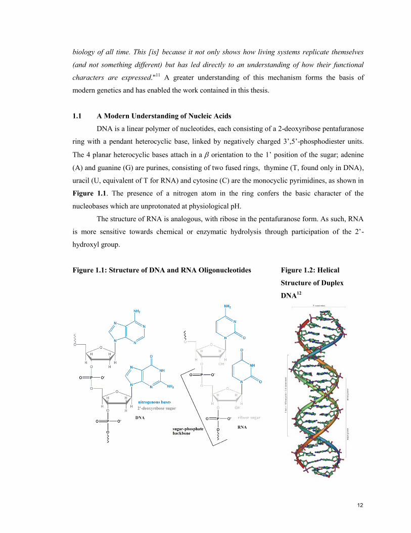
DNA is a linear polymer of nucleotides, each consisting of a 2-deoxyribose pentafuranose
ring with a pendant heterocyclic base, linked by negatively charged 3‟,5‟-phosphodiester units.
The 4 planar heterocyclic bases attach in a orientation to the 1‟ position of the sugar; adenine
(A) and guanine (G) are purines, consisting of two fused rings, thymine (T, found only in DNA),
uracil (U, equivalent of T for RNA) and cytosine (C) are the monocyclic pyrimidines, as shown in
Figure 1.1. The presence of a nitrogen atom in the ring confers the basic character of the
nucleobases which are unprotonated at physiological pH.
The structure of RNA is analogous, with ribose in the pentafuranose form. As such, RNA
is more sensitive towards chemical or enzymatic hydrolysis through participation of the 2‟-
hydroxyl group.
Figure 1.1: Structure of DNA and RNA Oligonucleotides Figure 1.2: Helical
Structure of Duplex
DNA12
12

DNA exists principally in a double-stranded form (ds), where 2 complementary
oligonucleotides hybridise in an anti-parallel fashion to form gently twisting ladder, as shown in
Figure 1.2. This association is non-covalent and achieved through hydrogen bonds between
complementary nitrogenous bases, called Watson-Crick base pairing, shown in Figure 1.3, as
well as the -stacking of neighbouring bases. The redundancy of structure confers fidelity to the
storage of genetic information, as damage in one strand can be repaired by „reading‟ the code of
the complementary strand.
Figure 1.3: Watson-Crick Base Pairing Specificity in DNA Nucleobases
N
HN
O
N
N
O
O
N
N
N
N O
HN
N
N
NN
N
NH
H
H
H
Adenine
Thymine
CytosineGuanine
H
H
RNA has a much shorter cellular lifetime than DNA, and as it is not the depository of
information between generations, it exists predominantly in the single stranded form (ss). When
RNA does form a duplex with itself, the 2‟-hydroxyl group cause changes in the parameters of the
helical structure, as outlined in Table 1.1. A hybrid duplex between RNA and DNA, or duplexes
containing modified bases generally have intermediate parameters.
Table 1.1: Characteristic Parameters Oligonucleotide Helicess
A form helices13
B form helices13
Classic Presentation RNA:RNA DNA:DNA
Helical sense Right Right
Residues per turn of the helix 11 10.4
Rise per base pair (bp) 2.56 Å 3.3 – 3.4 Å
Helix diameter 25.5 Å 23.7 Å
Major groove dimensions Narrow and deep Wide and deep
Minor groove dimensions Broad and shallow Narrow and deep
Sugar conformation C3‟-endo (North) C2‟-endo (South)
Base tilt 20° -6°
13
1.2 The Flow of Genetic Information
The process by which the information stored in DNA is expressed as functional proteins
is an elegant and elaborate system. The role of RNA in protein synthesis had been suspected since
1939, based on experiments carried out by Torbjörn Caspersson, Jean Brachet and Jack Schultz,
but was not confirmed until Hubert Chantrenne experiments on the messenger role played by
RNA in the synthesis of proteins in ribosome in 1965.14
In the initial step, transcription, a short
section of the DNA duplex is unwound and the sequence of a section of exposed single-stranded
DNA is transcribed into precursor messenger RNA (pre-mRNA). The pre-mRNA is then spliced
to remove non-coding sections (introns) and the remaining coding sequences (exons) are ligated
to form a single strand of mature mRNA. The mRNA then migrates out of the nucleus to the
cytoplasm, where the start codon (AUG, 5‟-end of mRNA) is recognised by the ribosomal
machinery and the sequence of bases is translated into a nascent protein by the successive
addition of amino acids coordinated by transfer RNA (tRNA).
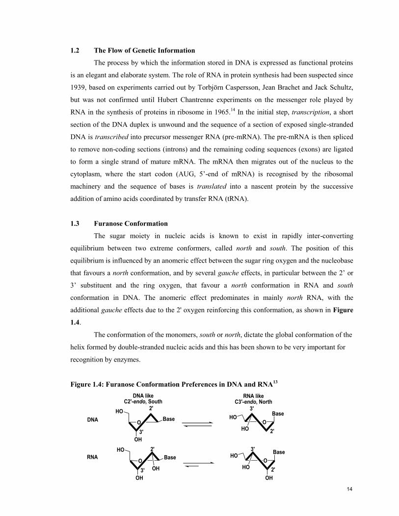
1.3 Furanose Conformation
The sugar moiety in nucleic acids is known to exist in rapidly inter-converting
equilibrium between two extreme conformers, called north and south. The position of this
equilibrium is influenced by an anomeric effect between the sugar ring oxygen and the nucleobase
that favours a north conformation, and by several gauche effects, in particular between the 2‟ or
3‟ substituent and the ring oxygen, that favour a north conformation in RNA and south
conformation in DNA. The anomeric effect predominates in mainly north RNA, with the
additional gauche effects due to the 2' oxygen reinforcing this conformation, as shown in Figure
1.4.
The conformation of the monomers, south or north, dictate the global conformation of the
helix formed by double-stranded nucleic acids and this has been shown to be very important for
recognition by enzymes.
Figure 1.4: Furanose Conformation Preferences in DNA and RNA13
Base
OH
HO
O
2'
3'HO
BaseHO
O
3'
2'
OH
OH
Base
OH
HO
O
2'
3'HO
BaseHO
O
3'
2'
RNA likeC3'-endo, North
DNA likeC2'-endo, South
DNA
RNA
14
1.4 Oligonucleotide-based Therapeutics
Greater understanding of the central role of nucleic acids in cellular functions has led,
naturally, to the desire to attack disease by the regulating gene expression of components in the
disease pathway. The specificity and universality of Watson-Crick base pairing has identified
chemically modified oligonucleotides as innovative therapies to achieve gene regulation or gene
silencing, through exploitation of the RNA interference (microRNA, siRNA) and antisense
pathways. A brief review of these latter two biological pathways is presented in the following
sections. The idea of oligonucleotide-based antisense therapy dates back to the 1960s when
Nirenberg et al. first demonstrated that hybridization of polyribouridylic acid with a
complementary oligoadenylate prevented the translation of the ribouridylic acid into
polyphenylalanine and established that oligonucleotide single-strandedness is required for protein
biosynthesis.15
Using modified oligonucleotides as drugs to target and degrade specific mRNA
sequences involved in disease pathways is attractive for several reasons: first it offers high
specificity due to Watson-Crick base pairing; second, less drug is necessary as degrading a single
mRNA will prevent the translation of multiple protein molecules; third, oligonucleotides can
silence disease-causing genes that encode so called “non-druggable targets”, which are not
amenable to conventional therapeutics such as small molecules, proteins or monoclonal
antibodies; and finally, the base sequences of the oligonucleotide drug can readily be adapted to
respond to mutations in the mRNA sequence.
1.5 Antisense Gene Silencing
The antisense approach to the down regulation of genes involves the activation of
ubiquitous enzyme ribonuclease H (RNase H), which has been identified in viruses, bacteria, and
eukaryotes,16
through the introduction of a DNA-like oligonucleotide. This strand is called an
antisense strand because it is anti-parallel to the sense mRNA target. When these 2 strands
hybridise to form a DNA/RNA hybrid duplex, the resulting complex can inhibit or prevent RNA
transport, splicing or translation and, crucially, may trigger degradation by RNase H.17, 18
RNase
H selectively cleaves the mRNA strand of DNA:RNA duplex,19
releasing the antisense strand to
find another complementary mRNA and amplify its effect in a catalytic manner.
As native DNA is highly susceptible to nucleases, and therefore can only exert an
antisense effect for a brief time,20, 21
several modifications have been investigated. These
modifications must attempt to overcome several hurdles, including specificity/selectivity of
mRNA recognition, biochemical stability (particularly against nucleases22
), affinity for target
15

mRNA,22
cell uptake,23
and finally, the ability to retain or improve RNase H activation.19
This
last requirement is highly exigent and has resulted in disqualifying many likely candidates.
1.6 RNA interference
The RNA interference (RNAi) pathway is a relative newcomer to the field of
oligonucleotide therapeutics, having been first reported by Andrew Fire and Craig Mello in
1998,24
but its huge impact and wide use were promptly recognised by the awarding of the 2006
Nobel Prize in physiology or medicine. This pathway likely evolved as a defence mechanism
against viral or bacterial invasions and has been identified in worm,24
plant25
and mammalian
cells.26
RNAi researchers have been able to take advantage of much of what was learned from the
antisense strategy, allowing for the rapid development of methods that exploit RNAi. Several new
reviews discuss recent advances, noting that although siRNA is still primarily used for gene
function analysis, RNAi-based technologies are rapidly advancing to the clinic.27, 28
The mechanism of RNAi, shown in Figure 1.5, involves the selection of a guide strand
from a short section of double stranded RNA and its use, through the RNA-induced silencing
complex (RISC), to recognise and degrade target RNA.
Figure 1.5: RNAi Pathway
dsRNA
RLCa.
b. c.
d.
target mRNAf.
e.
g.
RISC
Ago2
dsRNA
RLCa.
b. c.
d.
target mRNAf.
e.
g.
RISC
Ago2
The RNAi pathway is triggered by long double-stranded RNA (dsRNA)24
which is
cleaved into 21-25 nucleotides segments26, 29, 30
by an endogenous dsRNA specific RNase III
enzyme called Dicer.31
These 21-25 nucleotide lengths of dsRNA are referred to as small
interfering RNA (siRNA) and this is the length generally used for synthetic oligonucleotides
therapeutics.26
Using these shorter lengths avoids triggering the antiviral/interferon responses
a. Double stranded RNA is recognised by the
RISC loading complex (RLC) and transferred
to the ribonuclease Ago2. b. The guide strand
is then selected, and c. the passenger strand is
discarded, d. allowing for the active RNA
induced silencing complex (RISC) to assemble.
e. The guide strand is used to select target
mRNA with a complementary sequence, and f.
the complex cleaves the target mRNA. g. The
RISC is then re-activated and can go on to
catalytically cleave more mRNA.
16
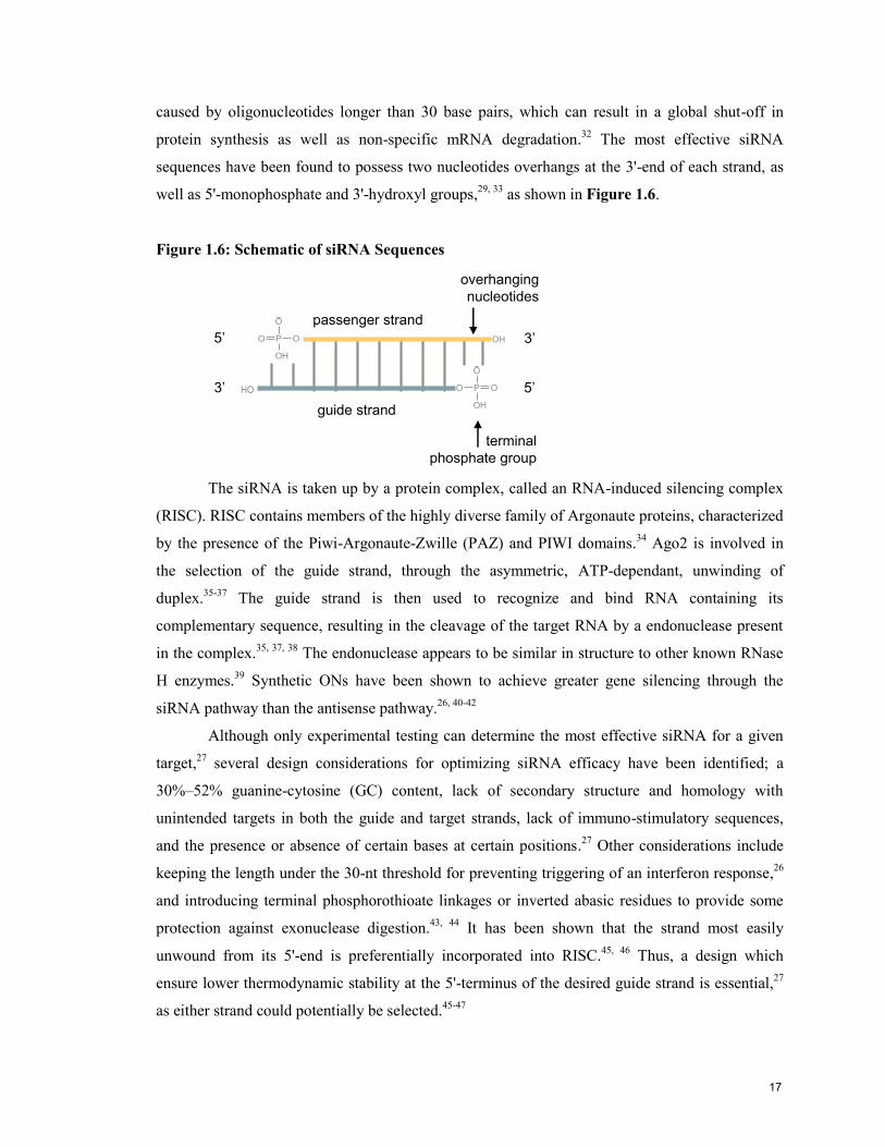
caused by oligonucleotides longer than 30 base pairs, which can result in a global shut-off in
protein synthesis as well as non-specific mRNA degradation.32
The most effective siRNA
sequences have been found to possess two nucleotides overhangs at the 3'-end of each strand, as
well as 5'-monophosphate and 3'-hydroxyl groups,29, 33
as shown in Figure 1.6.
Figure 1.6: Schematic of siRNA Sequences
P
O
OO
OH
5’ 3’
5’3’
guide strand
passenger strand
P
O
OO
OH
overhanging
nucleotides
terminal
phosphate group
OH
HO
The siRNA is taken up by a protein complex, called an RNA-induced silencing complex
(RISC). RISC contains members of the highly diverse family of Argonaute proteins, characterized
by the presence of the Piwi-Argonaute-Zwille (PAZ) and PIWI domains.34
Ago2 is involved in
the selection of the guide strand, through the asymmetric, ATP-dependant, unwinding of
duplex.35-37
The guide strand is then used to recognize and bind RNA containing its
complementary sequence, resulting in the cleavage of the target RNA by a endonuclease present
in the complex.35, 37, 38
The endonuclease appears to be similar in structure to other known RNase
H enzymes.39
Synthetic ONs have been shown to achieve greater gene silencing through the
siRNA pathway than the antisense pathway.26, 40-42
Although only experimental testing can determine the most effective siRNA for a given
target,27
several design considerations for optimizing siRNA efficacy have been identified; a
30%–52% guanine-cytosine (GC) content, lack of secondary structure and homology with
unintended targets in both the guide and target strands, lack of immuno-stimulatory sequences,
and the presence or absence of certain bases at certain positions.27
Other considerations include
keeping the length under the 30-nt threshold for preventing triggering of an interferon response,26
and introducing terminal phosphorothioate linkages or inverted abasic residues to provide some
protection against exonuclease digestion.43, 44
It has been shown that the strand most easily
unwound from its 5'-end is preferentially incorporated into RISC.45, 46
Thus, a design which
ensure lower thermodynamic stability at the 5'-terminus of the desired guide strand is essential,27
as either strand could potentially be selected.45-47
17

Although direct comparisons of different gene silencing methods are scarce, RNAi has
generally been found to be more effective than antisense or ribozymes. One study found RNAi,
which also produced more sustained silencing, to be 100-1000 fold more efficient than an
phosphorothioate modified antisense optimized for the same target mRNA.48
1.7 Chemical Modifications of Oligonucleotide for Therapeutics
Six key criteria have been identified for a successful oligonucleotide-based therapeutic:
(i) it can be synthesized easily and in bulk; (ii) is stable in vivo; (iii) is able to enter the target cell;
(iv) is retained by the target cell; (v) is able to interact with the cellular target; and (vi) it should
not interact in a non-sequence-specific manner with other macromolecules.18
This thesis work considers factors related to the final 2 points in RNAi; particularly that
the oligonucleotide forms stable and highly specific bonds with target mRNA, and are recognised
and cleaved by the requisite enzymes in the RNAi pathway. Unfortunately the requirements of
these 2 criteria are often in opposition, as constraining the sugar conformation favours the first but
increased rigidity, due to sterics or electronics, interferes with the second. This theme will be
further developed in the following sections.
1.8 Oligonucleotide Modifications in RNAi
Modifications to nucleotides can be classified under 3 categories; those involving the
heterocylic base, the sugar and the phosphodiester backbone. Many chemical modifications have
been explored within the more mature field of antisense,49-51
with particular focus on
modifications at the C2‟ position of the sugar moiety.52
Many of the lessons learned with
antisense can be transferred to RNAi as long as the differences in their mechanism of actions
(cellular pathways) are considered. For both RNAi and antisense, an adequate melting
temperature (Tm) with a complementary mRNA strand is a primary requirement.50
Several
comprehensive reviews of chemical modification in RNAi are available49, 53, 54
and the following
sections present a selection of some relevant sugar and backbone modifications.
1.9 Backbone Modifications
The phosphorothioate backbone (PS), in which a sulphur atom replaces a non-bridging
oxygen atom in the natural phosphodiester (PO) backbone, appears to extend the biological
lifetime of synthetic oligonucleotides,55
however this beneficial effect is partly due to association
with serum and cell proteins which may contribute to its relatively high toxicity56, 57
and issues
regarding non-sequence specific interactions.57, 58
It is recognised as a substrate for RNase H59
18
which is essential to the antisense pathway and has been tested in human clinical trials.60, 61
The
PS-DNA drug VitraveneTM
, developed for the treatment of human cytomegalovirus (CMV)
induced retinitis, is the first and only antisense drug available in the clinic.62
Additionally, the
diastereomeric nature of the phosphorothioate (PS) linkage has proved useful as a probe for
studying the stereochemical course of enzymatic reactions,63
to locate protein-nucleic acid
interactions,64-66
probe for metal ion-phosphate interactions,65
investigate the structure of the
substrate metal-ATP complex,65
elucidate DNA structure, and for studying the involvement of
cAMP in biological systems.67
Unfortunately PS-DNA does not form strong duplexes with RNA68
and is therefore less commonly used for RNAi strategies.
Another common modification is DNA or RNA strands containing 2',5'-linked
phosphodiester linkages; 2‟,5‟-RNA is found in some biological processes such as RNA
splicing69, 70
and has also been investigated for the down-regulation of gene expression.71
For
instance, DNA/2‟5‟-RNA chimeras and 2‟5‟-DNA show less non-specific binding to plasma and
cellular proteins minimizing toxicity in the antisense application.71
A comparison of thermal
melting temperatures (Tm) reveals the low duplex stability72
of 2',5'-RNA for mRNA and therefore
lack of relevance to RNAi: RNA:RNA > DNA:DNA DNA:RNA > RNA:2',5'-RNA > 2',5'-
RNA: 2',5'-RNA > DNA: 2',5'-RNA (undetected).73
1.10 Sugar Modifications
The formation of oligonucleotide duplexes is enthalpically favoured due to the hydrogen
bonding interactions and stacking of the heterobases, although the increased order of the duplex
structure is entropically unfavoured. Pre-organisation of the nucleotide is believed to reduce the
entropy loss during duplex formation73
while favoring base stacking and base-base hydrogen
bonding (enthalpic gain).
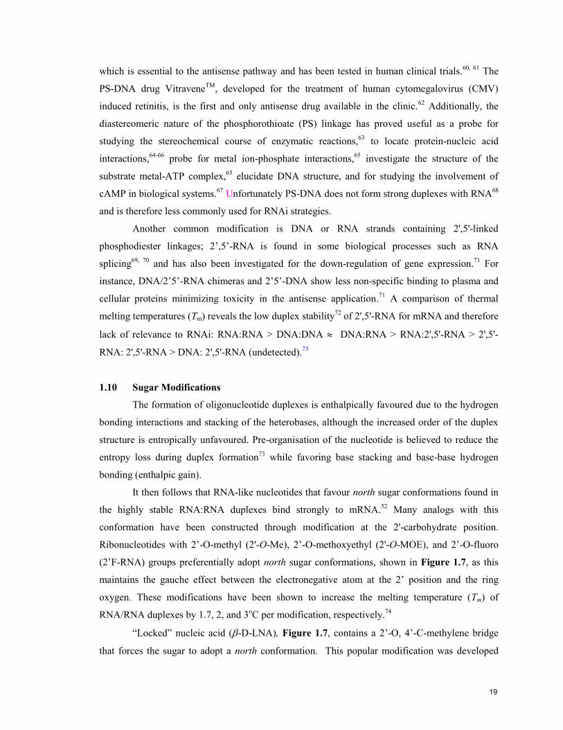
It then follows that RNA-like nucleotides that favour north sugar conformations found in
the highly stable RNA:RNA duplexes bind strongly to mRNA.52
Many analogs with this
conformation have been constructed through modification at the 2'-carbohydrate position.
Ribonucleotides with 2‟-O-methyl (2'-O-Me), 2‟-O-methoxyethyl (2'-O-MOE), and 2‟-O-fluoro
(2‟F-RNA) groups preferentially adopt north sugar conformations, shown in Figure 1.7, as this
maintains the gauche effect between the electronegative atom at the 2‟ position and the ring
oxygen. These modifications have been shown to increase the melting temperature (Tm) of
RNA/RNA duplexes by 1.7, 2, and 3oC per modification, respectively.
74
“Locked” nucleic acid (-D-LNA), Figure 1.7, contains a 2‟-O, 4‟-C-methylene bridge
that forces the sugar to adopt a north conformation. This popular modification was developed
19
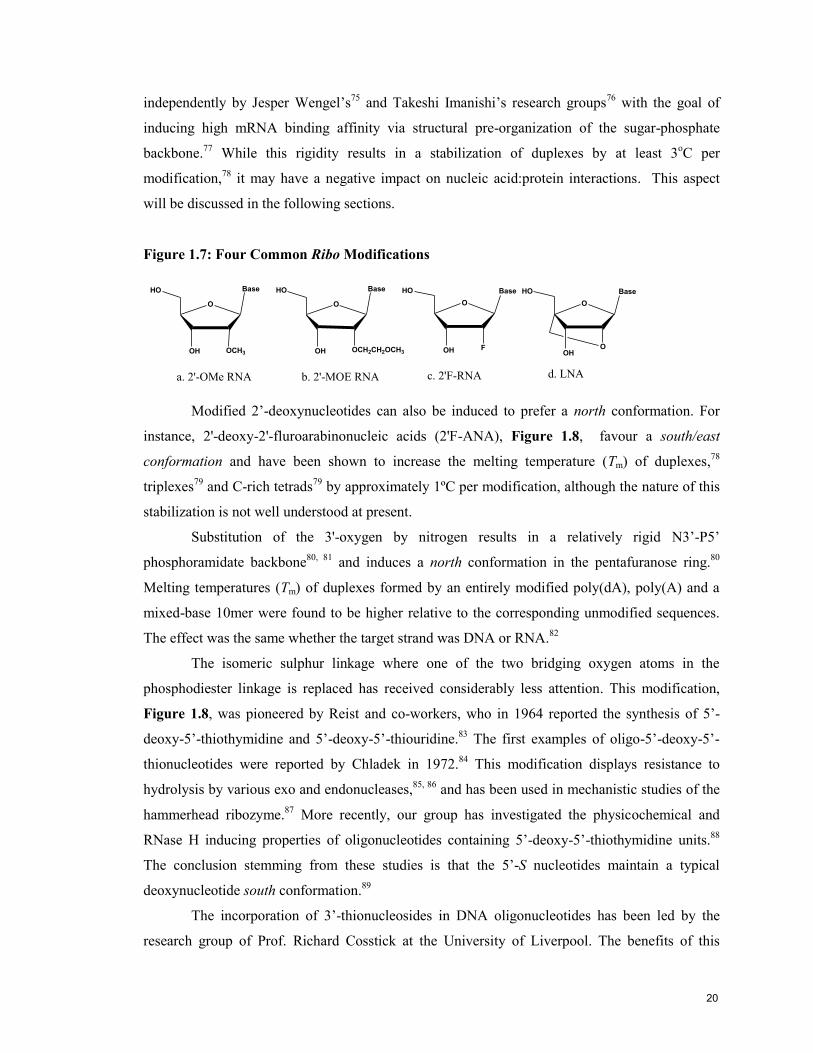
independently by Jesper Wengel‟s75
and Takeshi Imanishi‟s research groups76
with the goal of
inducing high mRNA binding affinity via structural pre-organization of the sugar-phosphate
backbone.77
While this rigidity results in a stabilization of duplexes by at least 3oC per
modification,78
it may have a negative impact on nucleic acid:protein interactions. This aspect
will be discussed in the following sections.
Figure 1.7: Four Common Ribo Modifications
Base
O
OCH3OH
HO
O
HO Base
FOH
a. 2'-OMe RNA c. 2'F-RNA
Base
O
OCH2CH2OCH3OH
HO
b. 2'-MOE RNA
O
HO Base
OOH
d. LNA
Modified 2‟-deoxynucleotides can also be induced to prefer a north conformation. For
instance, 2'-deoxy-2'-fluroarabinonucleic acids (2'F-ANA), Figure 1.8, favour a south/east
conformation and have been shown to increase the melting temperature (Tm) of duplexes,78
triplexes79
and C-rich tetrads79
by approximately 1ºC per modification, although the nature of this
stabilization is not well understood at present.
Substitution of the 3'-oxygen by nitrogen results in a relatively rigid N3‟-P5‟
phosphoramidate backbone80, 81
and induces a north conformation in the pentafuranose ring.80
Melting temperatures (Tm) of duplexes formed by an entirely modified poly(dA), poly(A) and a
mixed-base 10mer were found to be higher relative to the corresponding unmodified sequences.
The effect was the same whether the target strand was DNA or RNA.82
The isomeric sulphur linkage where one of the two bridging oxygen atoms in the
phosphodiester linkage is replaced has received considerably less attention. This modification,
Figure 1.8, was pioneered by Reist and co-workers, who in 1964 reported the synthesis of 5‟-
deoxy-5‟-thiothymidine and 5‟-deoxy-5‟-thiouridine.83
The first examples of oligo-5‟-deoxy-5‟-
thionucleotides were reported by Chladek in 1972.84
This modification displays resistance to
hydrolysis by various exo and endonucleases,85, 86
and has been used in mechanistic studies of the
hammerhead ribozyme.87
More recently, our group has investigated the physicochemical and
RNase H inducing properties of oligonucleotides containing 5‟-deoxy-5‟-thiothymidine units.88
The conclusion stemming from these studies is that the 5‟-S nucleotides maintain a typical
deoxynucleotide south conformation.89
The incorporation of 3‟-thionucleosides in DNA oligonucleotides has been led by the
research group of Prof. Richard Cosstick at the University of Liverpool. The benefits of this
20

modification, as enumerated by Cosstick, are that it is achiral, isopolar and isosteric with the
natural congener. This thesis examines for the first time the physicochemical and biological
properties of RNA strands incorporating 3‟-deoxy-3‟-thiothymidine units. As we will return to the
characteristics of this modification in Chapter 3 in more detail, this brief introduction will suffice
here.
A full slate of modified nucleosides have been introduced into siRNA duplexes. 2'-
fluororibose substitutions have been found to be generally well tolerated,90
although some
reduction of silencing has been reported.43
Incorporation of selected 2'-O-Me or 2'-deoxy
residues91, 92
or PS linkages43
maintained silencing with some increase in resistance to nucleases,33,
49 whereas full substitution significantly reduced silencing.
92 Some cytotoxicity has also been
reported with phosphorothioate (PS) linkages.93
Figure 1.8: Four 2’-Deoxy Modifications
O
HO Base
F
OH
a. 2'F-ANA
Base
O
HSH
HO
d. 3'-thio DNA
O
HO Base
NH2
b. 3'-amino DNA
H
Base
O
HOH
HS
c. 5'-thio DNA
1.11 Flexibility and Enzymatic Recognition
Correct strand pre-organisation is desirable because it leads to more stable and faithful
duplex hybridization, of which LNA‟s striking affinity towards RNA targets is an excellent
example. The rigid LNA backbone impedes RNase H enzymatic cleavage, whereas LNA/DNA
oligonucleotides are poor substrates at best.93
Chimeric oligonucleotides containing both 2‟-
FANA and DNA units were found to be more potent antisense molecules than a fully modified
2‟F-ANA oligomer.94
This suggests that the more flexible DNA units promote RNase H activity.
In fact, introduction of a single deoxynucleotide unit within a 2‟-FANA strand is sufficient to
impart flexibility and enhance RNase H activity.59, 95
Dr. Maria Mangos from our laboratory expanded on this theme by inserting acyclic
moieties such as 2‟,3‟-secouridine or butanediol, Figure 1.9, and found that both significantly
enhanced the target RNA cleavage by RNase H.95
Unfortunately, a significant drop in thermal
stability was observed with these sequences as compared to DNA and was believed to be a
consequence of the high flexibility of the linker. This underlines the importance of maintaining a
balance between flexibility and rigidity in synthetic therapeutic oligonucleotides. Flexibility may
be essential to accommodate that unnatural shape of modified nucleotides in tight enzymatic
21

pockets and a combination of pre-organization and flexibility appears to be the key to high
enzymatic efficiency.96-101
Figure 1.9: Butanediol and 2’,3’-Secouridine
O
O
P
O
OO
O
P OO
O
O
O
P
O
OO
O
P OO
OH
U
1.12 Thesis Objectives
Our research group has been interested in the design95, 100, 102-105
and characterization of
the structural and biological properties95
of synthetic nucleic acids, particularly for use in
antisense and more recently the RNAi strategies. Previous work in our laboratory have
demonstrated that a combination of pre-organization and flexibility has a beneficial effect on
RNase H efficiency and the current work attempts to extends this to the RNAi pathway. In doing
so, this thesis reports improvements to the synthesis of 3‟-deoxy-3‟-thiothymidine and the first
incorporation of this modified nucleoside into RNA oligonucleotides. Because this modification
mimics the structure of ribonucleosides (north pucker), we thought it would be compatible with
the RNAi cellular pathway. As such, we hoped it would provide solutions to some of the
challenges facing siRNA therapeutics (e.g., enhanced cellular activity). By synthesizing a small
library of modified siRNAs, the impact of the 3‟-S modification in the thermal stability and gene
silencing activity of siRNA were examined for the first time. Particular substitutions at the scissile
position of the passenger strand of siRNA duplexes enabled a closer examination of the
mechanism of RISC cleavage.
1.13 References
1 Darwin, C., On The Origin of Species by Means of Natural Selection, or
The Preservation of Favoured Races in the Struggle for Life. First Edition ed.; John Murray: London, 1859;
'Vol.' p.
2 Mendel, G., Experiments on Plant Hybridization (German: Versuche über Pflanzen-Hybriden).
Verh. Naturforsch. Ver. Brünn 1866, 4, 3–47.
3 Miescher, F., Die Spermatozoen einiger Wirbelthiere, Ein Beitrag zur Histochemie,. Verh.
Nat.forsch. Ges. Basel 1874, 6, 138–208.
4 Mirksy, A. E., The discovery of DNA. Scientific American 1968, June, 78.
5 Miescher, F., Ueber die chemische Zusammensetzung der Eiterzellen. Die Histochemischen und
physiologischen Arbeiten. 1871, 2, 3-23.
22
6 Avery, T.; Macleod, C. M.; McCarty, M., Studies on the chemical nature of the substance inducing
transformation of pneumococcal types. I. induction of transformation by a deoxyribonucleic acid fraction
isolated from pneumococcus type III. J. Exp. Med. 1944, 79, (I), 37.
7 Vischer, E.; Zamenhof, S.; Chargaff, E., Microbial nucleic acids: the deoxypentose nucleic acids
of avian tubercle bacilli and yeast. J. Biol. Chem. 1949, 177, 429.
8 Franklin, R. E.; Gosling, R. G., Molecular Configuration in Sodium Thymonucleate. Nature
(London) 1953, 171, (April 25), 740-741.
9 Watson, J. D.; Crick, F. H. C., Molecular Structure of Nucleic Acids: A Structure for Deoxyribose
Nucleic Acid. Nature (London) 1953, April 25, (171), 737-738.
10 Watson, J. D.; Crick, F. H. C., Genetic implications of the structure of deoxyribonucleic acid.
Nature (London) 1953, 171, 964.
11 Pollock, M. R., The Discovery of DNA : An Ironic Tale of Chance, Prejudice and Insight. J. Gen.
Microbiol. 1970, 63, 1-20.
12 Ströck, M., overview of the structure of DNA. In http://en.wikipedia.org/wiki/User:Mstroeck, ed.;
DNA_helix.png, 'Ed.'^'Eds.' Wikipedia: Vienna, 2006; 'Vol.' p^pp Permission is granted to copy, distribute
and/or modify this document under the terms of the GNU Free Documentation License, Version 1.2 or any
later version published by the Free Software Foundation; with no Invariant Sections, no Front-Cover Texts,
and no Back-Cover Texts.
13 Blackburn, G. M.; Gait, M. J., Nucleic Acids in Chemistry and Biology. Second ed.; Oxford
University Press: New York, 1996; 'Vol.' p.
14 Chantrenne, H., The polyribosomes, agents of protein synthesis. Arch Biol 1965, 76, (2), 307-16.
15 Nirenberg, M. W.; Matthaei, J. H., The Dependence of Cell-Free Protein Synthesis in E. coli upon
Naturally Occurring or Synthetic Polyribonucleotides. Proc. Natl. Acad. Sci. U. S. A. 1961, 47, (10), 1588-
1602.
16 Crouch, R. J.; Toulmé, J. J. e., Ribonucleases H. ed.; John Libbey: Paris, 1998; 'Vol.' p 265.
17 Crooke, S. T., Progress in antisense technology. Ann. Rev. Med. 2004, 55, 61-95.
18 Stein, C. A.; Cheng, Y.-C., Antisense Oligonucleotides as Therapeutic Agents-Is the Bullet Really
Magical? Science 1993, 261, 1004-1012.
19 Crooke, S. T., Review: Molecular mechanisms of action of antisense drugs. Biochim. Biophys.
Acta 1999, 1489, 31-44.
20 Koziolkiewicz, M.; Gendaszewska, E.; Maszewska, M.; Stein, C. A.; Stec, W. J., The
mononucleotide-dependent, nonantisense mechanism of action of phosphodiester and phosphorothioate
oligonucleotides depends upon the activity of an ecto-5'-nucleotidase. Blood 2001, 98, 995-1002.
21 Vaerman, J. L.; Moureau, P.; Deldime, F.; Lewalle, P.; Lammineur, C.; Morschhauser, F.; Martiat,
P., Antisense Oligodeoxyribonucleotides Suppress Hematologic Cell Growth Through Stepwise Release of
Deoxyribonucleotides. Blood 1997, 90, 331-9.
22 Kool, E. T., Preorganization of DNA: Design Principles for Improving Nucleic Acid Recognition
by Synthetic Oligonucleotides. Chem. Rev. 1997, 97, 1473-1487.
23 Mesmaeker, A. d.; Haner, R.; Martin, P.; Moser, H. E., Antisense Oligonucleotides. Acc. Chem.
Res. 1995, 28, 366-374.
24 Fire, A.; Xu, S.; Montgomery, M. K.; Kostas, S. A.; Driver, S. E.; Mello, C. C., Potent and specific
genetic interference by double-stranded RNA in Caenorhabditis elegans. Nature (London) 1998, 391, 806-
811.
25 Hamilton, A. J.; Baulcombe, D. C., A species of small antisense RNA in posttranscriptional gene
silencing in plants. Science 1999, 286, (950-952).
26 Elbashir, S. M.; Harborth, J.; Lendeckel, W.; Yalcin, A.; Weber, K.; Tuschl, T., Duplexes of 21-
nucleotide RNAs mediate RNA interference in cultured mammalian cells. Nature (London) 2001, 411, 494-
498.
27 Dykxhoorn, D. M.; Lieberman, J., Running Interference: Prospects and Obstacles to Using Small
Interfering RNAs as Small Molecule Drugs. Annual Review of Biomedical Engineering 2006, 8, 377-402.
23
28 Martin, S. E. N. J. C., Applications of RNA Interference in Mammalian Systems. Annual Review
of Genomics and Human Genetics 2007, 8, 82-108.
29 Elbashir, S. M.; Lendeckel, W.; Tuschl, T., RNA interference is mediated by 21-and 22-nucleotide
RNAs. Genes Dev. 2001, 15, 188-200.
30 Zamore, P. D.; Tuschl, T.; Sharp, P. A.; Bartel, D. P., RNAi: Double-stranded RNA directs the
ATP-dependent cleavage of mRNA at 21 to 23 nucleotide intervals. Cell (Cambridge, Mass.) 2000, 101,
25-33.
31 Ketting, R. F.; Fischer, S. E. J.; Bernstein, E.; Sijen, T.; Hannon, G. J.; Plasterk, R. H. A., Dicer
functions in RNA interference and in synthesis of small RNA involved in developmental timing in C-
elegans. Genes Dev. 2001, 15, 2654-2659.
32 Stark, G. R.; Kerr, I. M.; Williams, B. R.; Silverman, R. H.; Schreiber, R. D., How cells respond to
interferons. Annu. Rev. Biochem. 1998, 67, 22-264.
33 Elbashir, S.; Martinez, J.; Patkaniowska, A.; Lendeckel, W.; Tuschl, T., Functional anatomy of
siRNAs for mediating efficient RNAi in Drosophila melanogaster embryo lysate. EMBO J. 2001, 20, 6877–
88.
34 Parker, J.; Barford, D., Argonaute: a scaffold for the function of short regulatory RNAs. Trends
Biochem. Sci. 2006, 31, 622–30.
35 Rand, T.; Petersen, S.; Du, F.; Wang, X., Argonaute2 cleaves the antiguide strand of siRNA during
RISC activation. Cell (Cambridge, Mass.) 2005, 123, 621–29.
36 Schwarz, D. S.; Hutvagner, G.; Haley, B.; Zamore, P. D., Evidence that siRNAs function as
guides, not primers, in the Drosophila and human RNAi pathways. Mol. Cell 2002, 10, 537-548.
37 Matranga, C.; Tomari, Y.; Shin, C.; Bartel, D.; Zamore, P., Passenger-strand cleavage facilitates
assembly of siRNA into Ago2-containing RNAi enzyme complexes. Cell (Cambridge, Mass.) 2005, 123,
607–20.
38 Mello, C. C.; Conte, D., Revealing the world of RNA interference. Nature (London) 2004, 431,
338-342.
39 Song, J.-J.; Smith, S. K.; Hannon, G. J.; Joshua-Tor, L., Crystal Structure of Argonaute and Its
Implications for RISC Slicer Activity. Science (Washington, DC, United States) 2004, 305, (5689), 1434-
1437.
40 Carstea, E. D.; Hough, S.; Wiederholt, K.; Welch, P. J., State-of-the-art modified RNAi
compounds for therapeutics. Idrugs 2005, 8, 642-647.
41 Far, R. K. K.; Sczakiel, G., The activity of siRNA in mammalian cells is related to structural target
accessibility: a comparison with antisense oligonucleotides. Nucleic Acids Res. 2003, 31, 4417-4424.
42 Hough, S. R.; Wiederholt, K. A.; Burrier, A. C.; Woolf, T. M.; Taylor, M. F., Why RNAi makes
sense. Nat. Biotechnol. 2003, 21, 731-732.
43 Czauderna, F.; Fechtner, M.; Dames, S.; Ayguen, H.; Klippel, A.; Pronk, G. J.; Giese, K.;
Kaufmann, J., Structural variations and stabilizing modifications of synthetic siRNAs in mammalian cells.
Nucleic Acids Res. 2003, 31, (11), 2705-2716.
44 Morrissey, D.; Blanchard, K.; Shaw, L.; Jensen, K.; Lockridge, J.; al., e., Activity of stabilized
short interfering RNA in a mouse model of hepatitis B virus replication. Hepatology 2005, 41, 1349-56.
45 Reynolds, A.; Leake, D.; Boese, Q.; Scaringe, S.; Marshall, W.; Khvorova, A., Rational siRNA
design for RNA interference. Nat. Biotechnol. 2004, 22, 326–30.
46 Khvorova, A.; Reynolds, A.; Jayasena, S., Functional siRNAs and miRNAs exhibit strand biaa.
Cell (Cambridge, Mass.) 2003, 115, 209–16.
47 Schwarz, D.; Hutvagner, G.; Du, T.; Xu, Z.; Aronin, N.; Zamore, P., Asymmetry in the assembly
of the RNAi enzyme complex. Cell (Cambridge, Mass.) 2003, 115, 199–208.
48 Bertrand, J.; Pottier, M.; Vekris, A.; Opolon, P.; Maksimenko, A.; Malvy, C., Comparison of
antisense oligonucleotides and siRNAs in cell culture and in vivo. Biochem. Biophys. Res. Commun. 2002,
296, 1000–1004.
49 Chiu, Y. L.; Rana, T. M., siRNA function in RNAi: A chemical modification analysis. RNA 2003,
9, 1034-1048.
24
50 Freier, S. M.; Altmann, K.-H., The ups and downs of nucleic acid duplex stability: structure-
stability studies on chemically-modified DNA:RNA duplexes. Nucleic Acids Res. 1997, 25, 4429-4443.
51 Kurreck, J., Antisense technologies. Improvement through novel chemical modifications. Eur. J.
Biochem. 2003, 270, (8), 1628-1644.
52 Manoharan, M., 2 '-Carbohydrate modifications in antisense oligonucleotide therapy: importance
of conformation, configuration and conjugation. Bba-Gene Struct Expr. 1999, 1489, 117-130.
53 Fattal, E.; Bochot, A., Ocular delivery of nucleic acids: Antisense oligonucleotides, aptamers and
siRNA. Advanced Drug Delivery Reviews 2006, 58, 1203-1223.
54 Manoharan, M., RNA interference and chemically modified small interfering RNAs. Curr. Opin.
Chem. Biol. 2004, 8, 570-579.
55 Agrawal, S.; Zhao, Q., Antisense therapeutics in neuropharmacology. Curr. Opin. Chem. Biol.
1998, 2, 519-528.
56 Agrawal, S.; Kandimalla, E. R., Antisense therapeutics: is it as simple as complementary base
recognition? Mol. Med. Today 2000, 6, 72-81.
57 Weidner, D. A.; Valdez, B. C.; Henning, D.; Greenberg, S.; Busch, H., Phosphorothioate
oligonucleotides bind in a non sequence-specific manner to the nucleolar protein C23/nucleolin. FEBS Lett.
1995, 366, 146-150.
58 Srinivasan, S. K.; Teway, H. K.; Iversen, P. L., Characterization of binding sites, extent of binding,
and drug interactions of oligonucleotides with albumin. Antisense Res Dev. 1995, 5, (2), 131-9.
59 Mangos, M. M. Factors Governing The Design, Selection and Cleavage of Sugar-Modified
Duplexes by Ribonuclease H. Ph.D. Thesis, McGill University, Montreal, 2005.
60 De Fabritiis, P.; Calabretta, B., Antisense oligodeoxynucleotides for the treatment of chronic
myelogenous leukaemia: Are they still a promise? Haematologica 1995, 80, 295-9.
61 Webb, A.; Cunningham, D.; Cotter, F.; Clarke, P. A.; Di Stephano, F.; Ross, P.; Corbo, M.;
Dziewanowska, Z., BCL-2 antisense therapy in patients with non-Hodgkin lymphoma. Lancet 1997, 349,
1137-41.
62 Crooke, S. T., Vitravene (TM) - another piece in the mosaic. Antisense Nucleic Acid Drug Dev.
1998, 8, Vii-Viii.
63 Rajagopal, J.; Doudna, J. A.; Szostak, J. W., Stereochemical course of catalysis by the
Tetrahymena ribozyme. Science 1989, 244, 692-694.
64 Milligan, J. F.; Uhlenbeck, O. C., Determination of RNA-protein contacts using thiophosphate
substitutions. Biochemistry 1989, 28, 2849-2855.
65 Eckstein, F., Nucleoside Phosphorothioates. Ann. Rev. Biochem. 1985, 54, 367-402.
66 Dahm, S. C.; Uhlenbeck, O. C., Role of divalent metal ions in the hammerhead RNA cleavage
reaction. Biochemistry 1991, 30, 9464.
67 Wallace, J. C.; Edmonds, M., Polyadenylated nuclear RNA contains branches. Proc. Natl. Acad.
Sci. USA 1983, 80, (950-954).
68 LaPlanche, L. A.; James, T. L.; Powell, C.; Wilson, W. D.; Uznanski, B.; Stec, W. J.; Summers,
M. F.; Zon, G., Phosphorothioate-modified oligodeoxyribonucleotides. III. NMR and UV spectroscopic
studies of the Rp-Rp, Sp-Sp, and Rp-Sp duplexes, [d(GGSAATTCC)]2, derived from diastereomeric O-
ethyl phosphorothioates. Nucleic Acids Res. 1986, 14, (22), 9081–9093.
69 Giannaris, P. A.; Damha, M. J., Oligoribonucleotides containing 2',5'-phosphodiester linkages
exhibit binding selectivity for 3',5'-RNA over 3',5'-ssDNA. Nucleic Acids Res. 1993, 21, 4742-4749.
70 Bhan, P.; Bhan, A.; Hong, M.; Hartwell, J. G.; Saunders, J. M.; Hoke, G. D., 2',5'-Linked oligo-3'-
deoxyribonucleoside phosphorothioate chimeras: thermal stability and antisense inhibition of gene
expression. Nucleic Acids Res. 1997, 25, 3310-3317.
71 Kandimalla, E. R.; Manning, A.; Zhao, Q. Y.; Shaw, D. R.; Byrn, R. A.; Sasisekharan, V.;
Agrawal, S., Mixed backbone antisense oligonucleotides: Design, biochemical and biological properties of
oligonucleotides containing 2'-5'-ribo- and 3'-5'-deoxyribonucleotide segments. Nucleic Acids Res. 1997,
25, 370-378.
25
72 Wasner, M.; Arion, D.; Borkow, G.; Noronha, A.; Uddin, A. H.; Parniak, M. A.; Damha, M. J.,
Physicochemical and Biochemical Properties of 2',5'-Linked RNA and 2',5'-RNA:3',5'-RNA \"Hybrid\"
Duplexes. Biochemistry 1998, 37, 7478-7486.
73 Egli, M., Conformational preorganization, hydration, and nucleic acid duplex stability. Antisense
Nucleic Acid Drug Dev. 1998, 8, 123-129.
74 Koshkin, A. A.; Singh, S. K.; Nielsen, P.; Rajwanshi, V. K.; Kumar, R.; Meldgaard, M.; Olsen, C.
E.; Wengel, J., LNA (locked nucleic acids): synthesis of the adenine, cytosine, guanine, 5-methylcytosine,
thymine and uracil bicyclonucleoside monomers, oligomerization, and unprecedented nucleic acid
recognition. Tetrahedron 1998, 54, (14), 3607-3630.
75 Singh, S. K.; Nielsen, P.; Koshkin, A. A.; Wengel, J., LNA (locked nucleic acids): Synthesis and
high-affinity nucleic acid recognition. Chemical Communications 1998, 455-456.
76 Obika, S.; Nanbu, D.; Hari, Y.; Andoh, J.-i.; Morio, K.-i.; Doi, T.; Imanishi, T., Stability and
structural features of the duplexes containing nucleoside analogues with a fixed N-type conformation, 2'-
O,4'- C-methyleneribonucleosides. Tetrahedron Lett. 1998, 39, 5401-5404.
77 Koshkin, A. A.; Nielsen, P.; Meldgaard, M.; Rajwanshi, V. K.; Singh, S. K.; Wengel, J., An RNA
mimic forming exceedingly stable LNA : LNA duplexes. J. Am. Chem. Soc. 1998, 120, 13252-13253.
78 Chastain, M.; Tinoco, I., Structural Elements in RNA. Prog. Nucleic Acid Res. Mol. Biol. 1991,
41, 131-177.
79 Gray, D. M.; Ratliff, R. L.; Vaughan, M. R., Circular-Dichroism Spectroscopy of DNA. Methods
Enzymol. 1992, 211, 389-406.
80 Gryaznov, S.; Chen, J.-K., Oligodeoxyribonucleotide N3'->P5' Phosphoramidates: Synthesis and
Hybridization Properties. J. Am. Chem. Soc. 1994, 116, (7), 3143-3144.
81 Kissman, H. M.; Weiss, M. J., The Synthesis of 1-(Aminodeoxy-ß-D-ribofuranosyl)-2-
pyrimidinones. New 3'- and 5'-Aminonucleosides. JACS 1958, 80, 2575 - 2583.
82 Ding, D.; Gryaznov, S. M.; Lloyd, D. H.; Chandrasekaran, S.; Yao, S.; Ratmeyer, L.; Pan, Y.;
Wilson, W. D., An oligodeoxyribonucleotide N3'->P5' phosphoramidate duplex forms an A-type helix in
solution. Nucleic Acids Res. 1996, 24, (2), 354-360.
83 Reist, E. J.; Benitez, A.; Goodman, L., The Synthesis of Some S‟Thiopentofuranosylpyrimidines.
J. Org. Chem. 1964, 29, (3), 554-558.
84 Chladek, S.; Nagyvary, J., Nucleophilic Reactions of Some Nucleoside Phosphorothioates. J. Am.
Chem. Soc. 1972, 94, 2079-2085.
85 Jahn-Hofmann, K.; Engels, J. W., Efficient solid phase synthesis of cleavable
oligodeoxynucleotides based on a novel strategy for the synthesis of 5'-S-(4,4'-dimethoxytrityl)-2'-deoxy-5'-
thionucleoside phosphoramidites. Helv. Chim. Acta 2004, 87, (11), 2812-2828.
86 Rybakov, V. N.; Rivkin, M. I.; Kumarev, V. P., Some substrate properties of analogues of
oligothymidylates with p-s-C-5' bonds. Nucleic Acids Res. 1981, 9, (1), 189-201.
87 Kuimelis, R. G.; McLaughlin, L. W., Ribozyme-Mediated Cleavage of a Substrate Analogue
Containing an Internucleotide-Bridging 5'-Phosphorothioate: Evidence for the Single-Metal Model.
Biochemistry 1996, 35, (16), 5308 -5317.
88 Tedeschi, A. L. Synthesis and Biological Evaluation of Oligonucleotides Containing 3'- and 5'-S-
Phosphorthiolate Internucleotide Linkages. M.Sc. Thesis, McGill University, Montreal, 2004.
89 Cosstick, R.; S.Vyle, J., Synthesis and properties of dithymidine phosphate analogues containing
3'-thiothymidine. Nucleic Acids Res. 1990, 18, (4), 829-835.
90 Parrish, S.; Fleenor, J.; Xu, S.; Mello, C.; Fire, A., Functional anatomy of a dsRNA trigger:
differential requirement for the two trigger strands in RNA interference. Mol. Cell 2000, 6, 1077–87.
91 Braasch, D. A.; Jensen, S.; Liu, Y.; Kaur, K.; Arar, K.; White, M. A.; Corey, D. R., RNA
Interference in Mammalian Cells by Chemically-Modified RNA. Biochemistry 2003, 42, (26), 7967-7975.
92 Harborth, J.; Elbashir, S.; Vandenburgh, K.; Manninga, H.; Scaringe, S.; al., e., Sequence,
chemical, and structural variation of small interfering RNAs and short hairpin RNAs and the effect on
mammalian gene silencing. Antisense Nucleic Acid Drug Dev. 2003, 13, 83–105.
93 Kurreck, J.; Wyszko, E.; Gillen, C.; Erdman, V. A., Design of antisense oligonucleotides stabilized
by locked nucleic acids. Nucleic Acids Res. 2002, 30, 1911-1918.
26
94 Lok, C.-N.; Viazovkina, E.; Min, K.-L.; Nagy, E.; Wilds, C. J.; Damha, M. J.; Parniak, M. A.,
Potent gene-specific inhibitory properties of mixed-backbone antisense oligonucleotides comprised of 2'-
deoxy-2'-fluoro-D-arabinose and 2'-deoxyribose nucleotides. Biochemistry 2002, 41, (10), 3457-67.
95 Mangos, M. M.; Min, K.-L.; Viazovkina, E.; Galarneau, A.; Elzagheid, M. I.; Parniak, M. A.;
Damha, M. J., Efficient RNase H-Directed Cleavage of RNA Promoted by Antisense DNA or 2'F-ANA
Constructs Containing Acyclic Nucleotide Inserts. J. Am. Chem. Soc. 2003, 125, (3), 654-661.
96 Noronha, A.; Wilds, C. J.; Lok, C.-N.; Arion, K. V. D.; Parniak, M. A.; Damha, M. J., Synthesis
and Biophysical Properties of Arabinonucleic Acids (ANA): Circular Dichroic Spectra, Melting
Temperatures, and Ribonuclease H Susceptibility of ANA·RNA Hybrid Duplexes. Biochemistry 2000, 39,
(24), 7050 -7062.
97 Damha, M.; Usman, N.; Ogilvie, K., The rapid chemical synthesis of arabinonucleotides.
Tetrahedron Lett. 1987, 28, (1515), 1633-1636.
98 Damha, M. J.; Giannaris, P. A.; Marfeys, P., Antisense L/D-Oligodeoxynucleotide Chimeras:
Nuclease Stability, Base-Pairing Properties and Activity at Directing Ribonuclease H. Biochemistry 1994,
33, 7877-7885.
99 Watts, J. K.; Sadalapure, K.; Choubdar, N.; Pinto, B. M.; Damha, M. J., Synthesis and
Conformational Analysis of 2'-Fluoro-5-methyl-4'-thioarabinouridine (4'S-FMAU). J. Org. Chem. 2006, 71,
(3), 921-925.
100 Wilds, C.; Damha, M., 2'-Deoxy-2'-fluoro-beta-D-arabinonucleosides and oligonucleotides (2'F-
ANA): synthesis and physicochemical studies. Nucleic Acids Res. 2000, 28, (18), 3625-35.
101 Sabatino, D.; Damha, M. J., Oxepane Nucleic Acids: Synthesis, Characterization, and Properties of
Oligonucleotides Bearing a Seven-Membered Carbohydrate Ring. J. Am. Chem. Soc. 2007, 129, (26), 8259-
8270.
102 Damha, M. J.; Wilds, C. J.; Noronha, A.; Brukner, I.; Borkow, G.; Arion, D.; Parniak, M. A.,
Hybrids of RNA and Arabinonucleic Acids (ANA and 2'F-ANA) Are Substrates of Ribonuclease H. J. Am.
Chem. Soc. 1998, 120, (49), 12976-12977.
103 Denisov, A.; Noronha, A. M.; Wilds, C. J.; Trempe, J.-F.; Pon, R. T.; Gehring, K.; Damha, M. J.,
Solution structure of an arabinonucleic acid (ANA)/RNA duplex in a chimeric hairpin: comparison with 2'-
fluoro-ANA/RNA and DNA/RNA hybrids. Nucleic Acids Res. 2001, 29, (21), 4284-4293.
104 Trempe, J.; Wilds, C. J.; Denisov, A. Y.; Pon, R. T.; Damha, M. J.; Gehring, K., NMR Solution
Structure of an Oligonucleotide Hairpin with a 2'F-ANA/RNA Stem: Implications for RNase H Specificity
toward DNA/RNA Hybrid Duplexes. J. Am. Chem. Soc. 2001, 123, (21), 4896 -4903.
105 Watts, J. K.; Choubdar, N.; Sadalapure, K.; Robert, F.; Wahba, A. S.; Pelletier, J.; Mario Pinto, B.;
Damha, M. J., 2'-Fluoro-4'-thioarabino-modified oligonucleotides: conformational switches linked to
siRNA activity. Nucl. Acids Res. 2007, 35, (5), 1441-1451.
27
Chapter 2: Experimental Materials and Methods
2.1 General Methods
2.1.1 General Reagents
Required solvents were obtained by refluxing and subsequent distillation from drying
agents under inert atmospheres: acetonitrile (CH3CN, Fisher), methylene chloride (CH2Cl2,
Fisher), and pyridine (C5H5N, Fisher) were dried over calcium hydride (CaH2, Aldrich) and
tetrahydrofuran (C4H4O, THF, Fisher) was dried over a sodium metal-benzophenone system
(Aldrich). Anhydrous N,N-diisopropylethylamine (DIPEA, Aldrich) and anhydrous dioxane
(Aldrich), as well as acetone, chloroform, diethyl ether, ethanol, glacial acetic acid, hydrochloric
acid, methanol, triethylamine (TEA) and concentrated aqueous ammonia were used as received.
Reagents such as sodium bicarbonate (NaHCO3), anhydrous sodium sulphate (Na2SO4),
p-anisylchlorodiphenylmethane (MMTCl), benzoyl chloride (BzCl), 4-dimethylaminopyridine
(DMAP), O-(benzotriazol-1-yl)-1,1,3,3-tetramethyluronium hexafluorophosphate (HBTU), and
mesoyl chloride (MsCl) were purchased from Sigma-Aldrich and used as received.
Thymidine, cytidine, and N,N-diisopropylaminophosphonamidic chloride phosphitylating
reagent were purchased from Chemgenes Corp. (Ashland, MA) and used as received.
2.1.2 Chromatography for Nucleosides and Intermediates
Analytical thin layer chromatography (TLC) was performed on Merck Kieselgel 60 F254
aluminium sheets coated with fluorescent indicator doped silica gel (0.2 mm x 20 cm x 20 cm).
Compounds were visualized by illumination with a handheld 254 nm UV lamp and by dipping the
TLC plate in a dilute sulphuric acid solution (3% in MeOH) with subsequent heating. Preparatory
scale flash column chromatography was performed using SilicycleTM
ultra-pure silica gel (230-
400 mesh) under constant nitrogen pressure (5-10psi). Chloroform with an increasing gradient of
methanol (0-15%) was used as eluant.
2.1.3 Instruments
Circular Dichroism Spectra. CD spectra were generated using a JASCO J-810
spectropolarimeter, with software supplied by the manufacturer, between 210-350 nm at a scan
rate 50 nm/min. The temperature-controlled sample chamber was used as required.
NMR Spectra. NMR spectra were recorded using 200 MHz, 400 MHz and 500 MHz
Varian Mercury spectrophotometers. 1H-NMR,
32P-NMR and 2D-NMR (
1H-
1H COSY) were used
to characterize compounds. Deuterated solvents, including acetonitrile-d3, chloroform-d3, and
dichloromethane-d2, were obtained from Cambridge Isotope Laboratories.
28
Mass Spectrometry.
Low-resolution electrospray ionization mass-spectrometry was performed on a ThermoQuest
Finnigan LCQ Duo mass spectrometer in the positive ion mode. Samples were routinely
dissolved in methanol.
UV-Visible Spectra. Absorbance spectra in the visible and UV ranges were obtained
using a Varian Cary I or Cary 300 spectrophotometer with software supplied by the manufacturer.
All spectra were obtained in water or aqueous buffers, and a Peltier temperature-controlled
sample chamber was used as required.
2.2 Synthesis of 3’-Deoxy-3’-Thiothymidine Nucleoside and its 3’-S-
Phosphorothioamidite Derivative
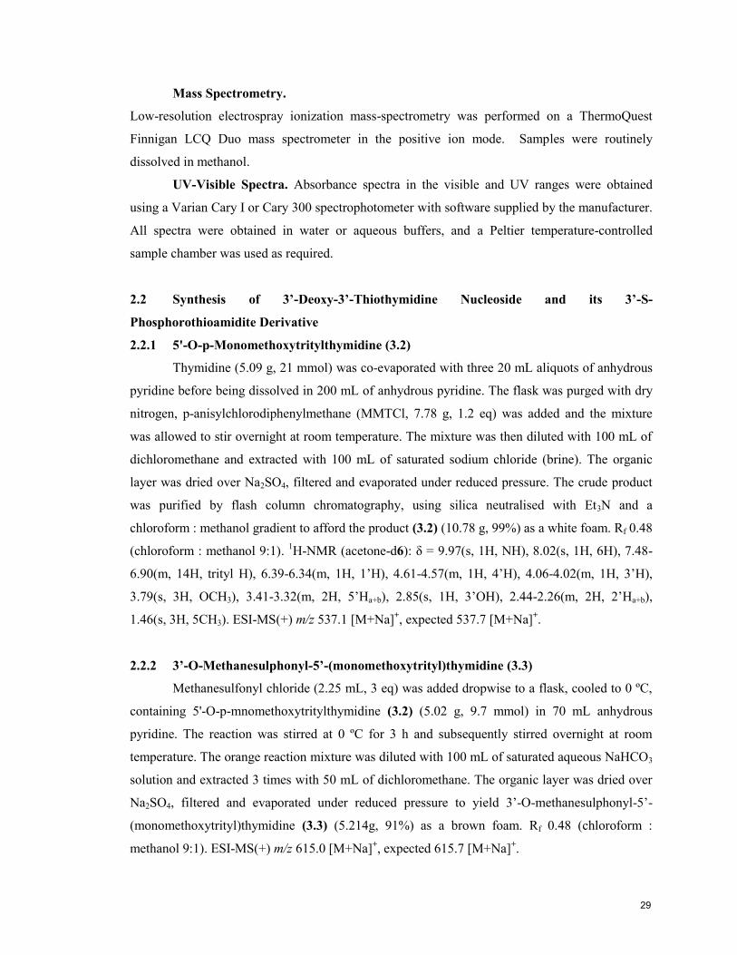
2.2.1 5'-O-p-Monomethoxytritylthymidine (3.2)
Thymidine (5.09 g, 21 mmol) was co-evaporated with three 20 mL aliquots of anhydrous
pyridine before being dissolved in 200 mL of anhydrous pyridine. The flask was purged with dry
nitrogen, p-anisylchlorodiphenylmethane (MMTCl, 7.78 g, 1.2 eq) was added and the mixture
was allowed to stir overnight at room temperature. The mixture was then diluted with 100 mL of
dichloromethane and extracted with 100 mL of saturated sodium chloride (brine). The organic
layer was dried over Na2SO4, filtered and evaporated under reduced pressure. The crude product
was purified by flash column chromatography, using silica neutralised with Et3N and a
chloroform : methanol gradient to afford the product (3.2) (10.78 g, 99%) as a white foam. Rf 0.48
(chloroform : methanol 9:1). 1H-NMR (acetone-d6): δ = 9.97(s, 1H, NH), 8.02(s, 1H, 6H), 7.48-
6.90(m, 14H, trityl H), 6.39-6.34(m, 1H, 1’H), 4.61-4.57(m, 1H, 4’H), 4.06-4.02(m, 1H, 3’H),
3.79(s, 3H, OCH3), 3.41-3.32(m, 2H, 5’Ha+b), 2.85(s, 1H, 3’OH), 2.44-2.26(m, 2H, 2’Ha+b),
1.46(s, 3H, 5CH3). ESI-MS(+) m/z 537.1 [M+Na]+, expected 537.7 [M+Na]
+.
2.2.2 3’-O-Methanesulphonyl-5’-(monomethoxytrityl)thymidine (3.3)
Methanesulfonyl chloride (2.25 mL, 3 eq) was added dropwise to a flask, cooled to 0 ºC,
containing 5'-O-p-mnomethoxytritylthymidine (3.2) (5.02 g, 9.7 mmol) in 70 mL anhydrous
pyridine. The reaction was stirred at 0 ºC for 3 h and subsequently stirred overnight at room
temperature. The orange reaction mixture was diluted with 100 mL of saturated aqueous NaHCO3
solution and extracted 3 times with 50 mL of dichloromethane. The organic layer was dried over
Na2SO4, filtered and evaporated under reduced pressure to yield 3’-O-methanesulphonyl-5’-
(monomethoxytrityl)thymidine (3.3) (5.214g, 91%) as a brown foam. Rf 0.48 (chloroform :
methanol 9:1). ESI-MS(+) m/z 615.0 [M+Na]+, expected 615.7 [M+Na]
+.
29
2.2.3 2,3’-Dideoxy-5’-(monomethoxytrityl)anhydrothymidine (3.4)
3’-O-methanesulphonyl-5’-(monomethoxytrityl)thymidine (3.3) (4.03g, 6.8 mmol) was
dissolved in an alkaline ethanolic solution (165 mL EtOH, 65 mL Et3N) and refluxed for 18 h.
The crude reaction mixture was evaporated under reduced pressure to a viscous oil, diluted with
80 mL water and extracted twice with 60 mL dichloromethane. The organic layer was dried over
Na2SO4, filtered and evaporated under reduced pressure to yield 2,3’-dideoxy-5’-
(monomethoxytrityl)anhydrothymidine (3.4) (3.212g, 95%) as a beige foam. Rf 0.37 (chloroform :
methanol 9:1). ESI-MS(+) m/z 497.1 [M+H]+, expected 497.5 [M+H]
+.
2.2.4 3’-Acetylthio-3’-deoxy-5’-(monomethoxytrityl)thymidine (3.5)1
Sodium thioacetate was prepared by adding thioacetic acid (1.2 mL, 1.1eq) drop wise to a
stirred solution of sodium hydride (0.3g, 1eq) in 10 mL of dry dioxane at 0 ºC. After stirring for
one hour, the sodium thioacetate solution was added to 2,3’-dideoxy-5’-
(monomethoxytrityl)anhydrothymidine (3.4, 1.5g, 3.0 mmol) in 10 mL of anhydrous dioxane. The
reaction was refluxed at 100 ºC for 3 h and then kept at 50 ºC for 16h. The crude mixture was then
diluted with 25 mL of saturated aqueous NaHCO3 and extracted four times with 20 mL of
dichloromethane. The organic layer was then washed with 50 mL of water, dried over Na2SO4,
filtered and evaporated under reduced pressure. The crude product was purified by flash column
chromatography, using silica neutralised with Et3N and a chloroform : methanol gradient to afford
the product (3.5) (1.54 g, 90%) as a brown foam. Rf 0.61 (chloroform : methanol 9:1). 1H-NMR
(CDCl3, 500 MHz): δ = 8.48(s, 1H, NH), 7.68(s, 1H, 6H), 7.44-7.25(m, 12H, trityl H), 6.85(d, 2H,
O-anisyl H), 6.28(t, 1H, 1’H), 4.65(m, 1H, 4’H), 4.19(m, 1H, 3’H), 3.79(s, 3H, OCH3), 3.70(s,
3H, SCOCH3), 3.50(m, 2H, 5’Ha+b), 2.78(m, 1H, 2’Ha), 2.45(m, 1H, 2’Hb), 1.49(s, 3H, 5CH3). See
Appendix 1 for full NMR spectrum. ESI-MS(+) m/z 595.1 [M+Na]+, expected 595.7 [M+Na]
+.
2.2.5 3’-deoxy-3’-S-5’-(monomethoxytrityl)thymidine (3.6)
3’-Acetylthio-3’-deoxy-5’-(monomethoxytrityl)thymidine (3.5) (500 mg, 0.874 mmol)
and dithiothreitol (266 mg, 1.5 eq) were dissolved in 35 mL EtOH and deoxygenated by bubbling
with argon gas for 45 minutes. The ethanolic solution was then cooled to 5ºC and 2 mL of 10 M
NaOH was added. Complete deacetylation was achieved after ~2 hours of stirring, and the
reaction was stopped by neutralization with a dilute aqueous HCl solution.
The solvent was removed in vacuo and the crude product was purified on a neutralised
silica gel column using a gradient of chloroform and methanol. Appropriate fractions were
30
combined and concentrated to dryness, then dissolved in dry benzene and sublimated to afford
(3.6) (427 mg, 92%) as a wonderfully fluffy pale yellow foam. Rf 0.43 (chloroform : methanol-
20:1). 1H-NMR (CDCl3, 500 MHz): δ = 10.1(s, 1H, NH), 7.98(s, 1H, 6H), 7.67-7.24(m, 14H,
trityl H), 6.18(dd, 1H, 1’H), 3.96-3.90(m, 2H, 3’+4’H), 3.77(s, 3H, OCH3), 3.52(dd, 1H, 5’Ha),
3.44 (dd, 1H, 5’Hb), 2.88(s, 1H, 3’SH), 2.73-2.64(m, 1H, 2’Ha), 2.46-2.34(m, 1H, 2’Hb), 1.55(s,
3H, 5CH3). See Appendix 1 for full NMR spectrum. ESI-MS(+) m/z 553.1 [M+Na] +
, expected
553.6 [M+Na]+.
2.2.6 3’-Deoxy-5’-Monomethoxytritylthymidine-3’-S-phosphorothioamidite (3.8)
3’-deoxy-3’-S-5’-(monomethoxytrityl)thymidine (3.6) (200 mg, 0.377 mmol was placed
in an oven dried amber amidite vial and anhydrous CH2Cl2 (2.1 mL), N,N-diisopropylethylamine
(1.3 eq, 85 µL), and N,N-diisopropylaminophosphonamidic chloride (0.95 eq, 80 µL) were added.
The reaction was shaken for 30 minutes and connected directly to the DNA synthesiser in order to
reduce the exposure of the sensitive thio moiety to atmospheric oxygen. An aliquot taken for
analysis and purified by a short hexane:EtOAc column had Rf 0.40 (chloroform : methanol- 20:1).
31P-NMR (CDCl3): 165.2, 163.6 ppm. See Appendix 1 for full NMR spectrum. ESI-MS(+) m/z
753.3 [M+Na] +
, expected 753.8 [M+Na]+.
2.3 Oligonucleotide Synthesis
2.3.1 General Reagents
Unilynker© CPG, the standard 2’-deoxy and ribo 5’-O-dimethoxytrityl-3’-O-(2-
cyanoethyl) N,N-diisopropylphosphoramidites and 2’ OMe amidites, as well as all reagents for
the DNA synthesiser were purchased from Chemgenes Corp. (Ashland, MA). Rasayan Inc.
(Encinitas, CA) supplied the 2’-deoxy-2’-fluoro-arabinothymidine 3’-O-(2-cyanoethyl) N,N-
diisopropylphosphoramidite.
In the solid phase oligonucleotide synthesis cycle, 5’-dimethoxytrityl and 5’-
monomethoxytrityl groups were removed with 3% (w/v) trichloroacetic acid in dichloromethane.
Activation of phosphoramidite coupling step was achieved with 0.25 M 4-ethylthiotetrazole in
ACN and any unreacted hydroxyl groups were capped with a combination of the Cap A solution
(10% (v/v) acetic anhydride and 10 % (v/v) dry 2, 4, 6-collidine in anhydrous THF) and the Cap
B solution (16% (v/v) dry N-methyl imidazole in anhydrous THF). A 0.1 M solution of iodine in
THF/pyridine/water (25/20/2, v/v/v) was employed to oxidise the phosphite triesters to the more
stable phosphotriesters.
31
2.3.2 Derivatization of Solid Support2
In a glass vial, oven baked at 170 ºC for 24h in order to reduce water absorbed on the
surface, 1g of long chain alkylamine controlled pore glass (500 Å, LCAA-CPG, Dalton Chemical
Laboratories) was shaken overnight with 10 mL of 3% trichloroacetic acid (TCA) in
dichloromethane. After filtration, the LCAA-CPG was washed successively with 30 mL of
dichloromethane, 80 mL of pyridine, and 50 mL of ether, and dried for several hours under
vacuum (<0.5 mbar).
The dry LCAA-CPG was then shaken overnight with 200 mg of succinic anhydride, 20
mg of 4-dimethylaminopyridine (DMAP) and 8 mL of dry pyridine. The LCAA-CPG was then
filtered for a second time, washed successively with the same solvents as above, and dried on the
filter for 30 min. In second oven-dried vial, 1g of the freshly succinylated LCAA-CPG was
combined with 0.1 mmol of the desired 5’-protected nucleotide, 0.1 mmol of HBTU, 0.1 mmol of
DMAP and 10 mL dry acetonitrile and shaken overnight. The derivatized, functionalised LCAA-
CPG was then filtered for a third time and washed successively as above. The loading of
nucleoside per g of CPG was determined by the absorption of liberated trityl groups and varied
between 80-110 µmol of nucleoside per gram of solid support. An amount of derivatized CPG,
prepared as above or purchased Unilynker (ChemGenes), appropriate for a 1 µmol synthesis was
loaded into an ABI-compatible synthesizer column with cellulose filters (ChemGenes) and
crimped closed with aluminium seals (ChemGenes).
2.3.3 General Automated Solid-Phase Synthesis of Oligonucleotides
Oligonucleotide syntheses were performed on an Applied Biosystems 3400 DNA
synthesizer using standard β-cyanoethyl phosphoramidite chemistry. CPG prepared as previously
described was acetylated with the manufacturer’s capping cycle to block free hydroxyl groups
still remaining on the surface. Unilynker CPG was used as received.
The 2’-deoxyribonucleoside phosphoramidites were prepared as 0.1 M solutions in
anhydrous acetonitrile and the ribonucleoside, 2’F-ANA, and 2’OMe as 0.15 M solutions in
anhydrous acetonitrile. The 3’-deoxy-3’-thiothymidine phosphorothioamidites were prepared as
0.15 M solutions in anhydrous dichloromethane.
The polymeric oligonucleotides were assembled step-wise from the phosphoramidite
monomers to create the desired sequence, shown in Figure 2.1. The 5’ hydroxyl of the growing
oligonucleotide is released and coupled to the P group of the incoming nucleoside, using the
reagents outlined in Table 2.1 below. Step 4a. is used for natural phosphate backbones, and 4b. to
generate phosphorothioate backbones.
32
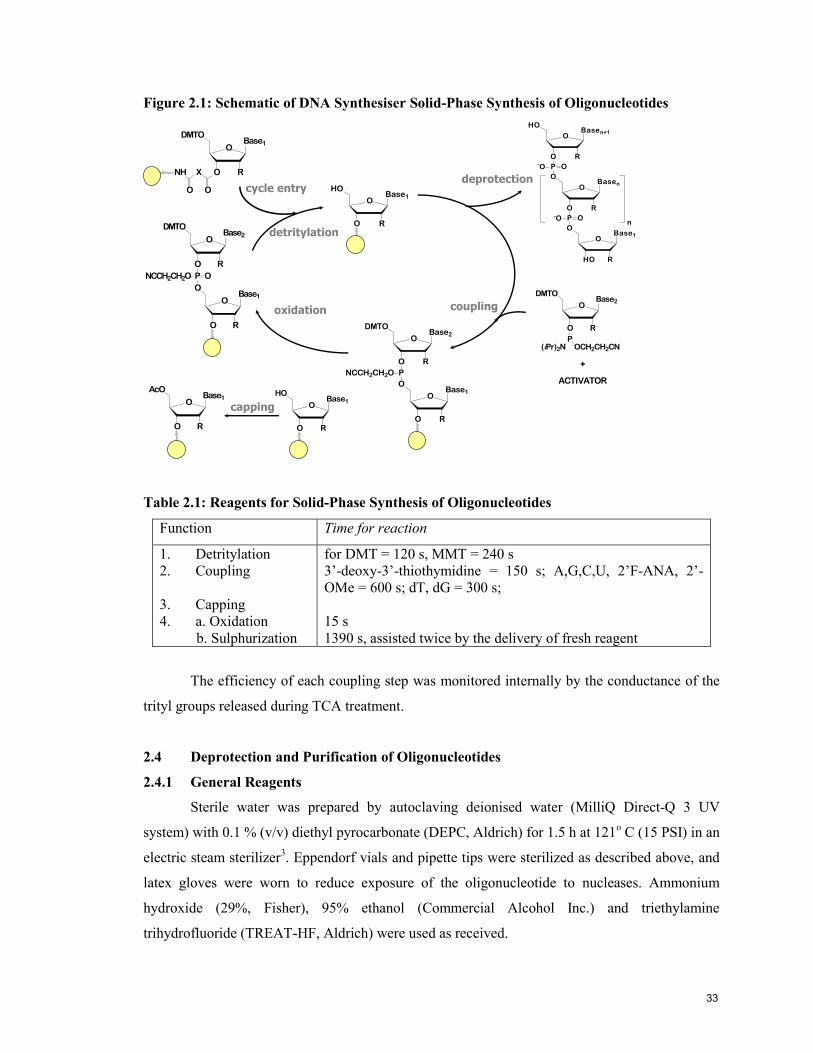
Figure 2.1: Schematic of DNA Synthesiser Solid-Phase Synthesis of Oligonucleotides
OBase1
RO
HO
OBase1
RO
DMTO
XNH
OO
OBase1
RO
HO
+
OBase1
RO
O
PNCCH2CH2O
O
OBase2
DMTO
R
coupling OBase2
RO
DMTO
POCH2CH2CN(iPr)2N
+
ACTIVATOR
detritylationO
Base1
RHO
O
P
O
OBasen
O
R
O-O
P
O
O-O
OBasen+1
HO
R
n
deprotection
OBase1
RO
O
PNCCH2CH2O
O
OBase2
DMTO
R
O
oxidation
OBase1
RO
AcO
capping
cycle entry
Table 2.1: Reagents for Solid-Phase Synthesis of Oligonucleotides
Function Time for reaction
1. Detritylation
2. Coupling
3. Capping
4. a. Oxidation
b. Sulphurization
for DMT = 120 s, MMT = 240 s
3’-deoxy-3’-thiothymidine = 150 s; A,G,C,U, 2’F-ANA, 2’-
OMe = 600 s; dT, dG = 300 s;
15 s
1390 s, assisted twice by the delivery of fresh reagent
The efficiency of each coupling step was monitored internally by the conductance of the
trityl groups released during TCA treatment.
2.4 Deprotection and Purification of Oligonucleotides
2.4.1 General Reagents
Sterile water was prepared by autoclaving deionised water (MilliQ Direct-Q 3 UV
system) with 0.1 % (v/v) diethyl pyrocarbonate (DEPC, Aldrich) for 1.5 h at 121o C (15 PSI) in an
electric steam sterilizer3. Eppendorf vials and pipette tips were sterilized as described above, and
latex gloves were worn to reduce exposure of the oligonucleotide to nucleases. Ammonium
hydroxide (29%, Fisher), 95% ethanol (Commercial Alcohol Inc.) and triethylamine
trihydrofluoride (TREAT-HF, Aldrich) were used as received.
33

Electrophoresis grade N,N’-methylene-bisacrylamide, ammonium persulphate (APS),
N,N,N’,N’-tetramethylethylenediamine (TEMED) and urea were obtained and used as received
from Amersham Biosciences (Arlington Heights, IL). Bromophenol blue (BPB), xylene cyanol
(XC), 2-Amino-2-(hydroxymethyl)-1,3-propanediol (Tris), boric acid, and ethylene-diamine
tetraacetate dihydrate (EDTA) were purchased from Bio-Rad (Mississauga, ON). Biological grade
formamide and Stains-All®
were obtained from Sigma. All aqueous eletrophoresis solutions were
made with deionized water (MilliQ Direct-Q 3 UV system) and sterile water was used for any
solution in direct contact with oligonucleotides.
Gel solutions of 20% and 24% acrylamide were prepared by mixing appropriate volumes
of 40% (w/v H2O) acrylamide stock solution (Amersham), 10 x TBE buffer (890 mM EDTA, 890
mM boric acid, 25 mM EDTA, pH 8.3) and water, with sufficient urea to produce a 7M
concentration of urea. Aqueous samples of oligonucleotide were mixed with an equal volume of
deionized formamide, prepared by stirring over a mixed bed ion-exchange resin (Bio-Rad AG
501-X8) for 30 min and filtration, prior to loading on the gels. Running dye markers contained
2% (w/v) of both XC and BPB, dissolved in a 2:1 (v/v) deionized formamide:10X TBE solution.
2.4.2 Cleavage from CPG and Deprotection of Oligomers
Freshly synthesised oligonucleotides were released from the CPG, and the phosphate
backbone and exocyclic amines of the nucleobases were liberated from their respective protecting
groups by treatment with a solution of aqueous ammonia (29%) in ethanol (3:1 v/v) for 24 h at 55
oC in a 1.5 mL screw-top microtube and then dried using a Speed-Vac
® concentrator at room
temperature. RNA-containing oligonucleotides were additionally shaken at room temperature for
48h with 100 μL/ μmole triethylamine trihydrofluoride (TREAT-HF, Aldrich) to remove the 2’-
O-tert-butyldimethylsilyl protecting groups and purified by precipitation from 1 mL of cold
butanol after the addition of 25 μL of 3M sodium acetate.
The dried oligonucleotides were then dissolved in sterile water, diluted as necessary to
accommodate the spectrophotometer and quantified by absorption at 260 nm for the subsequent
purification steps.
2.4.3 Analysis and Purification by Polyacrylamide Gel Electrophoresis (PAGE)
Fully deprotected oligonucleotides were analysed and purified by denaturing
polyacrylamide gel electrophoresis (PAGE) and/or HPLC. Gel solutions were polymerised with
10% (w/v) aqueous ammonium persulfate (APS) and TEMED, and run on a vertical slab
electrophoresis unit (Hoefer Scientific SE600, Fisher Scientific) with 0.75 mm spacers for
34

analytical gels and 1.5 mm spacers for preparatory runs. Oligonucleotide samples for analytical
gels were dissolved in 15 μL of deionized formamide:sterile water (2:1) and preparatory gels
samples in100-200 μL.
Analytical gels were run at 500 V until the faster BPB dye was about 4 cm from the
bottom of the gel plates and until it had almost run off the plate for preparatory runs. A dilute 0.5
x TBE running buffer (89 mM Tris/boric acid, 2.5 mM EDTA) was used for all runs. The gels
were then UV shadowed by placing them on fluorescent TLC plates wrapped in plastic film and
the results were captured by digital camera under the illumination of a handheld 254 nm UV
lamp.
Analytical gels were then placed in a StainsAll© solution for at least 24h to dye the
oligonucleotides and the images were recorded by digital camera. For preparative gels, the desired
oligonucleotide band was excised from the gel, shaken in 5 mL of sterile water for 24h in a
culture tube and the supernatant was dried down.
2.4.4 Reversed-Phase HPLC Analysis and Purification of Oligonucleotides
Analytical runs of reversed phase HPLC allowed for the investigation of the success of
oligonucleotide synthesis and preparatory runs were used to isolate the desired full length
material. A Waters 480/1525 HPLC chromatograph with a Phenomenex 3µm Oligo Reverse
Phase Clarity Column (50 x 4.60 um) and a two solvent system consisting of 100mM
triethylammonium acetate (A) in deionized water, and 100mM triethylammonium acetate in
acetonitrile (B) was used. The solvent system was adjusted from 5% B in A to 20% B in A over a
22 minute period, with flow rate of 1 mL/min was. The elution profile was recorded by UV-Vis
detection at 260 nm (the maximum absorbance of oligonucleotides) and 290 nm (to avoid
saturating the detector) using the manufacturer’s software. All solutions used for HPLC analysis
and purification were filtered through a 0.22 μM Nalgene filter to remove particulates.
2.4.5 Anion-Exchange HPLC Analysis of Oligonucleotides
Anion exchange HPLC was used to assess the success of oligonucleotide synthesis and to
characterise oligonucleotides purified by PAGE. A Waters 480/1525 HPLC instrument with a
Waters Protein-PakTM
DEAE 5PW (7.5 mm x 75 mm) column and a two solvent system
consisting of 1 M sodium perchlorate (NaClO4, Aldrich) (A) and deionized water (B). A flow rate
of 1 mL/min was maintained as the elution solvent was adjusted from 0% A to 30% A over 60
min, followed by 50% for 10 min and the elution profile was recorded by the manufacturer’s
35
software by UV-Vis detection at 260 nm. The run finished with a 100% B washing step reduce
anion carryover.
2.4.6 Desalting of Oligonucleotides
The crude oligonucleotides to be purified by either PAGE or HPLC were pre-purified
(desalted) by size exclusion chromatography on Sephadex G-25® columns (Amersham Pharmacia
Biotech) following the manufacturer’s instructions and the resulting fractions were quantified at
260 nm. The absorbance data was converted into molar quantities by using the molar extinction
coefficients ( 260) calculated for each sequence as 90% of the sum of the molar extinction
coefficients for the individual bases. The absorbance of the 3’-thio, 2’F-ANA and 2’-OMe
nucleosides were each assumed to be the same as those of their unmodified counterpart. Finally,
the pure oligonucleotides were ready for characterization.
2.5 Characterization of Oligonucleotides
2.5.1 Characterization of Oligonucleotides by Mass Spectrometry
Dry oligonucleotide samples were dissolved in 25µL of 10mM
ammonium acetate and a 3µL aliquot of this solution was injected on a small C18 trap and eluted
with a gradient of ACN/H2O in the mono-electrospray (Nano-ESI) inlet of a QTOF2 Mass
Spectroscopy instrument (Micromass) located at Concordia University. The negative ion mode
was selected for mass analysis, with the following parameters: Capillary Voltage: 3KV, Cone
Voltage: 20V, Extraction Voltage: 2V, Inlet Temperature: 80C, Collision Cell Voltage: 5V,
Collision Gas: Argon, MS resolution: 8000, MS accuracy: 50ppm.
2.5.2 UV-Thermal Denaturation of Oligonucleotides
Thermal denaturation profiles were obtained by monitoring the change in UV-absorbance
at 260 nm with increasing temperature on a Varian CARY 1 or 300 UV-Vis spectrophotometer
equipped with a Peltier temperature-controlled sample chamber, using software supplied by the
manufacturer.
Oligonucleotide duplexes were prepared by combining equimolar amount of
complementary strands, drying thoroughly and re-dissolving in 0.5-1 mL of a buffer consisting of
140 mM KCl, 1mM MgCl2, and 5 mM Na2HPO4 at pH 7.2. The duplexes were denatured by
heating to 90°C for 10-15 min, then allowed to hybridize as they slowly cooled at room
temperature for 2-3 h and then overnight at 4°C overnight. The cold samples were transferred to
36
cold quartz cells (Hellma QS-1.000), and after being sealed with Teflon-wrapped stoppers, were
allowed to equilibrate at 5°C in the cell holder of the spectrophotometer for 5 minutes.
The absorbance at 260 nm was measured at 0.5°C intervals as the temperature was
increased at a rate of 0.5°C/min. The thermal melting temperature (Tm) values were calculated as
the maximum of the first derivative plots of the absorbance versus temperature profiles, which
coincides with the point at which the oligonucleotides population exists as an equal mixture of
single-strands and duplexes.
2.5.3 Circular Dichroism Spectroscopy (CD)
Circular dichroic profiles were measured on a JASCO J-810 spectropolarimeter in quartz
cells (Hellma QS-1.000) cooled to 5°C, using software supplied by the manufacturer, between
210-350 nm at a scan rate of 50nm/min.
The same oligonucleotide samples used for the thermal denaturation studies described
above were used for CD measurements, after an identical heating and overnight cooling regime,
and 10 minutes of equilibration time in the cold sample holder. The CD spectra were corrected by
subtraction of a background scan of buffer, smoothed and were further corrected for concentration
to enable determination of the molar ellipticity. Molar ellipticity was calculated from the equation
[ ] = /cl, where is the relative intensity, c is the molar concentration of oligonucleotides and l
is the path length of the cell in centimetres.
2.6 Biological Studies4
These experiments were conducted by Dr. Francis Robert of the Department of Biochemistry,
McGill University.
2.6.1 RNAi Transfection
HeLa X1/5 cells, expressing the firefly luciferase gene, were maintained in Earle's
Minimum Essential Medium (EMEM) cell culture medium supplemented with 10% fetal bovine
serum (FBS), 2 mM L-glutamine, 1% non-essential amino acids, 1% MEM vitamins, 500 ml/ml
G418, and 300 mg/ml Hygromycin. For transfection, 24-well plates were seeded with 1.0x10⁵
cells per well. The following day, cells were incubated with increasing amounts of siRNAs
premixed with lipofectamine- plus reagent (Invitrogen) using 1 ml of lipofectamine and 4 ml of
the plus reagent per 20 pmol of siRNA (for the highest concentration tested). For the siRNA
titrations, each siRNA was diluted in buffer (30mM HEPES-KOH, pH 7.4, 100mM KOAc, 2mM
37
MgOAc2) and the amount of lipofectamine-plus reagent used relative to the siRNAs remained
constant.
Cell viability was determined by measuring the cell metabolic activity, as an indicator of
cellular toxicity resulting from siRNA transfection, using the Alamar Blue fluorimetric assay
(Medicorp, Montreal, QC) as per the manufacturer’s recommendations. The luciferase RLU
values were normalized to their respective Alamar blue RFU values and any toxicity due to the
experimental conditions was corrected.
2.6.2 Luciferase Activity Assay
Twenty-four hours after transfection, the cells were lysed in hypotonic lysis buffer
(15mM K3PO4, 1mM EDTA, 1% Triton, 2mM NaF, 1 mg/ml BSA, 1mM DTT, 100mM NaCl, 4
mg/ml aprotinin, 2 mg/ml leupeptin and 2 mg/ml pepstatin).
Luminescence was measured, immediately following addition of the luciferin substrate
solution, using a Fluostar Optima 96-well plate bioluminescence reader (BMG Labtech) and the
values were normalized to the protein concentration of the cell lysate as determined by the DC
protein assay (BioRad).
2.6.3 Luciferase mRNA Quantitation
Total cell RNA was extracted according to the RNeasy mini kit manufacturer’s protocol
(Qiagen, Mississauga, ON) and a 1 mg aliquot was used to prepare cDNA using random primers
and SuperScript II Reverse Transcriptase (Invitrogen, Burlington, ON).
Quantitative real-time PCR was performed using the LightCycler (Roche, Laval, QC),
SYBR green reagent, and gene-specific primers for luciferase (LUC5013 F1, 50-
acgctgggcgttaatcagag-30; LUC5013 R1, 50-gtcgaagatgttggggtgttg-30; TIB MOLBIOL, Berlin,
Germany) and the housekeeping gene GAPDH (huGAPD forward, 50-ggtggtctcctctgacttc-30;
huGAPD reverse, 50-ctcttcctcttgtgctcttg- 30; TIB MOLBIOL).
2.7 References
1 Elzagheid, M. I.; Mattila, K.; Oivanen, M.; Jones, B. C. N. M.; Cosstick, R.; Lönnberg, H.,
Hydrolytic Reactions of the cis-Methyl Ester of 3'-Deoxy-3'-thiothymidine 3',5'-Cyclic(phosphorothiolate).
Eur. J. Org. Chem. 2000, 1987-1991.
2 Damha, M. J.; Giannaris, P. A.; al., e., An improved procedure for derivatization of controlled-pore
glass beads for solid-phase oligonucleotide synthesis. Nucleic Acids Res. 1990, 18, (13), 3813-3821.
3 Blackburn, P.; Moore, S., The Enzymes. ed.; Academic Press: New York, 1982; 'Vol.' p.
4 Watts, J. K.; Choubdar, N.; Sadalapure, K.; Robert, F.; Wahba, A. S.; Pelletier, J.; Mario Pinto, B.;
Damha, M. J., 2'-Fluoro-4'-thioarabino-modified oligonucleotides: conformational switches linked to
siRNA activity. Nucl. Acids Res. 2007, 35, (5), 1441-1451.
38
Chapter 3: Synthesis of 3’-Deoxy-3’-thiothymidine and Its Incorporation into
Oligonucleotides
3.1 Relevance of 3’,5’-Linked 3’-S-Phosphorothiolate Linkages
Nucleic acids maintain stable tertiary structures and catalyze chemical reactions without
the diverse functional groups of proteins.1, 2
This economy prevents scientists from using site
directed mutagenesis to replace a functional group, as is done with proteins, since this only affects
hydrogen-bonding patterns. Thus, more elaborate methods involving the introduction of synthetic
nucleosides containing non-native functional groups have been developed. There are four distinct
positions in the phosphorus-sugar backbone where oxygen atoms are commonly substituted: 3’,
5’, and the two non-bridging phosphoryl oxygen atoms.
Sulphur is generally considered to be a conservative substitution for oxygen but there are
important differences between the two; primarily the harder, more electronegative character of
oxygen versus the softer, more polarisable character of sulphur. When the 3’-OH group of the
furanose ring is replaced with a thiol function, these differences have a significant effect on the
reactivity of the synthetic intermediates, the conformation of the sugar ring,3-6
,the stability of the
oligonucleotide7-11
as well as biological implications, as will be explored in this chapter and the
next. No studies directly examining the effect of bond length with 3’-S modification have
appeared in the literature, however other studies involving the replacement of an oxygen atom
with sulphur in nucleosides have found the difference in bond length to result in local changes but
not to distort the overall structure beyond the normal range of oligonucleotides.12-15
3.2 Synthesis of 3’-Deoxy-3’-thiothymidine
The advent of 3’-thio modified nucleoside in the literature begins with Richard Cosstick
et al.’s 1988 report of the synthesis of 5’-protected-3’-thiothymidine monomer and the solution
coupling via in situ phosphorylation to yield the d(TspT) dimer,16-18
Figure 3.1. Other approaches
to creating 3’-S-phosphorothiolate linkages include using phosphoramidites,19
phosphate
triesters20
and Michaelis-Arbusov type-reactions21
to create DNA-based dinucleotides and
oligonucleotides.
The synthetic pathway used to preparing the 3’-deoxy-3’-thiothymidine (3’S-dT)
monomer, Figure 3.2, used in this work was mainly adapted by Anna Lisa Tedeschi for our lab22
from an earlier reported synthesis.23
Improvements novel to this current work will be articulated
in the following discussion.
39

Figure 3.1: Cosstick’s Synthetic Scheme for 3’-Deoxy-3’-thiothymidine and its
Incorporation into a Dinucleotide
O
OMs
MMTO
NH
ON
O
O
SBz
MMTO
NH
ON
O
O
SH
MMTO
NH
ON
O
O
S
MMTO
NH
ON
O
O
OAc
NH
ON
O
a. b.
c.
PO O
O
NC
O
OH
HO
NH
ON
O
d.
a. In 2 steps, per Horwitz et al.24
b. NaSBz, dimethylformamide, 100°C
c. NH3, MeOH, room temp., 1h
d. (tetrazol-1-yl)2-PO[CH2]2CN, 4:1
tetrahydrofuran-2,6-lutidine, -78°C, 15
min ; 3’-O-acetylthymidine, 20 min; then
aqueous work-up.
Figure 3.2: 3’-Deoxy-3’-(thioacetyl)thymidine Synthetic Scheme
O
OH
HO
NH
ON
O
3.1
O
OH
MMTO
NH
ON
O
3.2
O
OMs
MMTO
NH
ON
O
3.3
OMMTO N
N
O
3.4
O
O
SAc
MMTO
NH
ON
O
3.5
a. b.
d.
c.
a. MMTCl (1.5 eq), pyridine, rt, 10h, 99%
yield.
b. MsCl (3 eq), pyridine, rt, ON, 91% yield.
c. Et3N, EtOH, reflux, 16h, 95% yield.
d. NaSAc, 1,4-dioxane, reflux 10h, 90%
yield.
40
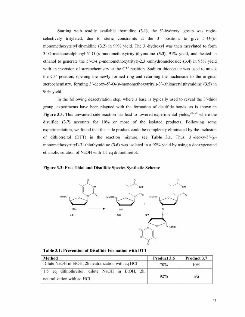
Starting with readily available thymidine (3.1), the 5’-hydroxyl group was regio-
selectively tritylated, due to steric constraints at the 3’ position, to give 5'-O-(p-
monomethoxytrityl)thymidine (3.2) in 99% yield. The 3’-hydroxyl was then mesylated to form
3’-O-methanesulphonyl-5’-O-(p-monomethoxytrityl)thymidine (3.3), 91% yield, and heated in
ethanol to generate the 5’-O-( p-monomethoxytrityl)-2,3’-anhydronucleoside (3.4) in 95% yield
with an inversion of stereochemistry at the C3’ position. Sodium thioacetate was used to attack
the C3’ position, opening the newly formed ring and returning the nucleoside to the original
stereochemistry, forming 3’-deoxy-5’-O-(p-monomethoxytrityl)-3’-(thioacetyl)thymidine (3.5) in
90% yield.
In the following deacetylation step, where a base is typically used to reveal the 3’-thiol
group, experiments have been plagued with the formation of disulfide bonds, as is shown in
Figure 3.3. This unwanted side reaction has lead to lowered experimental yields,22, 25
where the
disulfide (3.7) accounts for 10% or more of the isolated products. Following some
experimentation, we found that this side product could be completely eliminated by the inclusion
of dithiotreitol (DTT) in the reaction mixture, see Table 3.1. Thus, 3’-deoxy-5’-(p-
monomethoxytrityl)-3’-thiothymidine (3.6) was isolated in a 92% yield by using a deoxygenated
ethanolic solution of NaOH with 1.5 eq dithiothreitol.
Figure 3.3: Free Thiol and Disulfide Species Synthetic Scheme
O
SAc
MMTO
NH
ON
O
3.5
O
SH
MMTO
NH
ON
O
3.6
O
S
MMTO
NH
ON
O
3.7
O
S
OTMM
HN
ON
O
+
Table 3.1: Prevention of Disulfide Formation with DTT
Method Product 3.6 Product 3.7
Dilute NaOH in EtOH, 2h neutralization with aq HCl 70% 10%
1.5 eq dithiothreitol, dilute NaOH in EtOH, 2h,
neutralization with aq HCl 92% n/a
41
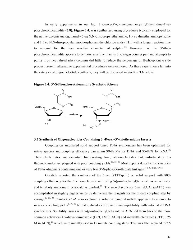
In early experiments in our lab, 3’-deoxy-5’-(p-monomethoxytrityl)thymidine-3’-S-
phosphorothioamidite (3.8), Figure 3.4, was synthesised using procedures typically employed for
the native oxygen analog, namely 5 eq N,N-diisopropylethylamine, 1.5 eq dimethylaminopyridine
and 1.5 eq N,N-diisopropylaminophosphonamidic chloride in dry THF with a longer reaction time
to account for the less reactive character of sulphur.22
However, as the 3’-thio-
phosphorothioamidite appears to be more sensitive than its 3’-oxygen counter part and attempts to
purify it on neutralised silica columns did little to reduce the percentage of H-phosphonate side
product present, alternative experimental procedures were explored. As these experiments fall into
the category of oligonucleotide synthesis, they will be discussed in Section 3.6 below.
Figure 3.4: 3’-S-Phosphorothioamidite Synthetic Scheme
O
SH
MMTO
NH
ON
O
O
S
MMTO
NH
ON
O
P N(iPr)2
O
various
NC
3.6 3.8
3.3 Synthesis of Oligonucleotides Containing 3’-Deoxy-3’-thiothymidine Inserts
Coupling on automated solid support based DNA synthesizers has been optimized for
native species and coupling efficiency can attain 99-99.5% for DNA and 95-98% for RNA.26
These high rates are essential for creating long oligonucleotides but unfortunately 3’-
thionucleosides are plagued with poor coupling yields.22, 25, 27
Most reports describe the synthesis
of DNA oligomers containing one or very few 3’-S-phosphorothiolate linkages.1, 5, 6, 18-20, 27-34
Cosstick reported the synthesis of the 5mer d(TTTspTT) on solid support with 80%
coupling efficiency for the 3’-thionucleoside unit using 5-(p-nitrophenyl)tetrazole as an activator
and tetrabutylammonium periodate as oxidant.35
The mixed sequence 6mer d(GATspATC) was
accomplished in slightly higher yields by delivering the reagents for the thioate coupling step by
syringe.6, 29, 35
Cosstick et al. also explored a solution based disulfide approach to attempt to
increase coupling yields5, 19, 31
but later abandoned it due to incompatibility with automated DNA
synthesizers. Solubility issues with 5-(p-nitrophenyl)tetrazole in ACN led them back to the more
common activators 4,5-dicyanoimidazole (DCI, 1M in ACN) and 4-ethylthiotetrazole (ETT, 0.25
M in ACN),27
which were initially used in 15 minute coupling steps. This was later reduced to 2.5
42

minutes for ETT34, 36
as no increase in yield was observed for extending the time beyond this point
but later reports reverted to the 15 minute coupling time without explanation.34, 36
They also
increased the 3’-thio-modified amidite concentration, to 0.15M in ACN, to enable the synthesis of
oligomers containing up to five 3’-S-phosphorothiolate linkages.25
In a recent oral presentation, R.
Cosstick discussed the possibility of using dimer building blocks already containing an internal
3’-S-phophorothiolate linkage presumably to get around poor coupling yields afforded by
nucleoside 3’-S-phosphorothioamidite monomers.37
An exploration of activators, oxidants and the preparation of the phosphorothioamidite
was undertaken as part of this work to maximise the coupling of the 3’-S-dT nucleoside on the
automated DNA synthesiser. In these experiments, and for all the oligonucleotides synthesised in
this work, 2’-deoxyribonucleoside 3’-O-phosphoramidites were prepared as 0.1 M solutions in
anhydrous acetonitrile and the ribonucleoside (RNA), 2’F-ANA, and 2’-OMe RNA monomers as
0.15 M solutions in anhydrous acetonitrile. The 3’-deoxythymidine-3’-S-phosphorothioamidites
were prepared as 0.15 M solutions in anhydrous dichloromethane. Commercially available, Cap
A, Cap B and 3% trichloroacetic acid in dichloromethane solutions were used.
3.4 Activators for 3’-Thio Coupling
A comparison of the efficacy of 3’-thio coupling of three activators, 4-ethylthiotetrazole
(ETT), 4,5-dicyanoimidazole (DCI) and 5-benzylthio-1-H-tetrazole (BTT) was performed by
creating a dT-12mer with a single 3’-thio insert in position 5. The lower pKa of ETT and BTT,
Table 3.2, results in faster rates of reaction, as does, conversely, the greater nucleophilicity of
DCI.37
Table 3.2: A Comparison of the pKa and Structure of Relevant Activators
Activator pKa27
Preparation Structure
5-Benzylthio-1H-Tetrazole 4.1 0.5 M in ACN NH
N
N
N
SCH2Ph
5-Ethylthio-1H-Tetrazole 4.3 0.25 M in ACN NH
N
N
N
SCH2CH3
4,5-Dicyanoimidazole 5.2 1 M in ACN
N
NH
CN
CN
43
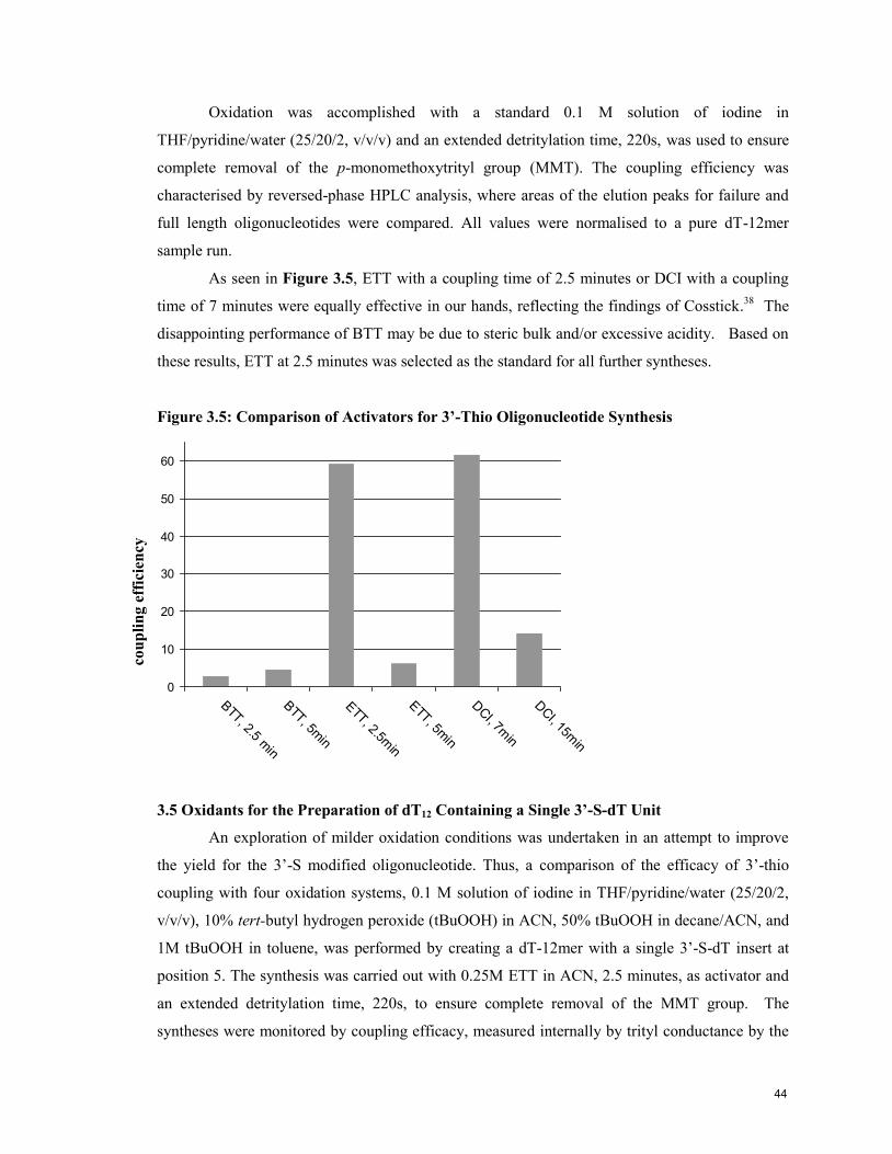
Oxidation was accomplished with a standard 0.1 M solution of iodine in
THF/pyridine/water (25/20/2, v/v/v) and an extended detritylation time, 220s, was used to ensure
complete removal of the p-monomethoxytrityl group (MMT). The coupling efficiency was
characterised by reversed-phase HPLC analysis, where areas of the elution peaks for failure and
full length oligonucleotides were compared. All values were normalised to a pure dT-12mer
sample run.
As seen in Figure 3.5, ETT with a coupling time of 2.5 minutes or DCI with a coupling
time of 7 minutes were equally effective in our hands, reflecting the findings of Cosstick.38
The
disappointing performance of BTT may be due to steric bulk and/or excessive acidity. Based on
these results, ETT at 2.5 minutes was selected as the standard for all further syntheses.
Figure 3.5: Comparison of Activators for 3’-Thio Oligonucleotide Synthesis
0
10
20
30
40
50
60
BTT, 2.5 m
in
BTT, 5m
in
ETT, 2.5m
in
ETT, 5m
in
DCI, 7m
in
DCI, 15m
in
3.5 Oxidants for the Preparation of dT12 Containing a Single 3’-S-dT Unit
An exploration of milder oxidation conditions was undertaken in an attempt to improve
the yield for the 3’-S modified oligonucleotide. Thus, a comparison of the efficacy of 3’-thio
coupling with four oxidation systems, 0.1 M solution of iodine in THF/pyridine/water (25/20/2,
v/v/v), 10% tert-butyl hydrogen peroxide (tBuOOH) in ACN, 50% tBuOOH in decane/ACN, and
1M tBuOOH in toluene, was performed by creating a dT-12mer with a single 3’-S-dT insert at
position 5. The synthesis was carried out with 0.25M ETT in ACN, 2.5 minutes, as activator and
an extended detritylation time, 220s, to ensure complete removal of the MMT group. The
syntheses were monitored by coupling efficacy, measured internally by trityl conductance by the
cou
pli
ng e
ffic
ien
cy
44
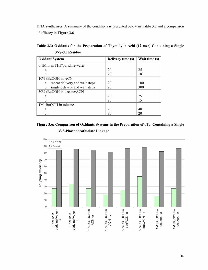
DNA synthesiser. A summary of the conditions is presented below in Table 3.3 and a comparison
of efficacy in Figure 3.6.
Table 3.3: Oxidants for the Preparation of Thymidylic Acid (12 mer) Containing a Single
3’-S-dT Residue
Oxidant System Delivery time (s) Wait time (s)
0.1M I2 in THF/pyridine/water
a.
b.
20
20
25
18
10% tBuOOH in ACN
a. repeat delivery and wait steps
b. single delivery and wait steps
20
20
100
300
50% tBuOOH in decane/ACN
a.
b.
20
20
25
15
1M tBuOOH in toluene
a.
b.
20
30
40
20
Figure 3.6: Comparison of Oxidants Systems in the Preparation of dT12 Containing a Single
3’-S-Phosphorothiolate Linkage
0
10
20
30
40
50
60
70
80
90
100
0.1
M I2 in
pyridin
e/w
ate
r -
a
0.1
M I2 in
pyridin
e/w
ate
r -
b
10%
tB
uO
OH
in
AC
N -
a
10%
tB
uO
OH
in
AC
N -
b
50%
tB
uO
OH
in
dec/A
CN
-a
50%
tB
uO
OH
in
dec/A
CN
- b
1M
tB
uO
OH
in
tolu
ene -
a
1M
tB
uO
OH
in
tolu
ene -
b
co
up
lin
g e
ffic
ien
cy
% 3'-S Step
% Overall
45

The best coupling efficiencies for the 3’-S step were with the 0.1M I2 (20s, then 25s) and
50% tBuOOH (20s, then 15s) system. Thus, a further comparison of these two oxidants was
performed with an oligoribonucleotide 21mer, 3'-ggCGAACUXCAGAAAUUAAUU-5', using
0.25M ETT at 2.5 minutes and 220s of detritylation. The syntheses were monitored by coupling
efficacy, measured internally by trityl conductance by the DNA synthesiser, as shown in Table
3.4. The lack of differentiation in the results led us to continue using the I2 system since it was
easier for practical reasons. This finding is supported by Joseph A. Piccirilli’s group27
.
Table 3.4: Comparison of Overall Coupling Efficacy for Mixed Sequence 21mers with 2
Oxidants
Oxidant Delivery Wait Comments X=U X=3’-S a
0.1M I2 in THF/pyr/water 20 s 25 s 97% 86%
50% tBuOOH in ACN 12 s 300 s Repeat delivery and wait. 96% 86% aAverage of three runs.
A good synthesis with relatively efficient coupling of the 3’-S-dT insert will still result in
considerable failure sequences where the insert does not couple, as seen around the 16-17 minute
mark in the HPLC trace in Figure 3.7.
Figure 3.7: HPLC trace of Crude Oligoribonucleotidesa
0
0.08
0.16
0.24
0 5 10 15 20 25 30 35 40 45
Minutes
Ab
so
rba
nc
e
5'-UUA AUT AAA GAC UUC AAG Cgg-3'
5'-UUA AUS AAA GAC UUC AAG Cgg-3'
aU,T,G,C = ribonucleosides, g = 2’-deoxyguanosine, S = 3’-deoxy-3’-thiothymidine.
S
46
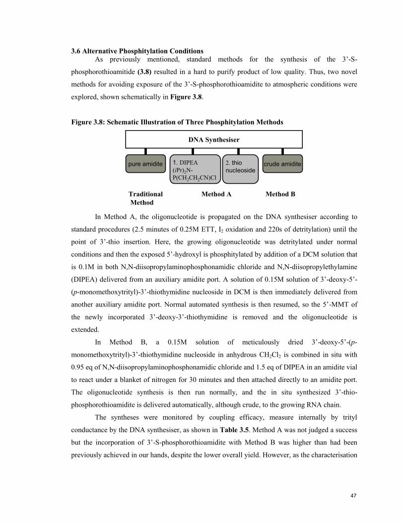
3.6 Alternative Phosphitylation Conditions
As previously mentioned, standard methods for the synthesis of the 3’-S-
phosphorothioamitide (3.8) resulted in a hard to purify product of low quality. Thus, two novel
methods for avoiding exposure of the 3’-S-phosphorothioamidite to atmospheric conditions were
explored, shown schematically in Figure 3.8.
Figure 3.8: Schematic Illustration of Three Phosphitylation Methods
Method A
DNA Synthesiser
pure amidite
Traditional
Method
2. thio
nucleoside
Method B
1. DIPEA
(iPr)2N-
P(CH2CH2CN)Cl
crude amidite
In Method A, the oligonucleotide is propagated on the DNA synthesiser according to
standard procedures (2.5 minutes of 0.25M ETT, I2 oxidation and 220s of detritylation) until the
point of 3’-thio insertion. Here, the growing oligonucleotide was detritylated under normal
conditions and then the exposed 5’-hydroxyl is phosphitylated by addition of a DCM solution that
is 0.1M in both N,N-diisopropylaminophosphonamidic chloride and N,N-diisopropylethylamine
(DIPEA) delivered from an auxiliary amidite port. A solution of 0.15M solution of 3’-deoxy-5’-
(p-monomethoxytrityl)-3’-thiothymidine nucleoside in DCM is then immediately delivered from
another auxiliary amidite port. Normal automated synthesis is then resumed, so the 5’-MMT of
the newly incorporated 3’-deoxy-3’-thiothymidine is removed and the oligonucleotide is
extended.
In Method B, a 0.15M solution of meticulously dried 3’-deoxy-5’-(p-
monomethoxytrityl)-3’-thiothymidine nucleoside in anhydrous CH2Cl2 is combined in situ with
0.95 eq of N,N-diisopropylaminophosphonamidic chloride and 1.5 eq of DIPEA in an amidite vial
to react under a blanket of nitrogen for 30 minutes and then attached directly to an amidite port.
The oligonucleotide synthesis is then run normally, and the in situ synthesized 3’-thio-
phosphorothioamidite is delivered automatically, although crude, to the growing RNA chain.
The syntheses were monitored by coupling efficacy, measure internally by trityl
conductance by the DNA synthesiser, as shown in Table 3.5. Method A was not judged a success
but the incorporation of 3’-S-phosphorothioamidite with Method B was higher than had been
previously achieved in our hands, despite the lower overall yield. However, as the characterisation
47

and biological activity of these 3’-thio modified oligoribonucleotides were the main objective of
this work, we did not seek to further optimize the approach.
Table 3.5: Comparison of Coupling Efficacy for Alternative Phosphitylation Methods with
dT-12mers
Method 3’-S Step Yeild Overall Yield
A 26% 25%
B 65% 56%
3.7 Phosphorothioamidite Stability
The stability of the 3’-S-phosphorothioamidite (3.8) was assessed by NMR to determine
if the poor coupling yields during synthesis were due, in part, to deterioration of the reactive 3’
group required for coupling. A quantitative 31
P NMR experiment, with an extended relaxation
time ( 2), was undertaken to record a spectrum each half hour for 24h of a sample of the 3’-S-
phosphorothioamidite at room temperature in CDCl3. Two representative spectra are presented in
Figure 3.9.
Figure 3.9: 31
P NMR of Phosphorothioamidite
a.) 0h
b.) 10h
48

For the first 8h of the experiment no changes were observed in the spectra, i.e. signals at
163 and 159 ppm remained in constant ratio, see Figure 3.9, but at 10h some additional signals of
low intensity were observed between 80 and 20 ppm although these did not increase appreciably
in intensity in the remaining 14h. Fresh samples of 3’-S-phosphorothioamidite were prepared in
acetone-d6, CDCl3 and ACN-d3 and quantitative 31
P NMR spectra were recorded immediately.
When a second spectrum of each was recorded 2 days later, the samples in acetone-d6 and CDCl3
were unchanged, but the ACN sample showed several new phosphorus species and a decline in
the intensity of the phosphorothioamidite signals. Thymidine 3’-S-phosphorothioamidite (3.8)
stored as a dry foam in the freezer for 2 months was unchanged. Thus, phosphorothioamidites
stored in the freezer and freshly prepared in CH2Cl2 were taken to be stable on the DNA
synthesiser for the ~8h run and no further concern about their stability was entertained.
3.8 Summary of Optimised Synthesis Conditions for Oligonucleotides Containing 3’-Deoxy-
3’-thiothymidine
The optimal conditions for synthesis of oligoribonucleotides containing 3’-S-
phosphorothiolate linkages, in our hands, is summarised in Table 3.6.
Table 3.6: Optimised Conditions for 3’-S-Phosphorothioamidite Coupling
Parameter Recommendation
Activator 0.25 M ETT in ACN, 2.5 minutes
Oxidant 0.1M I2 in THF/pyr/water (25/20/2, v/v/v)
3’-S-
Phosphorothioamidite
Preparation
0.18 M 5’-MMT-3’-deoxy-3’-thiothymidine, 0.17 M N,N-
diisopropylaminophosphonamidic chloride, and 0.23 M DIPEA in
CH2Cl2 reacted in amidite vial and delivered crude.
With these conditions, a small library of native and chimeric oligonucleotides was
assembled on the DNA synthesiser and their characterization will be addressed in Chapter 4.
Phosphorothioamidite coupling yields were adequate to produce sufficient samples for
characterisation and biological assays but would prove a serious issue for wider application of
these compounds.
3.9 References
1 Breaker, R. R.; Joyce, G. F., A DNA enzyme that cleaves RNA. Chem. Biol. 1994, 1, 223-229.
2 Gold, L.; Polisky, B.; Uhlenbeck, O.; Yarus, M., Annu. Rev. Biochem. 1995, 64, 763-797.
49
3 Watts, J. K.; Choubdar, N.; Sadalapure, K.; Robert, F.; Wahba, A. S.; Pelletier, J.; Mario Pinto, B.;
Damha, M. J., 2'-Fluoro-4'-thioarabino-modified oligonucleotides: conformational switches linked to
siRNA activity. Nucl. Acids Res. 2007, 35, (5), 1441-1451.
4 Inoue, N.; Minakawa, N.; Matsuda, A., Synthesis and properties of 4'-ThioDNA: unexpected
RNA-like behavior of 4'-ThioDNA. Nucleic Acids Res. 2006, 34, 3476–3483.
5 Beevers, A. P. G.; Fettes, K. J.; Sabbagh, G.; Murad, F. K.; Arnold, J. R. P.; Cosstick, R.; Fisher,
J., NMR and UV studies of 3'-S-phosphorothiolate modified DNA in a DNA:RNA hybrid dodecamer
duplex; implications for antisense drug design. Organic & Biomolecular Chemistry 2003, 2, (1), 114-119.
6 Beevers, A. P. G.; Witch, E. M.; Jones, B. C. N. M.; Cosstick, R.; Arnold, J. R. P.; Fisher, J.,
Conformational analysis of 3'-S-PO3-linked ribo- and deoxyribodinucleoside monophosphates. Magn.
Reson. Chem. 1999, 37, (11), 814-820.
7 Eckstein, F., Annu. Rev. Biochem. 1985, 54, 367-402.
8 Cosstick, R.; Eckstein, F., Synthesis of d(GC) and d(CG) Octamers Containing Alternating
Phosphorothioate Linkages: Effect of the Phosphorothioate Group on the B-Z Transition. Biochemistry
1985, 24, 3630-3638.
9 Burgers, P. M. J.; Eckstein, F., Diastereomers of 5'-O-adenosyl 3'-O-uridyl phosphorothioate:
chemical synthesis and enzymic properties. Biochemistry 1978, 18, 592-96.
10 Bhan, P.; Bhan, A.; Hong, M.; Hartwell, J. G.; Saunders, J. M.; Hoke, G. D., 2',5'-Linked oligo-3'-
deoxyribonucleoside phosphorothioate chimeras: thermal stability and antisense inhibition of gene
expression. Nucleic Acids Res. 1997, 25, 3310-3317.
11 Woolf, T. M.; Jennings, C. G.; Rebagliati, M.; Melton, D. A., The stability, toxicity and
effectiveness of unmodified and phosphorothioate antisense oligodeoxynucleotides in Xenopus oocytes and
embryos. Nucleic Acids Res. 1990, 18, (7), 1763–1769.
12 Thewalt, U.; Bugg, C. E., Effects of Sulfur Substituents on Base Stacking and Hydrogen Bonding.
The Crystal Structure of 6-Thioguanosine Monohydrate. J. Am. Chem. Soc. 1972, 94, (25), 8892-8898.
13 Xu, Y.; Kool, E. T., Chemical and enzymatic properties of bridging 5'-S-phosphorothioester
linkages in DNA. Nucleic Acids Res. 1998, 26, (13), 3159-3164.
14 Minakawa, N.; Kaga, D.; Kato, Y.; Endo, K.; Tanaka, M.; Sasaki, T.; Matsuda, A., Synthesis and
structural elucidation of 1-(3-C-ethynyl-4-thio-beta -D-ribofuranosyl)cytosine (4 -thioECyd). J. Chem. Soc.,
Perkin Trans. 1 2002, 2182 - 2189.
15 Boggon, T. J.; Hancox, E. L.; McAuley-Hecht, K. E.; Connolly, B. A.; Hunter, W. N.; Brown, T.;
Walker, R. T.; Leonard, G. A., The crystal structure analysis of d(CGCGAASSCGCG)2, a synthetic DNA
dodecamer duplex containing four 4 -thio-2 -deoxythymidine nucleotides. Nucleic Acids Res. 1996, 24, (5),
951-961.
16 Denisov, A. Y.; Hannoush, R. N.; Gehring, K.; Damha, M. J., A Novel RNA Motif Based on the
Structure of Unusually Stable 2',5'-Linked r(UUCG) Loops. J. Am. Chem. Soc. 2003, 125, (38), 11525-
11531.
17 Cosstick, R.; Vyle, J. S., Synthesis and phosphorus–sulphur bond cleavage of 3-thiothymidylyl(3-
5)thymidine. J. Chem. Soc. Chem. Commun. 1988, 992-993.
18 Cosstick, R.; S.Vyle, J., Synthesis and properties of dithymidine phosphate analogues containing
3'-thiothymidine. Nucleic Acids Res. 1990, 18, (4), 829-835.
19 Fettes, K.; O'Neil, I.; Roberts, S. M.; Cosstick, R., Solid-phase synthesis of oligodeoxynucleotides
containing 3'-S-phosphorothiolate linkages. Nucleosides, Nucleotides & Nucleic Acids 2001, 20, (4-7),
1351-1354.
20 Vyle, J.; Li, X.; Cosstick, R., New methods for the synthesis of 3'-S-phosphorothiolate
internucleoside linkages. Tetrahedron Lett. 1992, 33, (21), 3017-3020.
21 Li, X.; Cosstick, R., Application of an intramolecular Michaelis–Arbusov reaction to the synthesis
of nucleoside 3-S,5-O-cyclic phosphorothiolate triesters. J. Chem. Soc., Perkin Trans. 1 1993, 1091.
22 Tedeschi, A. L. Synthesis and Biological Evaluation of Oligonucleotides Containing 3'- and 5'-S-
Phosphorthiolate Internucleotide Linkages. M.Sc. Thesis, McGill University, Montreal, 2004.
23 Eleuteri, A.; Reese, C. B.; Song, Q., Synthesis of 3,5-dithiothymidine and related compounds. J.
Chem. Soc., Perkin Trans. 1 1996, 18, 2237 - 2240.
50
24 Horwitz, J. R.; Chua, J.; Noel, M., Nucleosides V: The monomesylates of 1-(2'-deoxy-beta-
Dlyxofuranosyl)thymidine. J. Org. Chem. 1964, 29, 2076.
25 Cosstick, R. In UK Stabilization of Multi-Stranded Nucleic Acid Structures Using 3’-S-
Phosphorothiolate Linkages, The XVII International Roundtable on Nucleosides, Nucleotides and Nucleic
Acids (IRTXVII), Bern, Switzerland, 2006; Haenni, D. L., 'Ed.'^'Eds.' Bern, Switzerland, 2006; p^pp.
26 Davis, R. H., Large-scale oligoribonucleotide production. Curr. Opin. Biotechnol. 1995, 6, (2),
213-217.
27 Sabbagh, G.; Fettes, K. J.; Gosain, R.; O'Neil, I. A.; Cosstick, R., Synthesis of
phosphorothioamidites derived from 3'-thio-3'-deoxythymidine and 3'-thio-2',3'-dideoxycytidine and the
automated synthesis of oligodeoxynucleotides containing a 3'-S-phosphorothiolate linkage. Nucleic Acids
Res. 2004, 32, (2), 495-501.
28 Li, X.; Andrews, D.; Cosstick, R., Synthesis of a Dinucleoside 3'S-Phosphorothiolate Containing
2'-Deoxy-3'-Thioadenosine. Tetrahedron 1992, 48, (13), 2729-2738.
29 Weinstein, L. B.; Earnshaw, D. J.; Cosstick, R.; Cech, T. R., Synthesis and Characterization of an
RNA Dinucleotide Containing a 3'-S-Phosphorothiolate Linkage. J. Am. Chem. Soc. 1996, 118, (43),
10341-10350.
30 Cosstick, R.; Vyle, J. S., Solid-phase synthesis of oligonucleotides containing 3'-thiothymidine.
Tetrahedron Lett. 1989, 30, 4693-4696.
31 Beevers, A. P. G.; Fettes, K. J.; O’Neil, I. A.; Roberts, S. M.; Arnold, J. R. P.; Cosstick, R.; Fisher,
J., Probing the effect of a 3A-S-phosphorothiolate link on the conformation of a DNA:RNA hybrid;
implications for antisense drug design. Chemical Communications 2002, 14, 1458–1459.
32 Cosstick, R.; Buckingham, J.; Brazier, J.; Fisher, J., Stabilization Of Multi-Stranded Nucleic Acid
Structures Using 3'-S-Phosphorothiolate Linkages. Nucleosides, Nucleotides and Nucleic Acids Research
2007, 26, 555 - 558.
33 Jayakumar, H. K.; Buckingham, J. L.; Brazier, J. A.; Berry, N. G.; Cosstick, R.; Fisher, J., NMR
studies of the conformational effect of single and double 3'-S-phosphorothiolate substitutions within
deoxythymidine trinucleotides. Magn. Reson. Chem. 2007, 45, (4), 340-345.
34 Bentley, J.; Brazier, J. A.; Fisher, J.; Cosstick, R., Duplex stability of DNA·DNA and DNA·RNA
duplexes containing 3'-S-phosphorothiolate linkages. Organic & Biomolecular Chemistry 2007, 5, 3698–
3702.
35 Vyle, J. S.; Connolly, B. A.; Kemp, D.; Cosstick, R., Sequence- and Strand-Specific Cleavage in
Oligodeoxyribonucleotides and DNA Containing 3’-Thiothymidine. Biochemistry 1992, 31, 3012-3018.
36 Buckingham, J.; Sabbagh, G.; Brazier, J.; Fisher, J.; Cosstick, R., Control of DNA Conformation
Using 3'-S-Phosphorothiolate-Modified Linkages. Nucleosides, Nucleotides and Nucleic Acids 2005, 24,
(5), 491-495.
37 Research, G. Technical Brief - ABOUT ACTIVATORS: Now and tomorrow. (May 5th),
38 Sun, S.; Yoshida, A.; Piccirilli, J. A., Synthesis of 3'-thioribonucleosides and their incorporation
into oligoribonucleotides via phosphoramidite chemistry. RNA 1997, 3, (11), 1352-1363.
51
Chapter 4: Physical and Biological Characterisation of Oligonucleotides Containing
3’-S-Phosphorothiolate Linkages
4.1 Introduction
Broad therapeutic use of RNAi depends on the development of synthetic oligonucleotides
able to strongly induce RISC, and with good nuclease resistance, sufficient cell penetration and
high affinity for the target mRNA.1 Our laboratory has investigated the importance of pre-
organization and flexibility in eliciting RNaseH in the antisense strategy,2, 3
and the rising
importance of RNAi has prompted the present investigation into the use of 3‟-deoxy-3‟-
thiothymidine (3‟-S-dT) inserts in RNA. This project was initiated based on promising early
experiments in our lab4 and other reports
5, 6 of the 3‟-thio modification in DNA. Because this
modification mimics the structure of ribonucleosides (north pucker), we thought it would be
compatible with the RNAi pathway while providing solutions to some of the challenges facing
siRNA therapeutics, e.g., the development of siRNA chemistries that enhance or maintain cellular
activity.
In the present chapter, the impact of this modification on the structure and activity of
siRNA duplexes will be investigated by thermal denaturing experiments, circular dichroism and
cell based assays, while drawing comparisons, when appropriate, to the known 3‟-S-DNA
oligonucleotides.
4.2 3’-Phosphorothiolate Linkages and Conformation
The conformation of nucleosides can be influenced by structural changes in the
carbohydrate moiety. The absence of a 2‟ substituent in 2‟-deoxynucleosides generally results in a
dynamic equilibrium between the north and south conformers, although the south conformer is
generally favoured.7,8
In this case, the electronegativity of its 3‟-OH substituent leads to a strong
gauche effect between the 3'-OH and the ring oxygen (O4‟). As the south pucker is favored, this
gauche effect must override the anomeric effect that stabilizes the north conformer. Substitution
at the 3‟ position with the less electronegative sulphur atom lessens the gauche effect, allowing
the anomeric effect to dominate and populate the north conformation.
Cosstick and co-workers investigated these effects with respect to 3‟-thio modification by
comparing the conformation of 3‟-S-phophorothiolate linked DNA (TspT) and RNA (IspU)
dinucleotides to the conformations of native (phosphodiester-linked) dinucleotide TpT using 1H
NMR spectroscopy. They found that in both the DNA and RNA dimers the sugar ring
conformations are affected by the 3‟-S-phosphorothiolate modification, with a large increase in
52

the percentage of north-type puckering. This change was ascribed to the decrease in the influence
of the gauche effect between the 3‟-S substituent and the O4‟-ring oxygen experienced when
replacing an oxygen atom by the less electronegative 3‟sulphur atom. Further NMR studies by
Cosstick‟s group on DNA trimers containing 3‟-S-dT units confirmed that the 3‟-S-
phosphorothiolate linkage shifts the conformation of its own sugar ring to north along with that of
its 3‟ neighbour.9 This phenomenon is possibly due to increased intramolecular base stacking.
Surprisingly, two consecutive 3‟-S-dT inserts do not affect the conformation of neighbouring
nucleotides, indicating that the maximum effect of 3‟-thio modifications can be achieved by non-
consecutive incorporation,10
up to the saturation point of 3 inserts, either contiguous or alternating
with native dT residues, in the 16mer sequence d(GCGTTTTTTTTTTGCG).11
Cosstick and co-
workers also reported that a single 3‟-thio insert in a DNA dodecamer maintains its preferred
north conformation and influences the conformation of its neighbour towards north, resulting in a
reproducible increase in melting temperature of 2.5°C.12
These observations are similar, although
less dramatic, than those observed with LNA modified duplexes. LNA is able to shift the
conformation of the neighbouring ribo and 2‟-deoxyribonucleosides to 90% north13
and favours
increased intrastrand base stacking.14, 15
The above findings demonstrating the RNA-like (north) conformation of 3‟-deoxy-3‟-
thionucleosides is of considerable interest in the search for suitable siRNA drugs, and suggest that
this modification can be used to modify siRNA without compromising its potency.
4.3 A Library of Modified RNA Duplexes Containing 3’-Thio Linkages
A concise library of chemically modified RNA duplexes was synthesised. The duplexes
were composed predominantly of native ribonucleotides, with inserts of 3‟-deoxy-3‟-
thiothymidine (3‟-S-dT), 2‟-deoxy-2‟-fluorouridine, 2‟-OMe-uridine, and 2‟-deoxynucleotides.
For our RNAi investigation, we selected a mixed sequence short interfering RNA (siRNA) duplex
shown to be effective in the down-regulation of the protein luciferase in modified HeLa cells. The
annealed duplexes have a complementary region of 19 base pairs and 2-nt DNA overhangs on the
3‟ ends. Incorporating 2‟-deoxynucleotide residues reduces synthesis costs and may possibly
decrease the exonuclease degradation of siRNA in cells.5 Heavier DNA modification can reduce
off-target effects.6
The small amounts of oligonucleotide produced by the low yielding syntheses in Chapter
3 were prioritized for thermal stability and RNAi analysis. When sufficient sample remained, the
oligonucleotide was also characterized by MS, see Table 4.1.
53
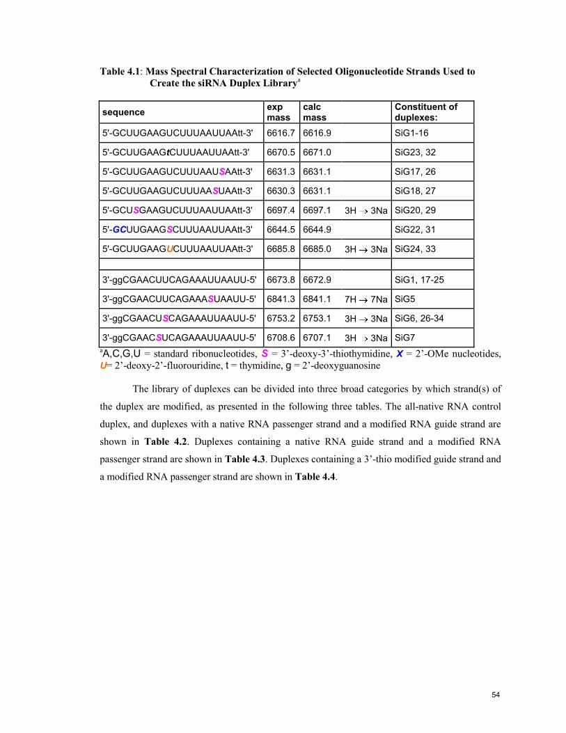
Table 4.1: Mass Spectral Characterization of Selected Oligonucleotide Strands Used to
Create the siRNA Duplex Librarya
sequence exp mass
calc mass
Constituent of duplexes:
5'-GCUUGAAGUCUUUAAUUAAtt-3' 6616.7 6616.9 SiG1-16
5'-GCUUGAAGtCUUUAAUUAAtt-3' 6670.5 6671.0 SiG23, 32
5'-GCUUGAAGUCUUUAAUSAAtt-3' 6631.3 6631.1 SiG17, 26
5'-GCUUGAAGUCUUUAASUAAtt-3' 6630.3 6631.1 SiG18, 27
5'-GCUSGAAGUCUUUAAUUAAtt-3' 6697.4 6697.1 3H 3Na SiG20, 29
5'-GCUUGAAGSCUUUAAUUAAtt-3' 6644.5 6644.9 SiG22, 31
5'-GCUUGAAGUCUUUAAUUAAtt-3' 6685.8 6685.0 3H 3Na SiG24, 33
3'-ggCGAACUUCAGAAAUUAAUU-5' 6673.8 6672.9 SiG1, 17-25
3'-ggCGAACUUCAGAAASUAAUU-5' 6841.3 6841.1 7H 7Na SiG5
3'-ggCGAACUSCAGAAAUUAAUU-5' 6753.2 6753.1 3H 3Na SiG6, 26-34
3'-ggCGAACSUCAGAAAUUAAUU-5' 6708.6 6707.1 3H 3Na SiG7
aA,C,G,U = standard ribonucleotides, S = 3‟-deoxy-3‟-thiothymidine, X = 2‟-OMe nucleotides, U= 2‟-deoxy-2‟-fluorouridine, t = thymidine, g = 2‟-deoxyguanosine
The library of duplexes can be divided into three broad categories by which strand(s) of
the duplex are modified, as presented in the following three tables. The all-native RNA control
duplex, and duplexes with a native RNA passenger strand and a modified RNA guide strand are
shown in Table 4.2. Duplexes containing a native RNA guide strand and a modified RNA
passenger strand are shown in Table 4.3. Duplexes containing a 3‟-thio modified guide strand and
a modified RNA passenger strand are shown in Table 4.4.
54

Table 4.2: An Inventory of Duplexes with a Native RNA Passenger Strand and a Modified
RNA Guide Stranda,b
Name Sequence Tm Tm hysteresis c
SiG1 3'-ggCGAACUUCAGAAAUUAAUU-5'
5'-GCUUGAAGUCUUUAAUUAAtt-3‟
61.0
61.0
61.0
1.0
1.0
1.0
SiG2 3'-ggCGAACUUCAGAAAUUAAUS-5'
5'-GCUUGAAGUCUUUAAUUAAtt-3' 61.1 +0.1 1.4
SiG3 3'-ggCGAACUUCAGAAAUUAASU-5‟
5'-GCUUGAAGUCUUUAAUUAAtt-3' 57.9 -3.1 2.8
SiG4 3'-ggCGAACUUCAGAAAUSAAUU-5'
5'-GCUUGAAGUCUUUAAUUAAtt-3' 56.8 -4.2 -0.3
SiG5 3'-ggCGAACUUCAGAAASUAAUU-5'
5'-GCUUGAAGUCUUUAAUUAAtt-3' 56.0 -5.0 2.1
SiG6 3'-ggCGAACUSCAGAAAUUAAUU-5'
5'-GCUUGAAGUCUUUAAUUAAtt-3' 61.3* +0.3 1.1
SiG7 3'-ggCGAACSUCAGAAAUUAAUU-5'
5'-GCUUGAAGUCUUUAAUUAAtt-3' 62.0 +1.0 1.0
SiG9 3'-ggCGAACUUCAGAAAUtAAUU-5'
5'-GCUUGAAGUCUUUAAUUAAtt-3' 61.1 +0.1 1.4
SiG10 3'-ggCGAACUUCAGAAAtUAAUU-5'
5'-GCUUGAAGUCUUUAAUUAAtt-3' 60.1 -0.9 1.3
SiG11 3'-ggCGAACUtCAGAAAUUAAUU-5'
5'-GCUUGAAGUCUUUAAUUAAtt-3' 61.9 +0.9 1.5
SiG12 3'-ggCGAACtUCAGAAAUUAAUU-5'
5'-GCUUGAAGUCUUUAAUUAAtt-3' 61.8 +0.8 1.5
SiG13 3'-ggCGAACUUtAGAAAUUCAUU-5'
5'-GCUUGAAGUCUUUAAUUAAtt-3' 52.5 -8.5 1.0
SiG14 3'-ggCGAACUUUAGAAAUUAAUU-5'
5'-GCUUGAAGUCUUUAAUUAAtt-3' 55.5 -5.5 2.6
SiG15 3'-ggCGAACUUSAGAAAUUAAUU-5'
5'-GCUUGAAGUCUUUAAUUAAtt-3' 59.9 -1.1 1.2
SiG16 3'-ggCGAACUSCAGAAAUUAAUU-5'
5'-GCUUGAAGUCUUUAAUUAAtt-3' 59.6 -1.4 1.1*
aA,C,G,U = standard ribonucleotides, S = 3‟-deoxy-3‟-thiothymidine, S = mismatch 3‟-deoxy-3‟-
thiothymidine, U = 2‟-OMe-uridine, U = mismatch uridine, t = mismatch thymidine, t =
thymidine, g = 2‟-deoxyguanosine bTm experiments on aqueous solutions of ~2 x 10
-6 M of each oligonucleotide strand, 140 mM
KCl, 1 mM MgCl2, 5 mM Na2HPO4 buffer (pH 7.2); uncertainty of ± 0.5 o
C. Tm calculated for
heating curve, relative to control, by the baseline method. c
Hysteresis between heating and
cooling profiles was calculated from the derivative Tm values. *Data from second thermal run.
55
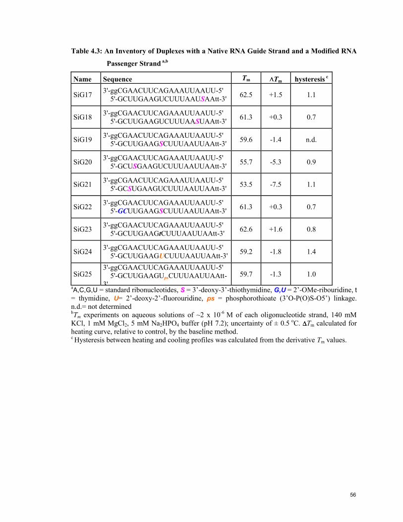
Table 4.3: An Inventory of Duplexes with a Native RNA Guide Strand and a Modified RNA
Passenger Strand a,b
Name Sequence Tm Tm hysteresis c
SiG17 3'-ggCGAACUUCAGAAAUUAAUU-5'
5'-GCUUGAAGUCUUUAAUSAAtt-3' 62.5 +1.5 1.1
SiG18 3'-ggCGAACUUCAGAAAUUAAUU-5'
5'-GCUUGAAGUCUUUAASUAAtt-3' 61.3 +0.3 0.7
SiG19 3'-ggCGAACUUCAGAAAUUAAUU-5'
5'-GCUUGAAGSCUUUAAUUAAtt-3' 59.6 -1.4 n.d.
SiG20 3'-ggCGAACUUCAGAAAUUAAUU-5'
5'-GCUSGAAGUCUUUAAUUAAtt-3' 55.7 -5.3 0.9
SiG21 3'-ggCGAACUUCAGAAAUUAAUU-5'
5'-GCSUGAAGUCUUUAAUUAAtt-3' 53.5 -7.5 1.1
SiG22 3'-ggCGAACUUCAGAAAUUAAUU-5'
5'-GCUUGAAGSCUUUAAUUAAtt-3' 61.3 +0.3 0.7
SiG23 3'-ggCGAACUUCAGAAAUUAAUU-5'
5'-GCUUGAAGtCUUUAAUUAAtt-3' 62.6 +1.6 0.8
SiG24 3'-ggCGAACUUCAGAAAUUAAUU-5'
5'-GCUUGAAGUCUUUAAUUAAtt-3' 59.2 -1.8 1.4
SiG25 3'-ggCGAACUUCAGAAAUUAAUU-5'
5'-GCUUGAAGUpsCUUUAAUUAAtt-
3'
59.7 -1.3 1.0
aA,C,G,U = standard ribonucleotides, S = 3‟-deoxy-3‟-thiothymidine, G,U = 2‟-OMe-ribouridine, t
= thymidine, U= 2‟-deoxy-2‟-fluorouridine, ps = phosphorothioate (3‟O-P(O)S-O5‟) linkage.
n.d.= not determined bTm experiments on aqueous solutions of ~2 x 10
-6 M of each oligonucleotide strand, 140 mM
KCl, 1 mM MgCl2, 5 mM Na2HPO4 buffer (pH 7.2); uncertainty of ± 0.5 o
C. Tm calculated for
heating curve, relative to control, by the baseline method. c Hysteresis between heating and cooling profiles was calculated from the derivative Tm values.
56
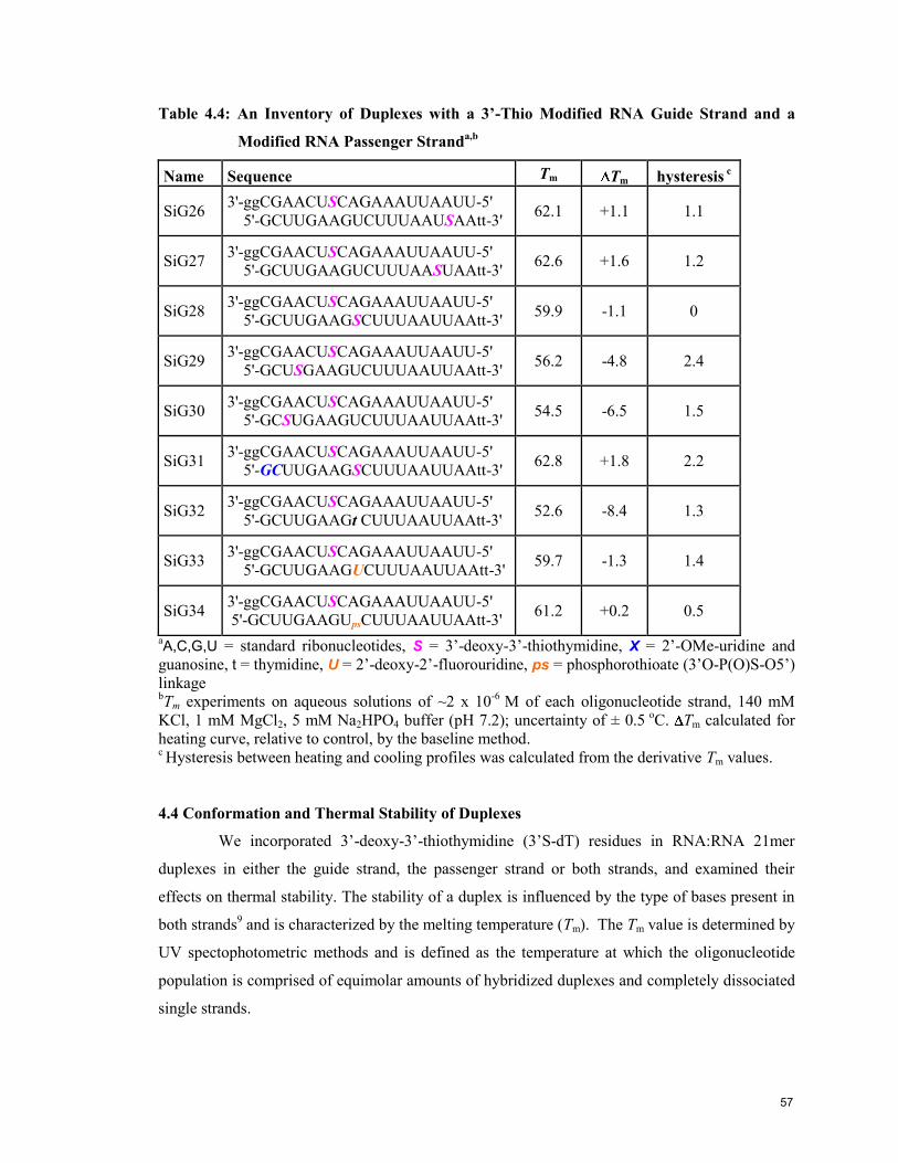
Table 4.4: An Inventory of Duplexes with a 3’-Thio Modified RNA Guide Strand and a
Modified RNA Passenger Stranda,b
Name Sequence Tm Tm hysteresis c
SiG26 3'-ggCGAACUSCAGAAAUUAAUU-5'
5'-GCUUGAAGUCUUUAAUSAAtt-3' 62.1 +1.1 1.1
SiG27 3'-ggCGAACUSCAGAAAUUAAUU-5'
5'-GCUUGAAGUCUUUAASUAAtt-3' 62.6 +1.6 1.2
SiG28 3'-ggCGAACUSCAGAAAUUAAUU-5'
5'-GCUUGAAGSCUUUAAUUAAtt-3' 59.9 -1.1 0
SiG29 3'-ggCGAACUSCAGAAAUUAAUU-5'
5'-GCUSGAAGUCUUUAAUUAAtt-3' 56.2 -4.8 2.4
SiG30 3'-ggCGAACUSCAGAAAUUAAUU-5'
5'-GCSUGAAGUCUUUAAUUAAtt-3' 54.5 -6.5 1.5
SiG31 3'-ggCGAACUSCAGAAAUUAAUU-5'
5'-GCUUGAAGSCUUUAAUUAAtt-3' 62.8 +1.8 2.2
SiG32 3'-ggCGAACUSCAGAAAUUAAUU-5'
5'-GCUUGAAGt CUUUAAUUAAtt-3' 52.6 -8.4 1.3
SiG33 3'-ggCGAACUSCAGAAAUUAAUU-5'
5'-GCUUGAAGUCUUUAAUUAAtt-3' 59.7 -1.3 1.4
SiG34 3'-ggCGAACUSCAGAAAUUAAUU-5'
5'-GCUUGAAGUpsCUUUAAUUAAtt-3' 61.2 +0.2 0.5
aA,C,G,U = standard ribonucleotides, S = 3‟-deoxy-3‟-thiothymidine, X = 2‟-OMe-uridine and
guanosine, t = thymidine, U = 2‟-deoxy-2‟-fluorouridine, ps = phosphorothioate (3‟O-P(O)S-O5‟)
linkage bTm experiments on aqueous solutions of ~2 x 10
-6 M of each oligonucleotide strand, 140 mM
KCl, 1 mM MgCl2, 5 mM Na2HPO4 buffer (pH 7.2); uncertainty of ± 0.5 o
C. Tm calculated for
heating curve, relative to control, by the baseline method. c Hysteresis between heating and cooling profiles was calculated from the derivative Tm values.
4.4 Conformation and Thermal Stability of Duplexes
We incorporated 3‟-deoxy-3‟-thiothymidine (3‟S-dT) residues in RNA:RNA 21mer
duplexes in either the guide strand, the passenger strand or both strands, and examined their
effects on thermal stability. The stability of a duplex is influenced by the type of bases present in
both strands9 and is characterized by the melting temperature (Tm). The Tm value is determined by
UV spectophotometric methods and is defined as the temperature at which the oligonucleotide
population is comprised of equimolar amounts of hybridized duplexes and completely dissociated
single strands.
57
These measurements were carried out with 2 μM of each strand suspended in an aqueous
buffer designed to mimic physiological conditions, adjusted to pH 7.2. The samples were first
heated at 95oC to ensure total dissociation of the strands, cooled slowly and stored for at least 10
hours at 4oC to facilitate complete annealing of the strands. The cold samples were transferred to a
pre-cooled UV spectrophotometer. The UV absorbance of the samples was measured at 260 nm
as they were gradually heated (0.5oC/min) to 95
oC and then re-cooled to 10
oC.
At low temperatures, UV absorption is lower due to π-stacking interactions between
neighbouring chromophores (purine and pyrimidine bases) in the intertwined strands, called a
hypochromic effect.16
As the temperature increases, the duplexes dissociate and the absorbance
increases, since there is much less π-stacking in single strands. The resulting sigmoidal „melting‟
curve (A260 vs temperature) typically shows a sharp change in absorbance over a narrow
temperature change (for example, see Figure 4.1). This reveals the cooperative nature of
oligonucleotide association. The first derivative of the curve is usually a good approximation of
the Tm, since the inflection point of the curve naturally occurs at the center of the transition. Tm
values can also be determined from the mid-point of the hyperchromic change, as will be
discussed below.
As seen in the representative samples in Figure 4.1, the sharp monophasic transitions
indicate the presence of a single cooperative complex in each sample. The Tm profile of each
duplex in the library is available in Appendix 2. The Tm values were calculated by the baseline
method, Tables 4.2-4.4, where the Tm is taken as the midpoint between the upper and lower
baselines defined by the low and high temperature regions of the Tm curve. The control
RNA:RNA duplex has a Tm of 61°C, and the differently modified duplexes show both stabilising
and destabilising effects.
The second ramp for both the heating and cooling stages found the Tm in each case to be
slightly lower than the first cycle (average of 0.3°C for each), which fits expectations because the
duplexes were not given enough time to reach their ideal lowest energy conformations. The
hysteresis between heating and cooling stages was generally small (average 1.4°C) and no clear
trends presented themselves among the groups. The runs were highly reproducible, as seen with
the control samples, which were run on 3 consecutive days.
Another method for calculating Tm is the “derivative method”, where the first derivative
of the curve is taken as the Tm value. Tm values calculated by the derivative method are available
in Appendix 2. There is generally good agreement between the 2 methods of calculating Tm, with
the hypochromic method typically calculating a value several degrees lower than the derivative
method. The thermodynamic parameters H, S, G, Keq and KTm were calculated using the
58
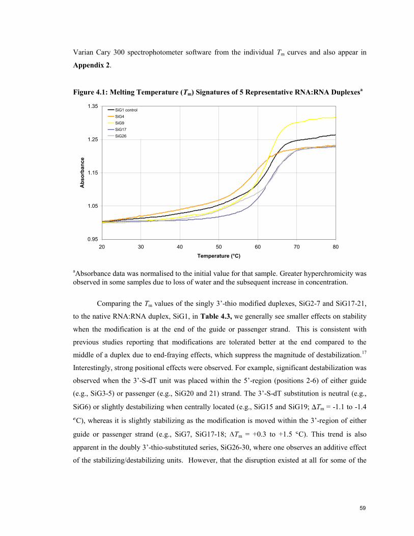
Varian Cary 300 spectrophotometer software from the individual Tm curves and also appear in
Appendix 2.
Figure 4.1: Melting Temperature (Tm) Signatures of 5 Representative RNA:RNA Duplexesa
0.95
1.05
1.15
1.25
1.35
20 30 40 50 60 70 80
Temperature (°C)
Ab
so
rba
nce
SiG1 control
SiG4
SiG9
SiG17
SiG26
aAbsorbance data was normalised to the initial value for that sample. Greater hyperchromicity was
observed in some samples due to loss of water and the subsequent increase in concentration.
Comparing the Tm values of the singly 3‟-thio modified duplexes, SiG2-7 and SiG17-21,
to the native RNA:RNA duplex, SiG1, in Table 4.3, we generally see smaller effects on stability
when the modification is at the end of the guide or passenger strand. This is consistent with
previous studies reporting that modifications are tolerated better at the end compared to the
middle of a duplex due to end-fraying effects, which suppress the magnitude of destabilization.17
Interestingly, strong positional effects were observed. For example, significant destabilization was
observed when the 3‟-S-dT unit was placed within the 5‟-region (positions 2-6) of either guide
(e.g., SiG3-5) or passenger (e.g., SiG20 and 21) strand. The 3‟-S-dT substitution is neutral (e.g.,
SiG6) or slightly destabilizing when centrally located (e.g., SiG15 and SiG19; Tm = -1.1 to -1.4
C), whereas it is slightly stabilizing as the modification is moved within the 3‟-region of either
guide or passenger strand (e.g., SiG7, SiG17-18; Tm = +0.3 to +1.5 C). This trend is also
apparent in the doubly 3‟-thio-substituted series, SiG26-30, where one observes an additive effect
of the stabilizing/destabilizing units. However, that the disruption existed at all for some of the
59
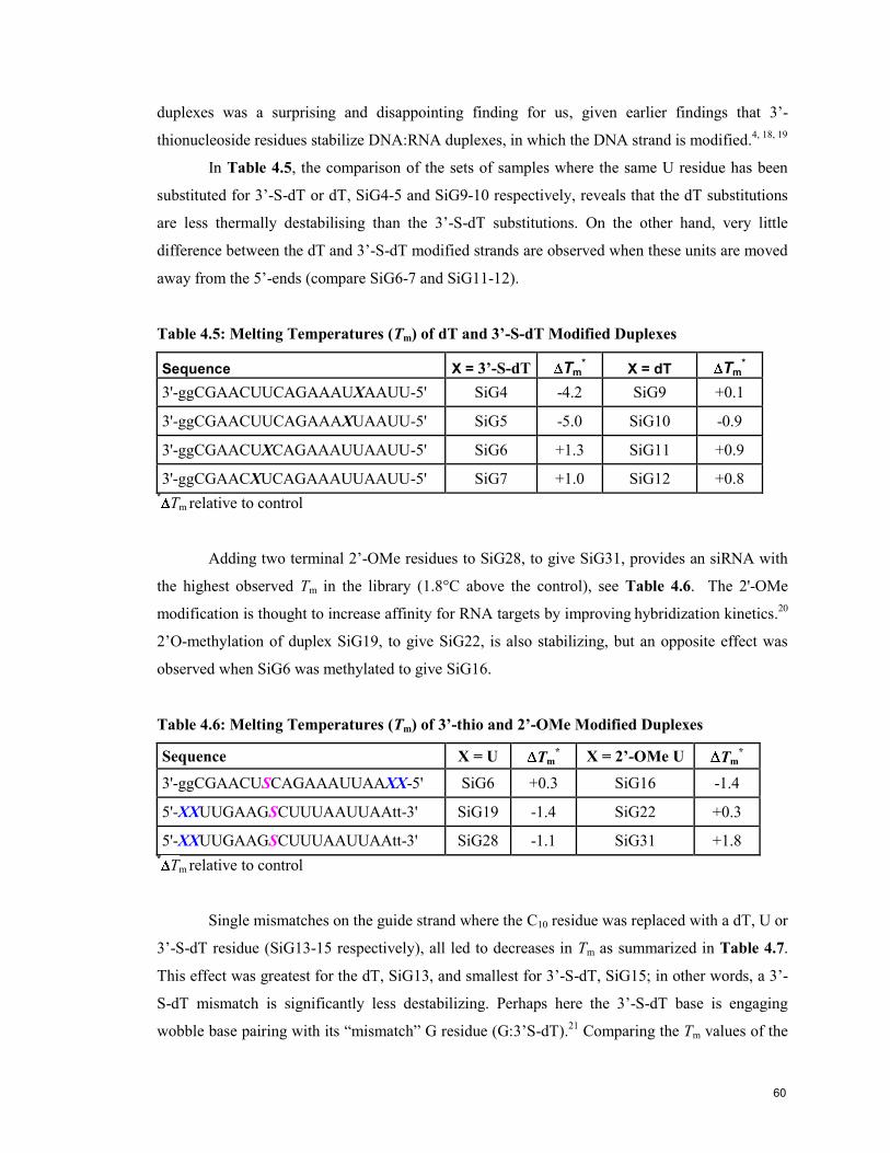
duplexes was a surprising and disappointing finding for us, given earlier findings that 3‟-
thionucleoside residues stabilize DNA:RNA duplexes, in which the DNA strand is modified.4, 18, 19
In Table 4.5, the comparison of the sets of samples where the same U residue has been
substituted for 3‟-S-dT or dT, SiG4-5 and SiG9-10 respectively, reveals that the dT substitutions
are less thermally destabilising than the 3‟-S-dT substitutions. On the other hand, very little
difference between the dT and 3‟-S-dT modified strands are observed when these units are moved
away from the 5‟-ends (compare SiG6-7 and SiG11-12).
Table 4.5: Melting Temperatures (Tm) of dT and 3’-S-dT Modified Duplexes
Sequence X = 3’-S-dT Tm*
X = dT Tm*
3'-ggCGAACUUCAGAAAUXAAUU-5' SiG4 -4.2 SiG9 +0.1
3'-ggCGAACUUCAGAAAXUAAUU-5' SiG5 -5.0 SiG10 -0.9
3'-ggCGAACUXCAGAAAUUAAUU-5' SiG6 +1.3 SiG11 +0.9
3'-ggCGAACXUCAGAAAUUAAUU-5' SiG7 +1.0 SiG12 +0.8 *
Tm relative to control
Adding two terminal 2‟-OMe residues to SiG28, to give SiG31, provides an siRNA with
the highest observed Tm in the library (1.8°C above the control), see Table 4.6. The 2'-OMe
modification is thought to increase affinity for RNA targets by improving hybridization kinetics.
20
2‟O-methylation of duplex SiG19, to give SiG22, is also stabilizing, but an opposite effect was
observed when SiG6 was methylated to give SiG16.
Table 4.6: Melting Temperatures (Tm) of 3’-thio and 2’-OMe Modified Duplexes
Sequence X = U Tm* X = 2’-OMe U Tm
*
3'-ggCGAACUSCAGAAAUUAAXX-5' SiG6 +0.3 SiG16 -1.4
5'-XXUUGAAGSCUUUAAUUAAtt-3' SiG19 -1.4 SiG22 +0.3
5'-XXUUGAAGSCUUUAAUUAAtt-3' SiG28 -1.1 SiG31 +1.8 *
Tm relative to control
Single mismatches on the guide strand where the C10 residue was replaced with a dT, U or
3‟-S-dT residue (SiG13-15 respectively), all led to decreases in Tm as summarized in Table 4.7.
This effect was greatest for the dT, SiG13, and smallest for 3‟-S-dT, SiG15; in other words, a 3‟-
S-dT mismatch is significantly less destabilizing. Perhaps here the 3‟-S-dT base is engaging
wobble base pairing with its “mismatch” G residue (G:3‟S-dT).21
Comparing the Tm values of the
60

C 3‟-S-dT mismatch at position 10, SiG15, to the U 3‟-S-dT substitution at position 9,
SiG6, we find the former to be slightly more destabilizing.
Table 4.7: Melting Temperatures (Tm) of Guide Strand Mismatches
*Tm
relative to control
In the interest of exploring the dynamics of RISC at the passenger strand cleavage site,22
we made several substitutions at this position. Substitutions with dT and the 3‟-S-dT were well
tolerated, SiG23 and SiG24 respectively, in terms of thermal stability but substitution with 2‟-F-
rU, SiG25, had a destabilizing effect.
There are no reports of 3‟-thio modified RNA duplexes in the literature. However,
Cosstick‟s group has measured the Tm values of 3‟-thio modified DNA (DNA*) hybridized to
complementary DNA or RNA. For DNA*:DNA duplexes, Cosstick et al. reported that the 3‟-
thio modification caused a decrease in Tm of ~1°C/modification compared to a native DNA:DNA
duplex. This finding was supported by experiments in our lab, although the observed effects were
smaller with the homopolymeric thymidine stands examined by Anna Lisa Tedeschi of our
laboratory.23
Cosstick‟s group also reported that 3‟-thio modifications in DNA*:RNA duplexes
resulted in an increase of ~1.4°C/modification relative to its native counterpart.17
Our data show
that 3‟-S-dT may have a stabilizing or destabilizing effect depending on its location in the
RNA:RNA duplex.
4.5 Analysis of Oligonucleotide Conformation by Circular Dichroism (CD) Spectroscopy
Circular dichroism (CD) spectroscopy provides useful information about the secondary
structure of oligonucleotide duplexes: the ability of nucleic acids to absorb circularly polarized
light, due to the chiral N-glycosidic bonds between the sugar and the nucleobases, increases when
they adopt a global helical formation.24
The CD instrument measures the molar ellipticity (differential absorbance of left and right
circularly polarized light) across a range of wavelengths, resulting in a signature curve for each
Sample Sequence Tm*
SiG6 U9 3‟-S-dT substitution 3'-ggCGAACUSCAGAAAUUAAUU-5' +0.3
SiG13 C10 dT mismatch 3'-ggCGAACUUtAGAAAUUCAUU-5' -8.5
SiG14 C10 U mismatch 3'-ggCGAACUUUAGAAAUUAAUU-5' -5.5
SiG15 C10 3‟-S-dT mismatch 3'-ggCGAACUUSAGAAAUUAAUU-5' -1.1
61
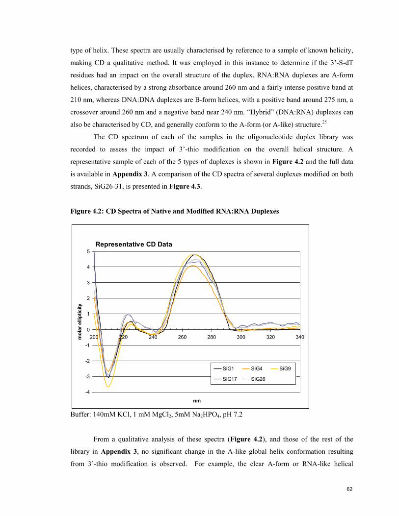
type of helix. These spectra are usually characterised by reference to a sample of known helicity,
making CD a qualitative method. It was employed in this instance to determine if the 3‟-S-dT
residues had an impact on the overall structure of the duplex. RNA:RNA duplexes are A-form
helices, characterised by a strong absorbance around 260 nm and a fairly intense positive band at
210 nm, whereas DNA:DNA duplexes are B-form helices, with a positive band around 275 nm, a
crossover around 260 nm and a negative band near 240 nm. “Hybrid” (DNA:RNA) duplexes can
also be characterised by CD, and generally conform to the A-form (or A-like) structure.25
The CD spectrum of each of the samples in the oligonucleotide duplex library was
recorded to assess the impact of 3‟-thio modification on the overall helical structure. A
representative sample of each of the 5 types of duplexes is shown in Figure 4.2 and the full data
is available in Appendix 3. A comparison of the CD spectra of several duplexes modified on both
strands, SiG26-31, is presented in Figure 4.3.
Figure 4.2: CD Spectra of Native and Modified RNA:RNA Duplexes
Representative CD Data
-4
-3
-2
-1
0
1
2
3
4
5
200 220 240 260 280 300 320 340
nm
mo
lar
ellip
tic
ity
SiG1 SiG4 SiG9
SiG17 SiG26
Buffer: 140mM KCl, 1 mM MgCl2, 5mM Na2HPO4, pH 7.2
From a qualitative analysis of these spectra (Figure 4.2), and those of the rest of the
library in Appendix 3, no significant change in the A-like global helix conformation resulting
from 3‟-thio modification is observed. For example, the clear A-form or RNA-like helical
62
formation of the control all-native RNA duplex, SiG1, is mirrored in several duplexes. These
include duplexes containing a 3‟-thio modification on the guide strand, SiG4, or on the passenger
strand, SiG17, a dT insert on the guide strand, SiG9, and 3‟-thio modifications on both the guide
and passenger strands, SiG26.
For most cases, the CD traces are nearly superimposable reflecting the uniformity of their
helical structures. The CD spectra of the doubly 3‟-thio modified duplexes where destabilization
was observed, SiG28-30, resemble that of the control, SiG1, except that their CD traces are less
intense around the 265 nm band, see Figure 4.3. This region is often associated with base
stacking. Likewise, duplexes containing mismatches were found to present a slight disruption of
the CD signature (Appendix 3), likely reflecting steric interactions with the opposing RNA
nucleotide strand.
Figure 4.3: CD Spectra of Guide and Passenger Strand Modified RNA:RNA Duplexes
-4
-3
-2
-1
0
1
2
3
4
5
200 220 240 260 280 300 320 340
wavelength (nm)
mo
lar
ellip
tic
ity
SiG1 SiG26 SiG27
SiG28 SiG29 SiG30
SiG31
4.6 Evaluation the Gene Silencing Activity of 3’-Thio Modified siRNA
The entire library of siRNA duplexes, Tables 4.1 to 4.3, was evaluated for the ability to
down-regulate the production of a target protein. A unique but practical method for doing this
involves using HeLa cells that have been modified to express the luciferase enzyme, taken from
the firefly genome, which emit light when exposed to the luciferin substrate.24
As this is a
property not found in the human genome, we are then able to directly measure the production of
this single protein, the target of the siRNA, without false signals from other proteins. These
assays were carried out in collaboration with Drs. Francis Robert and Jerry Pelletier
(Biochemistry Department, McGill University).
63

HeLa X1/5 cells, expressing the firefly luciferase gene, were maintained in supplemented
cell culture medium and seeded into 24-well plates. Cells were incubated with increasing amounts
of siRNA premixed with lipofectamine for twenty-four hours, at which point the cells were lysed,
exposed to a luciferin solution and the luminescence was measured.
As described above, earlier work with 3‟-S-dT units in DNA duplexes, including the
conformational work by Cosstick,24
had led us to hope that this modification would prove useful
in RNAi silencing assays. However, the 3‟-S-dT modification gave mixed results in the luciferase
knockdown assay, as seen in Figures 4.4 to 4.6. Luminescence, on the y-axis, represents the light
produced by the sample relative to that produced by untreated cells. Effective siRNA duplexes
result in lower luciferase production and thus have lower luminescence. That the luminescence
observed in the series dilutions of each sample reflects greater effects for more siRNA (dose
dependence) is a useful check that what we are observing is actually an RNAi effect.
Figure 4.4: RNAi Activity of Duplexes Modified Only on the Guide Strand
0.0
0.2
0.4
0.6
0.8
1.0
1.2
1.4
SiG1
control
SiG2 SiG3 SiG4 SiG5 SiG6 SiG7 SiG9 SiG10 SiG11 SiG12 SiG13 SiG14 SiG15 SiG16
0.4 nM
2 nM
10 nM
Comparing the RNAi efficacy of duplexes modified with 3‟-S-dT on the guide strand,
SiG2-7, and the dT modified guide strand series, SiG9-12, to the parent native siRNA duplex,
SiG1, in Figure 4.4, we see that 3‟-S-dT is poorly tolerated on the guide strand. The low RNAi
activity of SiG3-5 may be exacerbated by the fact that the 3‟-S-dT modifications in these
duplexes are found in the 5‟-proximal region shown to be associated with the surface of the Mid-
PIWI lobe of RISC,24
and the likely nucleation site for mRNA.26
When 3‟-S-dT on the guide
strand is combined with terminal 2‟-OMe residues, SiG16, the RNAi performance remained
lum
inescence
64
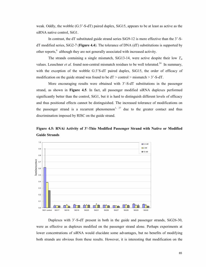
weak. Oddly, the wobble (G:3‟-S-dT) paired duplex, SiG15, appears to be at least as active as the
siRNA native control, SiG1.
In contrast, the dT substituted guide strand series SiG9-12 is more effective than the 3‟-S-
dT modified series, SiG2-7 (Figure 4.4). The tolerance of DNA (dT) substitutions is supported by
other reports,6 although they are not generally associated with increased activity.
The strands containing a single mismatch, SiG13-14, were active despite their low Tm
values. Leuschner et al. found non-central mismatch residues to be well tolerated.24
In summary,
with the exception of the wobble G:3‟S-dT paired duplex, SiG15, the order of efficacy of
modification on the guide strand was found to be dT > control > mismatch > 3‟-S-dT.
More encouraging results were obtained with 3‟-S-dT substitutions in the passenger
strand, as shown in Figure 4.5. In fact, all passenger modified siRNA duplexes performed
significantly better than the control, SiG1, but it is hard to distinguish different levels of efficacy
and thus positional effects cannot be distinguished. The increased tolerance of modifications on
the passenger strand is a recurrent phenomenon1, 27
due to the greater contact and thus
discrimination imposed by RISC on the guide strand.
Figure 4.5: RNAi Activity of 3’-Thio Modified Passenger Strand with Native or Modified
Guide Strands
0.0
0.1
0.2
0.3
0.4
0.5
0.6
0.7
0.8
0.9
1.0
SiG1 control SiG17 SiG18 SiG19 SiG20 SiG21 SiG26 SiG27 SiG28 SiG29 SiG30
0.4 nM
2 nM
10 nM
Duplexes with 3‟-S-dT present in both in the guide and passenger strands, SiG26-30,
were as effective as duplexes modified on the passenger strand alone. Perhaps experiments at
lower concentrations of siRNA would elucidate some advantages, but no benefits of modifying
both strands are obvious from these results. However, it is interesting that modification on the
lum
ines
cen
ce
65
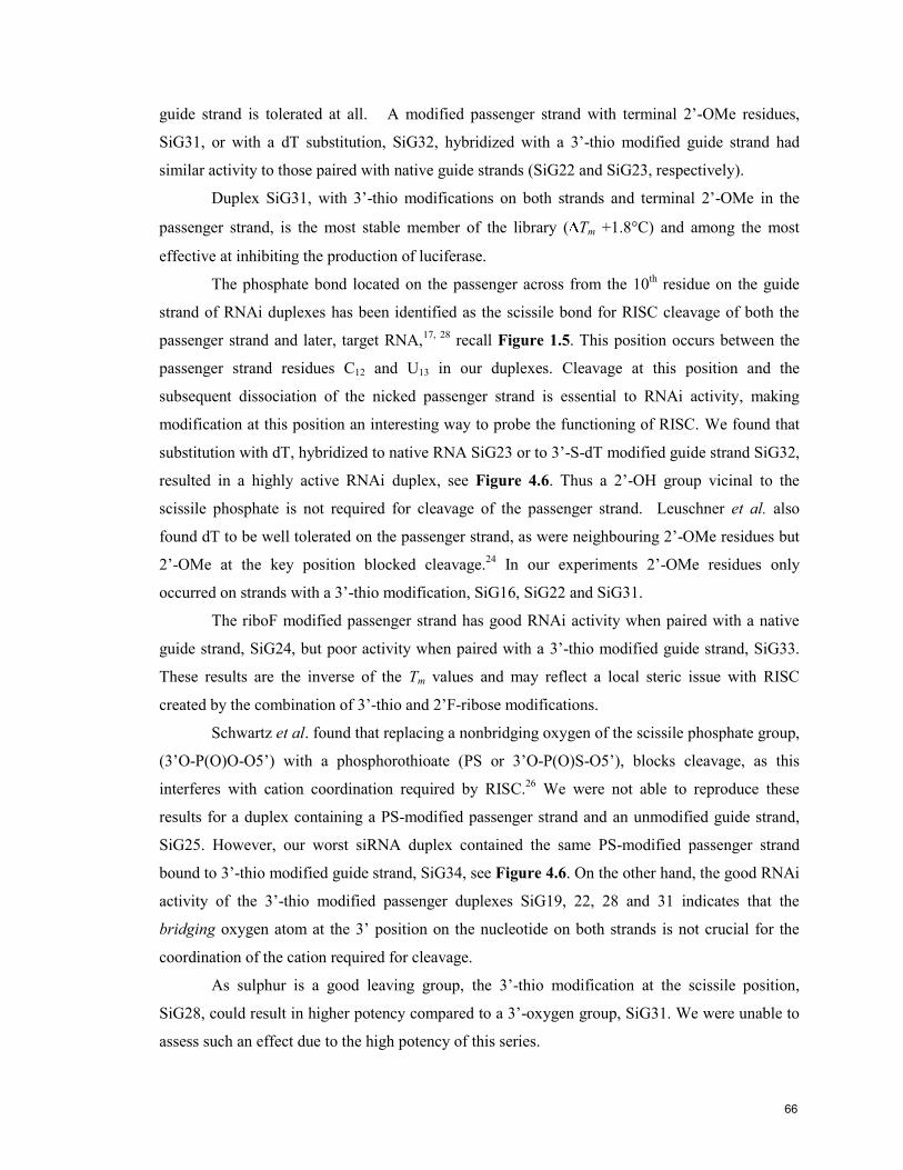
guide strand is tolerated at all. A modified passenger strand with terminal 2‟-OMe residues,
SiG31, or with a dT substitution, SiG32, hybridized with a 3‟-thio modified guide strand had
similar activity to those paired with native guide strands (SiG22 and SiG23, respectively).
Duplex SiG31, with 3‟-thio modifications on both strands and terminal 2‟-OMe in the
passenger strand, is the most stable member of the library ( Tm +1.8°C) and among the most
effective at inhibiting the production of luciferase.
The phosphate bond located on the passenger across from the 10th residue on the guide
strand of RNAi duplexes has been identified as the scissile bond for RISC cleavage of both the
passenger strand and later, target RNA,17, 28
recall Figure 1.5. This position occurs between the
passenger strand residues C12 and U13 in our duplexes. Cleavage at this position and the
subsequent dissociation of the nicked passenger strand is essential to RNAi activity, making
modification at this position an interesting way to probe the functioning of RISC. We found that
substitution with dT, hybridized to native RNA SiG23 or to 3‟-S-dT modified guide strand SiG32,
resulted in a highly active RNAi duplex, see Figure 4.6. Thus a 2‟-OH group vicinal to the
scissile phosphate is not required for cleavage of the passenger strand. Leuschner et al. also
found dT to be well tolerated on the passenger strand, as were neighbouring 2‟-OMe residues but
2‟-OMe at the key position blocked cleavage.24
In our experiments 2‟-OMe residues only
occurred on strands with a 3‟-thio modification, SiG16, SiG22 and SiG31.
The riboF modified passenger strand has good RNAi activity when paired with a native
guide strand, SiG24, but poor activity when paired with a 3‟-thio modified guide strand, SiG33.
These results are the inverse of the Tm values and may reflect a local steric issue with RISC
created by the combination of 3‟-thio and 2‟F-ribose modifications.
Schwartz et al. found that replacing a nonbridging oxygen of the scissile phosphate group,
(3‟O-P(O)O-O5‟) with a phosphorothioate (PS or 3‟O-P(O)S-O5‟), blocks cleavage, as this
interferes with cation coordination required by RISC.26
We were not able to reproduce these
results for a duplex containing a PS-modified passenger strand and an unmodified guide strand,
SiG25. However, our worst siRNA duplex contained the same PS-modified passenger strand
bound to 3‟-thio modified guide strand, SiG34, see Figure 4.6. On the other hand, the good RNAi
activity of the 3‟-thio modified passenger duplexes SiG19, 22, 28 and 31 indicates that the
bridging oxygen atom at the 3‟ position on the nucleotide on both strands is not crucial for the
coordination of the cation required for cleavage.
As sulphur is a good leaving group, the 3‟-thio modification at the scissile position,
SiG28, could result in higher potency compared to a 3‟-oxygen group, SiG31. We were unable to
assess such an effect due to the high potency of this series.
66
Figure 4.6: RNAi Activity of Modified Passenger Strand with Native or Modified Guide
Strands
0.0
0.2
0.4
0.6
0.8
1.0
1.2
SiG1 control SiG19 SiG22 SiG23 SiG24 SiG25 SiG28 SiG31 SiG32 SiG33 SiG34
0.4 nM
2 nM
10 nM
Flexibility can be very important in the confined space of enzymatic active sites.
Therefore, the anticipated increased flexibility of 3‟-deoxy-3‟-thiothymidine compared to the
native ribo U may be the only reason for the increased efficacy of 3‟-S-dT residues on the
passenger strand. However, the decreases in Tm are also due to this enhanced flexibility. Duplexes
with terminal 2‟-OMe residues, SiG22, or a dT substitution, SiG23, were equally effective.
In summary, 3‟-thio modifications in the passenger strand were found to be the most
effective in terms of RNAi activity (surpassing the native strand), whether paired with a native or
3‟-thio modified guide strand.
4.7 Conclusion
The thermal stability of RNA:RNA duplexes containing a single 3‟-deoxy-3‟-
thiothymidine (3‟-S-dT) residue was observed to be highly dependent on the location of the
modification. The RNAi activity of RNA*:RNA duplexes modified on the guide strand with ,3‟-
deoxy-3‟-thiothymidine was lower than that of the control duplex, but those modified on the
passenger strand were more active. Duplexes containing a 3‟-S-dT residue on both strands were
as active as those modified only on the passenger strand. Individual dT substitutions, single
mismatches or 2‟-OMe terminal residues did not have detrimental effects on RNAi activity.
Curiously, a guide strand modified siRNA duplex containing a G:3‟-S-dT “mismatch” was as
active as the unmodified duplex control. Comparison of the Tm data of this duplex to other
67
mismatch controls indicated that the 3‟-S-dT residue likely engages in wobble base pairing with a
G residue on the passenger strand.
Modification at the RISC cleavage position on the passenger strand resulted in an
interesting addition to the work of Schwartz et al. on the mechanism of RISC cleavage.26
As the
3‟-S-dT residue at this position did not have a detrimental effect on RNAi activity, it suggests that
the 3‟-nonbridging oxygen is not involved in the coordination of the cation.
4.8 References
1 Dykxhoorn, D. M.; Lieberman, J., Running Interference: Prospects and Obstacles to Using Small Interfering RNAs as Small Molecule Drugs. Annual Review of Biomedical Engineering 2006, 8, 377-402.
2 Patureau, B. Induction of RNase H Activity by Arabinose-Peptide Nucleic Acids. M.Sc. Thesis, McGill University, Montreal, 2005.
3 Mangos, M. Factors Governing the Design, Selection and Cleavage of Sugar-Modified Duplexes by Ribonuclease H. Ph.D. Thesis, McGill University, Montreal, 2005.
4 Tedeschi, A. L. Synthesis and Biological Evaluation of Oligonucleotides Containing 3'-
and 5'-S-Phosphorthiolate Internucleotide Linkages. M.Sc. Thesis, McGill University, Montreal, 2004.
5 Buckingham, J.; Sabbagh, G.; Brazier, J.; Fisher, J.; Cosstick, R., Control of DNA Conformation Using 3'-S-Phosphorothiolate-Modified Linkages. Nucleosides, Nucleotides and Nucleic Acids 2005, 24, (5), 491-495.
6 Beevers, A. P. G.; Fettes, K. J.; Sabbagh, G.; Murad, F. K.; Arnold, J. R. P.; Cosstick, R.;
Fisher, J., NMR and UV studies of 3'-S-phosphorothiolate modified DNA in a DNA:RNA hybrid dodecamer duplex; implications for antisense drug design. Organic & Biomolecular Chemistry 2003, 2, (1), 114-119.
7 Thibaudeau, C.; Plavec, J.; Carg, N.; Papchikhin, A.; Chattopadhyaya, J., How Does the Electronegativity of the Substituent Dictate the Strength of the Gauche Effect? J. Am. Chem. Soc. 1994, 116, 4038-4043.
8 Hantz, E.; Laruea, V.; Ladama, P.; Moyeca, L. L.; Gouyetteb, C.; Dinhb, T. H., Solution
conformation of an RNA–DNA hybrid duplex containing a pyrimidine RNA strand and a purine DNA strand. International Journal of Biological Macromolecules 2001, 28, (4), 273-284.
9 Jayakumar, H. K.; Buckingham, J. L.; Brazier, J. A.; Berry, N. G.; Cosstick, R.; Fisher, J.,
NMR studies of the conformational effect of single and double 3'-S-phosphorothiolate substitutions within deoxythymidine trinucleotides. Magn. Reson. Chem. 2007, 45, (4), 340-345.
10 Bentley, J.; Brazier, J. A.; Fisher, J.; Cosstick, R., Duplex stability of DNA·DNA and DNA·RNA duplexes containing 3'-S-phosphorothiolate linkages. Organic & Biomolecular Chemistry 2007, 5, 3698–3702.
11 Beevers, A. P. G.; Fettes, K. J.; O’Neil, I. A.; Roberts, S. M.; Arnold, J. R. P.; Cosstick, R.;
Fisher, J., Probing the effect of a 3A-S-phosphorothiolate link on the conformation of a DNA:RNA hybrid; implications for antisense drug design. Chemical Communications 2002, 14, 1458–1459.
12 Lauritsena, A.; Wengel, J., Oligodeoxynucleotides containing amide-linked LNA-type dinucleotides: synthesis and high-affinity nucleic acid hybridization. Chemical Communications 2002, (5), 530-531.
13 Nielsen, K. E.; Singh, S. K.; Wengel, J.; Jacobsen, J. P., Solution structure of an LNA hybridized to DNA: NMR study of the d(CTLGCTLTLCTLGC):d(GCAGAAGCAG) duplex containing four locked nucleotides. Bioconjugate Chemistry 2000, 11, 228-238.
14 Obika, S.; Nanbu, D.; Hari, Y.; Andoh, J.-i.; Morio, K.-i.; Doi, T.; Imanishi, T., Stability and structural features of the duplexes containing nucleoside analogues with a fixed N-type conformation, 2'-O,4'- C-methyleneribonucleosides. Tetrahedron Lett. 1998, 39, 5401-5404.
68

15 Epple, C.; Leumann, C., Bicyclo(3.2.19-DNA, a new DNA analog with a rigid backbone and flexibly linked bases: pairing properties with complementary DNA. Chemistry & Biology 1998, 5, 209-216.
16 Majlessi, M.; Nelson, N. C.; Becker, M. M., Advantages of 2'-O-methyl oligoribonucleotide probes for detecting RNA targets. Nucleic Acids Res. 1998, 26, 2224–2229.
17 Elbashir, S.; Martinez, J.; Patkaniowska, A.; Lendeckel, W.; Tuschl, T., Functional
anatomy of siRNAs for mediating efficient RNAi in Drosophila melanogaster embryo lysate. EMBO J. 2001, 20, 6877–88.
18 Brahms, J.; Brahms, S., Circular Dichroism of Nucleic Acids. In Fine Structure of Proteins and Nucleic Acids, ed.; Fasman, G. D., Timasheff, S. N., 'Ed.'^'Eds.' Dekker: 1970; 'Vol.' 4, p^pp 79.
19 Gray, D. M.; Hung, S. H.; Johnson, K. H., Absorption and circular dichroism spectroscopy of nucleic acid duplexes and triplexes. Methods Enzymol. 1995, 246, 19-34.
20 Elbashir, S. M.; Harborth, J.; Lendeckel, W.; Yalcin, A.; Weber, K.; Tuschl, T., Duplexes of 21-nucleotide RNAs mediate RNA interference in cultured mammalian cells. Nature (London) 2001, 411, 494-498.
21 Salazar, M.; al., e., The DNA strand in DNA.RNA hybrid duplexes is neither B-form nor A-form in solution. Biochemistry 1993, 32, (16), 4207-15.
22 Birmingham, A.; Anderson, E. M.; Reynolds, A.; Ileley-Tyree, D.; Leake, D.; Fedorov, Y.; Baskerville, S.; Maksimova, E.; Robinson, K.; Karpilow, J.; al., e., 30 UTR seed matches, but not overall identity, are associated with RNAi off-targets. Nat. Methods 2006, 3, 199–204.
23 Yuan, Y.-R.; Pei, Y.; Ma, J.-B.; Kuryavyi, V.; Zhadina, M.; Meister, G.; Chen, H.-Y.; Dauter, Z.; Tuschl, T.; Patel, D. J., Crystal structure of A. aeolicus Argonaute, a site-specific DNA-guided endoribonuclease, provides insights into RISC-mediated mRNA cleavage. Mol. Cell 2005, 19, 405–419.
24 Leuschner, P. J. F.; Ameres, S. L.; Kueng, S.; Martinez, J., Cleavage of the siRNA passenger strand during RISC assembly in human cells. EMBO reports 2006, 7, (3), 314-320.
25 Hohjoh, H., RNA interference (RNAi) induction with various types of synthetic oligonucleotide duplexes in cultured human cells. FEBS Lett. 2002, 521, (1-3), 195-199.
26 Schwarz, D.; Tomari, Y.; Zamore, P., The RNA-induced silencing complex is a Mg2+-dependent endonuclease. Current Biology 2004, 14, (9), 787-91.
27 Chiu, Y. L.; Rana, T. M., siRNA function in RNAi: A chemical modification analysis. RNA 2003, 9, 1034-1048.
28 Elbashir, S. M.; Lendeckel, W.; Tuschl, T., RNA interference is mediated by 21-and 22-nucleotide RNAs. Genes Dev. 2001, 15, 188-200.
69
Contribution to Knowledge
This thesis reports the first incorporation of 3’-thionucleosides into RNA
oligonucleotides, and examines their physicochemical and biological properties. Improvements to
the synthesis of 3’-deoxy-3’-thiothymidine (3’-S-dT), particularly the use of dithiotreitol (DTT) to
reduce the formation of disulfide bonds when revealing the 3’-thiol group, are described. In
addition, an in situ method for the preparation of nucleosides 3’-S-phosphorothioamidite
derivatives was developed to increase monomer coupling yields. The 3’-phosphorothioamidite
was found to be stable when stored as a dry foam in the freezer for at least 2 months or 24h at
room temperature in CH2Cl2.
Adjustments were made to the solid-phase synthesis cycle, particularly the coupling and
oxidation steps, to obtain sufficient amounts of the 3’-S modified RNA strands for the
characterisation and biological assays of a small library of native and modified RNA:RNA
(siRNA) duplexes.
Thermal denaturation studies revealed that the incorporation of one 3’-S-dT insert in
RNA:RNA duplexes can be either destabilizing or stabilizing depending on their location within
the duplex. There where cases when one or two 3’-S-dT incorporations, or chimeric
modifications (e.g., both 3’-S-dT and 2’-OMe rN) provided duplexes with greater thermal
stability relative to the control native siRNA duplex. CD signatures of the modified duplexes
were indicative of an A-like helix, as expected, although some perturbation in base stacking
interactions was detected in certain duplexes.
Duplexes modified with 3’-S-dT residues on the guide strand generally led to a significant
reduction in siRNA gene silencing activity, although a guide strand modified duplex containing a
G:3’S-dT mismatch exhibited similar activity to the control (unmodified) siRNA duplex. The Tm
data of this mismatch duplex suggests that the G and 3’S-dT bases are engaged in wobble base
pairing interactions. Duplexes containing the 3’S-dT modification on the passenger strand or on
both strands perform rather well, many surpassing the activity of the native siRNA control. Other
substitutions such as dT, or terminal 2’-OMe ribonucleotide residues did not have detrimental
effects on RNAi activity.
Specific substitutions at the scissile position of the passenger strand of siRNA duplexes
indicate that the non-bridging 3’-oxygen is not involved in the coordination of the metal cation for
RISC-induced cleavage.
70

Appendix 1: NMR Spectra of Selected Nucleosides
3’-acetyl-2’,3’-dideoxy-5’-monomethoxytrityl-Thymidine
2’,3’-dideoxy-3’-thio-5’-monomethoxytrityl-Thymidine
O
SH
MMTO
NH
ON
O
3.6
O
SAc
MMTO
NH
ON
O
3.5
71

2’,3’-dideoxy-3’-thiophosphoramidite-5’-monomethoxytrityl-Thymidine
O
S
MMTO
NH
ON
O
3.8
P N(iPr)2
OCN
72

Appendix 2: UV/Thermal Dissociation Profiles of RNA:RNA Duplexes
Figure A2.1: Tm Spectra of Guide Strand Modified RNA:RNA Duplexes
0.98
1.00
1.02
1.04
1.06
1.08
1.10
1.12
1.14
1.16
15 25 35 45 55 65 75
Temperature (°C)
Ab
so
rba
nc
eSiG1 SiG2
SiG3 SiG4
SiG5 SiG6*
SiG9 SiG10
SiG11 SiG12
*Data for SiG6 from the second heating ramp.
Figure A2.2: Tm Spectra of Guide Strand Mismatch RNA:RNA Duplexes
0.98
1.00
1.02
1.04
1.06
1.08
1.10
1.12
1.14
15 25 35 45 55 65 75
Temperature (°C)
Ab
so
rban
ce
SiG1 SiG13
SiG14 SiG15
73
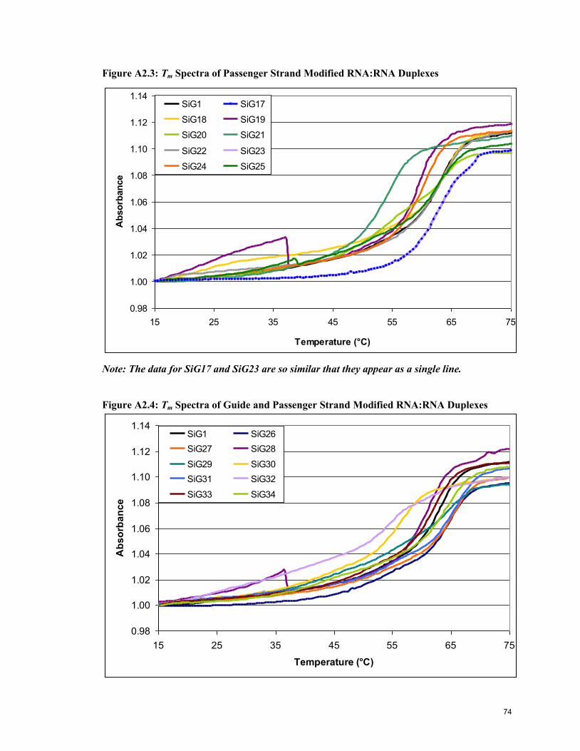
Figure A2.3: Tm Spectra of Passenger Strand Modified RNA:RNA Duplexes
0.98
1.00
1.02
1.04
1.06
1.08
1.10
1.12
1.14
15 25 35 45 55 65 75
Temperature (°C)
Ab
so
rban
ce
SiG1 SiG17
SiG18 SiG19
SiG20 SiG21
SiG22 SiG23
SiG24 SiG25
Note: The data for SiG17 and SiG23 are so similar that they appear as a single line.
Figure A2.4: Tm Spectra of Guide and Passenger Strand Modified RNA:RNA Duplexes
0.98
1.00
1.02
1.04
1.06
1.08
1.10
1.12
1.14
15 25 35 45 55 65 75
Temperature (°C)
Ab
so
rba
nc
e
SiG1 SiG26
SiG27 SiG28
SiG29 SiG30
SiG31 SiG32
SiG33 SiG34
74
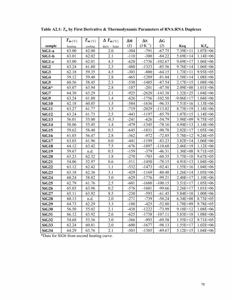
Table A2.1: Tm by First Derivative & Thermodynamic Parameters of RNA:RNA Duplexes
sample
Tm (°C)
heating
Tm (°C)
cooling
Tm (°C)
deriv – hypo
H
(J)
S
(J·K−1
)
G
(J) Keq KTm
SiG1-a 63.00 62.00 2.0 -304 -791 -67.75 7.39E+11 1.07E+06
SiG1-b 63.01 62.02 2.1 -110 -300 -84.22 5.69E+14 1.14E+06
SiG1-c 63.00 62.01 4.3 -620 -1736 -102.67 9.69E+17 1.06E+06
SiG2 63.24 61.80 2.3 -480 -1323 -85.56 9.76E+14 1.06E+06
SiG3 62.18 59.35 4.5 -303 -800 -64.15 1.73E+11 9.93E+05
SiG4 59.12 59.40 2.8 -465 -1289 -81.04 1.58E+14 1.08E+06
SiG5 60.56 58.45 2.3 -530 -1485 -87.54 2.17E+15 1.08E+06
SiG6* 65.07 63.94 2.8 -107 -201 -47.50 2.09E+08 1.01E+06
SiG7 64.30 63.29 2.1 -925 -2620 -143.38 1.32E+25 1.04E+06
SiG9 63.24 61.80 1.4 -626 -1756 -102.50 -9.06E+17 1.04E+06
SiG10 62.18 60.85 1.5 -584 -1636 -96.33 7.51E+16 1.13E+06
SiG11 63.27 61.77 3.5 -719 -2029 -113.82 8.73E+19 1.14E+06
SiG12 63.24 61.73 2.5 -443 -1197 -85.79 1.07E+15 1.14E+06
SiG13 56.01 55.00 -0.3 -241 -626 -54.79 3.98E+09 9.75E+05
SiG14 58.06 55.45 1.4 -479 -1345 -78.16 4.94E+13 1.14E+06
SiG15 59.62 58.40 0.5 -645 -1831 -90.78 2.02E+17 1.05E+06
SiG16 61.03 56.47 2.8 -362 -972 -72.85 5.78E+12 9.24E+05
SiG17 63.03 61.96 0.0 -441 -1199 -83.23 3.82E+14 1.06E+06
SiG18 64.12 63.42 7.5 -676 -1897 -110.68 2.46E+19 1.12E+06
SiG19 59.67 n.d. 0.5 -159 -379 -46.31 1.30E+08 9.71E+05
SiG20 63.23 62.32 1.8 -270 -703 -60.35 3.75E+10 9.67E+05
SiG21 54.06 52.97 0.6 -511 -1450 -78.15 4.91E+13 1.04E+06
SiG22 63.12 62.42 1.1 -532 -1473 -92.41 1.55E+16 1.04E+06
SiG23 63.18 62.36 3.1 -429 -1169 -80.48 1.26E+14 1.05E+06
SiG24 60.24 58.82 3.0 -629 -1776 -99.21 2.40E+17 1.10E+06
SiG25 62.79 61.76 2.5 -601 -1680 -100.15 3.51E+17 1.05E+06
SiG26 65.03 63.96 0.2 -576 -1601 -99.66 2.26E+17 1.01E+06
SiG27 65.11 63.92 8.5 -230 -593 -61.45 5.84E+10 1.00E+06
SiG28 60.13 n.d. 2.0 -271 -739 -50.24 6.34E+08 8.73E+05
SiG29 64.73 62.29 3.3 -180 -425 -52.80 1.78E+09 9.78E+05
SiG30 56.50 55.02 2.1 -438 -1222 -73.99 9.18E+12 1.08E+06
SiG31 66.12 63.92 2.6 -625 -1738 -107.11 5.83E+18 1.08E+06
SiG32 54.68 53.36 3.0 -366 -993 -69.58 1.55E+12 9.71E+05
SiG33 62.24 60.81 2.0 -600 -1677 -98.13 1.55E+17 1.02E+06
SiG34 64.29 63.76 2.1 -503 -1385 -89.67 5.12E+15 1.04E+06
*Data for SiG6 from second heating curve.
75
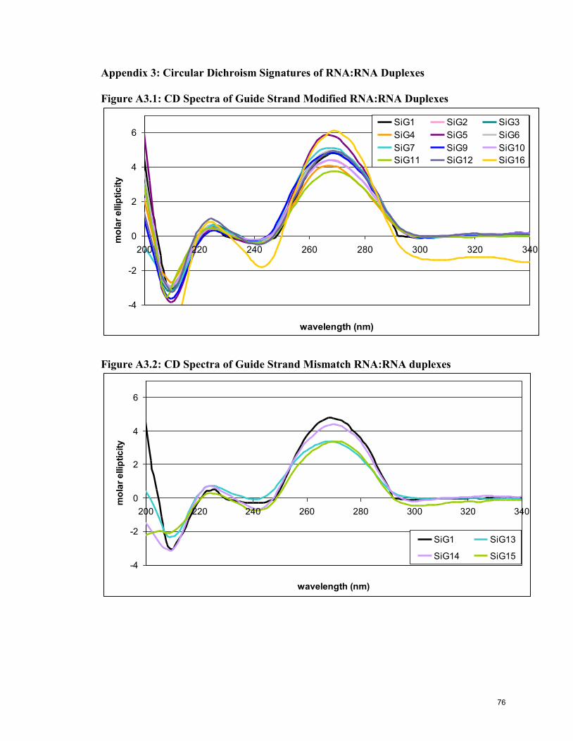
Appendix 3: Circular Dichroism Signatures of RNA:RNA Duplexes
Figure A3.1: CD Spectra of Guide Strand Modified RNA:RNA Duplexes
-4
-2
0
2
4
6
200 220 240 260 280 300 320 340
wavelength (nm)
mo
lar
ellip
tic
ity
SiG1 SiG2 SiG3
SiG4 SiG5 SiG6
SiG7 SiG9 SiG10
SiG11 SiG12 SiG16
Figure A3.2: CD Spectra of Guide Strand Mismatch RNA:RNA duplexes
-4
-2
0
2
4
6
200 220 240 260 280 300 320 340
wavelength (nm)
mo
lar
ellip
tic
ity
SiG1 SiG13
SiG14 SiG15
76

Figure A3.3: CD Spectra of Passenger Strand Modified RNA:RNA DCuplexes
-4
-3
-2
-1
0
1
2
3
4
5
6
200 220 240 260 280 300 320 340
wavelength (nm)
mo
lar
ellip
tic
ity
SiG1 SiG17 SiG18
SiG19 SiG20 SiG21
SiG22 SiG23 SiG24
SiG25
Figure A3.4: CD Spectra of Guide and Passenger Strand Modified RNA:RNA
Duplexes
-4
-3
-2
-1
0
1
2
3
4
5
200 220 240 260 280 300 320 340
wavelength (nm)
mo
lar
ellip
tic
ity
SiG1 SiG26 SiG27
SiG28 SiG29 SiG30
SiG31 SiG32 SiG33
SiG34
77