Syncope in the Emergency Department (ED) · 1 | P a g e Syncope in the Emergency Department (ED)...
11
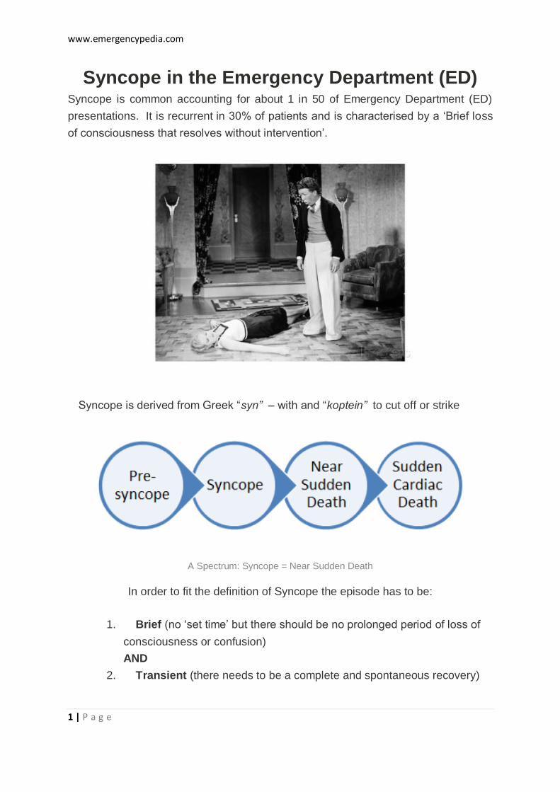
www.emergencypedia.com 1 | Page Syncope in the Emergency Department (ED) Syncope is common accounting for about 1 in 50 of Emergency Department (ED) presentations. It is recurrent in 30% of patients and is characterised by a ‘Brief loss of consciousness that resolves without intervention’. Syncope is derived from Greek “ syn” – with and “koptein” to cut off or strike A Spectrum: Syncope = Near Sudden Death In order to fit the definition of Syncope the episode has to be: 1. Brief (no ‘set time’ but there should be no prolonged period of loss of consciousness or confusion) AND 2. Transient (there needs to be a complete and spontaneous recovery)
Transcript of Syncope in the Emergency Department (ED) · 1 | P a g e Syncope in the Emergency Department (ED)...
www.emergencypedia.com
1 | P a g e
Syncope in the Emergency Department (ED) Syncope is common accounting for about 1 in 50 of Emergency Department (ED)
presentations. It is recurrent in 30% of patients and is characterised by a ‘Brief loss
of consciousness that resolves without intervention’.
Syncope is derived from Greek “syn” – with and “koptein” to cut off or strike
A Spectrum: Syncope = Near Sudden Death
In order to fit the definition of Syncope the episode has to be:
1. Brief (no ‘set time’ but there should be no prolonged period of loss of
consciousness or confusion)
AND
2. Transient (there needs to be a complete and spontaneous recovery)
www.emergencypedia.com
2 | P a g e
There is a ‘spectrum’ from pre syncope to syncope and from near-death to sudden
death. The presentation of Syncope to the ED can be an opportunity to diagnose
life-threatening underlying Cardiac Abnormalities. Patients and bystanders may
describe Syncope as a “blackout”, a “faint”, a “funny turn” or even a “seizure”.
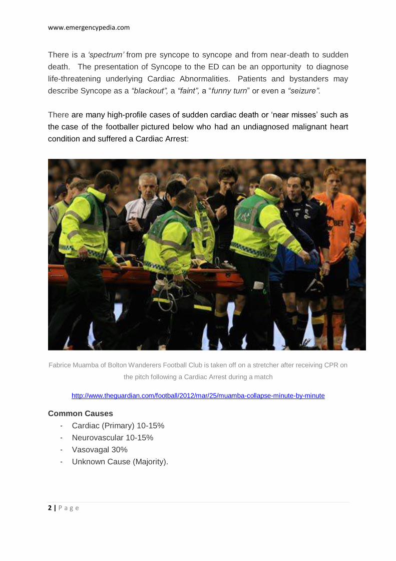
There are many high-profile cases of sudden cardiac death or ‘near misses’ such as
the case of the footballer pictured below who had an undiagnosed malignant heart
condition and suffered a Cardiac Arrest:
Fabrice Muamba of Bolton Wanderers Football Club is taken off on a stretcher after receiving CPR on
the pitch following a Cardiac Arrest during a match
http://www.theguardian.com/football/2012/mar/25/muamba-collapse-minute-by-minute
Common Causes
- Cardiac (Primary) 10-15%
- Neurovascular 10-15%
- Vasovagal 30%
- Unknown Cause (Majority).

www.emergencypedia.com
3 | P a g e
In true Syncope the important ‘rule out’ conditions to consider are generally
“Cardiac” rather than Neurological. ‘Stroke’ and other neurological problems
generally don’t cause true Syncope as defined above.
Pathogenesis of Syncope
Syncope is manifested by a short-lived period of Hypoperfusion which in turn leads
to a loss of consciousness due to neurological dysfunction affecting the cerebral
hemispheres and reticular activating system (RAS).
Factors that maintain cerebral perfusion include:
Cardiac output
Systemic vascular resistance
Mean arterial pressure
Intravascular volume
Cerebrovascular resistance with intrinsic auto-regulation
Metabolic regulation
Diagnosis of Syncope
There is a vast list of potential causes of Syncope. A list of 50 causes is probably
not really that helpful and would be difficult and impractical to memorise anyway. In
the Emergency Department, it is probably most important to make a judicious
assessment of the patient in order to rule in life threatening causes such as cardiac
arrhythmia and rule syncope ‘mimics’ such as Seizures. The two key steps are
confirming the patient indeed has true ‘Syncope’ (as per the definitions above) and if
they do have Syncope, is there are underlying ‘commonly deadly’ diagnosis?
While an alternative diagnosis (CVA, TIA, Seizure) should be considered and looked
for on neurological examination, they are unlikely to present with isolated syncope.
Diagnosis of neurological disorders in the ED are usually made from other features
of the presentation. Vascular Catastrophes such as Aortic Dissection may present
with syncope and it is important to consider these in the assessment (see the “Rule
of 15’s” below)
www.emergencypedia.com
4 | P a g e
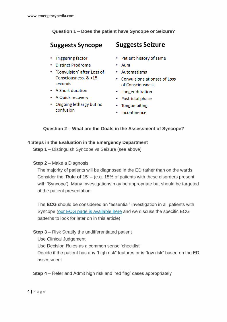
Question 1 – Does the patient have Syncope or Seizure?
Question 2 – What are the Goals in the Assessment of Syncope?
4 Steps in the Evaluation in the Emergency Department
Step 1 – Distinguish Syncope vs Seizure (see above)
Step 2 – Make a Diagnosis
The majority of patients will be diagnosed in the ED rather than on the wards
Consider the ‘Rule of 15‘ – (e.g. 15% of patients with these disorders present
with ‘Syncope’). Many Investigations may be appropriate but should be targeted
at the patient presentation
The ECG should be considered an “essential” investigation in all patients with
Syncope (our ECG page is available here and we discuss the specific ECG
patterns to look for later on in this article)
Step 3 – Risk Stratify the undifferentiated patient
Use Clinical Judgement
Use Decision Rules as a common sense ‘checklist’
Decide if the patient has any “high risk” features or is “low risk” based on the ED
assessment
Step 4 – Refer and Admit high risk and ‘red flag’ cases appropriately

www.emergencypedia.com
5 | P a g e
The Rule of 15%’s
Question 3 – What are the options for evidence based decision-making?
Decision Rules and Clinical Policies have been widely used in the evaluation of
Syncope.
Clinical Rules are useful for the following:
1. As a Checklist – a ‘check-listing’ of your own assessment of the
Syncope patient
2. Educating junior doctors and medical students about the key features of
a syncope assessment
3. For documentation of the Emergency evaluation in a structured manner
The Disadvantage of Clinical Decision Rules are that may rules can be
oversimplified or very broad (i.e. they may be under or overly sensitive). They are
unlikely to be as accurate as an experienced medical practitioner especially in
complex presentations such as Syncope. The rules described below are best used
to support clinical decision-making and documentation as described above
www.emergencypedia.com
6 | P a g e
Syncope Rules
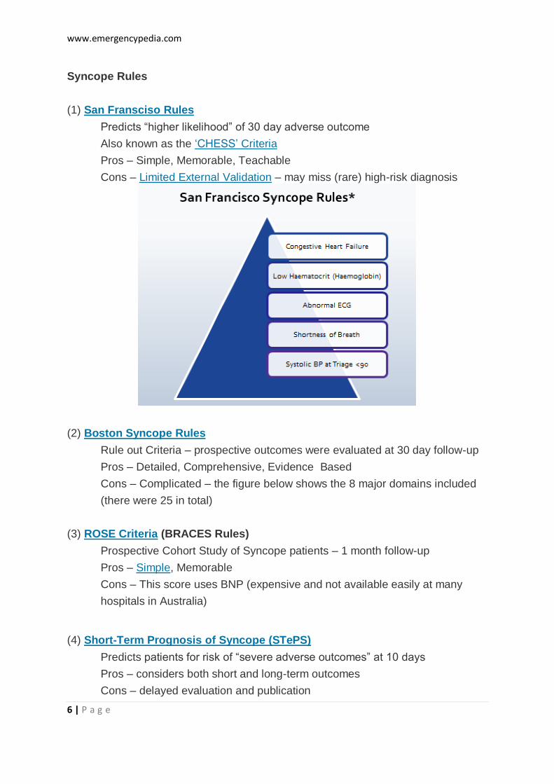
(1) San Fransciso Rules
Predicts “higher likelihood” of 30 day adverse outcome
Also known as the ‘CHESS’ Criteria
Pros – Simple, Memorable, Teachable
Cons – Limited External Validation – may miss (rare) high-risk diagnosis
(2) Boston Syncope Rules
Rule out Criteria – prospective outcomes were evaluated at 30 day follow-up
Pros – Detailed, Comprehensive, Evidence Based
Cons – Complicated – the figure below shows the 8 major domains included
(there were 25 in total)
(3) ROSE Criteria (BRACES Rules)
Prospective Cohort Study of Syncope patients – 1 month follow-up
Pros – Simple, Memorable
Cons – This score uses BNP (expensive and not available easily at many
hospitals in Australia)
(4) Short-Term Prognosis of Syncope (STePS)
Predicts patients for risk of “severe adverse outcomes” at 10 days
Pros – considers both short and long-term outcomes
Cons – delayed evaluation and publication
www.emergencypedia.com
7 | P a g e
Question 4 – What are the high risk features associated with Syncope?
Red Flags in the History
1. Old Age
2. Cardiac Disease and/or Syncope on exertion
3. Syncope while lying down (recumbent)
4. Family History (Sudden Death)
5. Positional Symptoms (may be more reliable than Orthostatic Blood Pressure and
Heart Rate)
6. Recurrent Syncope
7. Prolonged Loss of Consciousness
8. Medications that affect cardiac conduction
Examination Key Steps
1. Cardiac Exam
a. Blood Pressure
b. Pulse
c. Murmurs
2. Abdominal Exam
a. Ectopic Pregnancy
b. AAA
c. PR exam and Faecal Occult Blood (FOB) Testing
3. Neurological exam
a. Cranial Nerves, Limbs and Cerebellum
Investigations
1. ECG
a. Should be done in all patients
2. Full blood count and Cardiac Enzymes
a. Especially in older patients
3. Blood Sugar Level
4. Non-routine tests – should be used selectively:
a. Further Blood Tests, Orthostatic BP – generally not specific
http://www.ncbi.nlm.nih.gov/pubmed/2039097, Holter Monitoring – may
not add much to a ‘normal ECG’, CT head – has a low yield when used
non-selectively - http://www.ncbi.nlm.nih.gov/pubmed/11493131 , USS
Doppler and Echocardiography
www.emergencypedia.com
8 | P a g e
Question 5 – What are the key diagnoses to exclude on the ECG?
Acute Coronary Syndromes
STEMI, NSTEMI and Unstable Angina
ECG Changes associated with Pulmonary Embolism:
1. T wave inversion and RV Strain Pattern
2. Sinus Tachycardia
3. S1 Q3 T3
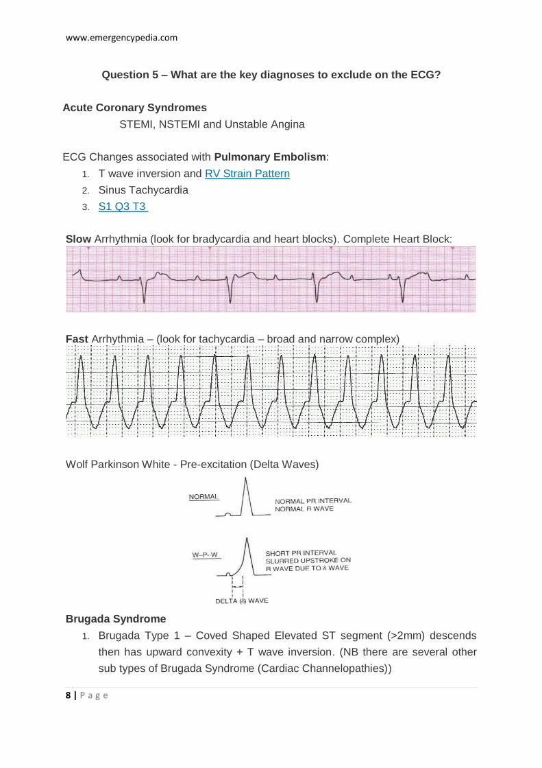
Slow Arrhythmia (look for bradycardia and heart blocks). Complete Heart Block:
Fast Arrhythmia – (look for tachycardia – broad and narrow complex)
Wolf Parkinson White - Pre-excitation (Delta Waves)
Brugada Syndrome
1. Brugada Type 1 – Coved Shaped Elevated ST segment (>2mm) descends
then has upward convexity + T wave inversion. (NB there are several other
sub types of Brugada Syndrome (Cardiac Channelopathies))
www.emergencypedia.com
9 | P a g e
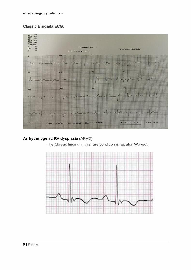
Classic Brugada ECG:
Arrhythmogenic RV dysplasia (ARVD)
The Classic finding in this rare condition is ‘Epsilon Waves’:
www.emergencypedia.com
10 | P a g e
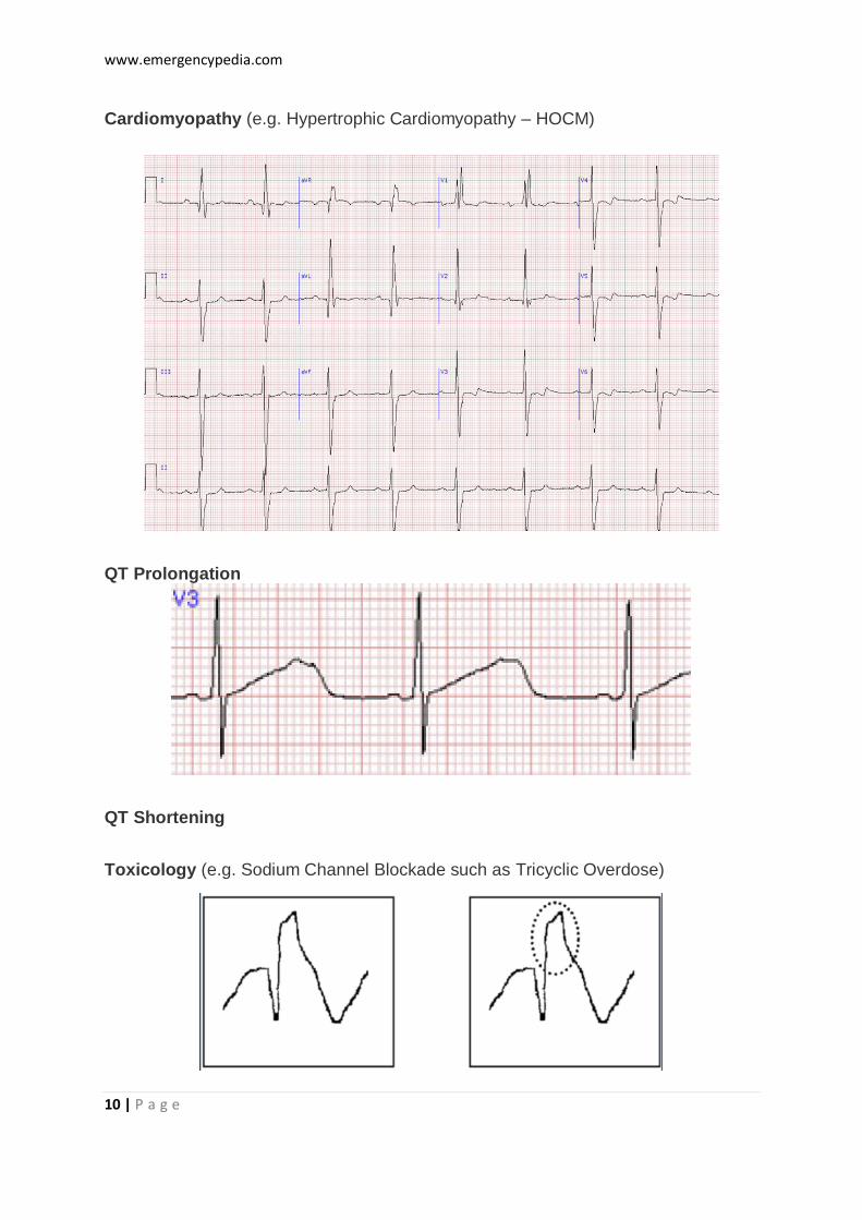
Cardiomyopathy (e.g. Hypertrophic Cardiomyopathy – HOCM)
QT Prolongation
QT Shortening
Toxicology (e.g. Sodium Channel Blockade such as Tricyclic Overdose)

www.emergencypedia.com
11 | P a g e
Electrolytes (e.g. Hyperkalaemia)
Take Homes
It is important to understand the definition of syncope (including brevity, loss of
consciousness and postural tone with spontaneous recovery) as well as associated
high-risk features. It is also important to develop skills in differentiating Syncope
from other presentations such as vertigo and seizures. Admission to hospital is often
required in patients with Syncope but does not always help make a definitive
diagnosis. Where in doubt, share the case with a colleague, use a clinical decision
rule and choose appropriate investigations to help risk stratify the patient and rule
out life threatening causes.
Practice Exam Question (2008)
Syncope – http://lifeinthefastlane.com/ccc/syncope
SEMEP – http://www.semep.co.uk/syncope.html