Supplementary Materials and Methods Exploratory toxicology...
17
Fully Human Anti-Hedgehog Antibodies 1 Supplementary Materials and Methods Exploratory toxicology study in male and female Wistar Rats Wistar (RCCHan) rats received an intravenous injection (via lateral tail vein) of vehicle (phosphate buffered saline), 10 mg/kg or 50 mg/kg of MEDI-5304 at a dose volume of 10 mL/kg on either the first (day 0) or seventh day (day 7) of the study. Study evaluations included body weights, food consumption, hematology, coagulation, and clinical chemistry measurements. All animals were necropsied 7 days after final dose administration. Organs were weighed before fixation and organ/body weight ratios were calculated (using the final body weight obtained prior to necropsy), as well as organ/brain weight ratios. All major organs were immersion-fixed in 10% neutral buffered formalin (NBF). Lungs were fixed by trans-tracheal inflation prior to immersion. Paired tissues were cut longitudinally (left) and transversely (right). The tissues were embedded in paraffin, sectioned, stained with hematoxylin and eosin, and examined microscopically by a veterinary pathologist. Calcified bone was de-calcified and processed as above. MedImmune is an Association for the Assessment and Accreditation of Laboratory Animal Care (AAALAC) accredited facility. Necropsy support and histopathology slide preparation were performed by Charles River Laboratories, Pathology Associates (PAI). The Institutional Animal Care and Use Committee (IACUC) reviewed and approved the study protocol in accordance with provisions of the USDA Animal Welfare Act and The Guide for the Care and Use of Laboratory Animals (NRC1996). Wistar (RCCHan) rats between 6-7 weeks of age from Harlan Laboratories (Indianapolis, IN) were assigned to study. Animals were identified with individual ear tags and each cage was be labeled with a cage card. Animals were group housed in polycarbonate cages, in a controlled environment (18-26°C and 30-70% relative humidity). Animals were fed Certified
Transcript of Supplementary Materials and Methods Exploratory toxicology...

Fully Human Anti-Hedgehog Antibodies
1
Supplementary Materials and Methods
Exploratory toxicology study in male and female Wistar Rats Wistar (RCCHan) rats
received an intravenous injection (via lateral tail vein) of vehicle (phosphate buffered saline),
10 mg/kg or 50 mg/kg of MEDI-5304 at a dose volume of 10 mL/kg on either the first (day
0) or seventh day (day 7) of the study. Study evaluations included body weights, food
consumption, hematology, coagulation, and clinical chemistry measurements. All animals
were necropsied 7 days after final dose administration. Organs were weighed before fixation
and organ/body weight ratios were calculated (using the final body weight obtained prior to
necropsy), as well as organ/brain weight ratios. All major organs were immersion-fixed in
10% neutral buffered formalin (NBF). Lungs were fixed by trans-tracheal inflation prior to
immersion. Paired tissues were cut longitudinally (left) and transversely (right). The tissues
were embedded in paraffin, sectioned, stained with hematoxylin and eosin, and examined
microscopically by a veterinary pathologist. Calcified bone was de-calcified and processed
as above. MedImmune is an Association for the Assessment and Accreditation of Laboratory
Animal Care (AAALAC) accredited facility. Necropsy support and histopathology slide
preparation were performed by Charles River Laboratories, Pathology Associates (PAI). The
Institutional Animal Care and Use Committee (IACUC) reviewed and approved the study
protocol in accordance with provisions of the USDA Animal Welfare Act and The Guide for
the Care and Use of Laboratory Animals (NRC1996).
Wistar (RCCHan) rats between 6-7 weeks of age from Harlan Laboratories (Indianapolis, IN)
were assigned to study. Animals were identified with individual ear tags and each cage was
be labeled with a cage card. Animals were group housed in polycarbonate cages, in a
controlled environment (18-26°C and 30-70% relative humidity). Animals were fed Certified

Fully Human Anti-Hedgehog Antibodies
2
Global Harlan Teklad Laboratory 2018 Diet and also given various cage-enrichment devices.
Animals were provided ad libitum access to filtered tap water via an automatic watering
system and/or water bottles. The water was routinely analyzed for contaminants and specific
microbes and met the EPA standards for drinking water.
Preliminary pharmacokinetics and nonclinical safety assessment study in Cynomolgus
Monkeys A preliminary pharmacokinetics and nonclinical safety assessment was conducted
at Charles River Laboratories (Reno, NV) in naive male and female chinese cynomolgus
monkeys that were between 2.5 to 3.9 years of age with body weights ranging from 2.3 to 2.7
kg. The parameters and end points evaluated in this study were clinical signs
(mortality/moribundity checks, cage side observations, and detailed clinical observations),
body weights, food consumption, skin biopsies, hematology, coagulation, and clinical
chemistry. Samples for serum drug levels and drug kinetic evaluations were also taken.
Skin punch biopsies were performed prestudy (day -4) and on day 4 following the first dose.
The skin biopsy area was cleansed with antiseptic and three punch biopsies of approximately
8 mm each were collected from the shaved dorsal area of each monkey. Skin samples were
collected into and stored in RNAlater prior to RNA isolation. The biopsy site was closed
with suture and/or tissue adhesive as needed, and local analgesics (i.e., lidocaine) were
administered. Animals were given an opiate analgesic and were allowed to recover in their
cages. Antesedan (a reversal agent for dexdomitor) was administered at veterinary discretion
after the procedure. No termination of the animals was done for the study. After study
completion, all animals were returned to the testing facility animal colony.

Fully Human Anti-Hedgehog Antibodies
3
Animals were housed individually in stainless-steel cages. Primary enclosures were as
specified in the United States Department of Agriculture (USDA) Animal Welfare Act (9
Code of Federal Regulations [CFR], Parts 1, 2, and 3) and as described in the Guide for the
Care and Use of Laboratory Animals. Temperatures of 64°F to 84°F (approximately 18°C to
29°C) with a relative humidity of 30% to 70% were maintained. A 12-hour light-12-hour
dark cycle was maintained. Ten or greater air changes per hour with 100% fresh air (no air
recirculation) were maintained in the animal room. Temperatures of 64°F to 84°F
(approximately 18°C to 29°C) with a relative humidity of 30% to 70% were maintained. A
12-hour light-12-hour dark cycle was maintained. Ten or greater air changes per hour with
100% fresh air (no air recirculation) were maintained in the animal room. Animals were fed
Purina Certified Primate Diet No. 5048 in amounts appropriate for the size and age of the
animals. This diet was supplemented with fruit or vegetables 2-3 times weekly. Animals
were provided reverse osmosis filter water that was passed through ultraviolet (UV) light
treatment. Processed water was available ad libitum to each animal via an automatic
watering device. The water was routinely analyzed for contaminants by Western
Environmental Testing Laboratory, Sparks, NV. No contaminants were known to be present
in the water at levels that would interfere with the results of this study. Animals were also
provided an enrichment device, food treat, and were periodically socialized to provide for
psychological enrichment. Socialization was limited to grooming bar access only.
Veterinary care was available throughout the course of the study and animals were examined
by the veterinary staff as warranted by clinical signs or other changes.
Determination of serum MEDI-504 levels in cynomolgus monkeys. Concentrations of
MEDI-5304 in cynomolgus monkey serum were determined using a qualified ELISA
method. The ELISA plates were coated with 1.0 μg/ml of sheep anti-human IgG (H+L
Fully Human Anti-Hedgehog Antibodies
4
specific, The Binding Site, Cat # A4003CUS01) in phosphate buffered saline (PBS)
overnight at 2°C to 8°C, washed with 0.05% Tween 20 in PBS, and blocked for at least 1 h at
room temperature with I-Block Buffer (Tropix, Bedford, MA). MEDI-5304 reference-
standard, quality-control (QC) and test sample dilutions were prepared in 10% cynomolgus
monkey serum (assay matrix) and added to blocked plates for 2 h at room temperature. Plates
were washed as above and incubated an additional 1 h at room temperature with 1:8000
dilution of HRP-goat anti human IgG (H+L-specific, Bethyl Labs, Cat# A80-319P). Unbound
detection antibody was removed by washing, and 100 μl of TMB substrate was added to
wells for 5 minutes. The development of color was stopped by addition of 50 μL of 2M
H2SO4. Plates were read within 15 minutes at 450 nm with SpectraMax Plus 384 plate reader
(Molecular Devices, Sunnyvale, CA). Concentrations of MEDI-5304 in QC and test-sample
dilutions on each plate were interpolated from the reference-standard curve for that plate. The
range of the assay for quantification of MEDI5304 in 100% cynomolgus monkey serum was
38 to 960 ng/ml.

Fully Human Anti-Hedgehog Antibodies
5
Supplementary Figures
Supplementary Figure S1. Binding kinetics of hedgehog antibodies to human and mouse
hedgehog proteins using surface plasmon resonance.

Fully Human Anti-Hedgehog Antibodies
6
A
B
Supplementary Figure S2. Hedgehog ligands stimulate mGLI1 reporter activity and
osteoblast differentiation in a dose-dependent manner.
Fully Human Anti-Hedgehog Antibodies
7
A
B
C
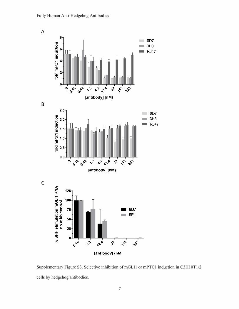
Supplementary Figure S3. Selective inhibition of mGLI1 or mPTC1 induction in C3H10T1/2
cells by hedgehog antibodies.
Fully Human Anti-Hedgehog Antibodies
8
vehicle MEDI-53040.0
0.5
1.0
1.5
hGli1mGli1
***
Rel
Exp
rn(v
. h
um
or
mu
HP
RT
1)
A
B
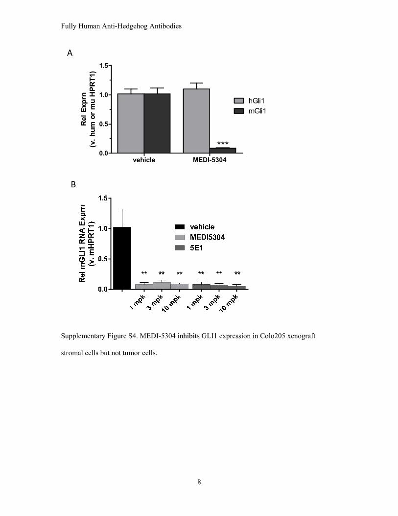
Supplementary Figure S4. MEDI-5304 inhibits GLI1 expression in Colo205 xenograft
stromal cells but not tumor cells.

Fully Human Anti-Hedgehog Antibodies
9
A
B
sHH
IHH
GLI-2
GLI-3
PTCH1
SMO
DYRK1
SUFU0
5
10
15
20
25
30
35R
ela
tiv
e e
xp
res
sio
n(r
ati
o C
SC
s:n
on
-CS
Cs
)
0
5
10
15
20
25
30
Sph
ere
num
ber
Supplementary Figure S5. Hedgehog pathway component RNA expression in primary
pancreatic tumor explant model P479 and effects of MEDI-5304 on tumorspheres derived
from pancreatic explant model 947.

Fully Human Anti-Hedgehog Antibodies
10
A
B
0 5 10 15 20 25 30-2
-1
0
1
2
3
1 mg/kg10 mg/kg30 mg/kg
Days post dosing
Ser
um
[M
ED
I-53
04]
log
(μg
/ml)
DoseCmax
(µg/ml)AUCall
(µg*d/ml)AUCINF
(µg*d/ml)CL
(ml/kg/d)t1/2
(d)
1 mg/kg 13.7 ± 4.6 26.7 ± 6.0 26.9 ± 6.0 38.6 ± 7.2 2.7 ± 0.3
10 mg/kg 137 ± 44 923 ± 106 1015 ± 156 10.0 ± 1.6 8.5 ± 2.7
30 mg/kg 650 ± 114 3396 ± 268 3968 ± 279 7.6 ± 0.5 10.4 ± 1.1
AUCALL: area under the concentration-time curve from time zero to the last measurable time point
AUCINF: area under the concentration-time curve from time zero to infinityCL: systemic clearanceCmax: observed peak concentration t1/2: terminal phase elimination half-life
Supplementary Figure S6. Pharmacokinetics of MEDI-5304 in rats.

Fully Human Anti-Hedgehog Antibodies
11
A B
0 7 1410
100
1000
10000
10 mg/kg50 mg/kg
days post dose
seru
m [
ME
DI-
5304
]( μ
g/m
l)
C
D
DoseCmax
(µg/ml)AUCINF
(µg*d/ml)CL
(ml/kg/d)t1/2
(d)
10 mg/kg 268 ± 1 833 ± 119 12.2 ± 1.8 6.1 ± 1.4
50 mg/kg 885 ± 5 6601 ± 866 7.7 ± 1.0 9.0 ± 1.0
vehicle MEDI-5304 50 mg/kg
Supplementary Figure S7. Exploratory toxicology of MEDI-5304 in rats
Fully Human Anti-Hedgehog Antibodies
12
ParametersGroup 2 Group 3 Group 4
0.3 mg/kg 3 mg/kga 1 mg/kg 10 mg/kga 3 mg/kg 75 mg/kga
n 4 4 4 4 4 4Cmax
(µg/mL)5.08 ± 0.54 89.7 ± 10.2 20.2 ± 1.0 290 ± 4.3 55.8 ± 4.9 1900 ± 270
AUClast
(day·µg/mL)16.7 ± 1.1 912 ± 84 80.6 ± 10.1 3400 ± 100 244 ± 25 22100 ± 2970
AUCinf
(day·µg/mL)18.5 ± 0.9 ND 98.2 ± 5.9 ND 342 ± 47 ND
t1/2
(d)4.12 ± 0.38 7.75 ± 0.32 5.76 ± 0.48 10.1 ± 0.7 7.74 ± 0.81 8.07 ± 0.48
CL(mL/kg/d)
16.4 ± 0.9 8.24 ± 1.08 10.3 ± 0.6 7.93 ± 0.34 9.45 ± 1.66 9.33 ± 1.47
A
Parameters are shown as mean ± SDa AUClast was calculated f rom the time interval 28 through 35 days post last dose on Day 28 and all PK parameters were derived f rom concentration-time prof ile data post last dose on Day 28.
B
0 7 14 21 28 35 420.1
1
10
100
1000
10000
Grp2 0.3mg/kgGrp3 1mg/kgGrp4 3mg/kgGrp2 3mg/kgGrp3 10mg/kgGrp4 75mg/kg
Time (day)
seru
m [
ME
DI-
5304
]( μ
g/m
l)
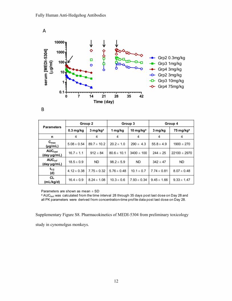
Supplementary Figure S8. Pharmacokinetics of MEDI-5304 from preliminary toxicology
study in cynomolgus monkeys.

Fully Human Anti-Hedgehog Antibodies
13
B
C
A
pretreated day 40.0
0.1
0.2
0.3
0.4
control3 mpk10 mpk75 mpk
** **
***Rel
Exp
rn G
LI1
(vs
cyn
o H
PR
T1)
pretreated day 40.0
0.1
0.2
0.3
0.4
control3 mpk10 mpk75 mpk
* ** **
Rel
Exp
rn P
TC
H1
(vs
cyn
o H
PR
T1)
pretreated day 40
2.0×10⁻³
4.0×10⁻³
6.0×10⁻³
8.0×10⁻³
1.0×10⁻²
control3 mpk10 mpk75 mpk
* ***
Rel
Exp
rn P
TC
H2
(vs
cyn
o H
PR
T1)
Supplementary Figure S9. Pharmacodynamics of MEDI-5304 in cynomolgus monkeys.
Fully Human Anti-Hedgehog Antibodies
14
Supplementary Figure Legends
Supplementary Figure S1. Representative sensograms of SPR experiments are depicted for
binding of human and mouse hedgehog proteins to anti-hedgehog monoclonal antibodies 6D7
and 3H8 affinity-captured or 5E1 directly immobilized onto the C1 chip. Dashed black lines
represent fits of the monoexponential binding model to the experimental data shown as full
gray lines. (A) 6D7 binding to human SHH (B) 6D7 binding to human/mouse IHH (C) 6D7
binding to mouse SHH (D) 3H8 binding to human SHH (E) 3H8 binding to human/mouse
IHH (F) 3H8 binding to mouse SHH (G) 5E1 binding to human SHH and (H) 5E1 binding to
human/mouse IHH.
Supplementary Figure S2. (A) Serially diluted human SHH C24II or human/mouse C28II
IHH was added to NIH3T3 cells stably expressing the mGLI1 luciferase reporter, and cells
were incubated for 24 hours. Fold stimulation in comparison to cells that did not receive
hedgehog ligand is plotted. (B) Serially diluted human SHH C24II or human/mouse C28II
IHH was added to C3H10T1/2 cells and incubated for 3 days. Alkaline phosphatase activity
was determined as a measure of osteogenesis.
Supplementary Figure S3. Hedgehog antibodies inhibit induction of endogenous mPTC1 by
hedgehog proteins. C3H10T1/2 cells were treated with 600 ng/ml (30 nM) SHH C24II (A) or
2 µg/ml (100 nM) IHH C28II (B) for 24 hours in the absence or presence of serially diluted
6D7, 3H8, or R347. Modulation of endogenous levels of mPTC1 RNA by hedgehog
stimulation and treatment with anti-hedgehog mAbs was determined by qRT-PCR (TaqMan)
using the 2- ∆∆Ct method. mPTC1 expression of each sample was normalized to mHPRT1
expression and fold stimulation was calculated relative to unstimulated cells in the absence of
Fully Human Anti-Hedgehog Antibodies
15
antibody. (C) C3H10T1/2 cells were treated with 600 ng/ml SHH C24II in the presence of
varying concentrations of 6D7 or 5E1 and treated for 72 h. Values plotted are per cent
mGLI1 levels (normalized to mHPRT1) induced by SHH treatment in the absence of
antibody. All values represent means ± SEM.
Supplementary Figure S4. (A) Colo205 colon tumor cells were implanted into athymic
mice and once tumors reached ~ 400 mm3 in size, tumor bearing animals were treated with
vehicle or 10 mg/kg MEDI-5304 for 24 h. RNA expression of human and mouse GLI1 was
determined by qRT-PCR using species-specific TaqMan primers/probes and normalized to
the corresponding expression of human or mouse HPRT1. Values represent means ± SEM.
Statistical analysis was performed using Student’s t test. A significance level of P < 0.001 is
indicated (***) when comparing antibody treatment to vehicle controls. (B) Animals bearing
Colo205 xenograft tumors received vehicle or 1, 3, or 10 mg/kg MEDI-5304 or 5E1, and
relative mGLI1 RNA levels were determined in excised tumors 24 h after dose administration
as described in (A). Statistical analysis was performed using one-way ANOVA and
Dunnett’s multiple comparison tests. Statistical significance of P < 0.01 is indicated (**).
Supplementary Figure S5. (A) Cancer stem cells from the P479 model were isolated by
FACS sorting from dissociated bulk tumor cells using the cell surface markers ESA, CD44,
and CD24. The RNA expression of various hedgehog pathway components was determined
in CSCs and bulk tumor cells with human specific TaqMan primers/probes. The ratio of
expression in CSCs to bulk tumors is shown. (B) Effect of MEDI-5304 on formation of
tumorspheres derived from the primary pancreatic model 947. Following FACS sorting,
single cell suspensions from patient pancreatic cancer xenografts were resuspended in
tumorsphere media. Cells were plated in the absence or presence of 5, 10 or 30 µg/ml MEDI-
Fully Human Anti-Hedgehog Antibodies
16
5304. Tumorspheres were cultured for 4 days and counted. No statistically significant
differences were observed.
Supplementary Figure S6. (A) Serum MEDI-5304 levels in rats. A single i.v. bolus dose of
1, 10, 30 mg/kg MEDI-5304 was administered to Hans Wistar rats. Each dosing group was
comprised of three males and three females. Blood samples were collected via tail vein
without anti-coagulant agent. Serum levels of MEDI-3504 were determined by ELISA
specific for human IgG. (B). Noncompartmental analysis of the preliminary rat PK study
depicted in (A) was performed on individual animal serum concentration data using
WinNonlin Professional (version 5.2, Pharsight Corp., Mountain View, CA).
Supplementary Figure S7. (A) The maxilla from a vehicle-treated rat whose incisor has a
normal odondoblast layer (boxed area). 20 x, HE. Inset: Higher magnification of the
odontoblast layer (arrow heads). 100 x, HE. (B) The maxilla from a rat treated with MEDI-
5304 (50 mg/kg) whose incisor exhibits disorganization of the odondoblast layer admixed
with dentin-like material (boxed area). 20 x, HE. Inset: Higher magnification of the
odontoblast layer (arrow heads). 100 x, HE. (C) MEDI-5304 pharmacokinetics. Serum levels
of MEDI-5304 were determined during the execution of the exploratory rat toxicology study.
Doses were given i.v. on day 0 and day 7 (arrows). Antibody levels were determined by
ELISA that detects total human IgG. (D). Noncompartmental analysis of the rat PK data was
conducted based on data collected post first dose on day 0 during the exploratory rat
toxicology study.
Supplementary Figure S8. (A) Serum levels of MEDI-5304 were determined during the
execution of the preliminary non-clinical pharmacokinetics safety study conducted in

Fully Human Anti-Hedgehog Antibodies
17
cynomolgus monkeys. Animals received either vehicle (Grp1), or 0.3 (Grp2), 1 (Grp3), or 3
mg/kg (Grp4) MEDI-5304. After 14 days, the dose each group received was increased to 3
(Grp2), 10 (Grp3), or 75 mg/kg (Grp4). These doses were given on days 14, 21, and 28.
Time of dose administration is denoted by the arrows. Pharmacokinetic parameters of
MEDI-5304 were determined in the varying dosing groups. (B). Noncompartmental analysis
of the monkey PK data collected during the monkey safety study.
Supplementary Figure S9. Pharmacodynamic effects of MEDI-5304 observed in skin of
cynomolgus monkeys. Skin punches were performed 4 days prior to antibody (pretreated)
dosing and on day 4. Three skin punches were obtained from each animal at time of
sampling. The effects of MEDI-5304 on GLI1 (A), PTCH1 (B) and PTCH2 (C) RNA levels
were determined with TaqMan primer/probe sets for the rhesus orthologs and validated to
have 100% amplification efficiency with cynomolgus brain RNA. Values are means ± SEM.
Statistical significance was evaluated using one-way ANOVA and Dunnett’s multiple
comparison tests. Significance levels of P < 0.05 (*), P < 0.01 (**), and P < 0.001 (***) are
indicated for comparisons with the vehicle control.