
STUDIES ON CASEIN KINASE FROM THE LACTATING RABBIT …
360
STUDIES ON CASEIN KINASE FROM THE LACTATING RABBIT MAMMARY GLAND BY David Patrick William Lennon, Bsc. Department of Biochemistry University College London Submitted to the University of London for the degree Doctor of Philosophy MAY 1990 1
Transcript of STUDIES ON CASEIN KINASE FROM THE LACTATING RABBIT …

STUDIES ON CASEIN KINASE FROM THE LACTATING RABBIT MAMMARYGLAND
BY
David Patrick William Lennon, Bsc. Department of Biochemistry University College London
Submitted to the University of London for the degreeDoctor of Philosophy
MAY 1990
1
ProQuest Number: 10631060
All rights reserved
INFORMATION TO ALL USERS The quality of this reproduction is dependent upon the quality of the copy submitted.
In the unlikely event that the author did not send a com p le te manuscript and there are missing pages, these will be noted. Also, if material had to be removed,
a note will indicate the deletion.
uestProQuest 10631060
Published by ProQuest LLC(2017). Copyright of the Dissertation is held by the Author.
All rights reserved.This work is protected against unauthorized copying under Title 17, United States C ode
Microform Edition © ProQuest LLC.
ProQuest LLC.789 East Eisenhower Parkway
P.O. Box 1346 Ann Arbor, Ml 48106- 1346

ABSTRACTThe mammary gland specific casein kinase is the enzyme responsible for the in vivo phosphorylation of caseins, the major class of milk proteins. It is distinct from other so-called "casein kinases", which are neither tissue specific nor catalyze the in vivo phosphorylation of caseins. The work here describes the identification, purification and characterization of a casein kinase from the lactating rabbit mammary gland.
Initially localization studies, which involved centrifugation of mammary gland sub-cellular fractions through sucrose gradients, showed that whilst a small proportion of the rabbit casein kinase was associated with a golgi enriched fraction, the bulk was present in a fraction shown by electron microscopic analysis to be a homogeneous casein micelle fraction.
In order to study further the enzyme from both sources, a sub-cellular fractionation procedure was developed for the co-isolatation of golgi and micelle fractions from mammary gland homogenates. Using these two fractions as starting sources, casein kinase was purified, essentially to homogeneity, by a combination of gel-filtration on Sephacryl-S-300 and affinity chromatography on casein- sepharose. Analysis of the purified material by SDS polyacrylamide gel electrophoresis followed by silver staining showed that in both cases the enzyme comprised a
2
polypeptide doublet of apparent molecular weights 63 and 67 KDaltons.
The enzyme was characterized with respect to a number of properties including assay parameters including: temperature, pH, substrate concentration, and the requirement for divalent cations and the effect of a number of activators and inhibitors was studied.
It was planned to generate primary sequence data (10-20 amino acids only) which could be used to generate oligonucleotides which in turn could be used in future studies. It was found however that the purification procedure was unsuitable to isolate protein for sequencing since frequently low levels of contaminants were present. To over-come this the purification procedure was modified and an hplc step incorporated. This method was found to be suitable for the isolation of larger quantities (70-100 pg) of pure enzyme. The enzyme was not suceptible to direct sequencing, suggesting the presence of a blocked N- terminus. Treatment of the enzyme by chemical and enzymatic means did not generate any peptide fragments more suitable for sequencing.
3

ACKNOWLEDGEMENTS
Firstly I should like to thank my supervisor Dr. A.P. Boulton for his advice and encouragement throughout the production of this thesis and for his help with some of this work.
Thanks are due to Mr Mark Turmaine at the electronmicroscopic unit University College London for hiscollaboration and expert technical help with the electronmicroscopic work presented here. I am grateful toProfessor Brian Ketterer for allowing me to use the facilities of his laboratories to carry out the work described in chapter 8, and thank Dr. Brian Coles who supervised this work. The advice of Dr. R.K. Craig is gratefully aknowledged.
I thank the Science and Engineering Research Council for financial support.
I would also like to thank all my colleagues at the Medical Molecular Biology Unit, at H.U.C. and more recently at the University of Nottingham who have helped in some way or other in the production of this thesis.
This thesis is dedicated to my parents

CONTENTS
Page n°Title page.................................................. 1Abstract.................................................... 2Acknowledgements........................................... 4Table of contents.......................................... 5List of figures........................................... 13List of tables............................................ 17Abbrevi at i ons..............................................18CHAPTER 1 GENERAL INTRODUCTION........................... 19
General preface......................................... 201.1 Mammary gland and milk biology.................... 211.1.1 Structure of the rabbit mammary gland ......211.1.2 The composition of milk ........ 26
1.1. 2.1 Introduction.................................261.1.2. 2 Non-protein components...................... 26
A) Water..................................... 26B ) Carbohydrate............................. 28C) Lipids....................................28E) Miscallaneous non-protein components.... 30
1.1. 2. 3 Milk proteins............................... 321.1.3 Casein micelles ................................ 381.1.4 The enzymes of milk............................. 46
1.1.4.1 Introduction................................ 461.1. 4. 2 Distribution and origin.....................461.1. 4. 3 Role of milk enzymes........................ 50
1.1.5 Milk protein expression......................... 531.1.6 Secretion of the major components of milk 57
1.1.6.1 Fat.......................................... 57
5
1.15.2 Protein * . . ................................ 58
1.1.6. 3 Miscellaneous components....................61
1.2 PROTEIN PHOSPHORYLATION............................. 63
1.2.1 General considerations.........................631.2.2 Casein phosphorylation.........................681.2.3 Role of phosphoprotein phosphate.............. 691.2.4 Protein kinases................................ 711.2.4.1 General considerations...................... 71
1.2.4.2 Cyclic adenosine monophosphate dependent protein kinase.............................. 73
1.2.4.3 Cyclic guanosine monophosphate dependent protein kinase.............................. 76
1.2.4.4 Protein kinase C .............................771.2.4.5 Calcium and calmodulin dependent protein
kinase........................................ 791.2.4. 6 Protein tyrosine kinases.....................791.2.4. 7 Casein kinases............................... 811.2.4.8 Mammary gland casein kinases................. 831.2.4.9 Calcium and calmodulin dependent casein kinases............................................... 86
1.2.4.10 Substrate-specificity determinants of protein kinases............................ 87
1.3 Aims................................................ 91
CHAPTER 2 MATERIALS AND METHODS......................... 92
2.1 Materials............................................932.1.1 Animals.......................................... 932.1.1 Radiochemicals...................................93
6
2.1.3 Chemicals and solvents.......................... 932.1.4 Materials for hplc.............................. 952.1.5 Materials for protein sequencing............... 96
2.2 Methods............................................. 972.2.1 Enzyme assays....................................97
2.2.1.1 casein kinase............................... 972.2.1.2 Galactosyltransferase....................... 982.2.1.3 Glucose 6-phosphatase....................... 982 .2 .1.4 Lactate dehydrogenase....................... 99
2.2.2 Protein assay....................................992.2.3 SDS polyacrylamide gel electrophoresis......... 992.2.4 Silver staining SDS polyacrylamide gels....... 1002.2.5 Isolation of total bovine caseins............. 1012.2.6 Preparation of casein-sepharose................102
2.2.7 Collection of rabbit milk and isolation of casein micelles.................................102
2.2.8 Routine method for the co-isolation of casein micelles and golgi enriched fraction from lactating rabbit mammary gland.................102
2.2.9 Procedure for the routine purification of rabbit mammary gland casein kinase fromthe casein micelle rich fraction.............. 104
2.2.9.1 Gel-filtration step on Sephacryl-S-300....1052.2.9.2 Casein-sepharose affinity
chromatography step........................ 1062.2.9.3 Determination of the optimal
concentration of sodium chloride required to elute casein kinase from casein-sepharose........................... 106
2.2.9.4 Effect of protein concentrationon solubilization of casein kinasefrom micelles.............................. 107
7

2.2.9.5 Purification of rabbit casein kinasefrom whole rabbit milk..................... 108
2.2.9.6 Purification of rabbit casein kinasefrom a golgi enriched fraction............ 108
2.2.10 Purification of rabbit casein kinase-other procedures...............................108
2.2.10.1 ATP-agarose chromatography step.......... 1082.2.10.2 Further purification of ATP-agarose
purified rabbit casein kinase............ 1092.2.11 Purification of rabbit casein kinase
prior to hplc analysis-modifications tothe routine method............................ 110
2.2.12 Purification of rabbit casein kinaseusing hplc..................................... 110
2.2.13 Determination of the intra-cellular localization of rabbit mammary glandcasein kinase..................................Ill
2.2.13.1 Analysis of a post-nuclear supernatant on a linear (5-20% w/v) sucrosegradient................................... Ill
2.2.13.2 Analysis of a crude microsomal fractionon a discontinuous sucrose gradient......112
2.2.13.3 Effect of incubating rabbit golgi enriched fractions with increasing concentrations of trypsin in the presence and absence of 0.1% (w/v)Triton X-100.............................. 113
2.2.13.4 Temperature induced phase separation of rabbit golgi enriched membrane components in Triton X114.................114
2.2.13.5 Distribution of casein kinase following treatment of rabbit golgi enriched fractions with 0.1 M sodium carbonate..................................114
2.2.14 Estimation of the sedimentation co-efficientof rabbit mammary gland casein k i n a s e ........ 114
2.2.15 Isolation of guinea-pig mammary glandgolgi enriched fraction and partial purification of casein kinase................ 115
2.2.16 Electron microscopy........................... 1162.2.17 Phosphorylation of synthetic peptides
8

SEEEEE and SEAEEE by mammary glandcasein kinases.................................116
2.2.18 Amino acid analysis........................... 1172.2.19 Protein fragmentation......................... 118
2.3 Buffers............................................ 119CHAPTER 3 DISTRIBUTION OF LACTATING RABBIT MAMMARY
GLAND CASEIN KINASE......................... 1203.1 Introduction....................................... 1213.2 Results............................................ 124
3.2.1 Use of sucrose gradient centrifugation to determine the intra-cellular localizationof casein kinase.............................. 124
3.2.2 Electron microscopic analysis of the white coloured sedimented material and casein micelles isolated from rabbit milk...........128
3.3 Discussion......................................... 136
CHAPTER 4 FURTHER ANALYSIS OF CASEIN KINASE RICH FRACTIONS ISOLATED FROM THE RABBIT MAMMARY GLAND................................ 139
4.1 Introduction......................................... 1404.2 Results .......................................... 142
4.2.1 Development of a procedure for the co-isolation of casein kinase rich fractions..................................... 142
4.2.2 Electron microscopic analysis of the particulate fractions resulting from the fractionation of a microsomal preparation by centrifugation through 1.2/0.5 Msucrose 1 ayers................................144
4.2.3 Localization of casein kinase withinthe golgi enriched fraction............... 151
4.2.3.1 The effect of trypsin on casein kinase activity in the presence and absence of 0.1% (w/v) Triton X-100.... 151
4.2.3.2 Temperature induced phase separation of rabbit golgi enriched membrane components with Triton X-114.......... 154
4.2.3.3 Distribution of casein kinase following the treatment of golgi enriched
9
fractions with 0.1M sodium carbonate...1574.3 discussion........................................... 159
CHAPTER 5 PURIFICATION OF A RABBIT MAMMARY GLANDCASEIN KINASE ................................165
5.1 Introduction...................................... 1665.2 Results ........................................... 169
5.2.1 ATP-agarose chromatography and sucrose gradient centrifugation steps.................169
5.2.2 Gel-exclusion and casein-sepharose affinity chromatography steps.......................... 170
5.2.3 Analysis of purified casein kinase by centrifugation on linear sucrose(5-20% w/v) gradients ........................ 186
5.2.4 Purification of casein kinase from whole rabbit milk.................................... 192
5.2.5 Effect of various reagents on the releaseof casein kinase from casein micelles........ 197
5.2.5.1 The effect of sodium chloride...........1985.2.5.2 The effect of EGTA...................... 1985. 2. 5. 3 The effect of Triton X-100..............201
5.3 Discussion.......................................... 2075.4 Summary............................................. 215
CHAPTER 6 PURIFICATION OF CASEIN KINASE FROM A GOLGI ENRICHED FRACTION ISOLATED FROM THE LACTATING RABBIT MAMMARY GLAND.............. 218
6.1 Introducton ......................................... 2196.2 Results.............................................. 2206.3 Discussion........................................... 227CHAPTER 7 CHARACTERIZATION OF A RABBIT MAMMARY GLAND
CASEIN KINASE................................ 229
7.1 Introduction.........................................230
10

7.2 Results ............................................. 2317.2.1 The effect of enzyme concentration
on enzyme activity.............................. 2317.2.2 The effect of increasing assay incubation
time on enzyme activity......................... 2317.2.3 The effect of changes of pH on enzyme
activity......................................... 2347.2.4 The effect of temperature on enzyme activity. ..2377.2.5 The effect of the nature and concentration
of the divalent cation on enzyme activity.....2377.2.6 The effect of calcium chloride at increasing
concentrations in the presence ofmagnesium chloride on enzyme activity.......242
7.2.7 Kinetic parameters.............................. 2457.2.7.1 V.ax and K. of casein kinase for
casein..................................... 2457.2.7.2 V.a> and K. of casein kinase for
ATP........................................ 2487.2.8 The effect of increasing concentrations of
sodium chloride, EGTA and triton X-100 onenzyme activity..................................257
7.2.9 The effect of the presence of N6 ,2'-0- dibutyryl-adenosine 3':5' cyclicmonophosphate on enzyme activity............. 264
7.2.10 The effect of 5'-fluorosulfonylbenzoyl- adenosine on enzyme activity..................265
7.2.11 The effect of heparin on enzyme activity..... 2727.3 Discussion........................................ 281
CHAPTER 8 HIGH PRESSURE LIQUID CHROMATOGRAPHY PURIFICATION AND ATTEMPTED PROTEIN SEQUENCING....................................288
8.1 Introduction....................................... 2898.2 Results.............................................. 291
8.2.1 High pressure liquid chromatography......... 2918.2.2 Protein sequencing ............................29 6:8.2.3 Protein fragmentation......................... 299
11

8.3Discussion............................................... 304
CHAPTER 9 GENERAL DISCUSSION........................... 3079.1 Distribution of rabbit casein kinase .............3089.2 Mechanisms to account for the distribution of
rabbit casein kinase .............................. 3099.3 Explanation for the presence of two polypeptide
species in samples of purified casein kina s e.....3109.4 Milk as the site of casein phosphorylation...... 3129.5 The bulk of casein kinase in the micelle rich
fraction-a misleading observation? ............... 3139.6 The golgi as the site of casein phosphorylation
in the lactating rabbit mammary gland............. 3159.7 Properties of rabbit casein kinase ............... 3159.8 Hplc and protein sequencing...................... 3179.9 Further studies.................................... 317
Appendix 1 Determinants of substrate specificity ofmammary gland casein kinases................ 320
Appendix 2 The secretion of casein micelles-anelectron microscopic study...................327
CHAPTER 10 REFERENCES....................................333
12

TABLE OF FIGURESFIGURE TITLE PAGE1 The structure of the lactating
rabbit mammary gland..............................232 Electron micrograph of a cross
section of the lactating mammary gland.......... 253 The structure of lactose..........................294 Proposed model of the casein micelle
structure......................................... 445 The secretion of a fat globule from the
mammary secretory cell........................... 606 The structures of phosphorylated amino acids...657 Structures of phospho-protein linkages...........678 Profile of casein kinase and galactosyl
transferase activity in fractions resultingfrom centrifugation of a rabbit mammarygland post-nuclear supernatant on a linear5-20% (w/v) sucrose gradient.................. 127
9 Profile of enzyme activity in fractionsresulting from the centrifugation of a rabbit mammary gland crude microsomal fraction on a discontinuous sucrose gradient... 130
10 Electron micrograph of the white coloured casein kinase rich sediment from the discontinuous sucrose gradient centrifugation..133
11 Electron micrograph of casein micellesisolated directly from rabbit milk.............135
12 Flow diagram of the procedure developed forthe routine co-isolation of golgi and casein micelle rich fractions.......................... 143
13 Electron microscopic analysis of thecasein kinase rich sediment resulting from centrifugation of a crude microsomal preparation through a 0.5/1.2 M sucrose gradient............................... 148
14 Electron micrograph of the endoplasmicreticulum rich fraction resulting from centrifugation of a crude microsomal preparation through a 0.5/1.2 M sucrose gradient....................................... . 150
15 Electron micrograph of the golgi enriched
13
16
17
18
19
20
21
22
23
24
25
26
27
28
fraction resulting from the centrifugationof a crude microsomal fraction through a0.5/1. 2 M sucrose gradient...................... 153The effect on casein kinase activity of incubating a golgi enriched fraction with increasing concentrations of trypsin in the presence and absence of Triton X-100........... 156SDS polyacrylamide gel electrophoresis analysis of ATP-agarose and sucrose gradient purified rabbit mammary gland casein kinase.... 172Profile of casein kinase activity and protein concentration eluting from Sephacryl-S-300.................................174Some characteristics of the Sephacryl-S-300- column used described in section 2.2.9.1......177
Profile of casein kinase eluting from casein -sepharose following successive additions ofsodium chloride.................................178The effect of protein concentration on thesolubilization of rabbit casein kinasecasein micelles.................................181
Profile of casein kinase eluting from casein- sepharose........................................ 183SDS polyacrylamide gel electrophoresis analysis of Sephacryl-S-300 and casein- sepharose purified rabbit mammary gland casein kinase.................................... 185SDS polyacrylamide gel electrophoresisanalysis of rabbit casein micelles............. 188Analysis of purified rabbit mammary gland casein kinase on linear sucrose gradients and SDS polyacrylamide gel electrophoresis 191Analysis of purified rabbit mammary gland casein kinase by centrifugation on linear sucrose gradients in the presence and absence of 2-mercaptoethanol...................194SDS polyacrylamide gel electrophoresisanalysis of casein kinase purified directlyfrom rabbit milk ................................196
Effect of incubating casein micelles in thepresence of increasing concentrations ofsodium chloride..................................200
14

29 Effect of incubating casein micelles in the presence of increasing concentrationsof EGTA ......................................... 203
30 Effect of incubating casein micelles in the presence of increasing concentrations ofTriton X-100.....................................206
31 Flow diagram of the routine method for thepurification of casein kinase...................217
32 Profile of casein kinase from a golgienriched fraction eluting from a column of Sephacryl-S-300..................................222
33 Profile of casein kinase from the golgieluting from casein-sepharose...................224
34 SDS polyacrylamide gel electrophoresisanalysis of purified golgi casein kinase.......226
35 The effect of increasing enzyme concentrationon the incorporation of 3 2 P into total bovine caseins by purified rabbit mammarygland casein kinase............................. 233
36 The effect of changes of assay incubationtime on the activity of rabbit mammarygland casein kinase............................. 236
37 The effect of increasing pH on the activityof purified rabbit mammary glandcasein kinase....................................238
38 The effect of assay temperature on theactivity of purified rabbit mammary glandcasein kinase....................................241
39 The effect of the nature and concentration of the divalent cation on the activity of the purified rabbit mammary gland caseinkinase............................................ 244
40 The effect of increasing concentrations of Ca2 + in the presence of Mg2 + on the activity of rabbit mammary gland caseinkinase............................................ 246
41 The effect of increasing amounts of totalbovine casein on the activity of the purifiedrabbit mammary gland casein kinase.............. 250
42 Estimation of V.ai and K» of casein kinasefor casein........................................ 252
43 The effect of increasing amounts of ATP onthe activity of the purified rabbit mammarygland casein kinase.............................. 253
15

44
45
46
47
48
49
5051
52
53
54
55
56
57
Estimation of Vmax and K. of casein kinasefor ATP........................................... 256The effect of increasing concentrations of sodium chloride on purified rabbit mammary gland casein kinase activity.....................259The effect of increasing concentrationsof EGTA on purified rabbit mammary glandcasein kinase activity........................... 261The effect of increasing concentrations of Triton X-100 on purified mammary gland casein kinase activity.......................... 263
The effect of increasing concentrations ofN6-2 O-dibutyrylcyclic adenosine 3 *:51-cyclic monophosphate on a cyclic AMPdependent protein kinase......................... 267The effect of increasing concentrations of N6-2 O-dibutyrylcyclic adenosine 3 1:5'-cyclic monophosphate on purified rabbitmammary gland casein kinase activity............ 270Comparison of the structures of ATP and FSBA....271The effect of increasing concentrations ofFSBA on purified rabbit mammary gland casein kinase activity...................................274The effect of increasing concentrations of FSBA on partially purified guinea-pig mammary gland casein kinase activity..................... 276The effect of heparin on the activity of the purified rabbit mammary gland casein kinase 278The effect of heparin on the activity of the partially purified guinea-pig mammary gland casein kinase..................................... 280Profile of material eluting from an hplc column and analysis of material eluting at 26 minutes from an identical experiment by SDS polyacrylamide gel electrophoresis ............. 294Electron micrographs of cross sections of the lactating rabbit mammary gland showing the presence of casein micelles associated with stacks of smooth membrane........................ 323Electron micrographs of cross sections of the lactating rabbit mammary gland showing the presence of casein micelles in secretory vesicles and their subsequent release at the cell surface...................................... 326
16

LIST OF TABLES
TABLE TITLE PAGE1 The gross composition of milk.................... 272 The carbohydrates of milk.........................293 The lipids of fresh milk:- an overview............ 314 The approximate salt composition of milk........ 335 The composition of the caseins................... 356 The composition of casein micelles...............417 Characteristics of casein micelles...............428 The enzymes of rabbit milk....................... 479 Casein kinase and galactosyl-transferase
content of the three fractions resulting from the fractionation of a crude microsomal preparation on a discontinuous sucrosegradient......................................... 146
10 Structure of some common protein kinases....... 21111 Amino acid composition of hplc purified
casein kinase....................................29712 Common procedures protein fragmentation........ 30113 Attempted protein sequencing and generation
of casein kinase protein fragments............ 30314 Summary of the properties of rabbit casein
kinase........................................... 31615 Determinants of substrate specificity of the
mammary gland casein kinase using synthetic peptides-an absolute requirement for an n+2 acidic residue................................... 329
16 Phosphorylation of synthetic peptides byrabbit mammary gland casein kinase............. 331
17

ABBREVIATIONSACP Amorphous calcium phosphateAP Alkaline phosphataseATP Adenosine triphosphateBSA Bovine serum albuminC-terminal Carboxy-terminal (of a peptide)CCP Colloidal calcium phosphatecDNA Copy deoxyribonucleic acidCpm Counts per minuteCyclic AMP Adenosine 3':51 cyclic adenosine
monophosphateCyclic GMP Guanosine 3 1:51 cyclic adenosine
monophosphate DEAE DiethylaminoethylcelluloseEC Enzyme commissionEDTA Ethylenediaminetetraacetic acidEGTA Ethylene-glycol-bis (3 aminoethyl
ether) N N'-tetra acetic acid ER Endoplasmic reticulumFSBA 5 1-Fluorosulphonylbenzoyladenosine"H" buffer Homogenization bufferHplc high pressure liquid chromatographyK. Michaelis constanta-LA a-LactalbuminLDH Lactate dehydrogenaseMDH Maiate dehydrogenaseMes 2-(N-Morpholino) ethanesulphonic acidMFGM Milk fat globule membraneMops 4-Morpholinepropane sulphonic acidmRNA Messenger ribonucleic acidn+2/n+3 Denotes the amino acid two/three
residues from a phosphorylatable (n) amino acid
N-terminal Amino terminal (of a peptide)NAD* Nicotinamide adenine dinucleotideNMR Nuclear magnetic resonancePi Inorganic phosphatePMSF PhenylmethylsulphonylfluorideP-Ser PhosphoserinePTH PhenylthiohydantoinRER Rough endoplasmic reticulumSDS Sodium dodecyl sulphateSRP Signal recognition peptideTris 2-Amino-2-(hydroxymethyl) propane
1,3-diol (tris)TFA Trifluoroacetic acidTFMS Trifluoromethanesulphonic acidUDP-galactose Uridine diphosphate galactoseV. ax Limiting rateWAP Whey acidic proteinXO Xanthine oxidaseThree and one letter abbreviations for amino acids have been used in accordance with the IUPAC-IUB recommendations out-lined in the Biochemical Journal (1984) 214, 345-373.

CHAPTER 1
GENERAL INTRODUCTION
19

GENERAL PREFACE
The purpose of this general introduction is to provide an overview of two areas, firstly of mammary gland biology, including mammary gland structure and milk composition, including topics such as caseins, casein micelles, and of milk enzymes which are of more direct interest to the work in this thesis. The second area reviewed here is"phosphorylation", including protein kinases andparticularly the transfer of phosphorous to casein.
There are over 4000 species within the class mammalia (quoted in Fox, 1989), the enzyme described in this thesis has been isolated from the rabbit, Oryctolagus cunicuius,
(class: mammalia, of the order: Lagomorpha), and wherepossible the mammary gland biology described in this introduction relates to this species. However in many areas little information pertaining to this species is available, and by necessity then it is the cow (Bos
taurus), being the best studied species, that much of this introduction relates to.
20

1.1 MAMMARY GLAND AND MILK BIOLOGY
1.1.1 The structure of the lactating rabbit mammarygland
Lactation, the production of milk, is the most important distinguishing characteristic of the mammalia. The milk is produced in specialized organs, the mammary glands. The rabbit has a total of five pairs located on the ventral aspect of the body. More specifically they are situated bilaterally in the thoracic region (two pairs), abdominal region (two pairs) and a pair of inguinal glands, each gland has a single teat with eight to ten teat ducts (or galactophores). There is no udder structure in this species and as such only weak support is given to the glands by skin and connective tissue.
The functional rabbit mammary gland, i.e. one capable of secreting milk, consists of clusters of specialized secretory cells (termed lobulo-alveoli cells), the apical membrane of which face a central lumen into which milk is secreted. Each cluster of cells constitute a single mammary alveolus with the lumen draining into a small ductule, which in turn drains into a larger segmental duct. A group of alveoli with ductules merging into a subsegmental duct are termed mammary lobules, see Schmidt (1971).
21

The subsegmental ducts eventually merge with each other to collect into a larger segmental duct and then into a larger mammary sinus (or cistern). Each region of mammary lobules in the rabbit lead into a separate sinus and connect to the exterior by its own teat duct, in other species, including cow, each region of mammary lobule connect eventually into a common sinus and a single teat duct. A diagramatic representation of these structures is presented in figure 1.
The mammary gland has a large dense network of blood vessels and is highly innervated (Linzell, 1953a and Schmidt, 1971). Each mammary alveolus is surrounded by specialized contractile cells (myo-epithelial cells) which are stimulated by the hormonal influence of oxytocin leading to contraction and hence milk expulsion, part of the so-called "milk-ejection" reflex (see Linzell, 1955; Mepham, 1976 and Tindal, 1978).
The ultra-structure, as reflected in electron microscopic studies of the lobulo-alveolar cells, is typical of a tissue involved in copious secretory activity. These electron micrographs show large quantities of the intracellular organelles involved in this process, including large quantities of rough endoplasmic reticulum (RER) and membrane bound vesicles containing material destined for release at the cell surface, see figure 2 and appendix 2 .
22

gland
Schematic representation of A) the gross anatomical structure of the mammary ductal system and the associated mammary lobules (L), B) mammary gland lobule and C) the mammary gland alveolus, figure lc adapted from Patton (1969).
Sinus
Ductule,
Segmental Duct
/Lum en
MyoepithelUHC ells
SecretoryCells
Ductule
23
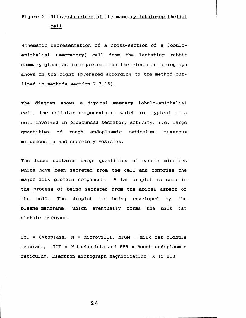
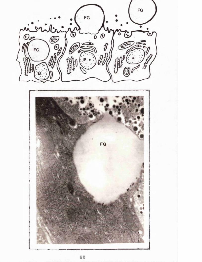
Figure 2 Ultra-structure of the mammary lobulo-epithelialcell
Schematic representation of a cross-section of a lobulo- epithelial (secretory) cell from the lactating rabbit mammary gland as interpreted from the electron micrograph shown on the right (prepared according to the method outlined in methods section 2.2.16).
The diagram shows a typical mammary lobulo-epithelial cell, the cellular components of which are typical of a cell involved in pronounced secretory activity, i.e. large quantities of rough endoplasmic reticulum, numerous mitochondria and secretory vesicles.
The lumen contains large quantities of casein micelles which have been secreted from the cell and comprise the major milk protein component. A fat droplet is seen in the process of being secreted from the apical aspect of the cell. The droplet is being enveloped by the plasma membrane, which eventually forms the milk fat globule membrane.
CYT = Cytoplasm, M = Microvilli, MFGM = milk fat globule membrane, MIT = Mitochondria and RER = Rough endoplasmic reticulum. Electron micrograph magnification= X 15 xlO3
2 4

C\.- i
25

1,1*2 The composition of milk
1,1.2*1 Introduction
The primary function of milk is to provide all the nutritional requirements of the neonate during the initial weeks or months of life. The composition of milks from different species varies enormously but all* by necessity of this nutritional role, contain protein* lipid, carbohydrate* minerals and water. Enzymes* co-enzymes* "carrier" proteins and immunoglobulins are also important constituents of milk* although their roles are probably other than nutritional.
A large literature and extensive reviews are available on various aspects of the composition of milk (example see Jenness, 1978, 1982; Jenness and Sloan, 1970; Morrison,1970; Christie, 1983 and Walstra and Jenness, 1984). The major components are listed in table 1 and the following section is intended as a brief review of these major components with an emphasis on the proteins, particularly the caseins and enzymes.
1.1.2.2 Non-protein components
A) WaterWater, constituting almost 90% by mass of milk, provides the medium in which milk constituents are either: 1)
2 6

Table 1 The gross composition of milk
By virtue of the nutritional role of milk its compostion is varied and contains large quantities of proteins, lipids, carbohydrates and salts and minerals (see section 1.1.2.1). Table 1 contains a list of the major components of bovine milk (data expressed as quantity/lKg milk). The table also indicates with which of the milk "compartments" the component is associated. The data is taken directly from Walstra and Jenness (1984).
Fai Globule Membrane Casein Micelle
Water 80 mg ?Proteus 330 mg Sreasem
G iv tr r id e s L ip id i f casein 261
tnglycendcs 38 g \ phospholipids 210 mg / proteose peptonC 0 4 g
dtglyccndes 01 ( \ cerebrosides 30 mg / Sainmonogiycendcs 10 mg \ gangiiosides 3 mg I Ca 800 mg
F a n » acids 25 mg neutral glycerides 4 1 phosphate 930 mg
S ir r o i l 100 mg sterols 15 mg I citrate 140 mg
Caroirnoids 0 .4 mg tiszvmes Mg. K . Na. Zn. etc. 130 mg
Vitamins A. D. E. K 2 mg 1 alkalinephosphatase
. \ Enzttmes
W ater 60 mg / unihinc osidase ♦ \ lipoprotein lipaseO lh rrs 30 mg / / many others S. plasmin ♦
Cu * m N jF a /rr ♦
Fe 100 Mg
Leukocvie
catalise
Lipoprotein Panicle
Polar lipids Protein
hnzymes
SerumWater 870 g Or/tan u at ids ProteanCarhohrdrates citrate 1600 mg casein ♦
lactose lormate 40 mg 0-lac toglobubn 3200 mgothers 0.1 g ? acetate 30 mg o-lactalbumin 1200 mg
Minerals lactate 30 mg serum albumin 400 mgCa .170 mg osalate 20 mg immunoglobulins 750 mgMg 75 mg others 20 mg proteose peptone 200 mg ?K 1540 mg Gasrs others 400 mgNa 460 mg osygen 6 mg Sonprotrtn nnrorrnous
i 'impoundsCl 1060 mg mirttgen 15 mg urea XIO mgphosphate I0KG mg Lipids peptides 200 mgsulfate 100 mg neutral glycendes ♦ amino acids 300 mgbicarbonate 100 mg fatty acids 15 mg others
Ira te elements phospholipids 110 mg Phosphoric esters 300 mg
Ln 400 Mg cerehrostdes 10 mg LnivmesFe 100 pg sterols 13 mg lactoperoaidase ♦Cu 20 m* others acid phosphatasemany others Vitamins many others
B vitamins 200 mg Alcohol 3 mgascorbic acid 20 mg
27

dissolved, for example lactose, minerals, water soluble vitamins, trace element 2 ) colloidally dispersed, some proteins; or 3) emulsified, for example lipids, fat soluble vitamins and sterols.
B ) CarbohydrateMilk contains a number of carbohydrates (see table 2) with lactose, a dissacharide of galactose and glucose (see figure 3) being quantitatively the most predominant in nearly all mammals. Although the milk of the duck-billed platypus (Ornithorhynchus anatinus) contains only traces of this sugar (Hooper, 1971-referenced in Jenness, 1978) and that of the Californian sea-lion (Zalophus californianus)
appears to be devoid of lactose (see Pi Ison and Kelly, 1962). Lactose is hydrolysed within the neonate digestive system, from whence the glucose and galactose, the latter later being converted to glucose, are absorbed and provide a source of energy.
C) LipidsLipid is an important dietary component of all mammals (see Gurr, 1983) and this is reflected in the large quantities of widely differing lipids secreted into milk (see table 3). The gross composition of milk lipids varies enormously with approximately 3-5% fat in ruminant milk (see Christie, 1983) and some of the highest, over 50%, being found in some species of seals (Amoroso et al, 1961; Ashworth, et al
1966 and Jenness and Sloan, 1970), presumably reflecting
28

Figure 3 The structure of lactose
Lactose, the major carbohydrate in the milk of most mammals, consists of a disaccharide composed of glucose and galactose. The structure of lactose is shown as figure 3.
Table 2 Carbohydrates of milk
Whilst the vast majority of the carbohydrate in milk is lactose a number of other carbohydrates are present quantitatively at very much lower levels. These carbohydrates are listed in table 2 .
Figure 3OH
OHOH
OHOH
Table 2Lactose
Free glucose Free galactose Amino sugarsNeutral oligosaccharides Acid oligosaccharides Nucleotide sugars
Tracesonly
29

the need of the neonate pup to lay down a fat store as insulation.
The milk of the black rhinoceros (Rhinocerotidae Diceros
bicornis) has virtually no fat content (Aschaffenburg et
al, 1961 and Gregory et al, 1964). Rabbit milk contains between 12 and 15% fat (Bergmann and Turner, 1937 and Davies et al, 1964).
The majority, approximately 98%, of milk lipid is in the form of triacylglycerides in globular form surrounded by a membrane, the milk fat globule membrane (MFGM). The remainder of milk fat includes free fatty acids, phospholipids, glycolipids and the fat soluble vitamins A, D, and E (see table 3).
There is tremendous inter-species difference in the fatty acids that comprise the triacylglycerides. Ruminant milk for example is rich in short chain, less than ten carbon atoms, fatty acids, whilst those of the non-ruminants contain larger quantities of longer chain acids. Rabbit milk contains mainly saturated fatty acids comprising eight and ten carbon atoms (see Mepham, 1976).
E) Miscellaneous Non-Protein ConstituentsMilk also contains a variety of water soluble vitamins; non-protein nitrogenous compounds example urea; gases such as carbon dioxide, nitrogen and oxygen; trace elements including zinc, aluminum, rubidium, manganese and other
30
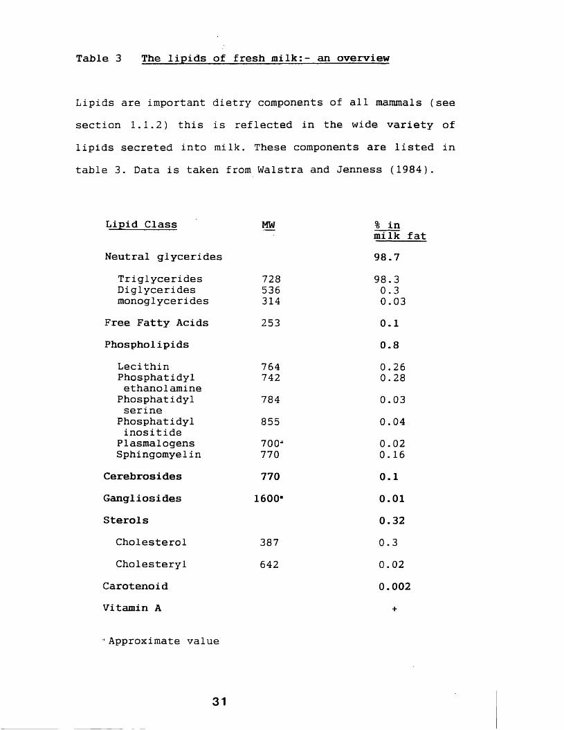
Table 3 The lipids of fresh milk:- an overview
Lipids are important dietry components of all mammals (see section 1.1.2) this is reflected in the wide variety of lipids secreted into milk. These components are listed in table 3. Data is taken from Walstra and Jenness (1984).
Lipid Class MW % inmilk fat
Neutral glycerides 98.7Triglycerides 728 98.3Diglycerides 536 0.3monoglycerides 314 0.03
Free Fatty Acids 253 0.1Phospholipids 0.8
Lecithin 764 0.26Phosphatidyl 742 0.28ethanolamine
Phosphatidyl 784 0.03serine
Phosphatidyl 855 0.04inositide
Plasmalogens 700a 0.02Sphingomyelin 770 0.16
Cerebrosides 770 0.1Gangliosides 1600" 0.01Sterols 0.32
Cholesterol 387 0.3Cholesteryl 642 0.02
Carotenoid 0.002Vitamin A +
- Approximate value
31

minerals notably calcium, potassium, phosphorous and sodium (see Table 4).
1.1.2.3 Milk proteins
Total protein in milk ranges from less than 10 g/1 in human to approximately 200 g/1 in the milk of some rabbits(Jenness, 1982). They may be broadly classified as caseins (the acid precipitable phospho-proteins) and the non-casein or whey proteins.
The major whey protein of ruminant milk is p-lactoglobulin, an albumin, which has also been isolated in a number of other artiodactyls including buffalo, goat and sheep, species where the amino acid sequence has been elucidated (see Jenness, 1982). It does, however, appear to be absent in virtually all non-artiodactyl species. The role of this protein remains unclear but suggested roles include: control of phosphate metabolism (Thompson and Farrell, 1974), involvement in the transfer of immunoglobulins from milk to the neonate (Butler, 1974), involvement in neonatal intestinal absorption of retinol (Pervaiz and Brew, 1985) or simply as a source of amino acids (Mepham, 1976). This protein also has considerable fatty acid binding properties (Perez et al, 1989).
In non-ruminant species the major whey protein is either the whey acidic protein (WAP) or a-lactalbumin (aLA). WAP which comprises a cysteine rich monomer of approximate
32
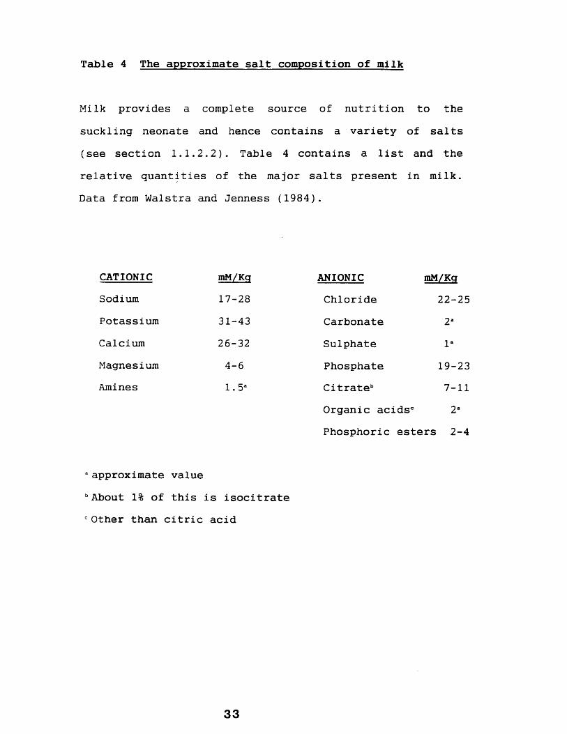
Table 4 The approximate salt composition of milk
Milk provides a complete source of nutrition to the suckling neonate and hence contains a variety of salts (see section 1.1.2.2). Table 4 contains a list and the relative quantities of the major salts present in milk. Data from Walstra and Jenness (1984).
CATIONIC mM/Kg ANIONIC mM/KqSodium 17-28 Chloride 22-25Potassium 31-43 Carbonate 2aCalcium 26-32 Sulphate laMagnesium 4-6 Phosphate 19-23Amines 1.5a Citrateb 7-11
Organic acidsc 2aPhosphoric esters 2-4
a approximate value bAbout 1% of this is isocitrate c Other than citric acid
33

apparent molecular weight of 14,000 Daltons, has been identified in the milks of rats and rabbits (Hennighausen and Sippel, 1982; Campbell and Rosen, 1984). The function of this protein is unknown, however the high cysteine content may serve to supplement the very low cysteine content of the caseins (see table 5).
With one exception a-lactalbumin has been found in all milks so far investigated. The exception being the milk of the Californian sea-lion (Zalophus californianus) in which it could not be detected by Johnson et al (1982) using an enzymatic assay. a-Lactalbumin is a polypeptide comprising 123 amino acids in all species (see Hall and Campbell,1986) except in the rabbit, 122 amino acids, (Hopp and Wood, 1979) and the rat, 140 amino acids (Prased et al,
1981). a-Lactalbumin acts as a co-enzyme, modifying the specificity of galactosyltransferase so as to favour the synthesis of lactose from UDP-galactose and galactose (see Hall and Campbell, 1986).
Apart from the major protein constituents, a number of minor protein constituents have been found in a number of species. Serum albumin, for example, a protein synthesized in the liver is present in the milk of all the approximately 200 species so far studied (Walstra and Jenness, 1984) and is presumably derived from the blood serum. No specific function of this protein in milk has been identfied.
34

Table 5 The composition of caseins
Caseins comprise around 80% of total milk protein and their principal role is nutritional (see section 1.1.2.3), this is reflected in the quantity and amino acid composition. Figure 5 (from Jenness, 1974)) lists the residues per mole of bovine caseins. Note the very low cysteine content of all caseins.
A m i n o A c id C o m p o s it io n s o f B o v i n e C a s e in s
Amino acid
Residues per mole
a^ -B K - A fi-A- y -A -'
Asp 7 25 4 4 4Asn 8 5 3Thr 5 20 14 9 8Ser S 21 12 11 10SerP 8 1 5 1Glu 25 56 13 17 10Gin 14 14 22 22Pro 17 15 20 35 34Gly 9 4 2 5 4Ala 9 11 15 5 5Val 11 19/20 11 19 17Cys/'2 0 2 0 0 0Met 5 5 2 6 6He 11 15 13 10 7Leu 17 18 8 22 19Tyr 10 14 9 4 4Phe 8 8 /9 4 9 9Lys 14 32 9 11 10His 5 5 3 5 5T ip o 2 1 1 1Arg 6 8 5 4 2
799 280/282 169 209 181
M W ( daltons) 23,616 33,700 19,023 23,982 20,497P 8 10 1 5 1
taken directly from Jenness, 1974
Other proteins such as lactoferrin (not present in rabbit milk - see Masson and Heremans, 1971), transferrin,immunoglobulins and at least forty enzymes (see section1.1.4) are commonly found in the milks of most species, but
6-7go up to make only about % of total milk protein (seetable 1). The majority of milk protein is howeverrepresented by the other class, the caseins.
Caseins
The term caseins is used to describe a group of milk specific proteins defined by the American Dairy Science Association as "a heterogeneous group of phosphoproteins precipitated from skimmed milk at pH 4.6 and 20°C". The majority of caseins in milk are found in the form of aggregates termed "casein micelles", the properties of which are described in section 1.1.3.
By far the best studied caseins are those isolated frombovine milk which consist of three principle species termedcu -, 3~, and k-casein. However many bovine casein variants have been reported. These are thought to result either from genetic polymorphism or differences in post-translational modifications (see Weller, 1979 and swaisgood, 1982).
The caseins of rabbit milk have been fairly well characterized, Testud and Ribadeu Dumas (1973) and Houdebine and Gaye (1975) have demonstrated the presence of three caseins and Dayal et al (1982) showed that these
36

comprised polypeptides of apparent molecular weights 31,000, 29,000 and 25,000 Daltons. In common with otherspecies rabbit caseins are post-translationally modified being both glycosylated and phosphorylated (Al-Sarraj et
al, 1978 and Dayal et al, 1982).
By examination of various properties of rabbit caseins, including phosphorous and carbohydrate content and cysteine content, Dayal et al (1982) concluded that the 31,000 and 29,000 Dalton species are analogous to bovine a- and k- casein respectively, a finding common to most other species. Studies at the nucleotide level show a remarkable similarity between all such caseins homologous to the bovine species (see Bonsing and MacKinlay, 1987).
Caseins comprise around 80% of total milk protein, including ruminant and rabbit milk, see Jenness (1973), and the principal role is undoubtedly nutritional, providing a rich source of amino acids to the suckling neonate (see table 4). However they contain very low levels of cysteine and this residue is probably provided by other milk proteins.
Caseins combine with calcium and phosphate to form casein micelles and in this form they act as carriers of these minerals to the neonate. The presence of the casein micelles has further been associated with the bioavailability of a variety of other minerals (reviewed by West, 1986).
37
Other roles suggested for caseins include the role of casein derived peptides as bioactive molecules. These peptides have been shown to be involved in a number of mechanisms for example in the stimulation of immune defence mechanisms in the neonate (Migliore-Samour and Jolles, 1988 and Migliore-Samour et al, 1989), as an opioid antagonist (Yoshikawa et al, 1986 and Chiba and Yoshikawa,1986), as an opioid agonist (Meisel and Friter, 1989) and as a fibrinogen inhibitor (Fiat et al, 1989).
It has already been mentioned that the majority of caseins in milk combine with calcium and phosphate to form a high molecular weight co-polymer termed the casein micelle. In this form the mother is able to store large quantities of these concentrated protein packages in the mammary gland ducts and can provide a high protein content milk to the suckling neonate. The micelle structure is also advantageous in that it protects the caseins from being precipitated by the high calcium content of milk.
In the following section the composition, structure and properties of casein micelles are discussed.
1.1.3 Casein micelles
Caseins are strongly interactive proteins capable of forming a variety of bonds with each other. Hydrophobic, hydrogen, electrostatic, disulphide and calcium bonding are
38

all possible (Thompson and Farrell, 1973 and Schmidt, 1982) and the majority of caseins in the milks of all species are found in the form of aggregates termed casein micelles.
In bovine milk all three principal species are found in the micelles and comprise over 90% of the structure (McMahon and Brown, 1984). Micelles are also very rich in both calcium and phosphate with smaller quantities of other inorganic ions and sugars (see table 6). The calcium and phosphate content of the micelles is mainly in a form termed colloidal calcium phosphate (CCP) which plays a vital role in the maintenance of micellar integrity. Removal of CCP to below a critical level causes dissociation of caseins, further removal causes complete dissociation of the micelles (Lin et al, 1972).
Casein micelles under the electron microscope appear as roughly spherical granular particles ranging in diameter from 20 to 680 nm (Schmidt and Payens, 1976; McGann, et al,
1980 and Donnelly et al, 1984) although it is now widely accepted that the micelles are built up from small "units" of casein aggregates of diameters 6 to 20 nm (see Knoop et
al, 1973 and Schmidt, 1982) termed casein sub-micelles (see also Schmidt, 1982; McMahon and Brown, 1984 and Ono et al, 1989).
Casein micelles have been the subject of considerable research, and much is known about their composition and characteristics (see Boulet et al, 1970 and McMahon and
3 9
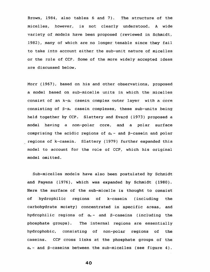
Brown, 1984, also tables 6 and 7). The structure of the micelles, however, is not clearly understood. A wide variety of models have been proposed (reviewed in Schmidt,1982), many of which are no longer tenable since they fail to take into account either the sub-unit nature of micelles or the role of CCP. Some of the more widely accepted ideas are discussed below.
Morr (1967), based on his and other observations, proposed a model based on sub-micelle units in which the micelles consist of an k-cu casein complex outer layer with a core consisting of p-cu casein complexes, these sub-units being held together by CCP. Slattery and Evard (1973) proposed a model having a non-polar core, and a polar surface comprising the acidic regions of cu - and p-casein and polar regions of k-casein. Slattery (1979) further expanded this model to account for the role of CCP, which his original model omitted.
Sub-micelles models have also been postulated by Schmidt and Payens (1976), which was expanded by Schmidt (1980). Here the surface of the sub-micelle is thought to consist of hydrophilic regions of k-casein (including the carbohydrate moiety) concentrated in specific areas, and hydrophilic regions of cu - and p-caseins (including the phosphate groups). The internal regions are essentially hydrophobic, consisting of non-polar regions of the caseins. CCP cross links at the phosphate groups of the cu - and p-caseins between the sub-micelles (see figure 4).
4 0
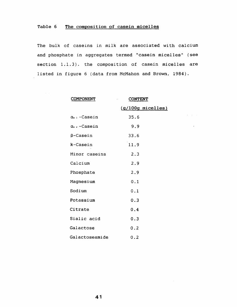
Table 6 The composition of casein micelles
The bulk of caseins in milk are associated with calcium and phosphate in aggregates termed "casein micelles" (see section 1.1.3). the composition of casein micelles are listed in figure 6 (data from McMahon and Brown, 1984).
COMPONENT CONTENT
cu i -Caseinas 2 -Casein3-Caseink-CaseinMinor caseinsCalciumPhosphateMagnesiumSodiumPotassiumCitrateSialic acidGalactoseGalactoseamide
(g/lOOg micelles)35.69.9
33.611.92.32.92.9 0.1 0.1 0.3 0.4 0.3 0.2 0.2
41
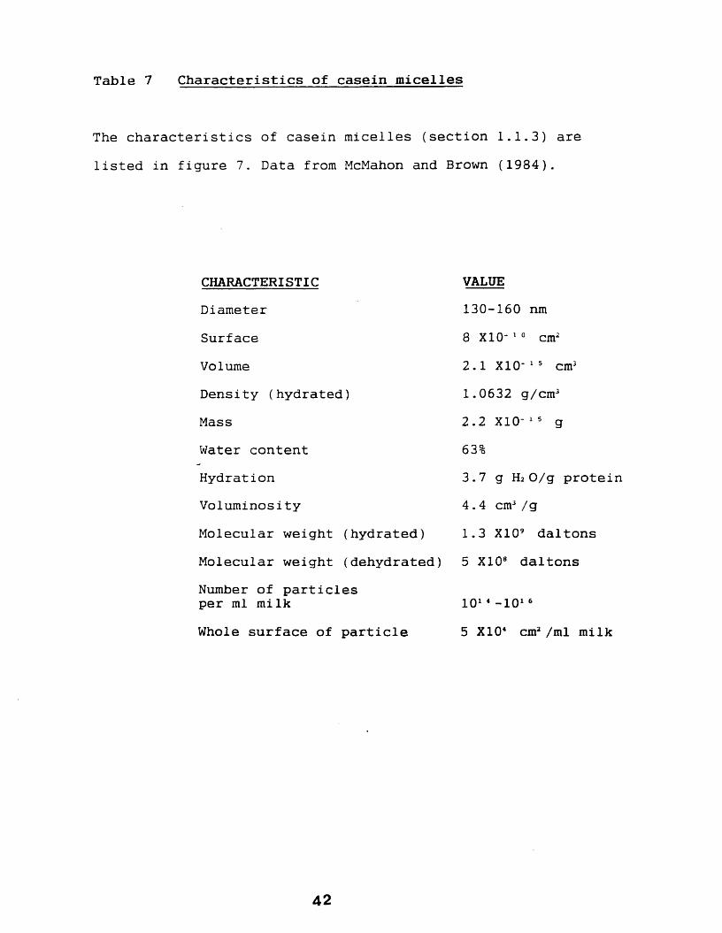
Table 7 Characteristics of casein micelles
The characteristics of casein micelles (section 1.1.3) are listed in figure 7. Data from McMahon and Brown (1984).
CHARACTERISTICDiameterSurfaceVolumeDensity (hydrated)MassWater content Hydration VoluminosityMolecular weight (hydrated)Molecular weight (dehydrated)Number of particles per ml milkWhole surface of particle
42
VALUE130-160 nm 8 X10- 1 0 cm2
2.1 X10- 1 5 cm3 1.0632 g/cm32.2 X10- 1 5 g 63%3.7 g H2O/g protein4 . 4 cm3 /g1.3 X109 daltons5 X108 daltons
101 4 - 101 65 X104 cm2 /ml milk
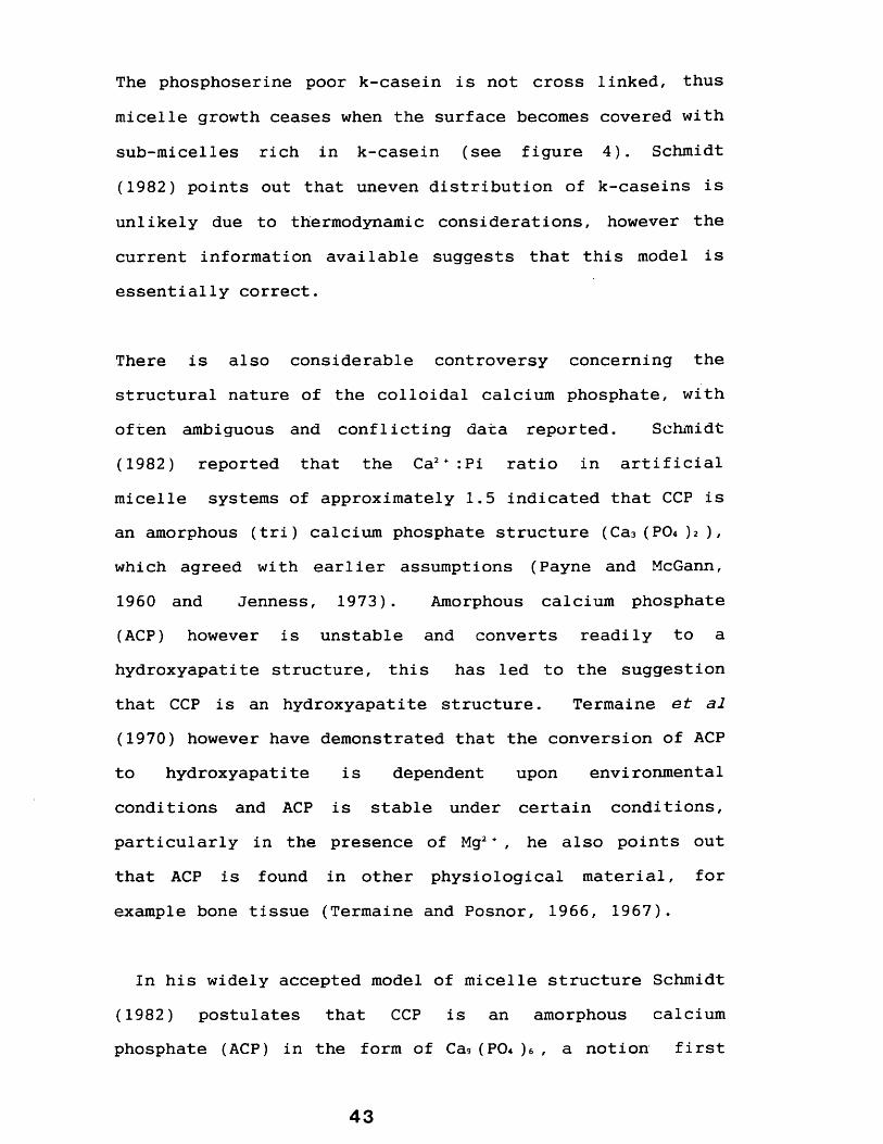
The phosphoserine poor k-casein is not cross linked, thus micelle growth ceases when the surface becomes covered with sub-micelles rich in k-casein (see figure 4). Schmidt (1982) points out that uneven distribution of k-caseins is unlikely due to thermodynamic considerations, however the current information available suggests that this model is essentially correct.
There is also considerable controversy concerning the structural nature of the colloidal calcium phosphate, with often ambiguous and conflicting data reported. Schmidt (1982) reported that the Ca2+ :Pi ratio in artificial micelle systems of approximately 1.5 indicated that CCP is an amorphous (tri) calcium phosphate structure (Ca3 (P04 )i ), which agreed with earlier assumptions (Payne and McGann, 1960 and Jenness, 1973). Amorphous calcium phosphate (ACP) however is unstable and converts readily to a hydroxyapatite structure, this has led to the suggestion that CCP is an hydroxyapatite structure. Termaine et al
(1970) however have demonstrated that the conversion of ACP to hydroxyapatite is dependent upon environmental conditions and ACP is stable under certain conditions, particularly in the presence of Mg2 + , he also points out that ACP is found in other physiological material, for example bone tissue (Termaine and Posnor, 1966, 1967).
In his widely accepted model of micelle structure Schmidt (1982) postulates that CCP is an amorphous calcium phosphate (ACP) in the form of Ca9 (PO4 )e , a notion first
43
Figure 4 Proposed model of the casein micelle structure
Schematic representation of the widely accepted model of the structure of the casein micelle proposed by D.G. Schmidt (see section 1.1.3).
these sub-units, note that the surface consists of subunits that contain a high proportion of k-casein which, according to Schmidts' theory, prevents further sub-unit association with this micelle. Figure 4c shows the proposed colloidal calcium phosphate links between the sub-micelles.
Diagrams are taken directly from Schmidt (1982).
Figure 4b, a completed micelle assembled from
submiceile
cV P0i'6
peptide chain
submiceile
c
key to fig. B
POj groups
t Ca0iP 0 ^ '6 c luster
44

postulated by Betts and Posner (1974). McGann et al (1983) also concluded that CCP is an amorphous calcium phosphate in combination with citrate and magnesium, conditions, as previously stated, where Termaine (1970) has shown ACP maybe stable.
The nature of the bonding between CCP and the caseins has also been the centre of some controversy. Ho and Waugh (1965) and Rose and Colvin (1966) first postulated that the site of binding of CCP in caseins is the organic (ester) phosphate (although the exact nature of the phosphate groups in caseins was unclear at this time).
More recently, advances have been made concerning the nature of the role of CCP in binding sub-micelles together and it's site of interactions. Aoki (1986) isolated casein aggregates cross-linked by CCP using high-performance gel chromatography in the presence of urea and showed (Aoki,1987) that the caseins were cross linked to CCP by esterphosphate groups to phosphoserine and phosphothreonine. He further demonstrated (Aoki, 1988) that the dissociation of caseins was related to the level of phosphorylation indicating that the strength of the micelle, as would be expected from his previous work, was dependent upon thephosphate available to form CCP linkages.
Bingham et al (1972) have however shown that when dephosphorylated cui were present then micelle structures could still be formed. Results which suggest that
4 5
interactions other, or including, than those postulated above may be involved in the formation of the casein micelle.
1.1.4 The enzymes of milk
1.1.4.1 Introduction
All milks have been shown to contain a wide variety of different enzymes (see Jenness, 1978), bovine milk being the best studied with the presence of over forty enzymesreported (see Shahani, 1966 and Jenness, 1978).
A number of studies have been made on the enzymes in rabbit milk, and although only a very few have been identified (see table 8), it would be expected that rabbit milkcontains most of the common enzymes found in the milks of other species. Although it should be noted that there are marked species differences both in the amounts and in some cases the presence of certain enzymes. Lysozyme, for example, is found at concentrations of 0.11 mg/ml in bovine milk (Chandan et al, 1968) but in primates and particularly in human milk it is a major whey protein found at concentrations of 400 mg/ml (Chandan et al, 1968).
1.1.4.2. Distribution and origin of milk enzyme
Enzymes in milk are not distributed in a homogeneousmanner, rather they are found to be associated with one or
46

Table 8 Enzymes of rabbit milk
A large range of enzymes have been identified in the milks of a number of species (section 1.1.4), few, however have been reported in the rabbit. These are listed below:
ENZYME REFERENCELysozyme Shahani et al (1962)L.D.H. Kjellberg and Karlson (1967)M.D.H. Kjellberg and Karlson (1967)Xanthine oxidase Larson (1974)Ribonuciease Chandan et al (1968); Kiermeier and
Kulman (1972)Galactosyl-transferase D.Lennon and A. Boulton (unpublished
observation)
4 7
more distinct phases, broadly classified as those associated with: the milk fat globule membrane, milkmicrosomal membranes, the water soluble phase, and those associated with soluble casein or the micelle complex.
In addition it should be noted that the distribution of an enzyme may not be stable, either due to physiological changes or to the experimental procedures employed in their study.
For example one of the lipases found in a soluble form in milk, the so-called naturally active lipase, can in freshly drawn milk become absorbed into milk fat globule membranes on cooling (Tarassuk and Frankel, 1957), and Morton (1953) reported that 30-40 % of alkaline phosphatase was associated with the MFGM. He suggests however that a greater proportion is originally bound when the milk is first secreted into the milk ducts, which then becomes dissociated during processing of milk, particularly during centrifugation.
The distribution of some milk enzymes depends on their origin. For example both xanthine oxidase (XO) and alkaline phosphatase (AP) in milk have been shown to be associated with the milk fat globule membrane (MFGM) (see Patton and Keenan, 1975), a structure which originates from mammary secretory epithelial cell plasma membrane (see Wooding, 1971 and Patton and Keenan, 1975). As both XO and
4 8
AP are originally plasma-membrane proteins it is not then surprising that they will be part of the MFGM.
Milk contains other membranes which also have enzymes closely associated with them. Morton (1954) demonstrated that AP and considerable XO activity were associated with membranes that were distinct from the MFGM, although Wooding (1971) disagrees that these membranes are not derived from the MFGM. These results were confirmed by Janolino and Swaisgood (1984), who also identified other enzymes (eg sulphydryloxidase and glutamyl transferase) asbeing associated with these membranes.
The distribution of milk lipase is of some interest to this project (see results chapters 3 and 4), as it has been reported to be an enzyme associated with casein (Harper et
al, 1956), an observation which has been confirmed by several groups. Skean and Overcast (1961) reported lipase activity associated with a-casein, and two groups have found that it may also be associated with the k-casein species (Yaguchi et al, 1964 and Fox et al, 1967). Whilst Gaffney et al (1966a, 1966b) detected lipase activity with a number of casein fractions, they also state that the lipase was previously found to be associated specifically with the (3-casein fraction citing their previous work (Saito et al, 1958) as reference, such an observation is however not clear from the original article.
49
Reviewing this work Downey and Murphy (1970) argued that the divergent nature of these observations was due to the tendancy of milk lipase to absorb onto protein precipitates during the fractionation of milk protein components.
A number of milk enzymes are thought to originate from the blood serum, for example aldolase and catalase. The levels of catalase and aldolase, were found to be markedly increased in the milks of animals with bacterial infections (Kitchen et al, 1970). These infections lead to inflammation of the mammary gland producing increases in vascular permeability leading to a resultant influx of serum proteins into milk. The presence of high levels of enzymes is thought to result from leakage of the serum as a consequence of this increase in permeability (Kitchen et al, 1970). Presumably there is some leakage of blood proteins into milk in the healthy animal as these enzymes, and other serum proteins, are usually present in normal mi lk.
The origin of other milk enzymes, notably proteolytic enzymes, may originate from the micro-organisms that inhabit healthy mammary gland alveoli, although milk does contain significant levels of indigenous proteolytic enzymes (Stores and Hull, 1956 and Harper et al, 1960).
1.1.4. 3 Roles for Milk Enzymes
50

It has been shown that many of the enzymes in milk are well characterized with respect to their origin, distribution and the reaction catalysed. One question remains a point of contention however, i.e. do these enzymes have a physiological role in milk ?.
Kitchen et al (1970) and Shahani (1973) suggested that the presence of enzymes in milk can be regarded as a "spilling over" from secretory cells and serum during the intense process of milk secretion. This probably occurs by exocytosis and not from cel 1-rupture as has been suggested in the literature (Shahani, 1966).
However this does not exclude a role for these enzymes once secreted into the milk. For example it was earlier described that XO being situated in the plasma membrane will ipso facto become associated with the MFGM, and here it seems it may have an important bacteriostatic function.
Green and Poli (1943) first showed that this enzyme can inhibit a variety of gram ( + ) and gram (-) bacteria. Reiter and Oram (1967) suggest that one possible mechanism for this may be the action of hydrogen peroxide, one of the products of the reaction catalysed by XO, as a bactericidal agent against microbial infection. Reiter and Oram (1967), however, also point out that milk contains only low concentration of the substrates present to produce bactericidal levels of hydrogen peroxide.
51
Bacteriostatic roles for the milk enzymes lactoperoxidase have also been postulated (Reiter and Oram, 1967; Jago and Morrison, 1963 and Gothefors and Marklund, 1975).
Recently it has become clear that XO probably exists intra- cel lularly as xanthine reductase and can be converted to the oxidase form by the action of sulphydryloxidase (Clare et al, 1981). Blakistone et al (1986) suggest that during conversion of the enzyme from reductase to the oxidase form which may occur in the alveoli duct could initiate oxidation of the lipid and membrane breakdown.
Human milk contains significant levels of proteolytic enzymes (Storrs and Hull, 1956) which may play an important role in the digestion of milk proteins, in the digestive system of the suckling young where digestive enzymes may not be fully functional. This would also explain why there is proteolytic activity associated with bovine caseins (Warner and Poli, 1945). However, Harper et al (1961) concluded that the levels of proteolytic enzymes in bovine milk were too low for it to have a significant role.
Milk therefore, contains large number of enzymes originating from a variety of sub-cellular sources. Although some of these enzymes may have a role, the function, if any, of most is still unclear.
52

The following sections deal with, in some detail, the expression of milk proteins and the secretion of the major milk components including casein micelles.
1.1.5 The expression of milk proteins
It is now well established that the expression, ie. transcription and translation processes, of a number of milk proteins is hormonally regulated. The principal hormones involved appear to be: prolactin (a peptidesynthesized in the anterior pituitary gland); glucocorticoids (steroid hormones such as cortisol, hydroxycortisone, etc) and progesterone (another class of steroid hormone).
The role of prolactin has been studied in some detail by a number of groups and has been shown to regulate the expression of caseins at three levels:
1) Using tissue culture techniques, Guyette et al (1979) demonstrated in the rat mamamry gland the presence of prolactin resulted in a 2-4 fold increase in the rate of transcription of casein mRNA. Similar effects have also been demonstrated in the rabbit (Teysott and Houdebine, 1980) and the mouse (Ball et al, 1988).
2) Prolactin has also been shown to increase the half-life of the casein mRNA molecules. Guyette et al (1979), for example, have shown a 17-25 fold increase in the half-life
53

of rat casein mRNA. A similar effect was demonstrated by Teysott and Houdbine (1980) in the rabbit.
3) It further appears that prolactin can also effect the preferential stimulation of casein mRNA translation (Teysott and Houdebine, 1981).
The effects of prolactin generally appear to be enhanced in the presence of glucocorticoids (Devinoy et al, 1978) but inhibited by progesterone (Teysott and Houdebine, 1981 and Houdebine and Gaye, 1975). The responsiveness of the mammary gland to prolactin may further be affected by external factors including seasonal variation (Foster et
al, 1989). Insulin too, may also play a role in enhancing casein expression (Choi et al, 1988). A number of other factors may also have a role in milk-protein expression including the extra-cellular matrix proteins (Blum et al,
1987).
Although less well characterized the expression of other milk proteins, including aLA, WAP, transferrin and lactoferrin, have been shown to be hormonally regulated by prolactin and glucocorticoids (Terada et al, 1988 and Vonderhaar and Ziska, 1989).
In common with the biosynthesis of most secretory proteins, milk proteins are synthesised on membrane bound polyribosomes for example aLA (Gaye et al, 1972); a-casein (Gaye et al, 1973); p-casein (Houdebine and Gaye, 1975) and
54
p-lactoglobulin (Gaye and Denamur, 1972). These observations have been confirmed in a number of other species including guinea-pig (see Craig et al, 1978) and the rabbit (Houdebine and Gaye, 1975).
It is now generally accepted that polyribosomes associated with the secretory protein mRNA are directed to the ER membrane by a series of "signals" and "receptors" (for recent review see Geiasch, 1989). Briefly, translation of secretory protein mRNA initially produces a short polypeptide (the signal region) which is recognised by a cytoplasmic receptor, the signal recognition particle (SRP). This causes a temporary arrest of translation during which the SRP/protein complex is directed to the RER membrane where it is recognised by integral membrane proteins. On reaching the RER translation resumes and the polypeptide is inserted across the RER membrane into the luminal space where the signal peptide is removed. Vectorial transport across the RER membrane therefore occurs co-translationally. Only an outline of this process has been given. For further information see reviews by Mepham et al (1982) and Pfeffer and Rothman (1987).
Once transported into the RER lumen further modifications can occur. One of which: glycosylation, involving theaddition of sugar moieties, is discussed briefly in the following section, and another: phosphorylation, which isdescribed at some length elsewhere.
5 5
N-glycosylation involves the transfer of a "core" oligosaccharide (glu)3-(Mann)9-(NAglu)2 via a lipid- phosphate carrier molecule (dolichol phosphate) to an asparagine residue, located at Asn-x-Thr/Ser of the protein undergoing glycosylation (Lennarz, 1983). This modification occurs in the ER probably whilst the polypeptide in being synthesized (Hanover and Lennarz, 1980). Further carbohydrate additions and trimming of these molecules also occurs in the endoplasmic reticulum, processes which are completed in the golgi (see Lennarz, 1983). Both rat (Gaye ea, 1982) and rabbit a-LA (Lingappa et al, 1978) are N- glycosylated.
In contrast O-glycosylation, involves the direct transfer of oligosaccharides to the hydroxyl group of either serine or threonine, without the involvement of a 1ipid-phosphate carrier molecule. It is generally agreed that 0- glycosylation is a post-translation event which occurs in the golgi-apparatus.
A number of caseins have been shown to contain carbohydrate moieties: for example bovine K-casein (Jolles et al, 1973 and Fournet et al, 1975); guinea pig caseins (see Craig et
al, 1978) and rabbit caseins (Suard et al, 1982) are all glycosylated. It has been reported that bovine k-casein contains an "0" linked sugar (Fiat et al, 1968-referenced in Jolles et al 1973) whilst there is some evidence to suggest that the rat k-casein may contain an N-glycosidic linkage attached to asparagine (Rosen and Shields, 1980).
56

The golgi also appears to be the site of further milk protein modifications including casein phosphorylation, as a number of groups have reported the identification of a casein kinase in the golgi apparatus of lactating mammary gland from several species (see section 1.2.4.8). This modification is discussed in detail in Section 1.2.
In the following section the mechanisms involved in the secretion of some of the milk components described above are described.
1.1.6 Secretion of the major milk compoments
1.1.6.1 Fat
A prominant feature of electron micrographs of cross sections of the lactating mammary gland are the large globules of fat seen in the lumen and in the cytoplasm of the secretory cell (see Figures 2 and 5). It has been known for a very long time that fat globules are surrounded by a membrane (reviewed in Hammersten, 1911) which has more recently been proved to be derived from the lactating secretory cell apical plasma membrane (Kitchen, 1974; Keenan et al, 1979).
The gross mechanism of secretion has now been well characterized. Following their formation the globules of
5 7

fat are transported to the apical membrane. Once reached the fat globule "pushes-up" the membrane (see figure 5) which finally envelopes it to become the milk fat globule membrane. The globule is thus secreted by a process akin to reverse pinocytocis.
A number of groups have noticed that large areas of cytoplasm are occasionally secreted within the milk fat globule membrane (Stockinger and Zarzicki, 1962; Kurosumi et al, 1968 and Wooding, 1971). This has led to some confusion as to whether this process should be regarded as "apocrine". The present consensus of opinion is that these cytoplasmic containing fat globules (known as "signets") are not very common and probably result from the premature "pinching off" of the plasma membrane, thus removing a section of cytoplasm, as a consequence of either contraction of myo-epithelial cells therefore squeezing the secretory cell or from internal "pressure" within the cell due to a build up of secretory components. It is generally felt that the term "apocrine" is therefore inappropriate with respect to fat secretion from the lactating mammary gland secretory cell.
1.1.6.2 Protein
Newly synthesized protein in the mammary gland have been traced to the golgi apparatus and prior to secretion accumulate in apical golgi cisternae (see Saacke and Heald,
5 8
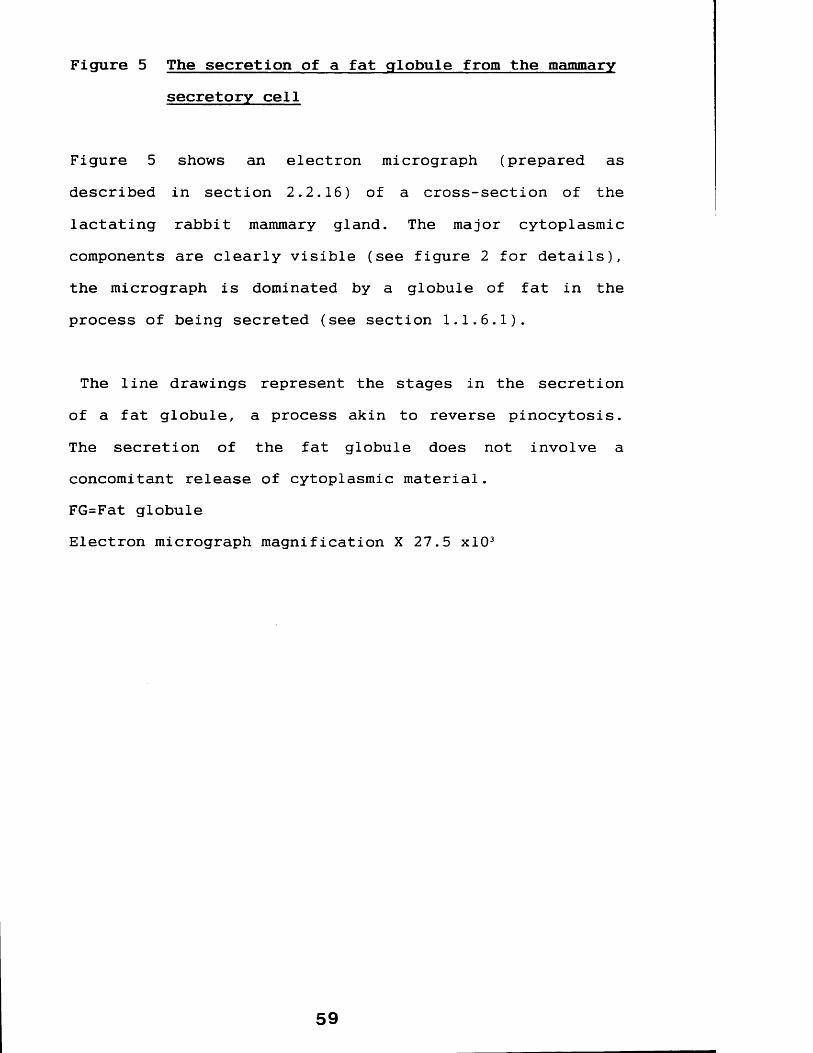
Figure 5 The secretion of a fat globule from the mammary secretory cell
Figure 5 shows an electron micrograph (prepared as described in section 2.2.16) of a cross-section of the lactating rabbit mammary gland. The major cytoplasmiccomponents are clearly visible (see figure 2 for details), the micrograph is dominated by a globule of fat in theprocess of being secreted (see section 1.1.6.1).
The line drawings represent the stages in the secretion of a fat globule, a process akin to reverse pinocytosis.The secretion of the fat globule does not involve aconcomitant release of cytoplasmic material.FG=Fat globuleElectron micrograph magnification X 27.5 xlO3
59

1974). Secretory vesicles are formed by the pinching off of these apical golgi cisternae thereby trapping the caseins. These vesicles are the site of the initiation of micelle bio-assembly and have been shown in numerous studies to contain casein micelles (for example see Franke et al 1976; Sasaki et al, 1978 and keenan et al, 1979). The secretory vesicles migrate to the apical membrane of the cell (see figure 2), fuse with it and release their contents into the lumen. Such a process can be followed by electron micrographic analysis of cross-sections of the lactating mammary gland (for example Helmien et al, 1968 and Franke et al, 1976) and have been demonstrated in the rabbit mammary gland (see this thesis: appendix 2).
The secretion of whey proteins has been less well characterized, probably since because they do not aggregate into large complexes, as do the caseins, they are not seen in electron micrographic analysis. It seems likely however that they are secreted within golgi derived vesicles as described above as a-lactalbumin and (3-lactoglobulin are present in secretory vesicle preparations (Sasaki et al,
1978).
1.1.6.3. Mi seal1aneous components
Lactose has also shown to be present in both the golgi (Kuhn and white, 1975) and in golgi derived secretory vesicles (Sasaki et al, 1978 and Keenan et al, 1979) and is the probable mode of secretion of: calcium (Baumrucker and
61

Keenan, 1978), potassium (Silcock and Patton, 1951) and citrate (Linzell and Peaker, 1971). Water probably follows the secretion of these components as a result of the osmotic pressure they exert.
So far this general introduction has discussed general mammary gland biology with emphasis on the proteins of milk. The following section discusses the phosphorylation of proteins (an important post-translational modification of caseins) the enzymes involved, protein kinases, with reference to the casein kinases.
62

1.2 PROTEIN PHOSPHORYLATION
1.2.1 General considerations
The first phosphoproteins to be described were phosvitin from egg yolk and the caseins from milk. Since then very many phosphoproteins have been described, and the term is now used to define many hundreds of proteins which contain amino acids with covalently bound phosphorous, even if the protein may only be transiently phosphorylated, or contain only a single phosphate group.
Protein bound phosphate may be classified as 0-, N- or acyl- linked phosphate. The most common in terms of abundance being the O-phosphates, which includes phosphoserine (see figure 6), the most common, and phosphothreonine (see figure 6). Eckhard et al (1979) reported that polyoma virus T antigen amino precipitates contained protein kinase activity. The proteinsphosphorylated by this enzyme were isolated and were shown by their electrophoretic behaviour and acid/alkali stability to contain 0-1inked phosphate at tyrosine, a previously unreported phosphorylated residue which has since been shown to be an increasingly important and common amino acid modification (see Hunter 1982).
Less common are the N-phosphates (phosphoamidates) represented by phosphohistidine, phospholysine and
63

phosphoarginine. Phosphoarginine has been reported from mammalian sources (Scott et al, 1976 and Levy-Favatier et
al, 1987) and in viral systems (Wilson and Consigh, 1985). More commonly found is protein bound phosphohistidine, phosphorylated at either the 1 or the 3 position of the imidazole ring, which has been reported in various fraction isolated from mammalian liver (eg. Zetterqvist and Engstrdm, 1966 and Blat and Harel, 1970).
Walinder et al (1968) reported the purification of a bovine liver protein which on incubation with 32P ATP was shown, by alkaline hydrolysis, to contain 1-32P, 3-32Pphosphohistidine and 3 2 P-phospholysine, phosphorylated at the Ne position, (see figure 6). The role of this protein is unknown, but Walinder et al (1968) postulate that it is an enzyme which is phosphorylated at these sites during the course of its reaction, similarly Todhunter and Purich (1974) reported that a glutamic acid (acyl phosphate) residue in E.Coli acetate kinase is transiently phosphorylated during the course of its reaction. It is noted that all caseins so far studied contain mainly phosphoserine with some also containing phosphothreonine.
A rather unusual protein bound phosphorylated amino acid has been described by Rhee (1984), i.e. the bacterial enzyme glutamine synthetase was shown to contain a 5 1 - nucleotidyl-O-tyrosyl residue, that is a adenylyl and uridylyl group attached in via phosphomonoester linkage to the hydroxy group of tyrosine residues (see figure 7c).
6 4
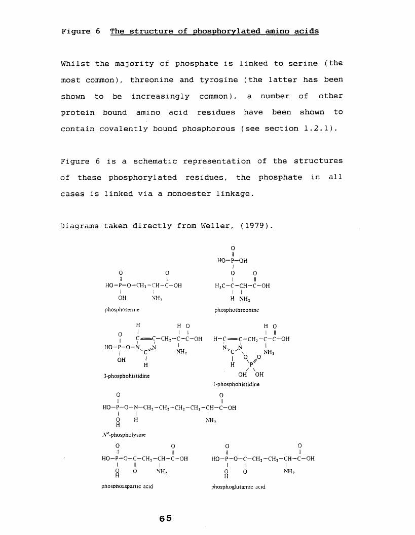
Figure 6 The structure of phosphorylated amino acids
Whilst the majority of phosphate is linked to serine (the most common), threonine and tyrosine (the latter has been shown to be increasingly common), a number of other protein bound amino acid residues have been shown to contain covalently bound phosphorous (see section 1.2.1).
Figure 6 is a schematic representation of the structures of these phosphorylated residues, the phosphate in all cases is linked via a monoester linkage.
Diagrams taken directly from Weller, (1979).
0IIH O - P - O HI
0 0 0 0II II I IIh o - p - o - c h 2- c h - c - o h h 3c - c - c h - c - o hI I II
OH N H 2 H N H 2
phosphosenne phosphothreonine
H H O H O0 i I II I IIII C = C - C H 2- C - C —OH h - c = c - c h 2- c - c - o h
I 1 I 1 I IHO —P—0 —N .N ' N * .N 1
1 ^ N H j ' C ' \ n h 2
OH I 1 ° \ PH H P
/ \3-phosphohistidine OH OH
1-phosphohistidine
0 0II IIHO—P—0 —N - C H , —CH 2- C H 2—CH2- C H - C - 0 H
I I I0 H NH ,H
A^-phospholvsine
0 0 0 0II II II II
H O —P—0 —C—CH i — C H —C —OH H 0 - P - 0 ~ C - C H 2- C H 2- C H - C - 0 H1 II I I II I
o 0 N H , 0 0 n h 2H H
phosphoaspartic acid phosphoglutamic acid
6 5
Whilst the majority of protein bound phosphate is linked to the protein as a monoester (Fig 7a), other linkages may be present. Curti and Porcellati (1960) and Porcellati and Curti (1960) for example reported that a phosphoserine present in certain proteins isolated from the central nervous system of the chicken was resistant to cleavage by purified phosphomonoesterases but was susceptible to cleavage by purified phosphodiesterases, indicating the this phosphate group was linked to the protein via a diester link (see figure 7).
Perlman (1954a, 1954b) in similar studies concluded that aproportion of phosphate in both a-casein (40%) and 13- casein (an unspecified amount) was linked to the amino acid residue via a diester bond (although not stated, Perlman probably used bovine caseins).
Ho et al (1969) using 31P Nuclear magnetic resonance (NMR) however found no evidence to substantiate the presence of such linkages in bovine as-B-casein. More recent evidence based on primary protein sequence data also seems to substantiate the observations of Ho et al (1969) (see West, 1989).
Other studies by Perlman (1955) and more recent studies using 31 P NMR techniques (Edmonson and James, 1979 and James et al, 1981) have concluded however that a number of
6 6
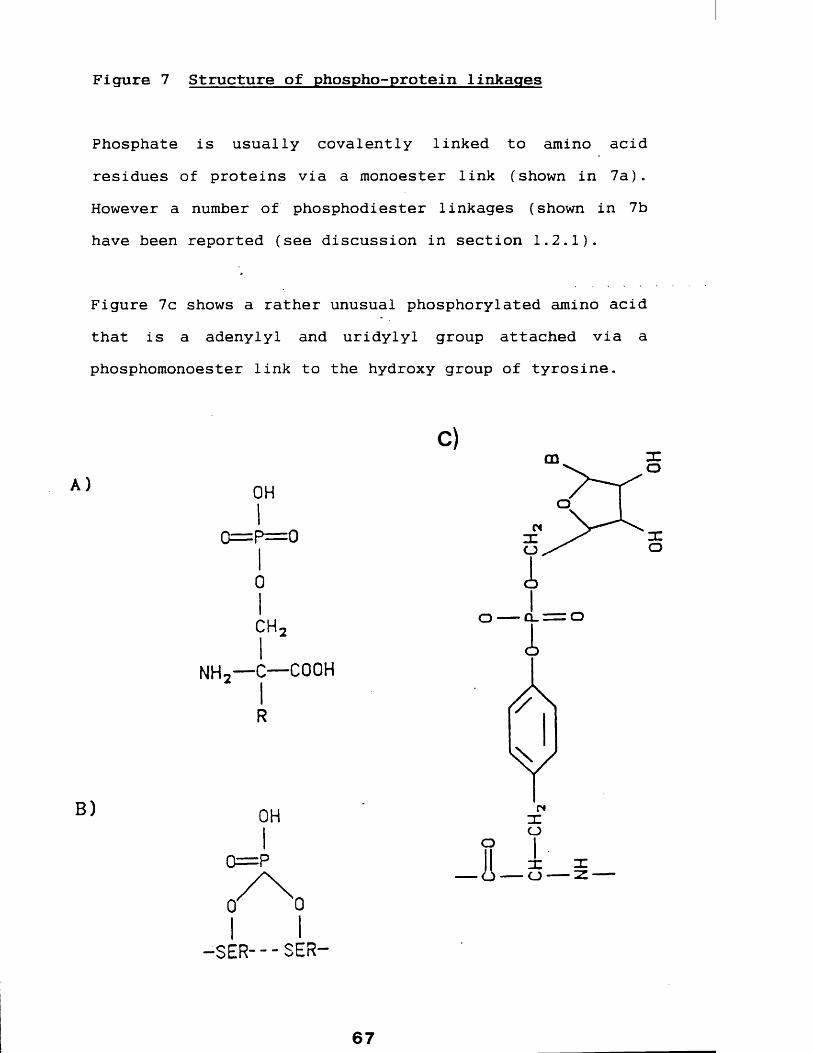
Figure 7 Structure of phospho-protein linkages
Phosphate is usually covalently linked to amino acid residues of proteins via a monoester link (shown in 7a). However a number of phosphodiester linkages (shown in 7b have been reported (see discussion in section 1.2.1).
Figure 7c shows a rather unusual phosphorylated amino acid that is a adenylyl and uridylyl group attached via a phosphomonoester link to the hydroxy group of tyrosine.
other proteins may contain such diester linked phosphate groups.
There is also indirect evidence to suggest that a phosphoserine residue of phosvitin might be converted to the enol phosphate derivative (Grant and Tarborsky, 1966).
In the following sections, the phosphorylation of casein is discussed, followed by a review of the role of protein phosphorylation including the role of the phosphate groups of caseins.
1.2.2 Casein phosphorylation
Turkington and Topper (1966) studied casein synthesis using mouse mammary gland explants and showed that the phosphorylation of caseins occurred post-translationally using a pool of unphosphorylated casein, results subsequently confirmed by Singh et al (1967) in lactating rat mammary gland tissue slices and confirmed by Mercier and Gaye (1980).
Mammary gland specific casein kinases have been isolated from golgi enriched fractions from a number of species and is thought to be responsible for the in vivo
phosphorylation of caseins. These enzymes are discussed in detail in a later section. The bovine caseins have been studied most extensively. The phosphorous of the cu i caseins is present as phosphoserine, with the cu i -D variant
6 8

containing an additional phosphate group linked to threonine. Bovine {3-caseins also contain phosphoserine as do the k-caseins. The phosphorous of caseins occurring only as phosphomonoesters (see previous discussion in section 1.2.1).
Dayal et al (1982) studied the characteristics of caseins isolated from rabbit, the species of interest in this thesis, and reported the phosphorous content of the three casein species identified. The 31,000 Dalton a casein contained approximately 4.5% w/w phosphate, whilst the "k- casein", of molecular weight 29,000 Daltons, contained only low phosphorous content (0.57% w/w). The phosphate content of the third casein, "X-casein", was not determined. It however stained blue with Stainsall but red after treatment with phosphatase, indicating that it is also a phosphoprotein, confirming the results of Al-Sarraj et al
(1978) who separated rabbit milk caseins and showed that they were all phosphoproteins.
Interestingly human |3-casein which has been shown to be present in five forms, each containing different levels of phosphorous, including one with no phosphate content (Groves and Gordon, 1970). All 3-caseins of the other species studied are, however, phosphorylated (see Jenness, 1978).
1.2.3. Role of phosphoprotein phosphate
69

Many enzymes contain covalently bound phosphorous only transiently. This can lead to the regulation of their function (reviewed in Weller, 1979), prehaps the best known example of this are those involved in the metabolism of glycogen. Under the influence of the hormone epinephrine (adrenaline) phosphorylase (3-kinase is activ ated via phosphorylation by the cyclic AMP dependent phosphorylase kinase kinase. The activated phosphorylase kinase in turn converts the inactive form of glycogen phosphorylase ("B" form) into the active form (phosphorylase "A") by transferring a phosphate group from ATP to the single phosphorylatable serine residue. Activated glycogenphosphorylase then can proceed to break down stored glycogen into glucose 1-phosphate which, after conversion into glucose 6-phosphate, is fed into the glycolytic metabolic pathway.
Many other enzymes which transfer phosphate groups themselves form transiently phosphorylated intermediates during the course of their reaction i.e autophosphorylation, the phosphate is only a "passenger" on the enzyme before being transferred to substrate protein, although the phosphorylation may cause conformational changes to the enzyme to allow the next stage to occur.
One interesting anomaly is the enzyme pepsin A [EC3.4.23.1] which contains a single phosphate at residue 68 (Stepanov et al, 1965 and Moravek and Koskta, 1973). The
70

enzyme is however neither regulated by phosphorylation nor is it a transient reaction intermediate (Weller, 1979).
Many non-enzyme proteins also contain phosphate which can serve functions other than those outlined above. They may serve as stores of phosphate. Phosvitin from the domestic fowl (Domesticus gall us) for example is the most highly phosphorylated naturally occurring protein known, containing some 9.7-10% phosphorous (Mecham and Olcott, 1949) and the caseins from milk, which are also very rich in phosphate (see table 5), provide rich sources of amino acids and this ion to the developing young.
Besides this role, phosphorylation of the caseins is thought to effect the bio-availability of a number of ions (see West, 1989 ) .
In the following sections the enzymes responsible for protein phosphorylation are reviewed, including those responsible for the in vivo phosphorylation of caseins.
1.2.4 Protein kinases
1.2.4.1 General considerations
Protein kinases are the enzymes responsible for catalysing the transfer of the phosphoryl groups of a nucleoside- triphosphate to form phosphoesters of amino acid residues of the substrate protein. All protein kinases so far
71

studied utilize adenosine triphosphate (ATP) as the nucleoside phosphoryl donor, although some are also able to use guanosine triphosphate (GTP). The phosphate group can be transferred to a variety of protein bound amino acids, the most common being serine and threonine, usually resulting in the formation of a phosphomonoester link.
In the last ten years there has been an "explosion" in the number of protein kinases identified, leading to the comment by Hunter (1987) that the mammalian genome may "encode as many as a thousand distinct protein kinases". Much of the increase of protein kinases identified results from the application of molecular biology rather than conventional purification techniques, particularly in the identification of a number of protein tyrosine kinase viral gene products (see Section.1.2.4.6).
There have been a number of attempts to rationalize protein kinase nomenclature, the terminology however is still often confusing and in some instances misleading. The enzyme commission number : [E.C. 2.7.1.37] for protein kinases (ATP: protein phosphotransferases) too can nolonger represent the full range of protein kinases.
Much of the confusion in classifications stems from the fact that historically some protein kinases have been named after a particular substrate used in in vitro assays. This has led, for example, to protein kinases being isolated from diverse sources, including plant tissues, which being
72

capable of phosphorylating caseins in vitro, are then classified as "casein kinases", clearly examples where casein would never be available as the physiological substrate.
Other kinases more appropriately named after a substrate include the myosin light chain kinase and opsin kinase where the prefixes relate the physiological substrate, this too can however be misleading in the cases where the enzyme may phosphorylate a number of substrates in vivo but is named after only one. Many ubiquitous enzymes with a wide range of putative physiological substrates are frequently named after a particular effector eg cyclic AMP/GMP dependent protein kinase and calmodulin dependent protein kinase, or simply a non-committal name eg. protein kinase C is used. Some of these major groups of protein kinases are described in the following sections.
1.2.4.2 Cyclic Adenosine 3'-5' Monophosphate dependent protein kinases
Cyclic adenosine monophosphate (cyclic AMP) has long been known to be an important intermediate in cellular function. It is activlated when hormones or neuro-transmitters, the first messengers, combine with specific cell surface receptors on target cells leading to the activation of adenyl cyclase which in turn stimulates the intra- cellular second messenger, cyclic AMP. Cyclic AMP will then stimulate the cellular response which includes the
73

activation of proteins which in many cases is a protein kinase.
Cyclic AMP dependent protein kinases contain both regulatory (R) and catalytic (C) subunits usually in the form R2 -Ci . The binding of cyclic AMP to the regulatory subunit of the inactive holoenzyme causes the dissociation of this complex into the active enzyme i.e.
R2 -(Cyclic AMP)4 + C2 where C2 then catalyses the phosphorylation of a specific
target protein.
Cyclic AMP dependent protein kinases are found virtually in all mammalian species and have a wide tissue distribution. Kato et al (1983) reported the characterization of three cyclic AMP dependent protein kinases from a plant cell line of Lemna paucicostata (duck-weed) which presumably are activated in similar manner to those in mammalian tissues, and are presumably controlled by the plant hormones and regulatory factors.
These plant kinases however differ from mammalian cyclic AMP dependent protein kinases in that binding of cyclic AMP to the regulatory unit does not result in dissociation of the holoenzyme (Kato et al, 1984) and are similar in this respect to the cyclic GMP activated protein kinases (see later section).
74

There are no reports of cyclic AMP in bacteria, although the catabolite gene activator protein of E.toli shows significant homology with the regulatory subunit of cyclic AMP protein kinase (Webber et al, 1982) although there is no suggestion as yet that the catabolite activator protein is a protein kinase.
There are in fact two major forms of cyclic AMP dependentprotein kinase, which were first distinguished on the basisof their elution characteristics from DEAE chromatography(Corbin et al, 1975). These different forms are due to twodistinct regulatory subunits termed Ri and R2 . Thesesubunits differ in size, Zoller et al (1979) for exampleshowed that the regulatory subunits of cyclic AMP dependent
rprotein kinase type-I and type-2, from pocine skeletal, muscle, were 47,000 and 55,000 Daltons respectively (as judged by SDS polyacrylamide gel electophoresis) and others (see review by Edelman et al, 1987) have elucidated the amino acid sequence and based on this data have calculated the Mr of Ri and R2 to be 42,804 and 45,004 Daltons respectively.
Both types contain a catalytic subunit and although a wide range of molecular weights have been reported (see review by Weller, 1979) they are generally within the range 38,000-42,000 Daltons (Roach, 1984) and thought to be the same for both type-I and type-2 enzymes, although Uhller et al (1986) have sequenced a cDNA clone for catalytic subunits and shown the presence of a second isoform. Both
7 5

forms were expressed in all tissues they examined. One was however expressed at a much higher level in the brain and could possibly represent a tissue specialized variant.
1.2.4.3 Cyclic Guanosine 3 f:5' Monophosphate dependent protein kinase
In 1970 Kuo and Greengard first reported the isolation and partial purification from lobster muscle of a cyclicguanosine monophosphate (cyclic GMP) dependent protein kinase. Since then a variety of mammalian tissues have been shown to contain these enzymes including brain, lung, heart, (see Weller, 1979 for review).
The enzyme isolated from a variety of sources has a molecular weight of approximately 150,000 Daltons (e.g. Shoji et al, 1977 and Flockerzi et al, 1978) and consists of two subunits thought to be identical of molecularweights generally 75,000-82,000 Daltons as judged by SDS polyacrylamide gel electrophoresis (eg Gill et al, 1976 and Flockerzi et al, 1978) and by amino acid sequencing (Takio et al, 1984). The binding of two molecules of cyclic GMP to each subunit results in the activation of the enzyme(Corbin and Doskeland, 1983). In contrast to the cyclicAMP dependent protein kinase no dissociation of sub-units occurs on activation (Takai et al, 1976 and Gill et al,
1976).
76

The physiological function of the enzyme is not wellunderstood. Significant levels are present in smoothmuscle (Walter, 1981), where it is postulated to beinvolved in the regulation of smooth muscle tone via the
Taction of vasodilators, such as the atrial naturetic factor, as the primary messenger. Some of the highest levels of cyclic GMP dependent protein kinase however are in the cerebellum (Lincon et al, 1976 and Lohmann et al,
1981) where it could be activated via the action of neurotransmitters as the primary messenger.
1.2.4.4 Protein kinase C
Alternative mechanisms exist to activate protein kinases, eg protein kinase C. Protein kinase C is an enzyme whose activity in dependent upon both calcium and phospholipids, such as phosphatidylserine. The sensitivity to those effectors is further enhanced by diacylglycerol (Kishimoto et al, 1980) an effect mimicked by the tumour promoting phorbol esters (e.g. see Mieder et al, 1983 and Sharkey et
al, 1984) leading to prolonged stimulation.
The enzyme comprises of a monomer of molecular weights in the range 77,000-91,000 Daltons (Roach, 1984; Edelman et
al, 1987 and Hoban and Owen, 1988) and it is now known that a tissue may contain several protein kinase C iso-enzymes. Woodget and Hunter (1987) for example isolated this enzyme from the rat brain which was shown to consist of two polypeptide species (78,000 and 80,000 Daltons) which were
77
not related to each other by proteolytic cleavage or by post-translational modification. Similarly Hoban and Owen recently (1988) reported the purification of two forms of protein kinase C from human osteosarcoma cells.
Further characterization of possible protein kinase C isoenzymes has been made using a molecular biological approach. Knopf et al (1986) for example have isolated cDNA clones which on expression were shown to encode three distinct protein kinase C isomers. In total four mammalian protein kinase C genes are have been identified, one of which gives rise to two alternatively spliced protein kinase C mRNA species (see Hunter, 1987) and therefore at least five iso-enzymes of this protein exist.
Initially described in the rat and bovine brain (Tatian et
al, 1977; Inoue et al, 1977) protein kinase C has now been localized in a wide variety of species and cell types (Kuo et al, 1980). The highest levels in rat tissue were found in spleen, central nervous tissue and in the vas deferens.(Kuo et al, 1980).
The role of this enzyme remains unclear. At least one possibility is that it may be involved in the control of cell growth. Cuadaro et al (1990), for example, have shown that in response to activators this enzyme confers growth advantages in cell lines. A role of protein kinase C in cell growth would possibly explain the tumour promoting
78

properties of substances that cause prolonged protein kinase C stimulation (see above).
1.2.4.5 Calcium and calmodulin dependent protein kinases
Whilst calcium and phospholipids are required for the activation of a distinct enzyme (protein kinase C). The presence of calcium is a requirement for a variety of other protein kinases, these include, those where the presence of a calcium binding protein, calmodulin is also required.
Several distinct calcium and calmodulin dependent protein kinases have been identified in a number of tissues. These include the skeletal muscle myosin light chain kinase (see Dabrowska et al, 1978 and Nairn and Perry, 1979); the skeletal muscle phosphorylase kinase (see Grand et al,
1981 and Cohen, 1983); the liver glycogen synthetase kinase (Payne and Soderling, 1983) and the so-called calcium/calmodulin dependent kinase type-1 from the brain (Nairn et al, 1985), type-2 which phosphorylates synapsin 1 (site 2) (McGuiness et al, 1985) and finally type-3 from the pancreas (Nairn et al, 1985).
A calmodulin and calcium dependent enzyme has also been identified in the plant kingdom i.e. from the pea (Pisum
rsatium) (Blowers and Trewavas, 1987).A
1.2.4.6 Protein tyrosine kinases
79

Phosphotyrosine has been found to be an increasingly important amino acid residue modification (see section1.2.1). So far discussed in this section have been the protein kinases that phosphorylate serine and threonine residues, however the number of enzymes responsible for phosphorylating tyrosine that have been characterized or putatively identified has increased in recent years (see review by Hunter, 1987).
Amongst these enzymes identified have been a number of viral gene products. The best studied being protein tyrosine kinase PP60 src of the Rous Sarcoma virus (see review by Jova and Hansafusa, 1987) which comprises a monomer of 60,000 Daltons able to utilise both ATP and GTP as the phosphoryl donor, with a requirement for Mg2 + and Mn2 f .
Further examples include products of the following genes: fgr (from Gardner-Rasheed feline sarcoma virus), mgc (from Avian Myelocytomatosis virus MC29) and sis from Simian Sarcoma virus (see review by Heldin and Westermark, 1984 for more complete list). It is now well established that viral protein kinases are able to induce tumour growth and are usually known as oncogenes.
Another class of protein tyrosine kinases whose functions are much more clearly defined are the so-called growth factor receptor tyrosine kinases (reviewed by Heldin and Westermark, 1984 and Yarden and Ullrich, 1988). This group
8 0
includes receptors for: epidermal growth factor, theinsulin-like growth factor, platelet-derived growth factor and insulin, which has considerable growth promoting activity. A number of receptor-derived oncogenes have also been identified (see Yarden and Ullrich 1988).
Yoshikawa et al (1989) have shown that tyrosine kinases including pp60 v-sarc and the insulin receptor kinase are able to phosphorylate bovine casein at tyrosine residues. An interesting if prehaps not a physiologically significant observation. Although the mammary gland membranes do contain a tyrosine specific protein kinase capable of phosphorylating caseins (Mitev and Sirakov, 1989). What is significant however is the correlation between tyrosine phosphorylation and growth, prehaps just as phosphorylation at serine is a link in the control of many metabolic functions, tyrosine phosphorylation may be a link in the control of cellular growth.
1.2.4.7 Casein kinases
As previously discussed large numbers of protein kinases are classified according to a particular substrate that they have been found to phosphorylate, regardless of the fact that this may not be the physiological substrate. Hathaway and Traugh (1979, 1982) and Tuazon et al (1979)isolated protein kinases from rabbit reticulocytes that were able to phosphorylate caseins and were accordingly classified as "casein kinases". As will be explained below
81

these are distinct from the mammary gland casein kinases (the true casein kinase) such as the one studied here.
These so-called casein kinases are further sub-divided into casein kinase type-I and casein kinase type-2, according to their order of elution from DEAE columns. These enzymes have subsequently been found in a wide range of species with wide tissue distribution.
A number of other protein kinases are able to catalyse the in vitro phosphorylation of caseins, they are distinct from both casein kinase type-1 and type-2 and are usually classified according to other substrates phosphorylated.
In a brief communication Singh (1988) reported the purification and characterization of a protein kinase from bovine kidney which was able to phosphorylate casein. This enzyme shared several properties with casein kinase type-I including the inhibititory effects of: Mn2 + , Zn2 * ,adenosine triphosphate and heparin and the stimulatory effect of Spermine. This enzyme was classified by Singh as "casein kinase type-3". As with type-1 and type-2 this enzyme should not be confused with the true casein kinases.
Casein kinase type-I exists as a monomer with molecular weights ranging from 30,000-42,000 Daltons (Hathaway and Traugh, 1982) and has been identified in a variety of sources including rat liver, calf thymus, skeletal muscle and plant sources.
82

Casein kinase type-2 also has wide species distribution (Nakajo et al, 1986) with highest levels (in the rat) being found in the spleen, fat cells and testis (Singh and Huang, 1985; Nakajo et al, 1986). It is also present in non-mammalian sources including amphibian oocytes (Kandror and Stepanov, 1984). The enzyme -consists of two subunits, a and (3, with subunit sizes rang^ing from 37,000-44,000 Daltons for the a subunit 24,000-28,000 Daltons (Edelman et al, 1987).
Further to the confusion with the true casein kinase, both casein kinase type-I and type-2 have been localized in the lactating mammary gland (Bingham, 1987), where they presumably control some cellular functions and do not phosphorylate casein. The following section is concerned with casein kinases in the lactating mammary gland, i.e the true casein kinase thought to be responsible for the in vivo phosphorylation of caseins and are distinct from the casein kinases described in this section. Although both specific and non-specific casein kinases are present in the mammary gland, those that involved in the physiological phosphorylation of casein will be referred to as "mammary gland casein kinases".
1.2.4.8 Mammary gland casein kinase
It has long been known that the majority of caseins from all species are phosphorylated (see Hammersten, 1911),
8 3

despite early attempts (Schmidt and Davidson, 1956 and Sundararajan, 1958) nothing was known concerning the nature or localization of the enzyme involved.
The first evidence for the identification of the mammary gland casein kinase was reported in the rat by Bingham and Farrell (1972) and Bingham et al (1974). They demonstrated that golgi enriched fractions, previously shown by Turkington and Topper (1966) and Singh (1967) to be the probable site of casein phosphorylation in the rat and mouse, contained a protein kinase capable of rephosphorylating previously dephosphorylated bovine a*-, 3- and k-caseins.
This enzyme differs in several ways from other protein kinases previously described in the lactating rat mammary gland (e.g. see Majumder and Turkington, 1971 and Waddy and MacKinlay, 1971), which either only very poorly phosphorylated caseins or in some cases were dependent upon activation by cyclic AMP.
Following the identification of this true mammary gland casein kinase, Chew and MacKinlay (1974) using with lactating bovine mammary gland, demonstrated that a protein kinase was released from homogenized tissue by increasing salt concentration in buffers. The enzyme was shown to be capable of preferentially phosphorylating previously dephosphorylated aB-casein at sites which are also phosphorylated in vivo.
84

Mackinlay et al (1977) later showed that this enzyme was located on the golgi and that it exhibited characteristics similar to the rat mammary gland casein kinase. They concluded that this enzyme was responsible for the in vitro phosphorylation of caseins during their passage through the golgi. Bingham and Groves (1979) and Syzmanski and Farrell (1982) have confirmed the presence of casein kinase in the lactating bovine mammary gland, although they found slight differences between the enzyme with respect to specificity of phosphorylation. Bingham and Neville (1982) have isolated golgi enriched fractions from the lactating mouse mammary gland which contained protein kinase activity capable of rephosphorylating dephosphorylated (3-casein and Pascall (1981) described the partial purification of a casein kinase from golgi enriched fractions from the lactating guinea-pig mammary gland. The enzyme was shown to be an integral membrane protein, with binding sites facing the lumen of the golgi, and is by far the best studied mammary gland casein kinase.
The enzyme was purified by Moore et al (1985) who showed it to be a homodimer of molecular weight 74,000 Daltons (as judged by SDS polyacrylamide gel electrophoresis) and that it was kinetically distinct from the so-called casein kinase type-I and type-II present in other tissues.
In addition Moore et al (1985) raised a polyclonal antisera against the purified enzyme and showed it to be a mammary
85

gland specific protein in that the antisera failed to inhibit guinea-pig protein kinases present in red blood cells (casein kinase type-I and type-2).
These results demonstrate that the golgi associated casein kinase in the lactating guinea-pig mammary gland is distinct from the less specific ubiquitous casein kinases and is probably the enzyme responsible for in vivo
phosphorylation of caseins.
There are further a group of protein kinases which may also be involved in the in vivo phosphorylation of caseins these are discussed in the following section.
1.2.4.9 Calcium and calmodulin dependent casein kinases
In a preceding section a number of calcium and calmodulin dependent protein kinases were described, recently an enzyme dependent upon these activators has been shown to be present in the lactating mammary gland of the rat (Brooks and Landt, 1984) and the cow (Brooks, 1987). These enzymes were found to phosphorylate previously dephosphorylated (3- and k-casein (1989) which has led to the suggestion that this enzyme is a true mammary gland casein kinase.
Brooks and Landt (1984) showed that the rat enzyme was associated with particulate material enriched in markers for both the plasma-membrane and the golgi. However, this work is not clear as in a subsequent paper (Brooks and
86

Landt, 1985) they showed that although the enzyme was associated with particulate material, it was not enriched for marker enzymes for golgi, plasma-membrane or any other organelle.
This enzyme is distinct from the other mammary gland casein kinases described above which are not dependant on calmodulin for activation. Furthermore this enzyme is not able to phosphorylate a-casein.
To summarize, whilst it is generally accepted that the physiological casein kinases associated with the golgi apparatus described in section 1.2.4.8 are the true physiological casein kinases, the postulated role of the calcium/calmodulin dependent casein kinase in this process can not be overlooked.
1.2.4.10 Substrate-Specificity Determinants of Kinases
In a study of the residues phosphorylated in caseins Mercier (see Mercier et al, 1971 and Mercier, 1981) examined the residues surrounding phosphorylated sequences and noted that in every case an acidic residue was located two amino acids (N terminally) from the phosphorylated residue in the following pattern: P-Ser-X-A, where P-Ser is the phosphorylated serine residue, X can be any amino acid and A the acidic residue. The position of this acidic
8 7
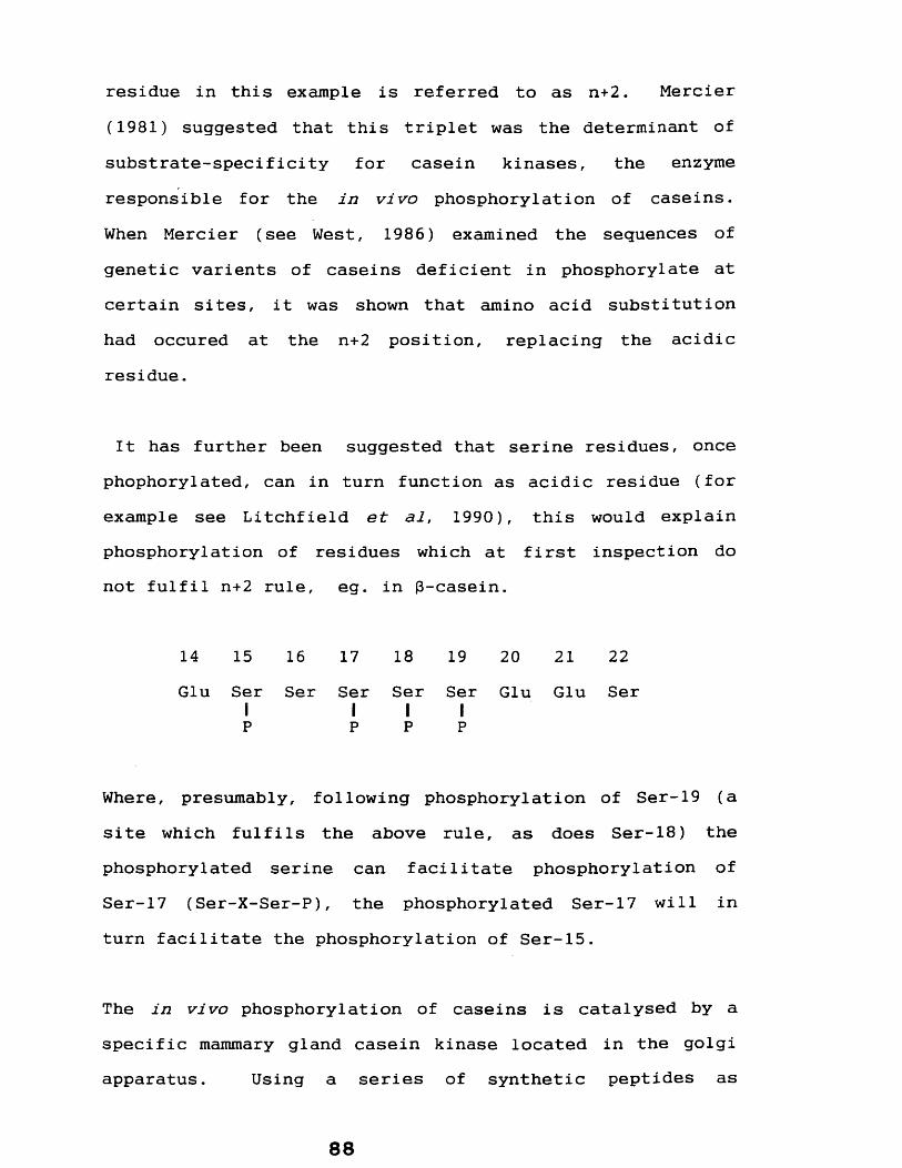
residue in this example is referred to as n+2. Mercier (1981) suggested that this triplet was the determinant of substrate-specificity for casein kinases, the enzyme responsible for the in vivo phosphorylation of caseins. When Mercier (see West, 1986) examined the sequences of genetic varients of caseins deficient in phosphorylate at certain sites, it was shown that amino acid substitution had occured at the n+2 position, replacing the acidic residue.
It has further been suggested that serine residues, once phophorylated, can in turn function as acidic residue (for example see Litchfield et al, 1990), this would explain phosphorylation of residues which at first inspection do not fulfil n+2 rule, eg. in p-casein.
14 15 16 17 18 19 20 21 22Glu Ser
IP
Ser Ser1P
Ser1P
Ser1P
Glu Glu Ser
Where, presumably, following phosphorylation of Ser-19 (a site which fulfils the above rule, as does Ser-18) the phosphorylated serine can facilitate phosphorylation of Ser-17 (Ser-X-Ser-P), the phosphorylated Ser-17 will in turn facilitate the phosphorylation of Ser-15.
The in vivo phosphorylation of caseins is catalysed by a specific mammary gland casein kinase located in the golgi apparatus. Using a series of synthetic peptides as
88

substrates, Meggio et al (1988, 1989) and guinea-pigmammary gland casein kinase described by Pascal1 et al
(1981) and Moore et al (1985) have shown that this enzyme has an absolute requirement for an acidic residue at position n+2 to the phosphorylated residue (n), agreeing with Merciers observations described here. They further showed that a cluster of acidic residues, including one at n+2 surrounding serine was required for optimal phosphorylation. It also appears that, at least for casein kinase type-2, the acidic residue does not necessarily have to be glutamic acid (Murakami et al, 1990).
It should be noted however that studies by Kuenzel et al
(1985), using synthetic peptide substrates and a mammary gland casein kinase from bovine showed that mammary gland casein kinase was able to phosphorylate serine when the acidic residue was at either n+2 or n+3 position. Meggio et al (1988) reported that guinea-pig mammary gland casein kinase could not phosphorylate, for e.g., Ser-Glu-Ala-Glu (acidic residue at n+3), which presumably reflects interspecies differences.
It is of interest to note, as shown by a number of groups (Donella Deanna et al, 1979 and Hoppe and Baydoun, 1981), that casein kinase type-2, discussed in an earlier section, is capable of partially phosphorylating casein residues which are phosphorylated in vivo. It has however been shown (Marin et al, 1986 and Meggio et al, 1988, 1989) that this enzyme has different specificity requirements to
89

guinea-pig mammary gland casein kinase in that it preferentially catalyses the phosphorylation at serines with acidic residue at position n+3 rather than at n+2.
Recent work by Meggio et al (1989) has shown that determinants such as the local structure may also be important. They have demonstrated that phosphorylation of serine by casein kinase type-2 is enhanced by the presence of (3-turn, it is expected that these investigations will be extended to include mammary gland casein kinase (L. Pinna, University of Padova, personal communication). Indeed in a study of the potential phosphorylation sites in k-casein from five species Holt and Sawyer (1988) predicted that all but one were on, or near, a predicted (3-turn.
Work by other groups indicates that other protein kinases have specificity determinants different from both mammary gland casein kinase and casein kinase type-2. For example, Kemp et al (1977) and Cohen et al (1978) have shown that the substrate specificity for cyclic AMP dependent protein kinase (see section 1.2.4.2) consists of basic, rather than acidic, residues C-terminally adjacent to phosphorylated residue.
It therefore appears that mammary gland casein kinase has distinct specificity requirements enabling it to efficiently phosphorylate caseins, its physiological role in the lactating mammary gland.
90

1.3 Aims
The work presented in this thesis describes the identification and characterization of a casein kinase from the lactating rabbit mammary gland. More specifically the aims of the project are as follows:
1) To identify and analyze the sub-cellular distribution of casein kinase in the lactating rabbit mammary gland.
2) To develop a method for the routine isolation of casein kinase rich fractions which can then be used as starting sources for the purification, to homogeneity, of the enzyme.
3) To characterize the enzyme in terms of molecular weight, sub-unit structure, intra-membrane location, optimal assay requirements including cation requirement and kinetic parameters (K» and V* a * ).
4) To generate protein sequence data using purified casein kinase preparations.
The results of this work are presented in the following chapters.
91

CHAPTER 2 MATERIALS AND METHODS

2.1 Materials
2.1.1 Animals
Lactating rabbits of the New Zealand white strain at peak lactation (optimally around 18 days post-partum) were obtained from Broadway Farms, Froxfield, Hants, U.K. Lactating guinea-pigs (3 days post-partum) of the Dunkin Hartley strain were from Porcellus Animal Breeding, Sussex, U.K.
2.1.2 Radi ochemi cals
Adenosine 5'-[2f_32P] triphosphate, tetra (triethyl- ammonium) salt 3000 Ci/mMol (111 TBq/mMol) was from New England Nuclear, Stevenage, Herts, U.K. Uridine diphospho- D-[U"14C] galactose, ammonium salt >200 mCi/mMol (>7.4 GBq/mMol) and D-[U-14C] glucose 6-phosphate, sodium salt >150 mCi/mMol (>5.5 GBq/mMol) were purchased from Amersham International PLC, Amersham, Bucks, U.K.
2.1.3 Chemicals and Solvents
All chemicals and solvents used were obtained from BDH chemicals, Poole, Dorset except those in the following lists and sections 2.1.4 and 2.1.5.
93

Saggatal (pentobarbitone) and propan-l-ol were from May & Baker, Dagenham, Essex, U.K.Ecoscint A from National Diagnostics, Manville, N.J., USA. Trasylol (aprotonin) from Bayer U.K. Ltd, West Sussex, UK. Potassium dichloride from Hopkins & Williams, Chadwell Heath, Essex, U.K.UDP-galactose, NADH & glucose 6-phosphate were from Boehringer (London) Lewes, E. Sussex, U.K.Cyanogen bromide from Aldrich Chemical Company,Gillingham, Dorset, U.K.Oxytocin was obtained from the pharmaceutical supplies department of the Middlesex Hospital (London W1). Sephacryl-S-300, superfine from Pharmacia Fine Chemicals, Uppsala, Sweden.Pre-Stained SDS-PAGE molecular weight standards (low range) from Bio-Rad Laboratories, Hemel Hemsted, Hearts, U.K.Bovine serum albumin (fraction) 5 from I.C.N. Immunobiologicals, Lisle, II., USA.Araldyte CY212 and 2,4,6-Tri (dimethylaminomethy1pheno1)from Agar Scientific Ltd., Essex, USA.Absolute alcohol was from Hayman LTD., Essex, U.K.
The following chemicals were obtained from the Sigma Chemical Company, Poole, Dorset, U.K.:
Cyanogen bromide activated sepharose 4B, lactate dehydrogenase (from pocine heart) type 18, ATP linked through the N6-amino group to beaded agarose, protein
94

kinase, 3 1:51 cyclic AMP dependent from rabbit muscle, protease type 8 from Staphylococcus Aureas (strain V8), N6-21-O-dibutyryladenosine 3 ’:5'-cyclic monophosphate-Na, Triton X-100, dextran (industrial grade), Heparin (lithium salt), phenylmethylsulphonyIfluoride, N,N'-tetraacetic acid, N,N,N1N '-tetramethylethylenediamine, 4-morpho- 1inepropane-sulphonic acid, dowex 1x8 (200-400 mesh), high molecular weight standard protein mixture SDS-6H, 5 '-p- fluorosulfonylbenzenoyladenosine, N,N,-dimethylformamide.
Precondensed Triton X-114, was a kind gift of Dr. A.P. Boulton, University College London. Bovine milk was commonly available commercial whole pasteurized milk. Synthetic peptides SEEEEE and SEAEEE were kind gifts of Prof. L.A. Pinna, University of Padova, Italy.
2.1.4 Materials for hplc.
Gilson (Gilson-Anachem, Luton, Beds, U.K. ) hplc systems using Gilson 714 software run on an IBM computer. Water was double distilled then processed through a "Mili-Q- system".
Acetonitrile (methyl cyanide) was of hplc grade (far UV) obtained from Romil Chemicals, Ltd., Leics, U.K. Trifluoroacetic acid was supplied by Koch Light Ltd., Haverhill, Suffolk, U.K.
9 5
Reverse phase supports were 300 A pore C-8 Brownlee aquabore 300 columns (60 x 2.1mm) obtained through Anachem (Luton, Beds, U.K.).
2.1.5 Materials for protein sequencing
An Applied Biosystems (Foster City, Cal., USA.) 470A gas- phase sequencer, coupled to a 120A PTH analyser (Applied Biosystems) containing a Brownlee C-18 column (220 x 2.1mm) were used.
All chemicals and solvents were supplied by Applied Biosystems and were of "protein sequencer" grade.
96
2.2 Methods
2.2.1 Enzyme assays
2.2.1.1 Casein kinase (ATP: protein phosphotransferase) E.C. 12.7.1.371
Casein kinase assays were carried out essentially as described by Pascall (1981). Briefly assays were usually performed in a final volume of 60 pi, each assaycontaining 62.5 mM Mops (pH 6.3), 10 mM MgCli, 50 pM [^32PATP], 50 pg total bovine casein (initially in some assays total guinea-pig caseins at the same concentration were used) and up to 20 pi of sample. Where necessary assays were adjusted to final volume with water. Any modifications to the assay are indicated in theappropriate sections.
The assay mixture was incubated at 37°C for 15-30 minutes and the reaction terminated by the additions of 50 pg BSA and 1 ml 10% (w/v) trichloroacetic acid containing 10 mMtetrasodium pyrophosphate and incubation at 80° C for 10-20 minutes. Precipitates were collected on glass-fibre discs at the pump as described by Pascall (1981). The incorporation of 3 2 P into protein was detected by scintillation counting usually using Ecoscint A as thefluor.
97

Cyclic AMP dependent protein kinase was assayed as described above with the addition of a range of concentrations of N6-21-O-dibutyryladenosine 3':5'-cyclic monophosphate-Na.
2.2.1.2 Galactosyltransferase (UDP-galactose: N-acetyl -qlucosamine-4-galactosyltransferase)EC T2.4.1.221
Galactosyltransferase, a marker enzyme for the membranes of the trans-golgi catalyses the following reaction:
UDP[14C] Galactose r N-acetyl-[1 4C ]-lactoseamine+ N-acetylglucoseamine + UDP
Galactosyltransferase activity was estimated by a modification of the method of Bergeron et al (1973) as described by Pascall et al (1981) following completion of the enzyme reaction unconverted UDP-[14C] galactose was removed by anion exchange on Dowex 1x8 (400 mesh) and the N-acetyl-C1 4C]-lactoseamine estimated by scintillation counting.
2.2.1.3 Glucose 6-phosphatase (D-qlucose 6-phosphate phosphohydrolase) EC f1.1.1.271
A radiochemical method was used which involved the estimation of [U-1 4 C] glucose released after removal of unconverted glucose 6-phosphate by ion-exchange chromatography on dowex 1x8 (400 mesh), see Pascall (1981) for experimental details.
9 8
2.2.1.4 Lactate dehydrogenase (L-lactate: NADoxidoreductase) EC fl.1.1.271
Lactate dehydrogenase was assayed by the method of Kornberg (1955). The reaction mixture contained in a final volume of 1 ml:- 30 mM potassium phosphate (pH 7.6), 0.13 mM NADH2 , 0.33 mM sodium pyruvate and 50 pi of thefraction to be assayed. Following the initiation of the reaction by the addition of the enzyme sample (ensuring that it rapidly mixed thoroughly with the other assay components) the oxidation of NADH2 to NAD* was followed by a decrease of absorption at 340 nm.
2.2.2 Protein assay
Protein was assayed according to the method of Hartree (1972), standards contained BSA. Protein eluting from hplc columns was estimated from absorption at 214 nm.
2.2.3 SDS-polyacrylamide gel electrophoresis
Electrophoretic separation of proteins was carried-out using a discontinuous-buffered gel system. These were prepared essentially as described by Maizel (1972) in a vertical slab gel (approximate dimensions 20 cm x 15 cm x1.5 mm) with a 4% (w/v) polyacrylamide stacking gel and a 10% (w/v) polyacrylamide resolving gel.
99

Prior to analysis samples were dialysed against 4 1 of ammonium hydrogencarbonate (about 10 mM) at 4°C, lyophilized and redissolved in sample buffer consisting of 15% (w/v) SDS, 15% (v/v) 2-mercaptoethanol and 20% (v/v)glycerol in 0.6% (w/v) Tris-HCl (pH 6.7). Bromophenol blue (0.4% w/v in sample buffer) was added (2-5 pi) and the mixtures incubated in a 100°C water-bath for a few minutes.
Prior to polyacrylamide gel electrophoresis hplc purified material was treated as follows. The sample was neutralized by the addition of a few microlitres of 0.5 M ammonium bicarbonate containing a few crystals of bromophenol blue (until the colour remained blue) and then the bulk of the acetonitrile was removed by evaporation under a stream of inert gas (either nitrogen or argon).
On cooling samples were loaded onto the stacking gel and electrophoresed at about 14 mA (15-30 minutes) and at about 40 mA for 2.5-3.5 hours i.e. until the dye front reached the bottom of the resolving gel.
2.2.4 Silver staining SDS polyacrylamide gels
Following electrophoresis the gels were soaked in 200 mis of fixative, comprising methanol, acetic acid and water in the proportions 10:5:85 by volume, for at least 20
100

minutes. The water used through out this process was double distilled and purified further by reverse osmosis.
Following a brief wash in water the gel was soaked in 100 mis of oxidiser (lOOmg potassium dichromate in 14.2 mis 70% nitric acid) for 10 minutes. The gels were then washed in water for 10 minutes (three changes) and stained by immersion in 100 mis water containing 0.2g silver nitrate for 30 minutes.
Gels were then rinsed, firstly in water, then in a few mis of developer ( which comprised 17.8g sodium bicarbonate in 600 mis water and 1.5 mis of 38% v/v formaldehyde) before being soaked in the remaining volume of developer until protein bands were visualized. This process was stopped by decanting the developer and soaking the gel in 5% (v/v) acetic acid. Gels of interest werephotographed as soon as possible.
The polyacrylamide gel presented in figure 17 was silver stained essentially as described by Wray et al (1981) with the modifications proposed by Eschenbruch and Burk (1985) as out-lined by Moore (1985).
2.2.5 Isolation of total bovine caseins
Total bovine caseins were isolated from whole (pasteurized) bovine milk according to the method of Soulier et al (1975).
101
2.2.6 Preparation of casein-sepharose
Total bovine caseins (50mg) was coupled to 5g of cyanogen bromide (type 4B) according to the method of Delpech et al (1986). Prior to use casein-sepharose was washed with 0. 1M Tris-HCl (pH 7.5) containing 0.15 M NaCl and, where indicated Triton X-100 (0.1% v/v).
2.2.7 Collection of rabbit milk and the isolation of casein micelles
Milk was removed from the glands of lactating New Zealand white rabbits by manual expression of the glands following the intra venous injection of saggatal (approximately 2ml/kg) and oxytocin (8 units).
Whole milk was centrifuged at 100,000xg and the resultant lower sedimented material (casein micelles), resuspended in "H" buffer (section 2.3) and glycerol (40 mg/ml approx) and stored at -20°C.
2.2.8 Routine method for the co-isolation of casein micelles and qolqi enriched fraction from the lactating rabbit mammary gland
Lactating rabbits were killed by the intra venous
injection of saggatal (approximately 2 ml/kg). Mammary
102

glands were quickly removed and placed into ice cold dissection buffer (section 2.3).
Connective tissue surrounding the gland was removed and the tissue weighed. The tissue was then chopped with scissors and minced in a blender for 1 minute. Theaddition of an equal volume to weight (i.e. 1ml:lg) of "H"buffer (section 2.3) was made before the gland was mincedfor an additional 1 minute.
The minced gland was homogenized in aliquots using the large probe of a polytron (2-4 minutes) with theoccasional interuption for the removal of "clumps'1 of fibrous tissue.
The homogenate was pooled and centrifuged at 5000xg for 10 mins at 4° C and the resulting supernatant (the post- nuclear supernatant) decanted through glass wool. An equal volume of "H" buffer (section 2.3) was added before a further centrifugation step at 10,000xg for 20 min at 4°C.
The supernatant was discarded and the sedimented material resuspend in "H" buffer (section 2.3) at not thanapproximately i-2.ml/2g starting tissue. This material was layered onto a 20 ml cushion of 1.2 M sucrose in "H" buffer (see section 2.3) and centrifuged at 100,000xg for 1 hour.
103
This step resulted in material collecting at the 1.2M/0.5M sucrose interface (shown to be a golgi enriched fraction) and two layers of sedimented material. The interface material was recovered with a pasteur pipette and stored at -20°C. The upper sedimented layer was separated from the lower sedimented material by gentle aggitation in a small volume of "H" buffer (section 2.3) and stored as above.
The lower layer was resuspended by vortexing in "H" buffer (section 2.3) and then diluted with approximately an equal volume of glycerol and stored at -20°C. Final protein concentrations were 2-18 mg/ml. This was the casein kinase rich casein micelle fraction.
2.2.9 Procedure for the routine purification of rabbit mammary gland casein kinase from the casein micelle rich fraction
A combination of gel-filtration on Sephacryl-S-300 and affinity chromatography on casein-sepharose were used for the routine purification of the rabbit mammary gland casein kinase and are described in the following section. Minor modifications were made through out the development of these procedures, some of which are indicated.
104

2.2.9.1 Gel-filtration step on Sephacryl-S-300
Aliquots (2-12 ml) of the casein micelle fraction isolated from mammary gland homogenates as described in section 2.2.8 were resedimented by centrifugation in a Beckman Ti50 rotor at 32,000 or 35,000 rpm (69,000 and 84,000xg respectively) for 15-30 min and resuspended in 2 ml Tris-HCl (pH 7.5 at 4°C) containing 0.1% (v/v) Triton X-100. EGTA was later found to be a useful addition to this buffer and was included at a final concentration of 25 mM in subsequent experiments.
The resuspended material was incubated on ice for at least 10 minutes before being layered gently onto a column of Sephacryl-S-300 (36-38 x 1.6 cm) and allowed to enter under gravity. Following this 1ml fractions were collected by eluting at a flow rate of 0.25 ml/min (initial experiments were carried out at 0.5 ml/min). Eluates were assayed for casein kinase activity (see section 2.2.1.1).
The void volume of the column was estimated by fractionating a solution of 0.6 M NaCl and 0.1% (v/v)Triton X-100 in 0.1 M Tris-HCl (pH 7.5) containing a few crystals of blue dextran (molecular weight 2 xlO6 Daltons) and following the elution by absorbance at 342 nm. The column was "sized" by fractionating lactate dehydrogenase (500 pg) and ovalbumin (100 mg) in the same buffer as described above, the elution of these substances was followed by enzyme activity (LDH) as described in section
105
2.2.1.4 and protein assay (ovalbumin) as described in section 2.2.2.
2.2.9.2 Casein-sepharose affinity chromatography step
The fractions eluting from the Sephacryl-S-300 stepdescribed above that contained the peak of casein kinase activity were pooled, diluted four-fold with 100 mM Tris- HCl (pH 7.5) and applied to casein-sepharose (2-15 ml bed volume) at a flow rate of 0.3 ml/min (recirculating) for at least 1 hour, occasionally "batch-binding" was carried out. Following extensive washing with 0.1 m Tris-HCl (pH7.5 at 4°C) containing 0.15 M NaCl and 0.1% (v/v) Triton X-100 (for at least 1 hour) bound material was eluted (under gravity) with 0.6 M NaCl in the same buffer.Fractions (2.25ml) were collected and aliquots (10-20 pi) assayed for casein kinase activity (section 2.2.1.1).
2.2.9.3 Determination of the optimal concentration of sodium chloride required to elute casein kinase from casein-sepharose
An aliquot of the micelle rich fraction (which wasprepared essentially as described in section 2.2.8) as described in section 2.2.9.1 in 0.01 M Tris-HCl pH 7.5 containing 0.1% (v/v) Triton X-100 and 0.5 M NaCl andcentrifuged at 69,000xg for 1 hour at 4°C. The resultant supernatant was adjusted to a final sodium chloride
106

concentration of 0.15 M with 0.01 M Tris-HCl (pH 7.5) and bound to a column of casein-sepharose (see section2.2.9.1). Following extensive washing (20 column volumes) with of 10 mM Tris-HCl (pH 7.5) containing 0.15 M NaCl and0.1% Triton X-100 bound material was eluted (under gravity) by the successive additions of 5 mis of 0.1 M Tris-HCl (pH 7.5 at 4°C) containing 0.1% (w/v) Triton X-100 and increasing concentrations of sodium chloride (0.2- 1.0 M) . Each eluate fractions were collected separately and the casein kinase activity of each eluate fraction determined as described in section 2.2.1.1.
2.2.9.4 The effect of protein concentration on the solubilization of rabbit casein kinase from the micelle rich fraction
A range of aliquots of a stock (9 mg/ml) casein micelle rich fraction (section 2.2.8) were resedimented by centrifugation (155,000xg for 30 min at 4°C) and resuspended in 500 pi of 0.5 M NaCl in 10 mM Tris-HCl (pH 7.5) and also containing 0.1% (v/v) Triton X-100 at yield a range of final protein concentrations between 1 and 20 mg/ml. Each sample was centrifuged at 155,000xg for 1 hour at 4°C. The resultant supernatants were removed and retained and the surface of the sedimented material washed in a small volume of 0.25 M sucrose in 10 mM Tris- HCl (pH 7.5) before being resuspended in 500 pi 10 mM tris-HCl (pH 7.5) containing 0.5 M NaCl and 1% (v/v)Triton X-100. Aliquots of the supernatants and the
1 0 7
resuspended sedimented material were assayed for caseinkinase activity as described in section 2.2.1.1. except
Triton X-10 0that 0.67% (v/v)iwas included in each assay.
2.2.9.5 Purification of rabbit casein kinase from whole rabbit milk
Casein micelles were isolated from rabbit milk as described in section 2.2.7 and casein kinase purified essentially as described in sections 2.2.9.1 and 2.2.9.2 except that the casein micelle solubilization buffer and the Sephacryl running buffers contained 1% (v/v) Triton X- 100.
2.2.9.6 Purification of rabbit casein kinase from qolqi enriched fraction
A golgi enriched fraction isolated as described in section2.2.8 was used as a starting source for the purification of casein kinase using the method as essentially as described in sections 2.2.9.1 and 2.2.9.2.
2.2.10 Purification of rabbit casein kinase:-other procedures
2.2.10.1 ATP-agarose affinity chromatography step.
108

An aliquot of casein micelle rich fraction (about 3.5mg) was prepared essentially as described in section 2.2.7 and resuspended in a 0.13 M Tris-HCl (pH 7.5) containing 0.5 M KC1, 0.1% (v/v) Triton X-100. Particulate material wasremoved by centrifugation at 100,000xg for 1 hour at 4°C. The resultant supernatant was removed and dialysed overnight against 4 litres of 10 mM Tris-HCl (pH 7.5) containing 5 mM magnesium acetate.
The dialysed supernatant was adjusted to about 0.1% Triton X-100 and lpl 100% (v/v) 2-mercaptoethanol added. Thesample was then applied to a column of ATP-agarose at a flow rate of lml/min (recirculating) for at least 1 hour at 4° C .
Following extensive washing in ATP-agarose buffer (see section 2.3) bound material was eluted at a flow rate of 1 ml/min with 0.5 M KC1 in the same buffer. Fractions (1 ml) were collected and assayed for casein kinase activity (section 2.2 .1.1) .
2.2.10.2 Further purification of ATP-agarose purified rabbit casein kinase
Casein kinase purified on ATP-agarose as described above (section 2.2.9.1) was layered onto a linear sucrose gradient (5-20% w/v) and centrifuged at 55,000xg for 16 hours at 4°C. Following fractionation (section 2.2.13.1)
109

each sample was assayed for casein kinase activity (section 2 .2 .1.1) .
2.2.11 Purification of rabbit casein kinase prior to hplc analysis:-modifications to the routine method
Rabbit casein kinase was purified essentially as described in sections 2.2.9.1 and 2.2.9.2 except for the following modifications. Triton X-100 was omitted from running, washing and eluting buffers. Material was bound to the casein-sepharose column over a considerably longer timei.e. overnight.
Enzyme from a series of purification preparations were pooled, dialysed against ammonium bicarbonate (about 10 mM) (4 litres overnight) and lyophilized. Samples were resuspended in a small volume of water (200-400 pi) and stored at 4°C prior to the hplc step.
2.2.12 Purification of casein kinase:- reverse phase high pressure liquid chromatography
Rabbit mammary gland casein kinase was purified essentially as described in section 2.2.9.1 and 2.2.9.2 with the modifications out-lined in section 2.2.11 .
When necessary samples were centrifuged briefly in a bench-top "microfuge" (at full speed) to remove any undissolved particulate material and up-to 200 pi loaded
110
into a rheodyne sample loop (200 pi capacity) using a "Hamilton" microbore syringe. When sample volume was greater than 200 pi convenient aliquots were loaded under isocratic conditions.
Reverse phase high pressure liquid chromatography was carried-out using the system mentioned in section 2.1.4 at a flow rate of 0.2 ml/min using a gradient of 10-65% (v/v) acetonitrile in water and containing either 0.06% (v/v) or 0.05% (v/v) trifluoroacetic acid in water (i.e. there are two resevoirs).
2.2.13 Determination of the intra-cellular localization of rabbit casein kinase
2.2.13.1 Analysis of a post-nuclear supernatant on a linear 5-20% (w/v) sucrose gradient
A post-nuclear supernatant was prepared from a lactating rabbit mammary gland homogenate as described in section2.2.8 and an aliquot representing about 2.3g (wet weight) of starting tissue was fractionated by centrifugation on a linear 5-20% (w/v) sucrose gradient as described byPascall (1981). Briefly the gradient (10 ml 20 to 50 % sucrose in 20 mM Tris-maleate pH 7.0 at 4°C) was overlaid onto a sucrose "cushion" (0.5ml of 60 % sucrose in the same buffer), the sample carefully overlaid onto the gradient and the sample centrifuged at 155,000xg for 90
111

min at 4° C without the brake). The gradient was fractionated via a glass capillary tube inserted to the bottom of the gradient and 0.75 ml fractions collected. The material that sedimented to the bottom of the gradient was resuspended in "H" buffer (section 2.3). Each fraction was assayed for both casein kinase activity (as section 2.2.1.1 except that 1% (v/v) Triton X-100 waspresent in each assay) and galactosyltransferase activity as described in section 2.2.1.2.
2.2.13.2 Analysis of a crude microsomal fraction on a discontinuous sucrose gradient
A crude microsomal fraction was prepared from lactatingrabbit mammary gland homogenate as described in section2.2.7 except for the following modifications. Nembutal was the anaesthetic used, and the gland was chopped with scissors and minced using a scalpel blade and homogenized using both a Potter and a Dounce homogenizer by "hand".
The crude microsomal fraction was analysed on a discontinuous sucrose gradient essentially as described by Pascall (1981) and Boulton et al (1984). Briefly thesample was resuspended in 3 mis of 2.1 M sucrose in 50 mM triethanolamine (pH to 7.5 at 4° C with HC1) which also contained 250 mM KC1 and 5 mM magnesium acetate andoverlaid with the following sucrose solutions prepared in
112

the same buffer: 1.9 M (3 mis), 1.3 M (3 mis) and 0.5 M (to top of tube).
The gradient was centrifuged at 100,000xg for 5 hours and fractionated as described in section 2.2.13.2. Fractions (including the sedimented material) were assayed for the following enzyme activities: casein kinase:- as described in section 2.2.1.1 except that each assay also contained 1% (v/v) Triton X-100, galactosyltransferase:- asdescribed in section 2.2.1.2 and glucose 6-phosphatase:- as described in section 2.2.1.3.
2.2.13.3 Effect of incubating rabbit golgi enriched fractions with increasing concentrations of trypsin in the presence and absence of 0.1%(w/v) Triton X-100.
Aliquots of rabbit mammary gland golgi enriched fractions (section 2.2.8) were incubated for 1 hour at "room temperature" with increasing concentrations of trypsin (0- 500 pg/ml) in the presence or absence of 0.1% v/v Triton X-100.
Following incubation the reactions were terminated by the addition of 200 units of trasylol and 20 pi aliquots of the mixture assayed for casein kinase activity (section2.2.2.1) in the presence of about 1% triton X-100.
113

2.2.13.4 Temperature induced phase separation of membranecomponents in Triton X-114
Temperature induced phase separation of an aliquot (about 20 mg) of golgi enriched fraction (see section 2.2.8) was carried out essentially as described by Pryde (1986). Resultant detergent and aqueous rich phases were assayed for casein kinase activity as described in section 2.2.1.1. The aqueous rich phase was not further analysed by dialysis and centrifugation as described in Pryde(1986).
2.2.13.5 Treatment of rabbit golgi enriched fractions with 0.1 M sodium carbonate
An aliquot (about 150pg) of rabbit golgi enriched fraction prepared as described in section 2.2.8 was resuspended in 2 ml of ice-cold 0.1 M sodium carbonate (at a minimum pH 11) and incubated on ice for 30 mins.
Following incubation samples were centrifuged (100,000xg for 1 hour) and aliquots (20 pi) of the resultant supernatant and sedimented material (resuspended in a buffer containing 0.1% (v/v) Triton X-100) were assayed for casein kinaseactivity as described in section 2.2.1.1.
2.2.14 Estimation of the sedimentation coefficient of rabbit casein kinase
Rabbit casein kinase was purified by a combination of gel- filtration on Sephacryl-S-300 and affinity chromatography on
114
casein-sepharose as described in sections 2.9.1 and 2.9.2 and an aliquot layered onto a linear sucrose gradient comprising 5-20% (w/v) sucrose prepared in a stock buffercontaining 0.1 M Tris-HCl (pH 7.5) and 0.1% (v/v) Triton X- 100 in and centrifuged for 35 hours at 150,000xg at 4° C (with the brake off). In a parallel tube 250pg of lactate dehydrogenase (resuspended in 0.6 M sodium chloride in 0.1 M Tris-HCl pH 7.5) replaced casein kinase.
Following centrifugation the gradient was fractionated (via a glass capillary tube inserted to the bottom of the gradient as described in section 2.2.13.1) and each fraction assayed for either casein kinase (section 2.2.1.1) or lactate dehydrogenase activity (section 2.2.1.4). The sedimentation coefficient for casein kinase was estimated according to the method of Martin and Ames (1961) (see section 5.2.3).
2.2.15 Isolation of guinea-pig mammary gland golgienriched fractions and Partial purification of the casein kinase
Golgi enriched fractions were isolated from the mammary gland of this species essentially as described in section2.2.8 and the casein kinase partially purified either as described by Pascall (1981) by a casein-sepharose step as described in section 2.2.9.2.
115

2.2.16 Electron microscopy
Samples for electron microscopic analysis were prepared as follows. Sedimented material or pieces of fresh tissue were fixed by immersion in 1% (v/v) paraformaldehyde and2% (w/v) glutaldehyde in 0.1 M sodium carcodylate (pH7.4). This step was followed by secondary fixation in 1% (w/v) osmium in the same buffer.
Material was dehydrated by gradual replacement of water by ethanol and the sample was then fixed in Araldite resin, which consisted of equal parts of Araldite CY212 and dodecenyl succinic anhydride, in the presence of a catalyst, 2,4,6, tri (dimethylamino methyl phenol).
The samples were sectioned with a diamond ultra microtome knife (70-100 nm sections) and the sections mounted on a grid and stained with lead-citrate. A representive range of sections from the whole sample were taken. Samples were viewed under a Phillips 300 1L electron microscope. Electron micrographs were taken of fields of view that were of interest.
2.2.17 Phosphorylation of synthetic peptides SEEEEE and SEAEEE by mammary gland casein kinases
Rabbit and guinea-pig casein kinases (purified, partially purified samples or aliquots of casein kinase rich fractions were used see sections 2.2.9.2 2.2.15) were
116

incubated at 37° C as described in section 2.2.1.1 except that total bovine caseins were replaced by a synthetic peptide (10-15 pg of either SEEEEE or SEAEEE) and the samples were incubated for at least 45 mins. Reactions were terminated by the addition of an equal volume of 2 M HC1 and incubated at 100°C for 10 min. Following this step an equal volume of 0.1% trifluoroacetic acid was added (occasionally a greater volume, lml, was added) and the sample applied to a pre-washed (see below) Waters associates "Sep-Pak" column (see Southern, 1987). The sample was allowed to enter under gravity and the columns washed with 5mls of 0.15 (v/v) TFA beforebound material was eluted with 2mls of acetonitrile (100%). 32P labeled peptides in the eluate were estimated by scintilation counting.
Pre-washing "Sep-paks":- prior to use "Sep-paks" were washed as follows: 2mls of the following solutions wereapplied to the column in the following order: 0.1% (v/v)TFA, 25% (v/v) acetonitrile, 50% (v/v) acetonitrile, 100% (v/v) acetonitrile, 50% (v/v) acetonitrile, 25% (v/v)acetonitrile and finally 4mls of 0.1% (v/v) TFA. Allacetonitrile solutions were prepared in 0.1% (v/v) TFA.
2.2.18 Amino acid analysis
The amino acid composition of hplc purified casein kinase was performed by Dr. A. Aitken (NIMR, Mill Hil, London
117

NW7) using an applied biosystems automated amino acid analyser. Residues were analysed as the PTH derivative.
2.2.19 Protein fragmentation
Hplc purified casein kinase was digested with trypsin, V8 protease or cyanogen bromide as described in chapter 8. Digests were analysed as described in section 2.2.12 except the tryptic digests which were analysed on a 10-60% gradient (conditions as in section 2.2.12) with a "runtime" of 65 mins.
118
2.3 Buffers
The following buffers were commonly used:"H" buffer0.05 M Tris-maleate1 mM magnesium acetate1% (w/v) dextran5 mM 2-mercaptoethanol0.5 M sucrosepH 6.75 at 4° C100 pM pmsf (added before use)"Cushion buffer1*0.05 M Tris-maleate 1 mM magnesium acetate1.2 M sucrose pH to 6.75 at 4°C 100 pM added before use."Dissection buffer"0.05 M Tris-maleate lmM magnesium acetate 0.25 M sucrose pH 7.0 at 4° CATP-agarose binding buffer5 mM Tris-HCl pH 7.5 (at 4°C)1 mM EGTA5 mM magnesium acetate 1 mM 2-mercaptoethanol 0.1% (v/v) Triton X-100 0.1 mM sodium vanadate
119
CHAPTER 3DISTRIBUTION OF THE LACTATING RABBIT
MAMMARY GLAND CASEIN KINASE
120

3.1 Introduction
That casein phosphorylation occurs post- translational ly has been demonstrated by a number of groups including Turkington and Topper (1966) and Singh et al (1967), observations more recently confirmed by Mercier and Gaye (1980). The earliest experiments concerned with the identification of the enzymes responsible include those carried out by Schmidt and Davidson (1956). They demonstrated the presence of an enzyme capable of preferentially phosphorylating caseins in the lactating rabbit mammary gland and showed it to be associated with particulate material of unidentified origin, indeed at that time little was known concerning the sub-cellular localization of this enzyme in any species.
Casein kinase has since been shown to be associated with golgi-enriched fractions in the lactating mammary glands of a number of species, i.e the rat (Bingham et
al, 1972; Bingham and Farrell, 1974 and West and Clegg, 1983, 1984); cow (MacKinlay et al, 1977 and Szymanski and Farrell, 1982); guinea-pig (Pascall et al, 1981 and Moore et al, 1985) and mouse (Bingham and Neville, 1981). It is now widely accepted that the golgi is the intra-cellular site of casein phosphorylation. However it should be noted that Bulachev and Medvedev (1978) report the localization of a casein kinase in the membranes of the smooth endoplasmic reticulum isolated from the ovine mammary gland, and the work of Brooks (see section
121
1.2.4.9) which suggests that a calcium and calmodulin dependent casein kinase is associated with fractions other than the golgi may also be involved in the in vivo
phosphorylation of caseins.
A number of approaches have been used in the identification of the sub-cellular localization of casein kinase, these approaches generally involve the preparation of sub-cellular fractions (e.g. crude microsomal fraction) by differential centrifugation, and, following further centrifugation steps on sucrose gradients, the association of the enzyme with the specific components of the sub- cellular fraction is analyzed for e.g. by using marker enzymes. Two approaches to the "further centrifugation steps" are generally used.
The first approach is based on floatation of membranes through a discontinuous sucrose gradient i.e. "steps" of sucrose layers of differing molarity are overlayed on the sample. During centrifugation the particulate components of the sample will float and "rest" at the layer of most equivalent density. This technique was used successfully by Boulton et al. (1984) to demonstrate the association of casein kinase with the golgi enriched fraction with a lactating guinea-pig mammary gland microsomal fraction.
The second approach involves the fractionation of the microsomal membranes by centrifugation through linear
122

sucrose gradients (see Morre, 1971 and Fleisher, 1974 for reviews of this method). Indeed Pascall (1981), using this technique, showed the close association of the guinea-pig casein kinase with the golgi enriched membrane fraction.
Both techniques were used here to determine the intracellular location of the lactating rabbit mammary gland casein kinase.
123
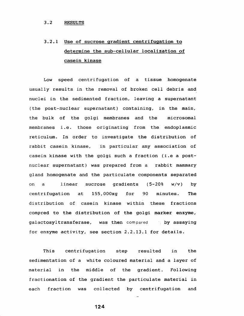
3.2 RESULTS
3.2.1 Use of sucrose gradient centrifugation to determine the sub-cellular localization of casein kinase
Low speed centrifugation of a tissue homogenate usually results in the removal of broken cell debris and nuclei in the sedimented fraction, leaving a supernatant (the post-nuclear supernatant) containing, in the main, the bulk of the golgi membranes and the microsomalmembranes i.e. those originating from the endoplasmic reticulum. In order to investigate the distribution of rabbit casein kinase, in particular any association of casein kinase with the golgi such a fraction (i.e a post- nuclear supernatant) was prepared from a rabbit mammary gland homogenate and the particulate components separated on a linear sucrose gradients (5-20% w/v) bycentrifugation at 155,000xg for 90 minutes. The distribution of casein kinase within these fractionscompred to the distribution of the golgi marker enzyme, galactosyltransferase, was then compared by assayingfor enzyme activity, see section 2.2.13.1 for details.
This centrifugation step resulted in the sedimentation of a white coloured material and a layer ofmaterial in the middle of the gradient. Followingfractionation of the gradient the particulate material in each fraction was collected by centrifugation and
124

assayed for both casein kinase and galactosyltransferase activity. The results of these assays are presented in figure 8. These show that the bulk of the casein kinase activity (28.7%) was present in the white sedimented material, the remainder being distributed throughout the gradient, with approximately 18.4% present in fractions 7- 10 (inclusive) in the centre of the gradient. These fractions also coincide with the edge of the galactosyltransferase activity peak which was contained in fractions 6-9 inclusive.
In an attempt to confirm the results described above a crude "microsomal" fraction (which should contain the golgi membranes and those of the endoplasmic reticulum) was prepared from a rabbit mammary gland homogenate (section 2.2.9) and analyzed by centrifugation through a discontinuous sucrose gradient (section 2.2.13.2). This resulted in the sedimentation of material, white in appearance, to the bottom of the tube and the presence of two distinct layers in the gradient.
Following fractionation (section 2.2.13.1) of the gradient and resuspension of the white material, each sample was assayed for casein kinase,galactosyltransferase and glucose 6-phosphatase activities as described in section 2.2.1. The results presented in figure 9 show that the white material contained about 22% of the total casein kinase activity, with successively decreasing amounts (about 51% in total) of this enzyme
125
Figure 8 Profile of casein kinase and galactosyltransferase activity in fractions resulting from centrifugation of a rabbit mammary gland post-nuclear supernatant on a linear 5-20% (w/v) sucrose gradient
A post-nuclear supernatant was prepared from a rabbit mammary gland tissue homogenate by differential centrifugation as decribed in section 2.2.8 and an aliquot equivalent to about 2. 3g of starting tissue was fractionated by centrifugation on a linear sucrose gradient as described in section 2.2.13.1.
Following centrifugation the particulate material in each fraction was collected by centrifugation in a "bench-top" microfuge (at high speed), resuspended in "H" buffer and assayed for casein kinase and galactosyltransferase activity as described in sections 2.2.1.1 and 2.2.1.2 respectively. The material that sedimented to the bottom of the gradient was similarly resuspended and assayed for these enzymes.
P=Pellet
126

Casein
Kinase
Activity
pmol
/mi
n/ml
(•)
120
90
60
30
5P 10
60
45
30
15
Fraction N2
127
Gala
ctos
yltr
ansf
eras
e Activity
cpm
x10
(O)

also present in fractions 1-4 (inclusive) within the 2.1 M sucrose layer. A small peak of casein kinase containing about 8% was recovered in fractions 14 and 15 towards the top of the gradient.
A single peak containing 46.8% ofgalactosyltransferase activity was present at the 1.9/1.3 M sucrose interface (fractions 10, 11 and 12). A peak ofglucose 6-phosphatase was present in the 1.9 M sucrose layer (indicated by the arrow in figure 9), glucose 6- phosphate activity was also associated with the galactosyltransferase peak.
3.2.2 Electron microscopic comparison of the white coloured sedimented material and casein micelle isolated from rabbit milk
In order to identify the white coloured sedimented material, which was shown in both preceding experiments to be rich in casein kinase activity, electron microscopic analysis was carried out. The sample used was the material which was isolated from the discontinuous gradient centrifugation step described in the preceding experiment. This analysis showed (see figure 10) that the material consisted entirely of roughly spherical, electron dense particles of granular appearance and ranging in diameter of between 71 to 385nM.
128

Figure 9 Profile of enzyme activities in fractionsresulting from the centrifugation of a crude microsomal preparation isolated on a discontinuous sucrose gradient
A crude microsomal fraction was prepared from lactating rabbit mammary gland homogenates essentially as described in section 2.2.8 (also see section 2.2.13.2) and fractionated by floatation through a discontinuous sucrose gradient (consisting of 2.1 M, 1.9 M, 1.3 M amd 0.5 M sucrose layers) as described in section 2.2.13.2.
The gradient was fractionated and an aliquot of each (also an aliquot of the resuspended sedimented material) was assayed for the following enzyme activities: caseinkinase, galactosyltransferase and glucose 6-phosphatase (see section 2.2.1 for details). The arrow indicates a peak of glucose 6-phosphate (see section 3.2.1 for further detaiIs).
P=Pellet • = Casein kinase activity A = Galactosyltransferase activity
129

Enzy
me
Activity
28
24
20
16
12
8
4
P 4 8 16 1812
Fraction Number
130

This material was compared with electron micrographs ofcasein micelles isolated directly from rabbit milk(section 2.2.7), which were shown to be identical in appearence to the material described above, see figure 11 .
131

Figure 10 Electron micrograph of the white colouredcasein kinase rich sediment from the discontinuous sucrose gradient centrifugation analysis
The white coloured casein kinase rich sedimented material resulting from analysis of a crude microsomal preparation by floatation through a discontinuous sucrose gradient (see section 2.2.13.2) was analysed by electron microscopy (as described in section 2.2.16).
A typical micrograph is shown opposite.Magnification X 53.2 xlO3
132

m»•#
##• 0 9 m*■ •#■
* • #
133
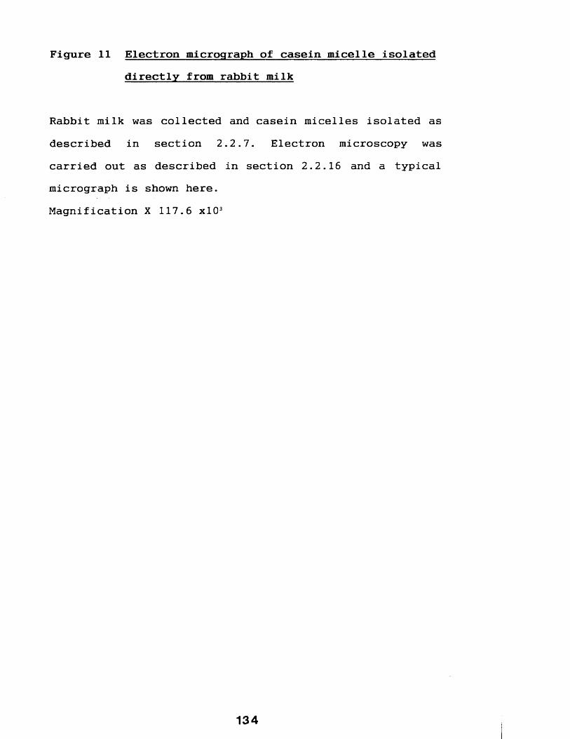
Figure 11 Electron micrograph of casein micelle isolateddirectly from rabbit milk
Rabbit milk was collected and casein micelles isolated as described in section 2.2.7. Electron microscopy was carried out as described in section 2.2.16 and a typical micrograph is shown here.Magnification X 117.6 xlO3
134

135
3.3 DISCUSSION
The result of the analysis of a post-nuclearsupernatant preparation on linear sucrose gradients showed that nearly 29% of the recovered casein kinase activitywas present in the white sediment, little enzyme activitywas however associated with the single peak of galactosyltransferase activity, an enzyme which has shown to be a marker for mammary gland golgi fractions (Keenan et al, 1974).
These results, were confirmed in a subsequent experiment using a discontinuous centrifugation step and a crude microsomal preparation as the starting source.
Electron micrographic analysis of the casein kinase rich sedimented material showed it to comprise ofparticles identical in appearence to the casein micelles of rabbit milk. It is concluded that this fraction is a homogenous preparation of casein micelles. Electron micrographs of the cross section of the mammary gland (see figure 2) shows that few micelles are present in the cytoplasm of the secretory cells and the micelles isolated here probably originate from the milk stored within the mammary gland tissue ductules, sub-segmental ducts and secretory cell luminal spaces (see figure 1). Following disruption of the tissue by homogenization the micelles, being of sufficient density, co-sediment with the
136

microsomal and golgi membranes which explains why they are present in the starting materials for the two experiments described in section 3.2.1.
All previous reports (see section 1.2.4.7), with one exception (Bulachev and Medvedev, 1978), have shown that casein kinase is localized in golgi enriched fractions. Pascall (1981), using a post-nuclear fraction isolated from the lactating guinea-pig mammary gland as a starting source in an identical experiment showed that the bulk of casein kinase activity was associated with the broad peak of galactosyltransferase activity, and therefore prehaps the most striking observation of these results is the association of 22-29% of kinase activity with the casein micelle rich fraction with very little associated with the golgi fractions.
These casein kinase rich micelles are eventually secreted into milk (see section 1.1.6.2), and as expected casein kinase activity is detectable in whole freshly drawn rabbit milk (data not shown). Whilst a number of enzymes have been identified in milk (see section 1.1.4) this is the first report of a casein kinase in milk. Indeed following a search of the literature it seems that no other protein kinase has been reported in milk, although both Giri (1958) and Kinsella (1972) have both demonstrated the presence of non-protein kinases in bovine milk.
137

Further more, whilst the enzymes of milk have been shown to be associated with a number of "phases" (see section 1.1.4.1), the association an enzyme with the micelle fraction is rather unusual. Only a lipase (Gaffney et al, 1966b and Downey and Murphy, 1970) and a proteolytic enzyme (Warner and Poli, 1945) have been shown to be similarly associated, (see section 1.1.4.2 for detailed discussion).
In general then, it is of interest that the rabbit casein kinase has an unusual distribution and, more specifically, one different from the other mammary gland casein kinases.
In the following chapter the casein kinase rich fractions described here are further characterized, initially by developing a procedure for the routine coisolation of both fractions which will form the basis of further studies.
138

CHAPTER 4
FURTHER ANALYSIS OF CASEIN KINASE RICH FRACTIONS ISOLATED FROM THE LACTATING
RABBIT MAMMARY GLAND
139

4.1 INTRODUCTION
Part of the work in chapter three demonstrated that the casein micelle rich fraction co-isolated with the post-nuclear and crude microsomal fractions and were of sufficient density to sediment below other particulate microsomal material. This work also indicated that whilst the bulk, in terms of the material containing the highest levels, of the casein kinase was associated with the micelle rich fraction, some was also associated with galactosyltransferase rich material probably of golgi origin.
The aims of the work presented in the chapter aretwo fold. Firstly, to develop a method for the routineisolation of the two casein kinase rich fractions, which can be used as starting source for the study of theenzymes and secondly, to analyse further the fractions in terms of confirming the identity of the casein kinase containing membranes which were tentatively identified as golgi membranes, and the localization of the enzyme within these membranes.
The majority of procedures for the routinepreparation of golgi fractions have been developed to use liver homogenates as a starting source, although some have been successfully modified for use with the lactating
14 0
mammary gland (Keenan et al, 1972a and Keenan et al,
1972b).
Pascal 1 et al (1981) and Moore et al (1985), who isolated golgi enriched fractions from the lactating guinea-pig mammary gland, utilized, with modifications, the method used by Hino et al (1978) to prepare golgi fractions from rat liver. This method involved the separation of the components of a "crude" microsomal preparation through a discontinuous sucrose gradient (comprising 0.5M and 1.2M sucrose layers). Rough endoplasmic reticulum (RER) membranes, being of sufficient density, sedimented through the 1.2 M sucrose layer, whilst the golgi fractions collected at the sucrose gradient interface.
An adaptation of the method was used here in the coisolation of both casein kinase rich fractions. This procedure and the further characterization of the resulting fractions are described here.
141

4:2 RESULTS
4.2.1 Development of a procedure for the coisolation of casein kinase rich fractions
Lactating New Zealand white rabbits were killed by intra venous injection of saggatal (about 2ml/kg) mammary glands quickly placed into ice cold dissection buffer ("D" buffer-see section 2.3). The glands were minced and homogenized as described in section 2.2.8. Differential centrifugation of the resulting homogenate resulted in the sedimentation of a crude microsomal fraction which was then layered onto a discontinuous sucrose gradient (comprising of 0.5 M and 1.2 M sucrose layers).
Centrifugation of this fraction (at 100,000xg) resulted in the separation of the following three distinct fractions:
1) material, opaque in appearance, collected at the 0.5 M and 1. 2M sucrose interface.
2) A "lower" sedimented layer consisting of tightly packed material, white in colour.
3) An "upper" brown coloured sedimented material.The sedimented fractions were easily separated by
gentle aggitation of the upper layer with a pasteur pipette. This procedure is summarized as a flow diagram in figure 12.
142

Figure 12 Flow diagram of the procedure developed for theroutine co-isolation of golgi and casein micelle rich fractions from the lactating rabbit mammary gland
A procedure was developed for the routine co-isolation of casein kinase rich fractions identified in chapter 3 . The fractions being the golgi and casein micelles. Details of this method may be found in sections 2.2.8 and chapter 4.
Lactating rabbits killed with saggatal (approx 2ml/kg) and the mammary gland dissected into "H" buffer1
Remove connective tissue. Chop and mince gland in "H" buffer (equal volume to weight) with blender for 2 min1
Homogenize minced gland using the large probe of a polytron for 2-4 minute at 3/4 to full speed
ICentrifuge homogenate at 50 00 xg
for 10 minute at 4° CIFilter the resultant post-nuclear supernatant through
glass wool. Add equal volume of "H" buffer. Centrifuge at 50,000 xg for 20 minutes at 4°C
■ iResuspend the resulting microsomal pellet in "H" buffer (up to about lml/2mg starting tissue). Layer onto 20 ml
cushion of 1.2 M sucrose in cushion buffer. Centrifuge at 100,000 xg for 1 hour at 4°C
IRemove material collecting at the sucrose gradient
interface-this is the golgi enriched fraction. Resuspend the white coloured sedimented material in "H" buffer-this
is the kinase rich casein micelle fraction

Analysis of the casein kinase content of each fraction was carried out as described in section 2.2.1.1, and typical results showed that the bulk (about 80%) of this enzyme was associated with the lower white material, see table 9. A smaller proportion (18%) was present in the interface material and only negligable amounts (2.6%) in the upper brown sedimented material.
The results in table 9 also show that about 46% of galactosyltransferase was present in this upper layer with the majority of the remainder (48%) being accounted for in the interface material. Little (5.5%) of this enzmme being present in the lower white sedimented material..
4.2.2 Electron microscopic analysis of the three fractions resulting from the centrifugation of a crude microsomal fraction through a 1.2 M sucrose step
Electron microscopic analysis was carried out on all three fractions according to method described in section 4.2.1. The lower white fraction was shown to contain a homogeneous population of granular, roughly spherical particles (figure 13) which are identical in appearance to the casein micelle rich fractions described in the preceding chapter. In contrast the upper sedimented material was shown to be heterogeneous in composition (figure 14), comprising mainly of two components, i.e.
1 4 4

Table 9 Casein kinase and galactosyltransferase contentof the three fractions resulting from the fractionation of a crude microsomal preparation on a discontinuous sucrose gradient
A crude microsomal preparation was isolated as described in section 2.2.8 and layered onto a discontinuous gradient comprising 0.5 M and 1.2 M sucrose layers. Following centrifugation three layers were isolated (see section3.2.1) and both casein kinase and galactosyltransferase activities of the three fractions determined (see section2.2 .1).
Results of the centrifugation of the "microsomal fraction" through a 0.5/1.2 M sucrose gradient are expressed as a percentage of the total recovered activity in the three particulate fractions recovered by this step.
145
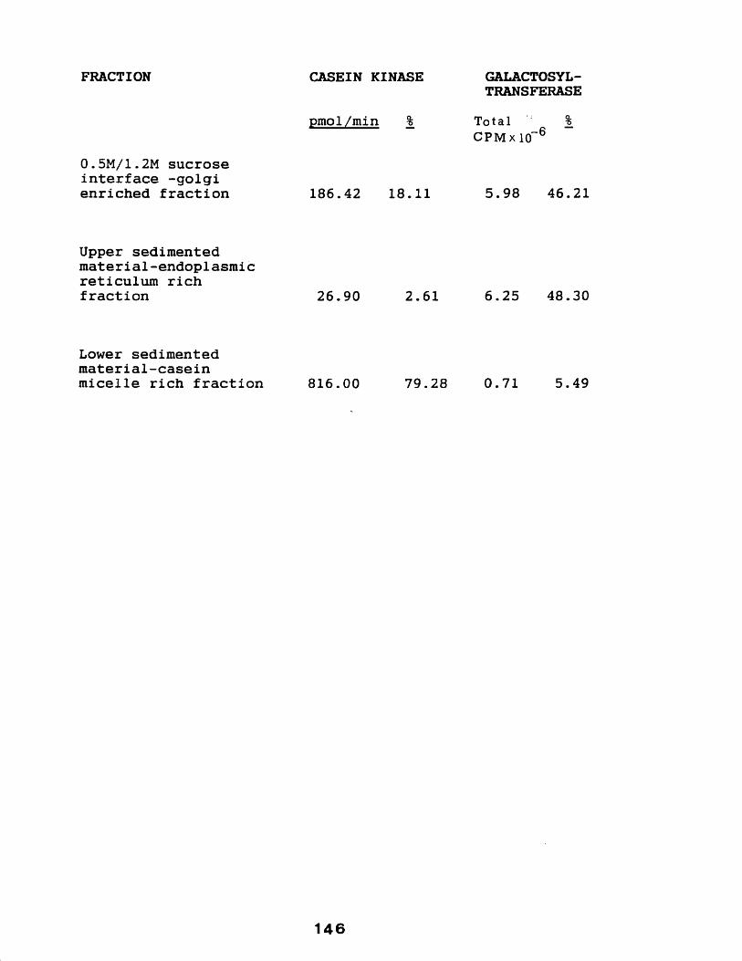
FRACTION CASEIN KINASE GALACTOSYLTRANSFERASE
0.5M/1.2M sucrose interface -golgi enriched fraction
pmol/min %
186.42 18.11
Total %CP Mx 10“6
5.98 46.21
Upper sedimented material-endoplasmic reticulum rich fraction 26.90 2.61 6.25 48.30
Lower sedimented material-caseinmicelle rich fraction 816.00 79.28 0.71 5.49
146
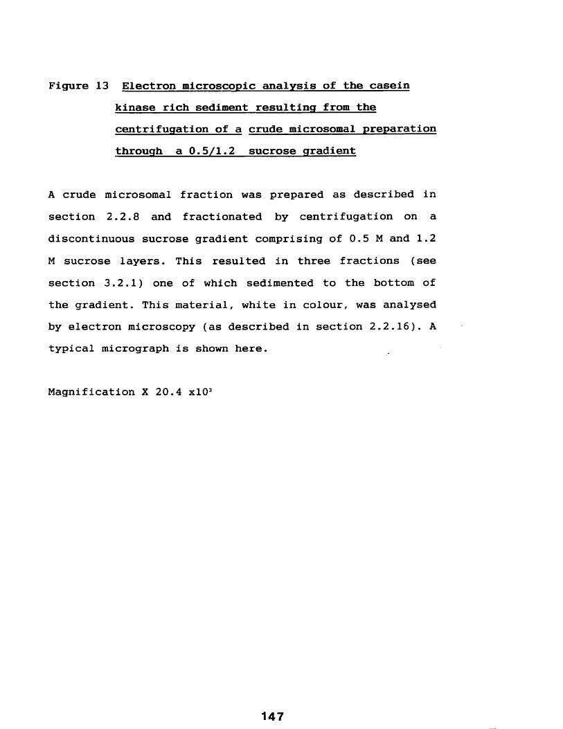
Figure 13 Electron microscopic analysis of the casein kinase rich sediment resulting from the centrifugation of a crude microsomal preparation through a 0.5/1.2 sucrose gradient
A crude microsomal fraction was prepared as described in section 2.2.8 and fractionated by centrifugation on a discontinuous sucrose gradient comprising of 0.5 M and 1.2 M sucrose layers. This resulted in three fractions (see section 3.2.1) one of which sedimented to the bottom of the gradient. This material, white in colour, was analysed by electron microscopy (as described in section 2.2.16). A typical micrograph is shown here.
Magnification X 20.4 xlO3

*
148
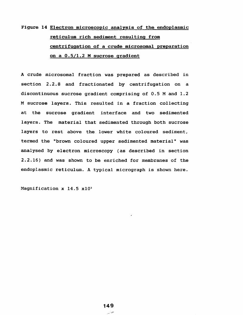
Figure 14 Electron microscopic analysis of the endoplasmicreticulum rich sediment resulting from centrifugation of a crude microsomal preparation on a 0.5/1.2 M sucrose gradient
A crude microsomal fraction was prepared as described in section 2.2.8 and fractionated by centrifugation on a discontinuous sucrose gradient comprising of 0.5 M and 1.2 M sucrose layers. This resulted in a fraction collecting at the sucrose gradient interface and two sedimented layers. The material that sedimented through both sucrose layers to rest above the lower white coloured sediment, termed the "brown coloured upper sedimented material" was analysed by electron microscopy (as described in section 2.2.16) and was shown to be enriched for membranes of the endoplasmic reticulum. A typical micrograph is shown here.
Magnification x 14.5 xlO3
1 49

9
h*c
r*#'r ▼
150
numerous casein micelle-like particles and a large population of "rough" vesicles. These "rough" vesicles as well as a larger proportion of "smooth" membranes were present in the electron micrographs of the interface material (figure 15).
4.2.3 Localization of casein kinase within the golgi enriched fraction
4.2.3.1 The effect of trypsin on casein kinaseactivity in the presence and absence of 0.1% (v/v) Triton X-100.
In order to investigate whether casein kinase activity is localized on the cytoplasmic or the luminal side of the golgi vesicles described in the preceding section, the ability of the proteolytic enzyme trypsin to inactivate the enzyme in these membranes was investigated.
Aliquots of this membrane material (isolated as described in section 2.2.8) were incubated in the presence of increasing concentrations (0 to 500 pg/ml) of trypsin in the presence or absence of 0.1% (v/v) of Triton X-100 as described in section 2.2.13.3. Following termination of the reaction (with the addition of 200 units of trasylol) the casein kinase activities remaining in each sample were estimated by assaying aliquots (20 pi), each assay contained about 1% (v/v) Triton X-100.
151

Figure 15 Electron micrograph of the golgi membranefraction resulting from the centrifugation of a crude microsomal preparation through a 0.5 M/1.2 M sucrose gradient
A crude microsomal fraction was prepared as described in section 2.2.8 and fractionated by centrifugation on a discontinuous sucrose gradient comprising of 0.5 M and 1.2 M sucrose layers. This resulted in three fractions (see section 3.2.1) one of which collected at the interface between the two sucrose layers, this material shown to be a golgi enriched fraction, was analysed by electron microscopy as described in section 2.2.16. A typical micrograph is shown here.
Magnification X 124 xlO3
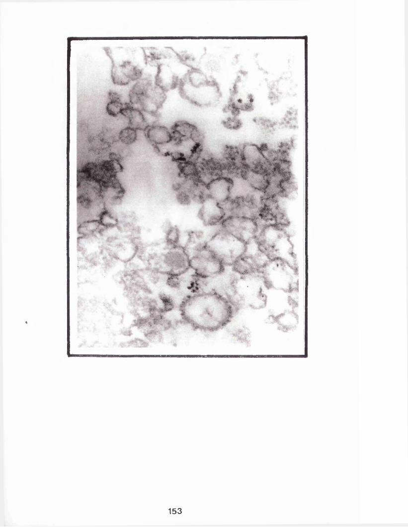
153
The results (figure 16) showed that in the absence of detergent trypsin had negligable effect on the casein kinase with over 85% activity remaining at the highest trypsin concentration used. In contrast with the presence of detergent in incubations little activity remained (<10%) even at the lowest concentration of trypsin (5 jig/ml).
4.2.3.2. Temperature induced phase separation ofrabbit golgi enriched membrane components with Triton X-114
Triton X-100, an anionic detergent, has been widely used in the solubilization of proteins from membranes. Triton X-114, a member of the same group of detergents, although equally as efficient at membrane solubilization (Egham et al, 1976) has found little application since it fails to maintain all the membrane components at room temperature (Pryde, 1976). A number of researchers have recently taken advantage of the properties of this detergent. Solubilization of membranes in Triton X-114 at 0°C followed by warming to 30°C results in the partition of the sample into a detergent rich and an aqueous phase. Each phase can be separated by a centrifugation step which results in the partition of the integral membrane protein components in the detergent phase whilst those associated with the luminal space are mainly distributed in the aqueous phase (Bordier, 1981; Pryde and Phillips, 1986; Pryde, 1986).
154
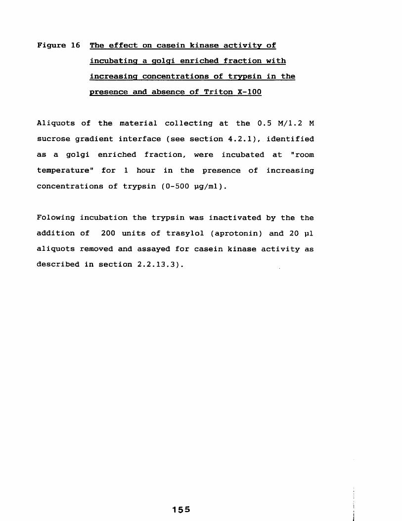
Figure 16 The effect on casein kinase activity ofincubating a golgi enriched fraction with increasing concentrations of trypsin in the presence and absence of Triton X-100
Aliquots of the material collecting at the 0.5 M/1.2 M sucrose gradient interface (see section 4.2.1), identified as a golgi enriched fraction, were incubated at "room temperature" for 1 hour in the presence of increasing concentrations of trypsin (0-500 pg/ml).
Folowing incubation the trypsin was inactivated by the the addition of 200 units of trasylol (aprotonin) and 20 pi aliquots removed and assayed for casein kinase activity as described in section 2.2.13.3).
155

ooin
oo
inr*-
oin
inCM
co (O CM
(%) Ai\ Aj;ov OSBUI^ uieseo
156
Trypsin
(jjg / m
i )

It was of interest to determine into which phase casein kinase, present in the rabbit mammary gland golgi vesicles, will associate with following treatment with Triton X-114. Aliquots (approximately 20 mg) of this material were treated with this detergent essentially according to Pryde (1986) and Pryde and Phillips (1986), see section 2.2.13.4.
The results of this preliminary study indicatedshowed that the vast majority of casein kinase activity (at least 90%) was present in the aqueous supernatant and the bulk of the remaining activity was present in thesedimented material.
4.2.3.3 Distribution of casein kinase following thetreatment of the golgi enriched membranefraction with 0.1M sodium carbonate
Fujiki et al (1982) reported that the treatment of membranes with 0.1M sodium carbonate resulted, following a centrifugation step, in the distribution of all but integral membrane protein components with the supernatant. Here aliquots of golgi enriched membrane fractions (prepared as described in section 2.2.8) were treated with 0.1 M Sodium carbonate (pH >11) and following centrifugation (at 54,000xg), both the resultantsedimented material and supernatant were assayed for
157
casein kinase activity in order to assess the distribution of the enzyme.
The results showed that the vast majority (approximately 90%) of the casein kinase activity was present in the supernatant, with the remainder (approximately 10%) present in the sedimented material.

4.3 DISCUSSION.
Rabbit mammary glands were removed and crude microsomal fractions were prepared according to the method described in section 2.2.8. which was an adaptation of the procedure described by Pascall et al (1981), itself based on the method of Hino et al (1978), used to prepare golgi enriched fractions from the lactating guinea-pig mammary gland.
Here, since the rabbit mammary gland was extremely fibrous, it was necessary to homogenize the gland by a rather harsh method using a "polytron" (a procedure infact recommended by a number of groups-see Keenan et al, 1974), whereas Pascall et al (1981) used a glass/Teflon homogenizer. Although treatment of the rabbit tissue with dispase, a collagenase, aided the "break-up" of this fibrous tissue sufficiently enough to facilitate the use of less harsh homogenization procedure (not described here) was not routinely employed.
In order to remove the bulk of broken cell debris centrifugation of the homogenate was carried out at a short (10 min) low speed (5000xg), compared with the procedure of Pascall et al (1981) who used 10,000xg for 20 minutes, in order to avoid the high density casein kinase rich casein micelle fraction sedimenting with the cell debris.
159
Centrifugation of the resultant microsomal fraction on a discontinuous sucrose gradient resulted in the isolation of three fractions which were analysed by assaying both casein kinase and galactosyltransferase content and by electron micrographic analysis.
The lower sedimented material, white in appearance contained the bulk of the recovered casein kinase activity, a result in agreement with previous observations (chapter 3) and was shown by electron microscopic analysis to consist of particles (see figure 13) identical to the casein micelles of rabbit milk (compare with figure 11). This material contained negligable (5.5%) galactosyltransferase activity, also in agreement with the results in the previous chapter.
The "upper" sedimented material contained 46.3% of the total galactosyltransferase activity recovered in the three particulate fractions and was shown (figure 14) to be heterogeneous in composition, comprising mainly of "rough" vesicles identical in appearance to the membranes that comprise the rough endoplasmic reticulum (RER), some of the casein micelles described above were also present.
The recovery of a such high percentage of the galactosyltransferase activity (in fact the majority) in this RER fraction appears to be inconsistent with the notion that this is a golgi "marker" enzyme. Pascall (1981) similarly found that the bulk of this enzyme was
160

associated with a RER fraction isolated from the lactating guinea-pig mammary gland. This anomaly may be explained by the apparent heterogeneity of the golgi apparatus both in terms of morphology and function which has been noted by a number of groups (Bretz et al, 1980; Bergernon et al, 1973) resulting in the finding that some golgi membranes may be of sufficient density to co-sediment with the RER membranes (Bergernon, 1973). Such observations explains the presence of golgi-marker enzyme in the RER fraction.
The material collecting at the 0.5M and 1.2M sucrose interface was shown to contain a smaller than expected proportion (46.2%) of the recovered golgi "marker" enzyme galactosyltransferase (see above). Electron microscopy showed this fraction to comprise mainly of "smooth" membranes with a small proportion of "rough" (i.e. with associated ribosomes) vesicles originating from the endoplasmic reticulum. The general lack of acuity of these electron micrographs is probably a direct effect of the fixation procedure on the membranes (M. Turmaine, Electron Microscopy Facility, University College London- personal communication).
The equivalent fraction isolated by Pascall et al
(1981) from the lactating guinea-pig mammary gland was analysed in a similar fashion and they concluded that this material was a golgi enriched fraction. Examination of electron micrographs of this material (see Pascall, 1981) reveals the presence of both smooth membranes and "rough"
161
vesicles. An observation in agreement with the results here. He further showed that the bulk of the recovered galactosyltransferase activity was not associated with this fraction, again in agreement with results here. Based on these same two criteria used by Pascall (1981), it is concluded that the interface material isolated here is similarly a golgi-enriched fraction.
A small proportion (18%) of casein kinase activity was present in this golgi enriched fraction, which, based on the observations of other workers (see section 1.2.4.7) is the most probable site of in vivo casein phosphorylation (see Conclusions-Chapter 9 for full discussion of this result). The distribution of the casein kinase was investigated further. Casein kinase activity in this fraction was completely inactivated by low levels (5 pg/ml) of the proteolytic enzyme trypsin when incubated in the presence of detergent (0.1% v/v Triton X-100), but was unaffected by this enzyme in the absence of Triton X- 100 at the same concentration.
These results indicate that casein kinase is either an integral membrane protein facing the interior of the golgi membrane vesicles and therefore protected from proteolysis by trypsin, or is present in soluble form within the golgi vesicle lumen where it would be similarly protected.
162
On the basis of similar studies Fleisher (1981) showed that both galactosyltransferase andsialyltransferase are located on the luminal side of golgi membrane vesicles and Pascall (1981) showed that the guinea-pig casein kinase also faces the golgi lumen. Since caseins are present within the golgi lumen (Fiske et al, 1967; Pascall, 1981) the identification here of the rabbit casein kinase as being on the luminal side of the golgi vesicles is consistent with the notion that the caseins are phosphorylated post-translationally during their passage through this organelle.
In a preliminary study it was shown that following treatment of the golgi enriched membrane fraction with both sodium carbonate and Triton X-114 the majority of casein kinase was present in the supernatant following a centrifugation step. This data suggests that the bulk of the rabbit casein kinase is not present as an integral membrane protein but rather in a soluble form probably within the golgi lumen. A result in contrast to the findings of similar localization studies on the guinea-pig casein kinase (Moore, 1985; Moore et al, 1985) who showed following similar treatment that this enzyme was present in the membrane rich fraction.
In summary, a procedure for the co-isolation of the casein kinase rich fractions (summarized in figure 12) was developed and was adopted as the method for the routine isolation of the casein kinase containing fractions. In
163

the following chapters this method is used to isolate the casein micelle rich fraction as the starting source for the purification of the enzyme responsible for the casein kinase activity.
164

CHAPTER 5
PURIFICATION OF A RABBIT MAMMARY GLAND CASEIN KINASE FROM THE CASEIN
MICELLE RICH FRACTION

5.1 INTRODUCTION
The advancements in the development of techniques of protein purification in recent years, particularly those employing the principles of partition chromatography, have resulted in a vast increase in number of protein kinases that have been purified (see Hunter, 1982). Affinity chromatography in particular has proved especially useful since it utilizes a feature common to many biologically active molecules, that is, their specificity to a particular substrate.
Although a number of mammary gland casein kinases have been identified (see section 1.2.4.8 for complete list) there have been few attempts at purification.
MacKinlay et al (1977), for example, reported the purification of the bovine mammary gland casein kinase by precipitation of the enzyme using successive additions of sodium deoxycholate to golgi enriched fractions. The extent of purification was not checked by analysis on polyacrylamide gels and probably is better described as a "casein kinase enriched fraction". Szymanski and Farrell(1982) also isolated a golgi enriched fraction from the same species and "isolated" the enzyme by solubilization in detergent (0.4% v/v Triton X-100). This procedure again resulted only in an enzyme "enriched" preparation.
166

The first attempt at developing a procedure for purification to homogeneity was carried out by Pascall et
al (1981) using the guinea-pig mammary gland. They isolated golgi enriched fractions and following solubilization in 0.1% (v/v) Triton X-100 and 0.5M KC1used affinity chromatography on ATP-agarose. This resulted in fractions enriched for casein kinase activity and shown to have a significantly reduced casein component. This procedure however proved unsuitable for use as a preparative procedure, since low levels of caseins contaminants co-purify with the enzyme (A. Moore, University College London, personal communication).
An improved purification procedure for the guinea pig casein kinase was developed by Moore et al (1985). They solubilized golgi enriched fractions, using 0.5 M KC1 and 0.1% (w/v) Triton X-100, and used a combination ofcentrifugation and linear sucrose gradients (5-20% w/v) and an improved ATP-agarose affinity step. This resulted in the purification of a single polypeptide of apparent molecular weight 74,000 Daltons, as judged by silver stained SDS polyacrylamide gel electrophoresis, and an approximate overall purification factor of 2300 with typically a yield of 2-5 pg of casein kinase from approx 20g (wet weight) of starting tissue (see Moore 1985).
The work in this chapter describes the development of a procedure for the purification of a casein kinase from the lactating rabbit mammary gland. The micelle rich
167

material isolated according to the method developed in the preceding chapter was used as the starting source.
ATP, a co-enzyme for most protein kinases, has proved a useful ligand in affinity chromatography of a number of such enzymes including the guinea-pig mammary gland casein kinase (Moore et al, 1985). The usefulness of this affinity step here in the isolation of a rabbit mammary gland casein kinase was investigated, either essentially as described by Pascall et al (1981) (data not presented here) or by the method of Moore et al (1985), which was found to be more successful. This step was followed by centrifugation on linear sucrose gradients, 5-20% (w/v).
Following these initial experiments a method involving the use of another affinity step, using casein as the ligand and Sephacryl-S-300 gel exclusion chromatography was developed. An improved procedure for the release of casein kinase from the micelles was also developed. Other steps involving affinity chromatography using various ligands were investigated, this work is not presented in any detail. The remainder of this work is presented in the following sections.
168
5.2 RESULTS
5.2.1 ATP-agarose chromatography and sucrosegradient centrifugation steps used in the purification of mammary gland casein kinase
Samples of casein micelle rich fraction were prepared as described in the chapter (section 2.2.8) and an aliquot (about 3.5mg) applied to a column of ATP-agarose at a flow rate of 1 ml min-1 for 1 hour (recirculating the sample). Following wash steps, bound material was eluted with ATP- agarose binding buffer (see section 2.3) containing 0.5 M KC1 at the same flow rate. This resulted in the elution of a single peak of casein kinase activity (not shown) and a proportion dialysed against ammonium bicarbonate, freeze dried and analysed by SDS polyacrylamide gel electrophoresis (see section 2.2.3). The protein was visualized by silver staining the gel (see section 2.2.4). The results show that the casein kinase rich eluate fractions were enriched for two major polypeptide species of molecular weight around 66,000 Daltons, with only a small proportion of lower molecular weight material (see figure 17).
A proportion (approx 17%) of the ATP-agarose purified material was layered onto linear sucrose gradient (5-20% w/v) and centrifuged at 55,000xg for 16 hours at 4°C. Following centrifugation the gradient was fractionated (section 2.2.13.1), and fractions analysed for casein
169

kinase activity (section 2.2.1.1). This resulted in the identification of a broad peak of enzyme activity (data not shown), which was analysed by SDS polyacrylamide gel electrophoresis and silver staining, which showed it to contain the same two polypeptide species as present in the casein kinase rich material that eluted from ATP-agarose (see figure 17). This step resulted in the further removal of low molecular weight proteins.
5.2.2 Gel exclusion and affinity chromatography steps
The previous experiments indicated that two polypeptides co-purified with casein kinase activity from both an ATP-agarose step and a subsequent sucrose gradient centrifugation step. Whilst not conclusive, these two proteins were putatively assumed to represent the rabbit casein kinase. The usefulness of a gel exclusion step prior to an affinity step in-order to remove low molecular weight contaminants was investigated. For a number of practical reasons the affinity step was changed from ATP to casein (the other enzyme substrate) as the ligand.
Aliquots of micelle rich fractions (section 2.2.8) were resuspended as described in section 2.2.9.1 (see also figure 21) and layered onto a column of Sephacryl-S- 300 and allowed to enter under gravity. Bound material was eluted with 0. 6M NaCl in 0.1 M Tris-HCl (pH 7.5 at 4°C) containing 0.1% (v/v) Triton X-100. The results(figure 18) show that casein kinase eluted in a single broad peak with peak casein kinase activity in this experiment present in fractions 32 to 37 inclusive (a
170

Figure 17 SDS polyacrylmide gel electrophoresis analysisof ATP-agarose and sucrose gradient purified rabbit mammary gland casein kinase
Casein kinase was purified from the micelle rich fraction using ATP-agarose and linear sucrose gradients (5-20% w/v) as described in sections 2.2.10.1 and 2.2.10.2 respectively. Following each step an aliquot of the pooled casein kinase rich fractions were retained and analysed by electrophoresis on 10% (w/v) SDSpolyacrylamide gels as described in section 2.2.3 and the gels silver stained as described in section 2.2.4.
Lane 1 contains aliquots of casein kinase rich material which eluted from ATP-agarose.
Lane 2 contains the casein kinase rich material from the sucrose gradient centrifugation step.
Molecular weight of 66,000 (BSA) is indicated on the right. The two arrows indicate the two polypeptides shown to be the casein kinase.

172

Figure 18 Profile of casein kinase activity and proteinconcentrations in fractions eluting from Sephacryl-S-300
Aliquots of casein micelle rich fraction were resuspended and applied to a column of Sephacryl-S-300 as described in section 2.2.9.1. The applied material was eluted at a flow rate of 0.5 ml/min in 0.1 M Tris-HCl (pH 7.5 ) containing 0.6 M NaCl and 0.1% (v/v) Triton X-100, and 1 ml fractions collected.
Casein kinase and protein concentration were assayed as described in sections 2.2.1.1 and 2.2.4 respectively.
173

140
| U J / 6 r f ( ) maiojd
£_0l x iiido A^iAj^ov aseu!>i uieseo
174
Fraction
N— (1
ml V
olum
e )
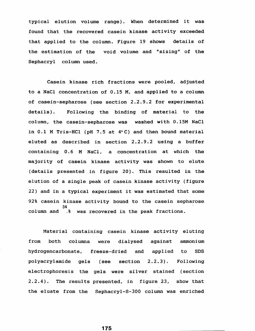
typical elution volume range). When determined it was found that the recovered casein kinase activity exceeded that applied to the column. Figure 19 shows details of the estimation of the void volume and "sizing" of the Sephacryl column used.
Casein kinase rich fractions were pooled, adjustedto a NaCl concentration of 0.15 M, and applied to a columnof casein-sepharose (see section 2.2.9.2 for experimentaldetails). Following the binding of material to thecolumn, the casein-sepharose was washed with 0.15M NaClin 0.1 M Tris-HCl (pH 7.5 at 4°C) and then bound materialeluted as described in section 2.2.9.2 using a buffercontaining 0.6 M NaCl, a concentration at which themajority of casein kinase activity was shown to elute(details presented in figure 20). This resulted in theelution of a single peak of casein kinase activity (figure22) and in a typical experiment it was estimated that some92% casein kinase activity bound to the casein sepharose
34column and ,% was recovered in the peak fractions.
Material containing casein kinase activity eluting from both columns were dialysed against ammonium hydrogencarbonate, freeze-dried and applied to SDS polyacrylamide gels (see section 2.2.3). Following electrophoresis the gels were silver stained (section 2.2.4). The results presented, in figure 23, show that the eluate from the Sephacryl-S-300 column was enriched

Figure 19 Some characterisics of the Sephacryl-S-300column used described in section 2.2.9.1
Some of the elution characteristics of the sephacryl-S-300 column used in the purification of rabbit casein kinase described in section 2.2.9.1 were determined. Solutions containing 500 pg of LDH (molecular weight 145,000 Daltons) and 100 mg ovalbumin (45,000 Daltons) were fractionated seperately as described in section 2.2.9.1 (the elution rate was 0.25 ml/min), LDH activity in the eluate fractions were determined as described in section2.2.1.4 and the presence of ovalbumin estimated by assaying for protein (section 2.2.2). The "void" volume of the column was estimated by determination of the volume required to elute the high molecular weight dye blue dextran (elution followed by absorption at 342 nm). For comparison casein kinase was isolated at the same flow rate (section 2.2.9.1) and the enzyme activity (section 2.2.1.1) in the eluate fractions determined.

Casein
Kinase
Activity
cpm
x10
L D H Ovalbumin40 “ \
35
30
co
25
20
15
10
\
Void Vo lu m e = 20 mis
i i i25 30 35 40 45
Fraction N & (1ml Volume)
177
Figure 20 Profile of casein kinase activity eluting fromcasein-sepharose using successive additions increasing concentrations of sodium chloride
An aliquot of casein micelle rich fraction, prepared as described in section 2.2.8, was resuspended as described in section 2.2.9.3, applied to a column of casein- sepharose, washed (see section 2.2.9.3) and eluted by successive additions of 5 ml volumes of 0.1 m Tris-HCl containing increasing amounts of sodium chloride. Eluate material was retained separately before each successive addition and an aliquot assayed for casein kinase activity as described in section 2.2.1.1.

Casein
Kinase
Activi
ty cpm
x1
0’̂
3
2
1
0-2 0-3 0-4 0-5 0-6 0-7 0*8 0*9 1-0
NaCl (M)
179
I *
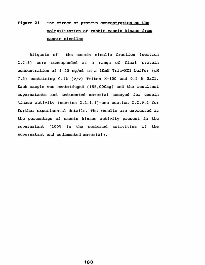
Figure 21 The effect of protein concentration on the solubilization of rabbit casein kinase from casein micelles
Aliquots of the casein micelle fraction (section2.2.8) were resuspended at a range of final protein concentration of 1-20 mg/ml in a lOmM Tris-HCl buffer (pH7.5) containing 0.1% (v/v) Triton X-100 and 0.5 M NaCl. Each sample was centrifuged (155,000xg) and the resultant supernatants and sedimented material assayed for casein kinase activity (section 2.2.1.1)-see section 2.2.9.4 for further experimental details. The results are expressed as the percentage of casein kinase activity present in the supernatant (100% is the combined activities of the supernatant and sedimented material).
X5O lO
80
> 60•4-̂o<
40
coV)COO
20
20151 05Protein (mg/ml)
181

Figure 22 Profile of casein kinase activity eluting fromcasein-sepharose
Casein kinase was purified using an affinity step on casein-sepharose as described in section 2.2.9.2. Bound material was eluted (the arrow indicates first eluate fraction) with 0.6 M sodium chloride in 0.1 M Tris-HCl (pH7.5) containing 0.1% (w/v) Triton X-100. An aliquot ofeach fraction was assayed for casein kinase activity as described in section 2.2.1.1.
182

> 20-
£ 1 0 -
1284Fraction
183

Figure 23 SDS polyacrylmide gel electrophoresis analysisof Sephacryl-S-300 and casein-sepharose purified rabbit mammary gland casein kinase
Casein kinase was purified from the micelle rich fraction using a combination of gel-exclusion chromatography on Sephacryl-S-300 and affinity chromatography on casein- sepharose as described in sections 2.2.9.1 and 2.2.9.2 respectively. Following each step an aliquot of the pooled casein kinase rich fractions were retained and analysed by electrophoresis on 10% (w/v) SDS polyacrylamide gels as described in section 2.2.3 and the gels silver stained as described in section 2.2.4.
Lane 1 contains aliquots of casein kinase rich material which eluted from Sephacryl-S-300.Lane 2 contains the casein kinase rich material from a casein-sepharose step.
Molecular weights, in Dalton xlO-3 , are indicated on the right.
184

1 2205
66
4529
I
185

for two polypeptide species of apparent molecular weighs63,000 and 67,000 Daltons, compare with figure 24 which contains an aliquot (about 35 pg) of starting material. This material generally also contained a number of low molecular weight contaminants.
Material eluting from the casein sepharose column was also enriched in both these polypeptide species with little evidence of the low molecular weight component (see figure 23).
5.2.3 Analysis of Sephacryl-S-300 and caseinsepharose purified casein kinase on linear 5-20 % (w/v) sucrose gradient centrifugation
Rabbit mammary gland casein kinase activity was isolated by a combination of chromatography on Sephacryl- S-300 followed by casein-sepharose as described above (section 5.2.2) and applied to a linear sucrose gradient 5-20% (w/v) prepared in a stock buffer of 0.1 M Tris-HCl (pH 7.5 at 4°C) containing 0.1% (v/v) Triton X-100.Identical sucrose gradients were layered with 250 pg of lactate dehydrogenase and both samples centrifuged in parallel for 35 hours at 150,000 xg at 4°C.
The results (presented in figure 25) show a single peak of casein kinase activity. Analysis of the fraction containing peak activity (fraction 18) by SDS polyacrylamide gel electrophoresis followed by silver
186
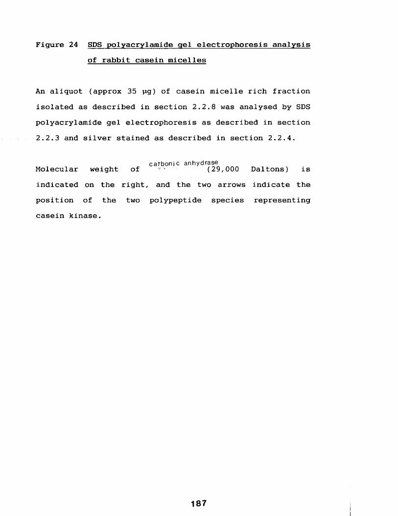
Figure 24 SDS polyacrylamide gel electrophoresis analysisof rabbit casein micelles
An aliquot (approx 35 pg) of casein micelle rich fraction isolated as described in section 2.2.8 was analysed by SDS polyacrylamide gel electrophoresis as described in section2.2.3 and silver stained as described in section 2.2.4.
c a fb o n ic anhydraseMolecular weight of - ■ (29,000 Daltons) isindicated on the right, and the two arrows indicate the position of the two polypeptide species representing casein kinase.
187
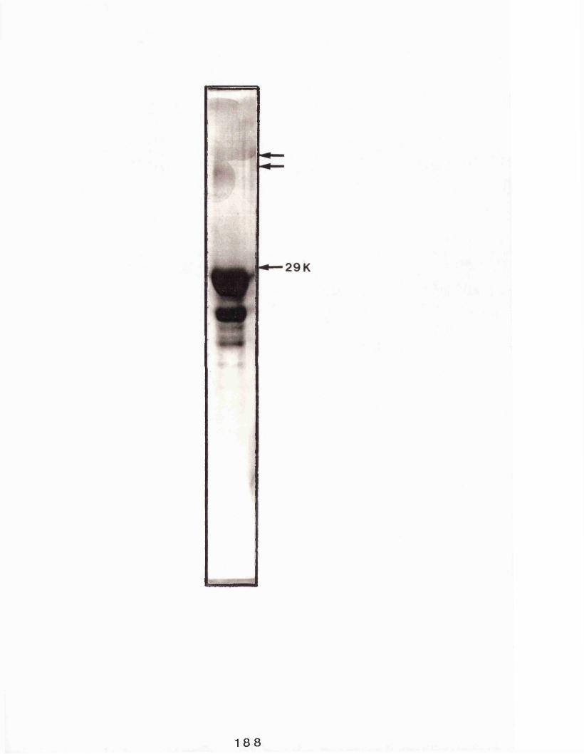
188

staining showed (figure 25) it to contain the same two polypeptide species described above.
Using this data the sedimentation coefficient (S) for the enzyme was estimated according to the method of Martin and Ames (1961) who stated that for two globular proteins (designated "1" and "2") the following relationship holds true:
Distance moved by protein 1 SiDistance moved by protein 2 S2
assuming that lactate dehydrogenase has a sedimentation coefficient of 7.4 (Pesce et al, 1967) the rabbit casein kinase activity has an estimated sedimentation coefficient of 3.9-4.28.
Martin and Ames (1961) further showed that for globular proteins the following relationship also holds true:
Si /S2 = (Mi/M2 )2/3
where Si and S2 are the sedimentation coefficients of lactate dehydrogenase and casein kinase respectively and Mi and M2 are their respective molecular weights.
Assuming a sub-unit molecular weight for lactate dehydrogenase of 145,000 Daltons (Peske et al, 1967) a
189
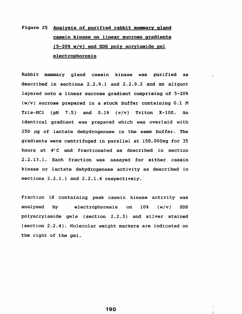
Figure 25 Analysis of purified rabbit mammary glandcasein kinase on linear sucrose gradients (5-20% w/v) and SDS poly acrylamide gel electrophoresis
Rabbit mammary gland casein kinase was purified as described in sections 2.2.9.1 and 2.2.9.2 and an aliquot layered onto a linear sucrose gradient comprising of 5-20% (w/v) sucrose prepared in a stock buffer containing 0.1 M Tris-HCl (pH 7.5) and 0.1% (v/v) Triton X-100. Anidentical gradient was prepared which was overlaid with 250 jig of lactate dehydrogenase in the same buffer. The gradients were centrifuged in parallel at 150,000xg for 35 hours at 4° C and fractionated as described in section 2.2.13.1. Each fraction was assayed for either casein kinase or lactate dehydrogenase activity as described in sections 2.2.1.1 and 2.2.1.4 respectively.
Fraction 18 containing peak casein kinase activity was analysed by electrophoresis on 10% (w/v) SDSpolyacrylamide gels (section 2.2.3) and silver stained (section 2.2.4). Molecular weight markers are indicated on the right of the gel.
190i

O Tfr
CM
Q
< OLX ujdo Ai!Ai)oy 0sbui^ u iaseo
191
Frac
tion
n
°
value of 55,000-64,000 Daltons for casein kinase is obtained.
In order to investigate whether 2-mercaptoethanol (a reducing agent which cleaves disulphide bridges) effects the distribution of casein kinase in a similar experiment to the one described above the following experiment was carried out. Two gradients, identical to those described above except that one also contained of the 2- mercaptoethanol (about 5 mM), were prepared. The gradients were layered with identical amounts of purified casein kinase and centrifuged in parallel as above for 42 hours. Both gradients were fractionated into 27 fractions (0.5 ml) and assayed for casein kinase activity as described in section 2.2.1.1 except that each assay was adjusted such that they all contained identical concentrations of 2- mercaptoethanol. The results (figure 26 shows that the presence of 2-mercaptoethanol did not alter the casein kinase activity profile).
5.2.4 Purification of casein kinase from caseinmicelles isolated directly from rabbit milk.
Milk was collected from New Zealand white rabbits by the manual expression of the mammary gland following an intra venous injection of oxytocin (8 units) and an overdose of pentobarbitone. Casein micelles were isolated from whole milk by centrifugation according to the method described in section 2.2.7 and casein kinase purified
192

Figure 26 Analysis of purified rabbit mammary gland casein kinase by centrifugation on linear sucrose gradients in the presence or absence of 2-mercaptoethanol
Rabbit mammary gland casein kinase was purified as described in section 2.2.9 and 0.5 ml aliquots analysed by centrifugation (42 hours at 150,000xg) on linear 5-20% (w/v) sucrose gradients (prepared in a stock buffer containing 0.1 M Tris-HCl pH 7.5 and 0.1% (v/v) Triton X- 100) in the presence or absence of 2-mercaptoethanol (approximately 5mM).
Following centrifugation the gradients were fractionated (section 2.2.13.1) and 20 pi each fraction (0.5 ml) assayed for casein kinase activity (section 2.2.1.1). Figure 26 shows a comparison of the resultant casein kinase activity profiles (the y-axis-casein kinase activity- is arbitarily shown as 0 to 12).
a + 2-mercaptoethanol #- 2-mercaptoethanol

Casein
Kinase
Acti
vity
top
25201510
Fraction N ?
194

Figure 27 SDS polyacrylamide gel electrophoresis analysisof casein kinase purified directly from rabbit milk
Whole rabbit milk was collected and casein micelles isolated as described in section 2.2.7 and casein kinase purified using a combination of Sephacryl-S-300 and casein-sepharose essentially as described in section 2.2.9.
Casein kinase rich material eluting from the final casein- sepharose step was analysed by SDS polyacrylamide gel electrophoresis on 10% (w/v) as described in section 2.2.3 and silver stained as described in section 2.2.4.
This material is shown in lane 1. For comparison casein kinase was purified from the micelle rich fraction (section 2.2.8) as described in sections 2.2.9.1 and2.2.9.2 and electrophoresed in parallel. This material is shown in lane 2. -
The position of casein kinase is indicated by the arrows.

196
essentially as described in section 2.2.9.1 and 2.2.9.2 with the modifications out-lined in section 2.2.9.5.
The enzyme eluted from the column of Sephacryl-S-300 typically in a volume of 35-41 mis (1 ml fractions) and as a single peak of activity from casein-sepharose (elution profiles are not shown). Analysis of the material from the casein sepharose step by SDS polyacrylamide gel electrophoresis (figure 27) showed it to consist of the same two polypeptide species previously described.
5.2.5 Effect of various reagents on release of kinase activity from casein micelles
Solubilization of casein micelles prior to application on Sephacryl-S-300 was carried out in a buffer which also included 0.6 M NaCl and 25 mM EGTA The following sections describe the rationale for including these agents in the solubilization buffer.
Casein sub-micelles, although voluminous, are tightly bound packages (see section 1.1.3) and it was of interest to investigate the effect of including additions of either sodium chloride, Triton X-100 or EGTA to incubations of micelle rich fractions on the amount of casein kinase released into the incubation supernatant.

5.2.5.1 Effect of sodium chloride
Zittle and Jasewicz (1962) demonstrated that a relatively low concentration of sodium chloride will cause the dissociation of a calcium-caseinate complex and it was of interest to investigate the effect of this salt on micelle disruption and any resulting release in casein kinase activity into solution. Aliquots (about 1 mg) of micelle rich fractions (section 2.2.8) were incubated for 1 hour in the presence of increasing concentrations (0-0.8 M) of sodium chloride at a final protein concentration of about 4 mg/ml at 4°C. Following this the incubation mixtures were centrifuged and casein kinase activity in the resultant supernatants were determined essentially as described in section 2.2.1.1.
The results (figure 28) show that increasing sodium chloride concentration resulted in an increase (compared with activity in absence of sodium chloride) of casein kinase activity up to 6.8 fold at 0.8 M, the highest NaCl concentration used.
5.2.5.2. Effect of EGTA.
Although the structure of the casein micelle is not fully understood (see section 1.1.3) it is well recognized that calcium is important in the maintenance of micelle structural integrity. Removal of this ion to below a

Figure 28 Effect of incubating casein micelles in the presence of increasing concentrations of sodium chloride.
Aliquots (about lmg) of rabbit casein micelles (see methods section 2.2.8) were incubated at a concentration of 4 mg/ml in the presence of increasing concentrations of sodium chloride (0-0.8M) for 1 hour on ice. Following this the samples were centrifuged in a "bench top microfuge" for 15 minutes (at full speed) and aliquots of the the resulting supernatants assayed for casein kinase activity essentially as described in section 2.2.1.1.
Casein
Kinase
Activity
Fold
increase
2 -
0-6 0-80-40-2NaCI (mM)
2 0 0

critical level results in complete dissociation of the micelle structure (Lin et al, 1972).
EGTA is a well known chelating agent which is capable of specifically forming a complex with calcium (Schmidt and Reilly, 1957) and it was of interest to assess the usefullness of the inclusion of this agent in buffers in order to disrupt casein micelles and hence solubilizing casein kinase.
Aliquots (about 1 mg) of casein micelle rich fractions (section 2.2.8) were incubated for 1 hour at 4°C in the presence of increasing concentrations (0-100 mM) of EGTA at a final protein concentration of about 4 mg/ml. Following this incubation mixtures were centrifuged and the casein kinase content of the resultant supernatants determined essentially according to the method described in section 2. 2.1.1.
The results (figure 29) show that following the addition of just 1 mM caused a 5.1 fold increase (compared with activity in the absence of EGTA) of casein kinase activity in the incubation supernatant. Further additions of EGTA up to 100 mM caused no further increase.
5.2.5.3 Effect of Triton X-100
Detergents are frequently used to solubilize membranes, utilizing their property of being able to

Figure 29 Effect of incubating casein micelles in thepresence of increasing concentrations of EGTA
Aliquots (about lmg) of rabbit casein micelles (prepared as described in section 2.2.8) were incubated at a final protein concentration of 4 mg/ml in the presence of increasing concentrations of EGTA (0-100 mM) for 1 hour on ice. Following this the samples were centrifuged in a "bench top microfuge" for 15 mins (at full speed) and aliquots of the the resulting supernatants assayed for casein kinase activity essentially as described in section 2 .2 .1.1.
2 0 2
Casein
Kinase
Activity
Fold
Increase
5
4
3
2
1
80604020E G T A (mM)
203
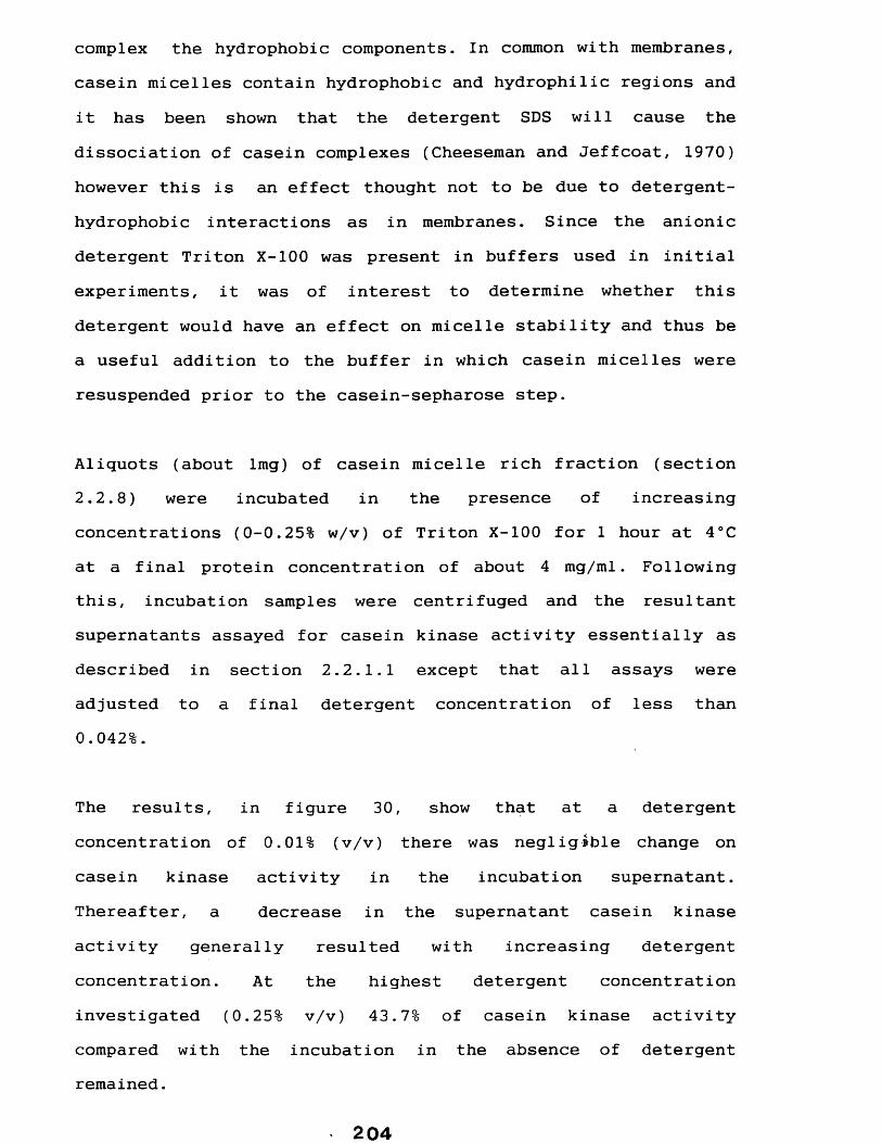
complex the hydrophobic components. In common with membranes, casein micelles contain hydrophobic and hydrophilic regions and it has been shown that the detergent SDS will cause the dissociation of casein complexes (Cheeseman and Jeffcoat, 1970) however this is an effect thought not to be due to detergent- hydrophobic interactions as in membranes. Since the anionic detergent Triton X-100 was present in buffers used in initial experiments, it was of interest to determine whether this detergent would have an effect on micelle stability and thus be a useful addition to the buffer in which casein micelles were resuspended prior to the casein-sepharose step.
Aliquots (about lmg) of casein micelle rich fraction (section2.2.8) were incubated in the presence of increasing concentrations (0-0.25% w/v) of Triton X-100 for 1 hour at 4°C at a final protein concentration of about 4 mg/ml. Following this, incubation samples were centrifuged and the resultant supernatants assayed for casein kinase activity essentially as described in section 2.2.1.1 except that all assays were adjusted to a final detergent concentration of less than 0.042%.
The results, in figure 30, show that at a detergentconcentration of 0.01% (v/v) there was negligible change oncasein kinase activity in the incubation supernatant.Thereafter, a decrease in the supernatant casein kinaseactivity generally resulted with increasing detergent concentration. At the highest detergent concentrationinvestigated (0.25% v/v) 43.7% of casein kinase activitycompared with the incubation in the absence of detergent remained.
204

Figure 30 Effect of incubating casein micelles in thepresence of increasing concentrations of TritonX-100
Aliquots (about lmg) of rabbit casein micelles (prepared as described in section 2.2.8) were incubated at about 4 mg/ml in the presence of increasing concentrations of Triton X-100 (0-0.25 % v/v) for 1 hour on ice. Following this the samples were centrifuged in a "bench top microfuge" for 15 mins at full speed and aliquots of the the resulting supernatants assayed for casein kinase activity essentially as described in section 2.2.1.1.

Casein
Kinase
Acti
vity
100
80
60
40
20
0-30-1 0*2Triton X-100 (%v/v)
206
5.3 DISCUSSION
The work in the preceding chapters identified a casein kinase from the lactating rabbit mammary gland associated with two particulate fractions identified as a casein micelle and golgi enriched fraction. The work involved here describes the development of a procedure for the purification of this enzyme using the micelle rich fraction as a starting source.
Pascall et al (1981) used affinity chromatography on ATP- agarose to partially purify the guinea-pig mammary gland casein kinase, whilst Moore et al (1985) used a centrifugation step on a 5-20% (w/v) sucrose gradientfollowed by an improved ATP-agarose step to purify the same enzyme essentially to homogeneity.
Initially an ATP-agarose step was used here which usually resulted in insignificant amounts of enzyme activity eluting from the column (data not shown), the improved ATP-agarose step used essentially as described by Moore et al (1985) was more effective and resulted in a single peak of casein kinase activity (not shown). This material was analysed by SDS polyacrylamide gel electrophoresis and was shown (figure 17) to be enriched for two polypeptide species of approximate apparent molecular weights in the region of 66,000 Daltons. Further purification of this material on a linear sucrose gradient resulted in a peak of casein kinase activity which was also shown to contain
207

the same two polypeptide species, with little evidence of any other material.
Based on this data it was considered likely, although not conclusive, that these two polypeptide species may represent the casein kinase activity.
The procedures used however were rather time consuming and limited with respect to the amount of material that could be purified at any one time. Other procedures were therefore investigated. Firstly a gel-filtration step, in order to remove the bulk of the low molecular weight components with molecular weights smaller than the putative casein kinase components. This step resulted in a single peak of casein kinase activity and, appears to elute later than the bulk of the other proteins, probably caseins, and is thus a useful step. In comparison with the starting material the casein kinase material was shown to be enriched for the two polypeptide species of interest. It did not remove all the low molecular weight proteins which are also probably caseins as their sizes would agree with the observations out-lined in section 1.1.2.3.
A futher affinity step using casein-sepharose resulted in the further enrichment of the polypeptides with little evidence of any other low molecular weight material contaminants. The two polypeptides were shown to have apparent molecular weights of 63,000 and 67,000 Daltons.
208

Further analysis of this material eluting from the casein- sepharose step by centrifugation on a linear sucrose gradient which confirmed that the single peak of casein kinase activity contained these same two polypeptides.
The results from these three steps further confirm that the two polypeptides of apparent molecular weights of63,000 and 67,000 Daltons represent the rabbit mammary gland casein kinase.
The enzyme differs in the respect from the guinea-pig mammary gland casein kinase, which is a homodimer of apparent molecular weight 74,000 Daltons, the other so- called "casein kinases" (see section 1.2.4.7) and other common protein kinases (see table 10).
Analysis of the purified enzyme on linear 5-20% (w/v)sucrose gradients resulted in a single peak of casein kinase activity which was shown to contain the two polypeptide species described above. This material in this peak had a sedimentation coefficient of 3.9-4.28 and molecular weight of 55,000-64,000 Daltons. These results show that the enzyme has a molecular weight considerably less than''the value expected if it comprised of a dimer of the two polypeptides and this suggests a monomer structure. Both polypeptide species were present in the single peak of casein kinase activity.

Table 10 Characterisics of some common protein kinases
Comparison of the molecular weights for some commonly occuring protein kinases including the ubiquitous multifunctional casein kinases (see section 1.2.4.7) and the guinea-pig mammary gland casein kinase.
Data taken from Roach (1984) and Moore et al (1985).

Protein kinase
Cyclic AMP dependentCyclic GMP dependentProtein kinase CCasein kinase ICasein kinase IIGuinea-pig mammary gland casein kinase
Structure
HeterotetramerHomodimerMonomerMonomerHeterotetramerHomodimer
Sub-unit Size(s) (KD)49-56/38-42748230-4043/2574
211

It is well known that 2-mercaptoethanol, a reducing agent, will reduce the disulphide bridges a common link between two polypeptides. Compared with a sucrose gradient set up as above, the presence of this agent in the sucrose gradient of a parallel experiment was shown to alter neither the position of the activity profile nor the ammount of casein kinase activity recovered. Results which indicate that disulphide bridges are not present and suggest the enzyme molecule is monomeric.
The presence of two polypeptides could be due to the existence of differentially post-translationally modified species of the same enzyme. Another explanation is the co-purification of a similar sized protein, although this is highly unlikely since both are present in casein kinase rich material resulting from a number of purification steps. The possibility of the isolation of two distinct proteirP kinases can not be ruled out.
It was also demonstrated that the rabbit casein kinase could be purified from micelles isolated directly from whole milk, although, as discussed in chapter 4, casein micelles were not routinely isolated directly from whole mi lk.
Further development of a procedure for the isolation of casein kinase involved investigating the effect of three agents on casein micelle disruption leading to release of casein kinase was investigated. Zittle and Jasewicz
212
(1962) have shown that a relatively low concentration of sodium chloride will cause the dissociation of a calcium- caseinate complex. They postulated that the disruption of this complex is due to the interference of the stabilizing repulsive effects of negatively charged ions by sodium chloride. Puri and Parkash (1965) and Jenness (1973) have also shown that sodium chloride will cause the dissociation of the micelle and suggest that this is due to the replacement of the micelle calcium (i.e. CCP) with sodium thus releasing calcium into solution.
Incubation of aliquots of casein micelles (at 4 mg/ml) in the presence of increasing concentrations of NaCl (0-0.8 M) resulted in an increase of casein kinase in the incubation supernatant. This increase in activity must result from an increase in the amount of enzyme present in the supernatant, since final assay incubations were adjusted in order to negate any possible direct effect on the assay of differing amounts of this salt. This result is in agreement with Puri and Parkash (1965) who found that increasing the sodium chloride to a maximum of 0.74 M resulted in the increased dissociation of tha micelle. They found further additions above this concentration resulted in no further increase.
It is likely that this increase in enzyme availability therefore reflects the disruption of the casein micelle structure presumably by one or both of the mechanisms
213

described above, with a concomitant release of enzyme closely associated with the micelle.
Incubation of casein micelles in the presence of EGTA at the same protein concentration also resulted in an increase in casein kinase activity present in the incubation supernatant. Assays again were adjusted to account for differences in EGTA concentrations and therefore negating its stimulatory effect on the casein kinase assay (see section 7.2.8) and the increase in activity therefore reflects an increase in the amount of enzyme present. A number of groups have shown that removal of calcium from casein micelles by dialysis results in the complete dissociation of micelle structure (Lin et al,
1972) presumably reflecting the importance of calcium in micelle assembly (see section 1.1.3). EGTA forms complexes specifically with calcium ions (Schmidt and Reilly, 1957), and therefore incubation of this compound with micelles, will result in the removal of this ion resulting in dissociation of the micelle. Similar effects on micellar integrity have been shown by using the less specific chelating agent EDTA (Lin et al, 1972 and Bloomfield andMorr, 1973 ) .
Detergents have proved useful tools in the solubilization of membrane proteins from the lipid components (see Hjelmeland and Chrambach, 1984). Dissociation of casein aggregates has been shown to occur following binding of the detergent SDS (Cheeseman and Jeffcoat, 1970) who
214

postulated that the detergent bound to hydrophilic proteins, and in doing so competed with casein molecules for these sites leading to disruption of the micelle structure. In contrast It was shown here that the addition of Triton X-100, up to 0.25% (v/v) resulted in a decrease of casein kinase activity in the incubation supernatant. The explanation for this effect is unclear.
It was concluded that 0.6 M sodium chloride and 25 mM EGTA were useful additions to the buffer in which the casein micelles are resuspended prior to the gel filtration step since they would appear to aid the release of the enzyme.
5.4 SUMMARY
To summarize, using casein micelles as a starting source, a number of purification procedures were used to isolate casein kinase rich fractions. In each case twopolypeptide species which were shown to have apparent molecular weights of 63,000 and 67,000 Daltons were isolated.
In particular the use of affinity columns indicated that both polypeptides bind ATP and casein, substrates for casein kinase and the data taken in toto would suggest that these represent two mammary gland casein kinases. It is not clear whether these two species represent the same enzyme and differences in apparent molecular weights
215

reflect differential post-translational modification or whether two distinct enzymes have been isolated.
The procedure adopted as the method for the routine purification of the enzyme comprises solubilization of casein micelles in a buffer containing 0.6 M sodium chloride and 25 mM EGTA (see materials and methods for details), followed by a combination of gel filtration on Sephacryl-S-300 and affinity chromatography on casein sepharose. This procedure is summarized in figure 31.
216
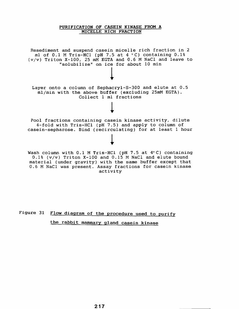
PURIFICATION OF CASEIN KINASE FROM A MICELLE RICH FRACTION
Resediment and suspend casein micelle rich fraction in 2 ml of 0.1 M Tris-HCl (pH 7.5 at 4 °C) containing 0.1%
(v/v) Triton X-100, 25 mM EGTA and 0.6 M NaCl and leave to "solubilize" on ice for about 10 minI
Layer onto a column of Sephacryl-S-300 and elute at 0.5 ml/min with the above buffer (excluding 25mM EGTA).
Collect 1 ml fractions
iPool fractions containing casein kinase activity, dilute
4-fold with Tris-HCl (pH 7.5) and apply to column of casein-sepharose. Bind (recirculating) for at least 1 hour
1Wash column with 0.1 M Tris-HCl (pH 7.5 at 4°C) containing 0.1% (v/v) Triton X-100 and 0.15 M NaCl and elute bound
material (under gravity) with the same buffer except that 0.6 M NaCl was present. Assay fractions for casein kinase
activity
Figure 31 Flow diagram of the procedure used to purify the rabbit mammary gland casein kinase

CHAPTER 6PURIFICATION OF CASEIN KINASE FROM A GOLGI
ENRICHED FRACTION ISOLATED FROM THE LACTATING RABBIT MAMMARY GLAND
218
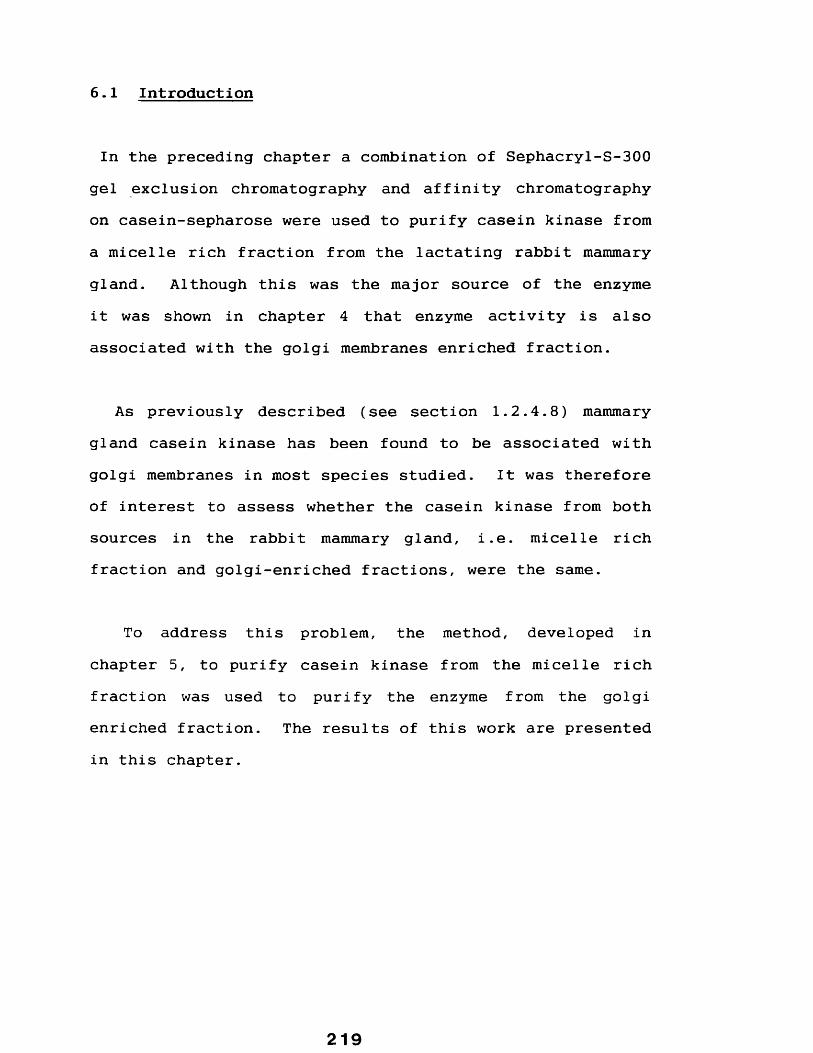
6.1 Introduction
In the preceding chapter a combination of Sephacryl-S-300 gel exclusion chromatography and affinity chromatography on casein-sepharose were used to purify casein kinase from a micelle rich fraction from the lactating rabbit mammary gland. Although this was the major source of the enzyme it was shown in chapter 4 that enzyme activity is also associated with the golgi membranes enriched fraction.
As previously described (see section 1.2.4.8) mammary gland casein kinase has been found to be associated with golgi membranes in most species studied. It was therefore of interest to assess whether the casein kinase from both sources in the rabbit mammary gland, i.e. micelle rich fraction and golgi-enriched fractions, were the same.
To address this problem, the method, developed in chapter 5, to purify casein kinase from the micelle rich fraction was used to purify the enzyme from the golgi enriched fraction. The results of this work are presented in this chapter.
219

6.2 RESULTS
A golgi enriched membrane fraction was prepared as described in methods section 2.2.8 and used as a starting source to purify casein kinase by a combination of gel- exclusion chromatography and casein-sepharose affinity chromatography essentially as described in sections2.2.9.1 and 2 . 2.9.2.
This resulted in the elutiOfl of a single distinct peak of casein kinase activity contained in fractions 39-44 inclusive, (figure 32). This material was pooled, adjusted to 0.15 M NaCl with 100 mM Tris-HCl pH 7.5 and applied to a column containing casein-sepharose essentially as described in materials and methods. Following washing bound material was eluted in buffer containing 0.6 M NaCl (see section 2.2.9.6). The bound casein kinase activity eluted as a single peak (see fig 33). This material was pooled and dialysed against 4 litres of ammonium bicarbonate (about 10 mM), freeze dried, resuspended in sample buffer and applied to 10% polyacrylamide gels (see section 2.2.3). Following electrophoresis gels were stained as described in section2.2.4 . The results show (see figure 34) that the casein kinase purified from the golgi enriched fraction contained the same two polypeptide species described in the preceding chapter.
220

Figure 32 Profile of casein kinase activity from a golgienriched fraction eluting from a column of Sephacryl-S-300
Aliquots of golgi enriched fraction isolated from the lactating rabbit mammary gland (see section 2.2.8) were resuspended in buffer containing 0.1% (v/v) Triton X-100 and 0. 6M NaCl and applied to a column of Sephacryl-S-300 and eluted in the same buffer at a flow rate of 0.25 ml min* 1 . Casein kinase activity in each eluate fraction was assayed as described in section 2.2.1.1.
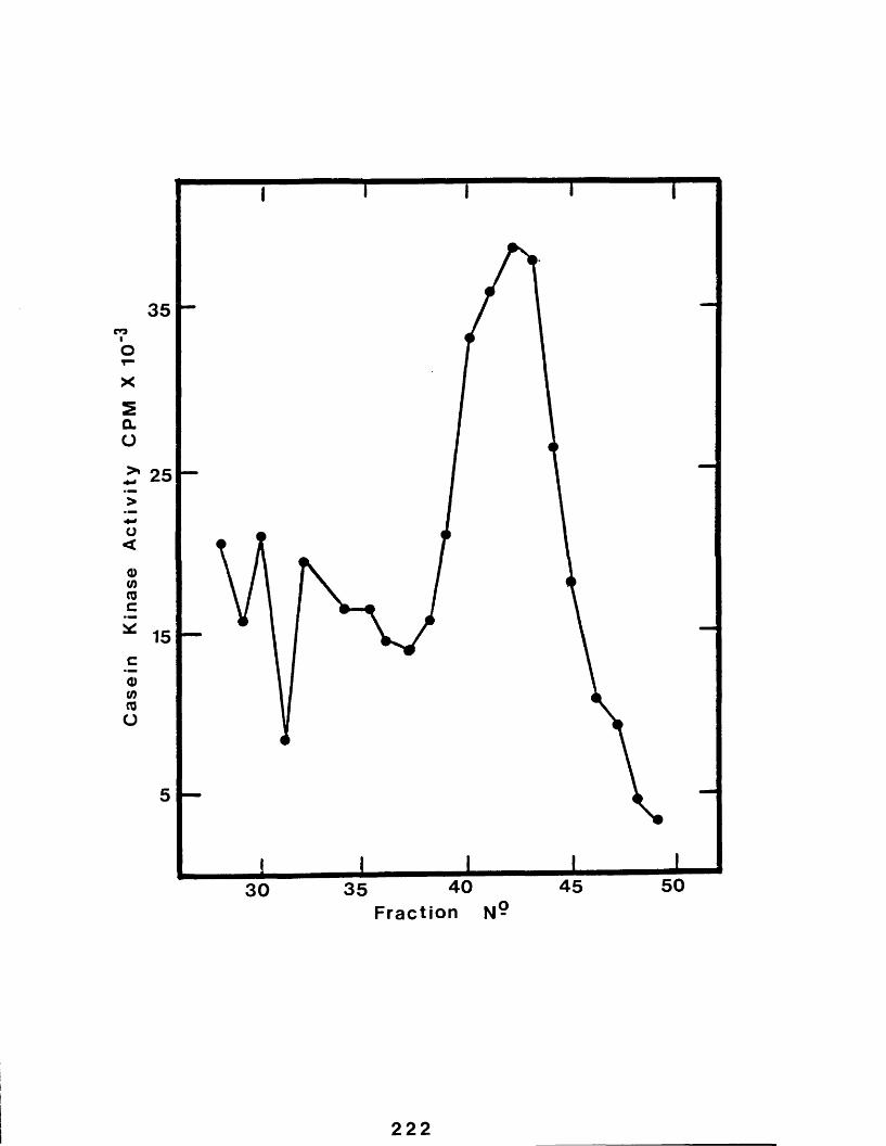
Casein
Kinase
Activity
CPM
X 10
35
25
15
!30 35 40
Fraction45 50
222

Figure 33 Profile of casein kinase activity isolatedfrom a golgi enriched fraction eluting from casein-sepharose
A golgi enriched fraction was isolated from the lactating rabbit mammary gland as described in section 2.2.8, and used as a starting source for the purification of casein kinase using the procedure developed in Chapter 5. Figure 33 shows the casein kinase activity (assayed as described in srection 2.2.1.1) eluting from casein-sepharose (see section 2.2.9.6).
223
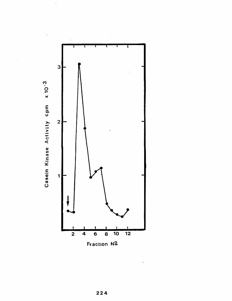
Casejn
Kinase
Activity
cpm
x 10
3
2
1
2 4 6 8 10 12Fraction N2
224

Figure 34 SDS polyacrylamide gel electrophoresis analysisof purified golqi casein kinase
Rabbit mammary gland golgi enriched fraction was isolated as described in section 2.2.8 and casein kinase purified using a combination of Sephacryl-S-300 and casein- sepharose essentially as described in sections 2.2.9.1 and 2 .2 .9 .2 .
Casein kinase rich material eluting from the final casein- sepharose step was analysed by SDS polyacrylamide gel electrophoresis on 10% (w/v) as described in section 2.2.3 and silver stained as described in section 2.2.4.
This material is shown in lane A. For comparison casein kinase was purified from the micelle rich fraction (section 2.2.8) as described in sections 2.2.9.1 and 2 . 2 . 9 . 2 , (lane B ).
225
226

6.3 DISCUSSION
In the previous chapter a combination of Sephacryl-S-300 gel filtration chromatography and casein-sepharose affinity chromatography were used to purify casein kinase from the micelle rich fraction. The same procedure was used to purify casein kinase from the golgi enriched fraction described in chapters 3 and 4 and resulted in the same two polypeptide species present in the micelle rich fraction.
Galactosyltransferase has similarly been shown to be present both in milk and a golgi enriched preparation (see review by Strous, 1986). The secreted form being derived from the membrane bound form by proteolysis hence the former has a lower molecular weight. It is clear that although casein kinase is has a similar distribution, the secreted form, being identical in molecular weight to the membrane form, is not derived by such proteolytic action.
This result further suggests that the secreted enzyme may be derived from the golgi associated enzyme possibly as a result of the same "spilling over" of cellular components in milk described by Kitchen (1970) to explain the presence of some the enzymes in milk (see section 1 .1 .4 .3 ).
227

In the following chapter some of the properties of casein kinase purified from the micelle rich fraction as described in chapter 5, are investigated.
228

CHAPTER 7
CHARACTERIZATION OF A RABBIT MAMMARY GLANDCASEIN KINASE

7.1 Int roduct i on
The experiments described in preceding chapters have identified a protein kinase from both a micelle rich fraction and a golgi enriched fraction which was capable of phosphorylating caseins. It has been demonstrated that a number of protein kinases from a variety of tissues are also able to phosphorylate caseins (sections 1.2.4.7). It was of interest therefore to characterize some of the properties of the purified enzyme to gain a clearer picture of any differences or similarities with other mammary gland casein kinases.
As part of the overall characterization of the purified rabbit enzyme, factors such as:- NaCl, EGTA and Triton X- 100 as well as parameters affecting the enzyme assay, for example:- assay time, substrate concentration and assay buffer pH, were also characterized.
The results of this work are presented in the following sections and, unless stated otherwise, the enzyme used was casein kinase, purified according to the procedure outlined in section 2.2.8.

7.2 RESULTS
7.2.1 The effect of enzyme concentration
To assess the effect of concentration of purified enzyme used in standard assays casein kinase was assayed over a range 0 to 20 pi as described in section 2.2.1.1. In all cases final assay volumes were adjusted to 60 pi with water.
The results (see figure 35) show that in the range 0 to 17 pl of enzyme added resulted in a directly proportional increase in the rate of casein phosphorylation. The plotted data however consists of a second linear phase when greater than 17 pl of enzyme were added. This also shows a proportional increase in phosphorylation rate but at a greater velocity than when less than 17 pl of enzyme were added.
7.2.2 The effect of increasing assay incubation time
The effect of increasing the 37° C assay incubation time on casein phosphorylation was investigated by setting up standard assays (as described in section 2.2.1.1) except that the 37°C incubation step was carried out over a range of time of 10 to 60 minutes.
2 31

Figure 35 The effect of increasing enzyme concentration onthe incorporation of 3 2 P into total bovine caseins by purified rabbit mammary gland casein kinase
Casein kinase was purified from the lactating rabbit mammary gland casein kinase as described in sections 2.2.9 and a range of this material (0-20 pl) included in standard assays (section 2.2.1.1).
232
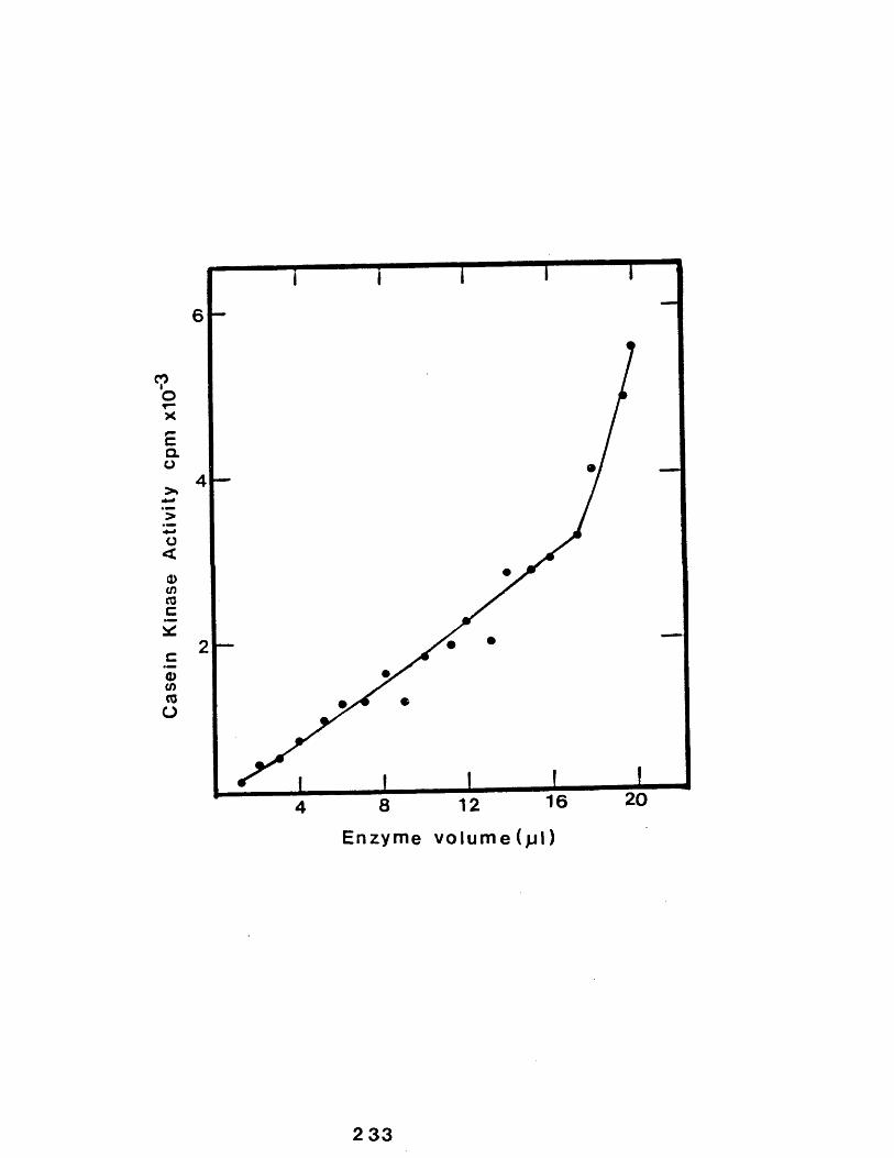
Casein
Kinase
Activi
ty cpm
x10COi
20168 12 Enzyme volume(jjl)
2 33

The results (see figure 36) demonstrate that over therange used increasing the incubation time resulted in a proportional increase in phosphorylation rate.
7.2.3 The effect of changes of pH on enzyme activity
The optimum pH conditions were determined by assaying purified casein kinase as described in section 2.2.1.1, except that the buffer was replaced by 15 mM 2-(N-morpholino) ethanolaminesulphonic acid-KOH (Mes-KOH) over the range pH 5 to pH 6, 15 mM 4-morphol inepropane-sulphonic acid-KOH (Mops-KOH) in the range pH 6 to pH 7.25and 15 mM Tris-HCl at a range of 7.25 to pH 8.0.
Further values of pH outside the range used (5-8) would be difficult for technical reasons since the substrate (casein) is sensitive to pH and precipitates below pH 4.7 (see "definition of caseins" section 1.1.2.3) at pH greater than 8.0 phosphoserine, resulting from the action of casein kinase, are labile (Weller, 1979) and the phosphate groups of p-casein are labile in alkali (Manson,1973).
Values were chosen to allow for overlap between ranges to account for any possible effect due to changes in the buffer system rather than pH.
234

Figure 36 The effect of changes of incubation time onthe activity of rabbit mammary gland casein kinase
Purified rabbit mammary gland casein kinase (sections2.2.8) was assayed as described in section 2.2.1.1 except that the incubation assay time at 37°C was varied over the range 10-60 min.
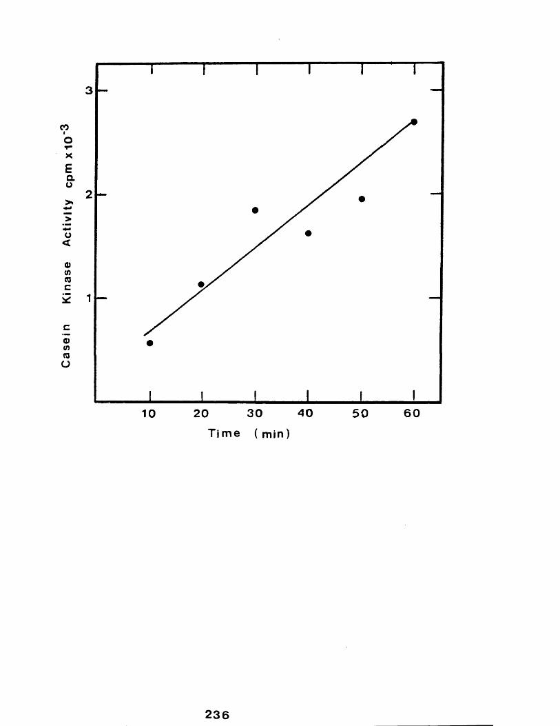
asein
Kinase
Activi
ty cp
mxIO
"3
2
1
10 20 30 40 50 60Time ( min)
236

The results (figure 37) show that caseins were poorly phosphorylated at pH 5 to pH 6 whilst the enzyme showed more activity at higher pH resulting in a broad optima of between pH 6.5 to pH 8, with peak activity at pH 7.25.
There is a slight discontinuity between phosphorylation using Mes or using Mops at pH 6, but only a small difference between values at pH 7.25 i.e. Mes (41026 cpm) and Tris (37943 cpm).
7.2.4 The effect of temperature on enzyme activity
The effect of assay temperature was assessed by setting up standard assays except that the incubation step at 37°C (see section 2.2.1.1) was replaced by incubating over the range 4°C to 60°C.
The results presented in figure 38 show a single sharp optima of enzyme activity over the range 22 to 45° C with maximum activity at 30°C.
7.2.5 The effect of the nature and concentration of the divalent cation
Enzymes which use ATP as a substrate generally have a requirement for a divalent cation, usually Mg2 + (Grandt et al, 1980). The ability of the mammary gland casein kinase to utilize one of three cations i.e Mg2 * , Ca2 + and Mn2 *
237

Figure 37 The effect of increasing pH on the activity ofrabbit mammary gland casein kinase
Purified rabbit mammary gland casein kinase (section2.2.9) was assayed essentially as described in section2.2.1.1 except that the following three different buffer systems were used. 15 mM 2-(N-morpholino)ethanesulphonic acid-KOH inm the range pH 5-6 ( • ); 15 mM 4-morpholino-propanesulphonic acid over a range of 6-7.25 ( A ) and 15 mM Tris-HCl at a range of 7.25-8.0 ( ■ ). In all casestotal bovine casein served as the phosphate acceptor.

Casein
Kinase
Activity
CPM
X 10'
P H Units
239

Figure 38 The effect of changes of assay temperature on the activity of purified rabbit mammary gland casein kinase
Purified rabbit casein kinase (sections 2.2.9.1 and 2.2.9.2) was assayed as described in section 2.2.1.1 except that the assays were incubated at the following temperatures: 4,15,22,30,37,45,50, and 60°C.

Casein
Kinase
Activity
CPM
X 10
’
25e*s
20
15
10
0 20 6040Temperature °C
241

over a range of concentrations (0 to 15 mM) was therefore investigated.
Standard assays were set up as described in section2.2.1.1 except that increasing concentrations (0 to 15 mM) of these cations, as the chloride, replaced the divalent cation usually present in the assays.
The results (figure 39) reveal that the rabbit mammary gland casein kinase is activated in the presence of Mg2 + over the whole range tested with a clear optima of activity at 7.5 mM. Negligable enzyme activity was detected in the presence of Ca2 + or Mn2 + at any concentration tested.
7.2.6 The effect of increasing concentrations of calcium chloride on enzyme activity in the presence of magnesium chloride
It was of interest to assess the effect of increasing concentrations of Ca2 + on the enzyme activity in the presence of Mg2+ for two reasons. Firstly Moore (1985) showed that the guinea-pig enzyme was inactivated by increasing Ca2 + concentrations and secondly it is known that the starting source for enzyme purification i.e the micelle rich fraction, is rich in Ca2+ (see section 1.1.3) and it was therefore important to investigate whether this would effect enzyme activity.
242

Figure 39 The effect of the nature and concentration ofthe divalent cation on the activity of the purified rabbit mammary gland casein kinase
Standard assays were set up as described in section2.2.1.1 except that increasing concentrations (0-15 mM) of one of the following divalent cation, as the chloride, replaced 10 mM magnesium chloride: Mg2 + (• ); Mn2 * ( orCa2 + ( 4 ) .
\
243

Casein
Kinase
Activity
CPM
X 10
151050
Divalent Cation (mM)
244

Assays were set up (essentially as described in section2.2.1.1) and included a range of Ca2+ (0 to 17 mM), as thechloride, were included. The results (figure 40) showed that increasing the concentration of Ca2 * greater than 86 pM resulted in an increased inhibition of casein kinase activity. Below this concentration there was no significant effect on enzyme activity.
7.2.7 Kinetic parameters
1.2.1.1 Casein
In order to determine the limiting rate (V.a. ) and the Michael is constant (Km ) values of the rabbit casein kinase for casein (total bovine caseins were used) as the substrate the following assays were set-up. Assays were set up containing 100 pm of J^32P ATP, 10 pl purified enzyme, 10 pl 60 mM MgCl2 , 3.75 pl 1M Mops and casein inthe range 0 to 80 pg in a final volume of 60.5 pl. Samples were then assayed essentially as described in section 2 .2 .1 .1 .
The results are presented graphically in figure 41 i.e velocity (cpm/min) against amount of substrate (pg/assay), and as the double reciprocal of these values in figure 42. Values for K, and V™ a * were estimated according to the method described by Lineweaver and Burke (1934) i.e. where an inverse plot of velocity versus substrate concentration
245

Figure 40 The effect of increasing concentrations of Ca2 *in the presence of Mg2 * on the activity of rabbit mammary gland casein kinase
Purified rabbit mammary gland casein kinase (section2.2.9) was assayed essentially as described in section2.2.1.1 except the assays were carried out in the presence of 8.6 mM magnesium chloride and a range of calcium chloride (0-17 mM).
246

Casein
Kinase
Acti
vity
+ 8*6 rnM Mg100
80
60
40
20
20 00010 100 200050
Calcium ( p M )
247
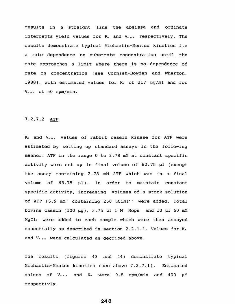
results in a straight line the absissa and ordinate intercepts yield values for K™ and V™ a> respectively. The results demonstrate typical Michaelis-Menten kinetics i.e a rate dependence on substrate concentration until the rate approaches a limit where there is no dependence of rate on concentration (see Cornish-Bowden and Wharton, 1988), with estimated values for Km of 217 pg/ml and for Vm a z of 50 cpm/min.
1.2.1.2 ATP
Km and Vm a * values of rabbit casein kinase for ATP were estimated by setting up standard assays in the following manner: ATP in the range 0 to 2.78 mM at constant specific activity were set up in final volume of 62.75 pl (except the assay containing 2.78 mM ATP which was in a final volume of 63.75 pl). In order to maintain constant specific activity, increasing volumes of a stock solution of ATP (5.9 mM) containing 250 pCiml" 1 were added. Total bovine casein (100 pg), 3.75 pl 1 M Mops and 10 pl 60 mM MgCl2 were added to each sample which were then assayed essentially as described in section 2.2.1.1. Values for Km and Vma * were calculated as decribed above.
The results (figures 43 and 44) demonstrate typical Michaelis-Menten kinetics (see above 7.2.7.1). Estimated values of Vm a x and K™ were 9.8 cpm/min and 400 pM respectivly.
24 8

Figure 41 The effect of increasing amounts of totalbovine casein on the activity of the purified rabbit mammary gland casein kinase
Purified rabbit mammary gland casein kinase (section2.2.9) was assayed as described in section 2.2.1.1 in the presence of increasing amounts of total bovine casein (0- 80 pg) .
The data is presented as velocity (cpm/min) versus substrate "concentration" (pg/assay).

Casein
Kinase
Activity
cpm/
min
60
40-
20 -
9060Casein (^g)
30
250
Figure 42 Estimation of V. ax and K. of casein kinase forcasein
The data presented in figure 41 was replotted as the reciprocal to yield the "double reciprocal plot", where V is the rate (cpm/min) and S the substrate concentration (pg/assay). V™ax was calculated from the absissa intercept and K* the ordinate intercept (not shown on this scale).

X1050
40
301
20
10
2 4 6 10X10
252

Figure 43 The effect of increasing amounts of ATP on theactivity of the purified rabbit mammary gland casein kinase
Purified rabbit mammary gland casein kinase (section2.2.9) was assayed as described in section 2.2.1.1 with the modifications out-lined in section 7.2.7.2 in the presence of increasing concentrations of ATP (0-2.78 mM).
The results presented as a plot of velocity (cpm/min) versus substrate concentration (mM) and are shown opposite.
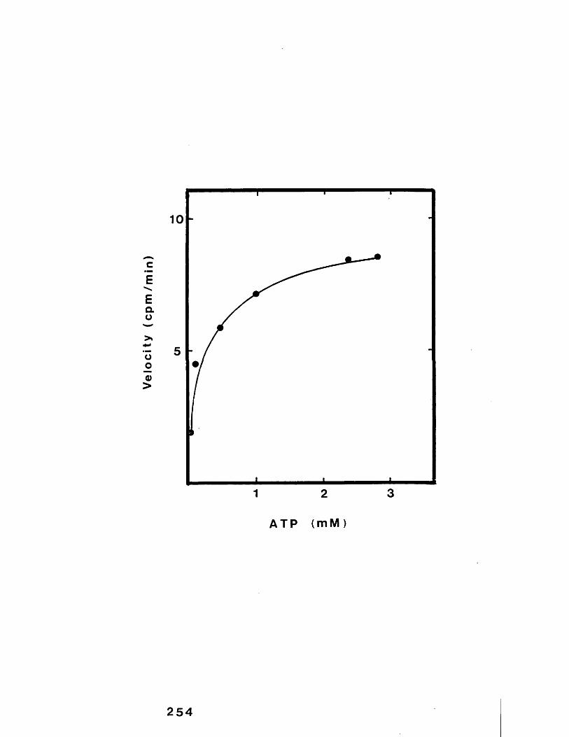
Velocity
(cpm
/min
)
10
5
1 2 3
ATP (mM)
254
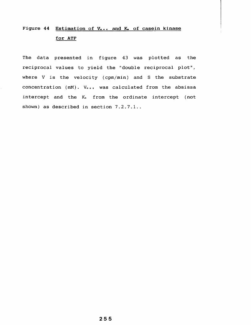
Figure 44 Estimation of V,a» and K- of casein kinase for ATP
The data presented in figure 43 was plotted as the reciprocal values to yield the "double reciprocal plot", where V is the velocity (cpm/min) and S the substrate concentration (mM). V™ ax was calculated from the absissa intercept and the Km from the ordinate intercept (not shown) as described in section 7.2.7.1..
25 5
20
x10V
10
0-5 1 2
256

7.2.8 The effect of increasing concentrations of Sodiumchloride, EGTA and Triton X-100 on enzyme activity
As NaCl, EGTA and Triton X-100 were often included in a number of casein kinase assays in the work presented in this thesis it was essential to investigate any possible effect of these additions.
Standard assays were set up as described in section2.2.1.1 with the addition of increasing amounts of either NaCl (0 to 0.5 M), EGTA (0 to 150 mM) or Triton X-100 at concentrations of 0 to 0.2% (v/v). However it should benoted that due to the inclusion of 0.6 M NaCl in elution buffers (see section 2.2.9.1) aliquots of purified enzyme (10 pi) contributed a further 0.1 M NaCl to final assay incubations (60 pi). Triton X-100 and EGTA however were not included in elution buffer of the final step during the purification of the casein kinase sample used in these experiments.
The results (figure 45) shows a small initial increase in kinase activity (at 0.2 and 0.3 M NaCl) with a loss of activity on increasing NaCl concentration above 0.3M.
Increasing concentrations of EGTA in assays resulted in an increased stimulation of casein phosphorylation, albeit poorly, to about 50% at 100 mM, compared with the activity in the absence of EGTA. At the highest concentration used
257
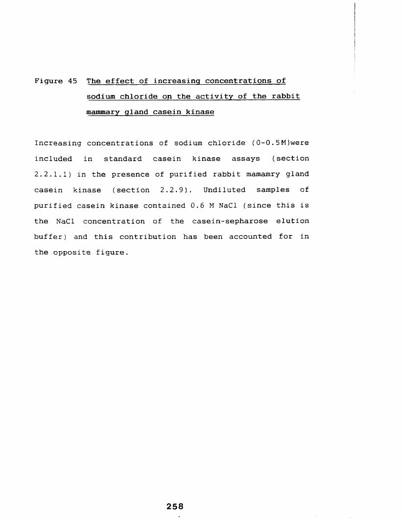
Figure 45 The effect of increasing concentrations ofsodium chloride on the activity of the rabbit mammary gland casein kinase
Increasing concentrations of sodium chloride (0-0.5M)were included in standard casein kinase assays (section2.2.1.1) in the presence of purified rabbit mamamry gland casein kinase (section 2.2.9). Undiluted samples of purified casein kinase contained 0.6 M NaCl (since this is the NaCl concentration of the casein-sepharose elution buffer) and this contribution has been accounted for in the opposite figure.

Casein
Kinase
Activity
CPM
X 10
0-2 0*60*4NaCl (M )
259

Figure 46 The effect of increasing concentrations of EGTAon the activity of rabbit mammary gland casein kinase
Purified rabbit mammary gland casein kinase (sections2.2.9) was assayed (section 2.2.1.1) in the presence of increasing concentrations of EGTA (0-150 mM).
Data is presented as a percentage of casein kinase activity compared with the activity in the absence of EGTA. Activity at 150 mM = 124.6%
2 6 0

Case
in
Kina
se
Act
ivit
y %
Incr
ease
140
130
110
70 80 9030 40 5010E G T A (rrM )
261

Figure 47 The effect of increasing concentrations ofTriton X-100 on purified rabbit mammary gland casein kinase activity
Purified rabbit mammary gland casein kinase (section2.2.9) was assayed in the presence of increasing concentrations of Triton X-100 (0-0.2% v/v).
262

100
80>
60
<D</)(0c2
40
c4)0)(0O
20| “
0.1 0*2Triton X-100 (%v/v)
263
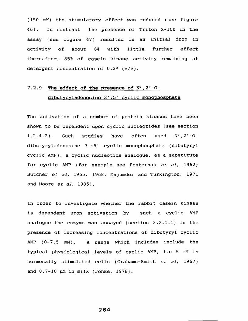
(150 mM) the stimulatory effect was reduced (see figure 46). In contrast the presence of Triton X-100 in the assay (see figure 47) resulted in an initial drop in activity of about 6% with little further effect thereafter, 85% of casein kinase activity remaining at detergent concentration of 0.2% (v/v).
7.2.9 The effect of the presence of N6 ,21-O-dibutyryladenosine 3 1:51 cyclic monophosphate
The activation of a number of protein kinases have been shown to be dependent upon cyclic nucleotides (see section1.2.4.2). Such studies have often used N6 ,21-0- dibutyryladenosine 3 1 :5 * cyclic monophosphate (dibutyryl cyclic AMP), a cyclic nucleotide analogue, as a substitute for cyclic AMP (for example see Posternak et al, 1962; Butcher et al, 1965, 1968; Majumder and Turkington, 1971and Moore et al, 1985).
In order to investigate whether the rabbit casein kinase is dependent upon activation by such a cyclic AMP analogue the enzyme was assayed (section 2.2.1.1) in the presence of increasing concentrations of dibutyryl cyclic AMP (0-7.5 mM) . A range which includes include the typical physiological levels of cyclic AMP, i.e 5 mM in hormonally stimulated cells (Grahame-Smith et al, 1967) and 0.7-10 pM in milk (Johke, 1978).
264

As a comparison 32 pg of a purified cyclic AMP dependent protein kinase from rabbit muscle (commercial preparation) replaced the mammary gland casein kinase in identical experiments.
The results (figure 48) show that the rabbit muscle enzyme was, as expected, increasingly stimulated by increased concentrations of dibutyryl cyclic AMP up to a maximum of about 7.5 fold at 20 pM. Although at concentrations greater than this the stimulatory effect was reduced. Furthermore when the concentration was greater than 2.5 mM the net effect on activity was inhibitory. In contrast the rabbit mammary gland casein kinase was inhibited over the whole range investigated (see figure 49).
7.2.10 The effect of 5'-Fluorosulfonylbenzoyladenosine
5'-Fluorobenzoyladenosine (FSBA) is an analogue of ATP (see figure 50) which binds in an irreversible manner to protein kinases (Zoller and Taylor, 1979; Zoller, 1979) and a number of other ATP binding proteins including:- pyruvate kinase (Wyatt and Colman, 1977) and the Fi-ATPase (Esch and Allison, 1978).
In order to assess the effect of increasing concentrations of FSBA on rabbit and, for comparison, the guinea-pig mammary gland casein kinase (partially purified according to the method out-lined in section 2.2.17) assays were
265

Figure 48 The effect of increasing concentrations of N6-21-O-dibutyrylcyclic adenosine 3':5'-cyclic monophosphate on the activity of a cyclic AMP dependent protein kinase
A cyclic AMP dependent protein kinase from rabbit muscle was obtained commercially and an aliquot (32pg) assayed as described in section 2.2.1.1 in the presence of increasing concentrations of dibutyryl cyclic AMP in the range 0-7.5 mM using total bovine caseins as the phosphate acceptor.
Results are expressed as a percentage change (i.e decrease and increases in activity were seen) relative to the assay at 0 pM dibutyryl cyclic AMP (100%).

CM T “CO 10 CO
^01 X e f iU B lJ Q % A j j A I J O V 0 S B U | > | U I 0 S B O
267
Dibutyryl
cyclic
AM
P ( p
M )
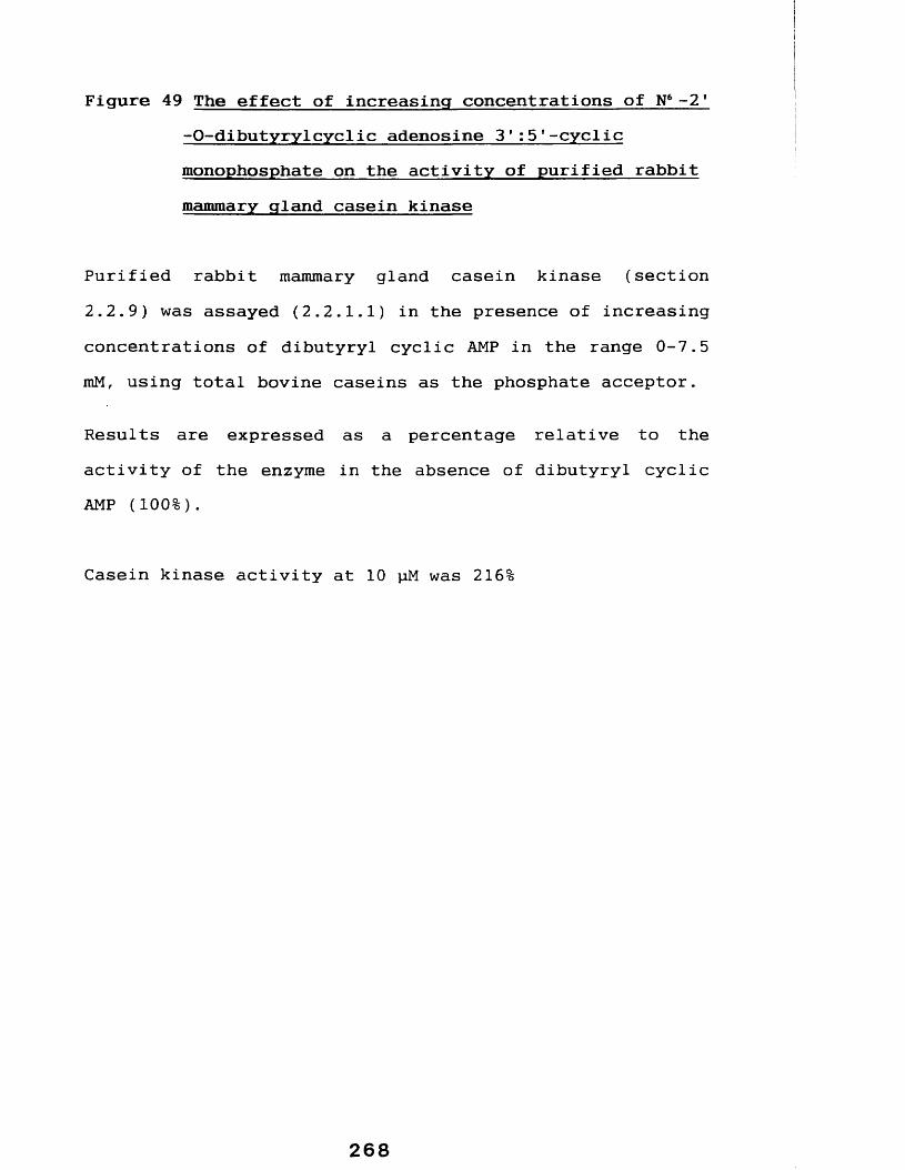
Figure 49 The effect of increasing concentrations of N6-21-O-dibutyrylcyclic adenosine S'iS'-cyclic monophosphate on the activity of purified rabbit mammary gland casein kinase
Purified rabbit mammary gland casein kinase (section2.2.9) was assayed (2.2.1.1) in the presence of increasing concentrations of dibutyryl cyclic AMP in the range 0-7.5 mM, using total bovine caseins as the phosphate acceptor.
Results are expressed as a percentage relative to the activity of the enzyme in the absence of dibutyryl cyclic AMP (100%).
Casein kinase activity at 10 pM was 216%
00
CM
% A j I A U O V 9SBUI>< U I 9 S B 0
269
Dibutyryl
cyclic
AM
P (p
M)

Figure 50 Comparison of the structures of FSBA and ATP
FSBA an analogue of ATP has been utilized in a number of studies as an inhibitor of protein kinases (section7.2.10) and was used in this study. The similarity between these two molecules is readily apparent on inspection of the structures.
2 70

•c— c h 2F— S
OHB) A T P
0II
0=P-II0
0
-0— P-II0
0II
*0— P— 0— C H 2
0
271

performed in the presence of increasing concentrations ofthis analogue (0-97.2 pm).
The results presented in figures 51 and 52 show that both enzymes are partially inhibited by increasing concentrations of FSBA.
7.2.11 Effect of heparin
Heparin, a heterogeneous polysaccharide, has been shown to inhibit a number of protein kinases (Moore et al, 1985; Feige et al, 1985) and (for the bovine adrenal "G" type "casein kinase") the inhibition is thought to arise by competition between casein and heparin for the casein binding site (Feige et al, 1985).
To investigate any possible effect on rabbit casein kinase, heparin (0 to 24 pg ml-1 ) was included in standard casein kinase assays (see section 2.2.1.1). In comparisson partially purified casein kinase from the guinea-pig mammary gland (see section 2.2.17), which is partially inhibited by heparin (Moore et al, 1985), replaced the rabbit enzyme in identical assays.
The results (see figures 53 and 54) show that the activities of both enzymes were only partially inhibited by heparin.
272
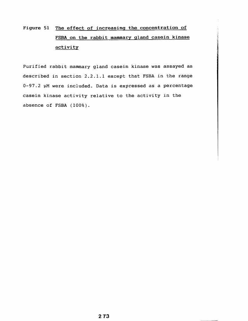
Figure 51 The effect of increasing the concentration of FSBA on the rabbit mammary gland casein kinase activity
Purified rabbit mammary gland casein kinase was assayed as described in section 2.2.1.1 except that FSBA in the range 0-97.2 pM were included. Data is expressed as a percentage casein kinase activity relative to the activity in the absence of FSBA (100%).

Casein
Kinase
Acti
vity
1005
80
60
40
20
60 80 1004020F S B A ( p M )
274
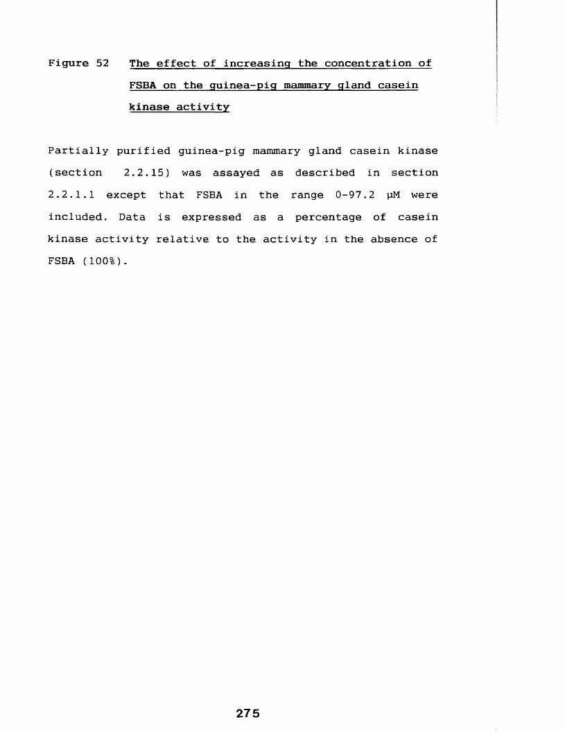
Figure 52 The effect of increasing the concentration ofFSBA on the guinea-pig mammary gland caseinkinase activity
Partially purified guinea-pig mammary gland casein kinase (section 2.2.15) was assayed as described in section 2.2. 1.1 except that FSBA in the range 0-97.2 pM were included. Data is expressed as a percentage of casein kinase activity relative to the activity in the absence of FSBA (100%).
275

Casein
Kinase
Acti
vity
100
80
60
40
20
20 40 60 80 100F S B A (pM)
276

Figure 53 The effect of heparin on the activity of thepurified rabbit mammary gland casein kinase
The rabbit mammary gland casein kinase was purified as described in section 2.2.9 and assayed essentially as described in section 2.2.1.1 in the presence of increasing concentrations of heparin (0-24 pg/ml).
2 77

Casein
Kinase
Activity
(%)
100
80
60
40
20
12 2 484 16 20
Heparin (pg/ml)
278

Figure 54 The effect of heparin on the activity of thepartially purified guinea-pig mammary gland casein kinase
The guinea-pig mammary gland casein kinase was partially purified as described in section and assayed essentially as described in section 2.2.1.1 in the presence of increasing concentrations of heparin (0-24 pg/ml).

Casein
Kinase
Activity
(%)
100
80
60
40
20
2416 2012
Heparin (Mg/ml)
280

7.3 DISCUSSION
Casein kinase was purified from the micelle rich fraction and used in a series of experiments aimed at characterizing some of it's properties.
Increasing the incubation time of the assay resulted in a proportional increase in the phosphorylation of casein, presumably reflecting the presence of excess substrates and other assay parameters. This also explains the increased phosphorylation seen with increasing enzyme concentrations, although the biphasic character of the result is not readily explained. An explanation may lie in certain features of primary protein sequence that constitute a casein kinase target residue. That is, phosphorylation of certain primary target residues which lie within defined regions (see section 1.2.4.10) can lead to the "activation" of further target residues which are not otherwise phosphorylated (see Meggio et al, 1989 and appendix 2). Now at higher enzyme concentrations (here greater than 17 pi) more of the readily phosphorylatable residues of a given casein molecule are phosphorylated, an event which will therefore "activate" still further sites rendering them accessible to the enzyme available resulting in an apparently higher phosphorylation rate.
The enzyme was shown to have a broad pH optimum of between pH 6.5 to pH 8, a range similar to the guinea-pig mammary
gland casein kinase (Moore, 1985) and, interestingly, similar to that of the rabbit mammary gland protein kinase associated with the unidentified particulate material which was described by Sundararajan et al (1958). Optimal enzyme activity was measured at temperatures between 30°C and 38°C which are close to the physiological body temperature, with little activity remaining when assays were incubated at greater than 45°C, temperatures presumably at which protein conformation, and therefore activity, is destroyed. These experiments also indicated that with respect to these parameters, the casein kinase assay conditions developed by Pascall (1981) were suitable for the rabbit mammary gland casein kinase (see materials and methods-chapter 2).
It was shown that Mg2 + stimulated the activity of the rabbit casein kinase and could not be substituted for by Ca2 + , which could replace Mg2 + for the guinea-pig casein kinase, albeit poorly (see Moore et al, 1985).
It was further shown that, in the presence of Mg2 + (8.6mM), increasing Ca2+ concentration above 86 pm resulted in the inhibition of the enzyme, an observation noted for the casein kinase type-1 and type-2 by Moore (1985). This data would suggest a direct inhibitory action of Ca2 + on either the enzyme or the substrates. One explanation is that in the in vitro assay Ca2 + may initiate the formation of casein aggregates, such as occurs during the in vivo bioassembly of casein micelles within golgi derived vesicles
282

(see section 1.1.3), there by rendering this substrate unavailable for phosphorylation.
It is of further interest to note that casein micelles, the starting source for the isolation of this enzyme, are rich in Ca2 + (see table 6) which would possibly rule out this as being the in vivo site of casein phosphorylation, therefore suggesting that this occurs in the other site of localization of the rabbit enzyme i.e. the golgi (see preceding chapters). Regions of the golgi too are rich in this ion, for example it is known to contain a Ca2 + pump (West and Clegg, 1984) and the golgi is the route of calcium secretion (section 1.1.6.3). The calcium in the golgi would therefore have some effect on casein kinase activity.
Further, Mn2 + could not substitute for Mg2 + a result in contrast to the finding of Pascall (1981) who showed that the guinea-pig mammary gland casein kinase activity was optimally stimulated by Mn2 + . Moore et al (1985) used the utilization of various divalent cations to distinguish between the guinea-pig mammary gland casein kinase and the ubiquitous casein kinase type-1 and type-2 (see section 1.2.4.2), the latter being inactivated by Mn2+ . The rabbit mammary gland casein kinase being inactive in the presence of this ion is thus similar to casein kinase type-2 in this respect.
283

Interestingly the casein kinase activity reported by Sundararajan (1958) from the lactating rabbit mammary gland had an absolute requirement for Mg2 + which could not be substituted for by Mn2 + . It is possible that the enzyme described by this group is the same as the casein kinase described here.
It is generally considered that mammary gland casein kinases are not dependent upon cyclic AMP for activation (Moore et al, 1985; Bingham, 1987) although other recent evidence suggests otherwise (see section 1.2.4.9). It was shown here that the rabbit casein kinase was not dependent upon dibutyryl cyclic AMP but was infact inhibited in the presence of this compound. Caseins were used as the enzyme substrate in these studies, Tao (1970) however, pointed out that stimulation by cyclic AMP was low with certain substrates, including caseins; an observation which may invalidate this result. However, a parallelexperiment using a commercially obtained cyclic AMP dependent protein kinase and casein as a substrate showedthat this enzyme was activated by dibutyryl cyclic AMPeven with casein as the substrate and therefore wouldnegaten the reservations raised by Tao.
At levels of greater than 20 pM of dibutyryl cyclic AMP the stimulation of this protein kinase was increasingly reduced, an observation previously reported by Iwai et al
(1972). They state that this effect was inhibitory. This is not strictly true since the overall effect was still
284

stimulatory. It was shown here that at even higher concentrations ( at and greater than 2.5 mM) the net effect was in fact inhibitory.
FSBA an ATP analogue was shown to inhibit both rabbit and guinea-pig mammary gland casein kinases presumably at the ATP binding sites. Whilst this observation is of limited value in itself further experiments employing 14C labelled FSBA could be useful in the determination of the primary sequence of ATP binding regions.
Heparin is a compound that has also been shown to inhibit a number of protein kinases and was used by Moore et al
(1985) as a criterion to distinguish the guinea-pig casein kinase (which is partially inhibited), from casein kinase type-2 which is inhibited and casein kinase type-1 which is not inhibited. It was confirmed here that the guinea- pig casein kinase was partially inhibited and further shown that the rabbit casein kinase was inhibited in a similar manner and thus distinct in this respect from the other so-called casein kinases.
The effect of a number of other agents on enzyme activity was also investigated. Concentrations of NaCl greater than0.3 M resulted in a minimum reduction of 39% of the initial casein kinase activity. It is of interest to note that Pascall (1981) showed the guinea-pig casein kinase to be inhibited by KC1. EGTA when present in assays at 10-150 mM appeared to stimulate enzyme activity. The explanation
285

for this effect is not readily apparent. Chernoff and Li (1983) reported that a phosphotyrosine protein phosphatase was stimulated by EGTA (albeit slightly) and suggested that this was due to the chelating of stimulatory ions. Such an explanation is not applicable here since EGTA will only chelate Ca2+ (Schmidt and Reilly, 1957) and therefore not affect Mg2 + the divalent cation essential for activation of rabbit casein kinase (see section 7.2.5).
It is noted that the starting source for enzyme purification, i.e. the casein micelles, are rich in Ca2 * and such ions co-purifying with the enzyme would be inhibitory (see previous results - this chapter), in this experiment the Ca2 + would in turn be removed by EGTA resulting in the effect demonstrated. This explanation however is not very likely as the purification procedure involes extensive wash steps and it is unlikely that the ion would be present in appreciable concentrations.
Triton X-100, over the range investigated, had no significant effect on enzyme activity an observation in contrast to the inhibitory effect of this detergent on the activity of the guinea-pig casein kinase (Pascall, 1981).
It is concluded that when EGTA, NaCl or Triton X-100 are included in standard assays the concentrations present,i.e. generally less than 0.2 M NaCl, negligable EGTA and 0.034% Triton X-100, have little effect on the casein kinase activity. In experiments where differing
286
components, particularly at high concentrations are used, then the final standard assays should be adjusted to contain equivalent concentrations of these components.
CHAPTER 8 HIGH PRESSURE LIQUID CHROMATOGRAPHY
PURIFICATION OF RABBIT CASEIN KINASE AND ATTEMPTED PROTEIN SEQUENCING
288

8.1 INTRODUCTION
Possible future studies in the study of rabbit casein kinase include the isolation of a cDNA clone for this enzyme which could be used for further studies (seechapter 9. There are two main approaches to screen a cDNA library in order to obtain such a clone. Firstly, antibodies can be used to screen an expression library(Young and Davis, 1983 and Huyatt et al, 1985). Secondly, using protein sequence, data oligonucleotides can be constructed and used to screen the library (Suggs et al,
1981 and Huyett et al, 1985). For either approach largequantities (at a minimum 100 pg) of protein would be requi red.
The aim of the work in this chapter was to prepare sufficient amounts of purified casein kinase (i.e in the region of 100 pg) and to generate protein sequence (10-20 residues only) using automated protein sequencing techniques, which in turn could be used in the future to isolate casein kinase clones as described above.
In chapter 5 of this thesis a procedure for the purification of casein kinase was described. This method as described was considered unsuitable for the isolation of the amount of enzyme required here since "scale-up" and occasionally smaller scale casein kinase preparations frequently contained low molecular weight contaminants (personal observation).
289

In an attempt to remove these contaminants a final high pressure (performance) liquid chromatography (hplc) step in addition to gel-exclusion and casein-sepharose affinity chromatography steps (essentially as described in chapter 5) was developed.
A successful hplc step would have a number of furtheradvantages. Firstly, the material eluting from the hplcstep is detected by following absorption at 214 nM and notenzyme activity, thus any loss of activity will not a,jepodize recovery of the sample. As a result casein kinase ft
resulting from a number of Sephacryl/casein-sepharose purification steps may be stored at -20°C, later pooled and recovered in a single hplc run. Thus larger quantities of enzyme are recovered in a small volume which, after minor adjustments to solvent content, are suitable for the automated protein sequencer or proteolytic/chemical cleavage. The use of the hplc step also means that the casein-sepharose wash step can be reduced (saving time and reducing enzyme losses) since the contaminants can be removed at the hplc step.
8.2 Results
8.2.1 High pressure liquid chromatography
Hplc is now recognised to be one of the most powerful and increasingly common techniques used in the isolation of peptides and proteins. Hplc is best viewed as a new way of approaching the well established liquid chromatography techniques. This technique utilizes rigid supports consisting of fine particles (usually silica) which give a large surface area and requires elution at high pressure. It can be used in gel-exclusion, ion-exchange, adsorption and bonded-phase modes.
Here bonded-phase hplc in the "reverse-phase" mode (where the polarity of the mobile phase is lower than that of the the stationary phase) has been utilized as a final step in the isolation of casein kinase from the lactating rabbit mammary gland using the micelle rich fraction as the starting source.
Rabbit casein kinase was purified essentially as described in section 2.2.9 with the modifications out-lined in section 2.2.11. Material from a number of experiments was pooled, dialysed against ammonium bicarbonate (about 10 mM) and lyophilized. The material was then resuspended in a small volume of water and analysed by hplc as described in section 2.2.12.
291
Material eluting fom the hplc was detected by following absorption at 214 nM and a typical elution profile is shown in figure 55. This shows that series of peaks present at a retention time of between 16 to 20 minutes and a single broad peak well removed from the bulk of the other material eluting from the column typically at a retention time of 26.2 minutes. Using this column (see materials section 2.1.4) this rather late retention time is characteristic of a protein hydrophobic in nature (B. Coles, CRC protein sequencing facility, University of London:- personal communication).
The material in this peak was analysed by SDS polyacrylamide gel electrophoresis (see section 2.2.3) which showed it to contain (see figure 55) the same two polypeptide species which were previously shown to be the casein kinase (see chapter 5, this thesis). There was no sign of lower molecular weight material except for a single "band" which was shown to be present in the empty adjacent gel running lanes and probably is an effect of the hplc solvents present in the sample.
This material was collected on a further hplc gradient (see section 2.2.19) and the amino acid content estimated (see section 2.2.19-carried out by A. Aitken at the NIMR, London), the results of which are presented in table 11. These results shows the enzyme to contain a high proportion of the aliphatic residues glycine and serine
2 9 2

Figure 55 Profile of material eluting from an hplc columnand analysis of the material eluting after 26 min by SDS polyacrylamide gel electrophoresis
Rabbit casein kinase was purified from the lactating rabbit mammary by a combination of gel-filtration on Sephacryl-S-300 and affinity chromatography using casein- sepharose as described in sections 2.2.9.1 and 2.2.9.2 with the modifications out-lined in section 2.2.11. A further procedure employing hplc (section 2.2.12) was then used as a further purification step. Figure 55 shows a typical profile of material eluting from the hplc column. The Y axis is absorption at a wavelengh of 214 nm, the scale is 0.5 absorbance units ("AU") at this wave lengh relative to bar height. The X axis is retention time ("RT") in minutes.
Material eluting from hplc column at about 26 min in an identical experiment was analysed by SDS polyacrylamide gel electrophoresis (2.2.3) and silver stained (2.2.4). the lane containing this material is shown to the right of a typical elution profile. Molecular weight markers are indicated on the right.
293
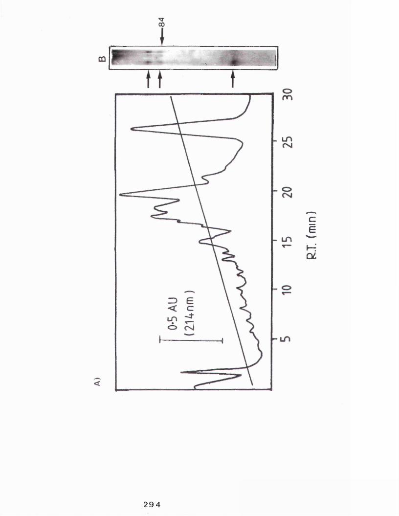
294
R.T.
fmm
)

with quite a high proportion of the hydrophobic aromatic ring containing residue tyrosine.
In a typical experiment four casein kinase preparations were carried out and stored at -20° C. This material was pooled and a proportion (95%) applied to hplc column resulting in the recovery of approximately 70-100 pg of casein kinase in a volume of about 600 pi.
The hplc purified casein kinase was used for the experiments in the following sections.
295
Recent advances in automated protein sequencing technology have resulted in highly reliable techniques which can be operated with as little as 10 pM of sample and can be used to sequence short (<40 amino acids) peptides.
Here this technique (using the Edman degradation reaction chemistry) was used in an attempt to sequence a short region of the N-terminus of the hplc purified casein kinase. Although sufficient protein was used, no sequence data was generated since no PTH residues (the end-products of each sequencing step) were detected by hplc analysis.
A different approach was therefore made to generate protein sequence data which is'outlined below.
296

Table 11 Amino acid analysis
Rabbit casein kinase was purified from the micelle rich fraction as decribed in section 2.2.11 and by the hplc step described in section 2.2.12 then by a further hplc step identical to procedure described in section 2.2.19 used for the isolation of tryptic digests. The amino acid content was determined by Dr. A. Aitken (see section 2.2.18 for details). The results are expressed as molar percentage. Obviously this percentage does not include the contribution of the residues not recovered by the procedure used.
297

AMINO ACID ANALYSIS OF HPLC PURIFIED CASEIN KINASEResidue Molar % Characteristic
Asp/Asx 2.40 Acidic/AmideGlu/Gln 10.70 Acidic/AmideSer 14.20 AliphaticGly 24.00 AliphaticArg 4.50 BasicThr 3.50 AliphaticAla 7.50 AliphaticTyr 11.40 AromaticVal 4.70 AliphaticMet 1.57 Sulphurlie 5.53 AliphaticPhe 2.05 AromaticLys 1.53 BasicLeu 6.40 AliphaticHis DestroyedCys DestroyedTyr DestroyedPhe Eluted with phenylthiourea

8.2.3 Protein fragmentation
Sequencing of a protein is often not posssible due to the presence of an in vivo modified N-terminal amino acid. To overcome this methodology is available allowing the chemical or proteolytic cleavage of the protein which, ideally, will generate peptides of reasonable size (at least 20-30 residues) easily isolated by hplc and containing a "free" N-terminal which are susceptible to the sequencing reaction.
Here two enzymatic (V8 protease and trypsin) and a chemical method (using cyanogen bromide) were used in an attempt to generate such peptides-see table 12 for specificity of these reagents. Hplc purified casein kinase were treated with these agents described in table 13 and section 2.2.19 and the resultant material analysed by hplc analysis (section 2.2.19).
These procedures, the results of which are summarized in table 13, generally resulted in only limited proteolysis (particularly with trypsin) or the generation of small peaks unsuitable for further analysis (hplc profiles not shown). Only two peptides were sequenced but did not yield useful sequence data, , . ;
299

Table 12 Common methods of protein fragmentation
A number of agents which cleave proteins are available to generate short peptides suitable for sequencing. A number, including those employed here, are reviewed in figure 12.
300

TJG• Q) (d O 01 t - i 03 '— ' ft, > VO
rd t*-* cn cuuX u
Q) *HP mqj£32GO
u a) a coi■H G■p o a gQ)uX -{ QJ £J■HP COo co ■n O
(0 CU 2
rHQJ+JQJ wo r-(0 CTiX3 »-• 30nH '"IQ fQ1r—I +Jaj qj•Hx cf0<u eQJ >i CO 23
ajT3r—lG0 £3 rdp (dCL, -P wo0 r-CO u G cn•H -H iH4H•PX -H CO VU 0 ̂qj a j E QJp ftQJ CO «.•p 00£3 IT3 QJ2 G QJ CTi0 -M CO *“1G G P G0 0 QJ -•H QJ a E ̂-P E QJ E 10a o Paj co ^ +ju QJ QJx •01 1Q) p rd X PrH > 1 QJP o rd P 0)0 QJ Q) rH•r“1 P rHC/3 Ehrd 0
2u 1
CO rd £3G -PrH • • •HQJ E mmh a P 00 >i QJ-p -P T3aj •HT3 QJ CJ u■H tdCO -P T3 X> i G U 1■p --H Oh O -h 0■H <d o x £3 E -iu G 1 rd U•H •H 01 O dJ -P I»W E P P 03 0•H P < *H iH • •u QJ \ -P 01 QJQJ -M TO CO a. ftQ 1 G >i QJ 0 >iC/3 U (0 J On 4-J -P
arH o<d XG ■p•HE 3pQJT3•P G1 (0
U COT3 COG 00 P£3 U
QJTJ -P•H Q) —-p 2 <Na woQJ 0 cncu +j i—i
X30£3■P0)2
G0■HU -P ■H CO 4J QJa O)> 1 -h P T3 H
■P -Pa C7i
■HCO T3 >
G QJQJ T3OJ -h 0 E G 0 CO P >i CQ U
301

Table 13 Attempted protein sequencing and generation ofcasein kinase protein fragments
Casein kinase was purified as described in sections 2.2.9.1 and 2.2.9.2 and a further hplc step and a number approaches made to generate short primary sequence data. Details of the methods used (although a number of expriments were carried out for each reagent a typical procedure is described) along with the results are included in table 13.

TREATMENT & METHOD HPLC ANALYSIS SEQUENCE
None-untreated hplc purified casein kinase used.
Single peak eluting at 26 min(approx)
None,possibly blocked N- Terminal.
CNBr-To hplc purified casein kinase add few crystals of CNBr and 4 x's volume of 70% formic acid-incubate at 4° C overnight at 37° C.
Few large peaks, Very many minor peaks after prolonged treatment
None
V8 protease-To hplcpurified casein kinase add 1/20 amount V8 in ammonium bicarbonate pH8.7 (50-100 mM final concentration). Incubate 37°C overnight.
Very many small peaks, occasionally few larger. Limited proteolysis with larger amounts of casein kinase.
None
Trypsin-To hplc purifi- Many small peaks. Few Noneed casein kinase add larger peaks. Majority1/50 amount trypsin, resistant to proteolysis,adjust to 50 mM ammonium bicarbonate (pH 8.5).Incubate at 37°C overnight.
8.3 Discussion
Following purification of casein kinase essentially as described in section a further hplc step was employed. A typical elution profile showed that number of protein peaks eluted during the middle of the gradient, these probably contain the bulk of contaminating caseins. A single large peak far removed from this other material eluted late in the run, typically at 26.2 minutes (this was found to be highly reproducible).
SDS polyacrylamide gel electrophoresis of this material showed it to contain the same two polypeptide species shown in the preceding chapters to be the casein kinase. This hplc step was then successful in all the initial requirements outlined in the above introduction (section 8.1) i.e following the application of casein kinase rich fractions from a number of preparations the hplc stepresulted in an esentially homogeneous preparation of casein kinase containing up to 100 pg in a small (under lml) volume.
Analysis of this material on an automated proteinsequencer did not result in any protein sequence i.e noPTH residues (the end products of the Edman degradationsequencing reaction) were detected.
304

One possible explanation for this is that the enzyme may contain a modified residue at the N-terminal. The presence of these modifications are well known to inhibit the initiation of the protein sequencing reaction. Such a modification may be due to the addition of a number of molecules including myristic acid (Aitken et al, 1982; 1984), glyceryl (Hantke and Brown, 1973), glucuronic acid (Kolaltukudy, 1984) or n-tetradecanoic acid which has been shown to be attached to a cyclic AMP dependent protein kinase (Carr et al, 1982). Most commonly these residues are acelated (Tsunasawa, 1984).
In order to overcome this problem attempts were made to cleave the protein using proteolytic digestion with enzymes and a chemical procedure using cyanogen bromide. Treatment of the hplc purified material was generally not successful in producing peptides suitable for sequencing, and attempted sequencing of two peptides (V8 digest of casein kinase) did not generate reliable protein sequence.
The reason for the lack of protein fragments is not clear. Amino acid analysis suggests that the casein kinase contains residues "recognised" by each of the reagents used, and indeed a number of small (in terms of amount) peptides were generated, it is possible that larger digest products were not suited to the hplc column matrix used for their separation and collection and were recovered in the initial eluate along with the hplc solvents. The resistance of the enzyme to trypic digestion is puzzling
305

as data presented in chapter 4 of this thesis shows that casein kinase is susceptible to -tryptic hydrolysis.
In summary the work of this chapter describes an improved procedure utilizing hplc techniques for the isolation of larger amounts of purified casein kinase. The work describes the difficulties in obtaining protein sequence data and leads to the conclusion that alternative procedures to generate this information might prove more useful (see chapter 9 for further discussion).
306
CHAPTER 9GENERAL DISCUSSION
307
9.1 Distribution of rabbit casein kinase.
In chapter 3 a casein kinase was identified in the lactating rabbit mammary gland and shown to be associated with two fractions which were subsequently identified as a homogeneous casein micelle fraction, containing the bulk of kinase activity, and a membrane fraction, shown to be a golgi enriched fraction, which contained substantially less of the enzyme activity. Casein kinase activity was also present in whole rabbit milk. This is the first report of casein kinase being either associated with casein micelles or secreted into milk in any form.
It was of interest to study the casein kinase from both sources. In order to do this a routine method for the rapid simultaneous isolation of both fractions was developed. Further analysis of these fractions confirmed their identities.
The enzyme was purified from the casein micelle fraction. Initially ATP-agarose affinity chromatography was used followed by a sucrose gradient centrifugation step. However a combination of gel-exclusion chromatography on Sephacryl-S-300 and an affinity chromatography step on casein-sepharose was adopted as the method of choice for the routine purification of the enzyme.
Both methods resulted in the enrichment of the same two polypeptide species and in conjunction with other
308

observations, it was concluded that the two polypeptide species isolated were the rabbit mammary gland casein kinase.
It was further shown that the casein kinase in the golgi enriched fraction similarly consisted of the same two polypeptide species. It is of interest to account for the unusual distribution of the rabbit casein kinase and possible mechanisms are discussed in the following section.
9.2 Mechanisms to account for the distribution of rabbit casein kinase
The distribution of the rabbit casein kinase is, therefore, similar to another enzyme present in the lactating mammary gland, galactosyltransferase. Thisenzyme has been shown to be both associated with the golgi and secreted into milk (Smith and Brew, 1977), although the secreted form is not associated with the casein micelles.
The secreted form of galactosyltransferase is derived from the membrane bound form by proteolysis and therefore has a slightly lower molecular weight (see Strous, 1986 for review). The rabbit casein kinase differs in this respect since both membrane and secreted forms are identical with respect to molecular weight.
309
The answer for the unusual distribution of the rabbit casein kinase probably lies in the fact that the micelle associated enzyme is most likely derived from the golgi membranes and becomes associated with the micelles possibly during their assembly and are therefore secreted from the cell. Thus the loss of the enzyme occurs simply as a result of the "spilling over" of cellular components during the intense secretory activity. Such a mechanism was postulated by Kitchen et al (1970) to account for the presence of a number of other enzymes in milk. It would be expected that casein kinase activity is present in the milk of other species also, indeed the author has detected such activity in other milks (data not shown).
As mentioned above the rabbit casein kinase isolated from both sites was shown to consist of two polypeptides. It is of interest to discuss the mechanisms that may account for this.
9.3 Explanation for the presence of two polypeptide species in samples of purified casein kinase
Results obtained from sucrose gradient centfifugation of purified casein kinase demonstrated a single peak of kinase activity with the highest single fraction containing both polypeptide species, a calculated molecular weight of the casein kinase peak was 55-64,000 Daltons. These results suggest that the active enzyme comprises a monomer. The presence of two polypeptide
310

species in the purified casein kinase preparations therefore would appear contradictory to this. A number of possible explanations are possible:
Firstly, the two polypeptides may represent differential modification of the same protein. Glycosylation (see section 1.1.5), for example, may account for as much as 50% of the mass of a protein (for example see Trimble and Malley, 1977). Differences in molecular weight of the same protein can also be attributed to differences in the level of phosphorylation or autophosphorylation.
Secondly, the smaller protein may be a produced by the proteolytic digestion of a region of the larger protein.
Another enzyme that exists in both membrane bound and secreted forms is the intestinal alkaline phosphatase (Yeldin et al, 1981). These arise from the expression of two distinct mRNA species (Seethram et al, 1987).
Finally, this may be accounted for by the co-isolation of proteins with different primary sequences. It is however unlikely that one of the polypeptides is a contaminant with no kinase activity (see chapter 5), but the co-purification of two distinct casein kinases cannot be ruled out.
One important question remains unclear, that is, is the casein micelle (and therefore milk i.e. the mammary
alveolar lumen) or the golgi the in vivo site of casein phosphorylation?.
9.4 The milk as the Site of casein phosphorylation
It is generally assumed that the in vivo site of casein phosphorylation is the golgi (see section 1.2.2 and 1.2.4.7.), and the identification of a golgi-associated rabbit casein kinase would appear then to substantiate this notion.
It is therefore interesting to consider the casein micelle associated enzyme in terms of function. In chapter 1 (see section 1.1.4.3) a number of enzymes were seen to have a function in milk. It is unlikely however that the casein kinase when associated with the micelle will phosphorylate casein for several reasons.
Firstly, casein micelle assembly is initiated in the golgi cisternae and golgi derived vesicles (see appendix 2), and therefore this process requires the presence of already phosphorylated casein. Casein phosphorylation must therefore occur before the micelle assembly is initiated, leaving either the golgi or endoplasmic reticulum as the only likely sites (see section 1.1.5 for discussion of casein expression). The results in table 9 show however that negligable casein kinase activity is associated with the endoplasmic reticulum.
312

Secondly, a supply of ATP as the phosphoryl donor is required for casein kinase activity (see sections 1.2.4 and 7.2.7.2) and it is highly unlikely that sufficient quantities are available in milk. The golgi membranes, on the other hand, contain specialized pumps which supply ATP into the lumen (Capasso et al, 1989).
Finally, results obtained here (see section 7.2.6) indicate that the enzyme would be inhibited by the high levels of calcium (although much is present as CCP-see section 1.1.3) present in milk (see table 4).
In conclusion it is unlikely that casein phosphorylation occurs in the casein micelles in the secreted milk, why then is the bulk of the casein kinase so distributed ?, this point is addressed below.
9.5 The bulk of casein kinase in the micelle rich fraction;- a misleading observation ?
It seems likely then that the in vivo site of casein phosphorylation is not the casein micelles in milk. Why then is casein kinase present at such high quantities in milk ?. A possible explanation is as follows: firstlyconsider the conclusion of section 9.2, that the casein kinase is found in milk due to a "spilling over" effect, now the mammary gland during peak lactation will accumulate a large store of casein micelles, and as such
the gland will contain a large ammount of the associated casein kinase. The identification of this fraction as the bulk source merely reflects this.
Therefore when considering the site of casein phosphorylation care has to be taken not to assign to much importance to the bulk of the casein kinase being present in the micelle rich fraction. A recently suckled gland would be expected to contain little milk and the bulk of the casein kinase would then appear to be associated with the golgi.
Indeed, it has been shown by the author (data not presented) that following the removal of milk by manual expression only small quantities of casein micelle rich fraction were recovered following a micelle/golgi preparation (see section 2.2.8) and the bulk of casein kinase activity was associated with the golgi enriched fraction.
It is of interest to note that the young rabbit will generally only suckle once every 24 hours (Deutsch, 1957), so the mammary glands used here may well have accumulated a large store of milk and therefore of casein kinase rich casein micelles.
The following section addresses the probable in vivo site of rabbit casein phosphorylation, that is the golgi.
3 1 4

9.6 The Golgi as the site of casein phosphorylation inthe lactating rabbit mammary gland
Although not conclusive in consideration of the above discussion it appears, that in agreement with the work in other species (see section 1.2.4.7), the golgi is the in
vivo site of casein phosphorylation in the rabbit.
Indeed the golgi has been shown to be the site of phosphorylation of a number proteins eg. proteoglycans (Gldss et al, 1980) and vitellogenin (Gottlieb and Wallace, 1981), and a number of other protein kinases have been shown to be present in the golgi membranes isolated from tissues other than the mammary gland (eg Capasso et
al, 1985 and Nigg et al, 1985). Therefore it is possible that the "casein11 kinase isolated here may in fact phosphorylate mammary gland proteins other than caseins. Further studies (out-lined in section 9.8) would be necessary to show conclusively whether or not this enzyme is mammary gland specific.
9.7 Properties of rabbit casein kinase
In chapter 7 some of the properties of the rabbit mammary gland casein kinase were investigated and any differences or similarities with other mammary gland casein kinases was discussed, some of these properties are summarized in table 14.
315

Table 14 Summary of the properties of rabbit casein kinase
Intra-cellular localization Golgi, also secreted into milk associated with casein micelles
Two Polypeptides 63,000-67,000 Daltons
SDS polyacrylamide gel analysis Appearence Molecular weight Sucrose gradient centrifugation analysis Sedimentation coefficient 3.9-4.28 SMolecular weight 55,000-64,000 DaltonsSubunit structure MonomericProperties of purified casein kinase Assay parameters:-Cation activation
pH optimum Temperature V.a, for casein K. for casein Vm a x for ATP K. for ATP
Mg2 + activated-optimally around 7.5 mMMn2 + inactiveCa2 + inactivepH 6.5-830-38° C50 cpm/min217 pg/ml9.8 cpm/min400 pm
Miscellaneous parametersDibutyryl cyclic AMPHeparinFSBAEGTANaClTriton X-100 A Autophosphorylated A Glycosylated
Not activated, > 2.5M inhibitedPartially inhibitedinhibitedInhibitedInhibited >0.2MLittle effectSome evidenceNot evident followingtreatment with Trifluoromethane-sulphonic acid
A Data not shown
316

Lastly, some further work on the rabbit casein kinase, presented in chapter 8, is summed-up.
9.8 Hplc and protein sequencing
Whilst the hplc step developed in chapter 8 was successful, no suitable protein sequence was generated due to the faliure of the direct sequencing of the protein or the faliure to produce suitable protein fragments. The reasons for the lack of data from the direct sequencing step has been disussed in detail (section 8.3). The lack of useful1 peptides is less clear, however the techniques used are "routine" and the lack of the required data is seen as a reflection of the properties of the enzyme rather than an indictment of the techniques.
Future studies could involve the "second approach" outlined in section 8.1, that is the production of polyclonal antisera using the hplc purified enzyme. This antisera would then be used to screen a cDNA expression library thereby avoiding the need for oligonucleotides based on protein sequence data.
9.9 Further studies
Studies with a casein kinase clone could involve the study of the genetic and primary sequences responsible for
317

the intra-cellular routing of the enzyme. Such studies usually involve the in vitro mutagenesis of the genetic sequence and the in vivo translation of the products within a surrogate system containing the appropriate organelles to study any changes in intra-cellular sorting. The Xenopus oocyte, which does not contain an endogenous protein kinase capable of phosphorylating caseins (Boulton et al, 1981), would be a suitable surrogate.
An anti-casein kinase antibody would have applications other than that mentioned above. For example, a true casein kinase would be expected to be mammary gland specific, the antisera could be used in immunofluoresence studies in order to substantiate such a notion. Indeed, Moore et al (1985) in such studies showed that the guinea- pig mammary gland casein kinase was localized in the mammary gland but not in liver, an organ which contains many other "casein kinases" (see section 1.2.4.7).
Further studies similar to those out-lined in section 1.2.4.10 and in appendix 2 involving the study of substrate specificity might further prove useful in the conformation that the enzyme described here is a true casein kinase.
The difference in the two polypeptide species shown to represent the rabbit casein kinase has not been resolved here. A more thorough study of differences in post-
318
t rans1at i onal might provide
modification, particularly some answers.
glycosylation,
APPENDIX 1 SECRETION OF CASEIN MICELLES-AN ELECTRON MICROSCOPIC STUDY
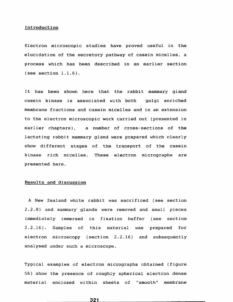
Int roduct i on
Electron microscopic studies have proved useful in the elucidation of the secretory pathway of casein micelles, a process which has been described in an earlier section (see section 1.1.6).
It has been shown here that the rabbit mammary gland casein kinase is associated with both golgi enriched membrane fractions and casein micelles and in an extension to the electron microscopic work carried out (presented in earlier chapters), a number of cross-sections of the lactating rabbit mammary gland were prepared which clearly show different stages of the transport of the casein kinase rich micelles. These electron micrographs are presented here.
Results and discussion
A New Zealand white rabbit was sacrificed (see section 2.2.8) and mammary glands were removed and small pieces immediately immersed in fixation buffer (see section 2.2.16). Samples of this material was prepared for electron microscopy (section 2.2.16) and subsequently analysed under such a microscope.
Typical examples of electron micrographs obtained (figure 56) show the presence of roughly spherical electron dense material enclosed within sheets of "smooth" membrane
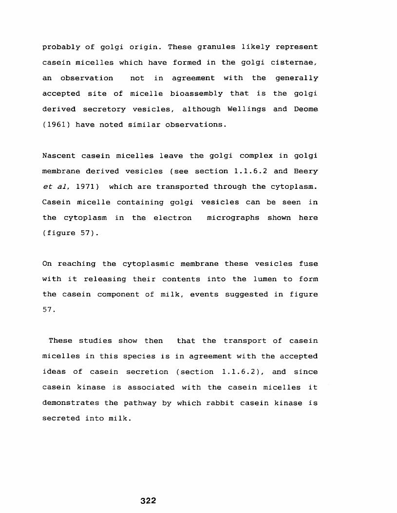
probably of golgi origin. These granules likely represent casein micelles which have formed in the golgi cisternae, an observation not in agreement with the generally accepted site of micelle bioassembly that is the golgi derived secretory vesicles, although Wellings and Deome (1961) have noted similar observations.
Nascent casein micelles leave the golgi complex in golgi membrane derived vesicles (see section 1.1.6.2 and Beery et al, 1971) which are transported through the cytoplasm. Casein micelle containing golgi vesicles can be seen in the cytoplasm in the electron micrographs shown here (figure 57).
On reaching the cytoplasmic membrane these vesicles fuse with it releasing their contents into the lumen to form the casein component of milk, events suggested in figure 57.
These studies show then that the transport of casein micelles in this species is in agreement with the accepted ideas of casein secretion (section 1.1.6.2), and since casein kinase is associated with the casein micelles it demonstrates the pathway by which rabbit casein kinase is secreted into milk.
322

Figure 56 Electron micrographs of cross sections of thelactating rabbit mammary gland showing the presence of casein micelles associated with the stacks of smooth membranes
Electron micrographs showing cross sections of the mammary epithelial cell from the lactating rabbit mammary gland were prepared as described in section 2.2.16.
The micrograph reveals typical sub-cellular components (section 1.1.1). Electron dense material with the general appearence of casein micelles are seen to be associated with stacks of smooth membrane probably of golgi origin.
Magnification A. X 50.7 xlO3B. X 10.2 xlO4
323

324

Figure 57 Electron micrographs of cross sections of thelactating rabbit mammary gland showing the presence of casein micelles in secretory vesicles and their subseguent release at the cell surface
Electron microscopic analysis was performed on cross sections of the lactating rabbit mammary gland as described in section 2.2.16. The micrographs shown in figure 57 shows stages in the release of casein micelles from golgi derived vesicles, across the plasma membrane into the apical lumen by process akin to reverse pinocytosis, (see section 1.1.6.2 and appendix 2 for detaiIs).
Magnification A. X 51.1 xlO3

326

APPENDIX 2DETERMINATION OF SUBSTRATE SPECIFICITY
FOR MAMMARY GLAND CASEIN KINASES
327
INTRODUCTION
A number of protein kinases of diverse origins are able to phosphorylate caseins to some extent (see section 1.2.4) which has led to confusion in nomenclature and to the confusion over the identity of the true mammary gland casein kinase. A number of groups using series of synthetic peptides have identified the primary sequence determinants which specify phosphorylatable residues recognized by a number of protein kinases which has led to a clearer understanding of the physiological substrates of a number of enzymes (see section 1.2.4.10)
RESULTS AND DISCUSSION
In a collaboration with the group of L. Pinna (University of Padova) it has been demonstrated that the determinants of substrate specificity for the guinea-pig mammary gland casein kinase include an absolute requirement for an acidic residue to be located two amino acids away (N- terminally) to the phosphorylated serine, see Meggio et al (1988), and Table 15. More recent studies have shown that local structural regions of the substrate are important determinants of substrate specificity (Meggio et al,
1989).
These studies have not as yet been extended to the mammary gland casein kinase in any other species. However in order to demonstrate if the rabbit casein kinase had a similar

Table 15 Determinants of substrate specificity of themammary gland casein kinase using synthetic peptides-an absolute requirement for an n+2 acidic residue
In appendix 2 studies were described in which the primary sequence determining whether a peptide is or is not phosphorylated were described. Table 15 lists the relative ease by which a mammary gland casein kinase (from the guinea-pig) is able to phosphorylate serine residues (S) of a number of synthetic peptides and demonstrates the importance of the acidic residue (glutamic acid) at the position n+2. (E = glutamic acid). The data is taken from Meggio et al (1988).
SUBSTRATE PHOSPHORYLATION_________________________ (% relative to SEEEEE)SEEEEE 100SEAEEE 0SAEEEE 110SEEEAE 67SEEAEE 93
329

requirement for an acidic residue as decribed above the ability of this enzyme and the guinea-pig casein kinase to phosphorylate two synthetic peptides SEEEEE and SEAEEE (kind gifts of L. Pinna) was compared.
Standard assays were set up except that 10-20 pg of either synthetic peptide was used in place of bovine caseins. In these experiments the enzyme was either purified or partially purified by a single casein- sepharose step or aliquots of micelle rich fraction were used (see section 2.2.17). Following incubation at 37°C (at least 0.75 hour) an equal volume of 2 M HC1 was added and the sample incubated at 100°C for 10 minutes. Phosphorylated peptides were isolated by ion-exchange chromatography using "sep-Paks" (see Southan, 1987) essentially as described by Kuenzel et al (1985), see section 2.2.17 for details.
The results showed (figure 16) that, in agreement with Meggio et al (1988), the guinea-pig mammary gland casein kinase preferentially phosphorylated the peptide SEEEEE to SEAEEE. In an average of 5 experiments the percentage phosphorylation of the peptide SEAEEE was only 33.6% relative to peptide- SEEEEE. It should be noted that this does not suggest an absolute requirement for an acidic residue at the position n+2 as described by Meggio et al
(1988), although they used an electrophoretic, and probably less accurate, method for the determination of phosphorylated residues; a fact which may account for
330

Table 16 Relative phosphorylation of two peptides bymammary gland casein kinases
A number of studies (see section 1.2.4.10) have utilized synthetic peptides to analyze determinants of substrate specificity. Two such peptides SEEEEE and SEAEEE were made available from L.A. Pinna (University of Padova) and were used as substrates for both the rabbit and guinea-pig mammary gland casein kinases as described in section 2.2.17. The results are shown in table 16. (S = serine, E = glutamic acid).
Guinea-pig casein kinase Rabbit casein kinasePhosphorylation Phosphorylation(% relative to SEEEEE) (% relative to SEEEEE)
8.0 56.728.2 117.536.3 148.072.7 129.423.0 55.0
350.0Mean: 33.6% Mean: 142.8
331

some discrepancy in the obsevations. In contrast to this the rabbit mammary gland casein kinase preferentially phosphorylated SEAEEE, demonstrating that it did not have an absolute requirement for an acidic residue at the position n+2.
This preliminary result suggests then that the determinants of substrate specificity for the two casein kinases investigated are distinct.
332

CHAPTER 10 REFERENCES
333

A
Aitken, A. fit Cohen, P. (1984) in "Methods in Enzymology" (Wold, F. & Moldave, K. eds.) vol. 106, pp205-210.Academic Press, Inc. New York, Publishers.Aitken, A., Cohen, P., Santikarn, S., Williams, D.H., Calder, A.G., Smith, A. fit Klee, C.B. (1982) F.E.B.S. Lett. 150, 314-318.Al-Sarraj, K. , White, D.A. fit Mayer, R.J. (1978) Biochem.J. 173, 877-883.Al-Sarraj, K. , White, D.A. fit Mayer, R.J. (1978) Int. J.Biochem. 9, 269-277.Amoroso, E.C., Goffin. A., Hailey, G. , Mathews, L.H. fitMathews, D.J. (1951) J. Physiol. 113, 4P-5P.Aoki, T., Kako, Y. fit Imamura, T. (1986) J. Dairy Res. 53, 53-59.Aoki, T., Yamada, N., Tomita, I., Kako, Y. fit Imamura, T. (1987) Biochim. Biophys. Acta. 911, 238-243.Aoki, T., Yamada, N. , Kako, Y. fit Imamura, T. (1988) J. Dairy Res. 35, 189-195.Aschaffenburg, R., Gregory, M.E., Rowland, S.J., Thompson, S.Y. fit Kon, V.M. (1961) Proc. Zoo. Soc. Lond. 137, 475-479.Ashworth, U.S., Ramaiah, G.D. fit Keyes, M.C. (1966) J. Dairy Sci. 49, 1206-1211.
BBall, R.K., Friss, R.R., Schoenenberger, C.A., Doppler, W. & Groner, B. (1988) The E.M.B.O J. 7, 2089-2095.
334

Baumrucker C.R. & Keenan, T.W. (1975) Expt. Cell Res. 90, 253-260.Beery, K.E. , Hood, L.F. & Patton, S. (1971) J. Dairy Sci. 54, 911-912.Bergeron, Borts, D. & Cruz, J. (1978) J. CellBiol. 76, 87-97.Bergeron, Ehreneich, J.H., Siekvitz, P. & Palade,G.E. (1973) J. Cell Biol. 59, 73-88.Bergmann, H.C. & Turner, C.W. (1937) J. Biol. Chem. 120, 21-27.Betts, F. & Posner, A.S. (1974) Trans. Am. Crystallogr. Assoc. 10, 73-84.Bingham, E.W. (1987) J. Dairy Sci. 70, 2233-2440.Bingham, E.W. & Farrell, jr., H.M. (1974) J. Biol. Chem. 249, 3647-3651.Bingham, E.W. & Farrell, jr., H.M. (1977) J. Dairy Sci. 60, 1199-1207.Bingham, E.W., Farrell, jr., H.M. & Basch, J.J. (1972) J. Biol. Chem. 247, 8193-8194.Bingham, E.W., Farrell, jr., H.M. & Carroll, R.J. (1972)Biochemistry 11, 2450-2454.Bingham, E.W. & Groves, M.L. (1979) J. Biol. Chem. 254, 4510-4515.Bingham, E.W. & Neville, M.C. (1982) Fed. Proc. 40, 1721. Bingham, E.W., Parris, N. & Farrell, jr., H.M. (1988) J. Dairy Sci. 71, 324-336.Blakeborough, P., Salter, D.N., Gurr, M.I. (1983) Biochem. J. 209, 505-512.
335

Blakistone, D.A., Aurand, L.W. fit Swaisgood, H.E. (1986) J. Dairy Sci. 69, 2803-2809.Blat, C. & Harel, L. (1970) Biochim. Biophys. Acta. 203, 271-282.Bloomfield, V.A. fit Morr, C.V. (1973) Neth. Milk Dairy J. 27, 103-120.Blowers, D.P. fit Trewaves, A.J. (1987) Biochem. Biophys. Res. Commun. 143, 691-696.Blum, J.L., Zeigler, M.E. fit Wicha, M.S. (1987) Expt. Cell Res. 173, 322-340.Bonsing, J. & MacKinlay, A.G. (1987) J. Dairy Res. 54, 447-461.Bordier, C. (1981) J. Biol. Chem. 254, 1604-1607.Boulet, M. , Yang, A. fit Riel, R.R. (1970) Can. J. Biochem. 48, 816-822.Boulton, A.P., Lane, C.D., Pascall, J.C. fit Craig, R.K.(1985) J. App. Biochem. 7, 79-85.Boulton, A.P., Pascall, J.C. fit Craig, R.K. (1984) Biochem. J. 222, 501-510.Bretz, R. , Bretz, H. fit Palade, G.E. (1980) J. Cell Biol. 84, 87-101.Brooks, C.L. (1987) J. Dairy Sci. 70, 2226-2232.Brooks, C.L. (1989) F.E.B.S. Lett. 243, 385-388.Brooks, C.L. fit Landt, M. (1984) Biochem. J. 224, 195-200. Brooks, C.L. fit Landt, M. (1985) Arch. Biochem. Biophys. 240, 663-673.Buchheim, W. fit Welsch, U. (1973) Neth. Milk Dairy J. 27, 163-180.
336

Bulachev, V.A. & Medvedev, I.K. (1978) Sel 1 Skokhozyaistavennaya Biologiya 13, 241-247.Butcher, R.W., Baird, C.E. & Sutherland, E.W. (1968) J.Biol. Chem. 243, 1705-1712.Butcher, R.W., Ho, R.J., Meng, H.C. fir Sutherland, E.W.(1965) J. Biol. Chem. 240, 4515-4523.Butler, J.E. (1974) in "Lactation: a ComprehensiveTreatise" (Larson, B.L. St Smith, V.R.) vol.3, pp217-255.Academic Press, Inc., New York, Publishers.
CCampbell, S.M. and Rosen, J.M. (1984) Nucleic acids Res.12, 8685-8697.Capasso, J.M., Abeijon, C. & Hirschberg, C.B. (1986) J.Biol. Chem. 260, 14879-14884.Capasso, J.M., Keenan, T.W., Abeijon, C. fir Hirshberg, C.B. (1989) J. Biol. Chem. 264, 5233-5240.Carr, S.A., Biemann, K., Shoj. S., Parmelee, D.C. fir
Titani, K. (1982) Proc. Natl. Acad. Sci. USA. 79, 6128-6131.Chandan, R.C., Parry,jr., P.M. & Shahani, L.M. (1968) J.Dairy Sci. 51, 606-607.Cheeseman, G.C. fir Jeffcoat, J. (1970) J. Dairy Res. 37, 245-257.Chew, L.F. & MacKinlay, A.G. (1974) Biochim. Biophys. Acta 359, 73-82.Chiba, H. , Tani, F. fir Yoshikawa, M. (1989) J. Dairy Res. 56, 363-366.
337

Choi, Y.J., Keller W.L., Berg, I.E., Park, C.S. & MacKinlay, A.G. (1988) J. Dairy Sci. 71, 2898-2903. Christie, W.W. (1983) in "Developments in Dairy Chemistry" (Fox, P.F. ed.), Vol.2, pp.1-35, Applied Science LTD. (Essex), Publishers.Clare, D.A, Blakistone, B.A., Swaisgood, H.E. & Horton, H.R. (1981) Arch. Biochim. Biophys. 211, 44-47.Cohen, S. (1983) in "Methods in Enzymology" (Corbin, J.D. & Hardman, J.C. eds.) vol.99, pp379-387. Academic Press, Inc. New York, Publishers.Corbin, J.D. & Doskeland, S.O. (1983) J. Biol. Chem. 258, 11391-11397.Comish-Bowden, A. & Wharton, C.W. (1988) Enzyme Kinetics (Rickwood, D. ed.) IRL press LTD (Oxford), Publishers. Craig, R.K., Boulton, A.P. & Campbell, P.N. (1978) Biochem. Soc. Trans. 6, 501-505.Craig, R.K., Mcllreavy, D. & Hall, R.D. (1978) Biochem. J. 173, 633-641.Cuadrado, A., Molloy, C.J. & Pech, M. (1990) F.E.B.S. Lett. 260, 281-284.Curti, B. & Porcellati, G. (1960) Boll. Soc. Biol. Sper. 36, 2056-2059.
DDabrowska, R., Sherry, J.M.F., Aromatorio, D.K. & Hartshorne, D.J. (1978) Biochemistry 17, 253-258.Davies, J.S., Widdowson, E.M. & McCance, R.A. (1964) Brit. J. Nutr. 18, 385-392.Delpech, M. , Levy-Favatier, F. & Kruh, J. (1986) Analyt. Biochem. 152, 100-106.
338
Deutsch, J.A. (1957) Brit. J. An. Behav. 5, 53-54.Devinoy, E.D., Houdebine, L-M. Sc Delouis, C. (1978) Biochim. Biophys. Acta 517, 360-366.Donella Deana, F., Meggio, F. & Pinna, L.A. (1979) Biochem. J. 179, 693-696.Donnelly, W.J., McNeill, G.P., Buchheim, W. fit McGann, T.C.A. (1984) Biochem. Biophys. Acta. 789, 136-143.Downey, W.K. & Murphy, R.F. (1970) J. Dairy Res. 37, 47-59.
EEdelman, A.M., Blumenthal, D.K. & Krebs, E.G. (1987) Ann. Rev. Biochem. 56, 567-617.Edmonson, D.E. & James, J.L. (1979) Proc. Natl. Acad. Sci. USA. 76, 3786-3789.Edge, A.S.B., Faltynek, C.R., Hof, L., Reichert,jr., L.E. & Weber, P. (1981) Analyt. Biochem. 118, 131-137.Eckhart, W. , Hutchinson, M.A. & Hunter, T. (1979) Cell, 18, 925-933.Emmens, M., Welling, G.W. fit Beitema, J.J. (1976) Biochem. J. 157, 317-323.Esch, F.S. and Allison, W.S. (1978) J. Biol. Chem. 253,6100-6106.Eschenbruck, M fit Burk, R.R. (1982) Analyt. Biochem. 125, 96-99.
FFeige, J-J., Pirollet, F. , Cochet, C. and Chambaz, E.M.(1980) F.E.B.S. Lett. 121, 139-142.Fevold, H.L. (1951) Advan. Prot. Chem. 6, 187-252.
339

Fiat, A-M., Alais, C. & Jolles, P. (1968) Chimica 22,137-139.Fiat, A-M., Levy-Toledano, S., Caen, J.P. & Jolles,P.(1989) J. Dairy Res. 56, 351-355.Fiske, S.W.L., Courtecuisse, V. & Haguenall, F. (1967) J. Natl. Cancer Inst. 39, 209-229.Fleer, E.A.M., Verheij, H.M. & De Haas, G.H. (1978) Eur.J. Biochem. 82, 261-269.Flockerzi, V., Speichermann, N. & Hofmann, F. (1978) J. Biol. Chem. 253, 3395-3399.Foster, A.M., Wilde, C.J., Vernon, R.G. & Pain, V.M.(1989) Abstracts of Biochemical Society Colloquia, 633r d meeting (London) pp62.Foulkes, J.G., Erikson, E. & Erikson, R.L. (1983) J. Biol. Chem. 258, 431-438.Fournet, B., Fiat, A-M., Montreuil, J. & Jolles, P. (1975)Biochimie 57, 161-165.Fox, P.F., Yagushi, M. & Tarassuk, N.P. (1967) J. Dairy Sci. 50, 307-312.Fox, P.F. (1989) in "Developments in Dairy Chemistry"(Fox, P.F. ed) Vol. 4, ppl-53. Elsevier Applied Science, London & New York. Publishers.Fujiki, Y., Hubbard, A.L. Fowler, S. & Lazarow, P.B.(1982) J. Biol. Chem. 93, 97-102.
GGaffney, P.J., Harper, W.J. & Gould, I.A. (1962) J. DairySci. 45, 646.Gaffney, P.J., Harper, W.J. & Gould, I.A. (1966) J. DairySci. 49, 921-924.
340

Gammeltoft, S. & Van Oberghen, E (1986) Biochem. J. 235, 1- 1 1 .
Gaye, P. and Denamur, R. (1970) Biochem. Biophys. Res. Commun. 41, 266-272.Gaye, P., Houdebine, L. & Denamur, R. (1973) Biochem. Biophys. Res. Commun. 51, 637-644.Gaye, P., Viennot, N. & Denamur, R. (1972) Biochim.Biophys. Acta. 262, 371-380.Gierach, L.M. (1989) Biochemistry 28, 923-930.Gill, G.N., Holdy, K.E., Walton, G.M. and Kanstein, C.B.(1976) Proc. Natl. Acad. Sci. USA. 73, 3918-3922.Giri, K.V., Rao, N. A. & Deb, A.K. (1958)Naturwissenschaften 45, 340.Glossl, J., Hoppe, W. & Kresse, H. (1986) J. Biol. Chem. 261, 1920-1923.Gothefors, L. & Marklund, S. (1975) Infect. Immun. 11, 1210-1215.Gottlieb, T .A. & Wallace, R.A. (1981) J. Biol. Chem. 256, 4116-4123.Grahame-Smith, D.G., Butcher, R.W., Ney, R.L. &Sutherland, E.W. (1967) J. Biol. Chem. 242, 5535-5541. Grand, R.J.A., Shenolikar, S & Cohen, P. (1981) Eur. J. Biochem. 113, 359-367.Granot, J., Mildvan, A.S. & Kaiser, E.T. (1980) Arch. Biochem. Biophys. 205, 1-17.Grant, C.T. & Taborsky, G. (1966) Biochemistry 5, 544-555. Gregory, M.E., Rowland, S.J., Thompson, S.Y. & Kon, V.M. (1965) Proc. Zoo. Soc. Lond. 145, 327-333.Gross, E. & Witkop, B. (1962) J. Biol. Chem. 237, 1856-

Groves, M.L. & Gordon, W.G. (1970) Arch. Biochem. Biophys. 140, 47-51.Gurr, M . (1983) in "Developments in Dairy" Chemistry (Fox, P.F. ed.), Vol.2, pp365-417, Applied Science Ltd. (Essex), Publishers.Guyette, W.A., Matusik, R.J. & Rosen, J.M. (1979) Cell, 17, 1013-1023.
HHantke, K. & Braun, V. (1973) Eur. J. Biochem. 34, 284-296.Hall, L. & Campbell, P.N. (1986) Essays in Biochemistry 22, 1-26.Hammersten, O. (1911) in "a Text-Book of Physiological Chemistry", (Mandel, J.A. translator) John Wiley & Sons (New York), Publishers.Hanover, J.A. & Lennarz, W.J. (1980) J. Biol. Chem. 255, 3600-3604.Harper, W.J., Gould, I.A. & Badami, M. (1956) J. Dairy Sci. 39, 910 (M7).Harper, W.J., Robertson jr., J.A., & Gould, I.A. (1960) J. Dairy Sci. 43, 1850-1851.Hatree, E.F. (1972) Analyt. Biochem. 48, 422-427.Hathaway, G.M., Lubben, T.H. & Traugh, J.A. (1980) J. Biol. Chem. 255. 8038-8041.Hathaway, G.M., Lubben, T.H. & Traugh, J.A. (1979) J. Biol. Chem. 254, 762-768.Hathaway, G.M. & Traugh, J.A. (1982) Curr. Top. Cell Regul. 21, 101-127.Heldin, C-H. & Westermark, B. (1984) Cell 37, 9-20.
342

Helminen, H.J. & Ericsson, J.L.E. (1968) J. Ultrastructure Res. 25, 193-213.Henninghausen, L.G. & Sippel, A.E. (1982) Nucleic Acids Res. 10, 2677-2684.Hino, Y. , Asano, A., Sato, R. & Shimizu, S. (1978) J.Biochem. (Japan) 83, 903-923.Hjelmeland, L.M. & Chrambach, A. (1984) In "Methods in Enzymology" (Jakoby, W.B. ed.) Vol. 104, pp305-318.Academic Press, Inc. (London) LTD. Publishers.Ho, C., Magnus son, J.A., Wilson, J.B. & Kurlan, R.J.(1969) Biochemistry 8, 2074-2086.Ho, C. & Waugh, D.F. (1965) J. Am. Chem. Soc. 87, 887-892. Hoban, C.J. & Owen, A.J. (1988) J. Cell Biol. 107, 719a. Holt, C. & Sawyer, L. (1988) Protein Engineering 2, 251-259.Hopp, T.P. & Woods, K.R. (1979) Biochemistry 18, 5182-5191.Hoppe, J. & Baydoun, H. (1981) Eur. J. Biochem. 87, 585-589.Hopper, K.E. (1971) PhD Thesis, Australian National University, Canberra.Houdebine, L.M. & Gaye, P. (1975) Mol. Cell. End. 3, 37-55.Hunter, T. (1982) Trends in Biol. Sci. 7, 246-249.Hunter, T. (1987) Cell 50, 823-829.Huynh, T.V., Young, R.A. & Davis, R.W. (1985) in "DNACloning: a Practical Approach" (Glover, D.M., ed.), Vol.l, pp49-78, IRL Press LTD. (Oxford), Publishers.
343
IInoue, M . , Kishimoto, A., Takai, Y. & Nishizuka, Y. (1977) J. Biol. Chem. 252, 7610-7616.Iwai, H., Inamasu, M. & Takeyama, S. (1972) Biochem. Biophys. Res. Commun. 46, 824-830.
JJahn, G.A., Djiane, J. & Houdebine, H-L. (1989) J. Steriod Biochem. 32, 373-379.James, J.L., Edmonson, D.E. & Husain, M. (1981) Biochemistry 20, 617-621.Janolino, V.G. & Swaisgood, H.E. (1984) J. Dairy Sci. 67, 1161-1168.Jenness, R. (1973) Neth. Milk Dairy J. 27, 251-257.Jenness, R. (1978) In "Lactation: a Comprehensive treatise" (Larson, B.L. & Smith, V.R. eds.) Vol.3, pp.3-107. Academic Press, Inc.(London) LTD., Publishers.Jenness, R. (1982) In "Developments in Dairy Chemistry" (Fox, P.F. ed. ) Vol.l, pp87-114. Applied Sci. LTD. (Essex), Publishers.Jenness, R. & Sloan, R.E. (1970) Dairy Sci. Abst. 32, 599-612.Johke, T. (1978) In "Lactation: a Comprehensive Treatise"(Larson, B.L. ed. ) vol.4, pp513-522. Academic Press Inc.(London) LTD., Publishers.Johnson, J.D., Christiansen, R.O. & Kretchmer, N. (1972) Biochem. Biophys. Res. Commum. 47, 393-397.Jolles, J., Fiat, A-M., Alais, C. & Jolles, P. (1973) F.E.B.S. Lett. 30, 173-176.Jove, R. & Hanafusa, H. (1987) Ann. Rev. Biochem. 3, 31-56.
KKandor, K.V. & Stepanov, A.S. (1984) F.E.B.S. Lett. 170, 33-37. 344

Kato, R., Uno, I., Ishikawa, T. fit Fujii, T. (1984) Plant & Cell Physiol. 25, 691-696.Keenan, T.W., Huang, C.M. & Morre, D.J. (1972a) J. DairySci. 55, 51-57.Keenan, T.W., Huang, C.M. & Morre, D.J. (1972b) J. DairySci. 55, 862-864.Keenan, T.W., Huang, C.M. & Morre, D.J. (1972c) J. DairySci. 55, 1577-1585.Keenan, T.W., Morre, D.J. & Huang, C.M. (1974) In"Lactation: a Comprehensive Treatise" (Larson, B.L &Smith, V.R.) Vol.2, pp 191-233. Academic Press, Inc.(London) LTD., Publishers.Keenan, T.W., Morre, D.J. & Cheetham, R.D. (1970) Nature (London) 228, 1105-1106.Keenan, T.W., Sasaki, M., Eigel, W.N., Morre, J.D., Franke, W.W., Zulak, I.M. & Bushway, A. A. (1979) Expt. Cell. Res. 124, 47-61.Keil-Dlouha, V., Zylba, N. , Imhoff, J-M., Tong, N-T. & Kreil, B. (1971) F.E.B.S. Lett. 16, 291-295.Kemp, B.E., Graves, D.J., Benjamini, E fit Kebbs, E.G.(1977) J. Biol. Chem. 252, 4888-4893.Kitchen, B.J., Taylor, G.C. fit White, I.C. (1970) J. Dairy Res. 37, 279-288.Kinsella, J.E. (1972) Int. J. Biochem. 3, 89-92.Kjelberg, B. fit Karlson, B.W. (1967) Comp. Biochem. Physiol. 22, 397-413.Knoop, A.M., Knoop, E. -fit Wiechen. A. (1973) Neth. Milk Dairy J. 27, 163-180.
345

Knopf, J.L., Myung-Ho, L ., Sultzman, L.A., Kriz, R.W. , Loomis, C.R., Hewick, R.M. Sc Bell, R.T. ( 1 9 8 6 ) Cell 4 6 ,
491-502.Kolattukudy, P.E. (1984) in "Methods in Enzymology" (Wold, F. Sc Moldave, K. eds.), Vol.106, pp 210-217. Academic Press, Inc. New York, Publishers.Kornberg, A. (1955) in "Methods in Enzymology" (Colowick, S.P. Sc Kaplan, N.O., eds.), Vol.l, pp 441-449. Academic Press Inc. New York, Publishers.Kroger, M. (1974) in "Lactation: a Comprhensive Treatise" (Larson, B.L. Sc Smith, V.R., eds.), Vol. 3, pp 135-157, Academic Press Inc. (London) LTD., Publishers.Kuenzel, E.A. & Krebs, E.G. (1985) Proc. Natl. Acad. Sci. USA. 82, 737-741.Kuenzel, E.A. Mulligan, J.A., Sommercorn, J. & Krebs, E.G. (1987) J. Biol. Chem. 262, 9136-9140.Kuo, J.F., Andersson, R.G.G., Wise, B.C., Mackerlova, L., Salomonsson, I., Brackett, N.L., Katoh, N. , Shoji, M. Sc Wrenn, R.W. (1980) Proc. Natl. Acad. Sci. USA. 77, 7039-7043.Kuo, J.F. Sc Greengard, P. (1970) J.Biol. Chem. 245, 2493-2498.Kurosumi, K. , Kobayashi, Y. Sc Baba, N. (1968) Exp. Cell Res. 50, 177-192.
L
Lennarz, W. (1983) in "Methods in Enzymology" (Fleischer, S Sc Fleischer, B. eds.) Vol. 98, pp91-97. Academic PressInc. (London) LTD., Publisher.
346

Levy-Favatier, F.f Delpech, M. & Kruh, J. (1987) Eur. J. Biochem. 166, 617-621.Lin, S.H.C., Leong, S.L., Dewan, R.K., Bloomfield, V.A. & Morr, C.V. (1972) Biochemistry 11, 1818-1821.Lincon, T.M., Hall, C.L., Park, C.R. & Corbin, J.D. (1976) Proc. Natl. Acad. Sci. USA. 73, 2559-2563.Lineweaver, H. & Burk, D. (1934) J. Am. Chem. Soc. 56, 658-666.Lingappa, V.R., Lingappa, J.R., Prasad, R., Ebner, K.E. & Blobel, G. (1978) Proc. Natl. Acad. Sci. USA. 75, 2338-2342.Linzell, J.L. (1953a) Brit. Vet. J. 109, 427-433.Linzell, J.L. (1953b) J. Physiol. 121, 390-402.Linzell, J.L. (1955) J. Physiol. 130, 257-267.Linzell, J.L. (1974) In "Lactation: a ComprehensiveTreatise" (Larson. B.L. & Smith, V.R. eds) Vol.2 ppl43- 225. Academic Press Inc. (London) LTD., Publisher.Linzell, J.L. & Peaker, M. (1971) Physiol. Rev. 51, 564-597.Litchfield, D.W., Arendt, A., Lozeman, F.J., Krebbs, E.G., Hargrave, P.A. & Palczewski, K. (1990) F.E.B.S. Lett. 261, 117-120.Lohmann, S.M., Walter, U., Miller, P.E. & Greengard, P. (1981) Proc. Natl. Acad. Sci. USA. 78, 653-657.
MMacKinlay, A.G., West, D.W. & Manson, W. (1977) Eur. J. Biochem. 76, 233-243.
347

Maizel, J.V. (1972) in "Methods in Virology" ppl79-224. (Maramorsch, K. & Kaprowski, H. eds), Academic Press, Inc., New York Publishers.Majumder, G.C. & Turkington, R.W. (1971) J. Biol. Chem. 246, 2650-2657.Manson, W. (1973) Neth. Milk Dairy J. 27, 181-187.Marin, O. , Meggio, F. , Borin, G. Chessa, G. & Pinna, L.A.(1986) Eur. J. Biochem. 160, 239-244.Martin, R.G. & Ames, B.N. (1961) J. Biol. Chem. 236, 1372- 1379.Masson, P.L. & Heremans, J.F. (1971) Comp. Biochem. Physiol. 39, 119-129.McGann, T.C.A., Donnelly. W.J., Kearney, R.D., & Buchheim, W. (1980) Biochem. Biophys. Acta. 630, 261-270.McGann, T.C.A., Keary, R.D., Bucheim, W. Posner, A.S., Betts, F. & Blumenthal, N.C. (1983) Calcif. Tiss. Res.(1983) 35, 821-823.McGuiness, T.L., Lai, Y. & Greengard, P. (1985) J. Biol. Chem. 260, 1096-1704.McMahon, D.J. & Brown, R.J. (1984) J. Dairy Sci. 67, 499-512.Mecham, D.K. & Olcott, H.S. (1949) J. Am. Chem. Soc. 71, 3670-3679.Meggio, F., Boulton, A.P., Marchiori, F., Borin, G., Lennon, D.P.W., Calderan, A. & Pinna, L.A. (1988) Eur. J. Biochem. 177, 281-284.Meggio, F. , Perich, P.W., Meyer, H.E., Hoffman-Posorshe, E., Lennon, D.P.W., Johns, R.B. & Pinna, L.A. (1989) Eur. J. Biochem. 186, 459-464.
348

Meisel, H. fit Frister, H. (1989) J. Dairy Res. 56, 343-345. Mepham,T. B. (1976) "The Secretion of Milk", Edward Arnold Ltd. London, Publishers.Mepham, T.B., Gaye, P. fit Mercier, J-C. (1982) in "Developments in Dairy Chemistry" (Fox, P.F. ed. ) Vol.l, ppll5-156. Applied Sci. LTD. (Essex), Publisher.Mercier, J-C. (1981) Biochimie 63, 1-17.Mercier, J-C. fit Gaye, P. (1980) Annal. N.Y. Acad. Sci. 343, 232-251.Merry, A.H., Dolphin, P.J., Munday, K.A. fit Akhtar, M. (1973) Biochem. J. 132, 459-463.Migliore-Samour, D. fit Jolles, P. (1988) Experimentia 44, 188-193.Migliore-Samour, D., Floc'H, F. fit Jolles, P. (1989) J.Dairy Sci. 56, 357-362.Miller, R.M., Huskins, D.W.L., Hasnains, S.S. fit Lagarde, P. (1981) Biochem. Biophys. Res. Commun. 99, 102-106. Mitev, V.I. fit Sirakov, L.M. (1989) Int. J. Biochem. 21, 337-340.Moore, A. (1985) Ph.D. Thesis, Univ. London.Moore, A., Boulton, A.P., Heid, H.W., Jarasch, E-D. fit Craig, R.K. (1985) Eur. J. Biochem. 152, 729-737.Moravek, L. fit Kostka, V. (1973) F.E.B.S. Lett. 43, 207-2 1 1 .
Morr, C.V. (1967) J. Dairy Sci. 50, 1744-1751.Morre, D.J. (1971) In "Methods in Enzymology" (Jakoby, W.B. ed. ) vol. 22, ppl30-148 Academic Press, Inc. NewYork, Publishers.Morton, R.K. (1954) Biochem. J. 57, 231-237.
349
Murakami, N. , Healy-Louie, G. & Elizinga, M. (1990) J. Biol. Chem. 265, 1041-1047.
NNairn, N.C., Bhagat, B. & Palfrey, H.C. (1985) Proc. Natl. Acad. Sci. USA. 82, 7939-7943.Nairn, N.C. & Greengard, P. (1987) J. Biol. Chem. 262, 7273-7281.Nairn, N.C., Hennings, jr., H.C. & Greengard, P. (1985) Ann. Rev. Biochem. 54, 931-976.Nairn, N.C. & Perry, S.V. (1979) Biochem. J. 179, 89-97. Nakajo, S., Hagiwara, K., Nakaya, K. & Nakamura, Y. (1986) Biochem. Int. 13, 701-707.Nardacci, N.J., Lee, J.W.C. & McGuire, W.C. (1978) Cancer Res. 38, 2694-2699.Nigg, E.A. , Schafer, G. , Hilz, H. & Eppenderger, H.M. (1985) Cell 41, 1034-1051.Nyman, P.O., Strid, L. & Westermark, G. (1968) Biochim. Biophys. Acta. 122, 554-556.
OOlins, G.M. & Bremel, R.D. (1984) Endocrinology 114, 1617- 1625.Ono, T., Kohno, H. , Odagri, S. & Takagi, T. (1989) J. Dairy Res. 56, 61-68.
PPal, K., Wechter, W.J. & Colman, R.F. (1975) J. Biol. Chem. 250, 8140-8147.Pascall, J.C. (1981) Ph.D. Thesis, Univ. London.Pascall, J.C., Boulton, A.P. & Craig, R.K. (1981) Eur. J. Biochem. 119, 91-99.
350
Patton, S. (1969) Scientific American 221, 59-68.Patton, S. & Keenan, T.W. (1971) Lipids 6, 58-61.Patton, S. fit Keenan, T.W. (1975) Biochim. Biophys. Acta. 415, 273-309.Payne, M.E. fit McGann, T.C.A. (1960) J. Dairy Res. 27, 9-17.Payne, M.E. fit Soderling, T.R. (1983) in "Methods in Enzymology" (Corbin, J.D. & Hardman, J.G. eds.) Vol. 99,pp 299-307. Academic Press, Inc. New York, Publishers.Peaker, M. (1978) In "Lactation: a Comprehensive Treatise" (Larson, B. ed. ) Vol.4, pp437-474. Academic Press, Inc.(London) LTD. Publisher.Perez, M.D., de Villegas, C.D., Sanchez, L., Aranda, P., Ena, J.M. fit Calvo, M. (1989) Biochem. J. 106, 1094-1097. Pervaiz, S. fit Brew, K. (1985) Science 228, 335-337.Pesce, A., Fondy, T.P., Stolzenbach, F., Castillo, F. fitKaplan, N.O. (1967) J. Biol. Chem. 242, 2151-2167. Perlmann, G.E. (1954a) Nature (London) 174, 273-274. Perlmann, G.E. (1954b) Biochim. Biophys. Acta. 13, 452-453.Perlmann, G.E. (1955) Adv. Prot. Chem. 10, 1-32.Pilson, M.E.Q. fit Kelly, A.L. (1962) Science 135, 104-105. Pffefer, S.R. & Rothman, J.E. (1985) Ann. Rev. Biochem. 56, 829-852.Porcellati, G. fit Curti, B. (1960) Boll. Soc. Biol. Sper. 36, 2053-2056.Posternack, T., Sutherland, E.W. fit Henion, W.K. (1962) Biochim. Biophys. Acta. 65, 558-560.
351

Prasad, R. , Butowski, R., Hamilton, J. & Ebner, K.F.(1981) Fed. Proc. 40, 1676 (787).Pryde, J.G. (1986) Trends Biol. Sci. 11, 160-163.Puri, B.R. & Parkash, S. (1965) J. Dairy. Sci. 48, 611-613.Pryde, J.G. & Philips, J.H. (1986) Biochem. J. 233, 525-533.Pyne, G.T. & McGann, T.C.A. (1960) J. Dairy Res. 27, 9-17.
RReiter, B. & Oram, J.D. (1967) Nature (London) 216, 328-330.Rhee, S.G. (1984) in "Methods in Enzymology" (Wold, F. & Moldave, K. eds.) Vol. 107, pp 183-200, Academic Press,Inc. New York, Publishers.Roach, P.J. (1984) in "Methods in Enzymology" (Wold, F. & Moldave, K. eds.) Vol. 107, pp 81-101, Academic Press,Inc. New York, Publishers.Rose, D. & Colvin, J.R. (1966) J. Dairy Sci. 49, 351-355. Rosen, J.M. & Shields, D. (1980) in "Testiculardevelopment, Structure and Function" (Steinberger, A. & Steinberger, E. eds.), pp 343-349, Raven Press, New York, Publishers.
SSaacke, R.G. & Heald, C.W. (1974) In "Lactation: a Comprehensive Treatise" (Larson, B.L. & Smith, V.R. ed)Vol. 2, ppl47-189. Academic Press Inc. (London) LTD. Publisher.
352

Saito, Z., Harper, W.J. & Gould, I.A. (1958) J. Dairy Sci. 41, 707-708.Sampath Kumar, K.S.V., Sundararajan, T.A. & Sarma, P.S. (1960) J. Biol. Chem. 235 , 679-683.Sasaki, M., Eigel, W.N. St Keenan, T.W. (1978) Proc. Natl. Acad. Sci. USA. 75, 5020-5024.Schmidt, D.G. (1980) Neth. Milk Dairy J. 34, 42-64. Schmidt, D.G. & Payens, T.A.J. (1976) in "Surface Colloids Science" (Matijevic, E. ed. ) vol.9, ppl65-229. John Wiley St Sons Inc. (New York), Publisher.Schmidt, G. St Davidson, H.M. (1956) Biochim. Biophys. Acta. 19, 116-120.Schmidt, G.H. (1971) in "Biology of Lactation", W.H. Freeman St Company (San Fransisco) Publishers.Schmidt, R.W. St Reilly, C.N. (1957) Anal. Chem. 29, 264-268.Schrirm, J., Gruber, M. & Ab, G. (1973) F.E.B.S. Lett.467, 167-169.Schwarz, J.H. St Lipmann, F. (1961) Proc. Natl. Acad. Sci. USA. 47, 1996-2004.Scott, L. , Kern, C.W., Halpern, R.M. St Smith, R.A. (1976)Biochem. Biophys. Res. Commun. 71, 459-465.Seethram, S., Ovitt, C., Strauss, A.W., Rubin, D. &
Alpers, D.H. (1987) Biochem. Biophys. Res. Commun. 145, 363-368.Sevrine, S., Nesterova, M.U., Sashchenko, C.P., Rasumoua, V.U., Tunitskaya, V.L. St Gucyaeu, N.N. (1975) Biochim. Biophys. Acta. 384, 413-422.
353

Severy, J-L., Teysott, B., Houdebine, L-M., Delouis, C. & Djiane, J. (1982) Biochimie 64, 133-140.Shahani, K.M. (1966) J. Dairy Sci. 49, 907-920.Shahani, K.M., Harper, W.J., Jensen, R.G, Parry,j.r., R.M. & Zittle, C.A. (1973) J. Dairy Sci. 56, 531-543.Sharkey, N.A., Leach, K.L. & Blumberg, P.M. (1984) Proc. Natl. Acad. Sci. USA. 81, 607-610.Shenolikar, S. & Cohen, P. (1978) F.E.B.S Lett. 86, 92-98. Shoji, M. , Patrick, J.G., Craig, W.D. & Kuo, J.F. (1977)Biochem. J. 161, 213-221.Silcock, W.R. & Patton, S. (1972) J. Cell. Physiol. 79, 151-154.Singh, T.J. (1988) J. Biol. Chem. 107, 280a.Singh, V.N., Dave, S.S. & Venkitasubramanian, T .A. (1967) Biochem. J. 104, 48c-50c.Skean, J.D. & Overcast, W.W. (1961) J. Dairy Sci. 44, 823- 832.Slatterly, C.W. (1976) J. Dairy Sci. 59, 1547-1556. Slatterly, C.W. (1979) J. Dairy Res. 46, 253-258.Slatterly, C.W. & Evard, R. (1973) Biochim. Biophys. Acta. 317, 529-538.Smith, C.A. & Brew, K. (1977) J. Biol. Chem. 252, 7294-7299.Soulier, S., Ribadeau-Dumas, B. & Denamur, R. (1975) Eur. J. Biochem. 50, 445-452.Southan, C. (1987) J. Chromatography 397, 399-404.Stepavov, V.M., Vakhitova, E.A., Egorov, C.A. & Avaeva, S.M. (1965) Biochim. Biophys. Acta. 110, 632-634.
354

Stockinger, L. & Zarzicki J. (1962) Zeitschrift fur Zel1forschung 57, 106-123.Storrs, A.B. & Hull, H.E. (1956) J. Dairy Sci. 39, 1097-1103.Strous, G.J. (1986) C.R.C. Crit. Rev. Biochem. 21, 119-151.Suard, Y.M.L., Tosi, M. & Kraehenbuhl, J-P. (1982) Biochem. J. 201, 81-90.Suggs, S.V., Wallace, R.B., Hirose, T., Kawashima, E.H. & Itakurg, K. (1981) Proc. Natl. Acad. Sci. USA. 78, 6613-6617.Sundararajan, T.A., Sampath kumar, K.S.V. & Sarma, P.S.(1958) Biochim. Biophys. Acta. 29, 449-450.Sundararajan, T.A., Sampath Kumar, K.S.V. St Sarma, P.S.(1960) J. Biol. Chem. 235, 673-678.Swaisgood, H.E. (1982) in "Developments in DairyChemistry" (Fox, P.A. ed.), Vol.l, ppl-59, Applied Science LTD. (Essex), Publishers.Szymanski, E.S. fit Farrell, jr., H.M. (1982) Biochim. Biophys. Acta 702, 163-172.
TTakai, Y., Nakaya, S., Inoue, M., Kishimoto, A.,Nishiyama, K. , Yamamura, H. St Nishizuka, Y. (1976) J. Biol. Chem. 251, 1481-1487.Taki, Y. , Nishiyama, K. , Yamamura, H. fit Nishizuka, Y.(1975) J. Biol. Chem. 250, 4690-4695.Takio, K. , Wade, R.D., Smith, S.B. Krebs, E.G., Walsh, K.A. & Titani, K. (1984) Biochemistry. 23, 4207-4218.
355
Tao, M. (1972) Biochem. Biophys. Res. Commun. 46, 56-61. Tarassuk, N.P. St Frankel, E.N. (1957) J. Dairy Sci. 40, 318-430.Terada, N. , Wakimoto, H. & Oka, T. (1987) J. Steroid Biochem. 29, 99-104.Termaine, J.D., Peckauskas, R.A. St Posner, A.S. (1970) Arch. Biochem. Biophys. 140, 318-325.Termaine, J.D. & Posner, A.S. (1967) Calcif. Tiss. Res. 1, 8-23.Testud, M. fit Ribadeau Dumas, B. (1973) Biochimie 55, 1085- 1083.Teyssot, B. fit Houdebine, L-M. (1980) Eur. J. Biochem. 110, 263-272.Teyssot, B. & Houdebine, L-M. (1981) Eur. J. Biochem. 117, 563-568.Thompson, M.P. (1971) in "The Milk Proteins" (McKenzie,H .A . ed) Vol.2 pp 117-174. Academic Press Inc. (London), Publishers.Thompson, M.P. fit Farrell, jr., H.M. (1973) Neth. Milk Dairy J. 27, 220-239.Thompson, M.P. fit Farrell, jr., H.M. (1974) in "Lactation: a Comprehensive Treatise" (Larson, B. fit Smith, V.L. eds.), Vol. 3, pp. 109-134. Academic Press, Inc. (London) LTD., Publishers.Todhunter, J.A. & Purich, D.L. (1974) Biochem. Biophys. Res. Commun. 60, 273-280.Trimble, R.B. fit Maley F. (1977) J. Biol. Chem. 252, 4409-4412.
356

Tsunasawa, S. & Sakiyama, F. (1984) in "Methods in Enzymology" (Wold, F. & Moldave, K. eds.) Vol. 106, ppl65- 170. Aacademic Press Inc. (New York), Publishers.Tuazon, P.T., Bingham, E.W. & Traugh, J.A. (1979) Eur. J. Biochem. 94, 497-504.Turkington. R.W. & Topper, Y.J. (1966) Biochim. Biophys. Acta 127, 366-372.
UUhller, M.D., Chrivia, J.C. & McKnight, G.S. (1986) J. Biol. Chem. 261, 15360-15363.
VVonderhaar, B.K. & Ziska, S.E. (1989) Ann. Rev. Physiol.51, 641-652.
WWaddy, C.T. & MacKinlay, A.G. (1971) Biochim. Biophys. Acta. 250, 491-500.Walinder, O. , Zetterqvist, O. & Engstrom, L. (1968) J. Biol. Chem. 243, 2793-2798.Walstra, P. & Jenness, R. (1984) Dairy Chemistry and Physics, John Wiley and Son Inc. New York, Publishers. Warner, R.C. & Polis, E. (1945) J. Am. Chem. Soc. 67, 529-532.Weber, I.T., Takio, K. , Titani, K. & Steitz, T.A. (1982) Proc. Natl. Acad. Sci. USA. 79, 7679-7683.Weller, M. (1979) Protein Phosphorylation, Pion Ltd. London, Publishers.Weilings, S.R. & Deome, K.B. (1961) J. Biophys. Biochem.
t
Cyt. 9. 479-484.West, D.W. (1980) J. Dairy Res. 53, 333-352.
357

West, D.W. fit Clegg, R.A. (1983) Eur. J. Biochem. 137, 215- 220.West, D.W. fit Clegg, R.A. (1984) Biochem. J. 219 , 181-187. White, M.D., Kuhn, N.J. & Ward, S. (1980) Biochem. J. 190, 621-624.Wilkinson, J.M. & Grand, R.J.A. (1978) Eur. J. Biochem.82, 493-501.Wilson, M.E. & Consighi, R.A. (1985) Virology 143, 516- 525.Wooding, F.B.P. (1971) J. Cell. Sci. 9, 805-821.Wooding, F.B.P. (1971) J. Ultrastruc. Res. 37, 388-400. Wooding, F.B.P., Peaker, M. St Linzell, J.L. (1970) Nature (London) 226, 762-764.Woodgett, J.R. fit Hunter, T. (1987) J. Biol. Chem. 262, 4836-4843.Wray, W., Boulik, A.S., Wray, V.P. fit Hancock, R. (1981) Analyt. Biochem. 118, 197-203.Wyatt, J.L. fit Colman, R.F. (1977) Biochemistry 16, 1333-1342.
YYaguchi, M., Tarassuk, N.P. fit Abe, N. (1964) J. Dairy Sci. 47, 1167-1172.Yeldin, S.T. , Young, G.P., Seethram, B., Seethram, S. fit Alpers, D.H. (1981) J. Biol. Chem. 256, 5620-5626.Yarden, O. fit Ullrich, M.D. (1988) Ann. Rev. Biochem. 57, 443-478.Yoshikawa, M. , Shimada, M. , Miyamura, T., Takeya, T. St Chiba, H. (1989) J. Dairy Res. 56, 399-402.
358
Yoshikawa, M. , Tani, F. , Ashikaga, T. & Chiba, H. (1986) Agric. Biol. Chem. 50, 2951-2954.Young, R.A. & Davis, R.W. (1983) Proc. Natl. Acad. Sci. USA. 80, 1194-1198.Young, R.W. & Davis, R.W. (1983) Science 222, 778-782.
ZZetterqvist, 0. & Engstrom, L. (1966) Biochim. Biophys.Acta 113, 520-530.Zittle, C.A. & Jasewicz, L.B. (1962) J. Dairy Sci. 45, 703--705.Zoller, M.J., Kerlavage, A.R. & Taylor, S.S. (1979) J. Biol. Chem. 254, 2408-2412.Zoller, M.J. Sc Taylor, S.S. (1979) J. Biol. Chem. 254,8363-8368.
359
![[CATALYST] · "Cortactin Phosphorylation by Casein Kinase 2 Regulates Actin-Related Protein 2/3 Complex Activity, Invado-podia Function, and Tumor Cell Invasion." Mol Cancer Res.](https://static.fdocuments.in/doc/165x107/600394b1f4f42556eb6a385e/catalyst-cortactin-phosphorylation-by-casein-kinase-2-regulates-actin-related.jpg)


![Arabidopsis Casein Kinase1 Proteins CK1.3 and CK1.4 ... · code Active Casein Kinases. (A) In vitro kinase assays by [g-32P]ATP autoradiography indicated that recombinant CK1.3 and](https://static.fdocuments.in/doc/165x107/5fd572d92d5adf1c9e637682/arabidopsis-casein-kinase1-proteins-ck13-and-ck14-code-active-casein-kinases.jpg)


