
Casein kinase 1 delta
7
structural communications Acta Cryst. (2013). F69, 1077–1083 doi:10.1107/S1744309113023403 1077 Acta Crystallographica Section F Structural Biology and Crystallization Communications ISSN 1744-3091 A monoclinic crystal form of casein kinase 1 d Nicholas A. Zeringo, Lea Murphy, Eric A. McCloskey, Loren Rohal and John J. Bellizzi III* Department of Chemistry, University of Toledo, 2801 West Bancroft Street, Toledo, OH 43606, USA Correspondence e-mail: [email protected] Received 16 July 2013 Accepted 20 August 2013 PDB Reference: casein kinase 1 , 4jjr Casein kinase 1 (CK1) is a regulatory enzyme in the mammalian circadian oscillator and represents a potential pharmacological target for modulating circadian rhythms. Crystal structures of four different polymorphs of CK1 have previously been determined and this article reports the crystallization and structure determination of a new crystal form belonging to space group P2 1 . Comparison of CK1 crystal structures reveals few conformational differences within the C-terminal lobe, but more significant movements of the -sheet region of the N-terminal lobe were observed. 1. Introduction The seven mammalian isoforms of casein kinase 1 (, , 1, 2, 3, and ") are Ser/Thr protein kinases that regulate diverse cellular processes including circadian oscillations of gene expression, Wnt and Hhg signaling and membrane trafficking (Knippschild et al., 2005; Vielhaber & Virshup, 2001; Price, 2006; Virshup et al. , 2007). The casein kinase 1 and " isoforms (CK1 and CK1") share a nearly identical catalytic domain followed by a divergent C-terminal tail that undergoes autophosphorylation and serves as an autoinhibitory domain (Graves & Roach, 1995; Cegielska et al., 1998). The rela- tionship between phosphoryl-group transfer by CK1/" and circadian rhythms was established by the identification of mutations in the catalytic domains of CK1 and CK1" that lead to altered circadian- rhythm period length (Lowrey et al. , 2000; Xu et al., 2005). CK1 and CK1" phosphorylate key components of the circadian oscillator to control the nuclear accumulation of feedback proteins (Lee et al. , 2009), making these enzymes potential targets for pharmacological modulation of circadian rhythms. Recent data indicate that CK1 is the primary regulator of circadian-rhythm period length (Etchegaray et al., 2009, 2010). The crystal structures of the catalytic domains of the fission yeast CK1 homolog Cki (Xu et al., 1995) and rat CK1 (Longenecker et al., 1996) were determined over 15 years ago, before the role of CK1 in circadian rhythms was discovered. More recently, crystal structures of human CK1 complexed with several inhibitors (Long et al., 2012a,b; Huang et al., 2012), as well as the first structures of CK1" (Long et al., 2012b), have been reported. Here, we describe a P2 1 crystal form of the catalytic domain of mouse CK1 (residues 1–299; identical in amino-acid sequence to rat and human CK1) and compare it with previously solved CK1 structures. 2. Materials and methods 2.1. Protein expression, purification and crystallization Amino acids 1–299 (which comprise the catalytic domain) are identical in human, mouse and rat CK1, so for simplicity all struc- tures discussed will subsequently be referred to as CK1 regardless of genetic source. The coding sequence for CK1 (1–299) was amplified from the murine gene and cloned into the bacterial expression vector pLIC-HT K , which encodes an N-terminal 6His tag and a tobacco etch virus (TEV) protease cleavage site, using ligation-independent cloning (Doyle, 2005). After observing inefficient tag cleavage owing to autophosphorylation of the serine in the TEV recognition site # 2013 International Union of Crystallography All rights reserved
-
Upload
eric-mccloskey -
Category
Documents
-
view
278 -
download
0
Transcript of Casein kinase 1 delta

structural communications
Acta Cryst. (2013). F69, 1077–1083 doi:10.1107/S1744309113023403 1077
Acta Crystallographica Section F
Structural Biologyand CrystallizationCommunications
ISSN 1744-3091
A monoclinic crystal form of casein kinase 1 d
Nicholas A. Zeringo, Lea
Murphy, Eric A. McCloskey,
Loren Rohal and
John J. Bellizzi III*
Department of Chemistry, University of Toledo,
2801 West Bancroft Street, Toledo, OH 43606,
USA
Correspondence e-mail:
Received 16 July 2013
Accepted 20 August 2013
PDB Reference: casein kinase 1 �, 4jjr
Casein kinase 1 � (CK1�) is a regulatory enzyme in the mammalian circadian
oscillator and represents a potential pharmacological target for modulating
circadian rhythms. Crystal structures of four different polymorphs of CK1�have previously been determined and this article reports the crystallization and
structure determination of a new crystal form belonging to space group P21.
Comparison of CK1� crystal structures reveals few conformational differences
within the C-terminal lobe, but more significant movements of the �-sheet
region of the N-terminal lobe were observed.
1. Introduction
The seven mammalian isoforms of casein kinase 1 (�, �, �1, �2, �3,
� and ") are Ser/Thr protein kinases that regulate diverse cellular
processes including circadian oscillations of gene expression, Wnt
and Hhg signaling and membrane trafficking (Knippschild et al., 2005;
Vielhaber & Virshup, 2001; Price, 2006; Virshup et al., 2007). The
casein kinase 1 � and " isoforms (CK1� and CK1") share a nearly
identical catalytic domain followed by a divergent C-terminal tail
that undergoes autophosphorylation and serves as an autoinhibitory
domain (Graves & Roach, 1995; Cegielska et al., 1998). The rela-
tionship between phosphoryl-group transfer by CK1�/" and circadian
rhythms was established by the identification of mutations in the
catalytic domains of CK1� and CK1" that lead to altered circadian-
rhythm period length (Lowrey et al., 2000; Xu et al., 2005). CK1� and
CK1" phosphorylate key components of the circadian oscillator to
control the nuclear accumulation of feedback proteins (Lee et al.,
2009), making these enzymes potential targets for pharmacological
modulation of circadian rhythms. Recent data indicate that CK1� is
the primary regulator of circadian-rhythm period length (Etchegaray
et al., 2009, 2010).
The crystal structures of the catalytic domains of the fission yeast
CK1� homolog Cki (Xu et al., 1995) and rat CK1� (Longenecker et al.,
1996) were determined over 15 years ago, before the role of CK1� in
circadian rhythms was discovered. More recently, crystal structures of
human CK1� complexed with several inhibitors (Long et al., 2012a,b;
Huang et al., 2012), as well as the first structures of CK1" (Long et al.,
2012b), have been reported. Here, we describe a P21 crystal form of
the catalytic domain of mouse CK1� (residues 1–299; identical in
amino-acid sequence to rat and human CK1�) and compare it with
previously solved CK1� structures.
2. Materials and methods
2.1. Protein expression, purification and crystallization
Amino acids 1–299 (which comprise the catalytic domain) are
identical in human, mouse and rat CK1�, so for simplicity all struc-
tures discussed will subsequently be referred to as CK1� regardless of
genetic source. The coding sequence for CK1� (1–299) was amplified
from the murine gene and cloned into the bacterial expression vector
pLIC-HTK, which encodes an N-terminal 6�His tag and a tobacco
etch virus (TEV) protease cleavage site, using ligation-independent
cloning (Doyle, 2005). After observing inefficient tag cleavage owing
to autophosphorylation of the serine in the TEV recognition site# 2013 International Union of Crystallography
All rights reserved

(ENLYFQ/S), site-directed mutagenesis (Liu & Naismith, 2008) was
used to change the serine to glycine, resulting in a construct that
underwent complete TEV protease digestion.
Rosetta2 (DE3) pLysS competent Escherichia coli cells (Novagen)
were transformed with the CK1� (1–299) expression plasmid and
grown in Terrific broth at 310 K to an OD600 of 0.8, at which point the
temperature was lowered to 289 K and expression was induced by
addition of IPTG to a final concentration of 1 mM. Cells were grown
for 18 h post-induction, harvested by centrifugation and frozen at
193 K. Pellets were resuspended in lysis buffer consisting of 20 mM
Tris pH 8.0, 500 mM NaCl, 1 mM tris-(2-carboxyethyl)phosphine,
1 mM phenylmethanesulfonyl fluoride, 1 mg ml�1 pepstatin A,
1 mg ml�1 leupeptin, 50 U ml�1 Benzonase nuclease (Novagen), lysed
by sonication and clarified by centrifugation at 20 000g for 20 min.
The clarified extract was applied onto a Co2+-charged 5 ml HiTrap
IMAC FF column (GE Biosciences) and 6�His-CK1� (1–299) was
eluted with buffer consisting of 20 mM Tris pH 8.0, 500 mM NaCl,
500 mM imidazole. After exchanging the buffer into 50 mM Tris pH
7.5, 200 mM NaCl, 5 mM octyl-�-d-glucoside, 1 mM EDTA, 1 mM
DTT using a HiTrap Desalting column (GE Biosciences),
structural communications
1078 Zeringo et al. � Casein kinase 1 � Acta Cryst. (2013). F69, 1077–1083
Figure 1Crystal structure of P21 CK1� (1–299). (a) Stereoview of chain A of P21 CK1� (1–299) colored from blue at the N-terminus to red at the C-terminus, with �-strands and�-helices labeled as in Longenecker et al. (1996). (b) Stereoview of chain B of P21 CK1� (1–299) colored from blue at the N-terminus to red at the C-terminus. The figureswere prepared using MacPyMOL (Schrodinger LLC).
Figure 2Conformational differences in the C-terminal lobe. (a) P21 CK1� (1–299) chain A (green), with residues 294–299 at the C-terminus colored yellow. (b) PDB entry 1cki chainA (red), with the C-terminal residues and the variable-conformation region of the activation loop highlighted in yellow. (c) The activation loop of P21 CK1� (1–299) chain B(cyan) adopts the more common conformation seen in other CK1� structures, rather than the alternative conformation seen in PDB entry 1cki.

6�His-CK1� (1–299) was treated with ProTEV Plus protease
(Promega) to remove the N-terminal 6�His tag. Finally, CK1� (1–
299) was purified using a HiLoad Superdex 200 16/60 size-exclusion
column (GE Biosciences) equilibrated with 50 mM Tris pH 7.5,
200 mM NaCl, 5 mM octyl-�-d-glucoside, 1 mM EDTA, 1 mM DTT.
Purified CK1� 1–299 was concentrated to 5 mg ml�1 for crystal-
lization using a 10 000 molecular-weight cutoff Vivaspin centrifugal
filter unit (GE Healthcare). Crystals were grown using the hanging-
drop vapor-diffusion method by equilibrating a drop consisting of
1 ml CK1� (1–299) in 50 mM Tris pH 7.5, 200 mM NaCl, 5 mM octyl-
�-d-glucoside, 1 mM EDTA, 1 mM DTT mixed with 1 ml reservoir
solution over a 0.7 ml reservoir consisting of 100 mM succinic acid
pH 7.0, 15%(w/v) PEG 3350. Crystals appeared after 4 d at room
temperature and reached maximum dimensions of 25 � 25 mm.
2.2. Data collection, structure determination and refinement
Crystals were cryoprotected in mother liquor containing 25%(v/v)
glycerol, mounted and flash-cooled in liquid N2. Diffraction data were
collected at 100 K on Advanced Photon Source beamline 21-ID-G to
structural communications
Acta Cryst. (2013). F69, 1077–1083 Zeringo et al. � Casein kinase 1 � 1079
Table 1Data-collection and refinement statistics.
Values in parentheses are for the highest resolution shell.
Space group P21
Wavelength (A) 0.97857Unit-cell parameters (A, �) a = 52.15, b = 102.25,
c = 64.59, � = 101.53Resolution (A) 40.66–2.41 (2.49–2.41)Total reflections 196881 (18518)Unique reflections 25572 (2405)Multiplicity 7.7 (7.7)Completeness (%) 99.2 (92.7)Rmerge† 0.094 (0.681)hI/�(I)i 15.0 (2.8)Optical resolution‡ (A) 1.84Wilson B factor (A2) 41.9Refinement statistics
Rwork§ 0.210 (0.276)Rfree 0.237 (0.313)No. of protein atoms 4726No. of waters 87R.m.s.d. bond lengths (A) 0.004R.m.s.d. bond angles (�) 0.96Average B factors (A2)
Protein atoms 32.2Waters 35.4
Ramachandran plot statistics, residues in} (%)Favored region 96.93Allowed region 3.07Outlier region 0.0
† Rmerge =P
hkl
Pi jIiðhklÞ � hIðhklÞij=
Phkl
Pi IiðhklÞ, where hI(hkl)i is the mean
intensity of symmetry-related reflections Ii(hkl). ‡ Calculated using SFCHECK(Vaguine et al., 1999). § Rwork =
Phkl
��jFobsj � jFcalcj
��=P
hkl jFobsj, where Fobs andFcalc are the experimental and calculated structure-factor amplitudes, respectively. Rfree
was calculated as for Rwork but using a random 5% of the data that were excluded fromrefinement. } Calculated using the MolProbity web server (Chen et al., 2010).
Figure 3Conformational differences in the N-terminal lobe. Chains were aligned by the residues of the C-terminal lobe (amino acids 88–281). The C-terminus of helix �A aligns wellin all structures, but rotation of the �-sheet, and in some cases pivoting of �A, are observed between different structures. R.m.s.d. values were calculated using Chimera(Pettersen et al., 2004). (a) Superposition of CK1� (1–299) chain B (cyan) with 1cki chain A (red). (b) Superposition of CK1� (1–299) chain B (cyan) with 3uys chain A (blue).(c) Superposition of CK1� (1–299) chain B (cyan) with 3uzp chain B (yellow). (d) Superposition of CK1� (1–299) chain B (cyan) with 4hgt chain B (orange). (e)Superposition of CK1� (1–299) chain B (cyan) with 4hnf chain A (purple).

a resolution of 2.41 A. Data were indexed and scaled using HKL-
2000 (Otwinowski & Minor, 1997). The structure of CK1� (1–299)
was solved by molecular replacement using Phaser (McCoy et al.,
2007) with chain A of PDB entry 3uzp (apo CK1� 1–294; Long et al.,
2012a) as a search model. Iterative model building using Coot
(Emsley et al., 2010) and refinement using PHENIX (Adams et al.,
2010) led to the final model with an Rwork of 21.0% and an Rfree
of 23.7%. Individual isotropic B factors were used throughout the
refinement process. Noncrystallographic symmetry was not used in
either the positional or the thermal refinement. Data-collection and
refinement statistics are presented in Table 1. MolProbity (Chen et al.,
2010) was used for model validation. Intermolecular interfaces were
analyzed with the PISA server (Krissinel & Henrick, 2007). The
coordinates and structure factors have been deposited in the RCSB
Protein Data Bank with PDB code 4jjr.
3. Results and discussion
3.1. Structure of CK1d (1–299)
CK1� (1–299) crystallized in space group P21 with two molecules
in the asymmetric unit. The N-terminal lobe of the canonical protein
kinase fold comprises a five-stranded antiparallel �-sheet (�1–�5)
and a single �-helix (�A), while the larger C-terminal lobe is
predominantly helical (Fig. 1). Residues not modelled owing to poor
or absent electron density were residues 1–2, 43–46, 171–173 and 217–
222 in chain A and residues 17–20, 218–223 and 294–299 in chain B.
Several residues in the middle of loop L-EF (in the C-terminal lobe)
are missing from the model in both chains; it has previously been
suggested that this loop is involved in the recognition and binding
of substrate and it has been found to be partially disordered in most
previously determined CK1� structures (Longenecker et al., 1996).
The missing residues 17–20 in chain B form part of the P-loop (loop
L-1,2), which is presumably more flexible in the absence of ligand in
the ATP-binding cleft. The backbones of chains A and B have a root-
mean-square displacement (r.m.s.d.) of 0.82 A (calculated over 273
C� atoms).
3.2. Comparisons to previously determined CK1d structures
Apo CK1� has previously been crystallized in two different space
groups: P212121 (CK1� 1–317; PDB entries 1cki and 1ckj; Longe-
necker et al., 1996) and P1 (CK1� 1–294; PDB entry 3uys; Long et al.,
2012a). There are four deposited structures of CK1� (1–294) with
inhibitors bound in the ATP-binding site: two different crystal forms
(P21 and P1) of a complex with the inhibitor PF670462 (PDB entries
3uzp and 3uyt; Long et al., 2012a), a C2 complex with the inhibitor
PF4800567 (PDB entry 4hgt; Long et al., 2012b) and a P21 complex
with a pyrimidinyl pyrrolopyridinone inhibitor (PDB entry 4hnf;
Huang et al., 2012). The conformation adopted by the two lobes of the
catalytic domain in P21 CK1� (1–299) is essentially the same as that
seen in the previously deposited structures, with overall C� r.m.s.d.
values for both chains of P21 CK1� (1–299) with all chains of previous
CK1� structures ranging from 0.6 to 1.3 A. All of the loops that are
not modeled in our structure are also missing in at least some of the
previously determined structures, suggesting that they are intrinsi-
cally flexible and disordered in the absence of bound substrate.
Alignment of P21 CK1� (1–299) with other CK1� structures by
superposition of the C-terminal lobe (residues 88–281) indicates that
the conformation of the C-terminal lobe is well conserved in all
structures but that there is movement of the N-terminal domain
relative to the C-terminal domain and conformational changes within
the N-terminal domain are observed in different structures. Differ-
ences observed in the C-terminal lobe are mainly confined to the
extreme C-terminus and a region of the activation loop (Figs. 2 and
3). In some structures, smaller displacements of part of loop L-EF and
at the N-terminus of �F (residues 210–225) can be seen (Fig. 3).
Residues 294–299 at the C-terminus of chain A in P21 CK1�(1–299) adopt an extended conformation that interfaces with the
N-terminal lobe of chain B in the asymmetric unit (Figs. 2a and 5a).
This is markedly different from the structure adopted by the corre-
sponding residues in PDB entry 1cki chain A (Longenecker et al.,
1996), in which these residues form an extra turn of helix and then
point in a different direction that is not involved in intermolecular
contacts in the crystal (Figs. 2b and Fig. 3a). In all other deposited
CK1� chains these residues are not present in the model, either
because electron density was not visible or because they were not
present in the crystallized construct.
Two conformations have previously been observed for residues
171–173 in the activation loop (Figs. 2b, 2c and 3a). One conformation
has been observed in chain A of PDB entries 1cki and 1ckj
(Longenecker et al., 1996), in which these residues bulge out and
away from helix �D (Fig. 2b) and are stabilized by an extensive
hydrogen-bonding network involving hydrogen bonds from the
backbone amide of Asn170 to the carbonyl of Gly187, from the
backbone amide of Asn172 to the O� atom of Asn170, from the side
chain of Arg172 to the carbonyl of Leu173 and from the side chain of
Lys154 to the O� atom of Asn172. In most CK1� structures, including
all chains of PDB entries 3uzp, 3uyt, 4hgt and 4hnf, as well as chain B
of PDB entries 1cki and 1ckj, this region of the activation loop is
found in a different conformation in which the backbone amide of
Lys171 forms a hydrogen bond to the carbonyl of Gly187. This
conformation permits divalent anions to bind via electrostatic
structural communications
1080 Zeringo et al. � Casein kinase 1 � Acta Cryst. (2013). F69, 1077–1083
Figure 4Catalytically important residues. Lys38 and Glu128 from 1cki chain A (red), 3uyschain A (blue), 3uzp chain B (yellow), 4hgt chain B (orange) and 4hnf chain A(purple) are superimposed on the structure of CK1� (1–299) chain B (cyan) afteralignment by the C-terminal lobes as in Fig. 3. The conformation of the putativegeneral base Glu128 does not change much between structures, but theconformational changes in the �-sheet shown in Fig. 3 lead to small shifts in theposition of the ATP-binding residue Lys38.
structural communications
Acta Cryst. (2013). F69, 1077–1083 Zeringo et al. � Casein kinase 1 � 1081
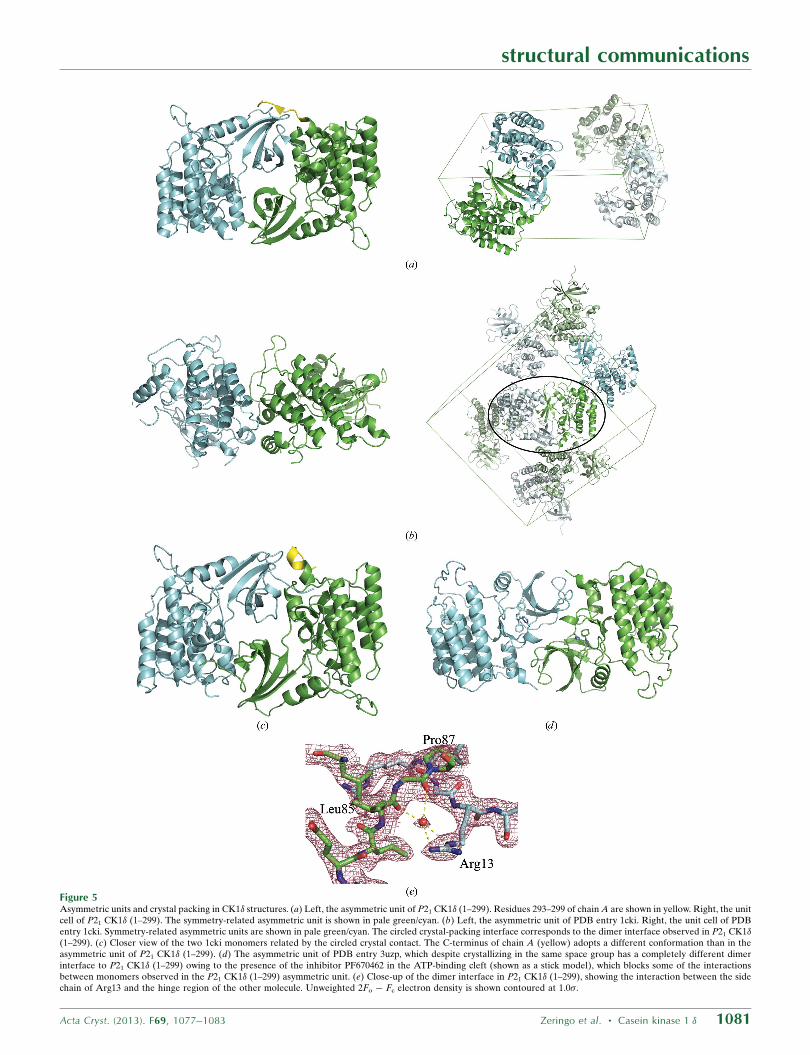
Figure 5Asymmetric units and crystal packing in CK1� structures. (a) Left, the asymmetric unit of P21 CK1� (1–299). Residues 293–299 of chain A are shown in yellow. Right, the unitcell of P21 CK1� (1–299). The symmetry-related asymmetric unit is shown in pale green/cyan. (b) Left, the asymmetric unit of PDB entry 1cki. Right, the unit cell of PDBentry 1cki. Symmetry-related asymmetric units are shown in pale green/cyan. The circled crystal-packing interface corresponds to the dimer interface observed in P21 CK1�(1–299). (c) Closer view of the two 1cki monomers related by the circled crystal contact. The C-terminus of chain A (yellow) adopts a different conformation than in theasymmetric unit of P21 CK1� (1–299). (d) The asymmetric unit of PDB entry 3uzp, which despite crystallizing in the same space group has a completely different dimerinterface to P21 CK1� (1–299) owing to the presence of the inhibitor PF670462 in the ATP-binding cleft (shown as a stick model), which blocks some of the interactionsbetween monomers observed in the P21 CK1� (1–299) asymmetric unit. (e) Close-up of the dimer interface in P21 CK1� (1–299), showing the interaction between the sidechain of Arg13 and the hinge region of the other molecule. Unweighted 2Fo � Fc electron density is shown contoured at 1.0�.
interactions with Lys154, Arg127 and Lys171. This is one of four
observed anion-binding sites that have potential roles as phosphate-
recognition sites for substrate binding and autoinhibition (Longe-
necker et al., 1996). In PDB entry 1ckj chain B, which was solved from
crystals soaked in 17 mM sodium tungstate, a WO42� ion is bound to
this site with a refined occupancy of 0.1 (Longenecker et al., 1996); in
PDB entries 3uys chains A and B and 3uyt chain A (which were
crystallized from sodium sulfate), an SO42� ion with a refined occu-
pancy of 1.0 occupies the same binding site (Long et al., 2012a). Chain
B of P21 CK1� (1–299) adopts the more common conformation seen
in the anion-bound structures (Fig. 2c), and in P21 CK1� (1–299)
chain A residues 171–173 were not modeled owing to poor electron
density.
Significant movement of the N-terminal domain �-sheet can be
seen when aligning different CK1� structures by the C-terminal
domain (Fig. 3). As helix �A in the N-terminal lobe (residues 49–59
superimposes well in most structures (Figs. 3a, 3c and 3d), these
movements represent rotations of the �-sheet within the N-terminal
lobe rather than movement of the entire N-terminal lobe relative to
the C-terminal lobe. The greatest displacements are observed for �1–
�3 (residues 1–50) and �4–�5 (residues 68–83); however, the extent
of movement varies greatly among the different structures (Fig. 3).
In PDB entries 3uzp (Fig. 3c) and 4hgt (Fig. 3d), the �-sheet
undergoes a clockwise rotation that brings �1 and �2 towards the
C-terminal lobe to form a more closed conformation over the
inhibitor bound in the ATP-binding cleft and results in a large
displacement of the C-terminal end of �3. In PDB entries 3uys
(Fig. 3b) and 4hnf (Fig. 3e) helix �A pivots on its C-terminal end,
rotating along with the �-sheet to adopt a more open conformation in
which �1 and �2 move farther away from the ATP-binding cleft. The
largest movement of the �-sheet can be seen when comparing PDB
entry 4hnf and P21 CK1� (1–299) (Fig. 3e). �1 is displaced by 4.3 A
and �2 by 3.5 A. The C-terminus of �3 in PDB entry 4hnf has shifted
to where the N-terminus of �4 in P21 CK1� (1–299) is. Loop L-3,4 is
shifted by 5.5 A between the two structures (Gly75 C� displacement).
Despite these conformational changes, the hinge connecting the two
lobes is virtually unmoved as C� of Leu85 is displaced by 0.4 A
between the two structures. These movements of the N-terminal lobe
lead to small displacements of the catalytically important ATP-
binding residue Lys38 (Fig. 4), but the conformation of the putative
catalytic base Glu128 is essentially unchanged.
3.3. Dimer interface and crystal packing
Even though CK1� (1–299) is a monomer in solution, the two
molecules in the asymmetric unit of the P21 CK1� (1–299) structure
share an extensive dimer interface that buries a total of 3530 A2,
involving 51 residues on each monomer and containing 15 hydrogen
bonds. The N-terminal lobe of each molecule fits between the
N-terminal lobe and C-terminal lobe of its partner (Fig. 5a). An
approximate noncrystallographic twofold axis is at the center of an
interface involving the side chains of Arg13, Lys14, Ile15 (on L-1,2),
Leu25 (on �2) and Glu34 (on �3), as well as the backbone carbonyls
of Leu85 and Pro87 (on the hinge connecting the two lobes). The
side chain of Arg13 extends partially into the ATP-binding groove
between the two lobes in the partner molecule to interact through a
bridging water molecule with the backbone carbonyls of Leu85 and
Pro87 (Fig. 5e). This penetration into the ATP-binding groove makes
this crystal form unsuitable for soaking experiments for inhibitor
screening. Side chains on helix �B in the C-terminal lobe interact with
side chains on �4–�5 in the N-terminal lobe of the other molecule and
the C-terminus of chain A interacts with strands �4 and �5 as well
as the N-terminus of chain B. Although there are no intermolecular
contacts between residues in the two C-terminal lobes, the N-terminal
ends of helices �F in the C-terminal lobes are pointing towards each
other, forming a semicircle.
Although this is a new crystal form, closer inspection revealed that
the interface between monomers in P21 CK1� (1–299) was nearly the
same as a crystal-packing interface observed in the P212121 crystal
form of apo CK1� (1–317) (Figs. 5b and 5c; PDB entry 1cki and 1ckj;
Longenecker et al., 1996), with the exception of the interactions made
by residues 294–299 at the C-terminus of chain A with the N-terminal
lobe of chain B. In PDB entry 1cki this crystal contact involves 50
residues on chain A and 49 on chain B, buries a total of 3380 A2 and
involves 18 hydrogen bonds and one salt bridge, in contrast to the
interface between molecules in the asymmetric unit that mainly
involves the C-terminal lobes and spans only 920 A2 (Fig. 5b). A
similar dimer interface to that observed in P21 CK1� (1–299) was
described for two molecules of CK1� (1–342) related to one another
by a crystallographic twofold axis in a C2221 crystal structure (coor-
dinates not deposited; Longenecker et al., 1998). Although this is an
extensive dimer interface that occurs in at least three different crystal
forms, size-exclusion chromatography indicates that CK1� is a
monomer in solution. It is unlikely that this dimer interface is
biologically relevant, as this interface blocks access of ATP and
peptide substrate to the active site.
A different P21 crystal form has previously been reported for the
CK1� (1–294)–inhibitor structures 3uzp (Long et al., 2012a) and
4hgt (Huang et al., 2012). As in the P21 CK1� (1–299) structure, the
N-terminal lobe of one monomer sits between the N-terminal and
C-terminal lobes of its partner, but does not penetrate as deeply into
the ATP-binding groove owing to the presence of the inhibitor
molecule (Fig. 5d). As a result, the interactions making up the dimer
interface in these structures are completely different from those in
P21 CK1� (1–299), as are the relative orientations of the two mole-
cules to one another.
The authors wish to thank Donald Ronning and members of
his research group for assistance with data collection and helpful
discussions. This work was supported by start-up funds from the
University of Toledo and a grant from the deArce Memorial
Endowment Fund in Support of Biomedical Research (to JJB) and
Undergraduate Summer Research Awards (to LM and EAM). Use
of the Advanced Photon Source, an Office of Science User Facility
operated for the US Department of Energy (DOE) Office of Science
by Argonne National Laboratory, was supported by the US DOE
under Contract No. DE-AC02-06CH11357. Use of the LS-CAT
Sector 21 was supported by the Michigan Economic Development
Corporation and the Michigan Technology Tri-Corridor (Grant
085P1000817).
References
Adams, P. D. et al. (2010). Acta Cryst. D66, 213–221.Cegielska, A., Gietzen, K. F., Rivers, A. & Virshup, D. M. (1998). J. Biol.
Chem. 273, 1357–1364.Chen, V. B., Arendall, W. B., Headd, J. J., Keedy, D. A., Immormino, R. M.,
Kapral, G. J., Murray, L. W., Richardson, J. S. & Richardson, D. C. (2010).Acta Cryst. D66, 12–21.
Doyle, S. A. (2005). Methods Mol. Biol. 310, 107–113.Emsley, P., Lohkamp, B., Scott, W. G. & Cowtan, K. (2010). Acta Cryst. D66,
486–501.Etchegaray, J.-P., Machida, K. K., Noton, E., Constance, C. M., Dallmann, R.,
Di Napoli, M. N., DeBruyne, J. P., Lambert, C. M., Yu, E. A., Reppert, S. M.& Weaver, D. R. (2009). Mol. Cell. Biol. 29, 3853–3866.
Etchegaray, J.-P., Yu, E. A., Indic, P., Dallmann, R. & Weaver, D. R. (2010).PLoS One, 5, e10303.
structural communications
1082 Zeringo et al. � Casein kinase 1 � Acta Cryst. (2013). F69, 1077–1083

Graves, P. R. & Roach, P. J. (1995). J. Biol. Chem. 270, 21689–21694.Huang, H. et al. (2012). ACS Med. Chem. Lett. 3, 1059–1064.Knippschild, U., Gocht, A., Wolff, S., Huber, N., Lohler, J. & Stoter, M. (2005).
Cell. Signal. 17, 675–689.Krissinel, E. & Henrick, K. (2007). J. Mol. Biol. 372, 774–797.Lee, H., Chen, R., Lee, Y., Yoo, S. & Lee, C. (2009). Proc. Natl Acad. Sci. USA,
69, 6–9.Liu, H. & Naismith, J. H. (2008). BMC Biotechnol. 8, 91.Long, A., Zhao, H. & Huang, X. (2012a). J. Med. Chem. 55, 956–960.Long, A., Zhao, H. & Huang, X. (2012b). J. Med. Chem. 55, 10307–10311.Longenecker, K. L., Roach, P. J. & Hurley, T. D. (1996). J. Mol. Biol. 257,
618–631.Longenecker, K. L., Roach, P. J. & Hurley, T. D. (1998). Acta Cryst. D54,
473–475.Lowrey, P. L., Shimomura, K., Antoch, M. P., Yamazaki, S., Zemenides, P. D.,
Ralph, M. R., Menaker, M. & Takahashi, J. S. (2000). Science, 288, 483–492.McCoy, A. J., Grosse-Kunstleve, R. W., Adams, P. D., Winn, M. D., Storoni,
L. C. & Read, R. J. (2007). J. Appl. Cryst. 40, 658–674.Otwinowski, Z. & Minor, W. (1997). Methods Enzymol. 276, 307–326.Pettersen, E. F., Goddard, T. D., Huang, C. C., Couch, G. S., Greenblatt, D. M.,
Meng, E. C. & Ferrin, T. E. (2004). J. Comput. Chem. 25, 1605–1612.Price, M. A. (2006). Genes Dev. 20, 399–410.Vaguine, A. A., Richelle, J. & Wodak, S. J. (1999). Acta Cryst. D55, 191–205.Vielhaber, E. & Virshup, D. M. (2001). IUBMB Life, 51, 73–78.Virshup, D. M., Eide, E. J., Forger, D. B., Gallego, M. & Harnish, E. V. (2007).
Cold Spring Harb. Symp. Quant. Biol. 72, 413–420.Xu, R., Carmel, G., Sweet, R. M., Kuret, J. & Cheng, X. (1995). EMBO J. 14,
1015–1023.Xu, Y., Padiath, Q. S., Shapiro, R. E., Jones, C. R., Wu, S. C., Saigoh, N., Saigoh,
K., Ptacek, L. J. & Fu, Y.-H. (2005). Nature (London), 434, 640–644.
structural communications
Acta Cryst. (2013). F69, 1077–1083 Zeringo et al. � Casein kinase 1 � 1083


![Neuropathology and cognitive performance in centenarians ... · of casein kinase 1 delta as a marker of GVD [43], and the distributionLewy bodies [6]and pTDP-43 accumulations [25].](https://static.fdocuments.in/doc/165x107/5f4134bf5e326525e658345a/neuropathology-and-cognitive-performance-in-centenarians-of-casein-kinase-1.jpg)


![Arabidopsis Casein Kinase1 Proteins CK1.3 and CK1.4 ... · code Active Casein Kinases. (A) In vitro kinase assays by [g-32P]ATP autoradiography indicated that recombinant CK1.3 and](https://static.fdocuments.in/doc/165x107/5fd572d92d5adf1c9e637682/arabidopsis-casein-kinase1-proteins-ck13-and-ck14-code-active-casein-kinases.jpg)
