Streptococcus pneumoniae Infection Increases Expression of ... · Streptococcus pneumoniae, also...
13
Med & Health Jun 2018; 13(1): 130-142 ORIGINAL ARTICLE 130 https://doi.org/10.17576/MH.2018.1301.13 Address for correspondence and reprint requests: Nurul Adhwa Rahman. Pengiran Anak Puteri Rashidah Sa'adatul Bolkiah Institute of Health Sciences, Universiti Brunei Darussalam, Jalan Tungku Link, Kampong Rimba, Gadong. Tel: +673 2463001 E-mail: [email protected]/[email protected] Streptococcus pneumoniae Infection Increases Expression of von Willebrand Factor (vWF) in a Mouse Brain Endothelium Model NURUL ADHWA R, SUWARNI D, SITI HANNA M Pengiran Anak Puteri Rashidah Sa’adatul Bolkiah Institute of Health Sciences, Universiti Brunei Darussalam, Brunei Darussalam ABSTRAK Faktor von Willebrand (vWF) adalah glikoprotein multimerik besar yang dikeluarkan secara normal oleh sel-sel endotelial otak dan ianya wujud sebagai protein beredar di dalam darah tetapi struktur asal dan matang vWF tidak terbahagi. Jangkitan pneumokokus boleh menyebabkan pembelahan protein, sehingga membentuk beberapa multimer dengan saiz yang berbeza. Kajian ini telah menguji hipotesis adakah jangkamasa jangkitan Streptococcus pneumoniae boleh menentukan pembentukan saiz vWF. Sel-sel endotelial otak tikus (bEnd.3) telah dijangkiti pada waktu tertentu dengan Streptococcus pneumoniae (serotype 19F). Mikroskopi immunofluoresensi telah dilakukan dan dianalisa dengan sewajarnya. Sel-sel juga diekstrak dan kandungan protein telah dipisahkan ke dalam saiz yang berbeza dengan menggunakan Western blot. Ekspresi vWF kemudian dianalisa dan dibandingkan antara sampel dan antara jangkamasa jangkitan tertentu. Pengeluaran vWF adalah didapati meningkat semasa jangkitan. Walaupun pelbagai perkara tidak spesifik boleh menyebabkan pembahagian vWF, kajian ini menunjukkan bahawa bentuk pembahagian vWF adalah berkemungkinan bergantung pada jangkamasa jangkitan bakteria ini. Kata kunci: faktor von Willebrand, mikroskopi immunofluoresensi, Streptococcus pneumoniae, Western blot ABSTRACT von Willebrand factor (vWF) is a large multimeric glycoprotein which is normally released by the brain endothelial cells and is therefore present as a circulating protein in the blood. The initial and mature structure of vWF is uncleaved; however, pneumococcal infection can cause the cleavage of the protein, thus forming
Transcript of Streptococcus pneumoniae Infection Increases Expression of ... · Streptococcus pneumoniae, also...
Med & Health Jun 2018; 13(1): 130-142
ORIGINAL ARTICLE
130
https://doi.org/10.17576/MH.2018.1301.13
Address for correspondence and reprint requests: Nurul Adhwa Rahman. Pengiran Anak Puteri Rashidah Sa'adatul Bolkiah Institute of Health Sciences, Universiti Brunei Darussalam, Jalan Tungku Link, Kampong Rimba, Gadong. Tel: +673 2463001 E-mail: [email protected]/[email protected]
Streptococcus pneumoniae Infection Increases Expression of von Willebrand Factor (vWF) in a
Mouse Brain Endothelium Model
NURUL ADHWA R, SUWARNI D, SITI HANNA M
Pengiran Anak Puteri Rashidah Sa’adatul Bolkiah Institute of Health Sciences, Universiti Brunei Darussalam, Brunei Darussalam
ABSTRAK
Faktor von Willebrand (vWF) adalah glikoprotein multimerik besar yang dikeluarkan secara normal oleh sel-sel endotelial otak dan ianya wujud sebagai protein beredar di dalam darah tetapi struktur asal dan matang vWF tidak terbahagi. Jangkitan pneumokokus boleh menyebabkan pembelahan protein, sehingga membentuk beberapa multimer dengan saiz yang berbeza. Kajian ini telah menguji hipotesis adakah jangkamasa jangkitan Streptococcus pneumoniae boleh menentukan pembentukan saiz vWF. Sel-sel endotelial otak tikus (bEnd.3) telah dijangkiti pada waktu tertentu dengan Streptococcus pneumoniae (serotype 19F). Mikroskopi immunofluoresensi telah dilakukan dan dianalisa dengan sewajarnya. Sel-sel juga diekstrak dan kandungan protein telah dipisahkan ke dalam saiz yang berbeza dengan menggunakan Western blot. Ekspresi vWF kemudian dianalisa dan dibandingkan antara sampel dan antara jangkamasa jangkitan tertentu. Pengeluaran vWF adalah didapati meningkat semasa jangkitan. Walaupun pelbagai perkara tidak spesifik boleh menyebabkan pembahagian vWF, kajian ini menunjukkan bahawa bentuk pembahagian vWF adalah berkemungkinan bergantung pada jangkamasa jangkitan bakteria ini.
Kata kunci: faktor von Willebrand, mikroskopi immunofluoresensi, Streptococcus pneumoniae, Western blot
ABSTRACT
von Willebrand factor (vWF) is a large multimeric glycoprotein which is normally released by the brain endothelial cells and is therefore present as a circulating protein in the blood. The initial and mature structure of vWF is uncleaved; however, pneumococcal infection can cause the cleavage of the protein, thus forming

131
Expression of vWF with Streptococcus pneumoniae Med & Health Jun 2018;13(1): 130-142
several multimers at different sizes. The present study tested the hypothesis that Streptococcus pneumoniae infection of brain endothelial cells in culture induces the release of vWF at certain sizes time-dependently. Immortalized mouse brain endothelial cells (bEnd.3) were infected at specific hours with Streptococcus pneumoniae strains (serotype 19F). Immunofluorescence microscopy was performed and quantified accordingly. Cells were also extracted, and the proteins were separated into different sizes by western blot. The expression of vWF was then analysed and compared for a potential relationship between the strains and between the specific time of infection. The expression of vWF was elevated during infection. Even though multiple and non-specific events can also cause the cleavage of vWF, this study showed that the cleavage pattern of vWF is potentially dependent on the incubation time of infection.
Keywords: immunofluorescence microscopy, Streptococcus pneumoniae, von Willebrand factor, western blot.
infection (Kayal et al. 1998). With this in mind, Streptococcus pneumoniae have been suggested to adhere directly to vWF and stimulated the secretion of free vWF (Lüttge et al. 2012). Streptococcus pneumoniae, also known as the pneumococcus (in singular) or pneumococci (in plural) is a Gram-positive diplococcus, of at least 0.5 µm in cell size. The pneumococcus is the main causative agent of multiple and invasive diseases in humans, such as pneumonia, sepsis and meningitis (Bogaert et al. 2004). Our study particularly focused on pneumococcal meningitis, which is widely considered to result from sepsis which may lead to the breakdown of the blood-brain barrier (BBB) and pneumococcal dissemination throughout the central nervous system (Hatcher et al. 2016). Moreover, pneumococcal meningitis has been reported to have a high mortality rate of 30% and neurologic sequelae in 30 to 50% of survivors
INTRODUCTION
von Willebrand factor (vWF) is a large multimeric plasma glycoprotein which is normally produced by endothelial cells (Ohmori et al. 1982). Its main functions in the human body include thrombus formation, blood coagulation and the maintenance of homeostasis (Hassan et al. 2012). vWF is present in two different forms: (1) small vWF dimers which are secreted into the plasma, and (2) granular vWF multimers which are stored cell-bound in Weibel–Palade bodies for rapid mobilization in response to cell stress condition (Wagner & Bonfanti 1991). An increased plasma concentration of vWF has been detected in individuals with bacterial infection and has been suggested to be a marker of endothelium activation (Freitas et al. 2012). In addition, vWF levels were also found to be significantly higher in patients who did not survive the
132
Med & Health Jun 2018;13(1): 130-142 Nurul Adhwa R. et al.
(Durand et al. 1993). It is important to remember that any short delays in the diagnosis and treatment can increase the rate of sequelae and mortality (Brouwer et al. 2012). In addition, pneumococcal strains have the ability to transform itself into a viable but non-culturable (VBNC) state when grown on an agar plate (Li et al. 2014). The mechanism of pneumococcal meningitis is not entirely caused by the presence or absence of pneumococcal cell-surface capsule as previously speculated, but other virulence factors also play a major role in the onset of the disease (Hathaway et al. 2016; Iovino et al. 2016). This also involves the response by the host innate immune system, which will
counteract pneumococcal infection by releasing a number of inflammatory molecules such as interleukin-6 and tumour necrosis factor (TNF-α) (Mook-Kanamori et al. 2011). These molecules will eventually build up their recruitment in the meninges, causing more brain injury, which then triggers the onset of the signs and symptoms of meningitis (Fassbender et al. 1997). Based on this mechanism, one of the least commonly targeted molecules in the study of pneumococcal meningitis are the vWF. Recent findings have focused on the potential collaborative role of vWF in the clearance of pneumococcal infection. When activated, vWF undergoes a two-step conformational
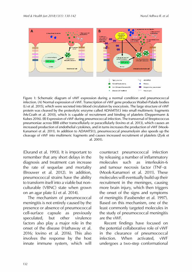
Figure 1: Schematic diagram of vWF expression during a normal condition and pneumococcal infection. (A) Normal expression of vWF. Transcription of vWF gene produces Waibel-Palade bodies (Li et al. 2015), which were secreted into blood circulation by exocytosis. The large structure of vWF protein was cleaved by the proteolytic enzyme called ADAMTS13 into small multimeric fragments (McGrath et al. 2010), which is capable of recruitment and binding of platelets (Deppermann & Kubes 2016). (B) Expression of vWF during pneumococcal infection. The transversal of Streptococcus pneumoniae across BBB either transcellularly or paracellularly (Iovino et al. 2013), which causes an increased production of endothelial cytokines, and in turns increases the production of vWF (Mook-Kanamori et al. 2011). In addition to ADAMTS13, pneumococcal pneumolysin also speeds up the cleavage of vWF into multimeric fragments and causes increased recruitment of platelets (Zysk et
al. 2001).

133
Expression of vWF with Streptococcus pneumoniae Med & Health Jun 2018;13(1): 130-142
change: (1) elongation from compact granular to linear form, and subsequently, (2) a tension-dependent local transition to a state with a high affinity for GPIbα, which is vWF binding site for platelets (Fu et al. 2017). Immobilised vWF can also bind to pneumococci directly, and thus allows vWF to bring together platelets and pneumococcal strains (Deppermann & Kubes 2016). vWF can then either activate the immune response by complement-mediated opsonization and phagocytosis (Andre et al. 2017), or they can be transported to the liver for pneumococcal clearance, as one of the host mechanism of action to combat pneumococcal infection (van’t Veer & van der Poll 2008). Figure 1 shows a proposed step-by-step events from adherence of pneumococci to the release of vWF during a pneumococcal infection. Nonetheless, despite this previous interest in vWF research, to the best of our knowledge, no study has investigated the expression pattern of vWF time-dependently which may be useful as a potential biomarker for S. pneumoniae. The main aim of this study was to investigate the potential application of vWF as a plasma biomarker of pneumococcal infection in the case of potential meningitis. In this study, bEnd.3 cells were applied as a brain endothelium model for the study of the pneumococcal breach of the blood-brain barrier in a time-dependent manner.
MATERIALS AND METHODS
CELL CULTURE
Immortalised mouse brain endothelial cell line (bEnd.3) was applied as a brain endothelium model and cultured in Dulbecco’s Modified Eagle’s Medium (DMEM) supplemented with 10% fetal bovine serum (FBS), 1% non-essential amino acid and 1% of penicillin-streptomycin at 37˚C in 5% CO2 (Williams et al. 1988; Montesano et al. 1990; Sikorski et al. 1993). The cells were grown on coverslips in 6-well plates at a seeding concentration of 1 X 10⁴ cells ml-1 and cultured for 3 days until 90-100% cell confluency. The cell passages used in this study were between passage 21 to passage 39.
BACTERIAL STRAINS USED IN THIS STUDY
Streptococcus pneumoniae clinical strains used in this study were isolated from gastric infection (designated as RSP1) and ear infection (designated as RSP2), collected from Raja Isteri Pengiran Anak Saleha (RIPAS) Hospital, Bandar Seri Begawan, Brunei Darussalam in March 2016. These strains have been maintained in 20% glycerol (v/v in cation-adjusted Mueller Hinton broth, supplemented with 10% lysed human blood v/v in sterile distilled water) at -20˚C. Streptococcus pneumoniae ATCC 49619 was used as the control strain. All of the strains in this study carried the same serotype 19F (Rahman et al. 2017).
INFECTION ASSAY
Pneumococcal strains were cultured in cation-adjusted Mueller-Hinton broth (supplemented with 5% lysed
134
Med & Health Jun 2018;13(1): 130-142 Nurul Adhwa R. et al.
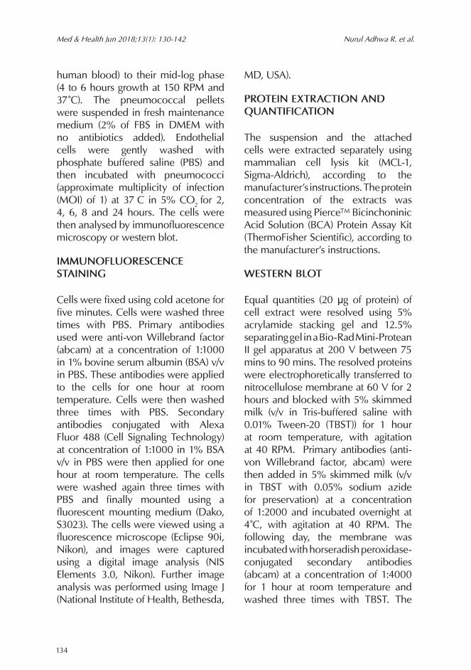
human blood) to their mid-log phase (4 to 6 hours growth at 150 RPM and 37˚C). The pneumococcal pellets were suspended in fresh maintenance medium (2% of FBS in DMEM with no antibiotics added). Endothelial cells were gently washed with phosphate buffered saline (PBS) and then incubated with pneumococci (approximate multiplicity of infection (MOI) of 1) at 37°C in 5% CO2 for 2, 4, 6, 8 and 24 hours. The cells were then analysed by immunofluorescence microscopy or western blot.
IMMUNOFLUORESCENCE STAINING
Cells were fixed using cold acetone for five minutes. Cells were washed three times with PBS. Primary antibodies used were anti-von Willebrand factor (abcam) at a concentration of 1:1000 in 1% bovine serum albumin (BSA) v/v in PBS. These antibodies were applied to the cells for one hour at room temperature. Cells were then washed three times with PBS. Secondary antibodies conjugated with Alexa Fluor 488 (Cell Signaling Technology) at concentration of 1:1000 in 1% BSA v/v in PBS were then applied for one hour at room temperature. The cells were washed again three times with PBS and finally mounted using a fluorescent mounting medium (Dako, S3023). The cells were viewed using a fluorescence microscope (Eclipse 90i, Nikon), and images were captured using a digital image analysis (NIS Elements 3.0, Nikon). Further image analysis was performed using Image J (National Institute of Health, Bethesda,
MD, USA).
PROTEIN EXTRACTION AND QUANTIFICATION
The suspension and the attached cells were extracted separately using mammalian cell lysis kit (MCL-1, Sigma-Aldrich), according to the manufacturer’s instructions. The protein concentration of the extracts was measured using Pierce™ Bicinchoninic Acid Solution (BCA) Protein Assay Kit (ThermoFisher Scientific), according to the manufacturer’s instructions.
WESTERN BLOT
Equal quantities (20 µg of protein) of cell extract were resolved using 5% acrylamide stacking gel and 12.5% separating gel in a Bio-Rad Mini-Protean II gel apparatus at 200 V between 75 mins to 90 mins. The resolved proteins were electrophoretically transferred to nitrocellulose membrane at 60 V for 2 hours and blocked with 5% skimmed milk (v/v in Tris-buffered saline with 0.01% Tween-20 (TBST)) for 1 hour at room temperature, with agitation at 40 RPM. Primary antibodies (anti-von Willebrand factor, abcam) were then added in 5% skimmed milk (v/v in TBST with 0.05% sodium azide for preservation) at a concentration of 1:2000 and incubated overnight at 4˚C, with agitation at 40 RPM. The following day, the membrane was incubated with horseradish peroxidase-conjugated secondary antibodies (abcam) at a concentration of 1:4000 for 1 hour at room temperature and washed three times with TBST. The

135
Expression of vWF with Streptococcus pneumoniae Med & Health Jun 2018;13(1): 130-142
blot was developed with enhanced chemiluminescent substrate (PierceTM ThermoFisher Scientific) for a maximum of 5 minutes at room temperature. Images were viewed using an imaging system (VersaDocTM, Bio-Rad), and captured using Quantity One software (Version 4.6.9, BioRad). The densities of the bands were determined using Image J (National Institute of Health, Bethesda, MD, USA).
DATA AND STATISTICAL ANALYSIS
All numerical data were expressed as the mean ± standard error of means from at least three independent experiments. The significance of strain differences was calculated, and a p-value of less than or equal to 0.05 was considered significant. Graphical presentation and statistical analysis were performed using the PRISM 7 software (GraphPad Software Inc., CA, USA).
RESULTS
The results were focused on the expression of vWF on infected brain endothelial cells as the brain endothelium model in the study of potential pneumococcal meningitis, with pneumococcal strains carrying the common serotype 19F. vWF detected in the suspension are labelled as immobilised or released vWF, while vWF from the whole-cell extracts are labelled as anchored vWF.
IMMUNOFLUORESCENCE EXPRESSION PATTERN OF VWF
Immunofluorescence microscopy was performed following 2, 4, 6, 8 and 24 hours of pneumococcal infection (Figure 2). Immobilised vWF were observed more notably in infected cells (Figure 2). However, the immobilised vWF did not represent the actual quantity of suspended or released
Figure 2: Representative immunofluorescence images of vWF post-pneumococcal infections time-dependently. All images were taken at 100X magnification with oil immersion as indicated by the
scale bar at the bottom right.

136
Med & Health Jun 2018;13(1): 130-142 Nurul Adhwa R. et al.
vWF, because the suspended medium was removed and washed prior to cell fixation step. The immunofluorescence expressions of vWF were observed to be upregulated following infection, as compared to the uninfected control (Figure 3). This increase of expression was highest at 4 hours, 6 hours and 24 hours for RSP1 and RSP2. Infected cells by the control strain ATCC 49619 also showed a consistently increased expression of vWF at 4 hours and 24 hours, when compared to the uninfected control. At 24 hours, vWF expression was observed to decline in both uninfected control and infected cells when compared to the earlier hours of infection. However, the infected cells still quantitatively
expressed higher vWF than the uninfected control. This decrease of expression may be due to the cellular exhaustion of vWF release to the surrounding. Starting from 2 hours post-infection, closely-packed tubules were observed in infected cells (Figure 4A and 4B). These tubules are also known as Weibel-Palade bodies. In addition, a string-like structure was also observed on the periphery of infected cells (Figure 4C).
WESTERN BLOT ANALYSIS OF VWF
Western blot analysis of both the suspension and the whole-cell extracts of the infected and uninfected cells
Figure 3: Immunofluorescence analysis of vWF at indicated hours of pneumococcal infections. Integrated density of vWF fluorescence (line graph) and its percentages to the uninfected control (bar graph). Each value represents at least three independent experiments, with at least three images were taken in each independent experiment. Uninfected (control) represents bEnd.3 cells which were included in every independent experiment. Significant differences (as shown by asterisks) were observed in comparison to the uninfected control (* represents p<0.05; ** represents p<0.005;
*** represents p<0.0005; Dunnett’s multiple comparison test, PRISM 7, GraphPad Inc.).

137
Expression of vWF with Streptococcus pneumoniae Med & Health Jun 2018;13(1): 130-142
revealed multiple differences in the levels of vWF proteins (Figure 5). Figure 5 shows that vWF bands in infected cell are more evident than the uninfected control. In addition, actin was only observed in the cell extracts (Figure 5). Actin was not present in the suspension of both uninfected control
and pneumococcal-infected cells (data not shown). This suggests that the endothelial cells did not detach during the infection. Quantitatively, the western blots were analysed in two ways utilising the integrated densities of the bands, from 250 kDa to 25 kDa, as analysed
Figure 4: The structures of vWF on pneumococcal-infected cells. Representative images of the expression of vWF on infected cells. These images were from RSP2-infected cells. (A) Weibel-Palade bodies were observed in infected cells starting from 2 hours onwards, most prominently at 24 hours. (B) A close-up schematic drawing of the Weibel-Palade bodies in A. (C) White arrows show string-like structures which represent the linear form of activated vWF to achieve more surface area for the attachment of pneumococci and other molecules such as platelets. The images were taken at 100X magnification with oil immersion as indicated by the scale bar at the bottom right.
Figure 5: Representative western blots of vWF and actin at indicated hours of incubation with Streptococcus pneumoniae. The protein content of the suspension and the cell extracts were run separately. Three independent experiments were performed, and consistent band separations were
observed at the indicated hours, post-infection.

138
Med & Health Jun 2018;13(1): 130-142 Nurul Adhwa R. et al.
by Quantity One software (Bio-Rad). Firstly, the number of vWF multimers were manually counted by their integrated density peaks, in comparison to the protein standard, to observe
the cleavage ability of pneumococcal strains (Figure 6). The immobilised vWF in the suspension was statistically significant at 4 hours and 6 hours post-pneumococcal infections, especially
Figure 6: The number of vWF multimer bands during pneumococcal infections, separated by western blot. (A) Immobilised vWF in suspensions. (B) Anchored vWF in cell extracts. Each value represents at least three independent experiments. The uninfected control represents bEnd.3 cells which were included in every independent experiment. Significant differences as shown by asterisks were observed in comparison to the uninfected control and ATCC 49619 (control strain) (* represents p<0.05, ** represents p<0.005, Dunnett’s multiple comparison tests, PRISM 7,
GraphPad Inc.).
Figure 7: The total integrated density of vWF using western blot analysis. (A) Immobilised vWF in suspensions. (B) Anchored vWF in cell extracts. Error bars represent standard error of means. Significant differences were observed as indicated (* represents p<0.05, ** represents p<0.005,
Tukey’s multiple comparison tests, PRISM 7, GraphPad Inc.).

139
Expression of vWF with Streptococcus pneumoniae Med & Health Jun 2018;13(1): 130-142
by RSP2, indicating that the clinical strains may have more cleavage ability than the control strain at 4 and 6 hours of infection. Overall, there are more multimers of immobilised vWF in the infected suspension than the uninfected control, at 4, 6 and 8 hours of infection. Secondly, the blots were analysed by the total integrated density of the bands, to observe the total amount of vWF (Figure 7). Similarly, infected cells showed a higher total integrated density of vWF in comparison to the uninfected control. Moreover, this difference was later found to be statistically insignificant.
DISCUSSION
The major findings of this study are that vWF was upregulated in the suspension as immobilised vWF, and was also found to accumulate in the endothelial cells as anchored vWF during pneumococcal infection. However, no
direct apparent and consistent pattern in the expression was observed during the time-dependent infection. In general, the expression of vWF during pneumococcal infection was found to be high between 4 hours until 6 hours of infection. The abundant expression of immobilised and small vWF fragments in infected suspensions signified a similar response to our current understanding of platelets activity during bacterial infection (Deppermann & Kubes 2016). When a cell undergoes injury, the cells release multiple proteins to send signals for platelet aggregation along the injured cells and also to create a matrix around the bacteria to prevent the bacteria from attaching to the cell surfaces. However, platelets are not able to directly bind to any bacteria. One of the ligands utilised by platelets for bacterial adherence is immobilised vWF. Therefore platelets will bind to the immobilised vWF and bacteria. On the other hand, because cultured
Figure 8: Schematic and hypothetical explanation of the expression of vWF during the time-dependent pneumococcal infection. Simplified and incomplete schematic diagram showing the release of vWF by pneumococcal-infected endothelial cells and its potential action on pneumococci from 2 hours to 24 hours. The figure is a continuation of the previously proposed theory of vWF
secretion in Figure 1. The figure was not drawn to scale.

140
Med & Health Jun 2018;13(1): 130-142 Nurul Adhwa R. et al.
endothelial cells are not exposed to fluid shear, uncleaved ultra-large vWF is likely to remain anchored to cell membranes (Freitas et al. 2012). Such anchorage would explain the increased expression of surface-associated vWF, as detected by immunofluorescence microscopy. However, these ultra-large vWF were not found by western blot, as they might be cleaved further into smaller fragments by the lysis buffer used in this study, as previously reported when vWF was treated with trypsin (Hamilton et al. 1985). Nonetheless, the drastic drop in the expression of vWF at 8 hours post-infection by immunofluorescence microscopy is still hypothetical. It is possible that at 8 hours post-infection, vWF was more involved in the aggregation of pneumococci as long strings of vWF, which were washed off before cell fixation. In reality, this binding of vWF with pneumococcal strains is to assist its binding with platelets, which later can transport itself to the liver for pneumococcal clearance (van’t Veer & van der Poll 2008). Figure 8 shows the schematic and hypothetical diagram to illustrate the results of this study. Meanwhile, the lowered expression at 24 hours post-infection might be due to the exhaustive release of vWF into the suspension, as seen by the decreasing pattern on the line graphs of the raw integrated density of the uninfected control and infected cells in Figure 3. In addition, this may also be due to the declining activity of the proteolytic metalloprotease, ADAMTS13, in the uninfected control when the severity of cell repair is
reduced (Crawley et al. 2005). On the other hand, the highly expressed anchored vWF during pneumococcal infection may be explained by a previous report which stated that the largest vWF multimers are stored in Weibel-Palade bodies and are released forcefully to the surrounding blood during cell stress conditions (Wagner & Bonfanti 1991). In addition, our study was focused on pneumococcal infection which carried the same serotype 19F. Both RSP1 and RSP2 showed a similar pattern of the expression of vWF. However, it showed a higher pattern as compared to control strain. Furthermore, RSP2 seems to have a stronger impact on the expression of vWF. This suggests that despite having the same serotype, the clinical strains proved to be more virulent than the control strain in infecting the brain endothelium model. Clinically, vWF has been used as a plasma biomarker for vWF disease, with a defect or deficiency in vWF, utilising few test systems targeting plasma vWF (Favaloro et al. 2016). It will be an interesting and cost-effective suggestion to offer these available test systems as a potential over-the-counter diagnostic kit for pneumococcal infections as well. Nonetheless, the data here should be interpreted carefully in light of several limitations, as this study shows very preliminary findings involving an in-vitro model. The limitations include whether they can recapitulate the human BBB, the species differences between a mouse and a human, or the differences in the absolute quantity of target proteins expressed in the cells (Haruki et al.

141
Expression of vWF with Streptococcus pneumoniae Med & Health Jun 2018;13(1): 130-142
2013), This also includes the multi-donor origin of the endothelial cells, which tends to be responsible for the different characteristics between batches (Bouïs et al. 2001). Further investigations utilising animal models should be performed following the same hours of infection as a comparative study to establish whether vWF can potentially be an indirect marker for pneumococcal meningitis in the blood sample.
CONCLUSION
Streptococcus pneumoniae infection of the mouse brain endothelium model has caused an increased expression of vWF. Immobilised vWF was also detected in the suspension, thus making vWF a potential indirect biomarker for pneumococcal meningitis infection. Nonetheless, even though multiple and non-specific events can also cause the cleavage of vWF, this study showed that the cleavage pattern of vWF is potentially dependent on the incubation time of infection.
ACKNOWLEDGEMENT
The authors would like to express their sincere gratitude to the Department of Microbiology laboratory of the Raja Isteri Pengiran Anak Saleha Hospital (RIPASH), Brunei Darussalam, especially to Mr Woo Boon Chu, for their kind assistance in providing the clinical strains. Nurul Adhwa Rahman was supported by the Graduate Research Scholarship (GRS), Universiti Brunei Darussalam. All experiments were supported by the Pengiran Anak
Puteri Rashidah Sa’adatul Bolkiah Institute of Health Sciences, Universiti Brunei Darussalam and the Ministry of Education, Brunei Darussalam. The authors also appreciate the technical help recieved from Mr Aswiradi Ahmad.
REFERENCES
Andre, G.O., Converso, T.R., Politano, W.R., Ferraz, L.F.C., Ribeiro, M.L., Leite, L.C.C., Darrieux, M. 2017. Role of Streptococcus pneumoniae Proteins in Evasion of Complement-Mediated Immunity. Front Microbiol 8(FEB): 224.
Bogaert, D., de Groot, R., Hermans, P.W.M. 2004. Streptococcus pneumoniae colonisation: The key to pneumococcal disease. Lancet Infect Dis 4(3): 144–54.
Bouïs, D., Hospers, G.A.P., Meijer, C., Molema, G., Mulder, N.H. 2001. Endothelium in vitro: a review of human vascular endothelial cell lines for blood vessel-related research. Angiogenesis 4(2): 91–102.
Brouwer, M.C., Thwaites, G.E., Tunkel, A.R., Van De Beek, D. 2012. Dilemmas in the diagnosis of acute community-acquired bacterial meningitis. The Lancet 380(9854): 1684–91.
Crawley, J.T.B., Lam, J.K., Rance, J.B., Mollica, L.R., O’Donnell, J.S., Lane, D.A. 2005. Proteolytic inactivation of ADAMTS13 by thrombin and plasmin. Blood 105(3): 1085–93.
Deppermann, C., Kubes, P. 2016. Platelets and infection. Semin Immunol 28(6): 536–45.
Durand, M.L., Calderwood, S.B., Weber, D.J., Miller, S.I., Southwick, F.S., Caviness, V.S., & Swartz, M.N. 1993. Acute Bacterial Meningitis in Adults - A Review of 493 Episodes. New England Journal of Medicine 328(1): 21–8.
Fassbender, K., Schminke, U., Ries, S., Ragoschke, A., Kischka, U., Fatar, M., Hennerici, M. 1997. Endothelial-derived adhesion molecules in bacterial meningitis: Association to cytokine release and intrathecal leukocyte-recruitment. J Neuroimmunol 74(1–2): 130–4.
Favaloro, E.J., Pasalic, L., Curnow, J. 2016. Laboratory tests used to help diagnose von Willebrand disease: an update. Pathology 48(4): 303–18.
Freitas, C., Assis, M.-C., Saliba, A.M., Morandi, V.M., Figueiredo, C.C., Pereira, M., Plotkowski, M.-C. 2012. The infection of microvascular endothelial cells with ExoU-producing Pseudomonas aeruginosa triggers the release of von Willebrand factor and platelet adhesion.
142
Med & Health Jun 2018;13(1): 130-142 Nurul Adhwa R. et al.
Mem Inst Oswaldo Cruz 107(6): 728–34. Fu, H., Jiang, Y., Yang, D., Scheiflinger, F., Wong, W.P.,
Springer, T.A. 2017. Flow-induced elongation of von Willebrand factor precedes tension-dependent activation. Nat Commun 8(1): 324.
Hamilton, K.K., Fretto, L.J., Grierson, D.S., McKee, P.A. 1985. Effects of plasmin on von Willebrand factor multimers. Degradation in vitro and stimulation of release in vivo. J Clin Invest 76(1): 261–70.
Haruki, H., Sano, Y., Shimizu, F., Omoto, M., Tasaki, A., Oishi, M., Koga, M., Saito, K., Takahashi, T., Nakada, T., Kanda, T. 2013. NMO sera down-regulate AQP4 in human astrocyte and induce cytotoxicity independent of complement. J Neurol Sci 331(1–2): 136–44.
Hassan, M.I., Saxena, A., Ahmad, F. 2012. Structure and function of von Willebrand factor. Blood Coagulation & Fibrinolysis 23(1): 11–22.
Hatcher, B.L., Hale, J.Y., Briles, D.E. 2016. Free Sialic Acid Acts as a Signal That Promotes Streptococcus pneumoniae Invasion of Nasal Tissue and Nonhematogenous Invasion of the Central Nervous System. Infection and Immunity 84(9): 2607–15.
Hathaway, L.J., Grandgirard, D., Valente, L.G., Täuber, M.G., Leib, S.L. 2016. Streptococcus pneumoniae capsule determines disease severity in experimental pneumococcal meningitis. Open Biology 6(3): 150269.
Iovino, F., Orihuela, C.J., Moorlag, H.E., Molema, G., Bijlsma, J.J.E. 2013. Interactions between Blood-Borne Streptococcus pneumoniae and the Blood-Brain Barrier Preceding Meningitis. PLoS ONE 8(7): e68408.
Iovino, F., Hammarlöf, D.L., Garriss, G., Brovall, S., Nannapaneni, P., Henriques-Normark, B. 2016. Pneumococcal meningitis is promoted by single cocci expressing pilus adhesin RrgA. J Clin Invest 126(8): 2821–6.
Kayal, S., Jaïs, J.P., Aguini, N., Chaudière, J., Labrousse, J. 1998. Elevated circulating E-selectin, intercellular adhesion molecule 1, and von Willebrand factor in patients with severe infection. Am J Respir Crit Care Med 157(3 Pt 1): 776–84.
Li, L., Mendis, N., Trigui, H., Oliver, J.D., Faucher, S.P. 2014. The importance of the viable but non-culturable state in human bacterial pathogens. Front Microbiol 5: 258.
Li, Y., Li, L., Dong, F., Guo, L., Hou, Y., Hu, H., Yan, S., Zhou, X., Liao, L., Allen, T.D., Liu, J.U. 2015. Plasma von Willebrand factor level is transiently elevated in a rat model of acute myocardial infarction. Exp Ther Med 10(5): 1743–9.
Lüttge, M., Fulde, M., Talay, S. R., Nerlich, A., Rohde, M., Preissner, K. T., Hammerschmidt, S., Steinert, M., Mitchell, T.J., Chhatwal, G.S., & Bergmann, S. 2012. Streptococcus pneumoniae
induces exocytosis of Weibel-Palade bodies in pulmonary endothelial cells. Cellular Microbiology 14(2): 210–25.
McGrath, R.T., McKinnon, T.A.J., Byrne, B., O’Kennedy, R., Terraube, V., McRae, E., Preston, R.J.S., Laffan, M.A., O’Donnell, J.S. 2010. Expression of terminal alpha2-6-linked sialic acid on von Willebrand factor specifically enhances proteolysis by ADAMTS13. Blood 115(13): 2666–73.
Montesano, R., Pepper, M.S.S., Möhle-Steinlein, U., Risau, W., Wagner, E.F.F., Orci, L. 1990. Increased proteolytic activity is responsible for the aberrant morphogenetic behavior of endothelial cells expressing the middle T oncogene. Cell 62(3): 435–45.
Mook-Kanamori, B.B., Geldhoff, M., van der Poll, T., van de Beek, D. 2011. Pathogenesis and Pathophysiology of Pneumococcal Meningitis. Clin Microb Rev 24(3): 557–91.
Ohmori, K., Fretto, L.J., Harrison, R.L., Switzer, M.E., Erickson, H.P., McKee, P.A. 1982. Electron microscopy of human factor VIII/Von Willebrand glycoprotein: effect of reducing reagents on structure and function. J Cell Biol 95(2 Pt 1): 632–40.
Rahman, N.A., Sharudin, A., Diah, S., & Muharram, S.H. 2017. Serotyping of Brunei pneumococcal clinical strains and the investigation of their capability to adhere and invade a brain endothelium model. Microb Pathog 110: 352–8.
Sikorski, E.E., Hallmann, R., Berg, E.L., Butcher, E.C. 1993. The Peyer’s patch high endothelial receptor for lymphocytes, the mucosal vascular addressin, is induced on a murine endothelial cell line by tumor necrosis factor-alpha and IL-1. J Immunol 151(10): 5239–50.
van’t Veer, C., van der Poll, T. 2008. Keeping blood clots at bay in sepsis. Nature Medicine 14(6): 606–8.
Wagner, D.D., Bonfanti, R. 1991. von Willebrand factor and the endothelium. Mayo Clin Proc 66(6): 621–7.
Williams, R., Courtneidge, S.A., Wagner, E.F. 1988. Embryonic lethalities and endothelial tumors in chimeric mice expressing polyoma virus middle T oncogene. Cell 52(1): 121–31.
Zysk, G., Schneider-Wald, B.K., Hwang, J.H., Bejo, L., Kim, K.S., Mitchell, T.J., Hakenbeck, R., Heinz, H.P. 2001. Pneumolysin is the main inducer of cytotoxicity to brain microvascular endothelial cells caused by Streptococcus pneumoniae. Infection and Immunity 69(2): 845–52.
Received: 12 Dec 2017Accepted: 26 Feb 2018