Streptococcus pneumoniae secretes hydrogen peroxide leading to ...
10
Streptococcus pneumoniae secretes hydrogen peroxide leading to DNA damage and apoptosis in lung cells Prashant Rai a,b , Marcus Parrish c , Ian Jun Jie Tay c , Na Li a,b , Shelley Ackerman c , Fang He d , Jimmy Kwang d , Vincent T. Chow a,b , and Bevin P. Engelward a,c,1 a Infectious Diseases Group, Singapore-MIT Alliance for Research and Technology, Singapore 138602; b Department of Microbiology, Yong Loo Lin School of Medicine, National University of Singapore, Singapore 117545; c Department of Biological Engineering, Massachusetts Institute of Technology, Cambridge, MA 02139; and d Animal Health Biotechnology, Temasek Life Sciences Laboratory, Singapore 117604 Edited by Hasan Yesilkaya, University of Leicester, Leicester, United Kingdom, and accepted by the Editorial Board May 11, 2015 (received for review December 17, 2014) Streptococcus pneumoniae is a leading cause of pneumonia and one of the most common causes of death globally. The impact of S. pneumoniae on host molecular processes that lead to detrimental pulmonary consequences is not fully understood. Here, we show that S. pneumoniae induces toxic DNA double-strand breaks (DSBs) in human alveolar epithelial cells, as indicated by ataxia telangiectasia mutated kinase (ATM)-dependent phosphorylation of histone H2AX and colocalization with p53-binding protein (53BP1). Furthermore, re- sults show that DNA damage occurs in a bacterial contact-indepen- dent fashion and that Streptococcus pyruvate oxidase (SpxB), which enables synthesis of H 2 O 2 , plays a critical role in inducing DSBs. The extent of DNA damage correlates with the extent of apoptosis, and DNA damage precedes apoptosis, which is consistent with the time required for execution of apoptosis. Furthermore, addition of catalase, which neutralizes H 2 O 2 , greatly suppresses S. pneumoniae-induced DNA damage and apoptosis. Importantly, S. pneumoniae induces DSBs in the lungs of animals with acute pneumonia, and H 2 O 2 production by S. pneumoniae in vivo contributes to its genotoxicity and virulence. One of the major DSBs repair pathways is nonhomologous end joining for which Ku70/80 is essential for repair. We find that deficiency of Ku80 causes an increase in the levels of DSBs and apoptosis, under- scoring the importance of DNA repair in preventing S. pneumoniae- induced genotoxicity. Taken together, this study shows that S. pneumoniae-induced damage to the host cell genome exacerbates its toxicity and pathogenesis, making DNA repair a potentially impor- tant susceptibility factor in people who suffer from pneumonia. DNA damage | Streptococcus pneumoniae | hydrogen peroxide | γH2AX | Ku80 O ne of the most common causes of community-acquired pneu- monia is Streptococcus pneumoniae, a commensal organism of upper respiratory tract (1). Pneumonia and other invasive pneu- mococcal diseases such as bacteremia, meningitis, and sepsis can be caused by S. pneumoniae, resulting in 1–2 million infant deaths every year (2). Secondary pulmonary infection by S. pneumoniae is commonly associated with higher mortality during major influenza pandemics (2). It is known that S. pneumoniae-induced cytotoxicity underlies pulmonary tissue injury during pneumonia and deter- mines the outcome of infection (3). Furthermore, toxicity to alveolar epithelium disintegrates pulmonary architecture as well as weakens the alveolar-blood barrier, facilitating systemic bacterial dissemi- nation. Although we have some understanding of bacterial viru- lence (4, 5), the underlying molecular processes in mammalian host cells that mediate tissue damage are not fully understood, which limits the development of mitigation strategies. There are significant data supporting a role for inflammation as a cause for cytotoxicity during infection. S. pneumoniae is known to induce a robust inflammatory response at the site of infection that culminates with infiltration and accumulation of inflammatory cells including neutrophils and macrophages (6–8). To defend against infection, activated inflammatory cells pro- duce high levels of genotoxic reactive oxygen and nitrogen spe- cies (RONS) including hydroxyl radical, superoxide, peroxide, nitric oxide, and peroxynitrite. RONS-induced DNA lesions such as base damage, single-strand breaks, and double-strand breaks (DSBs) can be cytotoxic and thus damaging to host tissue func- tion (9, 10). DSBs are one of the most toxic forms of DNA damage (11, 12). In response to DSBs, the ataxia telangiectasia mutated (ATM) kinase pathway is activated, leading to Ser-139 phosphorylation of histone H2AX, forming γH2AX. The pres- ence of γH2AX at DSBs recruits downstream DNA repair pro- teins, including 53BP1 and the Mre11/Rad50/Nbs1 (MRN) complex (13, 14). The major DSB repair pathway in nondividing cells is nonhomologous end-joining (NHEJ) (15). Early in NHEJ, Ku70/Ku80 heterodimer binds the damaged DNA ends. Ku80 plays a vital role in further recruitment and binding of the cata- lytic DNA-PKcs subunit (16). The DNA strands are then pro- cessed by nuclease activity of the MRN complex, and the DNA- PK holoenzyme recruits additional enzymes that complete the repair process (17). Despite the presence of efficient DSB re- pair, under conditions of excessive RONS, DNA damage can lead to cell death. Although studies have been done to explore the damaging po- tential of RONS associated with the host response (18, 19), the possibility that S. pneumoniae might directly induce oxidative damage to DNA had not been explored. Studies focused on re- spiratory, as well as intestinal pathogens (20, 21), call attention to the importance of microbial-induced DNA damage as an important dimension of pathogenicity. For example, Pseudomonas aeruginosa has been shown to induce oxidative DNA damage in lung cells accompanied by significant tissue injury (22). These findings Significance Streptococcus pneumoniae is the most common cause of pneumonia, a leading cause of death globally. Limitations in anti- biotic efficacy and vaccines call attention to the need to develop our understanding of host–pathogen interactions to improve mit- igation strategies. Here, we show that lung cells exposed to S. pneumoniae are subject to DNA damage caused by hydrogen peroxide, which is secreted by strains of S. pneumoniae that carry the spxB gene. The observation that S. pneumoniae secretes hy- drogen peroxide at genotoxic and cytotoxic levels is consistent with a model wherein host DNA damage and repair modulate pneumococcal pathogenicity. Author contributions: P.R., V.T.C., and B.P.E. designed research; P.R., M.P., I.J.J.T., N.L., S.A., and F.H. performed research; F.H. and J.K. contributed new reagents/analytic tools; P.R., N.L., V.T.C., and B.P.E. analyzed data; and P.R. and B.P.E. wrote the paper. The authors declare no conflict of interest. This article is a PNAS Direct Submission. H.Y. is a guest editor invited by the Editorial Board. Freely available online through the PNAS open access option. 1 To whom correspondence should be addressed. Email: [email protected]. This article contains supporting information online at www.pnas.org/lookup/suppl/doi:10. 1073/pnas.1424144112/-/DCSupplemental. www.pnas.org/cgi/doi/10.1073/pnas.1424144112 PNAS | Published online June 15, 2015 | E3421–E3430 MICROBIOLOGY PNAS PLUS
Transcript of Streptococcus pneumoniae secretes hydrogen peroxide leading to ...
Streptococcus pneumoniae secretes hydrogen peroxideleading to DNA damage and apoptosis in lung cellsPrashant Raia,b, Marcus Parrishc, Ian Jun Jie Tayc, Na Lia,b, Shelley Ackermanc, Fang Hed, Jimmy Kwangd,Vincent T. Chowa,b, and Bevin P. Engelwarda,c,1
aInfectious Diseases Group, Singapore-MIT Alliance for Research and Technology, Singapore 138602; bDepartment of Microbiology, Yong Loo Lin School ofMedicine, National University of Singapore, Singapore 117545; cDepartment of Biological Engineering, Massachusetts Institute of Technology, Cambridge,MA 02139; and dAnimal Health Biotechnology, Temasek Life Sciences Laboratory, Singapore 117604
Edited by Hasan Yesilkaya, University of Leicester, Leicester, United Kingdom, and accepted by the Editorial Board May 11, 2015 (received for reviewDecember 17, 2014)
Streptococcus pneumoniae is a leading cause of pneumonia and oneof the most common causes of death globally. The impact ofS. pneumoniae on host molecular processes that lead to detrimentalpulmonary consequences is not fully understood. Here, we show thatS. pneumoniae induces toxic DNA double-strand breaks (DSBs) inhuman alveolar epithelial cells, as indicated by ataxia telangiectasiamutated kinase (ATM)-dependent phosphorylation of histone H2AXand colocalization with p53-binding protein (53BP1). Furthermore, re-sults show that DNA damage occurs in a bacterial contact-indepen-dent fashion and that Streptococcus pyruvate oxidase (SpxB), whichenables synthesis of H2O2, plays a critical role in inducing DSBs. Theextent of DNA damage correlates with the extent of apoptosis, andDNA damage precedes apoptosis, which is consistent with the timerequired for execution of apoptosis. Furthermore, addition of catalase,which neutralizes H2O2, greatly suppresses S. pneumoniae-inducedDNA damage and apoptosis. Importantly, S. pneumoniae induces DSBsin the lungs of animals with acute pneumonia, and H2O2 productionby S. pneumoniae in vivo contributes to its genotoxicity and virulence.One of themajor DSBs repair pathways is nonhomologous end joiningfor which Ku70/80 is essential for repair. We find that deficiency ofKu80 causes an increase in the levels of DSBs and apoptosis, under-scoring the importance of DNA repair in preventing S. pneumoniae-induced genotoxicity. Taken together, this study shows thatS. pneumoniae-induced damage to the host cell genome exacerbatesits toxicity and pathogenesis, making DNA repair a potentially impor-tant susceptibility factor in people who suffer from pneumonia.
DNA damage | Streptococcus pneumoniae | hydrogen peroxide | γH2AX |Ku80
One of the most common causes of community-acquired pneu-monia is Streptococcus pneumoniae, a commensal organism of
upper respiratory tract (1). Pneumonia and other invasive pneu-mococcal diseases such as bacteremia, meningitis, and sepsis can becaused by S. pneumoniae, resulting in 1–2 million infant deathsevery year (2). Secondary pulmonary infection by S. pneumoniae iscommonly associated with higher mortality during major influenzapandemics (2). It is known that S. pneumoniae-induced cytotoxicityunderlies pulmonary tissue injury during pneumonia and deter-mines the outcome of infection (3). Furthermore, toxicity to alveolarepithelium disintegrates pulmonary architecture as well as weakensthe alveolar-blood barrier, facilitating systemic bacterial dissemi-nation. Although we have some understanding of bacterial viru-lence (4, 5), the underlying molecular processes in mammalian hostcells that mediate tissue damage are not fully understood, whichlimits the development of mitigation strategies.There are significant data supporting a role for inflammation
as a cause for cytotoxicity during infection. S. pneumoniae isknown to induce a robust inflammatory response at the site ofinfection that culminates with infiltration and accumulation ofinflammatory cells including neutrophils and macrophages (6–8).To defend against infection, activated inflammatory cells pro-duce high levels of genotoxic reactive oxygen and nitrogen spe-
cies (RONS) including hydroxyl radical, superoxide, peroxide,nitric oxide, and peroxynitrite. RONS-induced DNA lesions suchas base damage, single-strand breaks, and double-strand breaks(DSBs) can be cytotoxic and thus damaging to host tissue func-tion (9, 10). DSBs are one of the most toxic forms of DNAdamage (11, 12). In response to DSBs, the ataxia telangiectasiamutated (ATM) kinase pathway is activated, leading to Ser-139phosphorylation of histone H2AX, forming γH2AX. The pres-ence of γH2AX at DSBs recruits downstream DNA repair pro-teins, including 53BP1 and the Mre11/Rad50/Nbs1 (MRN)complex (13, 14). The major DSB repair pathway in nondividingcells is nonhomologous end-joining (NHEJ) (15). Early in NHEJ,Ku70/Ku80 heterodimer binds the damaged DNA ends. Ku80plays a vital role in further recruitment and binding of the cata-lytic DNA-PKcs subunit (16). The DNA strands are then pro-cessed by nuclease activity of the MRN complex, and the DNA-PK holoenzyme recruits additional enzymes that complete therepair process (17). Despite the presence of efficient DSB re-pair, under conditions of excessive RONS, DNA damage canlead to cell death.Although studies have been done to explore the damaging po-
tential of RONS associated with the host response (18, 19), thepossibility that S. pneumoniae might directly induce oxidativedamage to DNA had not been explored. Studies focused on re-spiratory, as well as intestinal pathogens (20, 21), call attention tothe importance of microbial-induced DNA damage as an importantdimension of pathogenicity. For example, Pseudomonas aeruginosahas been shown to induce oxidative DNA damage in lung cellsaccompanied by significant tissue injury (22). These findings
Significance
Streptococcus pneumoniae is the most common cause ofpneumonia, a leading cause of death globally. Limitations in anti-biotic efficacy and vaccines call attention to the need to developour understanding of host–pathogen interactions to improve mit-igation strategies. Here, we show that lung cells exposed toS. pneumoniae are subject to DNA damage caused by hydrogenperoxide, which is secreted by strains of S. pneumoniae that carrythe spxB gene. The observation that S. pneumoniae secretes hy-drogen peroxide at genotoxic and cytotoxic levels is consistentwith a model wherein host DNA damage and repair modulatepneumococcal pathogenicity.
Author contributions: P.R., V.T.C., and B.P.E. designed research; P.R., M.P., I.J.J.T., N.L.,S.A., and F.H. performed research; F.H. and J.K. contributed new reagents/analytic tools;P.R., N.L., V.T.C., and B.P.E. analyzed data; and P.R. and B.P.E. wrote the paper.
The authors declare no conflict of interest.
This article is a PNAS Direct Submission. H.Y. is a guest editor invited by the EditorialBoard.
Freely available online through the PNAS open access option.1To whom correspondence should be addressed. Email: [email protected].
This article contains supporting information online at www.pnas.org/lookup/suppl/doi:10.1073/pnas.1424144112/-/DCSupplemental.
www.pnas.org/cgi/doi/10.1073/pnas.1424144112 PNAS | Published online June 15, 2015 | E3421–E3430
MICRO
BIOLO
GY
PNASPL
US
raise the possibility that S. pneumoniae may also induce DNAdamage as a means for triggering host cell cytotoxicity. Althoughinduction of cell death is key to pathogenicity, the underlyingmechanisms by which S. pneumoniae induces apoptosis (23–25)and necrosis (26) in host cells is not yet well understood. Al-though it is known that certain pneumococcal proteins elicita potentially cytotoxic inflammatory response (4, 27), here weasked whether S. pneumoniae or its secreted factors could di-rectly generate DNA damage responses that could contribute tocell death. One such secreted factor could be hydrogen peroxide(H2O2), produced by action of pyruvate oxidase (encoded byspxB) in S. pneumoniae (28), H2O2 secreted by S. pneumoniaecould potentially contribute to pneumococci-induced oxidativestress and elicit DNA damage response during infection. Weused in vitro approaches to control H2O2 levels, and we alsoknocked out the spxB gene to reveal the impact of pneumococcalH2O2 in cells and animals. Specifically, we show that S. pneu-moniae indeed has the potential to induce significant levels ofDNA damage via secretion of H2O2 in vitro, that the levels ofinduced DNA damage contribute significantly to S. pneumoniae-induced toxicity, and that the spxB gene contributes to geno-toxicity associated with disease severity in an animal model.Furthermore, we found that the key NHEJ repair protein Ku80plays an important role in suppressing S. pneumoniae-inducedgenotoxicity and cytotoxicity. Together, the studies describedhere show that H2O2 secreted by S. pneumoniae is both genotoxicand cytotoxic and call attention to DNA damage and repair aspreviously unidentified factors in pneumococcal pathogenesis.
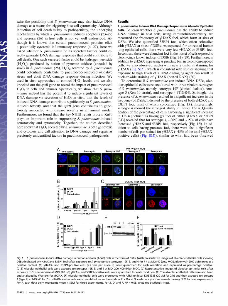
ResultsS. pneumoniae Induces DNA Damage Responses in Alveolar EpithelialCells. To learn whether S. pneumoniae has the ability to induceDNA damage in host cells, using immunohistochemistry, wemeasured the frequency of γH2AX foci, which form at sites ofDSBs. We also quantified 53BP1 foci, which often colocalizewith γH2AX at sites of DSBs. As expected, for untreated humanlung epithelial cells, there were very few γH2AX or 53BP1 foci.In contrast, there were abundant foci in the nuclei of cells exposed tobleomycin, a known inducer of DSBs (Fig. 1A) (29). Furthermore, inaddition to γH2AX appearing as punctate foci in bleomycin-exposedcells, we also observed nuclei with nearly uniform staining forγH2AX (Fig. S1C), which is consistent with studies showing thatexposure to high levels of a DNA-damaging agent can result innuclear-wide staining of γH2AX (pan-γH2AX) (30).To determine if S. pneumoniae can induce DNA DSBs, alve-
olar epithelial cells were cocultured with three virulent serotypesof S. pneumoniae, namely, serotype 19F (clinical isolate), sero-type 3 (Xen 10 strain), and serotype 4 (TIGR4). Strikingly, thepresence of S. pneumoniae resulted in a significant increase in thefrequency of DSBs, indicated by the presence of both γH2AX and53BP1 foci, most of which colocalized (Fig. 1A). Interestingly,serotype 4 showed the strongest ability to induce DSBs. Quanti-fication of the percentage of cells harboring a significant increasein DSBs [defined as having ≥5 foci of either γH2AX or 53BP1(31)] revealed that for serotype 4, ∼30% and ∼15% of cells haveincreased γH2AX and 53BP1 foci, respectively (Fig. 1B). In ad-dition to cells having punctate foci, there were also a significantnumber of cells pan-stained for γH2AX (∼45% of the total γH2AX-positive cells) (Fig. S1D), similar to what had been observed
Fig. 1. S. pneumoniae induces DNA damage in human alveolar (A549) cells in the form of DSBs. (A) Representative images of alveolar epithelial cells showingDSBs (indicated by γH2AX and 53BP1 foci) after exposure to S. pneumoniae serotypes 19F, 3, and 4 for 7 h at MOI 40 (LowMOI). Bleomycin (100 μM) serves as apositive control. (B) γH2AX- and 53BP1-positive cells (≥5 foci per nucleus) were quantified for each condition and expressed as percentage positive.(C–E) Alveolar epithelial cells were exposed to serotypes 19F, 3, and 4 at MOI 200–400 (High MOI). (C) Representative images of alveolar epithelial cells afterexposure to S. pneumoniae at MOI 300. (D) γH2AX- and 53BP1-positive cells were quantified for each condition. (E) The alveolar epithelial cells were also lysedand analyzed by Western for γH2AX. (F) Alveolar epithelial cells were pretreated with ATM inhibitor KU55933 (20 μM for 2 h) and then exposed to serotype4 (type 4) at MOI 40 for 7 h. γH2AX-positive cells were quantified for each condition. For B and D, each data point represents mean ± SEM for four experiments.For F, each data point represents mean ± SEM for three experiments. For B, D, and F, *P < 0.05, unpaired Student’s t test.
E3422 | www.pnas.org/cgi/doi/10.1073/pnas.1424144112 Rai et al.
following exposure to bleomycin. Unlike cells showing clear re-pair foci, where γH2AX and 53BP1 mostly colocalize, 53BP1 didnot stain in γH2AX pan-stained cells, which is consistent withprevious observations (30, 32, 33). For serotypes 19F and 3, therewas a greater frequency of γH2AX-positive cells compared withuninfected cells, although the ability of these serotypes to induceDSB foci was clearly reduced compared with serotype 4 (Fig.1B). To determine if observations are specific to the A549 celltype, we performed studies of lung adenoma cells (LA-4). ForLA-4 cells, we similarly observed that S. pneumoniae inducesDSBs (Fig. S2). These data, as well as analogous results in vivo(see below), indicate that the results of these studies are notspecific to one cell line. Finally, we found that induction of DNAdamage depends on the multiplicity of infection (MOI). At lowerMOI of 3–5, there was no significant increase in γH2AX or53BP1 foci for any of the strains (Fig. S1A).To further explore the potential for S. pneumoniae to induce
DNA damage, alveolar epithelial cells were exposed to a higherconcentration of S. pneumoniae. Given that up to ∼108 CFU/mLof S. pneumoniae have been reported to be present in infectedhuman lungs (34), alveolar epithelial cells are likely to be ex-posed to high levels of S. pneumoniae during acute infections atfocal lung regions. Therefore, we used MOI 200–400 for in-fection of all three serotypes. We found that all three serotypesinduced DNA damage in more than 20% of cells, with serotype 4inducing DNA damage in over 50% of the cells (Fig. 1D). Incontrast to the experiments at MOI of 30–50, most cells thatwere positive for γH2AX were pan-stained (Fig. 1C), suggestingthat, at higher MOI, elevated levels of DNA damage were in-duced (30). As an alternative approach, we also analyzed thelevels of H2AX phosphorylation by Western. Consistent withimmunofluorescence analysis, we observed significantly increasedγH2AX protein levels in lysates of epithelial cells exposed to allthree S. pneumoniae serotypes, with the highest levels being as-sociated with serotype 4 (Fig. 1E). To further explore the possi-bility that H2AX phosphorylation was induced as a response toDSBs, we targeted the canonical DNA damage response pathwaycentrally regulated by ATM kinase (35). When cells were pre-treated with specific ATM kinase inhibitor (KU55933) and thenexposed to serotype 4, the frequency of cells with γH2AX staining(both punctate and pan) decreased significantly with respect tomock-treated cells (Fig. 1F), indicating activation of the ATMpathway in response to S. pneumoniae. Taken together, these re-sults demonstrate that S. pneumoniae is able to induce DSBs inhuman alveolar epithelial cells, that DNA damage responses de-pend in part on ATM activity, and that the potency of S. pneumoniae-induced DSBs is serotype-dependent.
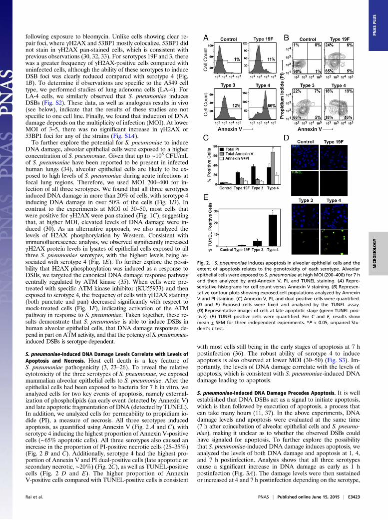
S. pneumoniae-Induced DNA Damage Levels Correlate with Levels ofApoptosis and Necrosis. Host cell death is a key feature ofS. pneumoniae pathogenicity (3, 23–26). To reveal the relativecytotoxicity of the three serotypes of S. pneumoniae, we exposedmammalian alveolar epithelial cells to S. pneumoniae. After theepithelial cells had been exposed to bacteria for 7 h in vitro, weanalyzed cells for two key events of apoptosis, namely external-ization of phospholipids (an early event detected by Annexin V)and late apoptotic fragmentation of DNA (detected by TUNEL).In addition, we analyzed cells for permeability to propidium io-dide (PI), a measure of necrosis. All three serotypes inducedapoptosis, as quantified using Annexin V (Fig. 2 A and C), withserotype 4 inducing the highest proportion of Annexin V-positivecells (∼65% apoptotic cells). All three serotypes also caused anincrease in the proportion of PI-positive necrotic cells (25–35%)(Fig. 2 B and C). Additionally, serotype 4 had the highest pro-portion of Annexin V and PI dual-positive cells (late apoptotic orsecondary necrotic, ∼20%) (Fig. 2C), as well as TUNEL-positivecells (Fig. 2 D and E). The higher proportion of AnnexinV-positive cells compared with TUNEL-positive cells is consistent
with most cells still being in the early stages of apoptosis at 7 hpostinfection (36). The robust ability of serotype 4 to induceapoptosis is also observed at lower MOI (30–50) (Fig. S3). Im-portantly, the levels of DNA damage correlate with the levels ofapoptosis, which is consistent with S. pneumoniae-induced DNAdamage leading to apoptosis.
S. pneumoniae-Induced DNA Damage Precedes Apoptosis. It is wellestablished that DNA DSBs act as a signal to initiate apoptosis,which is then followed by execution of apoptosis, a process thatcan take many hours (11, 37). In the above experiments, DNAdamage levels and apoptosis were evaluated at the same time(7 h after coincubation of alveolar epithelial cells and S. pneumo-niae), making it unclear as to whether the observed DSBs couldhave signaled for apoptosis. To further explore the possibilitythat S. pneumoniae-induced DNA damage induces apoptosis, weanalyzed the levels of both DNA damage and apoptosis at 1, 4,and 7 h postinfection. Analysis shows that all three serotypescause a significant increase in DNA damage as early as 1 hpostinfection (Fig. 3A). The damage levels were then sustainedor increased at 4 and 7 h postinfection depending on the serotype,
Fig. 2. S. pneumoniae induces apoptosis in alveolar epithelial cells and theextent of apoptosis relates to the genotoxicity of each serotype. Alveolarepithelial cells were exposed to S. pneumoniae at high MOI (200–400) for 7 hand then analyzed by anti-Annexin V, PI, and TUNEL staining. (A) Repre-sentative histograms for cell count versus Annexin V staining. (B) Represen-tative contour plots showing exposed cell populations analyzed by AnnexinV and PI staining. (C) Annexin V, PI, and dual-positive cells were quantified.(D and E) Exposed cells were fixed and analyzed by the TUNEL assay.(D) Representative images of cells at late apoptotic stage (green TUNEL posi-tive). (E) TUNEL-positive cells were quantified. For C and E, results showmean ± SEM for three independent experiments. *P < 0.05, unpaired Stu-dent’s t test.
Rai et al. PNAS | Published online June 15, 2015 | E3423
MICRO
BIOLO
GY
PNASPL
US
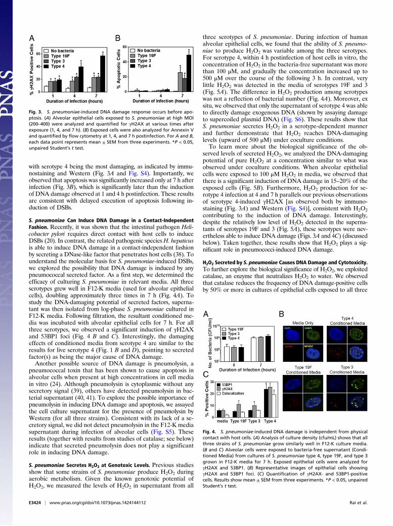
with serotype 4 being the most damaging, as indicated by immu-nostaining and Western (Fig. 3A and Fig. S4). Importantly, weobserved that apoptosis was significantly increased only at 7 h afterinfection (Fig. 3B), which is significantly later than the inductionof DNA damage observed at 1 and 4 h postinfection. These resultsare consistent with delayed execution of apoptosis following in-duction of DSBs.
S. pneumoniae Can Induce DNA Damage in a Contact-IndependentFashion. Recently, it was shown that the intestinal pathogen Heli-cobacter pylori requires direct contact with host cells to induceDSBs (20). In contrast, the related pathogenic species H. hepaticusis able to induce DNA damage in a contact-independent fashionby secreting a DNase-like factor that penetrates host cells (38). Tounderstand the molecular basis for S. pneumoniae-induced DSBs,we explored the possibility that DNA damage is induced by anypneumococcal secreted factor. As a first step, we determined theefficacy of culturing S. pneumoniae in relevant media. All threeserotypes grew well in F12-K media (used for alveolar epithelialcells), doubling approximately three times in 7 h (Fig. 4A). Tostudy the DNA-damaging potential of secreted factors, superna-tant was then isolated from log-phase S. pneumoniae cultured inF12-K media. Following filtration, the resultant conditioned me-dia was incubated with alveolar epithelial cells for 7 h. For allthree serotypes, we observed a significant induction of γH2AXand 53BP1 foci (Fig. 4 B and C). Interestingly, the damagingeffects of conditioned media from serotype 4 are similar to theresults for live serotype 4 (Fig. 1 B and D), pointing to secretedfactor(s) as being the major cause of DNA damage.Another possible source of DNA damage is pneumolysin, a
pneumococcal toxin that has been shown to cause apoptosis inalveolar cells when present at high concentrations in cell mediain vitro (24). Although pneumolysin is cytoplasmic without anysecretory signal (39), others have detected pneumolysin in bac-terial supernatant (40, 41). To explore the possible importance ofpneumolysin in inducing DNA damage and apoptosis, we assayedthe cell culture supernatant for the presence of pneumolysin byWestern (for all three strains). Consistent with its lack of a se-cretory signal, we did not detect pneumolysin in the F12-K mediasupernatant during infection of alveolar cells (Fig. S5). Theseresults (together with results from studies of catalase; see below)indicate that secreted pneumolysin does not play a significantrole in inducing DNA damage.
S. pneumoniae Secretes H2O2 at Genotoxic Levels. Previous studiesshow that some strains of S. pneumoniae produce H2O2 duringaerobic metabolism. Given the known genotoxic potential ofH2O2, we measured the levels of H2O2 in supernatant from all
three serotypes of S. pneumoniae. During infection of humanalveolar epithelial cells, we found that the ability of S. pneumo-niae to produce H2O2 was variable among the three serotypes.For serotype 4, within 4 h postinfection of host cells in vitro, theconcentration of H2O2 in the bacteria-free supernatant was morethan 100 μM, and gradually the concentration increased up to500 μM over the course of the following 3 h. In contrast, verylittle H2O2 was detected in the media of serotypes 19F and 3(Fig. 5A). The difference in H2O2 production among serotypeswas not a reflection of bacterial number (Fig. 4A). Moreover, exsitu, we observed that only the supernatant of serotype 4 was ableto directly damage exogenous DNA (shown by assaying damageto supercoiled plasmid DNA) (Fig. S6). These results show thatS. pneumoniae secretes H2O2 in a serotype-dependent mannerand further demonstrate that H2O2 reaches DNA-damaginglevels (upward of 500 μM) under coculture conditions.To learn more about the biological significance of the ob-
served levels of secreted H2O2, we analyzed the DNA-damagingpotential of pure H2O2 at a concentration similar to what wasobserved under coculture conditions. When alveolar epithelialcells were exposed to 100 μM H2O2 in media, we observed thatthere is a significant induction of DNA damage in 15–20% of theexposed cells (Fig. 5B). Furthermore, H2O2 production for se-rotype 4 infection at 4 and 7 h parallels our previous observationsof serotype 4-induced γH2AX [as observed both by immuno-staining (Fig. 3A) and Western (Fig. S4)], consistent with H2O2contributing to the induction of DNA damage. Interestingly,despite the relatively low level of H2O2 detected in the superna-tants of serotypes 19F and 3 (Fig. 5A), these serotypes were nev-ertheless able to induce DNA damage (Figs. 3A and 4C) (discussedbelow). Taken together, these results show that H2O2 plays a sig-nificant role in pneumococci-induced DNA damage.
H2O2 Secreted by S. pneumoniae Causes DNA Damage and Cytotoxicity.To further explore the biological significance of H2O2, we exploitedcatalase, an enzyme that neutralizes H2O2 to water. We observedthat catalase reduces the frequency of DNA damage-positive cellsby 50% or more in cultures of epithelial cells exposed to all three
Fig. 3. S. pneumoniae-induced DNA damage response occurs before apo-ptosis. (A) Alveolar epithelial cells exposed to S. pneumoniae at high MOI(200–400) were analyzed and quantified for γH2AX at various times afterexposure (1, 4, and 7 h). (B) Exposed cells were also analyzed for Annexin Vand quantified by flow cytometry at 1, 4, and 7 h postinfection. For A and B,each data point represents mean ± SEM from three experiments. *P < 0.05,unpaired Student’s t test.
Fig. 4. S. pneumoniae-induced DNA damage is independent from physicalcontact with host cells. (A) Analysis of culture density (cfu/mL) shows that allthree strains of S. pneumoniae grow similarly well in F12-K culture media.(B and C) Alveolar cells were exposed to bacteria-free supernatant (Condi-tioned Media) from cultures of S. pneumoniae type 4, type 19F, and type 3grown in F12-K media for 7 h. Exposed epithelial cells were analyzed forγH2AX and 53BP1. (B) Representative images of epithelial cells showingγH2AX and 53BP1 foci. (C) Quantification of γH2AX- and 53BP1-positivecells. Results show mean ± SEM from three experiments. *P < 0.05, unpairedStudent’s t test.
E3424 | www.pnas.org/cgi/doi/10.1073/pnas.1424144112 Rai et al.
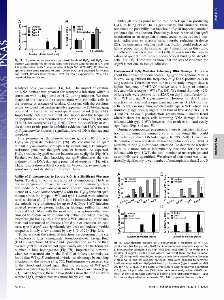
serotypes of S. pneumoniae (Fig. 6A). The impact of catalaseon DNA damage was greatest for serotype 4 infection, which isconsistent with its high level of H2O2 during infection. We thenincubated the bacteria-free supernatant with epithelial cells inthe presence or absence of catalase. Consistent with the cocultureresults, we found that catalase greatly suppresses the DNA-damagingpotential of bacteria-free serotype 4 supernatant (Fig. S7A).Importantly, catalase treatment also suppressed the frequencyof apoptotic cells as measured by Annexin V assay (Fig. 6B) andTUNEL for serotype 4 (Fig. S7B). Given the specificity of cat-alase, these results provide definitive evidence that H2O2 secretedby S. pneumoniae induces a significant level of DNA damage andapoptosis.In S. pneumoniae, the pyruvate oxidase gene (spxB) produces
H2O2 via pyruvate metabolism (28). We constructed an spxBmutant S. pneumoniae serotype 4 by introducing a kanamycin-resistance gene into the spxB gene of bacteria. As expected,H2O2 produced by spxB mutant bacteria was negligible (Fig. 6C).Further, we found that knocking out spxB eliminates the vastmajority of the DNA-damaging potential of serotype 4 (Fig. 6D).These results show a direct correlation between S. pneumoniae’sgenotoxicity and its ability to produce H2O2.
Ability of S. pneumoniae to Secrete H2O2 Is a Significant VirulenceFactor. To determine the relevance of pneumococcal H2O2 indisease progression in vivo, we used an acute pulmonary infec-tion model of S. pneumoniae in mice, and we compared the vir-ulence of S. pneumoniae serotype 4 with the H2O2-deficient spxBmutant strain. Both type 4 WT and type 4 ΔspxB were adminis-tered at similar cfu (2–3 × 10⁷ cfu) via the intratracheal route, andthe animals were monitored for up to 3 d. Type 4 WT infectioninduced severe symptoms, including lethargy, ruffled fur, andhunched back. Mice with the most severe symptoms either suc-cumbed to disease or were humanely euthanized when reachingexcess weight loss (≥20%). For type 4 WT, almost all of the ani-mals had succumbed to disease after day 2 of infection. In con-trast, type 4 ΔspxB was significantly less toxic and induced morbidsymptoms in only a few animals by day 3 (3 of 24) (Fig. 7A).To learn about the extent of infection, we determined the cfu
of bacteria in lung homogenate, broncho-alveolar lavage fluid(BALF), and blood. At days 2 and 3 postinfection, we found that,overall, spxB mutation did not significantly alter the bacterial cellnumber in lung homogenate (Fig. 7B). Importantly, however,when we measured cfu in the BALF of infected animals, wefound that WT spxB conferred a virulence advantage by enablinginvasion into the airways (Fig. 7C). Furthermore, we assessed cfuin the blood and found again that the presence of WT spxBconfers an advantage for invasion into the blood circulation (Fig.7D). Taken together, these in vivo studies show that the ability tosecrete H2O2 renders bacteria more highly virulent.
Although results point to the role of WT spxB in producingH2O2 as being critical to its genotoxicity and virulence, thereremained the possibility that knockout of spxB impacted anothervirulence factor: adhesion. Previously, it was reported that spxBinactivation in an acapsular pneumococcal strain reduced bac-terial adherence to alveolar cells, thereby reducing virulence(28). To determine whether spxB inactivation could reduce ad-hesive properties of the capsular type 4 strain used in this study,an adhesion assay was performed (28). It was found that inacti-vation of spxB did not reduce pneumococcal binding to alveolarcells (Fig. S8). These results show that the loss of virulence forΔspxB is not due to loss of adhesion.
Pneumococcal H2O2 Mediates Pulmonary DNA Damage. To learnabout the impact of pneumococcal H2O2 on the genome of cellsin vivo, we quantified the frequency of γH2AX-positive cells inlung sections. Consistent with our in vitro study, images show ahigher frequency of γH2AX-positive cells in lungs of animalsinfected with serotype 4 WT (Fig. 8A). We found that only ∼2%of lung cells were positive for γH2AX on day 1 postinfection forboth WT and ΔspxB S. pneumoniae. However, on day 2 post-infection, we observed a significant increase in γH2AX-positivecells (> 8%) in mice lung infected with type 4 WT, which wasstatistically significantly higher than that of type 4 ΔspxB (Fig. 8A and B). At day 3 postinfection, results show a similar trendwherein there are more cells harboring DNA damage in miceinfected with type 4 WT; however, this result is not statisticallysignificant (Fig. 8 A and B).During pneumococcal pneumonia, there is persistent infiltra-
tion of inflammatory immune cells in the lungs that couldthemselves produce DNA-damaging RONS (6–8). Hence, in-flammation-driven collateral damage to pulmonary cell DNA isplausible during S. pneumoniae infection. To determine whetherthere is a more robust inflammatory response for the miceinfected with type 4 WT, the total number of macrophages andneutrophils were quantified. We observed that there was a sta-tistically significantly lower number of neutrophils at days 2 and 3
Fig. 5. S. pneumoniae produces genotoxic levels of H2O2. (A) H2O2 pro-duction was quantified in the bacteria-free culture supernatant at 1, 4, and7 h postinfection with S. pneumoniae at high MOI (200–400). (B) Alveolarepithelial cells were exposed to pure 100 μM H2O2 and analyzed for γH2AXand 53BP1. Results show mean ± SEM for three experiments. *P < 0.05,unpaired Student’s t test.
Fig. 6. DNA damage induced by S. pneumoniae is mediated by its H2O2
production. (A) Analysis of γH2AX foci in alveolar epithelial cells exposed toS. pneumoniae serotype 4 at high MOI (200–400) with (+) or without (−)catalase (1 mg/mL). Cells are considered positive if there are five or morefoci. (B) Using similar conditions, apoptotic cells were quantified via AnnexinV staining. (C and D) Alveolar epithelial cells were exposed to serotype4 wild-type (type 4) and H2O2-deficient ΔspxB mutant (type 4 ΔspxB) at MOI300 for 7 h. (C) H2O2 in the bacteria-free culture supernatant was quantifiedat 1, 4, and 7 h postinfection. (D) Infected cells were analyzed for γH2AX foci.For A–D, control indicates absence of bacteria, and results show mean ± SEMfor three independent experiments. *P < 0.05, unpaired Student’s t test.
Rai et al. PNAS | Published online June 15, 2015 | E3425
MICRO
BIOLO
GY
PNASPL
US
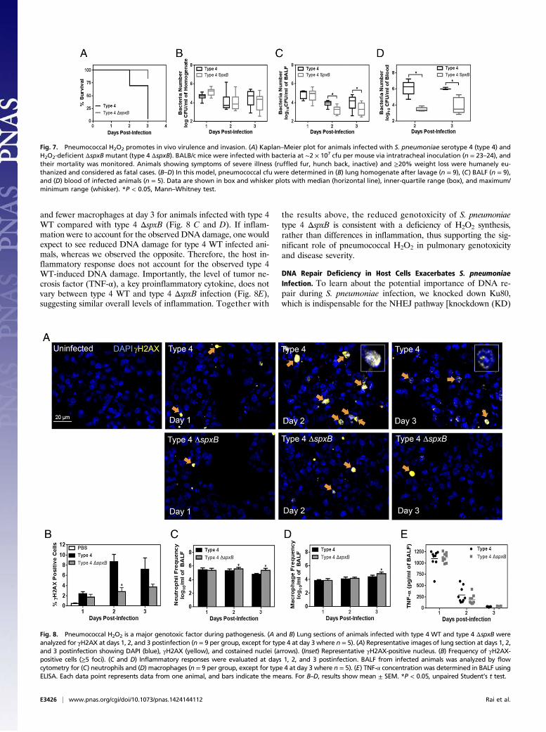
and fewer macrophages at day 3 for animals infected with type 4WT compared with type 4 ΔspxB (Fig. 8 C and D). If inflam-mation were to account for the observed DNA damage, one wouldexpect to see reduced DNA damage for type 4 WT infected ani-mals, whereas we observed the opposite. Therefore, the host in-flammatory response does not account for the observed type 4WT-induced DNA damage. Importantly, the level of tumor ne-crosis factor (TNF-α), a key proinflammatory cytokine, does notvary between type 4 WT and type 4 ΔspxB infection (Fig. 8E),suggesting similar overall levels of inflammation. Together with
the results above, the reduced genotoxicity of S. pneumoniaetype 4 ΔspxB is consistent with a deficiency of H2O2 synthesis,rather than differences in inflammation, thus supporting the sig-nificant role of pneumococcal H2O2 in pulmonary genotoxicityand disease severity.
DNA Repair Deficiency in Host Cells Exacerbates S. pneumoniaeInfection. To learn about the potential importance of DNA re-pair during S. pneumoniae infection, we knocked down Ku80,which is indispensable for the NHEJ pathway [knockdown (KD)
Fig. 7. Pneumococcal H2O2 promotes in vivo virulence and invasion. (A) Kaplan–Meier plot for animals infected with S. pneumoniae serotype 4 (type 4) andH2O2-deficient ΔspxB mutant (type 4 ΔspxB). BALB/c mice were infected with bacteria at ∼2 × 10⁷ cfu per mouse via intratracheal inoculation (n = 23–24), andtheir mortality was monitored. Animals showing symptoms of severe illness (ruffled fur, hunch back, inactive) and ≥20% weight loss were humanely eu-thanized and considered as fatal cases. (B–D) In this model, pneumococcal cfu were determined in (B) lung homogenate after lavage (n = 9), (C) BALF (n = 9),and (D) blood of infected animals (n = 5). Data are shown in box and whisker plots with median (horizontal line), inner-quartile range (box), and maximum/minimum range (whisker). *P < 0.05, Mann–Whitney test.
Fig. 8. Pneumococcal H2O2 is a major genotoxic factor during pathogenesis. (A and B) Lung sections of animals infected with type 4 WT and type 4 ΔspxB wereanalyzed for γH2AX at days 1, 2, and 3 postinfection (n = 9 per group, except for type 4 at day 3 where n = 5). (A) Representative images of lung section at days 1, 2,and 3 postinfection showing DAPI (blue), γH2AX (yellow), and costained nuclei (arrows). (Inset) Representative γH2AX-positive nucleus. (B) Frequency of γH2AX-positive cells (≥5 foci). (C and D) Inflammatory responses were evaluated at days 1, 2, and 3 postinfection. BALF from infected animals was analyzed by flowcytometry for (C) neutrophils and (D) macrophages (n = 9 per group, except for type 4 at day 3 where n = 5). (E) TNF-α concentration was determined in BALF usingELISA. Each data point represents data from one animal, and bars indicate the means. For B–D, results show mean ± SEM. *P < 0.05, unpaired Student’s t test.
E3426 | www.pnas.org/cgi/doi/10.1073/pnas.1424144112 Rai et al.
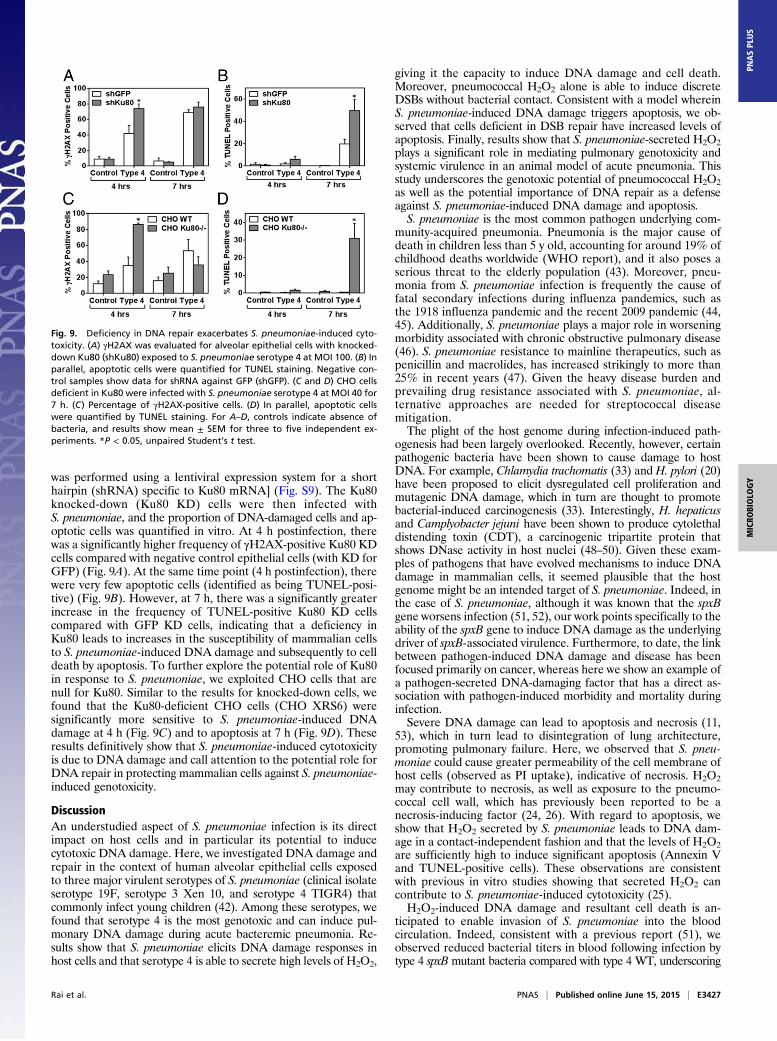
was performed using a lentiviral expression system for a shorthairpin (shRNA) specific to Ku80 mRNA] (Fig. S9). The Ku80knocked-down (Ku80 KD) cells were then infected withS. pneumoniae, and the proportion of DNA-damaged cells and ap-optotic cells was quantified in vitro. At 4 h postinfection, therewas a significantly higher frequency of γH2AX-positive Ku80 KDcells compared with negative control epithelial cells (with KD forGFP) (Fig. 9A). At the same time point (4 h postinfection), therewere very few apoptotic cells (identified as being TUNEL-posi-tive) (Fig. 9B). However, at 7 h, there was a significantly greaterincrease in the frequency of TUNEL-positive Ku80 KD cellscompared with GFP KD cells, indicating that a deficiency inKu80 leads to increases in the susceptibility of mammalian cellsto S. pneumoniae-induced DNA damage and subsequently to celldeath by apoptosis. To further explore the potential role of Ku80in response to S. pneumoniae, we exploited CHO cells that arenull for Ku80. Similar to the results for knocked-down cells, wefound that the Ku80-deficient CHO cells (CHO XRS6) weresignificantly more sensitive to S. pneumoniae-induced DNAdamage at 4 h (Fig. 9C) and to apoptosis at 7 h (Fig. 9D). Theseresults definitively show that S. pneumoniae-induced cytotoxicityis due to DNA damage and call attention to the potential role forDNA repair in protecting mammalian cells against S. pneumoniae-induced genotoxicity.
DiscussionAn understudied aspect of S. pneumoniae infection is its directimpact on host cells and in particular its potential to inducecytotoxic DNA damage. Here, we investigated DNA damage andrepair in the context of human alveolar epithelial cells exposedto three major virulent serotypes of S. pneumoniae (clinical isolateserotype 19F, serotype 3 Xen 10, and serotype 4 TIGR4) thatcommonly infect young children (42). Among these serotypes, wefound that serotype 4 is the most genotoxic and can induce pul-monary DNA damage during acute bacteremic pneumonia. Re-sults show that S. pneumoniae elicits DNA damage responses inhost cells and that serotype 4 is able to secrete high levels of H2O2,
giving it the capacity to induce DNA damage and cell death.Moreover, pneumococcal H2O2 alone is able to induce discreteDSBs without bacterial contact. Consistent with a model whereinS. pneumoniae-induced DNA damage triggers apoptosis, we ob-served that cells deficient in DSB repair have increased levels ofapoptosis. Finally, results show that S. pneumoniae-secreted H2O2plays a significant role in mediating pulmonary genotoxicity andsystemic virulence in an animal model of acute pneumonia. Thisstudy underscores the genotoxic potential of pneumococcal H2O2as well as the potential importance of DNA repair as a defenseagainst S. pneumoniae-induced DNA damage and apoptosis.S. pneumoniae is the most common pathogen underlying com-
munity-acquired pneumonia. Pneumonia is the major cause ofdeath in children less than 5 y old, accounting for around 19% ofchildhood deaths worldwide (WHO report), and it also poses aserious threat to the elderly population (43). Moreover, pneu-monia from S. pneumoniae infection is frequently the cause offatal secondary infections during influenza pandemics, such asthe 1918 influenza pandemic and the recent 2009 pandemic (44,45). Additionally, S. pneumoniae plays a major role in worseningmorbidity associated with chronic obstructive pulmonary disease(46). S. pneumoniae resistance to mainline therapeutics, such aspenicillin and macrolides, has increased strikingly to more than25% in recent years (47). Given the heavy disease burden andprevailing drug resistance associated with S. pneumoniae, al-ternative approaches are needed for streptococcal diseasemitigation.The plight of the host genome during infection-induced path-
ogenesis had been largely overlooked. Recently, however, certainpathogenic bacteria have been shown to cause damage to hostDNA. For example, Chlamydia trachomatis (33) and H. pylori (20)have been proposed to elicit dysregulated cell proliferation andmutagenic DNA damage, which in turn are thought to promotebacterial-induced carcinogenesis (33). Interestingly, H. hepaticusand Camplyobacter jejuni have been shown to produce cytolethaldistending toxin (CDT), a carcinogenic tripartite protein thatshows DNase activity in host nuclei (48–50). Given these exam-ples of pathogens that have evolved mechanisms to induce DNAdamage in mammalian cells, it seemed plausible that the hostgenome might be an intended target of S. pneumoniae. Indeed, inthe case of S. pneumoniae, although it was known that the spxBgene worsens infection (51, 52), our work points specifically to theability of the spxB gene to induce DNA damage as the underlyingdriver of spxB-associated virulence. Furthermore, to date, the linkbetween pathogen-induced DNA damage and disease has beenfocused primarily on cancer, whereas here we show an example ofa pathogen-secreted DNA-damaging factor that has a direct as-sociation with pathogen-induced morbidity and mortality duringinfection.Severe DNA damage can lead to apoptosis and necrosis (11,
53), which in turn lead to disintegration of lung architecture,promoting pulmonary failure. Here, we observed that S. pneu-moniae could cause greater permeability of the cell membrane ofhost cells (observed as PI uptake), indicative of necrosis. H2O2may contribute to necrosis, as well as exposure to the pneumo-coccal cell wall, which has previously been reported to be anecrosis-inducing factor (24, 26). With regard to apoptosis, weshow that H2O2 secreted by S. pneumoniae leads to DNA dam-age in a contact-independent fashion and that the levels of H2O2are sufficiently high to induce significant apoptosis (Annexin Vand TUNEL-positive cells). These observations are consistentwith previous in vitro studies showing that secreted H2O2 cancontribute to S. pneumoniae-induced cytotoxicity (25).H2O2-induced DNA damage and resultant cell death is an-
ticipated to enable invasion of S. pneumoniae into the bloodcirculation. Indeed, consistent with a previous report (51), weobserved reduced bacterial titers in blood following infection bytype 4 spxB mutant bacteria compared with type 4 WT, underscoring
Fig. 9. Deficiency in DNA repair exacerbates S. pneumoniae-induced cyto-toxicity. (A) γH2AX was evaluated for alveolar epithelial cells with knocked-down Ku80 (shKu80) exposed to S. pneumoniae serotype 4 at MOI 100. (B) Inparallel, apoptotic cells were quantified for TUNEL staining. Negative con-trol samples show data for shRNA against GFP (shGFP). (C and D) CHO cellsdeficient in Ku80 were infected with S. pneumoniae serotype 4 at MOI 40 for7 h. (C) Percentage of γH2AX-positive cells. (D) In parallel, apoptotic cellswere quantified by TUNEL staining. For A–D, controls indicate absence ofbacteria, and results show mean ± SEM for three to five independent ex-periments. *P < 0.05, unpaired Student’s t test.
Rai et al. PNAS | Published online June 15, 2015 | E3427
MICRO
BIOLO
GY
PNASPL
US
the role of spxB and its H2O2 production in development of ef-fective sepsis in our animal model. Previously, Regev-Yochay et al.demonstrated the competitive advantage of having spxB in a na-sopharyngeal colonization model (52). It has been reported thatspxB inactivation in acapsular pneumococcal serotype 2 strainreduces its adherence to alveolar epithelial cells (28). As this couldplay a role in virulence, we assayed bacterial adherence (28) andfound that inactivation of spxB did not reduce the adherence ofthe capsular serotype 4 strain (Fig. S8), indicating that a reductionin adherence does not explain the observed reduction in virulence.It is likely that the reported role of spxB in adhesion is specific toacapsular strains (54, 55).We also report an important and previously unidentified role
of the DNA repair protein Ku80 in suppressing S. pneumoniae-induced genotoxicity. This observation is consistent with theknown role of Ku80 in protecting alveolar epithelial cells (andother cell types) from gamma-irradiation–induced DSBs (56, 57).Overall, our data suggest a genotoxic model of pneumococcalpathogenesis whereby pneumococcal spxB-derived H2O2 induceshost DNA damage that overwhelms the Ku80-dependent NHEJrepair pathway, leading to cell death. Ultimately, via increasedDNA damage, the resultant cell death and tissue damage couldenable pneumococci to become more virulent and invasive.In previous studies of pathogen-induced DNA damage, DSBs
have been visualized by immunofluorescence detection of γH2AX(20–22, 33, 58). In our analysis of γH2AX foci, we found that arelatively high MOI produces sufficient H2O2 in the media to reachgenotoxic levels. At a significantly lowerMOI of 3–5, S. pneumoniaewas not able to secrete significant amounts of H2O2 (Fig. S1B) andhence did not induce any significant increase in DNA damage.However, during pulmonary infection in vivo, S. pneumoniae isknown to cause focal pneumonia (59). Consequently, it is antici-pated that there could be a significant DNA-damaging effect fromS. pneumoniae in vivo at sites of locally high MOI.Interestingly, during S. pneumoniae infection of alveolar epi-
thelial cells, we observed two patterns of γH2AX staining dependingon the MOI used. Low MOI (30–50) yielded foci of γH2AX with53BP1 in almost half of the total γH2AX-positive population,whereas the other half portrayed pan-γH2AX phosphorylationwithout any 53BP1 staining. At higher MOI (200–400), only pan-γH2AX staining without 53BP1 was observed. During infectionof animals, we observed that pan-γH2AX constituted about 30%of the total γH2AX analyzed in the lung sections (Fig. S10). Thepan-γH2AX staining has been reported to occur in human fibro-blast cells subjected to Adeno-associated virus (58, 60), Chlamydia(33), and UV and ionizing radiation (30, 61, 62). Recently, suchnuclear-wide γH2AX has been shown to occur in highly DNA-damaged cells and is mediated by ATM kinase (30). Here, wefound that most of the pan-γH2AX phosphorylation was depen-dent on ATM kinase and hence constituted part of the DNAdamage response cascade induced by S. pneumoniae.Although H2O2 clearly plays a significant role in the induction
of DNA damage and downstream responses to S. pneumoniae,we also found evidence for H2O2-independent induction of DSBs.During bacterial incubation with alveolar epithelial cells, we ob-served ∼60% γH2AX-positive cells. Interestingly, only 30% ofcells showed significant DNA damage when incubated with su-pernatant alone. It is possible that mammalian cell contact withS. pneumoniae causes DNA damage in host cells, independent ofbacterial H2O2. One possibility is that direct contact of mammaliancells with S. pneumoniae could activate surface proteins in alveolarepithelial cells and produce signals that impact oxidative status andDNA damage response pathways. Indeed, S. pneumoniae is shownto activate the cJun-NH2-terminal kinase (25) pathway, which hasthe potential to phosphorylate H2AX (63). Direct contact-inducedDNA damage is further supported by the observation that deletionof spxB (which is necessary for secretion of H2O2) in bacteria doesnot completely eliminate its ability to induce DNA damage. Thus,
although it is clear that pneumococcal H2O2 plays an importantrole in inducing DNA damage, there remain alternative mecha-nisms by which S. pneumoniae can contribute to DNA damage.To learn more about possible alternative mechanisms for
S. pneumoniae-induced DNA damage, we studied the commonpneumococcal toxin, pneumolysin. At high levels (e.g., ∼20 μg/mL),pneumolysin has been shown to induce apoptosis in alveolarcells in vitro (24). Although pneumolysin is an intracellularprotein without any signal peptide for secretion (39), it is knownto be released during bacterial lysis (64), and there are reportsof pneumolysin in culture supernatant of certain strains, evenwithout bacterial lysis (40, 41). We therefore tested for the pres-ence of extracellular pneumolysin in culture supernatants. Wewere unable to detect extracellular pneumolysin, which is consis-tent with its lack of a signal peptide for secretion. Thus, in thestudies presented here, it is unlikely that alveolar cells experiencepneumolysin at levels that are sufficient to induce DNA damage.Whether pneumolysin can induce DNA damage when the bacteriaundergo lysis, either via autolysis or via action of bactericidal an-tibiotics, is an interesting question for future studies.Given the low levels of H2O2 produced by serotypes 19F
and 3, the observation that catalase could suppress the ability ofthese strains to induce DNA damage was unexpected. However,it is important to consider the approach that was used to estimateH2O2 secretion, namely to sample the media. For strains thathave low-level production of H2O2, it may be that H2O2 is rap-idly diluted in the media. However, if S. pneumoniae settle toform a layer above the human cells, the local concentration ofH2O2 is anticipated to be far greater. Catalase would be antici-pated to counteract genotoxicity of H2O2 under these conditions.Moreover, the fact that serotypes 19F and 3 are not as cytotoxicas serotype 4 is consistent with H2O2 being a dominant mecha-nism for the induction of DNA damage and apoptosis.The rise of antibiotic-resistant bacteria calls attention to the
need for alternative strategies for mitigating disease. Further-more, although the capsular polysaccharide-based vaccines havebeen able to reduce the prevalence of vaccine-targeted invasiveserotypes in the past 2 decades (65), the serotypes not coveredby these vaccines are still prevalent and invasive especially inpatients with cardiopulmonary comorbidities or compromisedimmunity (66). Developing our understanding of the molecularprocesses that modulate the progression of pneumococcal dis-ease is therefore an important step in advancing alternative treat-ment approaches for S. pneumoniae infection. Although immune-cell–induced RONS play a role in fighting infections, theseinflammatory chemicals can also lead to collateral tissue damage.Furthermore, bacterially secreted H2O2 may exacerbate tissuedamage caused by inflammation-induced RONS. Importantly,S. pneumoniae strains that secrete H2O2 clearly must have mech-anisms to tolerate H2O2, and these mechanisms may render themresistant to H2O2 produced by immune cells. Thus, the use ofH2O2-neutralizing antioxidants, in concert with antibiotic regime,may be appropriate during severe pneumococcal pneumonia.Indeed, antioxidants have been shown to confer positive outcomeduring pneumococcal meningitis in a rat model (67). Constant se-cretion of such oxidants by serotypes colonizing the upper re-spiratory tract (URT) can potentially damage the URT epithelia,destabilizing its normal barrier function [e.g., ciliary velocity andmucus production (68)] and facilitating carriage of S. pneumoniae tobecome more invasive. Given that certain strains of S. pneumoniaeare resistant to H2O2 (69), and that H2O2 increases diseasepathogenicity, our data suggest that determining the status of thespxB gene in pneumococcal isolates could prove helpful inguiding the use of antioxidants in disease treatment.In this study, we have shown that S. pneumoniae creates high
levels of H2O2 and that the levels of H2O2 are sufficiently high toinduce DNA damage and apoptosis. We have shown that sup-pressing the levels of pneumococcal H2O2 either by treatment
E3428 | www.pnas.org/cgi/doi/10.1073/pnas.1424144112 Rai et al.
with catalase or by knocking out the gene necessary for H2O2biosynthesis suppresses S. pneumoniae-induced DNA damageand apoptosis. Furthermore, H2O2 secreted by S. pneumoniae playsa key role in pathogenesis, as shown by an acute pneumonia animalmodel. Importantly, human alveolar epithelial cells knocked downfor an essential component of the dominant DSB repair pathwayare more sensitive to S. pneumoniae-induced DNA damage andapoptosis, highlighting DNA repair as a potentially importantsusceptibility factor. In conclusion, the results of this study pointto a role for S. pneumoniae-induced DNA damage in diseasepathology and open doors to new avenues for developing ther-apeutic strategies that either suppress DNA damage or enhanceDNA repair during infection.
Materials and MethodsMouse Strains and Model. This study was carried out in strict accordance withthe National Advisory Committee for Laboratory Animal Research guidelines(Guidelines on the Care and Use of Animals for Scientific Purposes) in facilitieslicensed by the Agri-Food and Veterinary Authority of Singapore, the reg-ulatory body of the Singapore Animals and Birds Act. The protocol wasapproved by the Institutional Animal Care and Use Committee (IACUC),National University of Singapore (permit nos. IACUC 117/10 and 54/11). Fe-male BALB/c mice (7–8 wk) were purchased from InVivos Pte Ltd. and housedin an animal vivarium at the National University of Singapore. Mice wereinoculated via intratracheal dosing of S. pneumoniae at 2–3 × 10⁷ cfu in50 μL sterile PBS. BALF was drawn from the right lung using 800 μL sterilePBS, and blood was collected via cardiac puncture before harvesting thelung tissues. BALF and blood was plated onto blood agar on the same day,with appropriate dilution. The left lung was fixed in paraformaldehyde andlater embedded in paraffin. The right lung was frozen in liquid nitrogen andlater homogenized in 2 mL PBS and plated for cfu count.
Cell Culture and Bacterial Strains. S. pneumoniae serotypes and strains wereserotype 3 (Xen 10 A66.1), serotype 4 (TIGR4), and serotype 19F (clinicalisolate). Bacteria were cultured in brain heart infusion (BHI) broth (Sigma#53286), supplemented with 10% (vol/vol) heat-inactivated horse serum at37 °C. The human lung alveolar carcinoma (type II pneumocyte) A549 cellline was maintained in F12-K medium (Gibco) with 15% (vol/vol) FBS at 37 °Cwith 5% CO2. For experiments, pneumococci were at midlog phase (OD600
of 0.3–0.35), and A549 cells were at 70–80% confluency. The spxB gene wascloned into pGEMT, and the kanR gene was introduced at the HindIII site ofthe spxB gene (70). To create type 4 ΔspxB, plasmid was transformed intobacteria in the presence of 200 ng CSP-1 peptide.
Construction of Ku80 KD A549. Escherichia coli bacterial glycerol stockharboring Ku80 shRNA lentiviral constructs was purchased from Sigma(SHCLNG-NM_021141,TRCN 0000295856). Lentiviral constructs were pack-aged via cotransfection with pMD2.G and psPAX2 into 293T cells usingX-tremeGENE9 DNA transfection reagent (Roche) to produce lentiviral parti-cles. A549 cells were transduced with viral particles in the presence of 10 μg/mLPolybrene (Sigma) and were selected in 2 μg/mL puromycin (Sigma) 2 d aftertransduction. A549 cells were analyzed for Ku80 knockdown by Westernanalysis of cell lysates with an anti-Ku80 antibody (Santa Cruz).
Infection of Cells and Treatments. Stocks of S. pneumoniae were thawed andgrown in BHI medium supplemented with horse serum. Log-phase bacteriawere centrifuged at 4,000 × g for 10 min and resuspended in F12-K mediumbefore incubation with MOI 200–400 (high MOI), MOI 30–50 (low MOI), andMOI 3–5 for up to 7 h at 37 °C. Bacterial colonies were counted after platingonto soy blood agar and incubating at 37 °C for ∼24 h. Catalase (Sigma#C9322) treatment was done at 1 mg/mL in F12-K medium for 7 h.
Flow Cytometry. For flow cytometry of BALF cells, neutrophils were gatedat Gr-1+ CD11b+ population, whereas macrophages were gated at F4/80+
CD11b+ population. For apoptosis and necrosis assays, cultured cells wereincubated with Annexin V- PeCy7 (eBioscience #88–8103-74) and then withPI (2 μg/mL). Formaldehyde-fixed cells were stored in PBS.
Immunofluorescence.After incubationwith bacteria, cells were permeabilizedwith 0.2% Triton X-100 in PBS for 10min, blockedwith 3% (wt/vol) BSA in PBSfor 40 min, and washed once with PBS. Primary antibodies against γH2AX(Ser-139) (Millipore #05–636) and 53BP1 (Santa Cruz #sc-22760) were used at1:100 dilutions in PBS and incubated for 1 h at room temperature withcoverslip. For TUNEL staining, the labeling enzyme (Roche #1 1684795 910)was incubated similarly for 1 h at 37 °C. Secondary antibody (Invitrogen)labeled with either Alexa 488 or 564 were used for γH2AX and 53BP1.SlowFade (Invitrogen) mounted slides were stored at −20 °C. For staining of5-μm lung sections, antigen was retrieved using Dako retrieval buffer, thesections were blocked and incubated with anti-γH2AX antibody overnightand next day was stained with secondary antibody and mounted. All of thestained slides were examined under confocal microscope, and at least 9images (at 3 × 3 sites) were taken of each well under 60× magnification forcell culture slides, and 20 random images of each lung section was takenunder 40× magnification.
DNA Damage Quantification. Olympus FV10 2.0 viewer was used for imagingand counting in the dark room. At least 200 cells were counted. Cells werecategorized as those with (i) more than five distinct foci of γH2AX or 53BP1per nucleus regardless of colocalization, (ii) nuclei with colocalized foci ofγH2AX and 53BP1 counted as “overlap,” and (iii) pan-staining of γH2AX.Cells that showed colocalization of FITC and DAPI were counted as TUNEL-positive cells. For images from lung sections, DAPI was machine-countedusing IMARIS software, and Zeiss Zen software was used to examine andcount γH2AX-positive cells. All counting was done in a blinded fashion, andnuclei were selected by DAPI alone and subsequently analyzed for γH2AX.
Supernatant Assay. To prepare bacterial supernatant, log-phase bacteria weregrown in F12-K medium. After centrifugation, the supernatant was filtered(0.2 μm) and immediately incubated with A549 cells.
H2O2 Assay. To measure H2O2 levels, conditioned media was centrifuged,filtered (0.2 μm), and stored at −80 °C for analysis using a hydrogen peroxideassay kit according to the manufacturer’s instructions (Biovision #K265-200).
Western Analysis. Treated cells were washedwith PBS and incubated for 10minwith 200 μL of 1× lysis buffer [50 mM Tris·HCl, pH 6.8, 25mMdithiothrietol, 2%(wt/vol) SDS, 10% (vol/vol) glycerol]. The lysate was centrifuged at 17,000 × gfor 10 min at 4 °C, and the supernatant was denatured at 100 °C for 10 minand stored at −20 °C. For culture supernatants, after filtration, supernatantswere concentrated 10× using Amicon ultrafilter units (0.5 mL, 3 K) and dena-tured by heating. Protein concentration was quantified using the BioRad DCprotein assay kit, and lysates were electrophoresed in 15% SDS/PAGE. Analysiswas done using anti-γH2AX (Millipore #05–636) antibody or with anti-pneumolysinantibody and with secondary antibody conjugated with HRP (Dako) and laterdeveloped by adding Amersham ECL prime reagent (GE Life Science).
ACKNOWLEDGMENTS. We thank Prof. Andrew Camilli (Tufts University)for the gift of the TIGR4 strain and Dr. Yamada Yoshiyuki and Dr. OrsolyaKiraly for their valuable insights. This publication is made possible by theSingapore National Research Foundation and is administered by Singapore-Massachusetts Institute of Technology (MIT) Alliance for Research andTechnology.
1. McCullers JA (2006) Insights into the interaction between influenza virus and pneu-
mococcus. Clin Microbiol Rev 19(3):571–582.2. van der Poll T, Opal SM (2009) Pathogenesis, treatment, and prevention of pneu-
mococcal pneumonia. Lancet 374(9700):1543–1556.3. Kazzaz JA, et al. (2000) Differential patterns of apoptosis in resolving and non-
resolving bacterial pneumonia. Am J Respir Crit Care Med 161(6):2043–2050.4. Srivastava A, et al. (2005) The apoptotic response to pneumolysin is Toll-like receptor
4 dependent and protects against pneumococcal disease. Infect Immun 73(10):
6479–6487.5. del Mar García-Suárez M, et al. (2007) The role of pneumolysin in mediating lung
damage in a lethal pneumococcal pneumonia murine model. Respir Res 8(1):3.
6. Dallaire F, et al. (2001) Microbiological and inflammatory factors associated with the
development of pneumococcal pneumonia. J Infect Dis 184(3):292–300.7. Xu F, et al. (2008) Modulation of the inflammatory response to Streptococcus pneu-
moniae in a model of acute lung tissue infection. Am J Respir Cell Mol Biol 39(5):
522–529.8. Balamayooran G, Batra S, Fessler MB, Happel KI, Jeyaseelan S (2010) Mechanisms of
neutrophil accumulation in the lungs against bacteria. Am J Respir Cell Mol Biol 43(1):
5–16.9. Knaapen AM, Güngör N, Schins RPF, Borm PJA, Van Schooten FJ (2006) Neutrophils
and respiratory tract DNA damage and mutagenesis: A review. Mutagenesis 21(4):
225–236.
Rai et al. PNAS | Published online June 15, 2015 | E3429
MICRO
BIOLO
GY
PNASPL
US
10. Cooke MS, Evans MD, Dizdaroglu M, Lunec J (2003) Oxidative DNA damage: Mech-anisms, mutation, and disease. FASEB J 17(10):1195–1214.
11. Kaina B (2003) DNA damage-triggered apoptosis: Critical role of DNA repair, double-strand breaks, cell proliferation and signaling. Biochem Pharmacol 66(8):1547–1554.
12. Vamvakas S, Vock EH, Lutz WK (1997) On the role of DNA double-strand breaks intoxicity and carcinogenesis. Crit Rev Toxicol 27(2):155–174.
13. Ward IM, Minn K, Jorda KG, Chen J (2003) Accumulation of checkpoint protein 53BP1at DNA breaks involves its binding to phosphorylated histone H2AX. J Biol Chem278(22):19579–19582.
14. Nakamura AJ, Rao VA, Pommier Y, Bonner WM (2010) The complexity of phos-phorylated H2AX foci formation and DNA repair assembly at DNA double-strandbreaks. Cell Cycle 9(2):389–397.
15. Helleday T, Lo J, van Gent DC, Engelward BP (2007) DNA double-strand break repair:From mechanistic understanding to cancer treatment. DNA Repair (Amst) 6(7):923–935.
16. Falck J, Coates J, Jackson SP (2005) Conserved modes of recruitment of ATM, ATR andDNA-PKcs to sites of DNA damage. Nature 434(7033):605–611.
17. Cromie GA, Connelly JC, Leach DRF (2001) Recombination at double-strand breaks andDNA ends: Conserved mechanisms from phage to humans. Mol Cell 8(6):1163–1174.
18. Dominis-Kramari�c M, et al. (2011) Comparison of pulmonary inflammatory and an-tioxidant responses to intranasal live and heat-killed Streptococcus pneumoniae inmice. Inflammation 34(5):471–486.
19. Högen T, et al. (2013) Adjunctive N-acetyl-L-cysteine in treatment of murine pneu-mococcal meningitis. Antimicrob Agents Chemother 57(10):4825–4830.
20. Toller IM, et al. (2011) Carcinogenic bacterial pathogen Helicobacter pylori triggersDNA double-strand breaks and a DNA damage response in its host cells. Proc NatlAcad Sci USA 108(36):14944–14949.
21. Sun G, et al. (2008) Mycoplasma pneumoniae infection induces reactive oxygenspecies and DNA damage in A549 human lung carcinoma cells. Infect Immun 76(10):4405–4413.
22. Wu M, et al. (2011) Host DNA repair proteins in response to Pseudomonas aeruginosain lung epithelial cells and in mice. Infect Immun 79(1):75–87.
23. Ali F, et al. (2003) Streptococcus pneumoniae-associated human macrophage apo-ptosis after bacterial internalization via complement and Fcgamma receptors corre-lates with intracellular bacterial load. J Infect Dis 188(8):1119–1131.
24. Schmeck B, et al. (2004) Streptococcus pneumoniae-induced caspase 6-dependentapoptosis in lung epithelium. Infect Immun 72(9):4940–4947.
25. N’Guessan PD, et al. (2005) Streptococcus pneumoniae R6x induced p38 MAPK andJNK-mediated caspase-dependent apoptosis in human endothelial cells. ThrombHaemost 94(2):295–303.
26. Zysk G, Bejo L, Schneider-Wald BK, Nau R, Heinz H (2000) Induction of necrosis andapoptosis of neutrophil granulocytes by Streptococcus pneumoniae. Clin Exp Im-munol 122(1):61–66.
27. Colino J, Snapper CM (2003) Two distinct mechanisms for induction of dendritic cellapoptosis in response to intact Streptococcus pneumoniae. J Immunol 171(5):2354–2365.
28. Spellerberg B, et al. (1996) Pyruvate oxidase, as a determinant of virulence in Strep-tococcus pneumoniae. Mol Microbiol 19(4):803–813.
29. Chen J, Ghorai MK, Kenney G, Stubbe J (2008) Mechanistic studies on bleomycin-mediated DNA damage: Multiple binding modes can result in double-stranded DNAcleavage. Nucleic Acids Res 36(11):3781–3790.
30. Meyer B, et al. (2013) Clustered DNA damage induces pan-nuclear H2AX phosphor-ylation mediated by ATM and DNA-PK. Nucleic Acids Res 41(12):6109–6118.
31. Sharma GG, et al. (2010) MOF and histone H4 acetylation at lysine 16 are critical for DNAdamage response and double-strand break repair. Mol Cell Biol 30(14):3582–3595.
32. de Feraudy S, Revet I, Bezrookove V, Feeney L, Cleaver JE (2010) A minority of foci orpan-nuclear apoptotic staining of gammaH2AX in the S phase after UV damagecontain DNA double-strand breaks. Proc Natl Acad Sci USA 107(15):6870–6875.
33. Chumduri C, Gurumurthy RK, Zadora PK, Mi Y, Meyer TF (2013) Chlamydia infectionpromotes host DNA damage and proliferation but impairs the DNA damage re-sponse. Cell Host Microbe 13(6):746–758.
34. Piroth L, et al. (1999) Development of a new experimental model of penicillin-resistantStreptococcus pneumoniae pneumonia and amoxicillin treatment by reproducing hu-man pharmacokinetics. Antimicrob Agents Chemother 43(10):2484–2492.
35. Burma S, Chen BP, Murphy M, Kurimasa A, Chen DJ (2001) ATM phosphorylates histoneH2AX in response to DNA double-strand breaks. J Biol Chem 276(45):42462–42467.
36. Chan A, Reiter R, Wiese S, Fertig G, Gold R (1998) Plasma membrane phospholipidasymmetry precedes DNA fragmentation in different apoptotic cell models. Histo-chem Cell Biol 110(6):553–558.
37. Zhang XP, Liu F, Wang W (2011) Two-phase dynamics of p53 in the DNA damageresponse. Proc Natl Acad Sci USA 108(22):8990–8995.
38. Liyanage NP, et al. (2010) Helicobacter hepaticus cytolethal distending toxin causescell death in intestinal epithelial cells via mitochondrial apoptotic pathway. Heli-cobacter 15(2):98–107.
39. Walker JA, Allen RL, Falmagne P, Johnson MK, Boulnois GJ (1987) Molecular cloning,characterization, and complete nucleotide sequence of the gene for pneumolysin, thesulfhydryl-activated toxin of Streptococcus pneumoniae. Infect Immun 55(5):1184–1189.
40. Benton KA, Paton JC, Briles DE (1997) Differences in virulence for mice amongStreptococcus pneumoniae strains of capsular types 2, 3, 4, 5, and 6 are not attrib-utable to differences in pneumolysin production. Infect Immun 65(4):1237–1244.
41. Balachandran P, Hollingshead SK, Paton JC, Briles DE (2001) The autolytic enzymeLytA of Streptococcus pneumoniae is not responsible for releasing pneumolysin.J Bacteriol 183(10):3108–3116.
42. Jefferies JM, Macdonald E, Faust SN, Clarke SC (2011) 13-valent pneumococcal con-jugate vaccine (PCV13). Hum Vaccin 7(10):1012–1018.
43. Weycker D, Strutton D, Edelsberg J, Sato R, Jackson LA (2010) Clinical and economicburden of pneumococcal disease in older US adults. Vaccine 28(31):4955–4960.
44. Fleming-Dutra KE, et al. (2013) Effect of the 2009 influenza A(H1N1) pandemic oninvasive pneumococcal pneumonia. J Infect Dis 207(7):1135–1143.
45. Morens DM, Taubenberger JK, Fauci AS (2008) Predominant role of bacterial pneumoniaas a cause of death in pandemic influenza: Implications for pandemic influenzapreparedness. J Infect Dis 198(7):962–970.
46. Domenech A, et al. (2011) Serotypes and genotypes of Streptococcus pneumoniaecausing pneumonia and acute exacerbations in patients with chronic obstructivepulmonary disease. J Antimicrob Chemother 66(3):487–493.
47. Appelbaum PC (2002) Resistance among Streptococcus pneumoniae: Implications fordrug selection. Clin Infect Dis 34(12):1613–1620.
48. Fahrer J, et al. (2014) Cytolethal distending toxin (CDT) is a radiomimetic agent andinduces persistent levels of DNA double-strand breaks in human fibroblasts. DNARepair (Amst) 18:31–43.
49. Young VB, et al. (2004) In vitro and in vivo characterization of Helicobacter hepaticuscytolethal distending toxin mutants. Infect Immun 72(5):2521–2527.
50. Fox JG, et al. (2004) Gastroenteritis in NF-kappaB-deficient mice is produced withwild-type Camplyobacter jejuni but not with C. jejuni lacking cytolethal distendingtoxin despite persistent colonization with both strains. Infect Immun 72(2):1116–1125.
51. Orihuela CJ, Gao G, Francis KP, Yu J, Tuomanen EI (2004) Tissue-specific contributionsof pneumococcal virulence factors to pathogenesis. J Infect Dis 190(9):1661–1669.
52. Regev-Yochay G, Trzcinski K, Thompson CM, Lipsitch M, Malley R (2007) SpxB is asuicide gene of Streptococcus pneumoniae and confers a selective advantage in an invivo competitive colonization model. J Bacteriol 189(18):6532–6539.
53. Hegedus C, et al. (2008) Protein kinase C protects from DNA damage-induced necroticcell death by inhibiting poly(ADP-ribose) polymerase-1. FEBS Lett 582(12):1672–1678.
54. Talbot UM, Paton AW, Paton JC (1996) Uptake of Streptococcus pneumoniae by re-spiratory epithelial cells. Infect Immun 64(9):3772–3777.
55. Sanchez CJ, et al. (2011) Changes in capsular serotype alter the surface exposure ofpneumococcal adhesins and impact virulence. PLoS ONE 6(10):e26587.
56. Nussenzweig A, Sokol K, Burgman P, Li L, Li GC (1997) Hypersensitivity of Ku80-deficient cell lines and mice to DNA damage: The effects of ionizing radiation ongrowth, survival, and development. Proc Natl Acad Sci USA 94(25):13588–13593.
57. Yang QS, et al. (2008) ShRNA-mediated Ku80 gene silencing inhibits cell proliferationand sensitizes to gamma-radiation and mitomycin C-induced apoptosis in esophagealsquamous cell carcinoma lines. J Radiat Res (Tokyo) 49(4):399–407.
58. Schwartz RA, Carson CT, Schuberth C, Weitzman MD (2009) Adeno-associated virusreplication induces a DNA damage response coordinated by DNA-dependent proteinkinase. J Virol 83(12):6269–6278.
59. Gray BM, Converse GM, III, Dillon HC, Jr (1980) Epidemiologic studies of Streptococcuspneumoniae in infants: Acquisition, carriage, and infection during the first 24 monthsof life. J Infect Dis 142(6):923–933.
60. Fragkos M, Breuleux M, Clément N, Beard P (2008) Recombinant adeno-associatedviral vectors are deficient in provoking a DNA damage response. J Virol 82(15):7379–7387.
61. Marti TM, Hefner E, Feeney L, Natale V, Cleaver JE (2006) H2AX phosphorylationwithin the G1 phase after UV irradiation depends on nucleotide excision repair andnot DNA double-strand breaks. Proc Natl Acad Sci USA 103(26):9891–9896.
62. Stiff T, et al. (2006) ATR-dependent phosphorylation and activation of ATM in re-sponse to UV treatment or replication fork stalling. EMBO J 25(24):5775–5782.
63. Lu C, et al. (2006) Cell apoptosis: Requirement of H2AX in DNA ladder formation, butnot for the activation of caspase-3. Mol Cell 23(1):121–132.
64. Paton JC, Andrew PW, Boulnois GJ, Mitchell TJ (1993) Molecular analysis of thepathogenicity of Streptococcus pneumoniae: The role of pneumococcal proteins.Annu Rev Microbiol 47:89–115.
65. Richter SS, et al. (2013) Pneumococcal serotypes before and after introduction ofconjugate vaccines, United States, 1999–2011(1.). Emerg Infect Dis 19(7):1074–1083.
66. Luján M, et al. (2013) Effects of immunocompromise and comorbidities on pneu-mococcal serotypes causing invasive respiratory infection in adults: Implications forvaccine strategies. Clin Infect Dis 57(12):1722–1730.
67. Auer M, Pfister LA, Leppert D, Täuber MG, Leib SL (2000) Effects of clinically usedantioxidants in experimental pneumococcal meningitis. J Infect Dis 182(1):347–350.
68. Wright DT, et al. (1994) Interactions of oxygen radicals with airway epithelium. En-viron Health Perspect 102(Suppl 10):85–90.
69. Pericone CD, Park S, Imlay JA, Weiser JN (2003) Factors contributing to hydrogenperoxide resistance in Streptococcus pneumoniae include pyruvate oxidase (SpxB) andavoidance of the toxic effects of the fenton reaction. J Bacteriol 185(23):6815–6825.
70. Bättig P, Mühlemann K (2008) Influence of the spxB gene on competence inStreptococcus pneumoniae. J Bacteriol 190(4):1184–1189.
E3430 | www.pnas.org/cgi/doi/10.1073/pnas.1424144112 Rai et al.