Streptococcus pneumoniae induces expression of...
33
Streptococcus pneumoniae induces expression of the antibacterial CXC chemokine MIG/CXCL9 via MyD88-dependent signaling in a murine model of airway infection. Eliasson, Mette; Mörgelin, Matthias; Farber, Joshua M; Egesten, Arne; Albiger, Barbara Published in: Microbes and Infection DOI: 10.1016/j.micinf.2010.03.014 2010 Document Version: Peer reviewed version (aka post-print) Link to publication Citation for published version (APA): Eliasson, M., Mörgelin, M., Farber, J. M., Egesten, A., & Albiger, B. (2010). Streptococcus pneumoniae induces expression of the antibacterial CXC chemokine MIG/CXCL9 via MyD88-dependent signaling in a murine model of airway infection. Microbes and Infection, 12, 565-573. https://doi.org/10.1016/j.micinf.2010.03.014 Creative Commons License: CC BY-NC General rights Copyright and moral rights for the publications made accessible in the public portal are retained by the authors and/or other copyright owners and it is a condition of accessing publications that users recognise and abide by the legal requirements associated with these rights. • Users may download and print one copy of any publication from the public portal for the purpose of private study or research. • You may not further distribute the material or use it for any profit-making activity or commercial gain • You may freely distribute the URL identifying the publication in the public portal Take down policy If you believe that this document breaches copyright please contact us providing details, and we will remove access to the work immediately and investigate your claim. Download date: 30. Jan. 2020
Transcript of Streptococcus pneumoniae induces expression of...

LUND UNIVERSITY
PO Box 117221 00 Lund+46 46-222 00 00
Streptococcus pneumoniae induces expression of the antibacterial CXC chemokineMIG/CXCL9 via MyD88-dependent signaling in a murine model of airway infection.
Eliasson, Mette; Mörgelin, Matthias; Farber, Joshua M; Egesten, Arne; Albiger, Barbara
Published in:Microbes and Infection
DOI:10.1016/j.micinf.2010.03.014
2010
Document Version:Peer reviewed version (aka post-print)
Link to publication
Citation for published version (APA):Eliasson, M., Mörgelin, M., Farber, J. M., Egesten, A., & Albiger, B. (2010). Streptococcus pneumoniae inducesexpression of the antibacterial CXC chemokine MIG/CXCL9 via MyD88-dependent signaling in a murine modelof airway infection. Microbes and Infection, 12, 565-573. https://doi.org/10.1016/j.micinf.2010.03.014
Creative Commons License:CC BY-NC
General rightsCopyright and moral rights for the publications made accessible in the public portal are retained by the authorsand/or other copyright owners and it is a condition of accessing publications that users recognise and abide by thelegal requirements associated with these rights.
• Users may download and print one copy of any publication from the public portal for the purpose of private studyor research. • You may not further distribute the material or use it for any profit-making activity or commercial gain • You may freely distribute the URL identifying the publication in the public portalTake down policyIf you believe that this document breaches copyright please contact us providing details, and we will removeaccess to the work immediately and investigate your claim.
Download date: 30. Jan. 2020
1
Streptococcus pneumoniae induces expression of the
antibacterial CXC chemokine MIG/CXCL9 via MyD88-
dependent signaling in a murine model of airway infection
Mette Eliassona, Mathias Mörgelinb, Joshua M. Farberd, Arne Egestena, Barbara
Albigere*
Lund University and Lund University Hospital, Department of Clinical Sciences, a Section for Respiratory Medicine, and Allergology bClinical and Experimental Infection Medicine, e Dermatology and Venerology, 221 84 Lund, Sweden. d National Institute of Health, National Institute of Allergy and Infectious Diseases, Laboratory of Molecular Immunology, Bethesda, USA. *Corresponding author: Barbara Albiger, PhD Lund University and Lund University Hospital Biomedical Center, Department of Clinical Sciences Division of Dermatology and venerology Tornavägen 10 SE-221 84 Lund Sweden Tel: 00 46 (0)46 222 30 63 Fax: 00 46 (0)46 15 77 56

2
Abstract (148 words): MIG/CXCL9 belongs to the CXC family of chemokines and participates in the
regulation of leukocyte trafficking and angiogenesis. Certain chemokines, including
human MIG/CXCL9, exerts strong antibacterial activity in vitro, although the
importance of this property in vivo is unknown. In the present study, we investigated
the expression and a possible role for MIG/CXCL9 in host defence during mucosal
airway infection caused by Streptococcus pneumoniae in vivo. We found that
intranasal challenge of C57BL/6 wild-type mice with pneumococci elicited production
of high levels of MIG/CXCL9 in the lungs via the MyD88-dependent signaling
pathway. Whereas both human and murine MIG/CXCL9 showed efficient killing of
Streptococcus pneumoniae in vitro, MIG/CXCL9 knockout mice were not more
susceptible to pneumococcal infection. Our data demonstrate that, in vivo this
chemokine probably has a redundant role, acting together with other antibacterial
peptides and chemokines, in innate and adaptive host defense mechanisms against
pneumococcal infections.
Key words: Gram-positive bacteria; Streptococcus pneumoniae; Innate immunity;
Antimicrobial chemokines; Toll-Like Receptors; Epithelial cells.
Abbreviations: TLRs, Toll-like receptors; IFN, interferon; LTA, lipotechoic acid; Cfu,
colony forming units; wt, wild-type KO, knock out; NAL, nasopharyngeal lavage; p.i.,
post infection; i.n., intranasal; Human primary bronchial epithelial cells, HBEC;

3
1. Introduction
Streptococcus pneumoniae (the pneumococcus) is an encapsulated Gram-positive
bacterium with ninety-one different capsules or serotypes identified so far. This
bacterium is a common colonizer of the nasopharynx in children, but can also cause
life-threatening diseases such as pneumonia and invasive pneumococcal diseases
(i.e. sepsis and meningitis), killing more than 2 millions people in the world every
year and leaving severe sequelae in many children [1].
The Toll-Like Receptors (TLRs) play an important role in host defense against
microbial infections including pneumococci [2]. They recognize conserved bacterial
structures or also called pathogen-associated molecular patterns (PAMPs). Upon
recognition of their cognate ligands, TLRs can initiate two distinct signaling
cascades, the MyD88-dependent or the MyD88-independent, leading to either the
production of proinflammatory cytokines or the production of type I interferons (IFNs)
[2]. Pneumococci express several PAMPs such as lipoteichoic acids (LTA),
pneumolysin, and hypomethylated DNA containing CpG motifs that are ligands for
TLR2, TLR4 and TLR9 respectively [3-5]. Recent studies showed that mice deficient
in TLR2 are only marginally affected in their susceptibility to invasive pneumococcal
disease, while both TLR4 and TLR9 play important roles in controlling pneumococcal
colonization and the early clearance of bacteria from the lower respiratory tract. We
have shown that MyD88-deficiency in mice results in uncontrolled bacterial growth in
the airways leading to rapid bacterial dissemination into the bloodstream resulting in
systemic infection [4]. The increased susceptibility of MyD88-deficient mice is
illustrated by an impaired local production of proinflammatory cytokines and
chemokines [4].
Chemokines is a large family of peptides unified by their ability to exert receptor-

4
dependent regulation of leukocyte-trafficking during both health and disease [6].
They are divided into four groups, C, CC, CXC, and CX3C respectively, depending
on the arrangement of conserved cysteine-motifs in their NH2-terminal region. In
addition to their interactions with leukocytes, chemokines regulate other events
during inflammation, for example angiogenesis and angiostasis, and yet some have
potent antibacterial activity themselves [7]. The monokine induced by IFN-γ,
MIG/CXCL9 belongs to the CXC chemokine family and is expressed by epithelial
cells and granulocytes during Th1-polarized inflammation [8]. It exhibits strong
antibacterial activity in vitro against several pathogens [9, 10]. Recently, we found
MIG/CXCL9-dependent antibacterial activity against S. pyogenes at the surface of
IFN-γ stimulated pharyngeal epithelial cells in vitro [9]. This led us to investigate the
expression and a possible role for MIG/CXCL9 in vivo during mucosal airway
infection caused by Streptococcus pneumoniae.
In this study, two murine models of pneumococcal pneumonia (non-lethal resolving
and acute pneumonia) were used to investigate potential key roles for MIG/CXCL9 in
host defense functions. We show that MIG/CXCL9 is expressed in the lungs upon
pneumococcal infection in a MyD88-dependent manner. We also demonstrate that in
vitro MIG/CXCL9 has antibacterial activity against S. pneumoniae. However, we
were unable to demonstrate that MIG/CXCL9 is essential for microbial clearance and
host survival in vivo.
5
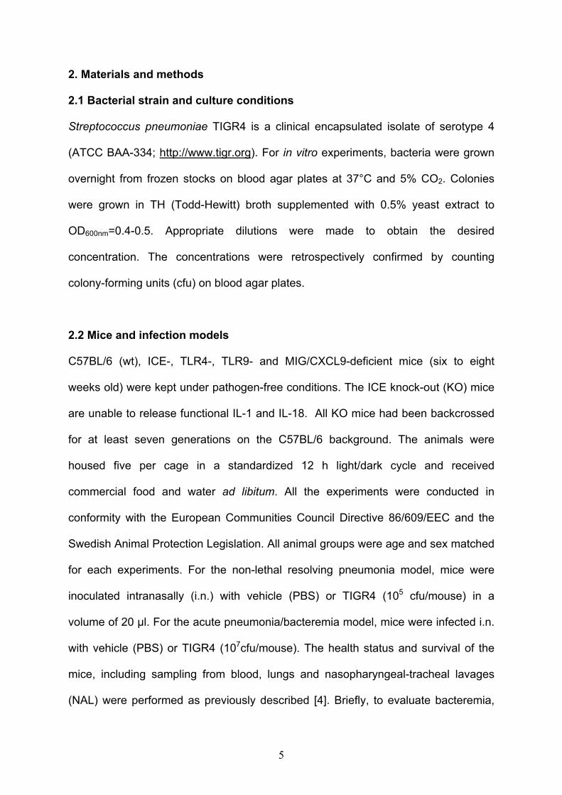
2. Materials and methods
2.1 Bacterial strain and culture conditions
Streptococcus pneumoniae TIGR4 is a clinical encapsulated isolate of serotype 4
(ATCC BAA-334; http://www.tigr.org). For in vitro experiments, bacteria were grown
overnight from frozen stocks on blood agar plates at 37°C and 5% CO2. Colonies
were grown in TH (Todd-Hewitt) broth supplemented with 0.5% yeast extract to
OD600nm=0.4-0.5. Appropriate dilutions were made to obtain the desired
concentration. The concentrations were retrospectively confirmed by counting
colony-forming units (cfu) on blood agar plates.
2.2 Mice and infection models
C57BL/6 (wt), ICE-, TLR4-, TLR9- and MIG/CXCL9-deficient mice (six to eight
weeks old) were kept under pathogen-free conditions. The ICE knock-out (KO) mice
are unable to release functional IL-1 and IL-18. All KO mice had been backcrossed
for at least seven generations on the C57BL/6 background. The animals were
housed five per cage in a standardized 12 h light/dark cycle and received
commercial food and water ad libitum. All the experiments were conducted in
conformity with the European Communities Council Directive 86/609/EEC and the
Swedish Animal Protection Legislation. All animal groups were age and sex matched
for each experiments. For the non-lethal resolving pneumonia model, mice were
inoculated intranasally (i.n.) with vehicle (PBS) or TIGR4 (105 cfu/mouse) in a
volume of 20 µl. For the acute pneumonia/bacteremia model, mice were infected i.n.
with vehicle (PBS) or TIGR4 (107cfu/mouse). The health status and survival of the
mice, including sampling from blood, lungs and nasopharyngeal-tracheal lavages
(NAL) were performed as previously described [4]. Briefly, to evaluate bacteremia,

6
blood samples (5 µl) were obtained from the tail vein at various time-points and used
for serial plating to quantify viable bacteria by cfu. The lungs were removed and
homogenized in 1 ml of PBS containing a complete cocktail protease inhibitor
(Roche Diagnostics Scandinavia) and used in serial dilutions to quantify viable
bacteria by plating on blood agar plates. The homogenates were centrifuged at 4°C
for 30 min at 5000 rpm and the supernatants were stored at -80°C for later cytokine
and chemokine analysis. NAL were performed in animals post-mortem with a 20-
gauge catheter inserted into the proximal trachea, flushing the nasopharynx through
the trachea and the nares with 200 µl PBS and used in serial plating to quantify
viable bacteria.
2.3. Proteins and antibodies
Recombinant human and murine MIG/CXCL9 were purchased from Peprotech,
Rocky Hill, NJ. The polyclonal goat antibody against MIG/CXCL9, the goat isotype
control, human and murine TNF-α as well as human and murine IFN-γ were obtained
from R&D Systems, Abingdon, UK.
2.4. In vitro killing assays
2.4.1. Bronchial epithelial killing
Human primary bronchial epithelial cells (HBEC) were obtained from Lonza
Copenhagen. HBEC were seeded in 24-well tissue culture plates and cultured to
confluence (1 x 10⁶cells/well) in MEM supplemented with 10% heat-inactivated fetal
bovine serum and penicillin/streptomycin (100 U/ml and 0.1 ng/ml respectively) at
37°C in an atmosphere containing 5% CO2 with 100% relative humidity. HBEC were
washed with MEM and then incubated in medium alone (control) or stimulated with
7
IFN-γ (10 ng/ml), TNF-α (10 ng/ml) or IFN-γ (10 ng/ml) + TNF-α (10 ng/ml) for 24 h.
Subsequently, 50 µl of bacteria (TIGR4; 2x10⁶/ml in incubation buffer; MOI=0.1) was
layered on top of the epithelial cells. This was followed by centrifugation at 300 x g
for 10 minutes to promote cell-bacteria interaction and incubation for 1 h at 37°C.
Trypsin (2.5 mg/ml in PBS) was used to detach the cells from the wells and Triton X-
100 (0.025% in PBS) was added to lyse the cells and release internalized bacteria.
Bacterial killing was determined by viable counts after plating on blood agar plates.
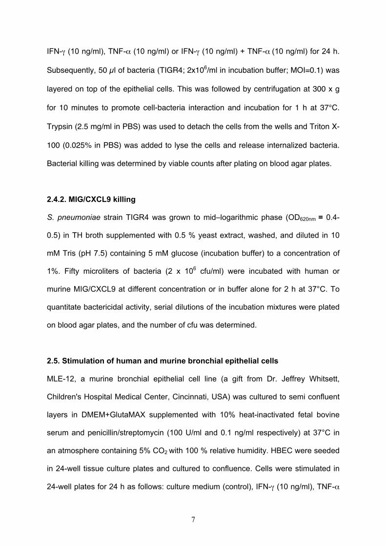
2.4.2. MIG/CXCL9 killing
S. pneumoniae strain TIGR4 was grown to mid–logarithmic phase (OD620nm = 0.4-
0.5) in TH broth supplemented with 0.5 % yeast extract, washed, and diluted in 10
mM Tris (pH 7.5) containing 5 mM glucose (incubation buffer) to a concentration of
1%. Fifty microliters of bacteria (2 x 10⁶ cfu/ml) were incubated with human or
murine MIG/CXCL9 at different concentration or in buffer alone for 2 h at 37°C. To
quantitate bactericidal activity, serial dilutions of the incubation mixtures were plated
on blood agar plates, and the number of cfu was determined.
2.5. Stimulation of human and murine bronchial epithelial cells
MLE-12, a murine bronchial epithelial cell line (a gift from Dr. Jeffrey Whitsett,
Children's Hospital Medical Center, Cincinnati, USA) was cultured to semi confluent
layers in DMEM+GlutaMAX supplemented with 10% heat-inactivated fetal bovine
serum and penicillin/streptomycin (100 U/ml and 0.1 ng/ml respectively) at 37°C in
an atmosphere containing 5% CO2 with 100 % relative humidity. HBEC were seeded
in 24-well tissue culture plates and cultured to confluence. Cells were stimulated in
24-well plates for 24 h as follows: culture medium (control), IFN-γ (10 ng/ml), TNF-α

8
(10 ng/ml) and IFN-γ (10 ng/ml) + TNF-α (10 ng/ml). The supernatants were
collected and the MIG/CXCL9 concentration was quantified using ELISA.
2.6. Determination of cytokine and chemokine levels
TNF-α, IFN-γ and MIG/CXCL9 of lung homogenates were measured using
commercial ELISA kits (R&D Systems, Abingdon, UK). According to the
manufacturer, the ELISAs are specific and do not recognize related peptides. The
detection limit for the ELISA was set to 30 pg/ml and the samples were diluted so
that values could be calculated from the linear part of the standard curves.
2.7. Modeling a predictive structure of murine and human MIG/CXCL9
2.7.1. Sequence alignment
The pairwise sequence comparison and alignment between mouse and human
CXCL9 were generated using ClustalW [11].
2.7.2 Molecular modeling Model structures of full length human and mouse MIG/CXCL9 (accession number
Q07325 and P18340, respectively) were generated as comparative homology
models using the NMR structure of truncated human MIP2-α (Gro-β) with Protein
Data Bank code 1QNK as template [12]. Residues 23-125 of human MIG/CXCL9
were aligned against the sequence of MIP2-α creating an alignment where MIP2- α
residues 39-107 matched to human MIG/CXCL9 residues 27-96 with only one gap at
threonine 40 of human MIG/CXCL9. The sequence identity was 49 %. The modeling
was performed using the program MODELLER version 9v7 [13-15]. Ten models
were generated using the automodel class and the model with the lowest DOPE
assessment score were selected. The NH2- and COOH-terminal parts of human
9
MIG/CXCL9 (residues 23-26 and 97-125, respectively) lack template and were thus
modeled in an extended conformation. Human and mouse MIG/CXCL9 are very
similar in sequence with an identity of 68 %. Hence, the model of mouse MIG/CXCL9
was generated in a similar manner. Residues 22-126 were aligned to the template
with MIP2-α residues 39-107 matching residues 26-95 of mouse MIG/CXCL9 with a
gap at threonine 39 of mouse CXCL9. Molecular representations of the models were
made using PyMOL [16].
2.8. Imaging techniques
2.8.1. Histology
Tissues were fixed in 4% paraformaldehyde, embedded in paraffin and cut to a
thickness of 4 µm. Deparaffinized tissue sections were incubated with polyclonal
goat antibodies against MIG/CXCL9 or goat isotype control antibody at a final
concentration of 5 µg/ml. The staining procedure was performed using a DAKO
TechMate 500/1000 Instrument and the manufacturer's detection kit (DAKO
ChemMate Detection Kit Peroxidase/DAB). The sections were weakly counterstained
with Mayer's hematoxylin solution.
2.8.2. Negative staining and transmission electron microscopy
Bacteria were incubated for 2 h at 37°C in incubation buffer, in the absence or
presence of MIG/CXCL9 (2.4 µg/ml). Negative staining of the bacteria was
performed with 0.75% uranyl formate. Specimens were examined in a Jeol 1200 EX
transmission electron microscope operated at 60 kV accelerating voltage.
2.9. Statistical evaluation

10
Significant differences were evaluated using the Student’s t test. The survival was
analyzed by Kaplan-Meier log rank test.

11
3. Results
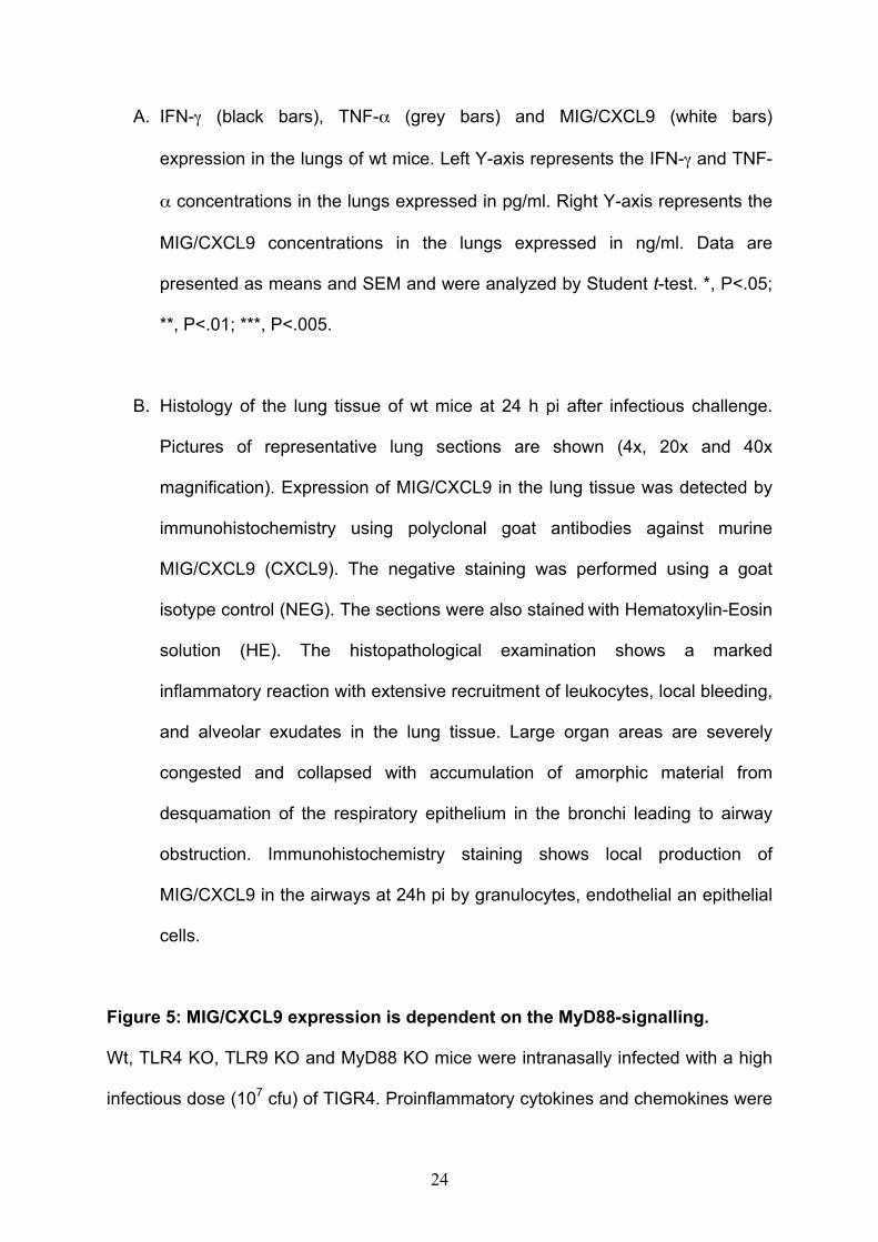
3.1 IFN-γ stimulated human bronchial epithelial cells are bactericidal
Human bronchial epithelial cells (HBEC) express high levels of MIG/CXCL9 in
response to IFN-γ stimulation [8]. We confirmed that HBEC produce high levels of
MIG/CXCL9 in response to IFN-γ, but not in response to TNF-α stimulation.
Moreover, a twofold increase in MIG/CXCL9 production was observed when IFN-γ
and TNF-α were combined, highlighting a synergistic effect of IFN-γ and TNF-α on
MIG/CXCL9 expression (Fig.1A). After stimulation with IFN-γ and TNF-α and IFN-
γ combined, but not with TNF-α alone, HBEC also exhibited antibacterial activity as
compared to non-stimulated control cells (Fig.1B). The antibacterial activity of HBEC
correlated with high levels of MIG/CXCL9 production suggesting a direct causality
between MIG/CXCL9 production and the antibacterial activity. Controls experiments
showed that neither IFN-γ nor TNF-α themselves or combined have any
antimicrobial activity per se (data not shown).
Taken together, our data demonstrate that during an inflammatory state, HBEC are
antibacterial against S. pneumoniae, possibly through the production of MIG/CXCL9.
3.2. Human and murine MIG/CXCL9 kill Streptococcus pneumoniae efficiently
in vitro
To test whether MIG/CXCL9 is antibacterial against S. pneumoniae, we performed in
vitro killing assays. Human MIG/CXCL9 displayed a strong and dose-dependent
antibacterial activity against S. pneumoniae TIGR4 strain (Fig. 2A), which can be
blocked with a polyclonal anti-MIG/CXL9 antibody (data not shown). Investigation of
bacterial morphology using electron microscopy and negative staining showed that
MIG/CXCL9 exposure caused lysis and leakage of intracellular content (Fig. 2B).
12
Sequence alignments showed that human and murine MIG/CXCL9 are highly
conserved with an overall of 67 % identity and 80 % similarity on the amino acid level
(Fig. 2C). Structural prediction revealed that both molecules have a similar three-
dimensional structure consisting of three anti-parallel β-sheets and amphipatic α-
helix with unordered tail regions (Fig. 2D). Recombinant murine MIG/CXCL9 showed
a potent antibacterial activity against TIGR4 (Fig. 2A), demonstrating that the
antibacterial function is conserved between the murine and human molecules.
3.4. Cytokine-stimulated murine bronchial epithelial cells express high levels
of MIG/CXCL9
Murine lung epithelial cells (MLE-12) produced high levels of MIG/CXCL9 in
response to IFN-γ, but not TNF-α stimulation. A 10-fold increase in MIG/CXCL9
production was observed when IFN-γ and TNF-α were combined (Fig. 3)
demonstrating the same synergistic effect of IFN-γ and TNF-α on MIG/CXCL9
expression as described above for the HBEC. Our data confirm that inflamed murine
bronchial epithelial cells are capable of producing MIG/CXCL9.
3.5. MIG/CXCL9 is produced in the lungs during pneumococcal pneumonia
Wild-type (wt) mice were infected intranasally (i.n.) with TIGR4 strain. We found that
TNF-α, IFN-γ and MIG/CXCL9 were expressed in the lungs during infection in vivo.
Both TNF-α and IFN- γ expression was maximal at 12 h post infection (pi) and
remained stable up to 24 h pi, while MIG/CXCL9 expression peaked 24 h after
inoculation; a sequential expression confirming that IFN-γ and TNF-α production
precede that of MIG/CXCL9 (Fig. 4A). Histopathological examination and
immunohistochemistry staining showed the presence of professional immune cells

13
into the lung tissue and the local production of MIG/CXCL9 in the airways during
infection (Fig. 4B). Thus, TNF-α, IFN-γ and MIG/CXCL9 are expressed in the lungs
upon pneumococcal infection, suggesting important roles of these proinflammatory
mediators in host protection.
3.6. MyD88-signalling is required for MIG/CXCL9 production in the lungs
The central adaptor MyD88 of the TLRs signaling has a key role during
pneumococcal pneumonia [3, 4]. Upon pneumococcal challenge, the local
expression of IL-12 but not IFN-γ was dependent on the MyD88 signaling pathway
(Fig. 5A and 5B). Next, we investigated the contribution of MyD88 and TLRs
signaling in pneumococci-induced MIG/CXCL9 expression. MyD88 KO mice showed
significantly lower levels of MIG/CXCL9 in the lungs as compared with wt mice (Fig.
5C). We found no significant difference in MIG/CXCL9 production in the lungs of the
single TLRs KO (Fig. 5C). Taken together, our data show that MIG/CXCL9
production in the lungs upon pneumococcal challenge is dependent mainly on the
MyD88 signaling pathway.
3.7. MIG/CXCL9 is not essential for survival in airway infection caused by
Streptococcus pneumoniae
To study the mucosal antibacterial function of MIG/CXCL9 in vivo, we infected wt
and MIG/CXCL9 KO mice i.n. with a low infectious dose of TIGR4 (105 cfu), which
leads to a non-lethal resolving pneumonia and colonization up to 15 days [3]. No
difference in survival or nasopharyngeal colonization was observed between wt and
KO mice (Fig. 6). In an acute pneumonia model, wt and MIG/CXCL9 KO mice were
infected i.n. with a high infectious dose of TIGR4 (107 cfu). In this model, the bacteria
14
can disseminate from the lungs to the bloodstream and the animal succumbs to an
overwhelming systemic infection. The survival of the mice in the two groups was
almost identical over time following initiation of the infection (Fig. 6). Bacterial loads
in the blood, in the NAL and in the lungs were equivalent in both groups (data not
shown). Thus, the absence of MIG/CXCL9 does not affect clearance of pneumococci
from the upper and lower respiratory tract or susceptibility to invasive infection in a
murine model of S. pneumoniae infection.
15
4. Discussion
In this work, we investigated the expression and the possible importance for
MIG/CXCL9 in mucosal host defense during respiratory infections caused by S.
pneumoniae.
The respiratory epithelium is continuously exposed to inhaled particles and
pathogens. It is an important source of mediators of lung inflammation such as
chemokines, which are crucial for the recruitment of leukocytes into sites of infection.
Therefore, they constitute an important first line of defense against airborne
infections [17]. Human bronchial epithelial cells (HBEC) express high levels of
MIG/CXCL9 in response to IFN-γ stimulation [8]. We showed that primary HBEC
stimulated with IFN-γ exhibit antibacterial activity against S. pneumoniae.
Furthermore, during pneumococcal infection, IFN-γ and MIG/CXCL9 are expressed
in the lungs of wt mice, suggesting potential key roles of these inflammatory
mediators in host defense.
The sequence and the predicted three-dimensional structure of murine and human
MIG/CXCL9 are highly conserved. In vitro, both human and murine MIG/CXCL9 kill
pneumococci efficiently, demonstrating that the antibacterial activity is also
conserved and may be an important feature of this molecule.
Even though MIG/CXCL9 expression is critically dependent on IFN- γ and the
interferon-dependent transcription factor, STAT1, the MIG/CXCL9 gene contains NF-
kB responsive elements and its transcription is enhanced by a synergistic interaction
between STAT1 and NF-kB [18, 19]. We could confirm in vitro that TNF-α synergizes
with IFN-γ to induce production of MIG/CXCL9 by bronchial epithelial cells. In vivo,
the local production of IL-12 but not IFN-γ is impaired in the MyD88-deficient mice,

16
which led us to hypothesize that the expression of MIG/CXCL9 might be dependent
of the MyD88-signaling. Indeed, MIG/CXCL9 expression is reduced in MyD88 KO
mice. Thus, our results in MyD88 KO mice emphasize the importance of the
enhancement of MIG/CXL9 expression via TLR/IL-1R-dependent activation of the
NF-kB pathway. MyD88 is also involved in the IL-1/IL-18 R signaling but we
excluded a possible role for IL-1/IL-18 as mediators for MIG/CXCL9 production since
ICE KO mice express high amounts of MIG/CXCL9 (data not shown). However, the
MIG/CXCL9 expression is not blunted in TLR4 and TLR9 KO mice, suggesting either
redundancy between the TLRs or the involvement of a yet unidentified non-
redundant MyD88-dependent receptor. Recently, it was demonstrated that Group A
streptococci induce cytokines and chemokines production in a MyD88-dependent
manner without the involvement of TLR2, TLR4 and TLR9 and that Group B
streptococci induce type I IFN production via TLR7-dependent, MyD88-dependent
signaling [20-22].
However, murine MIG/CXCL9 did not decrease bacterial dissemination or
colonization and MIG/CXCL9 KO mice are not more susceptible to infection than wt
animals. We do not know, whether the levels of MIG/CXCL9 found in tissue
homogenates are bactericidal, how these molecules are exposed and whether they
can exert antibacterial activity in vivo. One plausible explanation is that in vivo this
chemokine have a redundant role, acting together with other antibacterial peptides
and chemokines, in innate and adaptive host defense mechanisms counteracting
bacterial infections. There are two closely related chemokines that belong to the
CXC family, IP-10/CXCL10 and I-TAC/CXCL11 respectively. They share the ability
with MIG/CXCL9 to signal through CXC chemokine receptor 3 (CXCR3). They also
exhibit antibacterial activity in vitro and might compensate for the absence of
17
MIG/CXCL9 in vivo. In a murine model of Klebsiella pneumoniae pneumonia, IP-
10/CXCL10 but not MIG/CXCL9 promoted Type 1 immunity [23]. Infection of CXCR3
KO mice with the pneumococcal TIGR4 strain showed a higher colonization level in
the nasopharynx of CXCR3 KO as compared to wild type animals despite no
significant difference in survival (data not shown). A persistent colonization might
lead to the development of invasive disease. Another indication of redundancy is that
recent Genbank submissions revealed a single bp deletion in the coding sequence
(13th codon) for I-TAC/CXCL11, a gene close to MIG/CXCL9 and IP-10/CXCL10 on
mouse chromosome 5 of the C57BL/6 mice. The C57BL/6 I-TAC/CXCL11 sequence
is predicted to produce a truncated, non-functional protein. The GenBank sequence
of I-TAC/CXCL11 in the 129sv strain has an I-TAC/CXCL11 ORF without the
deletion. Because the MIG/CXCL9 gene was targeted in ES cells from the 129sv
strain, and because of the close linkage of MIG/CXCL9 and I-TAC/CXCL11, we have
sequenced the relevant I-TAC/CXCL11 sequences in our wt and MIG/CXCL9 KO
mice and we found out that the MIG/CXCL9 KO mice have the 129sv strain
sequence for I-TAC/CXCL11, predicted to encode a full length I-TAC/CXCL11. One
consequence might be that because MIG/CXCL9, IP-10/CXCL10 and I-TAC/CXCL11
share CXCR3, a functional I-TAC/CXCL11 in the MIG/CXCL9 KO mice
underestimates the effects of knocking out MIG/CXCL9 in experimental comparisons
with the C57BL/6 wt mice. The potential roles of IP-10/CXCL10 and I-TAC/CXCL11
in pneumococcal infections are under investigation.
MIG/CXCL9 is also a chemaoattractant for T cells and NK cells to inflammatory sites
where IFN-γ is produced. Thus, we cannot exclude a role of MIG/CXCL9 in recruiting
CD4+ T cells to the site of infection. Clearance of pneumococci is dependent upon
the rapid recruitment and presence of CD4+ T cells at the time of infection
18
suggesting a previously underestimated role for the cellular players of the adaptive
immune system in the control and clearance of this pathogen [24, 25].
In conclusion, we could identify an antimicrobial role for MIG/CXCL9 in vitro but not
in vivo, most likely explained by an important redundancy among key molecules
regulating host defense.

19
6. Acknowledgements
We would like to thank Elise Nilsson for technical assistance with the histology. BA,
AE and ME were supported by grants from the Swedish Medical Research Council,
the Swedish Heart and Lung Foundation, the Swedish Society of Medicine, the
Medical Faculty at Lund University and the Foundations of Crafoord, Bergh, Ihre,
Hedberg, Kock, Marcus & Marianne Wallenberg, Lars Hiertas, Alfred Österlund,
Längmanska and OE & Edda Johansson.

20
References
[1] K.L. O'Brien, L.J. Wolfson, J.P. Watt, E. Henkle, M. Deloria-Knoll, N. McCall, E. Lee, K. Mulholland, O.S. Levine, T. Cherian, Burden of disease caused by Streptococcus pneumoniae in children younger than 5 years: global estimates, Lancet 374 (2009) 893-902. [2] B. Albiger, S. Dahlberg, B. Henriques-Normark, S. Normark, Role of the innate immune system in host defence against bacterial infections: focus on the Toll-like receptors, J Intern Med 261 (2007) 511-528. [3] B. Albiger, S. Dahlberg, A. Sandgren, F. Wartha, K. Beiter, H. Katsuragi, S. Akira, S. Normark, B. Henriques-Normark, Toll-like receptor 9 acts at an early stage in host defence against pneumococcal infection, Cell Microbiol 9 (2007) 633-644. [4] B. Albiger, A. Sandgren, H. Katsuragi, U. Meyer-Hoffert, K. Beiter, F. Wartha, M. Hornef, S. Normark, B.H. Normark, Myeloid differentiation factor 88-dependent signalling controls bacterial growth during colonization and systemic pneumococcal disease in mice, Cell Microbiol 7 (2005) 1603-1615. [5] R. Malley, P. Henneke, S.C. Morse, M.J. Cieslewicz, M. Lipsitch, C.M. Thompson, E. Kurt-Jones, J.C. Paton, M.R. Wessels, D.T. Golenbock, Recognition of pneumolysin by Toll-like receptor 4 confers resistance to pneumococcal infection, Proc Natl Acad Sci U S A 100 (2003) 1966-1971. [6] A. Viola, A.D. Luster, Chemokines and their receptors: drug targets in immunity and inflammation, Annu Rev Pharmacol Toxicol 48 (2008) 171-197. [7] M. Eliasson, A. Egesten, Antibacterial chemokines--actors in both innate and adaptive immunity, Contrib Microbiol 15 (2008) 101-117. [8] A. Sauty, M. Dziejman, R.A. Taha, A.S. Iarossi, K. Neote, E.A. Garcia-Zepeda, Q. Hamid, A.D. Luster, The T cell-specific CXC chemokines IP-10, Mig, and I-TAC are expressed by activated human bronchial epithelial cells, J Immunol 162 (1999) 3549-3558. [9] A. Egesten, M. Eliasson, H.M. Johansson, A.I. Olin, M. Morgelin, A. Mueller, J.E. Pease, I.M. Frick, L. Bjorck, The CXC chemokine MIG/CXCL9 is important in innate immunity against Streptococcus pyogenes, J Infect Dis 195 (2007) 684-693. [10] H.M. Linge, I. Sastalla, D.P. Nitsche-Schmitz, A. Egesten, I.M. Frick, Protein FOG is a moderate inducer of MIG/CXCL9, and group G streptococci are more tolerant than group A streptococci to this chemokine's antibacterial effect, Microbiology 153 (2007) 3800-3808. [11] J.D. Thompson, D.G. Higgins, T.J. Gibson, CLUSTAL W: improving the sensitivity of progressive multiple sequence alignment through sequence weighting, position-specific gap penalties and weight matrix choice, Nucleic Acids Res 22 (1994) 4673-4680. [12] Y.Q. Qian, K.O. Johanson, P. McDevitt, Nuclear magnetic resonance solution structure of truncated human GRObeta [5-73] and its structural comparison with CXC chemokine family members GROalpha and IL-8, J Mol Biol 294 (1999) 1065-1072. [13] N. Eswar, B. John, N. Mirkovic, A. Fiser, V.A. Ilyin, U. Pieper, A.C. Stuart, M.A. Marti-Renom, M.S. Madhusudhan, B. Yerkovich, A. Sali, Tools for comparative protein structure modeling and analysis, Nucleic Acids Res 31 (2003) 3375-3380.
21
[14] N. Eswar, B. Webb, M.A. Marti-Renom, M.S. Madhusudhan, D. Eramian, M.Y. Shen, U. Pieper, A. Sali, Comparative protein structure modeling using Modeller, Curr Protoc Bioinformatics Chapter 5 (2006) Unit 5 6. [15] N. Eswar, B. Webb, M.A. Marti-Renom, M.S. Madhusudhan, D. Eramian, M.Y. Shen, U. Pieper, A. Sali, Comparative protein structure modeling using MODELLER, Curr Protoc Protein Sci Chapter 2 (2007) Unit 2 9. [16] W.L. DeLano, The PyMOL Molecular Graphics System, DeLano Scientific, San Carlos, CA. , (2002). [17] K.S. Tsai, M.H. Grayson, Pulmonary defense mechanisms against pneumonia and sepsis, Curr Opin Pulm Med 14 (2008) 260-265. [18] S. Mahalingam, G. Chaudhri, C.L. Tan, A. John, P.S. Foster, G. Karupiah, Transcription of the interferon gamma (IFN-gamma )-inducible chemokine Mig in IFN-gamma-deficient mice, J Biol Chem 276 (2001) 7568-7574. [19] Y. Ohmori, R.D. Schreiber, T.A. Hamilton, Synergy between interferon-gamma and tumor necrosis factor-alpha in transcriptional activation is mediated by cooperation between signal transducer and activator of transcription 1 and nuclear factor kappaB, J Biol Chem 272 (1997) 14899-14907. [20] N. Gratz, M. Siller, B. Schaljo, Z.A. Pirzada, I. Gattermeier, I. Vojtek, C.J. Kirschning, H. Wagner, S. Akira, E. Charpentier, P. Kovarik, Group A streptococcus activates type I interferon production and MyD88-dependent signaling without involvement of TLR2, TLR4, and TLR9, J Biol Chem 283 (2008) 19879-19887. [21] G. Mancuso, M. Gambuzza, A. Midiri, C. Biondo, S. Papasergi, S. Akira, G. Teti, C. Beninati, Bacterial recognition by TLR7 in the lysosomes of conventional dendritic cells, Nat Immunol 10 (2009) 587-594. [22] M. Liljeroos, R. Vuolteenaho, S. Rounioja, B. Henriques-Normark, M. Hallman, M. Ojaniemi, Bacterial ligand of TLR2 signals Stat activation via induction of IRF1/2 and interferon-alpha production, Cell Signal 20 (2008) 1873-1881. [23] X. Zeng, T.A. Moore, M.W. Newstead, J.C. Deng, S.L. Kunkel, A.D. Luster, T.J. Standiford, Interferon-inducible protein 10, but not monokine induced by gamma interferon, promotes protective type 1 immunity in murine Klebsiella pneumoniae pneumonia, Infect Immun 73 (2005) 8226-8236. [24] K. Trzcinski, C.M. Thompson, A. Srivastava, A. Basset, R. Malley, M. Lipsitch, Protection against nasopharyngeal colonization by Streptococcus pneumoniae is mediated by antigen-specific CD4+ T cells, Infect Immun 76 (2008) 2678-2684. [25] R. Malley, K. Trzcinski, A. Srivastava, C.M. Thompson, P.W. Anderson, M. Lipsitch, CD4+ T cells mediate antibody-independent acquired immunity to pneumococcal colonization, Proc Natl Acad Sci U S A 102 (2005) 4848-4853.

22
Figure Legends:
Figure 1: Inflamed human bronchial epithelial cells exhibit antibacterial activity
against S. pneumoniae.
A. Human primary bronchial epithelial cells (HBEC) were stimulated with TNF-α
(10 ng/ml) or IFN-γ (10 ng/ml) alone or in combination for 24 h. Supernatant
from cells stimulated with culture medium was used as a control.
Supernatants were collected and the MIG/CXCL9 concentrations were
quantified using ELISA. The data shown represent mean ±SEM. Data were
analyzed by Student t-test. *, P<.05; **, P<.01; ***, P<.005
B. Unstimulated or stimulated human primary bronchial epithelial cells (HBEC)
were infected with S. pneumoniae TIGR4 for 2 h at a MOI of 0.1 (equivalent to
1 x 10⁵ cfu). The bactericidal activity was calculated by viable counts. The
data shown represent the mean values of at least three independent
experiments
Figure 2: Human and murine MIG/CXCL9 are conserved and kill pneumococci
efficiently in vitro.
A. Bactericidal activity of human (circle) and murine MIG/CXCL9 (square)
respectively against the invasive Streptococcus pneumoniae strain (TIGR4).
Bacteria (2 x 10⁶ cfu/ml) were incubated with increasing amounts of either
human or murine MIG/CXCL9 (0.12-12 µg/ml) for 2 h at 37°C. The
bactericidal activity was calculated on the basis of the number of cfu
recovered after exposure to MIG/CXCL9 and after incubation in buffer alone
(set as 100% survival). The mean values of at least three independent
experiments are shown.
23
B. Electron micrographs of S. pneumoniae (TIGR4) incubated in buffer alone (a)
or in presence of MIG/CXCL9 (2.4 µg/ml) (b-e). Bacteria exposed to
MIG/CXCL9 show disintegration and leakage of intracellular contents. Scale
bar; 0.5 µm.
C. Pairwise sequence comparison between human and mouse MIG/CXCL9. The
alignment was generated using ClustalW.
D. Ribbon representation of theoretical models of human (cyan) and mouse
(pink) MIG/CXCL9. Both molecules contain putative anti-parallel β-sheets in
the NH2-terminal region followed by an amphipatic α-helix and an unordered
tail COOOH-terminal regions
Figure 3: Synergistic induction of MIG/CXCL9 production by TNF-α and IFN-γ
in murine bronchial epithelial cells in vitro.
Murine bronchial epithelial cells (MLE-12) were stimulated with TNF-α (10 ng/ml) or
IFN-γ (10 ng/ml) alone or in combination for 24 h. Supernatant from cells stimulated
with culture medium was used as a control. Supernatants were collected and the
MIG/CXCL9 concentrations were quantified using ELISA. The data shown represent
mean ±SEM from three separate experiments. Data were analyzed by Student t-test.
*, P<.05; **, P<.01; ***, P<.005
Figure 4: MIG/CXCL9 is expressed in the airways during pneumococcal
infection in vivo.
Wt mice were intranasally infected with TIGR4 strain. Proinflammatory cytokines and
chemokines were measured in lung homogenates. Ten mice per time point were
used. Three control mice (C) were infected with 20 µl of control medium (PBS).
24
A. IFN-γ (black bars), TNF-α (grey bars) and MIG/CXCL9 (white bars)
expression in the lungs of wt mice. Left Y-axis represents the IFN-γ and TNF-
α concentrations in the lungs expressed in pg/ml. Right Y-axis represents the
MIG/CXCL9 concentrations in the lungs expressed in ng/ml. Data are
presented as means and SEM and were analyzed by Student t-test. *, P<.05;
**, P<.01; ***, P<.005.
B. Histology of the lung tissue of wt mice at 24 h pi after infectious challenge.
Pictures of representative lung sections are shown (4x, 20x and 40x
magnification). Expression of MIG/CXCL9 in the lung tissue was detected by
immunohistochemistry using polyclonal goat antibodies against murine
MIG/CXCL9 (CXCL9). The negative staining was performed using a goat
isotype control (NEG). The sections were also stained with Hematoxylin-Eosin
solution (HE). The histopathological examination shows a marked
inflammatory reaction with extensive recruitment of leukocytes, local bleeding,
and alveolar exudates in the lung tissue. Large organ areas are severely
congested and collapsed with accumulation of amorphic material from
desquamation of the respiratory epithelium in the bronchi leading to airway
obstruction. Immunohistochemistry staining shows local production of
MIG/CXCL9 in the airways at 24h pi by granulocytes, endothelial an epithelial
cells.
Figure 5: MIG/CXCL9 expression is dependent on the MyD88-signalling.
Wt, TLR4 KO, TLR9 KO and MyD88 KO mice were intranasally infected with a high
infectious dose (107 cfu) of TIGR4. Proinflammatory cytokines and chemokines were

25
measured in lung homogenates 24 h pi. Data are presented as means and SEM
and were analyzed by Student t-test. *, P<.05; **, P<.01; ***, P<.005.
A. IL-12 (black bars) and IFN-γ (white bars) concentrations in the lung tissue of
wt (n=5) and MyD88 KO (n=5). Control mice (n=3) were infected with 20 µl of
control medium (PBS).
B. MIG/CXCL9 expression in the lung tissue of wt (n=17) and TLR4 (n=9); TLR9
(n=16); MyD88 (n=9) KO mice. Control mice (n=3) were infected with 20 µl of
control medium (PBS).
Figure 6: MIG/CXCL9 deficient mice are not more susceptible to streptococcal
infections
Airway infection caused by Streptococcus pneumoniae in MIG/CXCL9 KO mice
compared with wt mice. Survival of wt (n=10-15) and MIG/CXCL9 KO (n=10-15)
mice after low (LD) and high (HD) infectious dose. The survival was analyzed by
Kaplan-Meier log rank test.
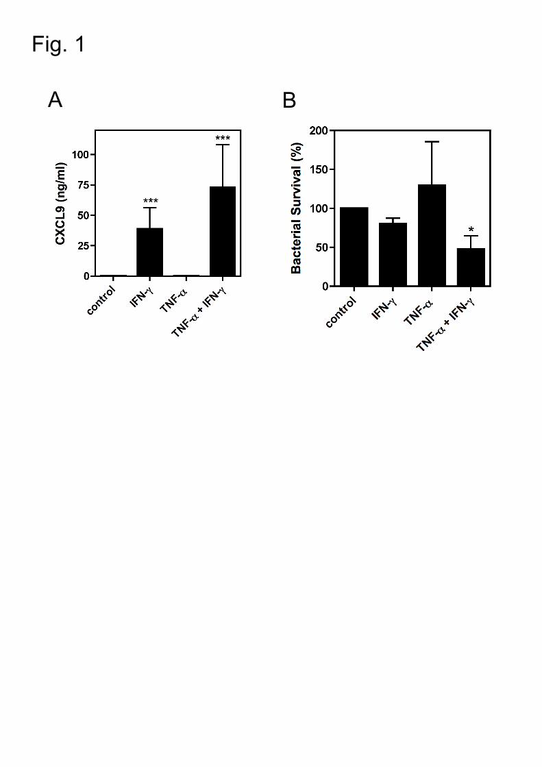
Fig. 1
A B
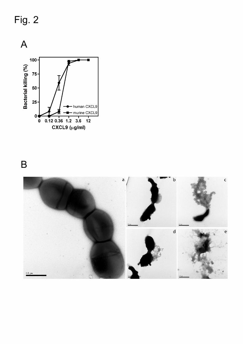
Fig. 2
A
B
C 10 20 30 40 50 60 70 ....|....| ....|....| ....|....| ....|....| ....|....| ....|....| ....|....|CXCL9_MOUSE -MKSAVLFLL GIIFLEQCGV RGTLVIRNAR CSCISTSRGT IHYKSLKDLK QFAPSPNCNK TEIIATLKNGCXCL9_HUMAN MKKSGVLFLL GIILLVLIGV QGTPVVRKGR CSCISTNQGT IHLQSLKDLK QFAPSPSCEK IEIIATLKNGClustal **.***** ***:* ** :** *:*:.* ******.:** ** :****** ******.*:* *********
80 90 100 110 120 ....|....| ....|....| ....|....| ....|....| ....|....| ....|..CXCL9_MOUSE DQTCLDPDSA NVKKLMKEWE KKINQKKKQK RGKKHQKNMK NRKPKTPQSR RRSRKTTCXCL9_HUMAN VQTCLNPDSA DVKELIKKWE KQVSQKKKQK NGKKHQK-KK VLKVRKSQ-R SRQKKTTClustal ****:**** :**:*:*:** *::.****** .****** * * :..* * *.:***
D
Murine CXCL9
Human CXCL9

Fig. 3
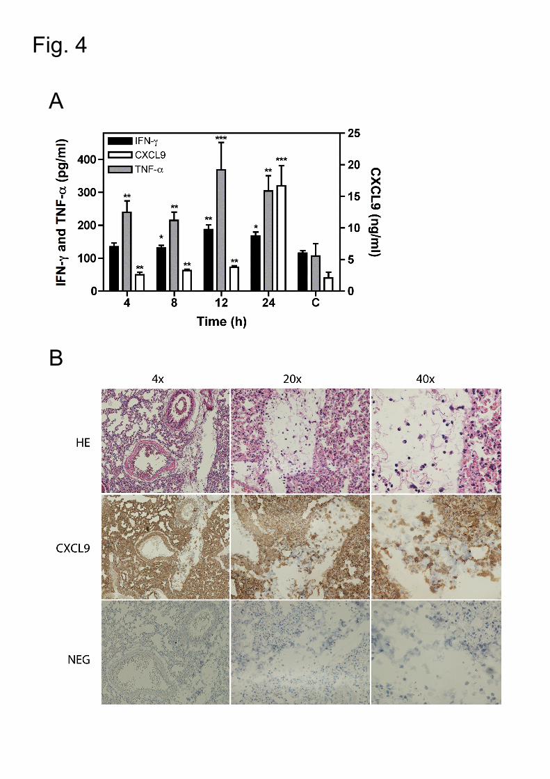
Fig. 4
A
B

Fig. 5
A B

Fig. 6