Genomes & Developmental Control Human embryonic stem cells ...
1
Maintenance of Human Pluripotent Stem Cells in
NutriStem® hPSC XF Media
Technical Manual
Version 03 - October 2016
Stem C
ells
2
NutriStem® hPSC XF
Cat# 05-100-1 NutriStem® hPSC XF, Xeno-free medium for feeder-free and feeder-dependent culture of hESC and hiPSC
Cat# 05-102-1 AF NutriStem® hPSC XF, without HSA Xeno-free medium for feeder-dependent culture of hESC and hiPSC
3
1. Introduction 4 NutriStem® hPSC XF Features 4 Precautions and Disclaimer 5
2. Media and Reagents 5 NutriStem® hPSC XF 5 Freezing Medium: CryoStem™ 6 EDTA Solution 0.5M 6 Laminin: LaminStem™ 521 6 MEF Medium 6 Collagenase IV Solution (1mg/ml) 7
3. General Methods 7 3.1 Maintenance of hPSC using NutriStem® hPSC XF medium 7 3.1.1 Observing culture under the microscope 7 3.1.2 Feeding hPSC using NutriStem® hPSC XF medium 8 3.1.3 Removing differentiated cells from hPSC culture 8 3.2 Thawing of hPSC 10 3.3 Cryopreservation of hPSC using CryoStem™ 11
4. Feeder-Free Culture Systems 13 4.1 Matrigel™-based feeder-free culture system 13 4.1.1 Coating plates with Matrigel™ 13 4.2 Passaging hPSC on Matrigel™ using NutriStem® hPSC XF medium 13 4.2.1 Enzymatic passaging of hPSC 13 4.2.2 Enzyme-free passaging of hPSC 16 4.3 Laminin-based feeder-free culture system 18 4.3.1 Coating plates with Laminin 521 18 4.3.2 Single cell passage of hPSC on Laminin 521 18
5. Feeder-Dependent Culture System 20 5.1 General Information 20 5.2 Observing culture under the microscope 20 5.3 Preparation of mitotically inactivated feeder cells (Mitomycin C-treated) 21 5.4 Preparation of feeder cell layer 21 5.5 Thawing procedure for hPSC on feeder cells 22 5.6 Passaging hPSC using NutriStem® hPSC XF medium 23 5.7 Transferring hPSC from feeders to a feeder-free culture system and vice versa 23
6. Adaptation to NutriStem® hPSC XF medium 24 6.1 Adapting hPSC to NutriStem® hPSC XF medium 24 6.1.1 Before beginning the adaptation 24 6.1.2 Points that might hinder successful adaptation 24 6.1.3 Adaptation protocol 25 6.2 Adapting hPSC to Laminin 521-based culture system using NutriStem® hPSC XF medium 26
7. References 27
Table of Contents
4
1. Introduction Traditional Human Embryonic Stem Cells (hESC) and Human Induced Pluripotent Stem Cells (hiPSC) culture methods require use of mouse or human fibroblast feeder layers, or feeder- conditioned media. These culture methods are labor-intensive, hard to scale, and it is difficult to maintain hPSC pluripotency due to undefined conditions. NutriStem® hPSC XF media were developed with a leading stem cell research group to enable maintenance and expansion of hESC and hiPSC in feeder-free culture or with feeder cells. NutriStem® hPSC XF media support the culture of undifferentiated hESC and hiPSC in serum-free conditions without any xeno-derived components on feeders such as Mouse Embryo Fibroblasts (MEF) and Human Foreskin Fibroblasts (HFF) as well as with Matrigel™ and recombinant laminin. The media contain Recombinant Human Basic Fibroblast Growth Factor (rh bFGF) and Recombinant Human Transforming Growth Factor Beta (rh TGFβ), and have been successfully tested to maintain the pluripotent nature of hESC and hiPSC. For long-term growth of hESC and hiPSC without feeder cells it is recommended to use NutriStem® hPSC XF with HSA (BI Cat# 05-100-1).Human Pluripotent Stem Cells (hPSC) must be monitored and cared for in order to maintain healthy, undifferentiated cultures. At the very least, the cultures must be fed every day by performing a complete medium change to replenish lost nutrients and keep the cultures free of unwanted differentiation factors. Although a small amount of differentiation is expected in human pluripotent stem cell cultures, the culture should be routinely cleaned up by manually removing or “picking” differentiated cells. Identifying and removing excess differentiation from hPSC cultures are essential techniques in the maintenance of a healthy population of cells.
NutriStem® hPSC XF Features
• Serum-Free Media (SFM), Xeno-Free (XF): all components are defined and xeno-free, including proteins.
• Sterile, 0.1μm membrane filtered under full aseptic conditions and practice in compliance with ISO 13408 (Aseptic Processing of Health Care Products).
• The proteins used: HSA (Human Serum Albumin), rh bFGF, rh TGFβ, human transferrin, and recombinant human insulin.
• A complete ready-to-use formulation (no additions required). • Contain L-alanyl-L-glutamine. • Do not contain antibiotics. • Enable culture of hESC and hiPSC without feeder cells. • Can be used with mouse or human feeder cells, Matrigel™, laminin, and matrix from
human cells. • Can be used with Laminin 521 (e.g., LaminStem™ 521 BI Cat# 05-753-1F) for single-cell
passage. • Intended for use in incubator with 5% CO2. • Support long-term growth of hESC and hiPSC, and maintain their ability to differentiate
without any signs of karyotype abnormalities.

5
Precautions and Disclaimer
1. For in vitro diagnostic use, and use as ancillary material in cell- and tissue-based therapies. 2. Do not use if a visible precipitate is observed in the medium. 3. Do not use NutriStem® hPSC XF and AF NutriStem® hPSC XF media beyond the expiration
date indicated on the product label.
2. Media and Reagents
Product Name Catalogue No.
NutriStem® hPSC XF 05-100-1
AF NutriStem® hPSC XF 05-102-1
Bio-Pure HSA, 10% Solution 05-720-1
CryoStem™ 05-710-1
DMEM/F12 (HAM) 1:1 01-170-1
Recombinant Trypsin-EDTA Solution 03-079-1
DPBS with Ca & Mg 03-020-1
DPBS without Ca & Mg 03-023-1
LaminStem™ 521 05-753-1
SBTI Solution (x50) 03-048-1
EDTA 0.5M Solution 01-862-1
Certified FBS Qualified for hPSC 04-002-1
0.1% Gelatin Solution 01-944-1
L-alanyl-L-glutamine, 200mM 03-022-1
Sodium Pyruvate, 100mM 03-042-1
NutriStem® hPSC XF medium (Cat# 05-100-1) Storage and Stability
NutriStem® hPSC XF is a ready-to-use medium and should be stored at -20°C. Upon thawing at room-temperature or at 2-8°C, the medium may be stored at 2-8°C for 2 weeks. In case smaller quantities are needed, use aseptic techniques to dispense into aliquots in order to avoid repeated freezing and thawing more than twice. NutriStem® hPSC XF must be warmed to room temperature (15-30°C) before use. To ensure stability of the medium, warm only the amount needed. Protect the medium from light. Shelf Life: Refer to product label for expiration date.When using AF NutriStem® hPSC XF medium (Cat# 05-102-1) for feeder-free culture, add 5% (v/v) of Bio-Pure HSA (Cat# 05-720-1) to the medium (0.5% (w/v) final). For example, 5.3ml of Bio-Pure HSA, 10% solution to 100ml of AF-NutriStem® hPSC XF. Bio-Pure HSA (05-720-1) is a xeno-free supplement specially formulated and qualified for hPSC.
6
Freezing Medium: CryoStem™ (Cat# 05-710-1)
CryoStem™ is a ready to use, chemically defined, animal component-free freezing medium designed for cryopreservation of hPSC as clumps or single cells. Store at 2-8°C. CryoStem™ contains no serum, but rather methylcellulose and 10% DMSO, and hence eliminates the risk of contamination with potential adventitious agents. Shelf Life: Refer to product label for expiration date.
EDTA Solution 0.5M (Cat# 01-862-1)
EDTA solution 0.5M, pH 8.0, is a concentrated stock solution for the preparation of 0.5mM EDTA dissociation solution. It is an enzyme-free, chemically defined, animal component-free solution. Prepare 0.5mM EDTA by combining 50μL of 0.5M EDTA, pH 8.0 with 50ml of DPBS without calcium and magnesium (BI Cat# 02-023-1). If required, filter the solution for sterility and store at room temperature.Shelf Life: Refer to product label for expiration date.
Laminin 521 (LaminStem™ 521)
LaminStem™ 521 (Laminin 521) is a chemically defined matrix for feeder-free culture of hPSC. LaminStem™ 521 facilitates self-renewal of hPSC in a chemically defined, feeder-free and xeno-free cell culture system.When used with NutriStem® hPSC XF medium, LaminStem™ 521 has been proven to promote cellular survival and expansion of hPSC after plating from single-cell suspension. When cultured with LaminStem™ 521, the hPSC grow as a monolayer and remain pluripotent without spontaneous differentiation.
Features:• Chemically defined • No treatment with apoptosis inhibitors needed• Supports long-term self-renewal of hPSC
MEF medium
Required materials:FBS Qualified for hPSC, Cat# 04-002-1 DMEM HG, Cat# 01-055-1 L-alanyl-L-glutamine 200 mM Solution, Cat# 03-022-1 Sodium pyruvate 100mM Solution, Cat# 03-042-1 β-Mercaptoethanol, 14.3M
7
Medium composition:Component Final Concentration For 100ml Complete Medium
DMEM HG 87.8ml
FBS 10% 10ml
Alanyl-Glutamine 200mM 2mM 1ml
Sodium Pyruvate 100mM 1mM 1ml
* β-Mercaptoethanol 50mM 0.1mM 0.2ml
*Note: This stock must be freshly prepared: β-Mercaptoethanol 50mM: add 3.5μl from 14.3M Stock to 996.5μl of DDW.
Collagenase IV Solution (1mg/ml)
Dissolve Collagenase Type IV in DMEM/F12 (1:1) medium to a concentration of 1mg/ml and filter for sterility. Collagenase solution may be frozen for long-term storage or stored at 2-8°C for up to 1 week.
Notes:• All solutions and equipment that come into contact with the cells must be sterile.• Always use proper aseptic techniques and work in a laminar flow hood.• hPSC are very sensitive to quality of medium and solutions. Therefore, it is highly recommended
to aliquot medium and solutions.• Always pre-warm all the required solutions to room temperature or 37°C.
3. General Methods
3.1 Maintenance of hPSC using NutriStem® hPSC XF medium
3.1.1 Observing the culture under the microscope
Observe the culture under the microscope and assess the overall culture quality. Note the following: absence of any visible contamination, overall colony size, quality and density, and any other observations. hPSC in feeder-free conditions tend to create less rounded colonies in comparison to their morphology on feeder cells. In addition, the colonies are thinner and in some lines less condensed. Spontaneously differentiated cells may resemble cobblestones or trophoblast-like cells. The differentiated cells may reside at the colony perimeter or in the center of the colony.There can also be differentiated colonies containing large clumps of hanging tissue, or small spheres of cells, like embryoid bodies. Different culture conditions yield different types of differentiated cells.
8
1. If the cultures do not require maintenance or passaging, they can be taken to the biological safety cabinet to be fed.
2. If the cultures contain more than 10% differentiating colonies, the cells should be “cleaned up” by manually removing the differentiated areas (see Section 3.1.3.).
3. If the cultures contain large or dense colonies, the cells should be passaged.
3.1.2 Feeding hPSC using NutriStem® hPSC XF medium
hPSC require daily medium change for optimal growth. It is possible to perform a double volume feed on the weekend and the next medium change two days later. Feeding the culture every other day continuously is not recommended. To skip work during the weekend, perform a culture passage on Friday, then weekend work is eliminated since medium change is not required on either Saturday or Sunday (24-48 hours following passage). If the culture is not ready for passage, perform a double volume feed to skip the weekend.
3.1.3 Removing differentiated cells from hPSC cultures
If the majority of the culture contains undifferentiated colonies and only some background differentiation, cleaning the differentiated colonies is preferable.
Protocol1. Under the microscope, scrape off the differentiated areas using a pipette tip.2. Aspirate the medium containing the floating differentiated cells.3. Wash the well once with warm (room temperature or 37°C) DMEM/F12 (1:1).4. Add the appropriate amount of warmed (room temperature or 37°C) NutriStem® hPSC XF
to each well. For cultures in a 6-well plate, add 3ml medium per well.5. Incubate the plate at 37°C and 5% CO2.
If the culture contains more differentiated colonies than pluripotent colonies, picking only the pluripotent colonies is preferable.
1. Aspirate the spent medium.2. Add pre-warmed (room temperature or 37°C) NutriStem® hPSC XF medium to each well.
For cultures in a 6-well plate, add 3ml medium per well.3. Under the microscope, using a pipette tip, pick up as many pluripotent colonies as possible,
leaving the differentiated colonies intact.4. Transfer the supernatant containing the floating colonies to a new coated well (Matrigel™
or Laminin 521). If the colonies are too big, pipette carefully up and down to break up the colonies. Do not make the colony clumps too small.
9
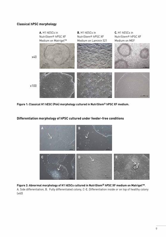
Classical hPSC morphology
Figure 1: Classical H1 hESC (P64) morphology cultured in NutriStem® hPSC XF medium.
Figure 2: Abnormal morphology of H1 hESCs cultured in NutriStem® hPSC XF medium on Matrigel™.A. Side differentiation, B. Fully differentiated colony, C-E. Differentiation inside or on top of healthy colony (x40)
A. H1 hESCs in NutriStem® hPSC XF Medium on Matrigel™
B. H1 hESCs in NutriStem® hPSC XF Medium on Laminin 521 0.5ug/cm2
C. H1 hESCs in NutriStem® hPSC XF Medium on MEF feeder layer
x40
x100
Differentiation morphology of hPSC cultured under feeder-free conditions
A
C
B
D E
10
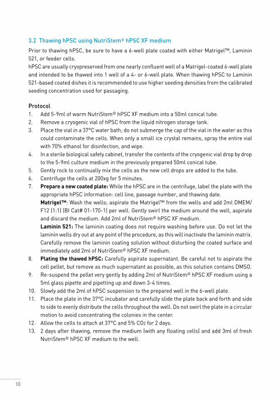
3.2 Thawing hPSC using NutriStem® hPSC XF medium
Prior to thawing hPSC, be sure to have a 6-well plate coated with either Matrigel™, Laminin 521, or feeder cells. hPSC are usually cryopreserved from one nearly confluent well of a Matrigel-coated 6-well plate and intended to be thawed into 1 well of a 4- or 6-well plate. When thawing hPSC to Laminin 521-based coated dishes it is recommended to use higher seeding densities from the calibrated seeding concentration used for passaging.
Protocol1. Add 5-9ml of warm NutriStem® hPSC XF medium into a 50ml conical tube.2. Remove a cryogenic vial of hPSC from the liquid nitrogen storage tank.3. Place the vial in a 37°C water bath; do not submerge the cap of the vial in the water as this
could contaminate the cells. When only a small ice crystal remains, spray the entire vial with 70% ethanol for disinfection, and wipe.
4. In a sterile biological safety cabinet, transfer the contents of the cryogenic vial drop by drop to the 5-9ml culture medium in the previously prepared 50ml conical tube.
5. Gently rock to continually mix the cells as the new cell drops are added to the tube.6. Centrifuge the cells at 200xg for 5 minutes.7. Prepare a new coated plate: While the hPSC are in the centrifuge, label the plate with the
appropriate hPSC information: cell line, passage number, and thawing date. Matrigel™: Wash the wells; aspirate the Matrigel™ from the wells and add 2ml DMEM/
F12 (1:1) (BI Cat# 01-170-1) per well. Gently swirl the medium around the well, aspirate and discard the medium. Add 2ml of NutriStem® hPSC XF medium.
Laminin 521: The laminin coating does not require washing before use. Do not let the laminin wells dry out at any point of the procedure, as this will inactivate the laminin matrix. Carefully remove the laminin coating solution without disturbing the coated surface and immediately add 2ml of NutriStem® hPSC XF medium.
8. Plating the thawed hPSC: Carefully aspirate supernatant. Be careful not to aspirate the cell pellet, but remove as much supernatant as possible, as this solution contains DMSO.
9. Re-suspend the pellet very gently by adding 2ml of NutriStem® hPSC XF medium using a 5ml glass pipette and pipetting up and down 3-4 times.
10. Slowly add the 2ml of hPSC suspension to the prepared well in the 6-well plate.11. Place the plate in the 37°C incubator and carefully slide the plate back and forth and side
to side to evenly distribute the cells throughout the well. Do not swirl the plate in a circular motion to avoid concentrating the colonies in the center.
12. Allow the cells to attach at 37°C and 5% CO2 for 2 days.13. 2 days after thawing, remove the medium (with any floating cells) and add 3ml of fresh
NutriStem® hPSC XF medium to the well.
11
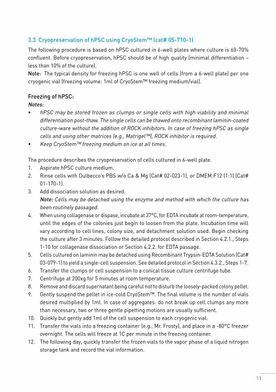
3.3 Cryopreservation of hPSC using CryoStem™ (cat# 05-710-1)
The following procedure is based on hPSC cultured in 6-well plates where culture is 60-70% confluent. Before cryopreservation, hPSC should be of high quality (minimal differentiation – less than 10% of the culture).Note: The typical density for freezing hPSC is one well of cells (from a 6-well plate) per one cryogenic vial (freezing volume: 1ml of CryoStem™ freezing medium/vial).
Freezing of hPSC:Notes: • hPSC may be stored frozen as clumps or single cells with high viability and minimal
differentiation post-thaw. The single cells can be thawed onto recombinant laminin-coated culture-ware without the addition of ROCK inhibitors. In case of freezing hPSC as single cells and using other matrices (e.g., Matrigel™), ROCK inhibitor is required.
• Keep CryoStem™ freezing medium on ice at all times.
The procedure describes the cryopreservation of cells cultured in 6-well plate. 1. Aspirate hPSC culture medium.2. Rinse cells with Dulbecco’s PBS w/o Ca & Mg (Cat# 02-023-1), or DMEM:F12 (1:1) (Cat#
01-170-1).3. Add dissociation solution as desired.
Note: Cells may be detached using the enzyme and method with which the culture has been routinely passaged.
4. When using collagenase or dispase, incubate at 37°C, for EDTA incubate at room-temperature, until the edges of the colonies just begin to loosen from the plate. Incubation time will vary according to cell lines, colony size, and detachment solution used. Begin checking the culture after 3 minutes. Follow the detailed protocol described in Section 4.2.1., Steps 1-10 for collagenase dissociation or Section 4.2.2. for EDTA passage.
5. Cells cultured on laminin may be detached using Recombinant Trypsin-EDTA Solution (Cat# 03-079-1) to yield a single-cell suspension. See detailed protocol in Section 4.3.2., Steps 1-7.
6. Transfer the clumps or cell suspension to a conical tissue culture centrifuge tube. 7. Centrifuge at 200xg for 5 minutes at room temperature. 8. Remove and discard supernatant being careful not to disturb the loosely-packed colony pellet.9. Gently suspend the pellet in ice-cold CryoStem™. The final volume is the number of vials
desired multiplied by 1ml. In case of aggregates: do not break up cell clumps any more than necessary, two or three gentle pipetting motions are usually sufficient.
10. Quickly but gently add 1ml of the cell suspension to each cryogenic vial.11. Transfer the vials into a freezing container (e.g., Mr. Frosty), and place in a -80°C freezer
overnight. The cells will freeze at 1C per minute in the freezing container.12. The following day, quickly transfer the frozen vials to the vapor phase of a liquid nitrogen
storage tank and record the vial information.
12
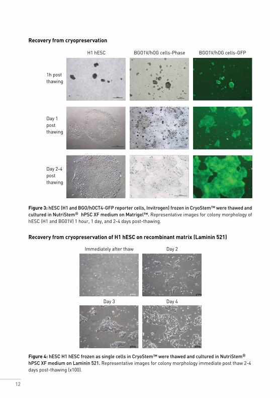
Recovery from cryopreservation
Figure 3: hESC (H1 and BGO/hOCT4-GFP reporter cells, Invitrogen) frozen in CryoStem™ were thawed and cultured in NutriStem® hPSC XF medium on Matrigel™. Representative images for colony morphology of hESC (H1 and BG01V) 1 hour, 1 day, and 2-4 days post-thawing.
Recovery from cryopreservation of H1 hESC on recombinant matrix (Laminin 521)
Figure 4: hESC H1 hESC frozen as single cells in CryoStem™ were thawed and cultured in NutriStem® hPSC XF medium on Laminin 521. Representative images for colony morphology immediate post thaw 2-4 days post-thawing (x100).
H1 hESC BGO1V/hOG cells-Phase BGO1V/hOG cells-GFP
Immediately after thaw
Day 3
Day 2
Day 4
1h post thawing
Day 1 post thawing
Day 2-4 post thawing
13
4. Feeder-Free Culture Systems
4.1 Matrigel™-based feeder-free culture system
4.1.1 Coating plates with Matrigel™
Matrigel™ preparation Matrigel™ should be thawed on ice, aliquoted and refrozen. For full instructions and recommendations on aliquot size, please refer to the insert supplied with the product.
Preparation of Matrigel™ Aliquots: 1. Thaw Matrigel™ overnight on ice at 2-8°C to avoid the formation of a gel. 2. Dilute Matrigel™ 1:1 with cold DMEM/F12 (1:1) medium (BI Cat# 01-170-1). Mix well with
a cold pipette. Keep mixture on ice. 3. Aliquot into pre-chilled 15ml tubes (for aliquot stability, please refer to the insert supplied
with the product). Store at -80°C.
Protocol1. Slowly thaw Matrigel™ aliquot on ice at 2-8°C to avoid the formation of a gel. 2. Dilute the Matrigel™ aliquot 1:20 with cold DMEM/F12 (1:1). 3. Add 1ml of Matrigel™ solution to each well of 6-well plate. 4. Incubate the plate for at least 1-2 hours at room temperature or overnight at 2-8°C. Plate
with Matrigel™ solution can be Parafilm® sealed and stored at 2-8°C for one week. Do not use the plate if the Matrigel™ solution does not completely cover the surface of the wells.
5. Before use, warm to room temperature, remove Matrigel™ solution, wash wells and immediately add NutriStem® hPSC XF medium.
4.2 Passaging hPSC on Matrigel™ using NutriStem® hPSC XF medium
Culture density is a critical aspect for maintaining healthy undifferentiated hPSC in NutriStem® hPSC XF medium. Cultures that are either too sparsely or too densely populated can lead to differentiation. Passage time should be determined by colony size and density regardless of the number of colonies in the well. Allowing colonies to get too large will result in differentiation. Colonies should not be allowed to reach a size or density high enough to touch one another for too long, as this will lead to increased differentiation. If the hPSC culture reaches confluency, split within the next 24 hours.
4.2.1 Enzymatic passaging of hPSC
We recommend using Collagenase IV at 1mg/ml. If using other enzyme-based dissociation solutions (Dispase or Accutase), optimal conditions should be determined by the user.
14
Protocol 1. Aspirate the culture medium from the well. 2. Wash the well once with warm DMEM/F12 (1:1) (BI Cat# 01-170-1). 3. Add 1ml per well of warmed Collagenase IV solution (1mg/ml). 4. Return the plate to the 37°C 5% CO2 incubator. Check the culture every 5-10 minutes.
Do not over-expose the cells to Collagenase IV. Observe the colonies under the microscope. Incubate until the edges of the colonies begin to curl up, but the colonies should remain attached to the plate surface.
5. Aspirate the Collagenase solution and wash very gently at least once with DMEM/F12 (1:1) (BI Cat# 01-170-1).
6. Add 2ml of pre-warmed NutriStem® hPSC XF medium to the well. 7. Gently scrape and wash the colonies off with a 5ml glass pipette. 8. Repeat the scraping and pipetting action 3-4 times until all the colonies have been removed
from the well surface. Pipette gently to avoid breaking up the colonies into too small clumps. 9. Transfer the detached colonies into a sterile conical tube. 10. Use another 2ml of medium to wash the well. Transfer the cell clumps into the same tube.
Note: The split ratio of NutriStem® hPSC XF medium-based culture is usually 1:6-1:8 every 3-5 days.
11. To obtain the desirable split ratio, increase the volume of the clump suspension in the tube with NutriStem® hPSC XF medium accordingly (if the desired splitting ratio is 1:6, increase the medium to a final volume of 6ml, thus 6 new wells can be seeded).
12. Prepare a new Matrigel™-coated 6-well plate by washing once with DMEM/F12 (1:1). Gently add 3ml NutriStem® hPSC XF medium to each well to be seeded.
13. Mix the clump suspension in the tube to break up cell clumps by gently pipetting up and down with a 5ml pipette.
14. Gently and evenly add 1ml of the suspended clumps to each new well to be seeded. 15. Place the plate in the 37°C 5% CO2 incubator and carefully move the plate back and forth
and side to side to evenly distribute the clumps throughout the well. 16. Allow the colonies to attach in the 37°C 5% CO2 incubator. 17. After 48 hours, change the medium daily with 2.5-3.0ml/well NutriStem® hPSC XF medium
until the colonies are large enough to passage. Notes:• It is possible to perform higher volume feed (4-5ml) on the weekend and the next
medium change two days later. • If the culture is at optimal density, the cells can be split every 3-5 days using a 1:6-1:8
splitting ratio. (Colonies from 1 well of a 6-well plate can be plated in 6-8 new wells of a 6-well plate). If the colonies are too dense or too sparse, adjust the splitting ratio accordingly.
15
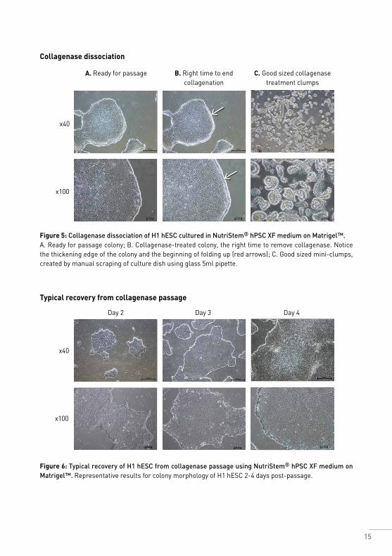
Collagenase dissociation
Figure 5: Collagenase dissociation of H1 hESC cultured in NutriStem® hPSC XF medium on Matrigel™.A. Ready for passage colony; B. Collagenase-treated colony, the right time to remove collagenase. Notice the thickening edge of the colony and the beginning of folding up (red arrows); C. Good sized mini-clumps, created by manual scraping of culture dish using glass 5ml pipette.
A. Ready for passage B. Right time to end collagenation
C. Good sized collagenase treatment clumps
x40
x100
Typical recovery from collagenase passage
Figure 6: Typical recovery of H1 hESC from collagenase passage using NutriStem® hPSC XF medium on Matrigel™. Representative results for colony morphology of H1 hESC 2-4 days post-passage.
Day 2 Day 3 Day 4
x40
x100
16
4.2.2 Enzyme-free passaging of hPSC
0.5mM EDTA dissociation solution: Prepare 0.5mM EDTA by combining 50μL of 0.5M EDTA (BI Cat# 01-862-1), pH 8.0 with 50ml of DPBS without calcium and magnesium (BI Cat# 02-023-1). If required filter the solution for sterility and store at room temperature.
Protocol This procedure describes the passage of the colonies as very small clumps. Volumes are for 1 well of 6-well plate.1. Wash cells twice with 2ml DPBS without calcium and magnesium (BI Cat# 02-023-1). 2. Add 1ml 0.5mM EDTA solution, swirl the vessel to coat the entire cell surface and quickly
discard.Note: Do not expose cells to the EDTA solution for more than needed for a quick wash at this point.
3. Add 1ml of 0.5mM EDTA solution and incubate for 3-4 minutes at room temperature. Do not move the plate during EDTA exposure.
4. Gently remove the EDTA solution and carefully add 1ml NutriStem® hPSC XF medium. 5. Detach and break colonies by gently pipetting up and down 3-4 times with a 1ml tip. Make
sure the pipetting washes the entire well.Note: Move quickly to the next step since the cell aggregates tend to re-attach to the well fast. When dissociating a few wells in parallel, gently swirl the cell aggregates in the plate during this step.
6. Plate the cell aggregates at the desired density in Matrigel™-coated wells with 4ml pre-warmed NutriStem® hPSC XF medium. Usually a splitting ratio of 1:8-1:10 every 4 days is required.
7. Place the plate in a 37°C 5% CO2 incubator. Move the plate several times back and forth and side to side to distribute the aggregates evenly in the well.
8. After 48 hours, change the medium daily with 2.5-3.0ml/well NutriStem® hPSC XF until the colonies are large enough to passage.
9. It is possible to perform higher volume feed (4-5ml) on the weekend and the next medium change two days later. Note: Do not move the plate during the first 48 hours post-split (this may increase differentiation of hPSC).
17
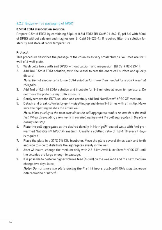
Enzyme- free passage using 0.5mM EDTA solution
Figure 7: EDTA passage of H1 hESC cultured in NutriStem® hPSC XF medium on Matrigel™.A. Ready for passage colony; B. 4 minutes treatment of 0.5mM EDTA solution; C. Good sized mini-clumps. (x100). Notice the gaps between the cells (white arrows). Mini clumps created
by 4 up/down pipettation of 1ml fresh medium.
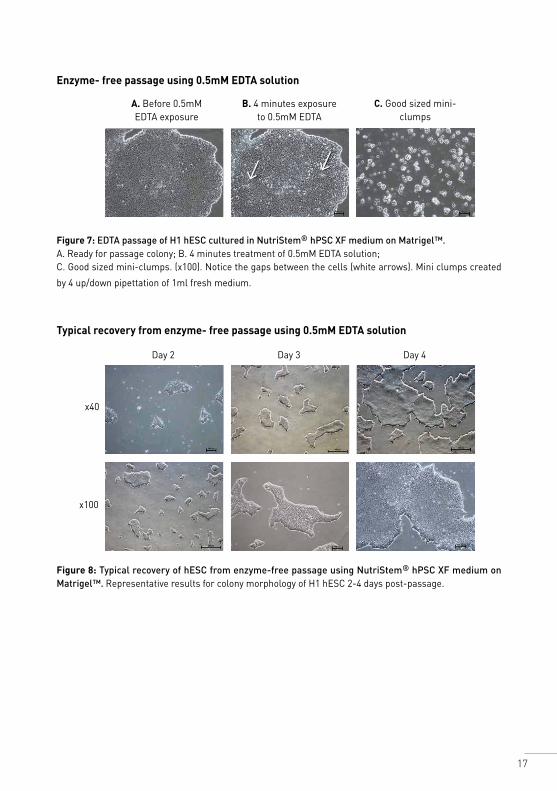
Typical recovery from enzyme- free passage using 0.5mM EDTA solution
Figure 8: Typical recovery of hESC from enzyme-free passage using NutriStem® hPSC XF medium on Matrigel™. Representative results for colony morphology of H1 hESC 2-4 days post-passage.
A. Before 0.5mM EDTA exposure
B. 4 minutes exposure to 0.5mM EDTA
C. Good sized mini-clumps
Day 2 Day 3 Day 4
x40
x100
18
4.3 Laminin-based feeder-free culture system
4.3.1 Coating plates with Laminin 521
Laminin 521 coating procedure at 0.5μg/cm2 in a 6-well plate. For a detailed protocol, follow laminin manufacturer instructions.
General coating protocol (0.5μg/cm2) 1. Slowly thaw Laminin 521 (0.1mg/ml) at 2-8°C. 2. Thawed laminin stock may be stored at 2-8°C under aseptic conditions. 3. Dilute 50μl Laminin 521 (0.1mg/ml) with 2ml DPBS with calcium and magnesium,
(300μl/12ml/6 wells). 4. Add 2ml/well of the diluted Laminin 521. Make sure the laminin solution is evenly spread
across the surface. Note that the laminin matrix will be inactivated if dried.5. Seal the plate with plastic film (e.g., Parafilm®) to prevent evaporation. 6. Incubate overnight at 2-8°C.
Notes: 1. Optimal coating concentration is cell-dependent and should be calibrated. 0.5-1 μg/cm2
should work well for most hPSC. 2. Coated plates may be stored aseptically at 2-8°C. Refer to the manufacturer’s instructions
for shelf life of coated plates.3. Rapid coating may be done at 37°C for 2 hours. 4.3.2 Single-cell passage of hPSC grown in NutriStem® hPSC XF on Laminin 521
This procedure describes the passage of hPSC from 1 well of 6-well plate.1. Wash cells twice with 2ml/well DPBS without calcium and magnesium. 2. Add 1ml of Recombinant Trypsin-EDTA Solution (BI Cat# 03-079-1). 3. Incubate at 37°C for 2-4 minutes. Exposure time may vary and should be adjusted.
Note: Do not over-expose the cells to the Recombinant Trypsin-EDTA Solution.4. Detach and break up colonies by pipetting 5-6 times up and down with a 1ml pipette. Make
sure the pipetting washes the entire well. 5. Add 4ml 1xSBTI to the Recombinant Trypsin-EDTA Solution. 6. Collect cells into a new sterile tube. 7. Centrifuge at 200g for 5 minutes. 8. Suspend the pellet with 1 ml of medium. 9. Count cells. 10. Plate cells at the desired density in Laminin 521 pre-coated wells with 3-4ml pre-equilibrated
medium. Usually, 10-20,000/cm2 for splitting every 4-5 days. 11. Place the plate in a 37°C 5% CO2 incubator. Move the plate several times back and forth
and side to side to evenly distribute the cells in the well. 12. Perform daily medium change after 48 hours with 2.5-3.0ml/well NutriStem® hPSC XF
until the colonies are large enough to passage. Do not move the plate during the first 48 hours post-split.
19
Notes: 1. It is critical not to move the seeded plates during the first 48 hours after splitting as this
may increase differentiation of hPSC. 2. Cells are ready to be passaged when cell culture is ≥60% confluent. Optimal seeding
densities will vary from one cell line to another and must be determined for your system. With optimal culture conditions and seeding density, most cell lines will reach confluency within 4-6 days and expand 10-25-fold.
3. It is possible to perform higher volume feed (4-5ml) for the weekend and skip 2 days without changing the medium. In this case, the cells must be passaged at the end of the week.
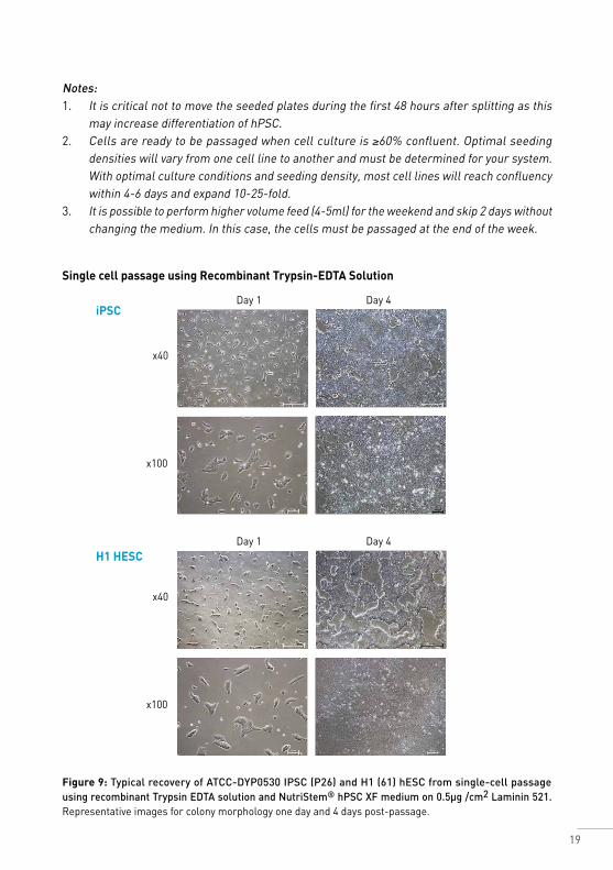
Single cell passage using Recombinant Trypsin-EDTA Solution
iPSC
H1 HESC
Figure 9: Typical recovery of ATCC-DYP0530 IPSC (P26) and H1 (61) hESC from single-cell passage using recombinant Trypsin EDTA solution and NutriStem® hPSC XF medium on 0.5µg /cm2 Laminin 521. Representative images for colony morphology one day and 4 days post-passage.
Day 1
Day 1
Day 4
Day 4
x40
x40
x100
x100
20
5. Feeder-Dependent Culture System
5.1 General information
NutriStem® hPSC XF medium is designed to support both feeder-dependent and feeder-free culture systems. If working with hESC or hiPSC in a feeder-dependent culture system it is possible to use the albumin-free version, AF NutriStem® hPSC XF medium (Cat# 05-102-1), for several passages. NutriStem® hPSC XF medium, which contains HSA (Cat# 05-100-1), is preferable for long-term growth.
5.2 Observing culture under the microscope
Observe the cells under the microscope and assess the overall feeder and hPSC culture quality. hPSC tend to create small condensed and round colonies when cultured on feeder cells in comparison to larger thicker colonies on Matrigel™. Spontaneously differentiated cells may resemble cobblestones or fibroblast-like cells. The differentiated cells may reside at the colony perimeter or in the center of the colony. There can also be differentiated colonies containing large clumps of swirled tissue, or little clumps of cells, like embryoid bodies. Different culture conditions yield different types of differentiated cells.
Notes: • Feeder cell quality: more than 90% viable and with normal fibroblast morphology, density,
and absence of any visible contamination.• hPSC quality: colony size, morphology, and density.
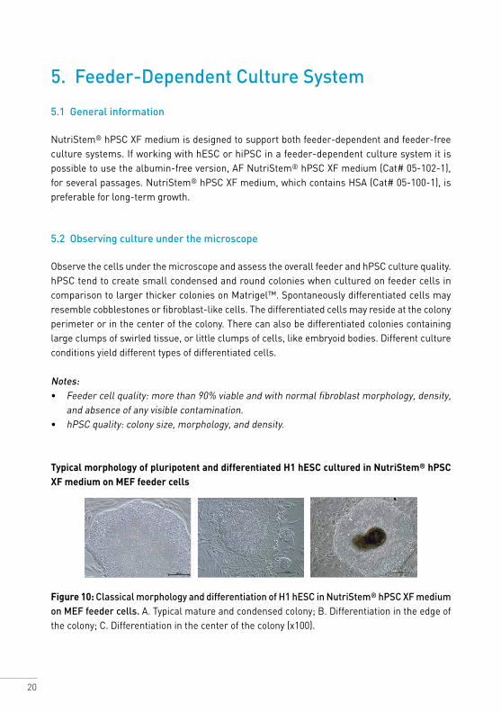
Typical morphology of pluripotent and differentiated H1 hESC cultured in NutriStem® hPSC XF medium on MEF feeder cells
Figure 10: Classical morphology and differentiation of H1 hESC in NutriStem® hPSC XF medium on MEF feeder cells. A. Typical mature and condensed colony; B. Differentiation in the edge of the colony; C. Differentiation in the center of the colony (x100).
21
5.3 Preparation of mitotically inactivated feeder cells (Mitomycin C-treated)
Feeder cells (MEF, HFF) should be mitotically inactivated by gamma irradiation or chemically treated with Mitomycin C to prevent proliferation. Inactivated feeder cells may be cultured immediately or cryopreserved for future use.
Inactivated feeder cell layers will only support hESC or hiPSC for five days from seeding.
1. MEF feeder cells should be passaged into T75 flasks twice and used on the third passage. 2. Discard the medium and add 8μg/ml Mitomycin C (6-7ml) into a culture flask and incubate
for two hours. 3. Wash three times with D-PBS without calcium and magnesium (02-023-1). 4. Add 3ml of Trypsin-EDTA solution and cover the entire culture-flask surface. 5. Incubate for 6 minutes. 6. Tap the side of the flask to loosen the cells. Add 10ml of feeder culture medium to neutralize
the trypsin. 7. Aspirate cell suspension into a conical tube. 8. Centrifuge for five minutes at 800g.
For cryopreservation of the feeder cells follow the CryoStem™ cryopreservation protocol, Section 3.3.For immediate use of cells follow the Preparation of Feeder Cell Layer protocol, Section 5.4.
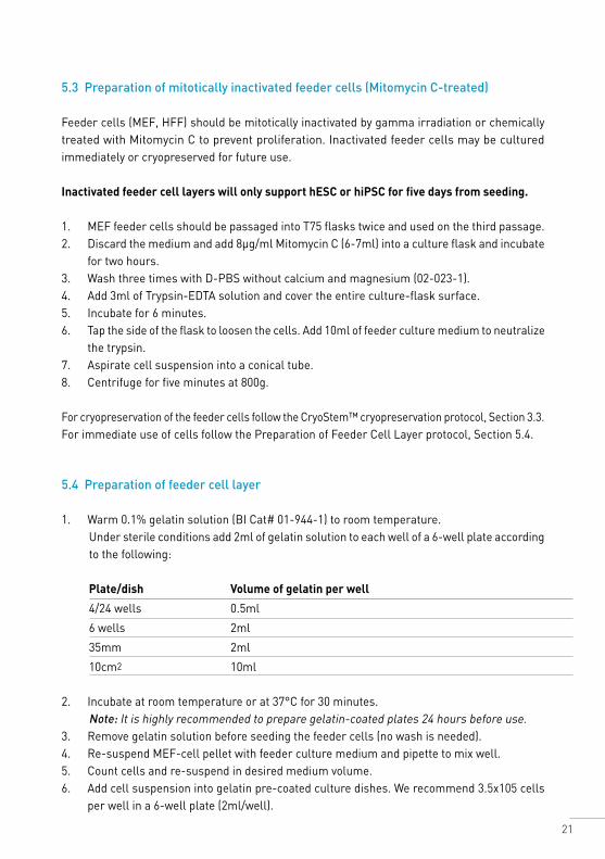
5.4 Preparation of feeder cell layer
1. Warm 0.1% gelatin solution (BI Cat# 01-944-1) to room temperature.Under sterile conditions add 2ml of gelatin solution to each well of a 6-well plate according to the following:
Plate/dish Volume of gelatin per well
4/24 wells 0.5ml
6 wells 2ml
35mm 2ml
10cm2 10ml
2. Incubate at room temperature or at 37°C for 30 minutes. Note: It is highly recommended to prepare gelatin-coated plates 24 hours before use.
3. Remove gelatin solution before seeding the feeder cells (no wash is needed).4. Re-suspend MEF-cell pellet with feeder culture medium and pipette to mix well. 5. Count cells and re-suspend in desired medium volume. 6. Add cell suspension into gelatin pre-coated culture dishes. We recommend 3.5x10⁵ cells
per well in a 6-well plate (2ml/well).
22
7. Incubate the feeder cells for at least two hours before plating the hPSC (cells must be attached and spread).
8. Before plating hPSC, change the feeder medium to NutriStem® hPSC XF medium.
Notes: 1. Feeder cell concentration can also be calculated as 3.5x10⁴ cells per cm2. 2. Do not use NutriStem® hPSC for MEF culture. Prepare MEF-covered plate using recommended
culture medium, and change the medium before plating the cells. 3. Plates may be used within 10 days from date of preparation. 4. It is highly recommended to prepare MEF plates 24 hours before use.
Thawing of cryopreserved inactivated feeder cells: 1. Remove vial containing cryopreserved inactivated feeder cells from liquid nitrogen storage
and thaw quickly in a 37°C water bath.2. When a small piece of ice remains, disinfect the vial using 70% ethanol.3. Pipette the contents of the vial and transfer the cells drop by drop into a conical tube
containing 5ml of feeder cell medium.4. Centrifuge for 5 minutes at 200g. Discard supernatant.5. Re-suspend cell pellet in feeder cell culture medium: 2ml for each well to be seeded.6. Add cell suspension into the gelatin-coated 6-well plate.
Notes: 1. Adding the medium drop by drop is crucial for cell recovery.2. MEF seeding density: for specific cell density please refer to the insert supplied with the
product. In general, 350,000 cells can be seeded in 1 well of a 6-well plate when using ongoing culture. 500,000 cells should be seeded in 1 well of a 6-well plate when thawing cryopreserved cells.
3. If the feeder cells are too sparse, they may not maintain the hPSC without differentiation, and the cells may not attach well. At a too high density, the feeder layer may detach from the plate, and the culture will be lost.
5.5 Thawing procedure for hPSC on feeder cells using NutriStem® hPSC XF medium
One day prior to thawing hPSC, plate a feeder layer of inactivated feeder cells (e.g., Mitomycin C-treated MEF) in one well of a gelatin-coated 6-well tissue culture plate in feeder culture medium. Incubate overnight at 37°C and 5% CO2.
Notes: 1. hESC or hiPSC should always be thawed into a plate of feeder cells less than five days old. 2. Check the feeders for quality before use (more than 90% viable and with normal fibroblast
morphology). 3. Follow thawing procedure according to section 3.2.
23
5.6 Passaging hPSC using NutriStem® hPSC XF medium Collagenase solution: Dissolve Collagenase Type IV in DMEM/F12 (1:1) (BI Cat# 01-170-1) to a concentration of 1mg/ml and filter for sterility with a 0.22μm pore size filter unit. Collagenase solution can be stored at 2-8°C for up to 2 weeks.
Protocol1. Remove medium from the well. 2. Add 1.0ml of collagenase solution. 3. Incubate at 37°C or at room temperature until the edges of the cell colonies begin to loosen
from the plate. Note: Incubation time will vary between cell lines and colony sizes. Begin checking the culture after 3 minutes. Do not over-incubate the culture, as hPSC are sensitive to enzymatic stress and may detach from the plate during the wash step.
4. Aspirate the dissociation solution and wash the cells twice with 2ml of sterile DMEM/F12 (1:1). 5. Add 1ml of culture medium and gently wash off and detach hPSC using a 5ml pipette. Most
of the feeder layer cells will remain on the plate. 6. Collect cell suspension and put into a conical tube. 7. Centrifuge for 3 minutes at 200g at room temperature. 8. Re-suspend hPSC using NutriStem® hPSC XF medium by gently pipetting up and down
with a 5ml pipette to break up clumps, and plate in feeder-covered plate with 2.5-3ml NutriStem® hPSC XF.
9. Change the medium daily with 2.5-3.0ml/well NutriStem® hPSC XF until the colonies are large enough to passage. Note: For effective separation of hPSC from the feeder cells, longer dissociation time is recommended (up to 1 hour).
5.7 Transferring hESC or hiPSC in NutriStem® hPSC XF medium from feeders to a feeder-free culture system and vice vers
NutriStem® hPSC XF medium is designed to support both feeder-dependent and feeder-free cultures. AF NutriStem® hESC XF medium is optional for short-term use on feeder cells such as MEF or HFF. When transferring to a feeder-free culture system use either AF NutriStem® hPSC XF medium (05-102-1) supplemented with 5% (v/v) of Bio-Pure HSA (05-720-1) or NutriStem® hPSC XF medium (05-100-1). AF NutriStem® hPSC XF supplemented with HSA is suitable for both feeder-dependent and feeder-free culture conditions.
Transferring hPSC from feeder-dependent culture to feeder-free culture: passage the cells directly to the Matrigel™-coated plate.Some residual feeder cells may be observed in the culture during the first 2 weeks, but in subsequent passages the inactivated feeder cells will be passaged out.
24
6. Adaptation
6.1 Adapting hPSC to Matrigel™-based NutriStem® hPSC XF medium
We recommend applying direct adaptation to NutriStem® hPSC XF medium on Matrigel™. Some lines will not adapt easily and will require gradual adaptation. hPSC may be transferred to NutriStem® hPSC XF medium from feeder or feeder-free cultures. Nevertheless, it is extremely important to start with a high quality culture (high density of cells, minimum background differentiation). The starter culture may be cryopreserved cells or an ongoing proliferating cell culture. Different hPSC lines may behave differently when transitioning to a different culture medium until they fully adapt.
6.1.1 Before beginning the adaptation
• Make a frozen stock of cells in the previous medium prior to adaptation.• Keep a culture going in each prior condition when starting the next level of adaptation as
a fallback in case the cells do not survive in the next passage.• In addition, it is best to keep a culture going in the previous culture medium (before
adaptation) as a “backup.”
6.1.2 Points that might hinder successful adaptation
• Timing of passage is critical. For best adaptation results, hPSC should be near confluency at the time of passage.
• Seeding density at passage is critical. If seeding ratio is too low, hPSC will differentiate. While adapting, hPSC decrease their proliferation rate. We recommend a split ratio of 1:2 for the first 3 passages of adaptation. Normally, a split ratio of between 1:6 and 1:8 is appropriate, but passaging at 1:2 ensures the higher density of cells needed when adapting to a feeder-free culture. A higher proliferation rate will be observed when the cells are fully adapted.
• Daily feeding is critical. Perform a complete medium change daily (double volume for the weekend) to replenish lost nutrients, and keep the cultures free of unwanted differentiation factors.
• Morphology change is normal. The edges of colonies may be less compact than the centers when adapting to NutriStem® hPSC XF medium. However, the hPSC are not differentiated but exhibit normal pluripotent stem-cell characteristics.
• Some differentiation may occur and should be removed manually prior to passaging.
General strategy for adaptation to NutriStem® hPSC XF mediumFrom other feeder-free media or from feeder-dependent media to NutriStem® hPSC XF medium: sub-culture the cells directly into NutriStem® hPSC XF medium on Matrigel™. Some residual feeders may be observed in the cultures during the adaptation process, but in subsequent passages the inactivated feeder cells will be passaged out.
25
6.1.3 Direct adaptation protocol
Direct adaptation can be performed both on ongoing proliferating cultures or cryopreserved cells.
A. Direct seeding strategy: hPSC are passaged directly into NutriStem® hPSC XF medium on Matrigel™. A 1:2 split ratio is suggested for the first 3 passages. The quality of the medium is critical for the success of adaptation. Use only fresh (no more than 2 weeks), protected from light, pre-warmed medium.1. Pre-warm the medium volume needed for this procedure.2. Detach the colonies in accordance with the passage protocol. 3. Centrifuge the cells at 200xg for 5 minutes.4. Carefully discard the supernatant.5. Re-suspend the pellet in NutriStem® hPSC XF medium; be careful not to break up the
colonies into too small clumps. The split ratio should be 1:2. Use 3ml medium for each well to be seeded.For example: When starting with 1 well of a 6-well plate, re-suspend the pellet in 6ml NutriStem® hPSC XF medium, and seed 3ml into 2 new wells of a 6-well plate pre-coated with Matrigel™.
6. Transfer 3ml of the colony suspension to a new Matrigel™-coated well.7. Place the plate in a CO2 incubator. Carefully slide the plate back and forth and side to side
to evenly distribute the cells throughout the well. Do not swirl the plate in a circular motion to avoid concentrating the colonies in the center. Incubate the plate in a CO2 incubator.
B. Freeze/thaw cycle strategy: hPSC are thawed directly into NutriStem® hPSC XF medium. It is possible to add ROCK inhibitor Y27632 (10μM) for the first overnight of adaptation. This will increase the survival of the cells.1. Pre-warm the medium volume needed for this procedure.2. Quickly thaw the cells in accordance with the thawing protocol (Section 3.2.).3. Re-suspend the pellet in NutriStem® hPSC XF medium; be careful not to break up the
colonies into too small clumps.4. Seed the cells into 1 well of a 6-well plate pre-coated with Matrigel™.5. Add the Y27632 directly to the well. The final concentration should be 10μM.6. Carefully slide the plate back and forth and side to side to evenly distribute the cells throughout
the well. Do not swirl the plate in a circular motion to avoid concentrating the colonies in the center. Incubate the plate in CO2 incubator. Do not move the plate for the next overnight.
7. The day after the thaw, replace the medium with fresh pre-warmed 3ml NutriStem® hPSC XF medium. Do not continue to add the ROCK inhibitor Y27632.
26
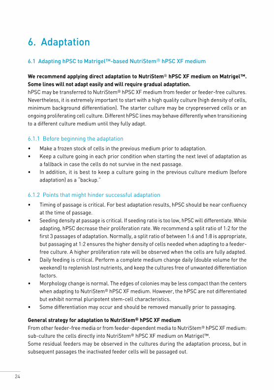
Direct adaptation to NutriStem® hPSC XF medium
After seeding
After passaging
Figure 11: Direct Adaptation: H1 hESC (P60) were passaged directly into NutriStem® hPSC XF medium on Matrigel™. A. 2 days post-seeding into NutriStem® hPSC XF medium; B. 3 days post-seeding into NutriStem® hPSC XF Medium; C. 3 days from P1 of adaptation; D. 3 days from P2 of adaptation (x100).
6.2 Adapting hPSC to Laminin 521-based culture system using NutriStem® hPSC XF medium
When transferring cells from another feeder-free matrix (e.g., Matrigel™) we recommend starting with a smaller well format (e.g., 96-well or 48-well format) and a higher seeding density (50,000-100,000 cells/cm2) for the first number of passages to let the cells adapt to the laminin matrix before increasing the well format and lowering the seeding density.
From feeder-free matrices The transfer from feeder-free matrices to Laminin 521 is often quite straightforward. Coat new plates with Laminin 521 according to the manufacturer’s instructions for use and perform a single-cell passage as described in BioLamina’s instructions for use (BL003). We recommend starting with a smaller well format (e.g., 24-well or 48-well format) and a higher seeding density (50,000-100,000 cells/cm2) for the first number of passages to let the cells adapt to the laminin matrix before increasing the well format and lowering the seeding density.
A B
D2 from seeding
D3 from P1
D3 from seeding
D3 from P2
C D
27
From feedersUsually it is sufficient to collect the pieces of undifferentiated hPSC colonies from the feeder plate, seed them directly on a Laminin 521-coated plate. When the cells reach about 70% confluence, perform an enzymatic single-cell passage for maintenance as described in BioLamina’s instructions for use (BL003). We recommend starting with a smaller well format (e.g., 96-well or 48-well format) and a higher seeding density (50,000-100,000 cells/cm2) for the first number of passages to let the cells adapt to the laminin matrix before increasing the well format and lowering the seeding density.Some hPSC lines are more difficult to adapt and will thus need parallel single-cell and colony passage for several passages to ensure single-cell survival as described above.
7. References NutriStem® and Cardiomyocytes
P. Menasché et al. Human embryonic stem cell-derived cardiac progenitors for severe heart failure treatment: First clinical case report. European Heart Journal (2015): ehv189.
L. Jacquet et al. Three Huntington’s Disease specific mutation-carrying human embryonic stem cell lines have stable number of CAG repeats upon in vitro differentiation into cardiomyocytes. PLOS One 10.5 (2015).
S. Rajasingh et al. Generation of functional cardiomyocytes from efficiently generated human iPSCs and a novel method of measuring contractility. PLOS One 10.8 (2015), e0134093.
W. Siqin et al. Spider silk for xeno-free long-term self-renewal and differentiation of human pluripotent stem cells. Biomaterials 35.30 (2014), 8496-8502.
E. Di Pasquale et al. Generation of human cardiomyocytes: A differentiation protocol from feeder-free human induced pluripotent stem cells. JoVE (Journal of Visualized Experiments) 76 (2013), e50429-e50429.
G. Foldes and M. Mioulane. High-content imaging and analysis of pluripotent stem cell-derived cardiomyocytes. Imaging and tracking stem cells. Humana Press (2013).
P.W. Burridge and E.T. Zambidis. Highly efficient directed differentiation of human induced pluripotent stem cells into cardiomyocytes. Pluripotent Stem Cells. Humana Press (2013).
R. S. Song et al. Generation, expansion, and differentiation of human induced pluripotent stem cells (hiPSCs) derived from the umbilical cords of newborns. Current Protocols in Stem Cell Biology (2013), 1C-16.
J.P. Awe et al. Stem Cell Research & Therapy (2013).
L. Warren et al. Highly efficient reprogramming to pluripotency and directed differentiation of human cells with synthetic modified mRNA. Cell Stem Cell 7.5 (2010), 618-630.
28
K. Tryggvason et al. Differentiation of pluripotent stem cells and cardiac progenitor cells into striated cardiomyocyte fibers using laminins LN-511, LN-521 and LN-221. US Patent 20,160,122,717 (2016).
Differentiation of Pluripotent Stem Cells
A.P. Reyes et al. Xeno-free and defined human embryonic stem cell-derived retinal pigment epithelial cells functionally integrate in a large-eyed preclinical model. Stem Cell Reports (2015).
M.V. Krivega et al. Cyclin E1 plays a key role in balancing between totipotency and differentiation in human embryonic cells. Mol. Hum. Reprod (2015).
S. Rajasingh et al. Generation of functional cardiomyocytes from efficiently generated human iPSCs and a novel method of measuring contractility. PLOS One (2015).
J. Lenzi et al. Differentiation of control and ALS mutant human iPSCs into functional skeletal muscle cells, a tool for the study of neuromuscolar diseases. Stem Cell Research (2016).
J. Bailly et al. Method for differentiation of pluripotent stem cells into multi-competent renal precursors. US Patent 20,160,145,578 (2016).
K. Tryggvason et al. Differentiation of pluripotent stem cells and cardiac progenitor cells into striated cardiomyocyte fibers using laminins LN-511, LN-521 and LN-221. US Patent 20,160,122,717 (2016).
Culture Methods of hESC and IPSC (Derivation, expansion, scaling up and suspensions)
M. Mikkola et al. A method for generating induced pluripotent stem cells. US Patent 20,160,068,818 (2016).
K. Alessandri et al. A 3D printed microfluidic device for production of functionalized hydrogel microcapsules for culture and differentiation of human neuronal stem cells (hNSC). Lab on a Chip (2016).
Y.Y. Lipsitz and P.W. Zandstra. Human pluripotent stem cell process parameter optimization in a small scale suspension bioreactor. BMC Proceedings (2015).
S. Eminli-Meissner et al. A novel four transfection protocol for deriving iPS cell lines from human blood-derived endothelial progenitor cells (EPCs) and adult human dermal fibroblasts using a cocktail of non-modified reprogramming and immune evasion mRNAs. Sientific Poster, REPROCELL (2015).
S. Wu et al. Efficient passage of human pluripotent stem cells on spider silk matrices under xeno-free conditions. Cellular and Molecular Life Sciences (2015).
S. Gregory et al. Autophagic response to cell culture stress in pluripotent stem cells. Biochemical and Biophysical Research Communications, doi:10.1016/j.bbrc.2015.09.080 (2015).
29
H. Tateno et al. Undifferentiated cell detection method and complex carbohydrate detection method. US Patent 20,150,204,870 (2015).
S. Herz. Optimization of RNA-based transgene expression by targeting Protein Kinase R. Dissertation for the degree of “fgreet rerum naturalium” (2015).
J. Lenzi et al. ALS mutant FUS proteins are recruited into stress granules in induced pluripotent stem cells (iPSCs) derived motoneurons. Disease Models & Mechanisms (2015).
N. Desai, P. Rambhia, and A. Gishto. Human embryonic stem cell cultivation: Historical perspective and evolution of xeno-free culture systems. Reproductive Biology and Endocrinology 13.1 (2015), 9.
T. Yokobori et al., Intestinal epithelial culture under an air-liquid interface: A tool for studying human and mouse esophagi. Diseases of the Esophagus (2015).
T. Cerbini et al. Transfection, selection, and colony-picking of human induced pluripotent stem cells TALEN-targeted with a GFP gene into the AAVS1 safe harbor. JoVE (Journal of Visualized Experiments) (2015).
L. Healy and L. Ruban. Derivation of induced pluripotent stem cells. Atlas of Human Pluripotent Stem Cells in Culture (2015).
N.Y. Thakar et al. TRAF2 recruitment via T61 in CD30 drives NFkB activation and enhances hESC survival and proliferation. Molecular Biology of the Cell (2015).
V. Bellamy et al. Long-term functional benefits of human embryonic stem cell-derived cardiac progenitors embedded into a fibrin scaffold. The Journal of Heart and Lung Transplantation (2014).
A. J. Schwab and A.D. Ebert. Sensory neurons do not induce motor neuron loss in a human stem cell model of spinal muscular atrophy. PLOS One (2014), 9(7), e103112.
G. Finesilver, M. Kahana, and E. Mitrani. Kidney-specific micro-scaffolds and kidney derived serum free conditioned media support in vitro expansion, differentiation, and organization of human embryonic stem cells. Tissue Engineering Part C: Methods (Not available, ahead of print) doi:10.1089/ten.TEC.2013.0574.
M. Amit and J. Itskovitz-Eldor. Novel methods and culture media for culturing pluripotent stem cells. US Patent 20,130,236,961 (2013).
M. Amit and J. Itskovitz-Eldor. Atlas of human pluripotent stem cells: Derivation and culturing. Stem Cell Biology and Regenerative Medicine (2012).
R. Bergström. Xeno-free culture of human pluripotent stem cells. Methods Mol Biol. (2011), 767:125-36.
S. Sugii et al., Human and mouse adipose-derived cells support feeder-independent induction of pluripotent stem cells. PNAS (2010), 107(8), 3558-3563.
J.Collins et al. Highly efficient reprogramming to pluripotency and directed differentiation of human cells with synthetic modified mRNA. Cell Stem Cell 7(5), 618-630 (2010).
30
Induction of Pluripotency of hESC and iPSC
S. Eminli-Meissner et al. A novel four transfection protocol for deriving iPS cell lines from human blood-derived endothelial progenitor cells (EPCs) and adult human dermal fibroblasts using a cocktail of non-modified reprogramming and immune evasion mRNAs. Scientific Poster, REPROCELL (2015).
U. Sahin et al. Method for cellular RNA expression. US Patent 20,150,314,018 (2015).
M. Brouwer et al. Choices for induction of pluripotency: Recent developments in human induced pluripotent stem cell reprogramming strategies. Stem Cell Reviews and Reports (2015).
H.X. Nguyen et al. Induction of early neural precursors and derivation of tripotent neural stem cells from human pluripotent stem cells under xeno-free conditions. Journal of Comparative Neurology (2014).
Different Basement Matrices
O. Simonson, Use of genes and cells in regenerative medicine. Karolinska Institutet (2015).
Nacalai USA Inc. Vitronectin-398™ (Xeno-free). Nacalai USA website.
BioLamina. Weekend free culture of human pluripotent stem cells on LN 521™. BioLamina news.
S. Rodin et al. Monolayer culturing and cloning of human pluripotent stem cells on laminin-521-based matrices under xeno-free and chemically defined conditions. Nature Protocols 9, 2354-2368 (2014), doi:10.1038/nprot.2014.159.
S. Rodin et al. Clonal culturing of human embryonic stem cells on Laminin-521/E-cadherin matrix in defined and xeno-free environment. Nature Communications 5, Article number: 3195 (2014).
StemAdhere™ Defined Matrix for hPSC. Primorigen Biosciences website.
I. Lenz et al. Automated 3D culture to undifferentiated hESC. (Scientific Poster).
J.L. Weber et al. The Corning® Synthemax™ Surface: A synthetic, xeno-free surface for long-term self-renewal of human embryonic stem cells in defined media. Presented at the 2010 World Stem Cell Summit.
Clinical Applications – Derivation and Expansion of hESC and IPSC
K. Jacobs et al. Higher-density culture in human embryonic stem cells results in DNA damage and genome instability. Stem Cell Reports (2016).
D. Keefe et al. A method for a single cell analysis of telomere length. US Patent 20,160,032,360 (2016).
A. P. Reyes et al. Xeno-free and defined human embryonic stem cell-derived retinal pigment epithelial cells functionally integrate in a large-eyed preclinical model. Stem Cell Reports, 6 (2016).
31
O. Simonson. Use of genes and cells in regenerative medicine. Karolinska Institutet (2015).
M. Di Salvio et al. Pur-alpha functionally interacts with FUS carrying ALS-associated mutations. Cell Death & Disease (2015).
L. de Oסate et al. Research on skeletal muscle diseases using pluripotent stem cells. doi:10.5772/60902, 2015.
P. Menasché et al. Towards a clinical use of human embryonic stem cell-derived cardiac progenitors: A translational experience.
T. Seki and K. Fukuda. Methods of induced pluripotent stem cells for clinical application. World Journal of Stem Cells (2015).
S. Abbasalizadeh and H. Baharvand. Technologies progress and challenges towards cGMP manufacturing of human pluripotent stem cells based therapeutic products for allogeneic and autologous cell therapies. Biotechnology Advances (2013).
Y. Luo et al. Stable enhanced green fluorescent protein expression after differentiation and transplantation of reporter human induced pluripotent stem cells generated by AAVS1 transcription activator-like effector nucleases. Stem Cells Translational Medicine (2014).
J. Durruthy-Durruthy et al. Rapid and efficient conversion of integration-free human induced pluripotent stem cells to GMP-grade culture conditions. PLOS One (2014).
H. Tateno et al. A medium hyperglycosylated podocalyxin enables noninvasive and quantitative detection of tumorigenic human pluripotent stem cells. Scientific Reports 4, Article number 4069 (2014).
J. P. Awe et al. Generation and characterization of transgene-free human induced pluripotent stem cells and conversion to putative clinical-grade status. Stem Cell Research & Therapy (2013), 4:87.
O. Hovatta. Infectious problems associated with transplantation of cells differentiated from pluripotent stem cells. Seminars in Immunopathology (2011).
S. Ström. Optimisation of human embryonic stem cell derivation and culture – towards clinical quality. Karolinska Institutet (2010).
U. Sahin et al. Diagnosis and therapy of cancer involving cancer stem cells. US Patent 20,160,159,901.

32
Biological Industries Israel Beit Haemek Ltd.Kibbutz Beit Haemek 2511500, Israel T.+972.4.9960595 F.+972.4.9968896 Email: [email protected]
w w w . b i o i n d . c o m


![10000005505-Maintenance of Human Pluripotent Stem Cells …€¦ · The maintenance and expansion of human pluripotent stem cells (human embryonic stem [ES] cells and human induced](https://static.fdocuments.in/doc/165x107/6033bf7fdddc672302645fcf/10000005505-maintenance-of-human-pluripotent-stem-cells-the-maintenance-and-expansion.jpg)